The humerus of South American caviomorph rodents: shape, function and size in a phylogenetic context
10
The humerus of South American caviomorph rodents: shape, function and size in a phylogenetic context C. C. Morgan 1,2 & A. Álvarez 1 1 Facultad de Ciencias Naturales y Museo, Universidad Nacional de La Plata, La Plata, Buenos Aires, Argentina 2 CONICET, Buenos Aires, Argentina Keywords functional morphology; geometric morphometrics; humerus; phylogenetic comparative methods; South American hystricomorphs. Correspondence Cecilia C. Morgan. Sección Mastozoología, Div. Zoología Vertebrados, Museo de La Plata. Paseo del Bosque s/n°, B1900FWA, La Plata, Argentina. Email: [email protected] Received 3 August 2012; revised 8 January 2013; accepted 14 January 2013 doi:10.1111/jzo.12017 Abstract South American caviomorph rodents comprise four major lineages encompassing wide taxonomic and ecological diversity, but the morphological diversity of their postcranial skeleton has not been thoroughly explored using phylogenetic com- parative methods. The main goal of this work is to analyze their humerus using geometric morphometrics in a phylogenetic context and attempt to tease apart the influence of locomotory preferences and shared evolutionary history on morpho- logical variation. We examined 28 genera in 9 families representing all major clades. Humeral shape was captured by 13 landmarks and four semilandmarks in 2D. In the morphospace of the first two principal components, most taxa were located near the origin along both axes. Fossorial octodontoids were apart from this central group. Most caviids were separated in one extreme of the morphos- pace; the specialized digging ctenomyid Ctenomys, the fossorial chinchillid Lagos- tomus and the generalized cavioid Cuniculus were located at the opposite end. Phylogenetic signal was significant. Regressions of shape on size were not signifi- cant; regression of shape on habit was significant for raw data and not significant after phylogenetic control. Humeral shape variation was primarily associated with the phylogenetic structure of the group; additionally, some morphological traits were associated with particular habits and interpreted as functional specializa- tions. This association between humeral shape and both phylogeny and habit at different hierarchical levels suggests early ecomorphological diversification of caviomorphs. Introduction As a major component of the forelimb skeleton, the humerus provides much functional information. The strong link between humeral morphology and locomotory and substrate preferences has been widely recognized; for example, runners tend to have longer, gracile humeri with narrow distal epiphy- ses, while diggers have shorter, more robust humeri with well- developed deltopectoral crest and broad distal epiphyses (see Hildebrand, 1988; Polly, 2007 and literature cited therein). Morphological variation is also expected to reflect shared evo- lutionary history (Felsenstein, 1985; Losos & Miles, 1994). Thus, humeral shape results from a complex interaction of factors, and phylogenetic context is essential to analyze and understand putative morphological adaptations. South American hystricomorph rodents (caviomorphs) are an excellent model to analyze such interactions, as the clade encompasses wide taxonomic and ecological diversity, distrib- uted in four major lineages traditionally considered as super- families: Erethizontoidea, Chinchilloidea, Octodontoidea and Cavioidea (Woods & Kilpatrick, 2005). They occupy different habitats and present varied habits: climbers such as the ereth- izontoid Coendou (prehensile-tailed porcupine) and the octo- dontoids Phyllomys and Dactylomys (spiny rats); cursors, as the cavioids Dasyprocta (agouti) and Dolichotis (mara); diggers including the chinchilloid Lagostomus (viscacha) and the specialized subterranean octodontoids Ctenomys (tuco- tuco) and Spalacopus (coruro); as well as generalized epigean forms that climb, dig and swim to some extent, such as the cavioids Cuniculus (paca) or Microcavia (mountain cavies). Their size also ranges widely from the small coruros, tuco tucos and some spiny rats (with a body mass as low as 80 g) medium-sized forms such as Myocastor (coypu; 7 kg), Doli- chotis (12 kg) or Lagostomus (6.5 kg), to the largest living rodent, the capybara Hydrochoerus (53 kg; Nowak, 1991). The postcranial skeleton of caviomorphs has been analyzed following different approaches (Lehmann, 1963; Biknevicius, 1993; Vassallo, 1998; Rocha-Barbosa et al., 2002, 2007; Weisbecker & Schmid, 2007; Samuels & Van Valkenburgh, 2008; Seckel & Janis, 2008; Morgan, 2009; Morgan & Verzi, 2011; Rocha-Barbosa & Casinos, 2011). In particular, Elissamburu & Vizcaíno (2004) and Candela & Picasso (2008) studied the humerus of a wide taxonomical sample, while Morgan & Verzi (2006), Steiner-Souza, de Freitas & Journal of Zoology Journal of Zoology. Print ISSN 0952-8369 Journal of Zoology •• (2013) ••–•• © 2013 The Zoological Society of London 1
-
Upload
independent -
Category
Documents
-
view
2 -
download
0
Transcript of The humerus of South American caviomorph rodents: shape, function and size in a phylogenetic context
The humerus of South American caviomorph rodents:shape, function and size in a phylogenetic contextC. C. Morgan1,2 & A. Álvarez1
1 Facultad de Ciencias Naturales y Museo, Universidad Nacional de La Plata, La Plata, Buenos Aires, Argentina2 CONICET, Buenos Aires, Argentina
Keywords
functional morphology; geometricmorphometrics; humerus; phylogeneticcomparative methods; South Americanhystricomorphs.
Correspondence
Cecilia C. Morgan. Sección Mastozoología,Div. Zoología Vertebrados, Museo de LaPlata. Paseo del Bosque s/n°, B1900FWA,La Plata, Argentina.Email: [email protected]
Received 3 August 2012; revised 8 January2013; accepted 14 January 2013
doi:10.1111/jzo.12017
AbstractSouth American caviomorph rodents comprise four major lineages encompassingwide taxonomic and ecological diversity, but the morphological diversity of theirpostcranial skeleton has not been thoroughly explored using phylogenetic com-parative methods. The main goal of this work is to analyze their humerus usinggeometric morphometrics in a phylogenetic context and attempt to tease apart theinfluence of locomotory preferences and shared evolutionary history on morpho-logical variation. We examined 28 genera in 9 families representing all majorclades. Humeral shape was captured by 13 landmarks and four semilandmarks in2D. In the morphospace of the first two principal components, most taxa werelocated near the origin along both axes. Fossorial octodontoids were apart fromthis central group. Most caviids were separated in one extreme of the morphos-pace; the specialized digging ctenomyid Ctenomys, the fossorial chinchillid Lagos-tomus and the generalized cavioid Cuniculus were located at the opposite end.Phylogenetic signal was significant. Regressions of shape on size were not signifi-cant; regression of shape on habit was significant for raw data and not significantafter phylogenetic control. Humeral shape variation was primarily associated withthe phylogenetic structure of the group; additionally, some morphological traitswere associated with particular habits and interpreted as functional specializa-tions. This association between humeral shape and both phylogeny and habit atdifferent hierarchical levels suggests early ecomorphological diversification ofcaviomorphs.
Introduction
As a major component of the forelimb skeleton, the humerusprovides much functional information. The strong linkbetween humeral morphology and locomotory and substratepreferences has been widely recognized; for example, runnerstend to have longer, gracile humeri with narrow distal epiphy-ses, while diggers have shorter, more robust humeri with well-developed deltopectoral crest and broad distal epiphyses (seeHildebrand, 1988; Polly, 2007 and literature cited therein).Morphological variation is also expected to reflect shared evo-lutionary history (Felsenstein, 1985; Losos & Miles, 1994).Thus, humeral shape results from a complex interaction offactors, and phylogenetic context is essential to analyze andunderstand putative morphological adaptations.
South American hystricomorph rodents (caviomorphs) arean excellent model to analyze such interactions, as the cladeencompasses wide taxonomic and ecological diversity, distrib-uted in four major lineages traditionally considered as super-families: Erethizontoidea, Chinchilloidea, Octodontoidea andCavioidea (Woods & Kilpatrick, 2005). They occupy differenthabitats and present varied habits: climbers such as the ereth-
izontoid Coendou (prehensile-tailed porcupine) and the octo-dontoids Phyllomys and Dactylomys (spiny rats); cursors, asthe cavioids Dasyprocta (agouti) and Dolichotis (mara);diggers including the chinchilloid Lagostomus (viscacha) andthe specialized subterranean octodontoids Ctenomys (tuco-tuco) and Spalacopus (coruro); as well as generalized epigeanforms that climb, dig and swim to some extent, such as thecavioids Cuniculus (paca) or Microcavia (mountain cavies).Their size also ranges widely from the small coruros, tucotucos and some spiny rats (with a body mass as low as 80 g)medium-sized forms such as Myocastor (coypu; 7 kg), Doli-chotis (12 kg) or Lagostomus (6.5 kg), to the largest livingrodent, the capybara Hydrochoerus (53 kg; Nowak, 1991).
The postcranial skeleton of caviomorphs has been analyzedfollowing different approaches (Lehmann, 1963; Biknevicius,1993; Vassallo, 1998; Rocha-Barbosa et al., 2002, 2007;Weisbecker & Schmid, 2007; Samuels & Van Valkenburgh,2008; Seckel & Janis, 2008; Morgan, 2009; Morgan & Verzi,2011; Rocha-Barbosa & Casinos, 2011). In particular,Elissamburu & Vizcaíno (2004) and Candela & Picasso (2008)studied the humerus of a wide taxonomical sample, whileMorgan & Verzi (2006), Steiner-Souza, de Freitas &
bs_bs_bannerJournal of ZoologyJournal of Zoology. Print ISSN 0952-8369
Journal of Zoology •• (2013) ••–•• © 2013 The Zoological Society of London 1
Cordeiro-Estrela (2010) and Elissamburu & De Santis (2011)focused on the specialized subterranean genus Ctenomys andrelated taxa; in each case, proposing adaptive explanationsfor the variation found. However, apart from Morgan (2009),there has been no attempt to tease apart the influence ofphylogeny and ecological factors on the morphological vari-ation of the caviomorph skeleton. Thus, the main goal of thiswork is to perform such an analysis of humeral shape usinggeometric morphometrics in a phylogenetic context. We firstgenerate a phylogenetic hypothesis using molecular data, andthen assess the contribution of phylogeny, locomotor modeand size to humeral shape variation in caviomorphs. Possiblefunctional interpretations are discussed in a phylogeneticframework.
Materials and methodsWe examined 28 genera in nine families (94 specimens,Table 1), including representatives of the four major lineagesof caviomorphs and of their ecological and morphologicaldiversity (more than 80% of living caviomorph families andabout 47% of living genera, Table 2).
Phylogenetic relationships among genera were studiedthrough Bayesian inference methods. Sequences from growthhormone receptor (GHR; 856 bp), transtyrethin hormone(TTH; 1142 bp), mitochondrial subunit 12S (12S; 992 bp) andcytochrome b (cytb; 1141 bp) genes were obtained fromGenBank (accession numbers in Supporting InformationAppendix S1). Genes were selected on the basis of their vari-ation in evolutionary rates (mitochondrial vs. nuclear) andtheir availability for the studied taxa. jModelTest 0.1 (Posada,2008) was employed to determine the most appropriate modelof sequence evolution for each gene; the best fit model forall genes was GTR + G. The Bayesian Inference method wasimplemented using MrBayes 3.1.2 (Ronquist & Huelsenbeck,2003). Two simultaneous analyses were run using the algo-rithm MCMC (Markov chain Monte Carlo) with 10 000 000generations; sampling frequency was 1000 and burn-in was setat 25%.
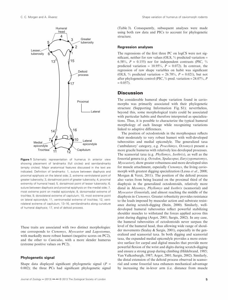
Morphological variation of the humerus was analyzedthrough geometric morphometric techniques. Specimens werephotographed in anterior view; each specimen was placed withthe plane formed by the diaphyseal axis and the transepi-condylar axis (Boileau & Walch, 1999) parallel to the cameralens, and with a ruler to record scale. Thirteen landmarks andfour semilandmarks (Fig. 1) were digitized using the softwaretpsDig 2.16 (Rohlf, 2010); all landmarks were digitized by thesame person (C. C. M.) and measurement error was assessedby the Procrustes analysis of variance (ANOVA) method(Klingenberg & McIntyre, 1998). The landmark + semiland-mark configurations were superimposed by GeneralizedProcrustes Analysis (Goodall, 1991; Rohlf, 1999) to removedifferences in location, orientation and scaling (i.e. nonshapevariation); semilandmarks were slid using the minimumbending energy criterion (Bookstein, 1997; Adams, Rohlf &Slice, 2004) using tpsRelw 1.49 (Rohlf, 2010). Centroid sizeswere saved for subsequent analyses. A principal componentanalysis of the aligned Procrustes coordinates averaged by
genus was performed to explore shape variation amongcaviomorph genera, using the software MorphoJ 1.04a(Klingenberg, 2011).
The influence of phylogeny on shape variation was evalu-ated using the univariate K statistic (Blomberg, Garland &Ives, 2003). We also tested the phylogenetic signal of all shapevariation, that is, the Procrustes shape coordinates, throughthe multivariate tree length test (Laurin, 2004; Klingenberg &Gidaszewski, 2010). Significance of both statistics wasassessed through 10 000 permutations. Analyses of phyloge-netic signal were performed using the Picante package(Kembel et al., 2010) for R (ver. 2.11.1, R Development CoreTeam, 2009) and MorphoJ software (Klingenberg, 2011),respectively.
To analyze the association between humeral morphologicalvariation and ecology, we built variables for two factorsclosely associated with the latter: size and habit (Hildebrand,1985; Reilly & Wainwright, 1994) and analyzed their relation-ship with shape variation by ordinary least squares (OLS)regression analyses. As size variable, we used the log-transformed centroid size averaged for each genus; centroidsize is used in geometric morphometrics as a measure of sizethat is uncorrelated with shape for small isotropic landmarkvariation (Mitteroecker & Gunz, 2009). The habit variablewas built from information available in the literature (sourcesdetailed in Table 2), with four habit categories represented bydummy variables. Because habit categories are not exclusiveand most caviomorphs are not greatly constrained to anyparticular locomotor mode (Elissamburu & Vizcaíno, 2004),those genera in which the relative involvement of the forelimbin running (cursorial), digging (fossorial) and/or climbing(scansorial/arboreal) activities is not predominant were classi-fied as generalized (‘ambulatory’). The arboreal Coendou waspooled with scansorial taxa in a single habit category. The firstthree principal components (PC) of the shape analysis (84.43%of the total variation) were retained as shape variables,based on both the Kaiser–Guttman and broken-stick criteria(Legendre & Legendre, 1998). Regressions were made usingraw data (OLS) and phylogenetic independent contrasts(PICs), to take into account the expected lack of indepen-dence among samples resulting from phylogenetic structure(Felsenstein, 1985; Rohlf, 2001). Regression significance wasassessed using permutation tests (10 000 rounds). The amountof variation accounted for by each regression model wasexpressed as a percentage of total variation, computed usingthe Procrustes metric (Drake & Klingenberg, 2008). Allregression analyses were made in MorphoJ. Morphofunc-tional interpretations were based on previous proposals(Hildebrand, 1985; Price, 1993; Vassallo, 1998; Elissamburu &Vizcaíno, 2004; Morgan & Verzi, 2006; Candela & Picasso,2008).
Results
Phylogeny
The phylogeny obtained (Fig. 2) was congruent with previouspartial hypotheses (Rowe & Honeycutt, 2002; Honeycutt,
Shape variation of humerus of caviomorph rodents C. C. Morgan and A. Álvarez
2 Journal of Zoology •• (2013) ••–•• © 2013 The Zoological Society of London
Rowe & Gallardo, 2003; Spotorno et al., 2004; Blanga-Kanfiet al., 2009) and with Upham & Patterson’s (2012) moreencompassing analysis, with some discrepancies probablydue to the fact that these authors used a partially different setof genes. The traditionally recognized clades within eachsuperfamily were recovered: Abrocomidae, Octodontidae,
Ctenomyidae and Echimyidae (Octodontoidea), Caviidae,Dasyproctidae and Cuniculidae (Cavioidea), Chinchillidae(=Chinchilloidea in this case) and Erethizontidae (=Erethizon-toidea). Posterior probabilities were moderate to high (>0.74);estimated parameters for Bayesian trees are given in Support-ing Information Appendix S2.
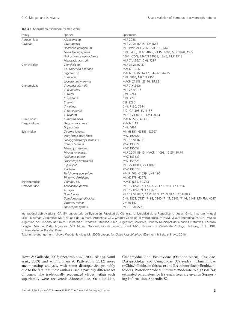
Table 1 Specimens examined for this work
Family Species Specimens
Abrocomidae Abrocoma sp. MLP 2038Caviidae Cavia aperea MLP 29.XII.00.15, 5.VI.00.8
Dolichotis patagonum MLP Prov. 213, 236, 250, 275, 642Galea leucoblephara CML 3430, 3432, 4875, 7136, 7240; MLP 1928, 1929Hydrochoerus hydrochaeris CZV1, CZV2, MACN 14038, 43.43, MLP 1915Microcavia australis MLP 7.VI.99.7, CML 7237
Chinchillidae Chinchilla sp. MLP 31.XII.02.37Ch. chinchilla boliviana MACN 13037Lagidium sp MACN 14.16, 14.17, 34–263, 44.25L. viscacia CML 5099, MACN 7350Lagostomus maximus MACN 21983, 23.14, 39.92
Ctenomyidae Ctenomys australis MLP 7.XI.95.6C. flamarioni MLP 28.V.01.5C. frater CML 7241C. sylvanus CML 7235C. lewisi CBF 2280C. opimus CML 7130, 7244C. rionegrensis 412, CA 393; EV 1137C. talarum MLP 1.VIII.00.11, 1.VIII.00.14
Cuniculidae Cuniculus paca MACN 22.5, 49396Dasyproctidae Dasyprocta azarae MACN 1.11
D. punctata CML 4695Echimyidae Clyomys laticeps MN 63851, 63853, 68967
Dactylomys dactylinus MVZ 190620Euryzygomatomys spinosus MLP 16.VII.02.11Isothrix bistriata MVZ 190629Mesomys hispidus MVZ 190653Myocastor coypus MLP 20.XII.89.15; MACN 14098, 15.20, 30.70Phyllomys pattoni MVZ 183139Proechimys brevicauda MVZ 153623P. poliopus MLP 22.II.00.7, 22.II.00.8P. roberti MVZ 197578Thrichomys apereoides MN 34406, 61659; UNB 190Trinomys dimidiatus MN 62273, 62278
Erethizontidae Coendou sp. MACN 6.34, 30.243Octodontidae Aconaemys porteri MLP 17.II.92.07, 17.II.92.2, 17.II.92.3, 17.II.92.4
A. sagei MLP 17.II.92.09, 17.II.92.10Octodon sp. MLP 12.VII.88.2, 12.VII.88.3, 12.VII.88.5, 12.VII.88.7Octodontomys gliroides CML 2872, 7137, 7138, 7140, 7144, 7145, 7146, 7148; MMPMa 4027Octomys mimax CM 06847Spalacopus cyanus MLP 10.XI.95.5
Institutional abbreviations: CA, EV, Laboratorio de Evolución, Facultad de Ciencias, Universidad de la República, Uruguay; CML, Instituto ‘MiguelLillo’, Tucumán, Argentina; MLP, Museo de La Plata, Argentina; CZV, Cátedra Zoología III Vertebrados, FCNyM, UNLP, Argentina; MACN, MuseoArgentino de Ciencias Naturales ‘Bernardino Rivadavia’, Buenos Aires, Argentina; MMPMa, Museo Municipal de Ciencias Naturales ‘LorenzoScaglia’, Mar del Plata, Argentina; MN, Museu Nacional, Rio de Janeiro, Brazil; MVZ, Museum of Vertebrate Zoology, Berkeley, USA; UNB,Universidade de Brasília, Brazil.Taxonomic arrangement follows Woods & Kilpatrick (2005) except for Galea leucoblephara (Dunnum & Salazar-Bravo, 2010).
C. C. Morgan and A. Álvarez Shape variation of humerus of caviomorph rodents
Journal of Zoology •• (2013) ••–•• © 2013 The Zoological Society of London 3

Morphometric analyses
Measurement error
According to the Procrustes ANOVA, the level of measure-ment error is negligible compared to individual variation(Supporting Information Appendix S3).
Principal components analysis
The first two PC summarized 77.43 % of the shape variation(PC1 = 61.04%; PC2 = 16.39%). In the morphospace of thesefirst two PC (Fig. 3), most of the taxa were located near theorigin along both axes. Within this central space, the scanso-rial echimyids Phyllomys and Mesomys were close to eachother and to the cursorial cavioid Hydrochoerus. Outside ofthis sector, the fossorial octodontoids (the echimyids Clyomysand Euryzygomatomys, and the octodontids Octodon, Spal-acopus and Aconaemys) formed a distinct group. Along themain axis of variation (PC1), the caviids (minus Hydroch-oerus) occupied extreme positive values, while the specializeddigging ctenomyid Ctenomys, the fossorial chinchillid Lagos-tomus and the generalized cavioid Cuniculus were located at
the negative end. Ctenomys also occupied the extreme negativevalues of PC2 along with the semiaquatic occasional diggerMyocastor, while the extreme positive values along this axiscorresponded to Cuniculus and another cavioid, the cursorialDasyprocta.
The taxa distributed in the central portion of the morpho-space share a humeral morphology characterized by moder-ately slender diaphysis, proximal epiphysis with medium-sizedtuberosities and humeral head almost level with the greatertuberosity, distal epiphysis with moderate entepicondyledevelopment, and a deltoid process located on the proximalthird of the diaphysis. In comparison, the humeri of fossorialoctodontoids (Clyomys, Euryzygomatomys, Octodon, Spal-acopus and Aconaemys) are more robust, with relativelybroader proximal and distal epiphyses. The scansorial Phyllo-mys and Mesomys also show more robust humeri, with thedeltoid process extending more distally along the diaphysis.The humeri of the taxa located at extreme positive values ofPC1 have narrow epiphyses, a high greater tuberosity thatsurpasses the level of the articular head, and a relatively moreproximal deltoid process. The taxa with the most negativevalues along PC1 have broader epiphyses, especially theentepicondyle, and a more distally extended deltoid process.
Table 2 Habit categories used in this study for analyzed taxa
Taxon Habit (for the genus) Body mass (g) Taxon Habit (for the genus) Body mass (g)
Cavioidea OctodontoideaCaviidae Abrocomidae
Cavia aperea Ambulatorya 6501 Abrocoma sp Ambulatorya 1482
Microcavia australis Digging h,i 2751 OctodontidaeGalea leucoblephara Ambulatory i 2351 Aconaemys sagei Digginga 1163
Dolichotis patagonum Cursorial b 12 0001 Spalacopus cyanus Digginga 1404
Hydrochoerus hydrochaeris Cursorial a 53 0001 Octodon degus Digginga 1505
Dasyproctidae Octodontomys gliroides Ambulatoryf 903
Dasyprocta azarae Cursoriala 27001 Octomys mimax Ambulatoryg 1036
Cuniculidae CtenomyidaeCuniculus paca Ambulatory a,e 11 0001 Ctenomys talarum Digginga 1407
EchimyidaeErethizontoidea Clyomys laticeps Diggingc 17210
Erethizontidae Dactylomys dactylinus Scansoriald 75010
Coendou prehensilis Scansoriala 45001 Euryzygomatomys spinosus Diggingc 18510
Isothrix negrensis Scansoriala 41010
Chinchilloidea Mesomys hispidus Scansorial a,d 16010
Chinchillidae Myocastor coypus Digginga 70001
Chinchilla lanigera Ambulatory 4351 Phyllomys pattoni Scansorialc,d 21210
Lagidium viscacia Ambulatory 15001 Proechimys albispinus Ambulatorya 1858
Lagostomus maximus Digginga 65001 Thrichomys apereoides Ambulatorya 3409
Trinomys albispinus Ambulatory a,d 17510
Sources of habit information: aNowak (1991); bSeckel & Janis (2008); cEisenberg & Redford (1999), dBonvincino, de Oliveira & D’Andrea (2008); ePérez(1992); fLessa et al. (2008); gSobrero et al. (2010); hUbilla (2008); iEbensperger & Blumstein (2006).Sources of body mass information: 1Canevari & Vaccaro (2007); 2Data from specimen labels (IADIZA); 3Vassallo & Echeverría (2009); 4Torres-Mura& Contreras (1998); 5Woods & Boraker (1975); 6Sobrero et al. (2010); 7Vassallo (1998); 8Pessôa & dos Reis (2002); 9dos Reis & Pessôa (2004);10Bonvincino, de Oliveira & D’Andrea (2008).The ‘scansorial’ category also includes the arboreal Coendou (see Materials and Methods). Mean body mass is expressed in grams; valuescorrespond to species included in these analyses (when such data were not available, the value corresponds to a congeneric species). Thesystematic arrangement follows Woods & Kilpatrick (2005) except for Myocastor, which is placed within the Echimyidae following Galewski et al.(2005).
Shape variation of humerus of caviomorph rodents C. C. Morgan and A. Álvarez
4 Journal of Zoology •• (2013) ••–•• © 2013 The Zoological Society of London
These traits are associated with two distinct morphologies:one corresponds to Ctenomys, Myocastor and Lagostomus,with markedly more robust humeri (negative scores on PC2),and the other to Cuniculus, with a more slender humerus(extreme positive values on PC2).
Phylogenetic signal
Shape data displayed significant phylogenetic signal (P =0.002); the three PCs had significant phylogenetic signal
(Table 3). Consequently, subsequent analyses were madeusing both raw data and PICs to account for phylogeneticstructure.
Regression analyses
The regressions of the first three PC on logCS were not sig-nificant, neither for raw values (OLS, % predicted variation =6.58%, P = 0.155) nor for independent contrasts (PIC, %predicted variation = 10.95%, P = 0.072). In contrast, theregression of raw shape variables on habit was significant(OLS, % predicted variation = 26.70%, P = 0.021), but notafter phylogenetic control (PIC, % pred. variation = 24.07%, P= 0.057).
DiscussionThe considerable humeral shape variation found in cavio-morphs was primarily associated with their phylogeneticstructure (Supporting Information Fig. S1); nevertheless,beyond this, some morphological traits could be associatedwith particular habits and therefore interpreted as specializa-tions. Thus, it is possible to characterize the typical humeralmorphology of each lineage while recognizing variationslinked to adaptive differences.
The position of octodontoids in the morphospace reflectstheir moderately to very robust humeri with well-developedtuberosities and medial epicondyle. The generalized taxa(‘ambulatory’ category, e.g. Proechimys, Octomys) present amore gracile humerus with relatively less developed processes.The scansorial taxa (e.g. Phyllomys, Isothrix), as well as thefossorial genera (e.g. Octodon, Spalacopus; Euryzygomatomys,Myocastor), show greater robustness and more developed sitesfor muscle attachment, especially Ctenomys, the living cavio-morph with greatest digging specialization (Lessa et al., 2008;Morgan & Verzi, 2011). The position of the deltoid processalso varies from being located on the proximal third of thediaphysis in the generalized octodontoids, relatively moredistal in Mesomys, Phyllomys and Isothrix (scansorial) andMyocastor (fossorial), and almost reaching the middle of thediaphysis in Ctenomys. Greater robusticity provides resistanceto the loads imposed by muscular action and substrate resist-ance during scratch-digging (Stein, 2000). Similarly, well-developed humeral tuberosities reflect powerful stabilizingshoulder muscles to withstand the forces applied across thisjoint during digging (Argot, 2001; Sargis, 2002). In any case,the humeral tuberosities of octodontoids never surpass thelevel of the humeral head, thus allowing wide range of shoul-der movements (Szalay & Sargis, 2001), especially in the gen-eralized and scansorial taxa. In both digging and scansorialtaxa, the expanded medial epicondyle provides a more exten-sive surface for carpal and digital muscles that provide morepowerful flexion of the wrist and digits during scratch-diggingand ensure a strong grasp during climbing (Hildebrand, 1985;Van Valkenburgh, 1987; Argot, 2001; Sargis, 2002). Similarly,the distal extension of the deltoid process observed in scanso-rial and some fossorial taxa enhances mechanical advantageby increasing the in-lever arm (i.e. distance from muscle
1
2
34
5
6
78 9
10
11 12
13
14
15
17
16
Proximal
Lateral
Deltoidprocess
Medialepicondyle
Lateralepicondyle
Greatertuberosity
Lessertuberosity
Humeralhead
Trochlea Capitulum
Figure 1 Schematic representation of humerus in anterior viewshowing placement of landmarks (full circles) and semilandmarks(empty circles). Major anatomical features discussed in the text areindicated. Definition of landmarks: 1, suture between diaphysis andproximal epiphysis on the lateral side; 2, extreme ventrolateral point ofgreater tuberosity; 3, dorsalmost point of greater tuberosity; 4, proximalextremity of humeral head; 5, dorsalmost point of lesser tuberosity; 6,suture between diaphysis and proximal epiphysis on the medial side; 7,most extreme point on medial epicondyle; 8, dorsomedial extreme oftrochlea; 9, dorsolateral extreme of capitulum; 10, most extreme pointon lateral epicondyle; 11, ventromedial extreme of trochlea; 12, vent-rolateral extreme of capitulum; 13–16, semilandmarks along curvatureof humeral diaphysis; 17, end of deltoid process.
C. C. Morgan and A. Álvarez Shape variation of humerus of caviomorph rodents
Journal of Zoology •• (2013) ••–•• © 2013 The Zoological Society of London 5

attachment to joint) of the deltoid and pectoral musclesthat contribute to forelimb retraction (Hildebrand, 1985;Fernández, Vassallo & Zárate, 2000; Stein, 2000).
The two subfamilies of Chinchillidae show divergent mor-phologies: the humerus of chinchillines Lagidium and Chin-chilla is gracile and shares the morphospace of the generalizedoctodontoids, while that of the lagostomine Lagostomus ismore similar to that of the digging octodontoids. This dispar-ity between the clades also agrees with their different habits:chinchillines use a half-bounding gait to move across rockyterrain (Lammers & German, 2002) that is not likely to exertstrong demand on the humerus, while the fossorial viscachasconstruct communal burrow systems presumably using theirforelimbs (Fernández, 1949), and are thus under similarmechanical requirements as other scratch-diggers.
The distinctive humeral morphology of cavioids isreflected by their position in the morphospace, mostly sepa-rated from other caviomorphs. In general, their humeri arecomparatively slenderer, with narrow epiphyses and stronglydeveloped greater tuberosity that surpasses the level of thehumeral head. The position of the deltoid process variesfrom markedly proximal in the caviine cavids (Cavia,
Microcavia and Galea) to similar to that of most othercaviomorphs in Dasyprocta, Dolichotis and Hydrochoerus.Cuniculus is quite different from the remaining cavioids: thehumerus is more robust, with a markedly distal deltoidprocess. In morphofunctional terms, the large greater tuber-osity restricts the range of shoulder movements to the par-asagittal plane; such restriction is characteristic of cursorialspecies (Argot, 2001; Sargis, 2002; Salton & Sargis, 2008).Concurrently, the proximally located deltoid process opti-mizes speed over force production, as expected for cursorialforms (Hildebrand, 1985; Polly, 2007). However, this struc-ture is relatively more distal in Hydrochoerus; this may berelated to different mechanical requirements linked to thelarge size of this rodent. The robust humerus of Cuniculus,with well-developed distal epiphysis and more distallyextended deltoid process, may reflect the wide functionalspectrum of this genus, which includes running, swimmingand digging (Pérez, 1992). Likewise, the greater robusticityobserved in Microcavia compared to other caviines couldalso be associated with its digging habit.
The only erethizontoid included in this analysis, the arbo-real Coendou, was close in the morphospace to the scansorial
Figure 2 Phylogenetic relationships of extant South American caviomorph rodents included in this study. Superfamilial and familial clades areindicated. Branch lengths represent molecular substitutions. Posterior probabilities different from 1 are indicated on corresponding nodes. Topologyis congruent with previous analyses (Huchon & Douzery, 2001; Rowe & Honeycutt, 2002; Honeycutt et al., 2003; Ledesma et al., 2009). Circlesbeside each taxon indicate habit: full gray, cursorial; full black, digging; full white, ambulatory; black border with gray fill, scansorial (includingarboreal). Scale: substitutions per site.
Shape variation of humerus of caviomorph rodents C. C. Morgan and A. Álvarez
6 Journal of Zoology •• (2013) ••–•• © 2013 The Zoological Society of London
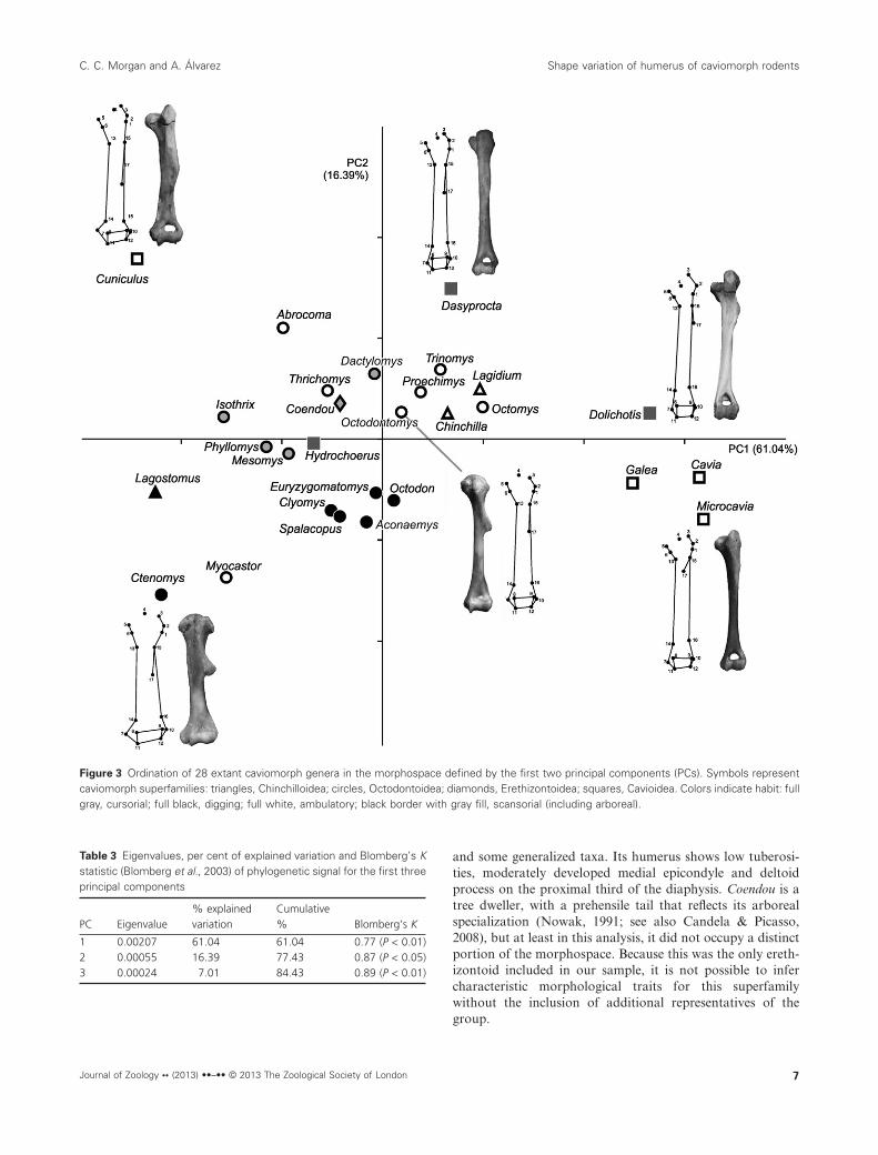
and some generalized taxa. Its humerus shows low tuberosi-ties, moderately developed medial epicondyle and deltoidprocess on the proximal third of the diaphysis. Coendou is atree dweller, with a prehensile tail that reflects its arborealspecialization (Nowak, 1991; see also Candela & Picasso,2008), but at least in this analysis, it did not occupy a distinctportion of the morphospace. Because this was the only ereth-izontoid included in our sample, it is not possible to infercharacteristic morphological traits for this superfamilywithout the inclusion of additional representatives of thegroup.
Figure 3 Ordination of 28 extant caviomorph genera in the morphospace defined by the first two principal components (PCs). Symbols representcaviomorph superfamilies: triangles, Chinchilloidea; circles, Octodontoidea; diamonds, Erethizontoidea; squares, Cavioidea. Colors indicate habit: fullgray, cursorial; full black, digging; full white, ambulatory; black border with gray fill, scansorial (including arboreal).
Table 3 Eigenvalues, per cent of explained variation and Blomberg’s Kstatistic (Blomberg et al., 2003) of phylogenetic signal for the first threeprincipal components
PC Eigenvalue% explainedvariation
Cumulative% Blomberg’s K
1 0.00207 61.04 61.04 0.77 (P < 0.01)2 0.00055 16.39 77.43 0.87 (P < 0.05)3 0.00024 7.01 84.43 0.89 (P < 0.01)
C. C. Morgan and A. Álvarez Shape variation of humerus of caviomorph rodents
Journal of Zoology •• (2013) ••–•• © 2013 The Zoological Society of London 7
ConclusionsThe association between humeral shape and both phylogenyand habit suggests an early ecomorphological diversification ofcaviomorph rodents accompanying their phylogenetic diver-gence. Our analysis showed that the major lineages of the cladecould be distinguished by characteristic humeral features, eventhose taxa that were not so close in the morphospace. Similarphylogenetic constraints on morphological patterns have beenobserved for several mammalian groups (e.g. scandentians,Sargis, (2002); carnivorans, Meloro & O’Higgins, 2011,Meloro et al., 2011; New World platyrrhines, Perez et al.,2011). At the same time, in a context of characteristic suprafa-milial morphologies, habits were associated with particularmorphological traits interpreted as specializations, in agree-ment with previous qualitative and quantitative analyses(Elissamburu & Vizcaíno, 2004; Candela & Picasso, 2008).Interestingly, Samuels and Van Valkenburgh’s (2008) analysisof locomotor adaptations in rodents showed that fossorial taxahad more robust humeri with well-developed muscular attach-ment sites, in agreement with our results; however, they did notfind major differences in humeral morphology among otherlocomotor habit categories. This discrepancy with the presentresults could be attributed to different methodologicalapproaches (indexes from linear measurements vs. geometricmorphometrics) and also probably to different taxonomicalrange of the respective samples.
To date, only a few studies have focused on macroevolution-ary processes driving the morphological variation of cavio-morph rodents. Craniomandibular shape variation has beenshown to have significant phylogenetic signal and simultane-ously a strong allometric component (Álvarez, 2012). In con-trast, variations in scapular shape agree with the phylogeneticpattern and are not associated with the different habits of theserodents (Morgan, 2009). It seems evident that complex factorshave disparate influence on different skeletal elements amongcaviomorphs, and may facilitate or constrain ecomorphologi-cal evolution within each lineage (Miles & Dunham, 1993;Losos & Miles, 1994). These results emphasize the importanceof performing further comparative analyses to achieve a betterperspective regarding the evolution of morphological disparityin South American caviomorphs.
AcknowledgementsWe thank D. Verzi for critical reading of the manuscript, S.I.Perez for methodological advice and two anonymous review-ers whose comments greatly improved this work. J. Aparicio,R. Bárquez, D. Flores, J. Marinho-Filho, A.I. Olivares, J.Oliveira, D. Romero and J. Vargas Mattos granted access tomaterials deposited in mammalogical collections under theircare. This work was funded by grants PICT 01744 (ANPCyT)and PIP 0270 (CONICET).
References
Adams, D.C., Rohlf, F.J. & Slice, D.E. (2004). Geometricsmorphometrics: ten years of progress following the ‘revolu-tion’. Ital. J. Zool. 71, 5–16.
Álvarez, A. (2012). Diversidad morfológica cráneo-mandibularde roedores caviomorfos en un contexto filogenético compara-tivo. PhD thesis, Universidad Nacional de La Plata,Argentina.
Argot, C. (2001). Functional-adaptive anatomy of the fore-limb in the Didelphidae, and the paleobiology of the Pale-ocene marsupials Mayulestes ferox and Pucadelphysandinus. J. Morphol. 247, 51–79.
Biknevicius, A.R. (1993). Biomechanical scaling of limb bonesand differential limb use in caviomorph rodents. J. Mamm.74, 95–107.
Blanga-Kanfi, S., Miranda, H., Penn, O., Pupko, T., DeBry,R.W. & Huchon, D. (2009). Rodent phylogeny revised:analysis of six nuclear genes from all major rodent clades.BMC Evol. Biol. 9, 71.
Blomberg, S.P., Garland, J.T. & Ives, A.R. (2003). Testing forphylogenetic signal in comparative data: behavioral traitsare more labile. Evolution 57, 171–745.
Boileau, P. & Walch, G. (1999). The three-dimensional geom-etry of the proximal humerus. Implications for surgicaltechnique and prosthetic design. J. Bone Joint Surg. 79B,857–865.
Bonvincino, C.R., de Oliveira, J.A. & D’Andrea, P.S. (2008).Guia dos roedores do Brasil, com chaves para géneros base-adas em caracteres externos. Rio de Janeiro: Centro Pan-Americano de Febre Aftosa – OPAS/OMS.
Bookstein, F.L. (1997). Landmark methods for forms withoutlandmarks: morphometrics of group differences in outlineshape. Med. Image Anal. 1, 225–243.
Candela, A.M. & Picasso, M.B.J. (2008). Functional anatomyof the limbs of Erethizontidae (Rodentia: Caviomorpha):indicators of locomotor behavior in Miocene porcupines.J. Morphol. 269, 552–593.
Canevari, M. & Vaccaro, O. (2007). Guía de mamíferos del surde América del Sur. Buenos Aires: L.O.L.A.
Drake, A.G. & Klingenberg, C.P. (2008). The pace of mor-phological change: historical transformation of skull shapein St. Bernard dogs. Proc. R. Soc. B 275, 71–76.
Dunnum, J.L. & Salazar-Bravo, J. (2010). Phylogeny, evolu-tion, and systematics of the Galea musteloides complex(Rodentia: Caviidae). J. Mamm. 91, 243–259.
Ebensperger, L.A. & Blumstein, D.T. (2006). Sociality in NewWorld hystricognath rodents is linked to predators andburrow digging. Behav. Ecol. 17, 410–418.
Eisenberg, J.F. & Redford, K.H. (1999). Mammals of the neo-tropics. Vol. 3: the central neotropics – Ecuador, Peru,Bolivia, Brazil. Chicago: University of Chicago Press.
Elissamburu, A. & De Santis, L. (2011). Forelimb proportionsand fossorial adaptations in the scratch-digging rodentCtenomys (Caviomorpha). J. Mamm. 92, 683–689.
Elissamburu, A. & Vizcaíno, S.F. (2004). Limb proportionsand adaptations in caviomorph rodents (Rodentia: Cavio-morpha). J. Zool. (Lond.) 262, 145–159.
Felsenstein, J. (1985). Phylogenies and the comparativemethod. Am. Nat. 125, 1–15.
Shape variation of humerus of caviomorph rodents C. C. Morgan and A. Álvarez
8 Journal of Zoology •• (2013) ••–•• © 2013 The Zoological Society of London
Fernández, M. (1949). Sobre la vizcacha (Lagostomus trichod-actylus Brooks), sus viviendas y su protección. Bol. Acad.Nac. Cienc. 38, 348–379.
Fernández, M.E., Vassallo, A.I. & Zárate, M. (2000). Func-tional morphology and paleobiology of the Pliocene rodentActenomys (Caviomorpha: Octodontidae): the evolution toa subterranean mode of life. Biol. J. Linn. Soc. 71, 79–90.
Galewski, T., Mauffrey, J.F., Leite, Y.L., Patton, J.L. &Douzery, E.J. (2005). Ecomorphological diversificationamong South American spiny rats (Rodentia; Echimyidae):a phylogenetic and chronological approach. Mol. Phylo-genet. Evol. 34, 601–615.
Goodall, C. (1991). Procrustes methods in the statisticalanalysis of shape. J. R. Stat. Soc. B 53, 285–339.
Hildebrand, M. (1985). Digging in quadrupeds. In Functionalvertebrate morphology: 89–109. Hildebrand, M., Bramble,D.M., Liem, K.F. & Wake, D.B. (Eds). Cambridge:Harvard University Press.
Hildebrand, M. (1988). Analysis of vertebrate structure. NewYork: John Wiley & Sons.
Honeycutt, R.L., Rowe, D.L. & Gallardo, M.H. (2003).Molecular systematics of the South American caviomorphrodents: relationships among species and genera in thefamily Octodontidae. Mol. Phylogenet. Evol. 26,476–489.
Huchon, D. & Douzery, E.J.P. (2001). From the Old Worldto the New World: a molecular chronicle of the phylogenyand biogeography of hystricognath rodents. Mol. Phylo-genet. Evol. 20, 238–251.
Kembel, S.W., Cowan, P.D., Helmus, M.R., Cornwell, W.K.,Morlon, H., Ackerly, D.D., et al. (2010). Picante: R toolsfor integrating phylogenies and ecology. Bioinformatics 6,1463–1464.
Klingenberg, C.P. (2011). MorphoJ. Manchester: Faculty ofLife Sciences, University of Manchester.
Klingenberg, C.P. & Gidaszewski, N.A. (2010). Testing andquantifying phylogenetic signals and homoplasy in mor-phometric data. Syst. Biol. 59, 245–261.
Klingenberg, C.P. & McIntyre, G.S. (1998). Geometric mor-phometrics of developmental instability: analyzing patternsof fluctuating asymmetry with Procrustes methods. Evolu-tion 52, 1363–1375.
Lammers, A.R. & German, R.Z. (2002). Ontogenetic allom-etry in the locomotor skeleton of specialized half-boundingmammals. J. Zool. (Lond.) 258, 485–495.
Laurin, M. (2004). The evolution of body size, Cope’s ruleand the origin of Amniotes. Syst. Biol. 53, 594–622.
Ledesma, K.J., Werner, F.A., Spotorno, A.E. & Albuja, L.H.(2009). A new species of mountain viscacha (Chinchillidae:Lagidium Meyen) from the Ecuadorean Andes. Zootaxa2126, 41–57.
Legendre, P. & Legendre, L. (1998). Numerical ecology.Amsterdam: Elsevier.
Lehmann, W.H. (1963). The forelimb architecture of somefossorial rodents. J. Morphol. 113, 59–76.
Lessa, E.P., Vassallo, A.I., Verzi, D.H. & Mora, M.S. (2008).Evolution of morphological adaptations for digging inliving and extinct ctenomyid and octodontid rodents. Biol.J. Linn. Soc. 95, 267–283.
Losos, J.B. & Miles, D.B. (1994). Adaptation, constraint, andthe comparative method: phylogenetic issues and methods.In Ecological morphology. Integrative organismal biology:60–98. Wainwright, P.C. (Ed.). Chicago: University ofChicago Press.
Meloro, C. & O’Higgins, P. (2011). Ecological adaptations ofmandibular form in fissiped Carnivora. J. Mamm. Evol. 18,185–200.
Meloro, C., Raia, P., Carotenuto, F. & Cobb, S.N. (2011).Phylogenetic signal, function and integration in the subu-nits of the carnivoran mandible. Evol. Biol. 38, 465–475.
Miles, D.B. & Dunham, A.E. (1993). Historical perspectives inecology and evolutionary biology: the use of phylogeneticcomparative analyses. Annu. Rev. Ecol. Syst. 24, 587–619.
Mitteroecker, P. & Gunz, P. (2009). Advances in geometricmorphometrics. Evol. Biol. 36, 235–247.
Morgan, C.C. (2009). Geometric morphometrics of thescapula of South American caviomorph rodents (Rodentia:Hystricognathi): form, function and phylogeny. Mamm.Biol. 74, 497–506.
Morgan, C.C. & Verzi, D.H. (2006). Morphological diversityof the humerus of the South American subterranean rodentCtenomys (Rodentia, Ctenomyidae). J. Mamm. 87, 1252–1260.
Morgan, C.C. & Verzi, D.H. (2011). Carpal-metacarpal spe-cializations for burrowing in South American octodontoidrodents. J. Anat. 219, 167–175.
Nowak, R.M. (1991). Walker’s mammals of the world. Balti-more: Johns Hopkins University Press.
Perez, S.I., Klaczko, J., Rocatti, G. & dos Reis, S.F. (2011).Patterns of cranial shape diversification during the phyloge-netic branching process of New World monkeys (Primates:Platyrrhini). J. Evol. Biol. 24, 1826–1835.
Pérez, E.M. (1992). Agouti paca. Mamm. Species 404, 1–7.Pessôa, M.L. & dos Reis, S.F. (2002). Proechimys albispinus.
Mamm. Species 693, 1–3.Polly, P.D. (2007). Limbs in mammalian evolution. In Fins into
Limbs. Evolution, development and transformation: 245–268.Hall, B.K. (Ed.). Chicago: University of Chicago Press.
Posada, D. (2008). jModelTest: phylogenetic model averaging.Mol. Biol. Evol. 25, 1253–1256.
Price, M.V. (1993). A functional-morphometric analysis offorelimbs in bipedal and quadripedal heteromyid rodents.Biol. J. Linn. Soc. 50, 339–360.
R Development Core Team (2009). R: a language and environ-ment for statistical computing. Vienna: R Foundation forStatistical Computing. See http://www.r-project.org/
Reilly, S.M. & Wainwright, P.C. (1994). Conclusion: ecologi-cal morphology and the power of integration. In Ecologicalmorphology. Integrative organismal biology: 339–354. Wain-wright, P.C. (Ed.). Chicago: University of Chicago Press.
C. C. Morgan and A. Álvarez Shape variation of humerus of caviomorph rodents
Journal of Zoology •• (2013) ••–•• © 2013 The Zoological Society of London 9
dos Reis, S.F. & Pessôa, M.L. (2004). Thrichomys apereoides.Mamm. Species 741, 1–5.
Rocha-Barbosa, O. & Casinos, A. (2011). Geometry and evo-lutionary parallelism in the long bones of cavioid rodentsand small artiodactyls. J. Biosci. 36, 1–9.
Rocha-Barbosa, O., Youlatos, D., Gasc, J.-P. & Renous, S.(2002). The clavicular region of some cursorsCavioidea (Rodentia: Mammalia). Mammalia 66, 413–421.
Rocha-Barbosa, O., Loguercio, M.F.C., Renous, S. & Gasc,J.-P. (2007). Comparative study on the forefoot and hind-foot intrinsic muscles of some cavioidea rodents (Mamma-lia, Rodentia). Zoology 110, 58–65.
Rohlf, F.J. (1999). Shape statistics: procrustes superimposi-tions and tangent spaces. J. Classif 16, 197–223.
Rohlf, F.J. (2001). Comparative methods for the analysis ofcontinuous variables: geometric interpretations. Evolution55, 2143–2160.
Rohlf, F.J. (2010). tps series software. Available at http://life.bio.sunysb.edu/morph
Ronquist, F. & Huelsenbeck, J.P. (2003). MRBAYES 3:Bayesian phylogenetic inference under mixed models. Bioin-formatics 19, 1572–1574.
Rowe, D.L. & Honeycutt, R.L. (2002). Phylogenetic relation-ships, ecological correlates, and molecular evolution withinthe Cavioidea (Mammalia: Rodentia). Mol. Biol. Evol. 19,263–277.
Salton, J.A. & Sargis, E.J. (2008). Evolutionary morphologyof the Tenrecoidea (Mammalia) forelimb skeleton. InMammalian evolutionary morphology: a tribute to FredrickS. Szalay: 51–71. Sargis, E.J. & Dagosto, M. (Eds). Dor-drecht: Springer.
Samuels, J.X. & Van Valkenburgh, B. (2008). Skeletal indica-tors of locomotor adaptations in living and extinct rodents.J. Morphol. 269, 1387–1411.
Sargis, E.J. (2002). Functional morphology of the forelimb oftupaiids (Mammalia, Scandentia) and its phylogeneticimplications. J. Morphol. 253, 10–42.
Seckel, L. & Janis, C. (2008). Convergences in scapula mor-phology among small cursorial mammals: an osteologicalcorrelate for locomotory specialization. J. Mamm. Evol. 15,261–279.
Sobrero, R., Campos, V.E., Giannoni, S.M. & Ebensperger,L.A. (2010). Octomys mimax (Rodentia: Octodontidae).Mamm. Species 42, 49–57.
Spotorno, A.E., Valladares, J.P., Marin, J.C., Palma, E. &Zuleta, C.R. (2004). Molecular divergence and phylogeneticrelationships of chinchillids (Rodentia: Chinchillidae).J. Mamm. 85, 384–388.
Stein, B.R. (2000). Morphology of subterranean rodents. InLife underground, the biology of subterranean rodents:19–61. Lacey, A.E., Patton, J.L. & Cameron, G.N. (Eds).Chicago: The University of Chicago Press.
Steiner-Souza, F., de Freitas, T.R.O. & Cordeiro-Estrela, P.(2010). Inferring adaptation within shape diversity of the
humerus of subterranean rodent Ctenomys. Biol. J. Linn.Soc. 100, 353–367.
Szalay, F.S. & Sargis, E.J. (2001). Model-based analysis ofpostcranial osteology of marsupials from the Palaeocene ofItaboraí (Brazil) and the phylogenetics and biogeographyof Metatheria. Geodiversitas 23, 139–302.
Torres-Mura, J.C. & Contreras, L.C. (1998). Spalacopuscyanus. Mamm. Species 594, 1–5.
Ubilla, M. (2008). Postcranial morphology of the extinctcaviine rodent Microcavia criolloensis (late Pleistocene,South America). Zool. J. Linn. Soc. 154, 795–806.
Upham, N.S. & Patterson, B.D. (2012). Diversification andbiogeography of the Neotropical caviomorph lineage Octo-dontoidea (Rodentia: Hystricognathi). Mol. Phylogenet.Evol. 63, 417–429.
Van Valkenburgh, B. (1987). Skeletal indicators of locomotorbehavior in living and extinct carnivores. J. Vert. Paleont.7, 162–182.
Vassallo, A.I. (1998). Functional morphology, comparativebehaviour, and adaptation in two sympatric subterraneanrodents genus Ctenomys (Caviomorpha: Octodontidae).J. Zool. (Lond.) 244, 415–427.
Vassallo, A.I. & Echeverría, A.I. (2009). Evolution of brainsize in a highly diversifying lineage of subterranean rodentgenus Ctenomys (Caviomorpha: Ctenomyidae). BrainBehav. Evol. 73, 138–149.
Weisbecker, V. & Schmid, S. (2007). Autopodial skeletaldiversity in hystricognath rodents: functional and phyloge-netic aspects. Mamm. Biol. 72, 27–44.
Woods, C.A. & Boraker, D.K. (1975). Octodon degus. Mamm.Species 67, 1–5.
Woods, C.A. & Kilpatrick, C. (2005). Infraorder Hystricog-nathi. In Mammal species of the world. A taxonomic andgeographic reference: 1538–1600. Wilson, D.E. & Reeder,D.M. (Eds). Baltimore: Johns Hopkins University Press.
Supporting InformationAdditional Supporting Information may be found in theonline version of this article at the publisher’s web-site:
Figure S1. Ordination of 28 extant caviomorph genera inthe morphospace defined by the first two principal compo-nents (PCs) with the obtained phylogeny superimposedaccording to reconstructed ancestral values.
Appendix S1. GenBank accession numbers for the 12S,cytb, GHR and TTH gene sequences for caviomorph rodentsanalysed in this work.
Appendix S2. Parameters from the Bayesian analysis ofgene sequences used in this work.
Appendix S3. Procrustes ANOVA test for measurementerror. Individual effect represents overall variation, and Error1 is the measurement error calculated for repeat measure-ments. SS: sum of squares; MS: mean squares, d.f.: degrees offreedom; F: F statistic; p: P-value.
Shape variation of humerus of caviomorph rodents C. C. Morgan and A. Álvarez
10 Journal of Zoology •• (2013) ••–•• © 2013 The Zoological Society of London