The frequency-dependent effect of ELF-EMF and MV at IS frequency on the growth, division and...
11
1 23 The Environmentalist ISSN 0251-1088 Environmentalist DOI 10.1007/s10669-011-9365-2 The frequency-dependent effect of extremely low-frequency electromagnetic field and mechanical vibration at infrasound frequency on the growth, division and motility of Escherichia coli K-12 Varsik Martirosyan, Levon Markosyan, Hrachik Hovhanesyan, Karlen Hovnanyan & Sinerik Ayrapetyan
-
Upload
independent -
Category
Documents
-
view
0 -
download
0
Transcript of The frequency-dependent effect of ELF-EMF and MV at IS frequency on the growth, division and...
1 23
The Environmentalist ISSN 0251-1088 EnvironmentalistDOI 10.1007/s10669-011-9365-2
The frequency-dependent effect ofextremely low-frequency electromagneticfield and mechanical vibration atinfrasound frequency on the growth,division and motility of Escherichia coliK-12Varsik Martirosyan, Levon Markosyan,Hrachik Hovhanesyan, KarlenHovnanyan & Sinerik Ayrapetyan

1 23
Your article is protected by copyright and
all rights are held exclusively by Springer
Science+Business Media, LLC. This e-offprint
is for personal use only and shall not be self-
archived in electronic repositories. If you
wish to self-archive your work, please use the
accepted author’s version for posting to your
own website or your institution’s repository.
You may further deposit the accepted author’s
version on a funder’s repository at a funder’s
request, provided it is not made publicly
available until 12 months after publication.
The frequency-dependent effect of extremely low-frequencyelectromagnetic field and mechanical vibration at infrasoundfrequency on the growth, division and motility of Escherichia coliK-12
Varsik Martirosyan • Levon Markosyan •
Hrachik Hovhanesyan • Karlen Hovnanyan •
Sinerik Ayrapetyan
� Springer Science+Business Media, LLC 2011
Abstract The aim of this work was to investigate the
frequency-dependent effects of extremely low-frequency
electromagnetic field (ELF-EMF) and mechanical vibra-
tion at infrasound frequency (MV at IS frequency or MV)
on growth and development of Escherichia coli K-12, by
using classical microbiological (counting colony forming
units), isotopic, spectrophotometric and electronmicro-
scopic methods. The frequency-dependent effects of MV
and ELF-EMF were shown that they could either stimulate
or inhibit the growth and the division of microbes
depending on the periods following exposure. However,
the mechanism through which the MV and ELF-EMF
effects affect the bacteria cell is not clear yet. It was sug-
gested that the aqua medium could serve a target through
which the biological effect of MV and ELF-EMF on
microbes could be realized. To check this hypothesis, the
frequency-dependent effects (2, 4, 6, 8, 10 Hz) of both MV
and ELF-EMF on the bacterial growth, division and their
motility in cases of exposure, the preliminary treated
microbes-free medium and microbes containing medium
were studied. Both MV and ELF-EMF effect on microbes
have frequency and post-exposure period duration-depen-
dent characters. The [3H]-thymidine involving experiments
shown that EMF at 4 Hz exposure has pronounced
stimulation effect on cell proliferation while 4 Hz MV has
inhibition effect. But at 8–10 Hz, the both EMF and MV
have inhibitory effects on cell proliferation. It is suggested
that 4 and 8 Hz EMF have different biological effects on
microbes.
Keywords MV at IS frequency � ELF-EMF � Escherichia
coli K-12 � Cell division � Motility
1 Introduction
There is vast literature devoted to the effect of physical and
chemical factors on the activity of various organisms,
whereas information on the effect of MV and ELF-EMF on
living organisms is limited. Depending on the frequency,
MV and ELF-EMF have stimulating or depressing effect
on the growth and division of microorganisms. However,
the mechanism by which MV and ELF-EMF affect the
bacteria cell is not clear yet.
Numerous hypotheses on the mechanisms of the bio-
logical effects of non-ionizing radiation (NIR) on cells and
organisms have been proposed. One of the most popular
hypotheses is the so-called ‘‘Water Hypothesis.’’ Since
water is a dominant component of environmental medium
and biological systems, and its molecule ionization is
extremely sensitive to different environmental factors, it
was suggested as a sensor for NIR, through which its
biological effects are realized (Klassen 1982; Ayrapetyan
and Markov 2006; Gudkova et al. 2005; Voikov 2006;
Chaplin 2011).
Previously it was shown that MV and ELF-EMF have
frequency-dependent effect on physicochemical properties
of water and water solutions. At frequency of 4 Hz the
effects were more pronounced (Stepanyan et al. 1999). It is
V. Martirosyan � S. Ayrapetyan (&)
UNESCO Chair in Life Sciences International Postgraduate
Educational Center, 31 Acharyan St., 0040 Yerevan, Armenia
e-mail: [email protected]
L. Markosyan � H. Hovhanesyan
Institutes of Microbiology of SPC Armbiotechnology,
NAS of Armenia, Yerevan, Armenia
K. Hovnanyan
Institute of Molecular Biology, Yerevan, Armenia
123
Environmentalist
DOI 10.1007/s10669-011-9365-2
Author's personal copy
known that such treatments of water lead to changes of the
CO2 solubility in water and at the presence of oxygen it
promotes the formation of reactive oxygen species (ROS),
such as singlet oxygen (1O2), super oxide radical (O2-),
peroxide anion (O2-2), hydroxyl radical (OH�) and hydro-
gen peroxide (H2O2), through which the modulation of cell
metabolic activity takes place (Klassen 1982; Chaplin
2011). To check this hypothesis, the effect of low fre-
quency of MV and EMF (2, 4, 6, 8, 10 Hz frequencies,
30 dB intensity and B = 0.4 mT intensity correspondingly,
for 30 min) on the bacterial growth and division was
studied. As the non-ionizing radiation leads to formation of
H2O2 and decreases the CO2 solubility in aqua medium, we
have decided to check the morphological changes of bac-
terial cells under the action of these biocides and visual-
izations of the ultra structural mechanisms actions of these
substances on E. coli K-12.
The biological effects of electromagnetic fields have
been studied, particularly on cells (mammalian, T-lym-
phocytes, tissue, tumors), biomolecules, chemical reactions
(influencing the electronic spin states of reaction interme-
diates) and microorganisms, as well as on other animal and
human cells (Polk and Postow 1996) and more recently, the
interaction with cell membranes (Baureus et al. 2003). It
has been reported that the stimulation or inhibition of the
growth of bacteria species and yeasts was dependent on the
field strength, frequency and bacterium kind (Moore 1979;
Ceon et al. 1987; Chacon et al. 1996; Ivanova et al. 1996;
Rao et al. 1997; Fojt et al. 2004).
On the other hand, the living cells are under continuous
effects of internal and external micro-mechanical factors
(such as gravity, tension, pressure and shear), intensities of
most of which are changed within the range of IS fre-
quency. At the cellular level, IS vibration (or MV at IS
frequency) leads to the changes of intracellular ion
homeostasis (Pei et al. 2006; Wei et al. 2007), enzyme
activities (Dang et al. 2007; Steplewski et al. 1964),
cytoskeleton organization, cell shape (Zhu et al. 2006),
growth (Wang et al. 2002), division (Naruse 2002), pro-
liferation (Wang et al. 2002), locomotion and survival.
So, the study of the action of MV and ELF-EMF on the
activity of microorganisms is of particular interest, since
microorganisms are very sensitive to various physical and
chemical factors.
2 Materials and methods
2.1 A special setup for the treatment of the bacterial
culture by ELF-EMF and MV
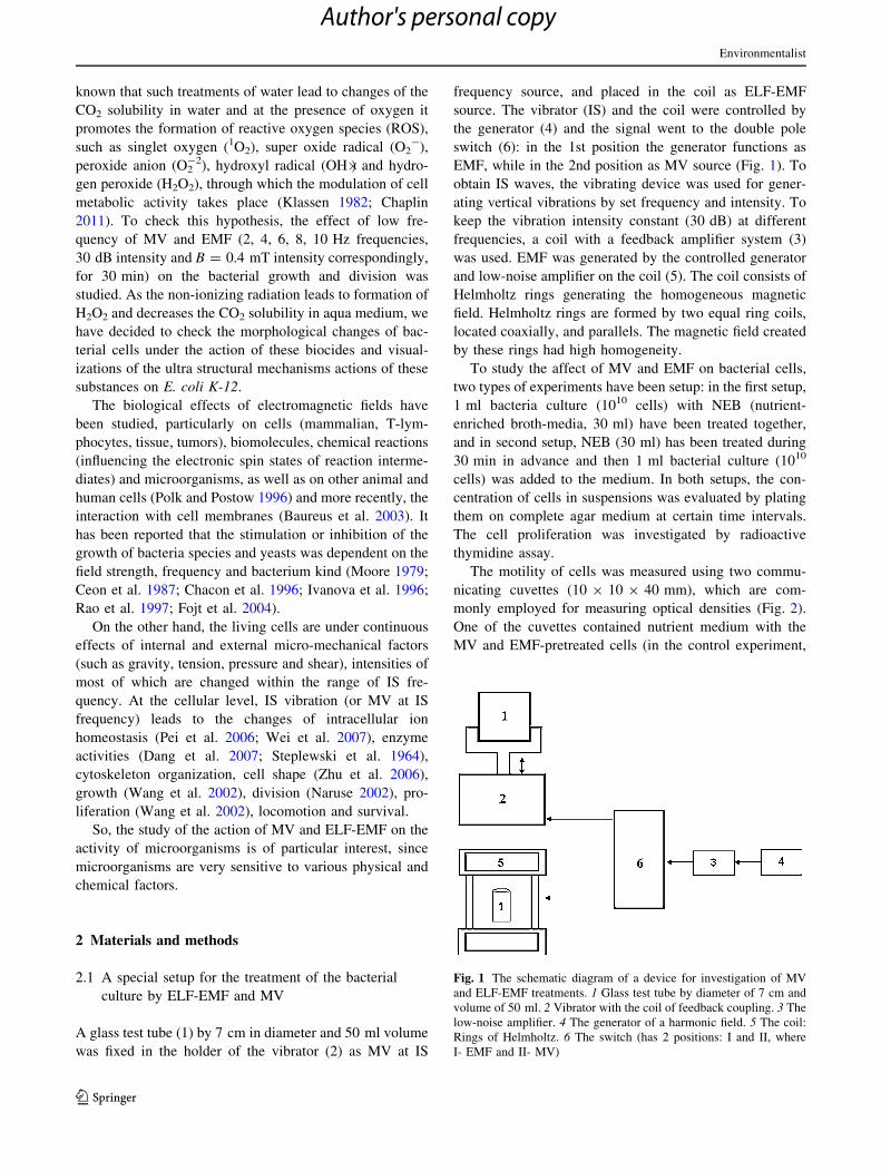
A glass test tube (1) by 7 cm in diameter and 50 ml volume
was fixed in the holder of the vibrator (2) as MV at IS
frequency source, and placed in the coil as ELF-EMF
source. The vibrator (IS) and the coil were controlled by
the generator (4) and the signal went to the double pole
switch (6): in the 1st position the generator functions as
EMF, while in the 2nd position as MV source (Fig. 1). To
obtain IS waves, the vibrating device was used for gener-
ating vertical vibrations by set frequency and intensity. To
keep the vibration intensity constant (30 dB) at different
frequencies, a coil with a feedback amplifier system (3)
was used. EMF was generated by the controlled generator
and low-noise amplifier on the coil (5). The coil consists of
Helmholtz rings generating the homogeneous magnetic
field. Helmholtz rings are formed by two equal ring coils,
located coaxially, and parallels. The magnetic field created
by these rings had high homogeneity.
To study the affect of MV and EMF on bacterial cells,
two types of experiments have been setup: in the first setup,
1 ml bacteria culture (1010 cells) with NEB (nutrient-
enriched broth-media, 30 ml) have been treated together,
and in second setup, NEB (30 ml) has been treated during
30 min in advance and then 1 ml bacterial culture (1010
cells) was added to the medium. In both setups, the con-
centration of cells in suspensions was evaluated by plating
them on complete agar medium at certain time intervals.
The cell proliferation was investigated by radioactive
thymidine assay.
The motility of cells was measured using two commu-
nicating cuvettes (10 9 10 9 40 mm), which are com-
monly employed for measuring optical densities (Fig. 2).
One of the cuvettes contained nutrient medium with the
MV and EMF-pretreated cells (in the control experiment,
Fig. 1 The schematic diagram of a device for investigation of MV
and ELF-EMF treatments. 1 Glass test tube by diameter of 7 cm and
volume of 50 ml. 2 Vibrator with the coil of feedback coupling. 3 The
low-noise amplifier. 4 The generator of a harmonic field. 5 The coil:
Rings of Helmholtz. 6 The switch (has 2 positions: I and II, where
I- EMF and II- MV)
Environmentalist
123
Author's personal copy
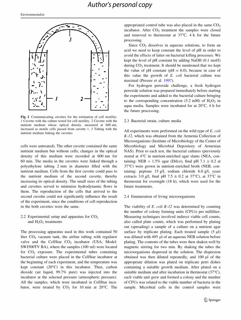
cells were untreated). The other cuvette contained the same
nutrient medium but without cells; changes in the optical
density of this medium were recorded at 600 nm for
60 min. The media in the cuvettes were linked through a
polyethylene tubing 2 mm in diameter filled with the
nutrient medium. Cells from the first cuvette could pass to
the nutrient medium of the second cuvette, thereby
increasing its optical density. The small sizes of the tubing
and cuvettes served to minimize hydrodynamic flows in
them. The reproduction of the cells that arrived to the
second cuvette could not significantly influence the result
of the experiment, since the conditions of cell reproduction
in the both cuvettes were the same.
2.2 Experimental setup and apparatus for CO2
and H2O2 treatments
The processing apparatus used in this work contained 50
liter CO2 vacuum tank, the airline tubing with regulator
valve and the CellStar CO2 incubator (USA, Model:
SWJ500TV BA), where the samples (100 ml) were located
for CO2 exposure. The experimental tubes containing
bacterial culture were placed in the CellStar incubator at
the beginning of each experiment, and the temperature was
kept constant (20�C) in this incubator. Then, carbon
dioxide (air liquid, 99.7% pure) was injected into the
incubator at the selected pressure (atmospheric pressure).
All the samples, which were incubated in CellStar incu-
bator, were treated by CO2 for 10 min at 20�C. The
appropriated control tube was also placed in the same CO2
incubator. After CO2 treatment the samples were closed
and removed to thermostat at 37�C, 4 h for the future
processing.
Since CO2 dissolves in aqueous solutions, to form an
acid we need to keep constant the level of pH in order to
avoid the effects of latter on bacterial killing processes. We
kept the level of pH constant by adding NaOH (0.1 mol/l)
during CO2 treatment. It should be mentioned that we kept
the value of pH constant (pH = 6.0), because in case of
this value the growth of E. coli bacterial culture was
maximal (Presser et al. 1997).
For hydrogen peroxide challenge, a fresh hydrogen
peroxide solution was prepared immediately before starting
the experiments and added to the bacterial culture bringing
to the corresponding concentration (5.2 mM) of H2O2 in
aqua media. Samples were incubated for at 20�C, 4 h for
the future processing.
2.3 Bacterial strain, culture media
All experiments were performed on the wild type of E. coli
K-12, which was obtained from the Armenia Collection of
Microorganisms (Institute of Microbiology of the Center of
Microbiology and Microbial Depository of Armenian
NAS). Prior to each test, the bacterial cultures (previously
stored at 4�C in nutrient-enriched agar slants (NEA, con-
taining: NEB ? 1.5% agar (Difco), final pH 7.1 ± 0.2 at
37�C) were grown in nutrient-enriched broth (NEB, con-
taining: peptone 15 g/l, sodium chloride 6.0 g/l, yeast
extracts 3.0 g/l, final pH 7.5 ± 0.2 at 37�C), at 37�C in
thermostat for overnight (18 h), which were used for the
future treatments.
2.4 Enumeration of living microorganisms
The viability of E. coli K-12 was determined by counting
the number of colony forming units (CFUs) per milliliter.
Measuring techniques involved indirect viable cell counts,
also called plate counts, which was performed by plating
out (spreading) a sample of a culture on a nutrient agar
surface by triplicate plating. Each treated sample (5 ll)
was diluted with 495 ll of an aqueous NEB solution before
plating. The contents of the tubes were then shaken well by
magnetic stirring for two min. By shaking the tubes the
microorganisms dispersed in the solution. The dispersion
obtained was then diluted repeatedly, and 100 ll of the
appropriate dilution was plated on triplicate petri dishes
containing a suitable growth medium. After plated on a
suitable medium and after incubation in thermostat (37�C),
each viable unit grew and formed a colony and the number
of CFUs was related to the viable number of bacteria in the
sample. Microbial cells in the control samples were
Fig. 2 Communicating cuvettes for the estimation of cell motility:
1 Cuvette with the culture tested for cell motility. 2 Cuvette with the
nutrient medium whose optical density, measured at 600 nm,
increased as motile cells passed from cuvette 1. 3 Tubing with the
nutrient medium linking the cuvettes
Environmentalist
123
Author's personal copy
counted by the same procedures described for treated
samples.
2.5 Electronmicroscopy assay
The methods of negative contrasting were done by 2%
phosphoric-tungsten acid solution at pH 6.8–7.0. For pre-
paring the electronic microscopic preparations of bacteria
suspensions after centrifugation at 3,000 rpm (CLR-1)
during 10 min bacterial sediment was fixed by 2.5%
solution of glutaraldehyde in 0.1 M cacodylate buffer
under pH 7.4 during 2 h under room temperature. After
three-phase washing in 0.1 M cacodylate buffer was fixed
under room temperature by 1% of solution tetraoxide
osmium in 0.1 M cacodylate buffer under pH 7.4 during
not less than 1 h. After washing in the same buffer,
dehydration of sample was conducted in the ethanol
increasing concentrations for 30, 50, 70, 96 and 100% or
acetone increasing concentrations and potting by mixture
of Araldite on Lufft. The bacterial sediment after poly-
merization in thermostat at 37� and 59�C, with further
dehydration, saturating was polymerized in Araldite prep-
arations. Ultrathin sections preparation was conducted by
using the ultra cut ‘‘Reichert Ultracut-Young.’’ Ultrathin
sections were stained in the uranyl acetate and lead citrate
(Reynolds-Veinable). These were observed with electronic
microscopes Tesla BS-500 (Czechoslovakia). The photo-
micrographies were scanned in permit of 900 pixels for
inch and processed on computer programme Corel Draw
versions 11 and Photoshop versions 8. Computer mor-
phometric and stereometric analysis of electronic micro-
scopic imaging was performed according to the program
‘‘Video Test Structure-5, nanotechnology.’’
2.6 Radioactive labeled [3H]-thymidine-based cell
proliferation assay (liquid scintillation counter)
For the measurement of the cell proliferation the bacteria
cultures were grown in 100 ml of NEB, overnight 18 h at
37�C. Then, 1 ml of overnight culture (1010 cells) was
added to the 30 ml NEB, in which content is 40 ll [3H]-
thymidine (1.3 lCi/ml) ([6-3H]-thymidine, PerkinElmer,
Boston, MA, 14.4 Ci/mmol specific activity). [3H]-thymi-
dine containing samples were treated by MV and ELF-
EMF at 30 min and then incubated in thermostat at 37�C,
for 18 h. The cells were then harvested by centrifugation
for 10 min at 5,000g. [3H]-thymidine uptake was stopped
by precipitation of each portion with ice-cold 10% tri-
chloroacetic acid (TCA), washed 2 times and then again
harvested by centrifugation. Then, 3 ml of Bray’s scintil-
lation cocktail counting solution was added to each sample
and the radioactivity was measured by using a Wallac
1,450 liquid scintillation counter.
2.7 Statistical analysis
Statistical analyses were conducted by SPSS 17 software.
The differences among the means of treatment were tested
by using the paired sample t test and expressed in the
figures with the help of asterisks (*). All experiments were
repeated at least three times. Values shown below are the
means of these experiments ± SE.
3 Results
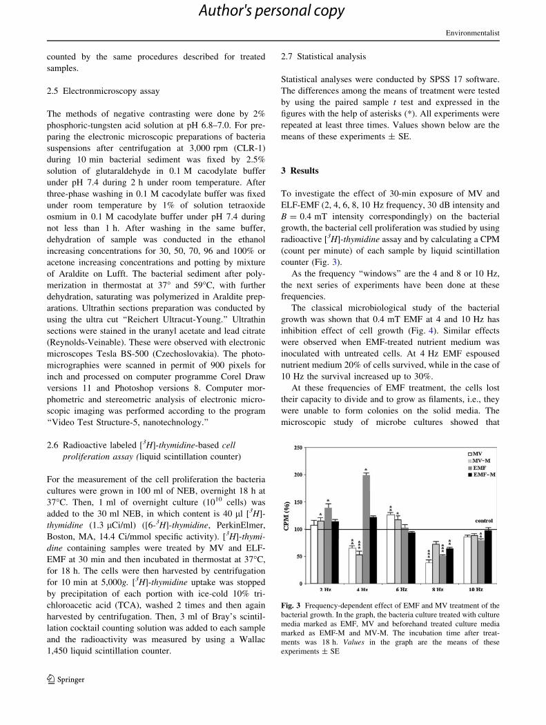
To investigate the effect of 30-min exposure of MV and
ELF-EMF (2, 4, 6, 8, 10 Hz frequency, 30 dB intensity and
B = 0.4 mT intensity correspondingly) on the bacterial
growth, the bacterial cell proliferation was studied by using
radioactive [3H]-thymidine assay and by calculating a CPM
(count per minute) of each sample by liquid scintillation
counter (Fig. 3).
As the frequency ‘‘windows’’ are the 4 and 8 or 10 Hz,
the next series of experiments have been done at these
frequencies.
The classical microbiological study of the bacterial
growth was shown that 0.4 mT EMF at 4 and 10 Hz has
inhibition effect of cell growth (Fig. 4). Similar effects
were observed when EMF-treated nutrient medium was
inoculated with untreated cells. At 4 Hz EMF espoused
nutrient medium 20% of cells survived, while in the case of
10 Hz the survival increased up to 30%.
At these frequencies of EMF treatment, the cells lost
their capacity to divide and to grow as filaments, i.e., they
were unable to form colonies on the solid media. The
microscopic study of microbe cultures showed that
Fig. 3 Frequency-dependent effect of EMF and MV treatment of the
bacterial growth. In the graph, the bacteria culture treated with culture
media marked as EMF, MV and beforehand treated culture media
marked as EMF-M and MV-M. The incubation time after treat-
ments was 18 h. Values in the graph are the means of these
experiments ± SE
Environmentalist
123
Author's personal copy
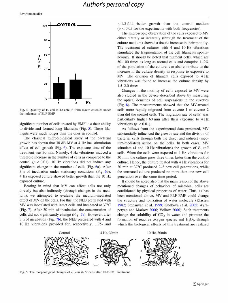
significant number of cells treated by EMF lost their ability
to divide and formed long filaments (Fig. 5). These fila-
ments were much longer than the ones in control.
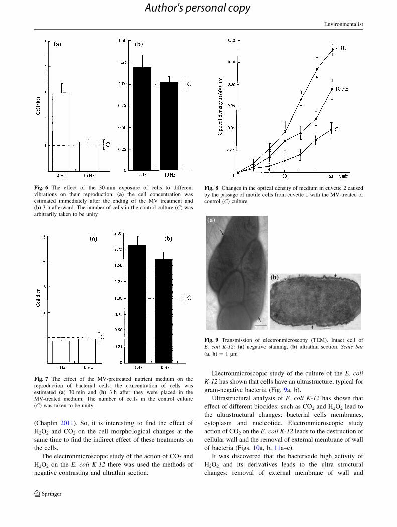
The classical microbiological study of the bacterial
growth has shown that 30 dB MV at 4 Hz has stimulation
effect of cell growth (Fig. 6). The exposure time of the
treatment was 30 min. Namely, 4 Hz vibrations induced a
threefold increase in the number of cells as compared to the
control (p \ 0.01); 10 Hz vibrations did not induce any
significant change in the number of cells (Fig. 6a). After
3 h of incubation under stationary conditions (Fig. 6b),
4 Hz exposed culture showed better growth than the 10 Hz
exposed culture.
Bearing in mind that MV can affect cells not only
directly but also indirectly (through changes in the med-
ium), we attempted to evaluate the medium-mediated
effect of MV on the cells. For this, the NEB pretreated with
MV was inoculated with intact cells and incubated at 37�C
(Fig. 7). After 30 min of incubation, the concentration of
cells did not significantly change (Fig. 7a). However, after
3 h of incubation (Fig. 7b), the NEB pretreated with 4 and
10 Hz vibrations provided for, respectively, 1.75- and
*1.5-fold better growth than the control medium
(p \ 0.05 for the experiments with both frequencies).
The microscopic observation of the cells exposed to MV
either directly or indirectly (through the treatment of the
culture medium) showed a drastic increase in their motility.
The treatment of cultures with 4 and 10 Hz vibrations
stimulated the fragmentation of the cell filaments sponta-
neously. It should be noted that filament cells, which are
50–100 times as long as normal cells and comprise 1–2%
of the population of the culture, can also contribute to the
increase in the culture density in response to exposure to
MV. The division of filament cells exposed to 4 Hz
vibrations was found to increase the culture density by
1.5–2.0 times.
Changes in the motility of cells exposed to MV were
also studied in the device described above by measuring
the optical densities of cell suspensions in the cuvettes
(Fig. 8). The measurements showed that the MV-treated
cells more rapidly migrated from cuvette 1 to cuvette 2
than did the control cells. The migration rate of cells’ was
particularly higher 60 min after their exposure to 4 Hz
vibrations (p \ 0.01).
As follows from the experimental data presented, MV
substantially influenced the growth rate and the division of
bacterial cells through both the direct and indirect (med-
ium-mediated) action on the cells. In both cases, MV
stimulate (4 and 10 Hz vibrations) the growth of E. coli
cells. When the cells were exposed to 4 Hz vibrations for
30 min, the culture grew three times faster than the control
culture. Hence, the culture treated with 4 Hz vibrations for
30 min at 37�C produced 2–3 new cell generations, while
the untreated culture produced no more than one new cell
generation over the same time period.
It should be noted also that the main reason of the above
mentioned changes of behaviors of microbial cells are
conditioned by physical properties of water. Thus, as has
been mentioned above, MV and ELF-EMF could change
the structure and ionization of water molecule (Klassen
1982; Stepanyan et al. 1999; Gudkova et al. 2005; Ayra-
petyan and Markov 2006; Voikov 2006). Such treatments
change the solubility of CO2 in water and promote the
formation of reactive oxygen species and H2O2, through
which the biological effects of this treatment are realized
Fig. 4 Quantity of E. coli K-12 able to form macro colonies under
the influence of ELF-EMF
Fig. 5 The morphological changes of E. coli K-12 cells after ELF-EMF treatment
Environmentalist
123
Author's personal copy
(Chaplin 2011). So, it is interesting to find the effect of
H2O2 and CO2 on the cell morphological changes at the
same time to find the indirect effect of these treatments on
the cells.
The electronmicroscopic study of the action of CO2 and
H2O2 on the E. coli K-12 there was used the methods of
negative contrasting and ultrathin section.
Electronmicroscopic study of the culture of the E. coli
K-12 has shown that cells have an ultrastructure, typical for
gram-negative bacteria (Fig. 9a, b).
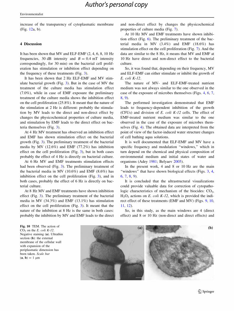
Ultrastructural analysis of E. coli K-12 has shown that
effect of different biocides: such as CO2 and H2O2 lead to
the ultrastructural changes: bacterial cells membranes,
cytoplasm and nucleotide. Electronmicroscopic study
action of CO2 on the E. coli K-12 leads to the destruction of
cellular wall and the removal of external membrane of wall
of bacteria (Figs. 10a, b, 11a–c).
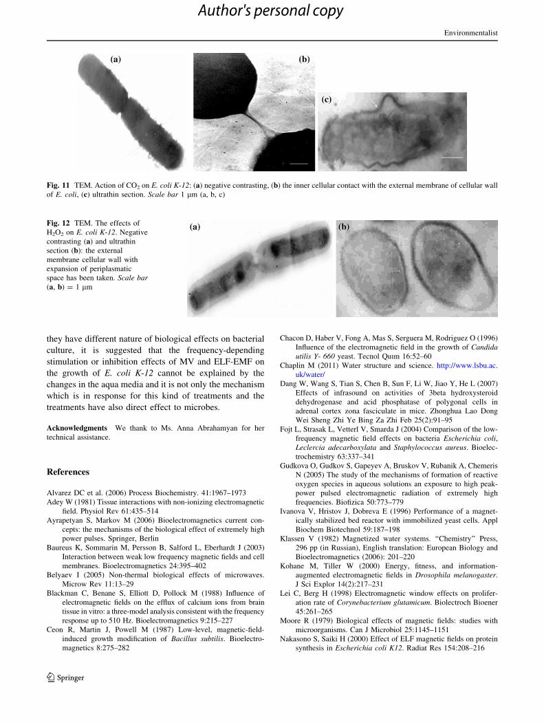
It was discovered that the bactericide high activity of
H2O2 and its derivatives leads to the ultra structural
changes: removal of external membrane of wall and
Fig. 6 The effect of the 30-min exposure of cells to different
vibrations on their reproduction: (a) the cell concentration was
estimated immediately after the ending of the MV treatment and
(b) 3 h afterward. The number of cells in the control culture (C) was
arbitrarily taken to be unity
Fig. 7 The effect of the MV-pretreated nutrient medium on the
reproduction of bacterial cells: the concentration of cells was
estimated (a) 30 min and (b) 3 h after they were placed in the
MV-treated medium. The number of cells in the control culture
(C) was taken to be unity
Fig. 8 Changes in the optical density of medium in cuvette 2 caused
by the passage of motile cells from cuvette 1 with the MV-treated or
control (C) culture
Fig. 9 Transmission of electronmicroscopy (TEM). Intact cell of
E. coli K-12: (a) negative staining, (b) ultrathin section. Scale bar(a, b) = 1 lm
Environmentalist
123
Author's personal copy
increase of the transparency of cytoplasmatic membrane
(Fig. 12a, b).
4 Discussion
It has been shown that MV and ELF-EMF (2, 4, 6, 8, 10 Hz
frequencies, 30 dB intensity and B = 0.4 mT intensity
correspondingly, for 30 min) on the bacterial cell prolif-
eration has stimulation or inhibition effect depending on
the frequency of these treatments (Fig. 3).
It has been shown that 2 Hz ELF-EMF and MV stim-
ulate bacterial growth (Fig. 3). But in the case of MV the
treatment of the culture media has stimulation effect
(7.0%), while in case of EMF exposure the preliminary
treatment of the culture media shows the inhibition effect
on the cell proliferation (25.8%). It meant that the nature of
the stimulation at 2 Hz is different: probably the stimula-
tion by MV leads to the direct and non-direct effect by
changes the physicochemical properties of culture media,
and stimulation by EMF leads to the direct effect on bac-
teria themselves (Fig. 3).
At 4 Hz MV treatment has observed an inhibition effect
and EMF has shown stimulation effect on the bacterial
growth (Fig. 3). The preliminary treatment of the bacterial
media by MV (12.6%) and EMF (77.2%) has inhibition
effect on the cell proliferation (Fig. 3), but in both cases
probably the effect of 4 Hz is directly on bacterial culture.
At 6 Hz MV and EMF treatments stimulation effects
had been observed (Fig. 3). The preliminary treatment of
the bacterial media in MV (10.6%) and EMF (8.6%) has
inhibition effect on the cell proliferation (Fig. 3), and in
both cases, probably the effect of 6 Hz is directly on bac-
terial culture.
At 8 Hz MV and EMF treatments have shown inhibition
effect (Fig. 3). The preliminary treatment of the bacterial
media in MV (34.3%) and EMF (13.1%) has stimulation
effect on the cell proliferation (Fig. 3). It meant that the
nature of the inhibition at 8 Hz is the same in both cases:
probably the inhibition by MV and EMF leads to the direct
and non-direct effect by changes the physicochemical
properties of culture media (Fig. 7).
At 10 Hz MV and EMF treatments have shown inhibi-
tion effect (Fig. 6). The preliminary treatment of the bac-
terial media in MV (3.4%) and EMF (18.6%) has
stimulation effect on the cell proliferation (Fig. 7). And the
data are similar to the 8 Hz, it means that MV and EMF at
10 Hz have direct and non-direct effect to the bacterial
culture.
So, it was found that, depending on their frequency, MV
and ELF-EMF can either stimulate or inhibit the growth of
E. coli K-12.
The nature of MV- and ELF-EMF-treated nutrient
medium was not always similar to the one observed in the
case of the exposure of microbes themselves (Figs. 4, 6, 7,
8, 9).
The performed investigation demonstrated that EMF
leads to frequency-dependent inhibition of the growth
(CFUs) and division of E. coli K-12 cells. The effect of
EMF-treated nutrient medium was similar to the one
observed in the case of the exposure of microbes them-
selves (Fig. 4). The obtained data are interpreted from the
point of view of the factor-induced water structure changes
of cell bathing aqua solutions.
It is well documented that ELF-EMF and MV have a
specific frequency and modulation ‘‘windows,’’ which in
turn depend on the chemical and physical composition of
environmental medium and initial states of water and
organisms (Adey 1981; Belyaev 2005).
In the present work, 4 and 8 or 10 Hz are the main
‘‘windows’’ that have shown biological effects (Figs. 3, 4,
6, 7, 8, 9).
It is concluded that the ultrastructural visualizations
could provide valuable data for correction of cytopatho-
logic characteristics of mechanism of the biocides: CO2,
H2O2 actions on E. coli K-12, which is provided the indi-
rect effect of these treatments (EMF and MV) (Figs. 9, 10,
11, 12).
So, in this study, as the main windows are 4 (direct
effect) and 8 or 10 Hz (non-direct and direct effects) and
Fig. 10 TEM. The action of
CO2 on the E. coli K-12.
Negative staining (a). Ultrathin
section (b): the external
membrane of the cellular wall
with expansion of the
periplasmatic dimension has
been taken. Scale bar(a, b) = 1 lm
Environmentalist
123
Author's personal copy
they have different nature of biological effects on bacterial
culture, it is suggested that the frequency-depending
stimulation or inhibition effects of MV and ELF-EMF on
the growth of E. coli K-12 cannot be explained by the
changes in the aqua media and it is not only the mechanism
which is in response for this kind of treatments and the
treatments have also direct effect to microbes.
Acknowledgments We thank to Ms. Anna Abrahamyan for her
technical assistance.
References
Alvarez DC et al. (2006) Process Biochemistry. 41:1967–1973
Adey W (1981) Tissue interactions with non-ionizing electromagnetic
field. Physiol Rev 61:435–514
Ayrapetyan S, Markov M (2006) Bioelectromagnetics current con-
cepts: the mechanisms of the biological effect of extremely high
power pulses. Springer, Berlin
Baureus K, Sommarin M, Persson B, Salford L, Eberhardt J (2003)
Interaction between weak low frequency magnetic fields and cell
membranes. Bioelectromagnetics 24:395–402
Belyaev I (2005) Non-thermal biological effects of microwaves.
Microw Rev 11:13–29
Blackman C, Benane S, Elliott D, Pollock M (1988) Influence of
electromagnetic fields on the efflux of calcium ions from brain
tissue in vitro: a three-model analysis consistent with the frequency
response up to 510 Hz. Bioelectromagnetics 9:215–227
Ceon R, Martin J, Powell M (1987) Low-level, magnetic-field-
induced growth modification of Bacillus subtilis. Bioelectro-
magnetics 8:275–282
Chacon D, Haber V, Fong A, Mas S, Serguera M, Rodriguez O (1996)
Influence of the electromagnetic field in the growth of Candidautilis Y- 660 yeast. Tecnol Quım 16:52–60
Chaplin M (2011) Water structure and science. http://www.lsbu.ac.
uk/water/
Dang W, Wang S, Tian S, Chen B, Sun F, Li W, Jiao Y, He L (2007)
Effects of infrasound on activities of 3beta hydroxysteroid
dehydrogenase and acid phosphatase of polygonal cells in
adrenal cortex zona fasciculate in mice. Zhonghua Lao Dong
Wei Sheng Zhi Ye Bing Za Zhi Feb 25(2):91–95
Fojt L, Strasak L, Vetterl V, Smarda J (2004) Comparison of the low-
frequency magnetic field effects on bacteria Escherichia coli,Leclercia adecarboxylata and Staphylococcus aureus. Bioelec-
trochemistry 63:337–341
Gudkova O, Gudkov S, Gapeyev A, Bruskov V, Rubanik A, Chemeris
N (2005) The study of the mechanisms of formation of reactive
oxygen species in aqueous solutions an exposure to high peak-
power pulsed electromagnetic radiation of extremely high
frequencies. Biofizica 50:773–779
Ivanova V, Hristov J, Dobreva E (1996) Performance of a magnet-
ically stabilized bed reactor with immobilized yeast cells. Appl
Biochem Biotechnol 59:187–198
Klassen V (1982) Magnetized water systems. ‘‘Chemistry’’ Press,
296 pp (in Russian), English translation: European Biology and
Bioelectromagnetics (2006): 201–220
Kohane M, Tiller W (2000) Energy, fitness, and information-
augmented electromagnetic fields in Drosophila melanogaster.
J Sci Explor 14(2):217–231
Lei C, Berg H (1998) Electromagnetic window effects on prolifer-
ation rate of Corynebacterium glutamicum. Biolectroch Bioener
45:261–265
Moore R (1979) Biological effects of magnetic fields: studies with
microorganisms. Can J Microbiol 25:1145–1151
Nakasono S, Saiki H (2000) Effect of ELF magnetic fields on protein
synthesis in Escherichia coli K12. Radiat Res 154:208–216
Fig. 11 TEM. Action of CO2 on E. coli K-12: (a) negative contrasting, (b) the inner cellular contact with the external membrane of cellular wall
of E. coli, (c) ultrathin section. Scale bar 1 lm (a, b, c)
Fig. 12 TEM. The effects of
H2O2 on E. coli K-12. Negative
contrasting (a) and ultrathin
section (b): the external
membrane cellular wall with
expansion of periplasmatic
space has been taken. Scale bar(a, b) = 1 lm
Environmentalist
123
Author's personal copy
Naruse Y (2002) Mechanical vibration model for chromosomes in
metaphase of mitosis and possible application to the interruption
of cell division. Biosystems 66:55–63
Pei Zh, Sang H, Li R, Xiao P, He J, Zhuang Zh, Zhu M, Chen J, Ma H
(2006) Infrasound-induced hemodynamics, ultrastructure, and
molecular changes in the rat myocardium. Environ Toxicol
22(2):169–175
Polk C, Postow E (1996) Handbook of biological effects of
electromagnetic field, 2nd edn. CRS Press, New York
Presser K, Ratkowsky D, Ross T (1997) Modeling the growth rate of
Escherichia coli as a function of pH and lactic acid concentra-
tion. Appl Environ Microbiol 63(6):2355–2360
Rao T, Sonolikar R, Saheb S (1997) Influence of magnetic field on the
performance of bubble columns and airlift bioreactor with
submerged microorganisms. Chem Eng Sci 52:4155–4160
Stepanyan R, Ayrapetyan G, Arakelyan A, Ayrapetyan S (1999)
Effect of mechanical oscillations on electrical conductivity of
water. Biophys J 44:199–204
Steplewski Z, Stoklosa E, Brazegowy A (1964) On the effect of
horizontal mechanical vibrations on the enzyme activity of the
reticuloendothelial system (RES) in the liver of white rats.
PubMed 5(20):580–586
Voikov V (2006) Biological significance of active oxygen-dependent
processes in aqueous systems. In: Pollack G, Cameron I,
Wheatley D (eds) Water and the cell. Springer, Dordrecht,
pp 285–298
Wang B, Long X, Liu Y, Duan C, Sakanishi A (2002) The effects of
mechanical vibration on the microstructure of Gerbera jamesonii
acrocarpous callus. Colloids Surf B Biointerfaces 23(1):1–5
Wei Y, Liu J, Shu Q, Huang X, Chen J (2007) Effects of infrasound
on ultrastructure of testis cell in mice. Environ Toxicol
Pharmacol 23(1):111–114
Zhu W, Zhou N, Qu J, Xu W, Kong L (2006) Effects of mechanical
vibration on cell density and cell morphology in the dynamic
microcellular foaming process. J Cell Plast 13:42–49
Environmentalist
123
Author's personal copy