The Free Energy Profile of Tubulin Straight-Bent Conformational Changes, with Implications for...
13
The Free Energy Profile of Tubulin Straight-Bent Conformational Changes, with Implications for Microtubule Assembly and Drug Discovery Lili X. Peng 1. , Monica T. Hsu 2. , Massimiliano Bonomi 3 , David A. Agard 4 , Matthew P. Jacobson 1,5 * 1 Department of Pharmaceutical Chemistry, University of California, San Francisco, San Francisco, California, United States of America, 2 Graduate Group in Biophysics, University of California, San Francisco, San Francisco, California, United States of America, 3 Department of Bioengineering and Therapeutic Sciences, University of California, San Francisco, San Francisco, California, United States of America, 4 Howard Hughes Medical Institute and Department of Biochemistry and Biophysics, University of California, San Francisco, San Francisco, California, United States of America, 5 Department of Biochemistry and Biophysics, University of California, San Francisco, San Francisco, California, United States of America Abstract ab-tubulin dimers need to convert between a ‘bent’ conformation observed for free dimers in solution and a ‘straight’ conformation required for incorporation into the microtubule lattice. Here, we investigate the free energy landscape of ab- tubulin using molecular dynamics simulations, emphasizing implications for models of assembly, and modulation of the conformational landscape by colchicine, a tubulin-binding drug that inhibits microtubule polymerization. Specifically, we performed molecular dynamics, potential-of-mean force simulations to obtain the free energy profile for unpolymerized GDP-bound tubulin as a function of the ,12u intradimer rotation differentiating the straight and bent conformers. Our results predict that the unassembled GDP-tubulin heterodimer exists in a continuum of conformations ranging between straight and bent, but, in agreement with existing structural data, suggests that an intermediate bent state has a lower free energy (by ,1 kcal/mol) and thus dominates in solution. In agreement with predictions of the lattice model of microtubule assembly, lateral binding of two ab-tubulins strongly shifts the conformational equilibrium towards the straight state, which is then ,1 kcal/mol lower in free energy than the bent state. Finally, calculations of colchicine binding to a single ab-tubulin dimer strongly shifts the equilibrium toward the bent states, and disfavors the straight state to the extent that it is no longer thermodynamically populated. Citation: Peng LX, Hsu MT, Bonomi M, Agard DA, Jacobson MP (2014) The Free Energy Profile of Tubulin Straight-Bent Conformational Changes, with Implications for Microtubule Assembly and Drug Discovery. PLoS Comput Biol 10(2): e1003464. doi:10.1371/journal.pcbi.1003464 Editor: Mark S. Alber, University of Notre Dame, United States of America Received August 26, 2013; Accepted December 16, 2013; Published February 6, 2014 Copyright: ß 2014 Peng et al. This is an open-access article distributed under the terms of the Creative Commons Attribution License, which permits unrestricted use, distribution, and reproduction in any medium, provided the original author and source are credited. Funding: This work was supported in part by NIH grant GM031627 (DAA). MTH would like to acknowledge an NSF Graduate Research Fellowship. The funders had no role in study design, data collection and analysis, decision to publish, or preparation of the manuscript Competing Interests: The authors have declared that no competing interests exist. * E-mail: [email protected] . These authors contributed equally to this work. Introduction Microtubules (MTs) are dynamic cytoskeletal polymers formed by the polymerization of ab-tubulin, a globular heterodimer comprised of two structurally related 55 kDa a- and b-subunits. Microtubules play a vital role in intracellular trafficking and cell division; these functions are influenced by the complex dynamics of the MT plus end, which undergoes stochastic periods of assembly and disassembly. The role of ab-tubulin conformational changes in the processes of assembly and disassembly has been the subject of great interest and some controversy [1]. Here we investigate the free energy landscape of ab-tubulin using molecular dynamics simulations, emphasizing implications for models of assembly, and modulation of the conformational landscape by colchicine, a tubulin-binding drug that inhibits microtubule polymerization. Tubulin has been shown to exist in two extreme conformations: a ‘‘straight’’ conformation observed in antiparallel zinc-induced tubulin sheets [2–6], which is compatible with incorporation into the MT lattice, and a ‘‘bent’’ conformation observed in the structure (T2R complex) of tubulin bound with colchicine, a MT-destabilizing drug, in a complex with the stathmin-like domain of RB3 (SLD-RB3) [7]. (The resolution of this T2R- colchicine structure was recently enhanced to 2.73 A ˚ [8].) X-ray crystallographic studies have also shown similarly bent tubulin structures in a complex with SLD-RB3 [9], bound to other MT- destabilizing drugs (vinblastine [8] and podophyllotoxin), as well as MT-stabilizing drugs (epothilone A and zampanolide) in a complex with tubulin tyrosine ligase (TTL) [10]. Distinguishing the straight and bent tubulin conformations are conformational rearrangements of the intermediate domains and an intra-dimer rotation: rotations of ,8u and ,11u, are required to superimpose the intermediate domains of the a- and b-subunits, respectively. In addition a ,12u intradimer rotation is required to superimpose both subunits of the straight and bent tubulin structures. Finally, the ‘‘straight’’ and bent tubulins are also distinguished by local rearrangements in the M and H1-S2 loop on both subunits, the b- subunit T7 loop and H8 helix, and the a-subunit T5 and H6-H7 loops [4,7,11]. Multiple rotational and translational motions in the a- and b- subunits differentiate the ‘‘straight’’ and ‘‘bent’’ conformations. We describe the ,12u intradimer conformational change of the PLOS Computational Biology | www.ploscompbiol.org 1 February 2014 | Volume 10 | Issue 2 | e1003464
Transcript of The Free Energy Profile of Tubulin Straight-Bent Conformational Changes, with Implications for...

The Free Energy Profile of Tubulin Straight-BentConformational Changes, with Implications forMicrotubule Assembly and Drug DiscoveryLili X. Peng1., Monica T. Hsu2., Massimiliano Bonomi3, David A. Agard4, Matthew P. Jacobson1,5*
1 Department of Pharmaceutical Chemistry, University of California, San Francisco, San Francisco, California, United States of America, 2 Graduate Group in Biophysics,
University of California, San Francisco, San Francisco, California, United States of America, 3 Department of Bioengineering and Therapeutic Sciences, University of
California, San Francisco, San Francisco, California, United States of America, 4 Howard Hughes Medical Institute and Department of Biochemistry and Biophysics,
University of California, San Francisco, San Francisco, California, United States of America, 5 Department of Biochemistry and Biophysics, University of California, San
Francisco, San Francisco, California, United States of America
Abstract
ab-tubulin dimers need to convert between a ‘bent’ conformation observed for free dimers in solution and a ‘straight’conformation required for incorporation into the microtubule lattice. Here, we investigate the free energy landscape of ab-tubulin using molecular dynamics simulations, emphasizing implications for models of assembly, and modulation of theconformational landscape by colchicine, a tubulin-binding drug that inhibits microtubule polymerization. Specifically, weperformed molecular dynamics, potential-of-mean force simulations to obtain the free energy profile for unpolymerizedGDP-bound tubulin as a function of the ,12u intradimer rotation differentiating the straight and bent conformers. Ourresults predict that the unassembled GDP-tubulin heterodimer exists in a continuum of conformations ranging betweenstraight and bent, but, in agreement with existing structural data, suggests that an intermediate bent state has a lower freeenergy (by ,1 kcal/mol) and thus dominates in solution. In agreement with predictions of the lattice model of microtubuleassembly, lateral binding of two ab-tubulins strongly shifts the conformational equilibrium towards the straight state, whichis then ,1 kcal/mol lower in free energy than the bent state. Finally, calculations of colchicine binding to a single ab-tubulindimer strongly shifts the equilibrium toward the bent states, and disfavors the straight state to the extent that it is no longerthermodynamically populated.
Citation: Peng LX, Hsu MT, Bonomi M, Agard DA, Jacobson MP (2014) The Free Energy Profile of Tubulin Straight-Bent Conformational Changes, withImplications for Microtubule Assembly and Drug Discovery. PLoS Comput Biol 10(2): e1003464. doi:10.1371/journal.pcbi.1003464
Editor: Mark S. Alber, University of Notre Dame, United States of America
Received August 26, 2013; Accepted December 16, 2013; Published February 6, 2014
Copyright: � 2014 Peng et al. This is an open-access article distributed under the terms of the Creative Commons Attribution License, which permitsunrestricted use, distribution, and reproduction in any medium, provided the original author and source are credited.
Funding: This work was supported in part by NIH grant GM031627 (DAA). MTH would like to acknowledge an NSF Graduate Research Fellowship. The fundershad no role in study design, data collection and analysis, decision to publish, or preparation of the manuscript
Competing Interests: The authors have declared that no competing interests exist.
* E-mail: [email protected]
. These authors contributed equally to this work.
Introduction
Microtubules (MTs) are dynamic cytoskeletal polymers formed
by the polymerization of ab-tubulin, a globular heterodimer
comprised of two structurally related 55 kDa a- and b-subunits.
Microtubules play a vital role in intracellular trafficking and cell
division; these functions are influenced by the complex dynamics
of the MT plus end, which undergoes stochastic periods of
assembly and disassembly. The role of ab-tubulin conformational
changes in the processes of assembly and disassembly has been the
subject of great interest and some controversy [1]. Here we
investigate the free energy landscape of ab-tubulin using molecular
dynamics simulations, emphasizing implications for models of
assembly, and modulation of the conformational landscape by
colchicine, a tubulin-binding drug that inhibits microtubule
polymerization.
Tubulin has been shown to exist in two extreme conformations:
a ‘‘straight’’ conformation observed in antiparallel zinc-induced
tubulin sheets [2–6], which is compatible with incorporation into
the MT lattice, and a ‘‘bent’’ conformation observed in the
structure (T2R complex) of tubulin bound with colchicine, a
MT-destabilizing drug, in a complex with the stathmin-like
domain of RB3 (SLD-RB3) [7]. (The resolution of this T2R-
colchicine structure was recently enhanced to 2.73 A [8].) X-ray
crystallographic studies have also shown similarly bent tubulin
structures in a complex with SLD-RB3 [9], bound to other MT-
destabilizing drugs (vinblastine [8] and podophyllotoxin), as well as
MT-stabilizing drugs (epothilone A and zampanolide) in a
complex with tubulin tyrosine ligase (TTL) [10]. Distinguishing
the straight and bent tubulin conformations are conformational
rearrangements of the intermediate domains and an intra-dimer
rotation: rotations of ,8u and ,11u, are required to superimpose
the intermediate domains of the a- and b-subunits, respectively. In
addition a ,12u intradimer rotation is required to superimpose
both subunits of the straight and bent tubulin structures. Finally,
the ‘‘straight’’ and bent tubulins are also distinguished by local
rearrangements in the M and H1-S2 loop on both subunits, the b-
subunit T7 loop and H8 helix, and the a-subunit T5 and H6-H7
loops [4,7,11].
Multiple rotational and translational motions in the a- and b-
subunits differentiate the ‘‘straight’’ and ‘‘bent’’ conformations.
We describe the ,12u intradimer conformational change of the
PLOS Computational Biology | www.ploscompbiol.org 1 February 2014 | Volume 10 | Issue 2 | e1003464

heterodimer, as described originally by Knossow et al., by the
movement of the H7 central helices in the a- and b-subunits (see
Figure 1). It is noteworthy that our method of defining the
curvature of a tubulin heterodimer differs from that used by Voth
and co-workers, who defined the ‘intrinsic bending angle’ by
calculating the intersection angle of two least-square fitted-vectors,
each defined through the center-of-masses of the N-terminal,
intermediate, and C-terminal domains of each subunit [12–14].
Voth et al. also characterized rotational motions of tubulin by the
twist angle between the a- and b-subunits. In Results and
Discussion, we quantitatively compare our method of calculating
the intradimer rotation angle with Voth’s method of calculating
the ‘intrinsic bending angle’.
The role of nucleotide state and tubulin conformation has been
debated in the context of two competing models of microtubule
assembly [15–17]. The allosteric model posits that GTP binding to
the exchangeable nucleotide-binding site (E-site) on the b-subunit
of soluble tubulin induces a ‘‘straight’’ conformation competent for
polymerization. On the other hand, the lattice model posits that
conformational changes in unpolymerized tubulin occur upon
recruitment into the growing lattice, and the role of GTP is to
increase affinity for the lattice by strengthening longitudinal
contacts. Both experimental and computational approaches have
been utilized to study this straight-to-bent conformational change
in tubulin associated with incorporation into the MT lattice. Using
small-angle X-ray scattering (SAXS), Rice et al. showed that,
under conditions where tubulin does not polymerize, soluble GTP-
and GDP-bound tubulin adopt conformations that were indistin-
guishable based on the SAXS profiles, and consistent with
structures of bent tubulin [1]. As aforementioned, the conforma-
tional landscape of tubulin has also been examined by molecular
dynamics simulations by Voth and co-workers [12–14]. In Voth et
al.’s unrestrained molecular dynamics simulations of up to 120 ns
in length, these authors reported that tubulin explores many
conformations including bent structures similar to that observed in
the T2R-colchicine complex [14]. This bend direction is in
agreement with those reported in Bennett et al.’s 20-ns MD
simulations of GTP- and GDP-bound unpolymerized tubulin [18].
These previously reported molecular dynamics simulations also
examined differences between GTP- and GDP-bound tubulin, an
issue that we do not consider here.
In this study we utilize free-energy calculations to characterize
the conformational landscape of a tubulin heterodimer, interpo-
lating between the ‘‘straight’’ structure from Zn2+-induced
protofilaments and ‘‘bent’’ tubulin from the T2R-colchicine
complex. In contrast to unrestrained molecular dynamics simula-
tions, we explore tubulin conformations only along a coordinate
connecting these two states, which we quantify using an intradimer
rotation angle (Figure 1). However, by performing umbrella
sampling and analysis using the weighted histogram analysis
method (WHAM), we can estimate the free energy associated with
deforming tubulin along this coordinate, providing quantitative
predictions concerning the relative free energies of the bent and
straight states. Specifically, this ‘‘potential of mean force’’ (PMF)
predicts that tubulin can exist in a continuum of conformations
ranging between straight and bent, but in agreement with existing
structural data, suggests that the bent states have lower free energy
and thus dominate in solution.
We also consider how a MT-disrupting drug modulates the
equilibrium between the straight and bent tubulin conformations
[19]. The oldest microtubule-disrupting drug, colchicine, was
discovered in 1889; its action on tubulin was elucidated in 1949
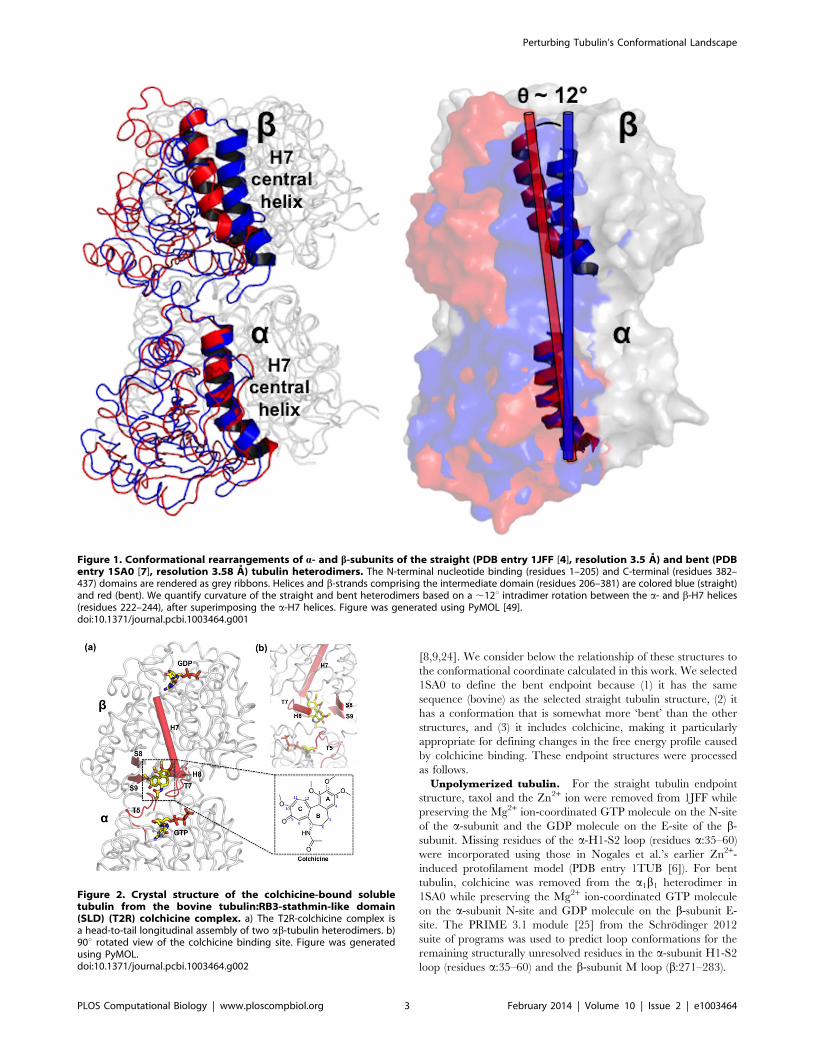
[11]. Crystal structures have revealed that colchicine binds close to
the interface between the a- and b-subunits (Figure 2), with the
binding site primarily on the b-subunit. Colchicine binds to the
soluble, unassembled form of tubulin, forming a poorly reversible
colchicine-tubulin complex. The binding site is sterically occluded
in the straight conformation, thus colchicine inhibits tubulin
polymerization, however the complex can be incorporated into the
microtubule lattice at both plus and minus ends [11,20]. It is
thought that colchicine binding also displaces the M loop on the b-
subunit (Phe b270 to Val b286), the structural element
instrumental in establishing the lateral contacts with the tubulin
molecule in the neighboring protofilament, further preventing
tubulin from adopting a polymerization-competent structure [7].
Substoichiometic concentrations of the tubulin-colchicine complex
are sufficient to inhibit microtubule growth, whereas high TC
concentrations lead to microtubule depolymerization [21]. Previ-
ous research has argued that colchicine binding strongly favors
binding the bent tubulin conformer, colchicine is sterically
hindered from binding the straight tubulin structure [22]. Our
computational results further support a conformational-bias mode
of action for colchicine; the PMF shows that colchicine binding
strongly disfavors the straight conformation.
Methods
Preparation of endpoint structures of conformationallandscapes
Selection of straight and bent tubulin endpoint
structures. Conformational change coordinates for tubulin
are bounded by the taxol-liganded straight tubulin heterodimer,
observed in zinc-induced protofilament sheets (PDB id 1JFF,
resolution 3.5 A, organism B. taurus), and the bent tubulin
structure observed in the a1b1 heterodimer of the T2R-colchicine
complex (PDB id 1SA0, resolution 3.58 A, organism B. taurus) (see
Figure S1 in Text S1). For the simulations of laterally-paired
tubulins, the endpoints of the conformational change coordinate
are terminated by laterally-paired straight and bent tubulins, each
with a-a and b-b protofilament contacts, as described in Wells et
al [23]. There are several other crystal structures that show tubulin
in a bent conformation, including some with better resolution
Author Summary
Microtubules are composed of ab-tubulins that play aninstrumental role in regulating intracellular trafficking andformation of the mitotic spindle during mitosis and celldivision. Structural studies have shown that tubulin existsin a ‘‘straight’’ conformation compatible with that in themicrotubule lattice and a ‘‘bent’’ conformation thought torepresent the unassembled state. There is current debateas to whether the straight-to-bent conformational changein tubulin is the cause or consequence of tubulin’sassembly into the microtubule lattice. Here, we use free-energy molecular dynamics simulations to qualitativelyunderstand the conformational landscape of tubulin in theunassembled state and upon lateral binding. We predictthat soluble tubulin exists primarily in a bent conforma-tion; our simulation results show that tubulin primarilyadopts an intermediately bent conformation in agreementwith structural data. We also show that lateral binding oftwo tubulins shifts the equilibrium in favor of the‘‘straight’’ state, supporting the hypothesis that thestraight-to-bent conformational change is the conse-quence of tubulin’s incorporation into the microtubulelattice via lateral interactions. We also show that colchicinebinding shifts the population of tubulin in favor of a bentstate, further implicating our work in drug discovery.
Perturbing Tubulin’s Conformational Landscape
PLOS Computational Biology | www.ploscompbiol.org 2 February 2014 | Volume 10 | Issue 2 | e1003464
[8,9,24]. We consider below the relationship of these structures to
the conformational coordinate calculated in this work. We selected
1SA0 to define the bent endpoint because (1) it has the same
sequence (bovine) as the selected straight tubulin structure, (2) it
has a conformation that is somewhat more ‘bent’ than the other
structures, and (3) it includes colchicine, making it particularly
appropriate for defining changes in the free energy profile caused
by colchicine binding. These endpoint structures were processed
as follows.
Unpolymerized tubulin. For the straight tubulin endpoint
structure, taxol and the Zn2+ ion were removed from 1JFF while
preserving the Mg2+ ion-coordinated GTP molecule on the N-site
of the a-subunit and the GDP molecule on the E-site of the b-
subunit. Missing residues of the a-H1-S2 loop (residues a:35–60)
were incorporated using those in Nogales et al.’s earlier Zn2+-
induced protofilament model (PDB entry 1TUB [6]). For bent
tubulin, colchicine was removed from the a1b1 heterodimer in
1SA0 while preserving the Mg2+ ion-coordinated GTP molecule
on the a-subunit N-site and GDP molecule on the b-subunit E-
site. The PRIME 3.1 module [25] from the Schrodinger 2012
suite of programs was used to predict loop conformations for the
remaining structurally unresolved residues in the a-subunit H1-S2
loop (residues a:35–60) and the b-subunit M loop (b:271–283).
Figure 1. Conformational rearrangements of a- and b-subunits of the straight (PDB entry 1JFF [4], resolution 3.5 A) and bent (PDBentry 1SA0 [7], resolution 3.58 A) tubulin heterodimers. The N-terminal nucleotide binding (residues 1–205) and C-terminal (residues 382–437) domains are rendered as grey ribbons. Helices and b-strands comprising the intermediate domain (residues 206–381) are colored blue (straight)and red (bent). We quantify curvature of the straight and bent heterodimers based on a ,12u intradimer rotation between the a- and b-H7 helices(residues 222–244), after superimposing the a-H7 helices. Figure was generated using PyMOL [49].doi:10.1371/journal.pcbi.1003464.g001
Figure 2. Crystal structure of the colchicine-bound solubletubulin from the bovine tubulin:RB3-stathmin-like domain(SLD) (T2R) colchicine complex. a) The T2R-colchicine complex isa head-to-tail longitudinal assembly of two ab-tubulin heterodimers. b)90u rotated view of the colchicine binding site. Figure was generatedusing PyMOL.doi:10.1371/journal.pcbi.1003464.g002
Perturbing Tubulin’s Conformational Landscape
PLOS Computational Biology | www.ploscompbiol.org 3 February 2014 | Volume 10 | Issue 2 | e1003464

These two modified endpoint structures were then energy-
minimized and equilibrated using the GROMACS 4.5.5 package
[26]. The GROMOS96 43a1 force field [27] was used in
conjunction with the particle mesh Ewald sum method [28] to
treat long-range electrostatic interactions. A time step of 2 fs was
used for all simulations. Parameters for GTP and GDP were
generated from the GlycoBioChem PRODRG2 server [29]. Each
endpoint structure was solvated in explicit SPC water molecules22
in the center of a periodic cubic box (dimensions of
125 A6125 A6125 A) and neutralized with NaCl. The GRO-
MACS 4.5.5 g_mindist module was used to ensure that there was
no overlap between the system with its periodic image during
equilibration. Each system was energy-minimized using steepest
descent while applying Ferguson’s flexible SPC water model [30]
constraints on the solvent. Periodic boundary conditions were
applied for each system throughout all minimization and
equilibration steps. Equilibration was performed in two stages:
first, the system was heated to 300 K, through use of velocity
rescaling, in the isothermal-isotropic (NVT) ensemble while
applying position restraints on the entire system. Each system
had attained 300 K by 100 ps, at which point the structure was
extracted and further equilibrated in the isothermal-isobaric (NPT)
ensemble (300 K, 1.013 bar) for up to 500 ps using the Parrinello-
Rahman barostat [31] implemented in GROMACS 4.5.5.
Laterally-bound tubulin pairs. For the straight pair end-
point, two straight tubulins laterally paired in the a-a, b-b manner
were taken from Wells et al.’s cryo-EM-derived ‘‘N’’ model of the
MT lattice [23], of which the atomic coordinates were obtained
through rigid fitting to Downing et al.’s 8 A density map of a 13-
protofilament MT [32]. To ensure that the interdimer contacts
represent those stabilizing a-a, b-b lateral contacts, Wells et al.
performed a multi-stage flexible fitting and equilibration procedure
on the ‘‘N’’ tubulin pair totaling 85 ns. For the bent pair endpoint,
we preserved these interdimer lateral contacts when globally aligning
the unpolymerized bent tubulins to the positions of the laterally-
bound straight tubulins. Both laterally-bound tubulin structures were
solvated with explicit SPC water molecules in the center of a periodic
box (dimensions of 300 A6300 A6300 A). Energy minimization
and equilibration procedures were performed as for the unpolymer-
ized tubulin described previously; the duration of the NVT and NPT
equilibration phases were 1 ns and 3 ns, respectively.
Colchicine-bound tubulin. The a1b1 heterodimer from the
T2R-colchicine complex was used as the bent endpoint, preserving
the colchicine molecule. Solvation, minimization, and equilibra-
tion of this structure were performed in analogous fashion as for
the unpolymerized bent tubulin. The GlycoBioChem PRODRG2
server [29] was used to generate parameters for colchicine. To
create a (hypothetical) structure of colchicine-bound straight
tubulin, we used a stepwise procedure involving successive
iterations of global alignment of tubulin and superimposition of
colchicine for tubulin structures intermediate between the bent to
straight endpoints, as described in greater detail in ‘‘Generating
the morphing path’’.
Selecting the reaction pathWe chose the path collective variable, or reaction path, to be the
angle required for superimposing the secondary structural
elements of the a- and b-subunit intermediate domains (helices
H6, H7, H8, H9, H10 and strands S7, S8, S9, and S10) of the
straight and bent tubulin molecules. The conformational space R
relative to this path can be expressed in terms of two collective
variables, s(R), which represents the progress of the dynamics along
the conformational change coordinate, and z(R), which represents
the progress away from the reaction path, as follows [33–36]:
s Rð Þ~ 1
P{1
PPi~1
i{1ð Þ:e{l R{R ið Þ½ �2
PPi~1
e{l R{R ið Þ½ �2ð1Þ
z Rð Þ~ 1
lln
XP
i~1
e{l R{R ið Þ½ �2 !
ð2Þ
where i is a discrete index ranging from 1 to P, the total number of
structures along the reaction coordinate, R{R ið Þ½ � is the mean
squared displacement of the 167 Ca atoms of the a- and b-
intermediate domain secondary structural elements, and l is a
prefactor in the exponential term that defines both s(R) and z(R).
(For the laterally-paired tubulins, R{R ið Þ½ � includes the two sets
of 167 Ca atoms in both dimers.) The value of l is chosen to be
proportional to the inverse of the mean square displacement
between two successive frames along the reaction coordinate;
here we set z Rð Þ~2:3:1
P
XP
i~1
R{R ið Þ½ �2 !{2
. It is important to
emphasize that umbrella sampling along this defined reaction
coordinate only allows us to explore the free energy differences for
tubulin conformations linearly interpolated along the defined
reaction path; this method does not allow us to explore bent
tubulin states whose a- and b-subunit intermediate domain
rotation deviates substantially from the defined reaction path.
Generating the morphing pathUnpolymerized tubulin and laterally-paired tubulins. The
thermally equilibrated straight and bent tubulin endpoint structures
were submitted to the Molmov morphing server [37] to generate
thirteen equidistantly-spaced, energy-minimized intermediate apo
tubulin structures along a linear path. These thirteen apo structures
were then liganded with GTP, GDP, and the Mg2+ ion as follows: 1)
each of the two endpoint structures was globally aligned with its
nearest apo intermediate; 2) GTP, GDP, and the Mg2+ ion were
superimposed onto the nearest intermediate structure, which was
then solvated, minimized, and equilibrated using the methods
described in the previous section. This cycle was iterated from both
endpoints for each successive tubulin intermediate. The final
snapshots of the fifteen equilibrated tubulin configurations (straight,
fully bent, and thirteen intermediately bent structures) were used as
the initial P = 15 umbrella nodes along the reaction path for the
calculating the potential of mean force for unpolymerized tubulin
(Figure S2a in Text S1). This procedure was executed in an
analogous fashion to produce the final snapshots for the thirteen
equilibrated laterally-bound tubulin pair configurations (straight pair,
fully bent pair, eleven intermediately bent pairs) for the initial P = 13
umbrella nodes in subsequent potential of mean force calculations
(Figure S2c in Text S1).
Colchicine-bound tubulin. The absence of a colchicine-
bound straight tubulin structure required us to use the equilibrated
straight endpoint structure, along with the equilibrated colchicine-
bound bent endpoint structure, to generate the colchicine-bound
tubulin intermediates. These two structures were submitted to
Molmov [37] to produce seven equidistantly-spaced, energy-
minimized intermediate apo tubulin structures. These seven apo
structures were complexed with colchicine, GTP, GDP, and the
Mg2+ ion as follows: 1) the equilibrated colchicine-bound fully bent
tubulin complex was globally aligned with its nearest apo
Perturbing Tubulin’s Conformational Landscape
PLOS Computational Biology | www.ploscompbiol.org 4 February 2014 | Volume 10 | Issue 2 | e1003464

intermediate; 2) colchicine, GTP, GDP, and Mg2+ from the
equilibrated colchicine-bound fully bent tubulin were then
superimposed onto the nearest apo intermediate; and 3) solvation,
minimization, and equilibration were performed on the successive
holo tubulin intermediate as described above. This cycle was
repeated for each successive intermediate from the bent to straight
endpoints until the colchicine-bound straight tubulin had been
fully equilibrated. The final snapshots for the nine equilibrated
colchicine-bound tubulin configurations (straight, fully bent, and
seven partially bent intermediates) were used as the initial P = 9
umbrella nodes along the reaction coordinate for calculating the
potential of mean force for colchicine-bound tubulin (Figure S2b
in Text S1).
Umbrella sampling, potential of mean force simulationsHarmonic biasing potentials are placed at each umbrella
position along a collective variable. The harmonic biasing
potentials for s(R) and z(R) are expressed as:
Vs(R)~1
2ks s(R){s(Ri)½ �2 ð3Þ
Vz(R)~1
2kz z(R){z(Ri)½ �2 ð4Þ
where ks and kz are the force constants for the harmonic restraints
for s and z, and Ri is each umbrella position i within the
configurational space R.
For the unpolymerized tubulin, we initially defined P = 15
equidistantly-spaced umbrella windows along the s(R) coordinate.
However, this discretization proved later insufficient to character-
ize the free energy along s(R), so we increased the number of
umbrella windows to P = 35 while restraining each umbrella node
by ks = 10 kcal/mol. To sufficiently characterize the free energy
landscape for the laterally-bound tubulin pair, we increased the
initial P = 13 equidistantly-spaced umbrella windows to P = 50
(Figure S3b in Text S1) while applying restraints of ks = 10 kcal/
mol. Because the domain rearrangements characterizing the
straight-to-bent conformational change are fully captured by the
s(R) collective variable, we did not place any harmonic restraints
on the z(R) path for the unpolymerized tubulin and laterally-
paired tubulin dimers.
We used the initial nine colchicine-bound tubulin morphing
intermediates to define P = 9 umbrella windows along s(R). To
achieve adequate sampling among successive umbrella windows
along s(R), we optimized ks to 10 kcal/mol for a total number of
P = 31 nodes. To prevent colchicine unbinding, especially from the
conformations with lower intradimer curvature (0u#h#6u), we
restrained the dynamics along the z(R) coordinate to kz = 10 kcal/
mol.
All simulations for the unpolymerized tubulin, laterally-paired
tubulins, and colchicine-bound tubulin were performed in the
canonical (NVT) ensemble at 1.013 bar and 300 K for 4 ns using
the PLUMED 1.3 [38]-implemented version of GROMACS
4.5.5. The last 3 ns of the production runs were used for
calculating the free energy profiles.
Generating free energy profiles using WHAMThe weighted histogram analysis (WHAM) [39,40] approach
was used to merge the data from the molecular dynamics
simulations and unbias the umbrella histograms in s(R). For the
unpolymerized tubulin and laterally-paired tubulins, we used the
g_wham module [41] of GROMACS 4.5.5 to calculate the free
energy profiles along the s(R) path, based on Eqn. 1, using a
resolution of 1000 bins and tolerance of 1e-6. We then estimated
the statistical uncertainty sPMF in the s(R) path accordingly [42]:
sPMF jð Þ~ Nb{1½ �{1:XNb
k~1
Wb,k jð Þ{SWb,k jð ÞTð Þ2h i
ð5Þ
where j is the reaction coordinate, Wb,k(j) (k~1, ::: , Nb)represents each of the Nb bootstrapped free energy profiles, and
SWb,k jð ÞT is the average of the Nb bootstrapped free energy
profiles. For each umbrella position ji, we generated a new
bootstrapped trajectory jb,i(t) yielding a new histogram hb,i(j).
WHAM was then executed on this new set of histograms for Nb
iterations to compute a bootstrapped free energy profile Wb,k(j).
(The g_wham module generates and aligns 1000 bootstrapped free
energy profiles Wb,k(j) at the initial position, ji = 0, in s(R) so that
the uncertainty at ji = 0 is zero.) The uncertainty values along the
s(R) coordinate are reported for the unpolymerized tubulin and
laterally-bound tubulin pair in Figures 3b and 4b, respectively.
For colchicine-bound tubulin, we calculated the two-dimen-
sional probability distribution and free-energy surface using the
wham-2d program (Figures S3a–b in Text S1). Results show that
sampling in the z(R) space remained quite close to the s(R) path,
even at s(R) positions corresponding to lower tubulin curvature,
confirming that the harmonic restraining potential Vz(R) was
sufficient to limit considerable deviations from the s(R) path. Using
GROMACS g_wham, we then projected this 2D free energy profile
onto s(R) at z(R) = 0, using 1000 bins and a tolerance of 0.001, and
computed the statistical uncertainty sPMF in the s(R) path using
the same bootstrapping methods described above.
Structural analysis of tubulin conformationsAnalyzing intradimer curvature of tubulin
heterodimers. All calculations of the intradimer rotation angles
of each bent tubulin heterodimer with respect to the straight were
performed using UCSF Chimera version 1.7 [43]. The Kabsch
and Sander algorithm [44] implemented in UCSF Chimera was
invoked to assign secondary structures for PDB structures that
lacked secondary structure assignments. Each bent heterodimer
was structurally superimposed onto the straight taxol-bound, zinc-
induced protofilament tubulin by the a-subunit H7 helices. The
structural alignment was performed in 3 different ways, using
either all atoms, only backbone atoms, or only Ca backbone atoms
of the a-H7 helix (see Supplemental Table S3 in Text S1 for exact
residues). The intradimer rotation was then defined by fitting a
plane to the a- and b-subunit H7 helices using least-squares fitting.
The intersection angle of the two vector planes represents the
intradimer rotation of each bent heterodimer.
To compute buried surface area, the crystal structures were first
pre-processed so that each heterodimer is only complexed with
GDP and GTP-Mg2+; any other protein chains (i.e. RB3-SLD,
TTL, D1), solvent molecules, and counter ions other than Mg2+
were also removed. The solvent-accessible surface area of each
tubulin heterodimer (SASAab) and the solvent-accessible surface
area of each a- and b-subunit, SASAa and SASAb, excluding the
other subunit, were calculated in PyMOL using a solvent probe
radius of 1.2 A and a solvent density of 4. The buried surface area
of each tubulin heterodimer was determined accordingly:
BSA = (SASAa+SASAb)2SASAab.
Calculating volume of colchicine binding pocket. For
each of the 31 umbrella positions along the conformational change
coordinate, five colchicine-bound tubulin structures corresponding
to the peak of each histogram were extracted from the last 3 ns of
Perturbing Tubulin’s Conformational Landscape
PLOS Computational Biology | www.ploscompbiol.org 5 February 2014 | Volume 10 | Issue 2 | e1003464

Figure 3. The (a) probability distribution, (b) free energy profile, and (c) buried surface area of unpolymerized tubulin as a functionof intradimer curvature. Intradimer rotation of the endpoint structures, the straight taxol-liganded, zinc-stabilized protofilament tubulin and benta1b1 heterodimer from the T2R-colchicine complex, are denoted at h= 0u (blue dash) and h,12u (red dash), respectively. Intradimer rotation ofselected intermediately bent tubulins are denoted as dotted lines: h,5u (GMPCPP-tubulin in double-tube layers), h,6u (a2b2 heterodimer of the T2R-TTL-ADP, T2R-TTL-zampanolide, and T2R-TTL complexes), h,6.9u (both a1b1 and a2b2 heterodimers of the GTP-tubulin-D1 DARPin structure), andh,10u (a1b1 heterodimer of T2R-TTL-zampanolide and T2R-TTL epothilone A complexes). Buried surface area values of tubulin heterodimer structureswith resolutions up to 3 A are shown in panel (c); the structures from PMF in (b) (open circles) and crystal structures of the straight (solid triangle) andbent (solid circles) heterodimers are denoted for each tubulin complex.doi:10.1371/journal.pcbi.1003464.g003
Perturbing Tubulin’s Conformational Landscape
PLOS Computational Biology | www.ploscompbiol.org 6 February 2014 | Volume 10 | Issue 2 | e1003464

the restrained molecular dynamics trajectories. The SiteMap
module [45] in the Schrodinger 2012 suite of programs was used
to calculate the volume of the colchicine binding pocket, using the
OPLS 2005 force field at a 0.7-A grid resolution and setting a 6 A
buffer region surrounding colchicine. The SiteScore for each
colchicine-tubulin complex was confirmed to be greater than 1.
For each of the 31 umbrella positions, the arithmetic mean and
standard deviation of the colchicine volume (ucolc) and binding
pocket volume (ucolc pocket) of the five representative structures
were calculated.
We also analyzed the hydrogen bonding, hydrophobic interac-
tions, and steric clashes of the colchicine-tubulin complexes at all
31 umbrella positions. For each of the five representative
structures from each umbrella position, we used the Maestro
command-line ‘‘Display Hydrophobic Interactions’’ script (to
identify the atoms of colchicine and tubulin involved in hydrogen
bonding and/or hydrophobic interactions. Steric clashes are
defined as two atoms separated by a distance that is less than
75% of the sum of their van der Waals radii.
Results/Discussion
Unpolymerized tubulin primarily adopts anintermediately bent conformation
Free energy profile of unpolymerized tubulin. Figure 3
shows the probability distribution, free energy profile, and buried
surface area computed from the potential-of-mean force calcula-
tions for unpolymerized tubulin (i.e., a single ab-heterodimer). We
had hypothesized that the PMF would show two prominent basins,
reflecting the straight and bent conformations of tubulin. This
hypothesis was only partly confirmed. The PMF does show a free
energy basin corresponding to the straight conformation, as
defined by 1JFF. This basin is separated by a relatively small free
energy barrier at ,3u intradimer rotation from a broad basin that
includes, at one end, the bent conformation corresponding to the
colchicine-bound structure (1SA0). Integrating the populations,
the straight basin (h,3.5u) accounts for approximately 20% and
the various bent states approximately 80%. Although the 1SA0
structure has an intra-dimer rotation that corresponds rather
precisely with a local minimum in the PMF, the global minimum
in the PMF has an intradimer rotation of ,6u. However, we note
that the free energy difference between this ‘intermediately bent’
global minimum and the ‘fully bent’ minimum at ,12u is small,
,1 kcal/mol, and only slightly larger than the estimates of the
statistical uncertainties (the true errors, which would include
limitations of the force field, are of course larger). Thus, we cannot
confidently conclude, on the basis of these calculations alone, that
the intermediately bent state is lower in free energy than the fully
bent state.
However, the broad and relatively featureless basin representing
the bent states is in itself a striking prediction, suggesting that
tubulin can exist in a nearly continuous range of different bent
intradimer rotation angles. Soluble GDP-tubulin was previously
thought to exist in only two states, the ‘‘straight’’ and ‘‘bent’’
structures represented by PDB ids 1JFF and 1SA0, respectively.
Our calculated PMF results are striking because our PMF suggests
a broad range of intermediately bent soluble GDP-tubulins whose
curvature correspond to specific energy wells in the PMF; this
relatively flat landscape is unexpected within the framework of this
two-state model. The number of crystal structures of tubulin has
been increasing rapidly in the past few years, and many of the
newer structures in fact have intradimer rotation angles, when
measured in the same manner as for the PMF, that correspond to
our ‘intermediately bent’ states (see Table 1). For example, the
cryo-EM structures of tubulin observed in double-layer tubes by
Nogales et al. [46], several structures of tubulin in complex with
tyrosine tubulin ligase, and a GTP-tubulin-D1 DARPin complex
[9] all have intradimer rotation angles in the range of 5–7u, similar
to the intermediately bent global free energy minimum observed
computationally. The combination of the potential of mean force
and the comprehensive analysis of crystal structures suggests that
tubulin can adopt a wide range of different ‘bent’ states, and we
hypothesize, but cannot currently prove, that tubulin in solution is
likely to exist in an ensemble of different states, dominated by a
variety of bent states. This hypothesis is also supported by other
molecular dynamics studies by Voth and co-workers [12–14], who
observed tubulin interconverting between different intradimer
rotation angles on the timescale of tens of nanoseconds.
As aforementioned, our metric for calculating tubulin’s curva-
ture focuses on the orientations of the a- and b-H7 helices. Voth et
al. accounted for tubulin’s curvature by the movement of the a-
and b-subunits with respect to one another. To reconcile these two
different calculation methods, we utilized Voth’s method to
measure the intrinsic bending angle of 15 equidistantly-spaced
structures along the reaction coordinate for the free energy
landscape of unpolymerized tubulin in Figure 3b. The results,
shown in Figure S5 in Text S1, indicate that the two metrics, while
not identical, correlate strongly with a slope close to 1.
Buried surface area of unpolymerized tubulin. It is
noteworthy that our method of describing conformational change
of tubulin is limited to the rotation of the heterodimer. Voth et al.
also calculated a twist angle to account for the twisting motions of
the a- and b-subunits with respect to one another, our reaction
coordinate does not account for this rotational motion. We
recognize limitations in this geometric descriptor, and have also
computed the buried surface area (BSA) of various intermediately
Figure 4. The (a) probability distribution and (b) free energyprofile for the laterally-paired tubulins. Probability histogramscorresponding to each umbrella window are shown in Figure S2c inText S1.doi:10.1371/journal.pcbi.1003464.g004
Perturbing Tubulin’s Conformational Landscape
PLOS Computational Biology | www.ploscompbiol.org 7 February 2014 | Volume 10 | Issue 2 | e1003464

Ta
ble
1.
Intr
adim
er
curv
atu
reo
f‘‘s
trai
gh
t’’
and
‘‘be
nt’
’tu
bu
linh
ete
rod
ime
rs.
Str
uct
ure
Re
solu
tio
n(A
)P
DB
IDS
ou
rce
Org
an
ism
Nu
cle
oti
de
Sta
teH
ete
rod
ime
r
Intr
ad
ime
r
Ro
tati
on
( 6)
a-s
ub
un
it
Ro
tati
on
( 6)
b-s
ub
un
it
Ro
tati
on
( 6)
Sp
ace
Gro
up
Re
f.
‘‘S
TR
AIG
HT
’’
Epo
thilo
ne
A-t
ub
ulin
*2
.89
1T
VK
B.
tau
rus
GD
Pab
1.9
60
.12
.86
1.7
1.5
60
.6P
21
[5]
Do
ceta
xel-
tub
ulin
*3
.70
1T
UB
S.sc
rofa
GD
Pab
4.8
63
.32
.36
0.4
1.9
60
.3P
21
[6]
‘‘B
EN
T’’
T2
R-T
TL-
zam
pal
on
ide
1.8
04
I4T
B.
tau
rus
GD
Pa
1b
11
0.5
62
.78
.76
1.1
13
.36
0.9
P2
12
12
1[1
0]
a2b
25
.86
0.2
4.9
61
.31
3.3
60
.9
T2
R-T
TL-
AD
P2
.00
4IH
JB
.ta
uru
sG
DP
a1b
19
.76
1.5
8.7
60
.81
4.6
60
.9P
21
21
21
[50
]
a2b
26
.16
0.4
4.3
61
.41
2.2
61
.0
T2
R-T
TL
2.2
04
I55
B.
tau
rus
GD
Pa
1b
19
.76
2.1
8.7
60
.91
2.2
64
.1P
21
21
21
[10
]
a2b
26
.16
0.2
6.2
61
.21
2.3
60
.9
T2
R2
.20
3R
YC
O.
ari
esG
DP
a1b
16
.96
0.4
9.8
63
.51
0.4
62
.1P
21
21
21
[24
]
a2b
26
.26
0.5
4.2
60
.31
1.0
60
.5
GT
P-t
ub
ulin
-D1
DA
RP
in2
.22
4D
RX
O.
ari
esG
TP
a1b
16
.86
0.3
11
.36
0.4
11
.96
1.3
P2
1[9
]
a2b
26
.86
0.3
10
.86
0.6
11
.96
1.2
T2
R-T
TL-
ep
oth
ilon
eA
2.3
04
I50
B.
tau
rus
GD
Pa
1b
11
0.0
62
.48
.56
1.0
13
.86
0.9
P2
12
12
1[1
0]
a2b
26
.16
0.4
6.0
61
.11
2.2
60
.7
T2
R2
.40
3R
YI
O.
ari
esG
DP
a1b
11
1.7
60
.68
.66
0.1
10
.86
0.5
P2
12
12
1[2
4]
a2b
26
.36
0.3
2.9
60
.31
1.0
60
.5
T2
R2
.52
3R
YF
O.
ari
esG
TP
a1b
11
0.6
60
.17
.86
0.6
11
.16
0.5
P2
12
12
1[2
4]
a2b
26
.16
0.1
4.3
60
.11
1.0
60
.5
T2
R-c
olc
hic
ine
-ust
iloxi
n2
.73
3U
T5
O.
ari
esG
DP
a1b
17
.76
0.6
9.8
60
.41
0.3
60
.3P
21
21
21
[8]
a2b
27
.76
0.1
2.5
60
.41
1.1
61
.0
T2
R2
.80
3R
YH
O.
ari
esG
MP
CP
Pa
1b
11
0.3
60
.27
.56
0.3
10
.86
0.5
P2
12
12
1[2
4]
a2b
26
.36
0.6
3.6
60
.21
1.1
60
.5
T2
R-v
inb
last
ine
3.4
74
EB6
O.
ari
esG
DP
a1b
12
0.4
62
.8**
10
.26
0.1
10
.66
0.5
P2
12
12
1[8
]
a2b
23
1.7
60
.5**
4.5
60
.11
2.3
60
.9
T2
R-c
olc
hic
ine
3.5
81
SA0
B.
tau
rus
GD
Pa
1b
11
26
0.1
9.2
60
.71
1.7
61
.6P
65
[7]
a2b
29
.26
1.0
7.1
61
.11
1.7
61
.6
T2
R3
.65
3H
KB
O.
ari
esG
DP
a1b
17
.76
1.8
7.6
61
.91
3.0
61
.8P
65
[20
]
a2b
21
6.5
60
.37
.56
0.4
13
.06
1.8
T2
R-p
od
op
hyl
loto
xin
4.2
01
SA1
B.
tau
rus
GD
Pa
1b
11
2.1
60
.69
.16
0.8
12
.96
1.3
P6
5[7
]
a2b
24
.86
0.1
6.8
61
.51
3.1
61
.0
For
the
RB
3-S
LD-b
ou
nd
stru
ctu
res,
the
ori
en
tati
on
so
fth
elo
ng
itu
din
ally
-pai
reda
1b
1an
da
2b
2h
ete
rod
ime
rsw
ith
resp
ect
toth
eM
Tp
lus
(+)
and
min
us
(2)
en
ds
are
sho
wn
inFi
gu
reS4
of
Te
xtS1
alo
ng
wit
hth
ere
lati
veo
rie
nta
tio
ns
of
the
D1
-(a
1b
1)
and
D1
-(a
2b
2)
dim
ers
inth
eG
TP
-tu
bu
lin-D
1D
AR
Pin
stru
ctu
res.
Cry
stal
stru
ctu
res
are
liste
din
ord
er
of
de
cre
asin
gre
solu
tio
n.
Re
sid
ue
sco
mp
risi
ng
thea
-an
db
-H7
he
lice
su
sed
for
ang
leca
lcu
lati
on
sar
elis
ted
inT
able
S3o
fT
ext
S1.
Each
ang
lew
asm
eas
ure
du
sin
gth
eb
ackb
on
eN
-Ca
-C-O
,Ca
,an
dal
lat
om
s,w
ith
the
stan
dar
dd
evi
atio
nsh
ow
nac
cord
ing
ly.
*Co
ntr
ol
calc
ula
tio
ns
of
the
‘‘str
aig
ht’
’tu
bu
linw
ere
pe
rfo
rme
do
nth
ee
po
thilo
ne
A-
and
do
ceta
xel-
bo
un
d,
Zn
2+ -s
tab
ilize
dp
roto
fila
me
nt
tub
ulin
wit
hre
spe
ctto
the
taxo
l-b
ou
nd
,Z
n2
+ -sta
bili
zed
pro
tofi
lam
en
ttu
bu
lin(a
-H7
:2
24
–2
42
,b
-H7
:2
24
–2
43
).**
Ou
tlyi
ng
valu
es
of
intr
adim
er
tub
ulin
curv
atu
reT
2R
-vin
bla
stin
est
ruct
ure
are
like
lyd
ue
tou
nra
velin
go
fth
eH
7h
elix
.d
oi:1
0.1
37
1/j
ou
rnal
.pcb
i.10
03
46
4.t
00
1
Perturbing Tubulin’s Conformational Landscape
PLOS Computational Biology | www.ploscompbiol.org 8 February 2014 | Volume 10 | Issue 2 | e1003464

bent tubulin structures, as an alternative way of quantifying
differences between ‘‘straight’’ and ‘‘bent’’ structures (see
Figure 3c). These BSA values in Figure 3c track very closely to
the conventionally defined intradimer rotation angles of our
predicted intermediately bent tubulin structures. Specifically, the
interface between the alpha and beta subunits becomes much
more extensive as tubulin adopts the straight conformation
consistent with incorporation into the microtubule. This suggests
a balance of forces between the energy required to straighten the
bent dimer and the interfacial packing energy gained in the
process. Along the bending coordinate used in our potential of
mean force calculations, the buried surface area varies nearly
linearly with respect to the geometrical parameter we have used to
quantify the intradimer rotation, as might be expected if our
reaction coordinate followed a minimum energy path along the
landscape. Most of the tubulin structures with resolutions better
than 3.0 A lie close to this line, with the exception of a few
structures in highly bent states. It should be emphasized that we
have only characterized the free energy landscape along a single
coordinate, and other relevant conformational states may exist ‘‘off
pathway’’. Nonetheless, the fact that the crystal structures that we
characterized as intermediately bent based on the geometric
intradimer angle also have intermediate values of intradimer
buried surface area supports the contention that tubulin can adopt
an intermediately bent state, and a two-state bent/straight model
is thus in certain respects overly simplistic. It is noteworthy that the
various X-ray crystallographic structures represented in Figure 3c
differ in their space group and resolution.
The a- and b-subunits of our bent endpoint structure have
intramonomer rotations of ,8u and ,11u, respectively. To
examine whether the rotation of the individual the a- and b-
subunits is related to the rotation of the entire heterodimer, we
calculated the rotation of each a- and b-subunit of the X-ray
crystallographic structures of tubulin shown in Table 1, as well as
for each tubulin intermediate along the reaction coordinate in
Figure 3. (These calculations were performed in accordance with
methods described in Knossow et al. [7].) As shown in Table S1 of
Text S1, the intramonomer rotations of the a- and b-subunits of
the interpolated tubulin structures increase monotonically but
nonlinearly with increasing heterodimer curvature. In the X-ray
crystallographic structures of bent tubulins, the b-subunit typically
rotates between ,11–14u, whereas the a-subunit intramonomer
rotation varies more widely between ,4–12u. Thus, while our
PMFs do not independently explore the intramonomer and the
intradimer rotations, the structural information suggests that the
intramonomer rotations in the a- and b-subunits may be partially
independent degrees of freedom.
Having characterized the surprisingly ‘flat’ free energy profile
for tubulin along a bending coordinate, we next examined how
this potential of mean force is modulated by (1) lateral association
of two tubulin heterodimers (with implications for the lattice model
of microtubule assembly), and (2) binding a small molecule drug,
colchicine (with implications for microtubule-binding therapeu-
tics).
Lateral association of tubulin heterodimers favors thestraight state
Figure 4 shows the free energy profile and probability
distribution for the laterally-paired tubulins. The region of the
conformational change coordinate ranging from h= 10u to h= 12uwas not successfully sampled because the lateral interdimer
contacts were not fully preserved during the NPT equilibration
of the lateral dimers corresponding to this range of curvature,
despite significant effort. These results suggest that unpolymerized
tubulin with .10u intradimer rotation is physically incapable of
forming both a-a and b-b lateral interactions, unless it transitions
into a lower curvature state.
The 1SA0 a1b1 heterodimer from the T2R-colchicine:tubulin
complex exhibits a ,8.3u twist angle, which is somewhat large
compared to the degree of twisting exhibited by the newer X-ray
crystallographic structures of tubulins, whose twist angles range
from ,5–7u (see Tables S2a–b in Text S1). We calculated the twist
angles of both heterodimers of thirteen laterally-paired tubulin
structures and found that, in general, they vary from 2–5u, which
are intermediate between the twist angles of the newer crystal
structures of bent tubulin, and the twist angle in the straight (taxol-
bound) structure. Therefore, because our structures of laterally-
paired tubulins along the conformational change coordinate do
not exhibit the extreme twisting motion prevalent in the 1SA0
a1b1 heterodimer, the lateral contacts in our interpolated
intermediate structures of laterally-paired tubulins should be
stable.
As aforementioned, the role of nucleotide state in affecting
tubulin conformation has been intensely debated in the context of
the lattice and allosteric models of microtubule assembly. The
lattice model postulates that soluble tubulin undergoes conforma-
tional changes only upon incorporation into the microtubule
lattice and independently of nucleotide state, whereas the allosteric
model posits that GTP binding pre-structures soluble tubulin into
the ‘‘straight’’ conformation compatible with that in the MT
lattice. Our PMF of the laterally-paired tubulins suggests that
lateral binding of tubulin heterodimers substantially modifies the
free energy profile, consistent with the lattice model. Consistent
with our expectations, based on the lattice model, lateral binding
strongly shifts the population toward the straight conformation
(,75%), which again is separated from the intermediately bent
conformation by a small free energy barrier (,2 kcal/mol), in this
case at ,4u. The global free energy minimum along the PMF has
an intradimer rotation angle (0.5u) very close to that observed in
structures of zinc-induced protofilament sheets (1JFF). Although
we find it geometrically infeasible for both tubulins to be in a fully
bent state, both can exist in an intermediately bent state when
laterally associated, and this state represents ,25% of the
population. (The two tubulins are constrained to maintain the
same intradimer angle across the PMF, i.e., we have not
investigated laterally associated straight-bent pairs, although
geometrically we expect these to be unfavorable.) Overall,
comparison of the ‘free’ and laterally-paired tubulin PMFs suggests
that lateral association shifts the bending equilibrium by
,1.5 kcal/mol toward the straight conformation, providing direct
(computational) support for the lattice model of microtubule
assembly.
Specifically, at the earliest stages of tubulin assembly, the lattice
model predicts that lateral contacts will be disadvantaged because
the binding interface is disrupted in the bent conformation. These
results suggest that even forming a single lateral pair is sufficient to
largely shift the equilibrium toward the straight conformation,
which then makes subsequent lateral additions more thermody-
namically favorable.
Implications of conformational changes in tubulin on MT
assembly. To begin to explore the functional consequences of
an assembly-dependent conformational change in tubulin that
underlies the lattice model, Rice et al. had used a simple two-step
nucleation-elongation model to approximate the kinetics of the
early stages of MT assembly. In keeping with experimental data,
kinetic parameters were set to capture the low affinity of subunit
addition during nucleation and a high affinity of subunit addition
during elongation: the monomer concentration was set to 10 mM,
Perturbing Tubulin’s Conformational Landscape
PLOS Computational Biology | www.ploscompbiol.org 9 February 2014 | Volume 10 | Issue 2 | e1003464

the bimolecular on-rate constant to kf = 16106 M21s21. Reverse
rate constants for nucleation kNuclr
� �and elongation kElon
r
� �such
were set such that their respective dissociation constants would be
KNucld = 1 mM and KElon
d = 1 mM, respectively, and that the
essence of the nucleation-elongation would be captured. The
kinetic equations characterizing MT assembly of a nucleus size of
N species (in which each species is represented by a heterodimer)
have been described.39 Using these kinetic parameters and a
nucleus size of N heterodimers, Rice et al. generated 500-second
polymerization profiles for a range of straightening penalties from
0 to 1.6 kBT (,1 kcal/mol). Using our data that the ensemble of
bent states are ,1 kcal/mol more energetically favorable than the
straight conformer, Rice et al.’s model suggests that a ,1 kcal/
mol straightening penalty increases the time required to assemble
into a 4-species nucleus by ,10-fold. That a modest straightening
penalty of 1 kcal/mol is sufficient to decrease the rate of MT
assembly highlights the sensitivity of MT assembly kinetics to the
initial stages of nucleation and elongation.
Colchicine binding strongly shifts the population ofsoluble tubulin toward bent structures
Free energy profile of colchicine-bound tubulin. Figure 5
shows the calculated free energy profile and probability distribu-
tion of the colchicine-bound tubulin complex as a function of the
intradimer rotation. The estimated statistical error in the relative
free energy profile ranges between 0.3–0.4 kcal/mol, confirming
adequate umbrella sampling and overlap throughout all umbrella
windows along the reaction coordinate, as shown in Figure S2b in
Text S1. The restraints used in generating the PMF prevented
colchicine from dissociating from tubulin. As expected, colchicine
binding strongly disfavors the straight conformation, and the free
energy decreases in a nearly linear manner, with no significant free
energy barriers, as the intradimer rotation angle increases, up to
the global free energy minimum observed at ,9u. These results
are consistent with previous findings that colchicine binds only to
the bent conformer, thus inhibiting tubulin assembly into the
microtubule lattice [7,11,20,21]. The intradimer rotation angles
observed in crystal structures of colchicine-bound tubulin range
from ,8–12u (Table 1), in good agreement with the PMF. It
should be noted that the T2R-colchicine-ustiloxin structure is
higher in resolution (2.7 A) than the T2R-colchicine structure
(3.6 A), which we used to define the most highly bent end-point
structure for the PMF. The two structures also have different space
groups.
Figure 6 depicts changes in the colchicine-binding pocket as a
function of the tubulin intradimer rotation angle. One reason for
colchicine strongly disfavoring the straight conformation is simply
that the binding pocket becomes much smaller (235 A3629 A3)
than for fully bent tubulin (937 A3631 A3 at h= 12u). The pocket
cannot collapse entirely in these simulations because colchicine is
restrained to remain in the binding pocket. The colchicine-bound
tubulin structures with ,8u intradimer curvature also appear to be
stabilized by hydrogen bonds with the backbone amide of a-Val
181 and side chain amine of b-Lys 350 (Figure S6 in Text S1), in
agreement with previous research that Val 181 is essential for
colchicine’s biological activity [47].
The results presented here, predicting a continuum of
intermediately bent tubulin states ranging from ‘‘straight’’ to
‘‘bent’’, has potential implications for microtubule-directed drug
discovery. In principle, it should be possible to identify compounds
that can bind selectively to various ‘‘intermediately bent’’ states, as
opposed to the strongly bent state stabilized by colchicine, or the
straight state presumably stabilized by taxol. More broadly, our
results suggest the potential utility of molecular dynamics methods
(including more advanced methods like Markov State Models,
which are not used here) to understanding how drugs or drug
candidates modulate the free energy landscape of the target
proteins. We expect this type of approach to be particularly
relevant to understanding and ultimately designing allosteric
modulators, and more broadly to how the chemical structure
and binding mode of a drug dictate the ways in which it modulates
the energy landscape.’’
Conclusions. Structural and functional studies have previ-
ously demonstrated the existence of the straight-bent equilibrium
for tubulin, and its relevance to microtubule assembly. Our new
contribution here is to compute the free energy along a
conformational coordinate connecting two limiting straight and
bent structures. We had hypothesized that we would observe two
free energy basins, corresponding to the straight and bent states,
with a free energy barrier separating these, and that the bent state
would correspond to the lowest (most favorable) free energy,
consistent with the lattice model of microtubule assembly. The
computed ‘potential of mean force’ along the chosen bending
coordinate largely supports this hypothesis, but also suggests that
the bent state is best understood as an ensemble of states with a
significant range of intradimer rotation angles (4–13u). The global
free energy minimum in the PMF lies at 6u intradimer rotation,
whereas the colchicine-bound structure that we (and others) have
used to define the ‘fully bent’ state lies at 12u, and is slightly higher
Figure 5. The (a) probability distribution and (b) free energyprofile for the colchicine-bound tubulin as a function ofintradimer rotation. Intradimer rotation of the endpoint structures,the ‘‘straight’’ taxol-liganded, zinc-stabilized protofilament tubulin and‘‘bent’’ a1b1 heterodimer from the T2R-colchicine complex, are denotedat h,0u (blue dash) and h,12u (red dash), respectively. Intradimerrotations observed in crystal structures of colchicine-bound tubulins aredenoted as dotted lines at h,7.8u (both a1b1 and a2b2 heterodimers ofT2R-colchicine-ustiloxin complex). Individual probability histogramscorresponding to each umbrella window is shown in Figure S2b inText S1.doi:10.1371/journal.pcbi.1003464.g005
Perturbing Tubulin’s Conformational Landscape
PLOS Computational Biology | www.ploscompbiol.org 10 February 2014 | Volume 10 | Issue 2 | e1003464

in free energy. Our colchicine simulations also support the
observation that colchicine biases towards conformations more
bent than the 6u rotation angle calculated for free tubulin. Recent
structures of tubulin co-crystallized with various small molecules
and macromolecules support the prediction that tubulin can adopt
a wide range of bent states.
Overall, the PMF predicts that the ensemble of bent states,
taken as a whole, has a free energy that is lower than that of the
straight state by about 1 kcal/mol, a modest value that is
nonetheless sufficient to have a significant impact on polymeriza-
tion kinetics, as represented (crudely) by a simple nucleation-
elongation model. We emphasize that the precise value should not
Figure 6. The colchicine binding pocket is sterically constrained in tubulin conformations with lower curvature. a) Ratios of thevolume of colchicine with respect to the volume of the predicted binding pocket for representative colchicine-bound tubulin heterodimers withcurvatures of h,0u, 2u, 6u, 8u, 10u, and 12u. b) SiteMap predictions of the colchicine binding site for colchicine-bound tubulin structures withintradimer curvature of h= 0u, 2u, 6u, 8u, 10u, and 12u. For each colchicine-bound tubulin structure, the representative structure with the maximumSiteScore is displayed. Black dots represent the predicted binding pocket of colchicine. Structural elements comprising the colchicine-binding domainare rendered as cartoon.doi:10.1371/journal.pcbi.1003464.g006
Perturbing Tubulin’s Conformational Landscape
PLOS Computational Biology | www.ploscompbiol.org 11 February 2014 | Volume 10 | Issue 2 | e1003464

be over-interpreted, because the statistical uncertainty estimated
for the PMF is ,0.5 kcal/mol; in addition, the underlying
molecular mechanics force field model also has limitations that
could lead to systematic errors, the magnitude of which are
difficult to estimate. An additional limitation is that the PMF only
probes one linear coordinate connecting two structures that we
chose as the extreme straight and bent states. There could exist
other ‘off-pathway’ states, involving conformational changes
orthogonal to and independent of the coordinate probed here,
which could also have functional relevance. It is also highly likely
that tubulin is capable of adopting bent conformations whose
curvature is in the direction opposite of that of 1SA0, as explored
by Voth and co-workers [13], and other intermediately bent
tubulins in the continuum of structures predicted in our PMF.
While unrestrained MD simulations do not have this limitation
[12,14,18], it is extremely difficult from them to converge, i.e., to
adequately sample the full conformational space. In future work,
Markov State Models could potentially be used to more
exhaustively characterize the free energy landscape, albeit with
substantial computational expense [48].
Nonetheless, the qualitative predictions of the PMFs agree well
with a variety of experimental observations, including the fact that
the intradimer rotation and surface area buried between the a and
b chains in experimental structures correspond well with low free
energy portions of the PMF. We also demonstrate that tubulin
binding to the drug colchicine, and binding to another tubulin in a
lateral interaction, both perturb the PMF in ways consistent with
expectations. Colchicine binding strongly biases the equilibrium
toward the bent state, to the extent that the straight state is
thermodynamically infeasible; we attribute this shift largely to
simple steric considerations, where the binding pocket shrinks as
tubulin straightens. Lateral tubulin binding shifts the conforma-
tional equilibrium in the opposite manner, such that the most
extreme bent states become infeasible and the straight conforma-
tion corresponds to the global free energy minimum (although
intermediately bent states may still be populated). This result is
consistent with the lattice model of assembly, and additionally
suggests that a single lateral interaction is sufficient to ‘‘pay’’ the
free energy cost of straightening tubulin.
Our PMFs of unpolymerized tubulin and laterally-paired
tubulins are based on tubulins bound with GDP at the b-subunit
E-site. We had chosen to use GDP-bound tubulin because these
were the only X-ray crystallographic structures available at the
commencement of this study, and it would have been risky to
artificially build in GTP. We have not considered in this work the
role of nucleotide state at this site, a factor that has been
considered in some prior MD simulations. Although SAXS
experiments reveal that GTP- and GDP-bound tubulin adopt
very similar, bent states in solution and that nucleotide choice does
not influence colchicine binding, we cannot rule out the possibility
that nucleotide state could modulate the PMF in some way, e.g.,
GTP shifting the equilibrium of the M-loop in a way that might
additional favor lateral interactions. In principle, free energy
simulations (PMFs or alchemical changes) could be used to address
this issue in future work.
Supporting Information
Text S1 Supporting Information. This section documents
the supplementary figures and tables referenced throughout the
text.
(DOCX)
Acknowledgments
Supercomputing resources from NSF XSEDE (at the Texas Advanced
Computer Center) and the UCSF QB3 cluster were used for the
simulations. The authors acknowledge Andrea Grafmuller and Eva Noya
for their technical input.
Author Contributions
Conceived and designed the experiments: LXP MTH DAA MPJ.
Performed the experiments: LXP MTH. Analyzed the data: LXP MTH
MB. Contributed reagents/materials/analysis tools: MTH. Wrote the
paper: LXP MTH MB DAA MPJ.
References
1. Rice LM, Montabana E, Agard DA (2008) The lattice as allosteric effector:
Structural studies of ab- and c-tubulin clarify the role of GTP in microtubule
assembly Proc Natl Acad Sci USA 105: 5378–5383.
2. Downing KH, Nogales E (1998) Tubulin structure: insights into microtubule
properties and functions. Curr Opin Struct Biol 8: 785–791.
3. Downing KH, Nogales E (1998) Tubulin and microtubule structure. Curr Opin
Struct Biol 10: 16–22.
4. Lowe J LH, Downing KH, Nogales E (2001) Refined structure of ab-tubulin at
3.5 A resolution. J Mol Biol 313: 1045–1057.
5. Nettles JH, Li H, Cornett B, Krahn JM, Snyder JP, Downing KH (2004) The
binding mode of epothilone A on alpha,beta-tubulin by electron crystallography.
Science 305: 866–869.
6. Nogales E, Wolf SG, Downing KH (1998) Structure of the alpha beta tubulin
dimer by electron crystallography. Nature 391: 199–203.
7. Ravelli RBG, Gigant B, Curmi PA, Jourdain I, Lachkar S, Sobel A, Knossow M
(2004) Insight into tubulin regulation from a complex with colchicine and a
stathmin-like domain. Nature 428: 198–202.
8. Ranaivoson FM, Gigant B, Berritt S, Joullie M, Knossow M (2012) Structural
plasticity of tubulin assembly probed by vinca-domain ligands. Acta
Crystallogr D Biol Crystallogr 68: 927–934.
9. Pecqueur L, Duelberg C, Dreier B, Jiang Q, Wang C, Pluckthun A, Surrey T,
Gigant B, Knossow M (2012) A designed ankyrin repeat protein selected to bind
to tubulin caps the microtubule plus end. Proc Natl Acad Sci USA 109: 12011–
12016.
10. Prota AE, Bargsten K, Zurwerra D, Field JJ, Dıaz JF, Altmann KH, Steinmetz
MO (2013) Molecular mechanism of action of microtubule-stabilizing anticancer
agents. Science 339: 587–590.
11. Skoufias DA, Wilson L (1992) Mechanism of inhibition of microtubule
polymerization by colchicine: inhibitory potencies of unliganded colchicine
and tubulin-colchicine complexes. Biochemistry 31: 738–746.
12. Gebremichael Y, Chu JW, Voth GA (2008) Intrinsic bending and structural
rearrangement of tubulin dimer: molecular dynamics simulations and coarse-
grained analysis. Biophys J 95: 2487–2499.
13. Grafmuller A, Noya E, Voth GA (2013) Nucleotide-dependent lateral and
longitudinal interactions in microtubules. J Mol Biol 425: 2232–2246.
14. Grafmuller A, Voth GA (2011) Intrinsic bending of microtubule protofilaments.
Structure 19: 409–417.
15. Janosi IM, Chetien D, Flyvbjerg H (2002) Structural microtubule cap: stability,
catastrophe, rescue, and third state. Biophys J 83: 1317–1330.
16. Desai A, Mitchison T (1997) Microtubule polymerization dynamics. Annu Rev
Cell Dev Biol 13: 83–117.
17. Howard J, Hyman A (2003) Dynamics and mechanics of the microtubule plus
end. Nature 422: 753–758.
18. Bennett MJ, Chik JK, Slysz GW, Luchko T, Tuszynski J, Sackett DL, Schriemer
DC (2009) Structural mass spectrometry of the alpha beta-tubulin dimer
supports a revised model of microtubule assembly. Biochemistry 48: 4858–4870.
19. Jordan MA, Wilson L (2004) Microtubules as a target for anticancer drugs. Nat
Rev Cancer 4: 253–265.
20. Dorleans A, Gigant B, Ravelli RB, Mailliet P, Mikol V, Knossow M (2009)
Variations in the colchicine-binding domain provide insight into the structural
switch of tubulin. Proc Natl Acad Sci USA 106: 13775–13779.
21. Panda D, Daijo JE, Jordan MA, Wilson L (1995) Kinetic stabilization of
microtubule dynamics at steady state in vitro by substoichiometric concentra-
tions of tubulin-colchicine complex. Biochemistry 34: 9921–9929.
22. Barbier P, Dorleans A, Devred F, Sanz L, Allegro D, Alfonso C, Knossow M, Peyrot
V, Andreu JM (2010) Stathmin and interfacial microtubule inhibitors recognize a
naturally curved conformation of tubulin dimers. J Biol Chem 285: 31672–31681.
23. Wells DB, Aksimentiev A (2010) Mechanical properties of a complete
microtubule revealed through molecular dynamics simulation. Biophys J 99:
629–637.
Perturbing Tubulin’s Conformational Landscape
PLOS Computational Biology | www.ploscompbiol.org 12 February 2014 | Volume 10 | Issue 2 | e1003464

24. Nawrotek A, Knossow M, Gigant B (2011) The determinants that govern
microtubule assembly from the atomic structure of GTP-tubulin. J Mol Biol 412:35–42.
25. Schrodinger (2012) PRIME. 3.1 ed. New York, NY.
26. Hess B, Kutzner C, van der Spoel D, Lindahl E (2008) GROMACS 4:Algorithms for Highly Efficient, Load-Balanced, and Scalable Molecular
Simulation. J Chem Theory Comput 4: 435–447.27. Schuler LD, Daura X, van Gunsteren WF (2001) An improved GROMOS96
force field for aliphatic hydrocarbons in the condensed phase. J Chem Theory
Comput 22: 1205–1218.28. Darden T, York D, Pedersen L (1993) Particle mesh Ewald: An N-log(N) method
for Ewald sums in large systems. J Chem Phys 98: 10089.29. Schuttelkopf AW, van Aalten DM (2004) PRODRG: a tool for high-throughput
crystallography of protein–ligand complexes. Acta Crystallogr D Biol Crystallogr60: 1355–1363.
30. Ferguson DM (2004) Parameterization and evaluation of a flexible water model.
J Comput Chem 16: 501–511.31. Parrinello M, Rahman A (1981) Polymorphic transitions in single crystals: a new
molecular dynamics method. J Appl Phys 52: 7182–7190.32. Li H, DeRosier DJ, Nicholson WV, Nogales E, Downing KH (2002)
Microtubule structure at 8 A resolution. Structure 10: 1317–1328.
33. Laio A, Parrinello M (2002) Escaping free-energy minima. Proc Natl Acad SciUSA 99: 12562–12566.
34. Branduardi D, Gervasio FL, Parrinello M (2007) From A to B in free energyspace. J Chem Phys 126: 054103.
35. Laio A, Gervasio FL (2008) Metadynamics: a method to simulate rare events andreconstruct the free energy in biophysics, chemistry and material science. . Rep
Prog Phys 71: 126601.
36. Barducci A, Bonomi M, Parrinello M (2011) Metadynamics. WIREs ComputMol Sci 1: 826–843.
37. Krebs WG, Gerstein M (2000) The morph server: a standardized system foranalyzing and visualizing macromolecular motions in a database framework.
Nucleic Acids Res 28: 1665–1675.
38. Bonomi M, Branduardi D, Bussi G, Camilloni C, Provasi D, Raiteri P, DonadioD, Marinelli F, Pietrucci F, Broglia RA, Parrinello M (2009) PLUMED: a
portable plugin for free-energy calculations with molecular dynamics. Comp
Phys Comm 180: 1961–1972.
39. Kumar S, Rosenberg JM, Bouzida D, Swendsen RH, Kollman PA (2004)
Multidimensional free-energy calculations using the weighted histogram analysis
method. J Comput Chem 16: 1339–1350.
40. Kumar S, Rosenberg JM, Bouzida D, Swendsen RH, Kollman PA (2004) THE
weighted histogram analysis method for free-energy calculations on biomole-
cules. I. The method. J Comput Chem 13: 1011–1021.
41. Hub JS, Groot BL, van der Spoel D (2010) g_wham - A free weighted histogram
analysis implementation including robust error and autocorrelation estimates.
J Comput Chem 6: 3713–3720.
42. Efron B (1979) Bootstrap methods: another look at the jackknife. Ann Statist 7:
1–26.
43. Meng EC, Pettersen EF, Couch GS, Huang CC, Ferrin TE (2006) Tools for
integrated sequence-structure analysis with UCSF Chimera. BMC Bioinfor-
matics 7.
44. Kabsch W, Sander C (1983) Dictionary of protein secondary structure: Pattern
recognition of hydrogen-bonded and geometrical features. Biopolymers 22:
2577–2637.
45. Schrodinger Suite 2012, SiteMap version 2.6. New York, NY.
46. Wang HW, Nogales E (2005) Nucleotide-dependent bending flexibility of
tubulin regulates microtubule assembly. Nature 435: 911–915.
47. Zhang Q, Peng Y, Wang XI, Keenan SM, Arora S, Welsh WJ (2007) Highly
potent triazole-based tubulin polymerization inhibitors. J Med Chem 50: 749–
754.
48. Bowman GR, Huang X, Pande VS (2009) Using generalized ensemble
simulations and Markov state models to identify conformational states. Methods:
197–201.
49. Schrodinger (2012) The PyMOL Molecular Graphics System. 1.5.0.4 ed. New
York, NY.
50. Prota AE, Mageira MM, Kuijpers M, Bargsten K, Frey D, Wieser M, Jaussi R,
Hoogenraad CC, Kammerer RA, Janke C, Steinmetz MO (2013) Structural
basis of tubulin tyrosination by tubulin tyrosine ligase. J Cell Biol 200:
259–270.
Perturbing Tubulin’s Conformational Landscape
PLOS Computational Biology | www.ploscompbiol.org 13 February 2014 | Volume 10 | Issue 2 | e1003464


![Straight Lines Slides [Compatibility Mode]](https://static.fdokumen.com/doc/165x107/6316ee6071e3f2062906978b/straight-lines-slides-compatibility-mode.jpg)