THE FINE STRUCTURE OF MEISSNER'S TOUCH ...
16
THE FINE STRUCTURE OF MEISSNER'S TOUCH CORPUSCLES OF HUMAN FINGERS NIKOLAJS CAUNA, M.D., and LEONARD L. ROSS, Ph.I). From the Departments of Anatomy, King's College, Newcastle t,pon Tyne, England, and Cornell University Medical College, New York ABSTRACT Thin slices of the finger pads of six individuals were fixed in buffered 1 per cent osmic acid, embedded in deaerated, nitrogcnated methacrylate, and cut into thin sections for electron microscopic study. Before embedding, the slices were trimmed so as to include several digital tactile corpuscles. Some thin sections were stained in l0 per cent aqueous phosphotungstic acid solution. The principal part of Meissner's corpuscle is made up of flattened laminar cells stretching across the corpuscle in irregular layers. The perinuclcar cytoplasm of these cells contains numerous small mitochondria, a sparse granular endo- plasmic reticulum, and a large number of small vesicles. Nerve fibers enter the side or base of the corpuscle, lose their myelin sheaths, and follow a meandering course between the laminar cell plates. The nerve endings enter into a close appositional relationship with the flattened portions of the laminar cells. In some areas the apposed axolemma and cell membranes are slightly thickened with small vesicles located along the cell membranc or on both surfaces. These regions are interpreted as synapses. The most prominent feature of the nerve endings is an extraordinary accumulation of small mitochondria which vary in size and internal density. The nerve endings also contain vacuoles, groups of dense concentric membranes, and small dense vesicles of irregular distribution. The laminar cells are separated from one another by a dense intercellular substance of uniform thickness which also envelops the entire corpuscle. This material contains randomly oriented collagen fibers and fine fibrils bound together by a dense material at nodal points recurring at regular intervals of approximately 120 rap. These findings are discussed in relation to the problems of the function of Meissncr's corpuscle, neural material loss and rcplaccment, and the presence of synapses. INTRODUCTION The structure and innervation of Meissner's tactile corpuscle has been reinvestigated in recent years by one of the authors (N. C.) using neuro- histological and cytological methods (1 5). It was demonstrated that the corpuscle is made up of a cylindrical column of flattened laminar cells interleaved with terminal nerve fibers and sur- rounded by an adventitial capsule that consists of fibrocytes and fine elastic and collagenous fibers. From two to nine myelinated nerve fibers enter the base and sides of the corpuscle from the underlying corial plexus, lose their myelin sheaths, and branch repeatedly within the corpuscle without forming intercommunications. They run a meandering This study was supported by United States Public Health Service Grant B-1523 (C 1). Received for publication, March 1, 1960. 467 Downloaded from http://rupress.org/jcb/article-pdf/8/2/467/1075010/467.pdf by guest on 02 June 2022
-
Upload
khangminh22 -
Category
Documents
-
view
0 -
download
0
Transcript of THE FINE STRUCTURE OF MEISSNER'S TOUCH ...

T H E F I N E S T R U C T U R E OF M E I S S N E R ' S
T O U C H C O R P U S C L E S OF H U M A N F I N G E R S
N I K O L A J S C A U N A , M . D . , and L E O N A R D L. R O S S , Ph.I ) .
From the Departments of Anatomy, King's College, Newcastle t, pon Tyne, England, and Cornell University Medical College, New York
A B S T R A C T
Thin slices of the finger pads of six individuals were fixed in buffered 1 per cent osmic acid, embedded in deaerated, nitrogcnated methacrylate, and cut into thin sections for electron microscopic study. Before embedding, the slices were tr immed so as to include several digital tactile corpuscles. Some thin sections were stained in l0 per cent aqueous phosphotungstic acid solution. The principal part of Meissner's corpuscle is made up of flattened laminar cells stretching across the corpuscle in irregular layers. The perinuclcar cytoplasm of these cells contains numerous small mitochondria, a sparse granular endo- plasmic reticulum, and a large number of small vesicles. Nerve fibers enter the side or base of the corpuscle, lose their myelin sheaths, and follow a meandering course between the laminar cell plates. The nerve endings enter into a close appositional relationship with the flattened portions of the laminar cells. In some areas the apposed axolemma and cell membranes are slightly thickened with small vesicles located along the cell membranc or on both surfaces. These regions are interpreted as synapses. The most prominent feature of the nerve endings is an extraordinary accumulation of small mitochondria which vary in size and internal density. The nerve endings also contain vacuoles, groups of dense concentric membranes, and small dense vesicles of irregular distribution. The laminar cells are separated from one another by a dense intercellular substance of uniform thickness which also envelops the entire corpuscle. This material contains randomly oriented collagen fibers and fine fibrils bound together by a dense material at nodal points recurring at regular intervals of approximately 120 rap. These findings are discussed in relation to the problems of the function of Meissncr's corpuscle, neural material loss and rcplaccment, and the presence of synapses.
I N T R O D U C T I O N
The structure and innervation of Meissner's tactile corpuscle has been reinvestigated in recent years by one of the authors (N. C.) using neuro- histological and cytological methods (1 5). It was demonstrated that the corpuscle is made up of a cylindrical column of flattened laminar cells
interleaved with terminal nerve fibers and sur- rounded by an adventitial capsule that consists of fibrocytes and fine elastic and collagenous fibers. From two to nine myelinated nerve fibers enter the base and sides of the corpuscle from the underlying corial plexus, lose their myelin sheaths, and branch repeatedly within the corpuscle without forming intercommunications. They run a meandering
This study was supported by United States Public Health Service Grant B-1523 (C 1). Received for publication, March 1, 1960.
467
Dow
nloaded from http://rupress.org/jcb/article-pdf/8/2/467/1075010/467.pdf by guest on 02 June 2022

course and , in y o u n g ind iv idua l s , t e r m i n a t e in
e x p a n s i o n s b e t w e e n the cells in close c o n t a c t w i th
the cell m e m b r a n e s . T h e fo rm of the ne rve e n d i n g s
u n d e r g o e s cha rac te r i s t i c c h a n g e s w i t h a d v a n c i n g
age, pa r t i cu l a r ly in i nd i v i dua l s e n g a g e d in m a n u a l
l abor (3 5). T h e p re sen t e lec t ron mic roscop ic s t udy was
u n d e r t a k e n to e x t e n d these ear l ier l ight mic ro -
scopic observa t ions . A r ecen t pub l i c a t i on f rom
a n o t h e r l a b o r a t o r y (26) descr ibes the fine s t r uc tu r e
of M c i s s n e r ' s corpusc le in the R h e s u s m o n k e y b u t
the a c c o m p a n y i n g m i c r o g r a p h s i l lus t ra te s t ruc-
tures l ack ing the l a m i n a r o r g a n i z a t i o n w h i c h is
the m os t cha rac te r i s t i c fea ture of the recep tor . T h e
obse rva t ions r eco rded here therefore a p p e a r to be
the first de ta i l ed desc r ip t ion of the fine s t r uc tu r e of
M e i s s n e r ' s corpusc les in m a n . T h e s e obse rva t ions
p rov ide n e w i n f o r m a t i o n on the re la t ion of the
nem 'a l a n d cel lu lar e l e m e n t s of the corpusc le a n d
on the n a t u r e of the a x o p l a s m i c o rgane l l e s in the
ne rve end ings . An in te rce l lu la r f ibrous c o m p o n e n t
is desc r ibed w h i c h has no t h i the r to been found in
a n y o t he r sensory receptor .
M A T E R I A L A N D M E T I I O D S
The mater ia l s tudied was obta ined from the pa lmar aspect of the finger-tips of four women 20 to 25 years
of age and of two men 19 and 44 years of age. The biopsies were taken wi thout anaesthesia. Using a razor blade, the superficial layer of the epidermis was first sliced off exposing the tips of the dermal papillae in an area approximate ly 3 mm. in diameter . A second cut was then made 0.3 to 0.5 ram. deeper, removing a thin slice including the papil lary layer of the cor ium in the center su r rounded by a r im of epidermis. The slices removed in this m a n n e r were immedia te ly immersed in the fixative which con- sisted of 1 per cent o smium tetroxide adjusted to pH 7.5 to 7.8 with veronal acetate buffer (19). After 30 minutes to 2!,~ hours fixation at I°C. the tissue was rinsed briefly in saline, dehydra ted in a graded series of alcohols, and infiltrated for 3 hours in three changes of a mixture of n-butyl and methyl methacry la te (8:1). T h e methacryla te mixture used for embedd ing contained 2 per cent luperco CDB as a catalyst and was degassed by evacuat ing with a p u m p and kept under ni t rogen for 2 hours prior to use. This is a modification of a technique originally proposed by Moore and Grimley (18). Polymerizat ion was al- lowed to proceed in an oven at (ill°C.
While in 70 per cent alcohol the tissue slices were cut under direct vision with a dissecting microscope
into smaller blocks each including one or more Meiss- ner 's corpuscles. Most of the blocks were t r immed so
as to favor the cut t ing of cross-sections of the cor- puscles but a few were cut longitudinal ly or obliquely. Sectioning was done with a Porter-Blum micro tome
E.rplanatio~l of Figures
~BBRF~VIATIONS USED IN FIGURF]S
C collagen fiber M mi tochondr ion D M dense mi tochondr ia M F myelin figure EP epidermis NE nerve en la rgement ER endoplasmic re t icu lum NS nar row segment of beaded nerve F fibrocyte ending G r ibonucleoprotein granule PCS pericellular substance GC Golgi complex SC Schwann cell ICS intercellular substance V vesicles LC laminar cell Va vacuole LCN l aminar cell nucleus NF neurof i lament LCP l aminar cell process
FIGURE l
A transverse section th rough the apical port ion of Meissner 's corpuscle showing lami- nar cells (lighter zones) and intercellular substance (darker bands) . The intcrce | lular substance (lC3') extends a round the surface of the corpuscle as per icorpuscuiar sub- stance (PCS). T he corpuscle is su r rounded by an adventi t ial capsule of connective tissue cells and collagen fibers. Nuclei of the l aminar ceils (LCN) are located at the per iphery of the cell body. Darkly s tained collagen fibers, singly or in groups are scat- tered in the dense intercellular substance. A nerve en la rgement (NE) and a nar row segment (NS) of a beaded nerve ending are sur rounded by cytoplasmic processes of the l aminar cells (LCt'). Female, 21 years. P T A stain. X 5400.
468 TIlE JOURNAL OF BIOPHYSICAl, AND BIOCIIEMICAL CYTOLOGY • VOLUME ~, 1960
Dow
nloaded from http://rupress.org/jcb/article-pdf/8/2/467/1075010/467.pdf by guest on 02 June 2022

(].~ITNA AND Ross Mei.~.~t~er's C~rl)~s~le F~'ne Str~¢('l,~re 469
Dow
nloaded from http://rupress.org/jcb/article-pdf/8/2/467/1075010/467.pdf by guest on 02 June 2022

(28) using either glass or diamond (16) knives. The micrographs were made on an RCA electron micro- scope, model EMU-3D, at magnifications of 2000 to 10,000. Greater magnifications were obtained by photographic enlargement.
To increase the contrast of the electron microscopic image some sections mounted on grids were stained in a lt) per cent aqueous phosphotungstic acid solu- tion according to the method of Watson (34).
O B S E R V A T I O N S
Laminar Cells." The cells of the principal par t of Meissner 's corpuscle appear as thin cytoplasmic laminae stretching across the corpuscle in ir- regular layers (Figs. l, 2, 4). Thei r f lat tened nuclei are usually found at the periphery of the cell body, which, at tha t point, reaches 4 to 6 # in thickness. Away from the nucleus, the cell becomes flattened into broad thin (300 m# or less) exten- sions of i rregular outline. In the apical portion of the corpuscle (Figs. 1, 4), the laminar cells have a ra ther regular distribution. In the deeper portions, they become th inner and increasingly irregular (Fig. 2). In the vicinity of a nerve ending, the l aminar ceils modify their prevail ing transverse or ientat ion and extend along the nerve, tbrming concentric layers a round it (Figs. 1 to 3).
The lanfinar cell nucleus is generally flattened, finely granular , and contains one or more nucleoli (Figs. 1, 2, 4). In the perinuclear cytoplasm are numerous small (100 to 200 nag) mi tochondr ia with a dense matr ix and sparse, randomly oriented cristae (Figs. 1, 3, 12). In the cytoplasmic ex- tensions the mi tochondr ia become less numerous and are rarely observed in those parts of the l aminar cells which surround the nerve endings
(Figs. 8 to 10). The perinuclear cytoplasm also contains a sparse
endoplasmic re t iculum in the form of widely scattered cisternae. Associated with the mem- branes are small (10 to 20 m#) dense granules identical in appearance to those described as ribo- nucleoprotein in other cell types (20-22, 25). Small groups of these granules not associated with membranes are found scattered in the per inuclear cytoplasm.
Another characterist ic feature of the l aminar cell is the presence of numerous small (40 to 50 m#) vesicles which are found in all regions of its cyto- plasm (Figs. 6, 7, 12). These vesicles are mem- brane- l imited structures whose contents have a density greater than tha t of the sur rounding cytoplasmic matrix. In tha t [)art of the cell which immediately surrounds a nerve ending the vesicles are lined up along that port ion of the cell mem- brane adjacent to the axolemma (Figs. 7, 9). In other areas, some of the vesicles appear to be confluent with the plasma membrane (Fig. 12). Nerve Endings." Nerve fibers derived fi'om the myel inated axons of the deep corial plexus enter the sides of the corpuscle or th rough its stalk from beneath. Non-myel inated fibers were not observed enter ing the corpuscle nor ending as a pert- corpuscular network in its capsule.
Nerve fibers enter ing the stalk of the corpuscle may retain their myelin sheaths for a short dis- tance. W h e n the myelin sheath is lost the Schwann sheath becomes irregular and terminates as the stalk gradually blends with the principal par t of the receptor. Nerve fibers which enter the side of Meissner 's corpuscle usually lose their myelin sheaths penet ra t ing the capsule and are sur- rounded only by a thin sleeve of Schwann cell cytoplasm. Once inside the receptor, the neur i lemmal sheath terminates and is replaced by a covering of l aminar cells. In those instances
FIGVRE Q
A transverse section through the middle portion of a Meissner's corpuscle. It is sur- rounded by a dense pericorpuscular substance (PCS) which is continuous with the intercellular substance tICS) inside the corpuscle. In certain areas the intercellular substance reveals periodic banding (indicated by x). A flattened fibrocyte (F) lies inside the adventitial capsule. The laminar cell nuclei (LCN) are disposed around the perimeter of the corpuscle and the flattened cytoplasmic processes (here cut horizon- tally) extend across the corpuscle and surround the nerve endings (indicated by y). The ner\e endings show mitochondria-filled enlargements (NE) and narrow segments (A~5'). Female, 2l years. )< 4600.
470 TIlE JOURNAL OF BIOPHYSI(!AL AND BIO(!IIEMH>\L CYTOLOGY " VOI,UME {41 1,0ti0
Dow
nloaded from http://rupress.org/jcb/article-pdf/8/2/467/1075010/467.pdf by guest on 02 June 2022

CAUNA ANI) Ross Melssner's Corpuscle Fine Structure 471
Dow
nloaded from http://rupress.org/jcb/article-pdf/8/2/467/1075010/467.pdf by guest on 02 June 2022

when a nerve fiber enters the side of the corpuscle and retains its myelin and Schwann cell sheaths, the myelin sheath is lost by a sequential peeling-off of the myelin lamellae from around the axon (Fig. 5). As this happens, the Schwann cell sheath becomes reduced and, when the last layer of myelin is lost, the neurilemmal sheath terminated and the investment of the ending is assumed by the laminar cells.
The nerve endings remain extracellular through- out their course, but the axolemma is always very closely apposed to the plasma membrane of the immediately surrounding laminar cell, being separated from it by an intercellular space of about 90 A. In some areas, the two apposing mem- branes appear slightly thickened (Fig. 9). Nerve fibers are not observed in direct relation to the perinuclear portion of laminar cells, nor in relation to the adjacent surfaces of two cellular laminae.
Nerve fibers approaching Meissner's corpuscle do not differ in structure from those found else- where in the corium. The axoplasm contains neurofilaments, small (40 to 50 m#) vesicles, and scattered mitochondria. On entering the corpuscle, either proximal or distal to the termination of the myelin sheath, certain structural alterations can be seen in the axoplasm (Figs. 5, 11). The most prominent of these is an accumulation of small (100 to 300 m/z) mitochondria which becomes so numerous and closely packed that they often crowd out and obscure the other axoplasmic constituents. It can be seen, however, that the axoplasm is denser and more granular than in more proximal regions of nerve fibers and it contains clumps of small vesicles (Fig. 11).
It has been shown in earlier histological studies (3) that two distinct tyFes of nerve endings can be distinguished on the basis of their intracorpuscular course. The first type occurs in corpuscles of adult males and in females engaged in heavy manual work. These "male" endings seldom ramify, but
follow a simple meandering course and end gradually. The second, "female" type endings, divide repeatedly and end in large flattened ex- pansions or, less frequently, in a succession of varicosities (3, 5). In the present electron micro- scopic study, what appear to be male type endings are packed with small dense mitochondria whose cristal structure is obscured by their internal density (Fig. 9). Some segments of these endings, probably near their termination, show a reduction in the number and the size of mitochondria (Fig. 10). Mitochondria found in these regions often appear smaller or vacuolated. In addition, the axoplasm in these regions contains a dense granu- lar substance of irregular distribution.
Of the four females included in this study, three of them possessed corpuscles with large expansive terminals (Fig. 1), while one had the rare case of beaded endings in which the nerve fibers undergo successive expansions and reductions in diameter before their terminations (Figs. 2, 3). In the narrower segments the axoplasm is characterized by a network of tubules about 10 m# in diameter representing the endoplasmic reticulum, scattered neurofilaments less than 5 m# in diameter, small (40 to 50 m#) vesicles, and large (100 to 200 m#) clear vacuoles (Figs. 3, 6). The mitochondria in these constricted segments are generally long (up to 1 #) and slender (75 to 150 mr0. Most of them exhibit randomly oriented cristae in a fairly dense matrix, but some contain numerous con- centric membranes.
Both the enlargements of the beaded endings and large expanded terminals of the other female corpuscles show a great similarity of structure. The expansions are almost completely filled with small mitochondria, most of which are long and slender with few, randomly oriented cristae (Figs. 2, 3, 7). Scattered among these long forms are small spherical mitochondria containing dense closely spaced concentric membranes. Examples
FIGIlRE 3
A field fi'om a transverse section of Meissner's corpuscle showing the complex rela- tionship between the laminar cells (LC) and nerve enlargements (NE). Nerve enlarge- ments and narrow segments (NS) differ in structure (see text). A laminar cell with its nuclcus (LCN) is shown in the center of the field. Its cytoplasm contains a Golgi com- plex (GC), many small vesicles (V), and small mitochondria (M). Note the dense mitochondria (DM) in the nerve endings. At x the intercellular substance is cut in such a plane that the periodic banding is shown. Female, 21 years. X 8800.
472 THE JOURNAL OF BIOPHYSICAL AND BIOCHEMICAL CYTOLOGY ' VOLUME 8, 1960
Dow
nloaded from http://rupress.org/jcb/article-pdf/8/2/467/1075010/467.pdf by guest on 02 June 2022

C.~TTN.~ AND Ross Meissner's Corpu.~cle Fine Structure '1,73
Dow
nloaded from http://rupress.org/jcb/article-pdf/8/2/467/1075010/467.pdf by guest on 02 June 2022

of mi tochondr ia with concentric disposition of in ternal membranes are also noted occasionally in the myel inated segments of the in t racorpuscular fiber (Fig. 11). Mi tochondr ia are seen which are transit ional in form between the regular and the dense types (Figs. 6, 7).
Packed between the mi tochondr ia are small (40 to 50 m#) vesicles randomly distr ibuted throughout the enlargement . In some instances, the vesicles of the l aminar cell are fewer in n u m b e r than those of the axoplasm. Also seen in the vari- cosities and terminal enlargements are large (100 to 200 m#) clear vacuoles and, occasionally, a complex group of dense circular membranes which resemble myelin figures. Several mito- chondr ia of the type having dense concentric membranes appear to be sequestered within this complex (Fig. 8).
While nerve endings are usually closely sur- rounded by laminar cell processes, a portion of the axolemma may be exposed to the intercel lular substance (Fig. 12). This is a rare occurrence, however, and it is usually possible to distinguish a nerve ending from a portion of the per inuclear cytoplasm of the l aminar cell by the fact tha t the lat ter is always surrounded by intercel lular substance. Capsule and Intercellular Substance." In phospho- tungstic acid (PTA)-sta ined preparat ions (Fig. 1),
Meissner 's corpuscle is sur rounded by a thin cap- sule consisting of mult iple layers of collagen fibers embedded in an amorphous matrix. On the basis of previous l ight microscopic studies (2), this capsule would be expected to consist of elastic fibers and ground substance. In unstained prepara- tions the intercel lular substance in the interior of the corpuscle appears as a relatively dense mater ia l which envelops all the laminar cells and their processes except where they are in relat ion to a nerve ending (Figs. 1 to 4). The intercellular spaces are of uniform thickness (100 to 150 m/~) in the bulk of the corpuscle, but increase in width in the more per ipheral regions. The intercellular material is ordinari ly devoid of recognizable structure, but in certain areas a periodic cross- band ing is discernible (Figs. 2, 3). Far greater detail is revealed in sections stained with PTA (Figs. 1, 13). Randomly oriented collagen fibers about 25 m# in diameter are scattered throughout the intercellular substance. In some areas of the intercellular substance, especially when the plane of the section is oblique or perpendicular to that of the in te r laminar space, a pa t tern of very fine (less than 10 m#) fibrils can be made out (Fig. 13). These fibrils are bound together by a dense mater ia l at nodal points recurr ing at regular in- tervals of approximately 120 m#. Sections which coincide with the plane of the in te r laminar space
FIGURE 4
An oblique-longitudinal section through the periphery of Meissner's corpuscle show- ing the stacking of the flattened leaves of the laminar cells. These flattened cytoplasmic processes (lighter bands) extend across the corpuscle, separated from one another by sheets of intercellular substance (darker hands). The nuclei (LCN) are located at the periphery of the corpuscle. A portion of the basal cell layer of the epidermis (EP) can be seen in the upper right hand corner of the micrograph, separated from the corpuscle by a wide zone of dermal connective tissue. A fibrocyte (F) lies in this zone. Female, 20 years. X 3500.
FIGURE 5
A iield from Meissner's corpuscle showing a myelinated nerve fiber entering the re- ceptor and losing its myelin and Schwann cell (SC) sheaths. At x is indicated the se- quential peeling-off of the myelin lamellae. At y is shown the site where the Schwann cell sheath terminates and the laminar cell processes (LCP) surround the nerve ending. The whole fiber with its sheaths is surrounded by numerous attenuated laminar cell processes separated from one another by intercellular substance (ICS). The medullated part of the nerve fiber contains a packet of mitochondria of ordinary appearance. The enlarged segment, surrounded by laminar cells, contains mitochondria of various sizes and densities, some with dense concentric membranes (DM). Female, 21 years. X 17,000.
47~ THE JOURNAL OF BIOPHYSICAL AND BIOCHEMICAL CYTOLOGY • ~'*OLUME 8, 1960
Dow
nloaded from http://rupress.org/jcb/article-pdf/8/2/467/1075010/467.pdf by guest on 02 June 2022

CAt'N,& AND Ross Meissner's Corpuscle Fine Structure 475
Dow
nloaded from http://rupress.org/jcb/article-pdf/8/2/467/1075010/467.pdf by guest on 02 June 2022

reveal tha t the fibrils radia te from the nodal points to produce a hexagonal pa t te rn reminiscent of tha t described by Jakus (17) in Descemet 's membrane .
These fibrils and the coarser collagen fibers are embedded in a dense amorphous mater ia l which is continuous with the layer of similar mater ia l a round the per iphery of the corpuscle (Figs. l, % 4 ) .
D I S C U S S I O N
Examinat ion of Meissner 's corpuscle with the electron microscope has clarified some aspects of the structure of this receptor which have been obscure for over 100 years (reviewed in 2-4). The present findings are in support of earlier l ight microscopic observations (4) tha t the principal cells of Meissner 's corpuscle are ar ranged in laminae and that the nerve endings are extra- cellular. T he electron microscope, however, has revealed a greater complexity in the a r rangement of these laminar cells than had previously been envisioned. This is especially true in the areas where l aminar cells surround the nerve endings. There is a remarkable similarity between the laminar cells and the Schwann cells in their rela- tion to the axons and in certain features of their fine structure. In the stalk region of the corpuscle a gradual transit ion can be observed from the Schwann cells to the laminar cells, much like tha t which has been described in the core of Pacinian corpuscles (6). O n this basis, it may be suggested that the l aminar cells are of lernmo- blastic origin, a concept originally advanced by
Krause nearly 80 years ago (reviewed in 4). This question, however, cannot be resolved completely by the present study. Observat ions of developing corpuscles are needed to clarify this point.
A structural componen t of Meissner 's corpuscle not previously adequately described is the a b u n d a n t intercellular material . Its functional role is problemat ical bu t one may speculate that in a receptor exposed to mechanical s t imulat ion the intercel lular mater ial may provide a firm support ing framework, prevent ing expansion or shearing motion of the cellular laminae. I t appears to be only loosely bound to the surfaces of adjacent cells, a fact which may explain the common ob- servation by light microscopy that the l aminar cells can easily be separated from one another except at the periphery of the corpuscle (4).
Only one other tissue is known which appears to possess a fibrillar s tructure similar to tha t in the in te r laminar matrix. This is Descemet 's m e m b r a n e as described by Jakus (17). This tissue is char- acterized by nar row dark bands separated by wide light bands traversed by fine filaments. While the average distance between the dark bands was 1070 A in Descemet 's m e m b r a n e compared to 1200 A in the intercellular substance of Meissncr 's corpuscle, the basic structural pat tern appears to be similar in the two organs. There has been, how- ever, a more exact definition of the character of Descemet 's membrane by diffraction and selective staining studies than is available for the inter- cellular substance. Jakus has suggested that this par t icular a r rangement of fibrils serves to keep Descemet 's membrane under the proper degree of tension so tha t it may act as a coll imating device.
FIGURE 6
A field showing the narrow segment of a nerve ending surrounded by laminar cell processes (LCP). The axoplasm contains mitochondria (M) of varying size and density. Several of the mitochondria (DM) show dense concentric membranes. The axoplasm also contains vesicles (V), a reticulated system of tubular elements (ER), neurofila- ments (NF), and large, clear vacuoles (Va). The surrounding laminar cells are packed with small, dense vesicles (V). Female, 22 years. X 27,000.
FIGURE 7
A field of a nerve enlargement and its surrounding laminar cell processes (LCP). The enlargement is filled with many small vesicles and mitochondria (M), some of the latter showing dense concentric membranes (DM) and some transitional forms be- tween the regular and the dense (y). The laminar cell shows many vesicles, in some areas (V) lined up along the laminar cell membrane apposing the axolemma. Female, 22 years. M 27,000.
-~76 THE JOURNAL OF BIOPHYSI(.~L AND BIOCHEMICAL CYTOLOGY • VOLUME 8, 1960
Dow
nloaded from http://rupress.org/jcb/article-pdf/8/2/467/1075010/467.pdf by guest on 02 June 2022

CAUNA AND Ross Meissner's Corpuscle Fine Structure 477
Dow
nloaded from http://rupress.org/jcb/article-pdf/8/2/467/1075010/467.pdf by guest on 02 June 2022
The similar structural organizat ion of the inter- cellular material of Meissner 's corpuscle may act to main ta in a tautness within the whole structure, which would enhance the responsiveness of the corpuscle to physical deformation.
The occurrence of an ext raordinary n u m b e r and variety of mi tochondr ia in the nerve endings was not unexpected. Earl ier l ight microscopic studies have demonst ra ted tha t there are wide variat ions in the type of nerve endings in the corpuscle, the neural pa t tern changing with age, occupation, and degree of use (3). In these earlier studies, evi- dence was found suggesting that nerve endings of certain receptors, including those of Meissner 's corpuscle, undergo a cont inual process of loss and replacement of neural mater ia l (5). Indeed, one of the reasons for under tak ing the present electron microscopic study was to extend our investigation of this process.
The morphological var ia t ion observed in the mi tochondr ia may signify a cont inual turnover of these organelles at the endings. The mi tochondr ia with dense concentric membranes may be in the early stages of involution, while the vacuolated forms and those wi th a part icular ly dense matr ix may be in a more advanced stage of this process. The dense granular and vesicular substance seen in some endings may represent the final stages in
the disintegration of the mitochondria . I t may be of significance in this regard that the relationships between the axolemma and the plasma membrane of the l aminar cells become less complex in those endings which show signs of mi tochondr ia l de- generation. Presumably, the mi tochondr ia lost are replaced by organelles carried centrifugally by axoplasmic flow from the perikaryon.
The concept of continual turnover at the nerve endings does have some exper imental support. Weiss and Hiscoe (35) have main ta ined that growth and centrifugal convection of axoplasm are not confined to the period of active elongation and enlargement but continue in the mature fiber. The concept of axoplasmic flow accompanied by turnover at the endings provides an explanat ion for the mechanism whereby the neural pat tern may change throughout the life of an individual.
On the other hand, the possibility exists that the accumulat ion of mi tochondr ia in the endings may be the morphological reflection of an extremely high rate of metabolism, possibly involved in the propagat ion of the impulse. On the basis of this interpretat ion, the mi tochondr ia with dense con- centric membranes may be specialized to perform a par t icular metabolic function and need not necessarily be regarded as degenerat ing forms.
Another observation which is deserving of com-
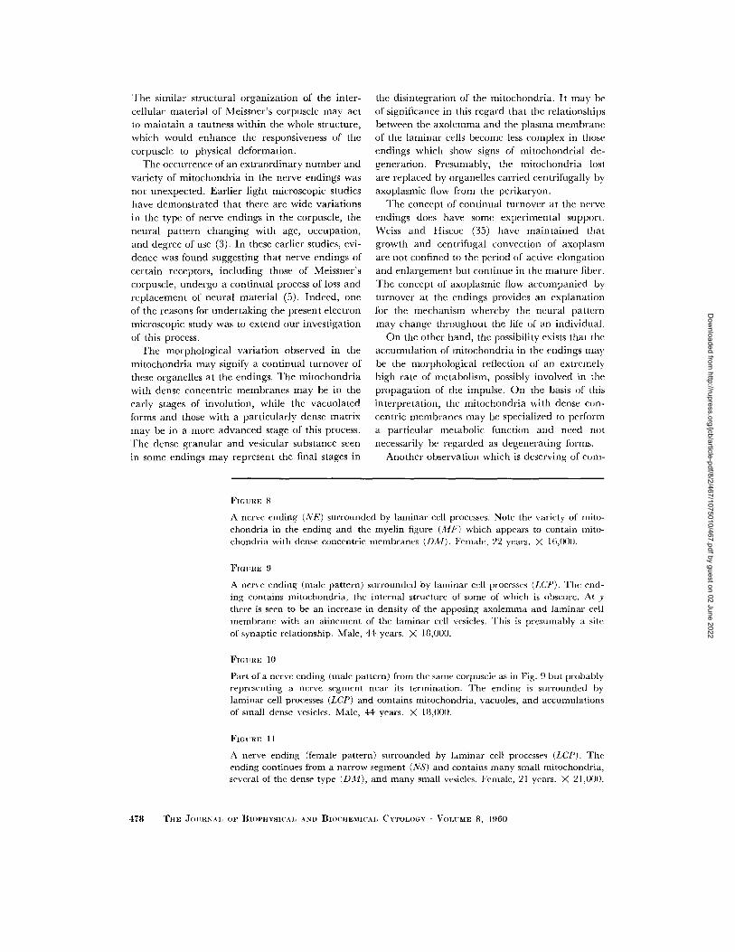
FIGURE ~'t
A nerve ending (NE) surrounded by laminar cell processes. Note the variety of mito- chondria in the ending and the myelin figure (A/F) which appears to contain mito- chondria with dense conccntric membranes (DM). Female, 22 year's. X 16,000.
FIGURE .(}
A nerve ending (male pattern) surrounded by laminar cell processes (LCP). The end- ing contains mitochondria, the internal structure of some of which is obscure. At y there is seen to be an increase in density of the apposing axolemma and laminar cell membrane with an alinement of the laminar cell vesicles. This is presumably a site of synaptic relationship, Male, 44 years. ) 18,000.
FIGURE 1(1
Part of a nerve ending (male pattern) from the same corpuscle as in Fig. 9 but probably representing a nerve segment near its termination. The ending is surrounded by laminar cell processes (LCP) and contains mitochondria, vacuoles, and accumulations of small dense vesicles. Male, 44 years. X 18,000.
FIGI RE l l
A nerve ending (female pattern) surrounded by laminar cell processes (LCP). The ending continues from a narrow segment (NS) and contains many small mitochondria, several of the dense type (DM), and many small vesicles. Female, 21 years. X 21,000.
478 THE JOURNAl, OF BIOPHYSICAL AND BIOCHEMICAI~ CYTOLOGY • VOLUME 8~ 1.(}60
Dow
nloaded from http://rupress.org/jcb/article-pdf/8/2/467/1075010/467.pdf by guest on 02 June 2022

CAUNA ..'*.NO ROSS Meis.s'ner's Corpuscle Fine Structure 479
Dow
nloaded from http://rupress.org/jcb/article-pdf/8/2/467/1075010/467.pdf by guest on 02 June 2022

ment is the presence of numerous small vesicles in the terminal axoplasm and in all parts of the l aminar cell cytoplasm. Because of their s t ructure and location it would be reasonable to assume tha t many of these vesicles are "synapt ic vesicles" and to postulate tha t certain areas of contact between the axolemma and the l aminar cell mem- brane consti tute a synaptic junct ion.
De Robert is and Bennet t (10) and Palay and Palade (23, 25) first advanced the theory tha t sub- microscopic vesicular components , termed "syn- aptic vesicles," are present in synapses and tha t these may be associated with product ion and release of acetylcholine or other neurohumora l transmitters. The rapidly growing body of observa- tions on these cytoplasmic vesicles has been reviewed by De Robert is (9) and by Palay (24). They have been found in all synapses so far studied (30). In certain ones their n u m b e r is reported to increase after prolonged exper imental st imulation (12) and decrease after denervat ion (8, 13, 29). The suggestion has been advanced (9) tha t these vesicles represent the "quan t a l units of acetylcho- line."
In most synapses such vesicles are found only in the presynaptic member . In the sensory recep- tors, however, their location varies according to the type. Vesicles are found in the presynaptic m e m b e r in the rod and cone cells (11) and carotid body chemoreceptors (31), in the postsynaptic member in the taste buds (7, 33), and in both members in the hair cells of the organ of Corti (32), the cristae ampullares (14, 15, 36), and in the Pacinian corpuscle (27). Meissner 's corpuscle, in this respect, belongs to the last group, which evi-
dently comprises receptors responding to mechani - cal st imulation.
An adequate explanat ion for the presence of vesicles on bo th sides of the presumed synaptic junc t ion in Meissner 's corpuscle is not immedia te ly available. These observations would tend to throw doub t on the concept of the uniformity of na ture of these vesicles. Vesicles in the presynaptic mem- ber may have an entirely different function from those in postsynaptic member . Indeed, many of the vesicles in bo th members probably have a more general metabol ic significance as pinocytotic vesicles. I t is appa ren t tha t the concept tha t vesicles found in synapses represent quan ta l units of acetyleholine is in need of fur ther investigation and reevaluat ion, par t icular ly in those incon- gruous instances where the vesicles are located in the postsynaptic member .
The authors wish to express their appreciation to Dr. Don. W. Fawcett for his many suggestions and his assistance in revising the manuscript. They wish also to acknowledge the able technical assistance of Miss Vira Levytska.
B I B L I O G R A P H Y
1. CAUNA, N., Nature and functions of the papil- lary ridges of the digital skin, Anat. Rec., 1954, 119,449.
2. CAUNA, N., Structurc and origin of the capsule of Meissner's corpuscle, Anat. Rec., 1956, 124, 77.
3. CAUNA, N., Nerve supply and nerve endings in Meissner's corpuscles, Am. o r. Anat., 1956, 99, 315.
FmURE 12
A field from a Meissner's corpuscle showing pgrtions of several laminar cells (LC) separated by the intracellular substance (ICS). The cell on the left exhibits a finely granular nucleus, small mitochondria, scattered granular endoplasmic reticulum (ER), groups of unassociated ribonucleoprotein granules (G), and groups of small vesicles (V), some of which (arrow) appear to be attached to the plasma membrane of th's cell. Note the two laminar cell processes (x and y) which are nothing more than two plasma membranes enclosing an occasional vesicle. A portion of a nerve ending (NE) is covered by a laminar cell process on one side but exposed to the intercellular sub- stance on the other side. Female, 22 years. X 21,000.
FIGURE 13
A field from a Meissner's corpuscle showing intercellular substance surrounded by laminar cells. The matrix of the substance contains darkly staining collagen fibers (C) and fine parallel fibrils bound together by a dense material at repeated intervals of approximately 1200 A (visible at x). Female, 21 years. PTA stain. X 21,000.
480 THE JOURNAL OF BIOPHYSICAL AND BIOCHEMICAL CYTOLOGY • VOLUME 8, 1960
Dow
nloaded from http://rupress.org/jcb/article-pdf/8/2/467/1075010/467.pdf by guest on 02 June 2022

CAUNA AND ROSS M¢issner's Corpuscle Fine Structure 481
Dow
nloaded from http://rupress.org/jcb/article-pdf/8/2/467/1075010/467.pdf by guest on 02 June 2022

4. CAUNA, N., Structure of digital touch corpuscles, Acta Anal., 1958, 32, 1.
5. CAUNA, N., The mode of termination of the sensory nerves and its significance, J. Comp. Neurol., 1959, 113, 169.
6. CAUNA, N., and MANNAN, G., Development and postnatal changes of digital Pacinian corpuscles in the human hand, J. Anal., 1959, 93, 271.
7. DE LORENZO, A. J., Electron microscopic ob- servations of the taste buds of the rabbits, J. Biophysic. and Biochem. Cytol., 1958, 4, 143.
8. DE ROBERTIS, E., Submicroscopic changes in the synapse after nerve section in the acoustic ganglion of the guinea-pig, J. Biophysic. and Biochem. Cytol., 1956, 2, 503.
9. DE ROBERTIS, E., Submicroscopic morphology and function of the synapse, Exp. Cell Research, 1958, suppl. 5, 347.
10. DE ROBERTIS, E., and BENNETT, H. S., Some features of the submicroscopic morphology of synapses in frog and earthworm, J. Biophysic. and Biochem. Cytol., 1955, 1, 47.
11. DE ROBERTIS, E., and FRANCHI, C. M., Electron microscope observations on synaptic vesicles in synapses of the retinal rods and cones, J. Biophysic. and Biochem. Cytol., 1956, 2, 307.
12. DE ROnERTIS, E., and VAZ FERREIRA, A., Sub- microscopic changes of the nerve endings in the adrenal medulla after stimulation of the splanchnic nerve, J. Biophysic. and Biochem. Cytol., 1957, 3, 611.
13. DE ROBERTIS, E., and VAZ FERREIRA, A., Elec- tron microscope study of the excretion of cathecol-containing droplets in the adrenal medulla, Exp. Cell Research, 1957, 12, 568.
14. ENGSTROM, H., On the double innervation of the sensory epithelia of the inner ear, Acta Oto- laryngol., 1958, 49, 109.
15. ENOSTROM, H., and WERSALL, J., The ultra- structural organization of the organ of Corti and of the vestibular sensory epithelia, Exp. Cell Research, 1958, suppl. 5, 460.
16. FERNANDEZ-MORAN, H., Applications of a dia- mond knife for ultra thin sectioning to the study of the fine structure of biological tissues and metals, J. Biopl~ysic. and Biochem. Cylol., 1956, 2, No. 4, suppl., 29.
17. JAKUS, M. A., Studies of the cornea. II. The fine structure of Descemet's membrane, J. BiG- physic, and Biochem. Cytol., 1956, 2, No. 4, suppl., 243.
18. MOORE, D. H., and GRIMLEY, P. M., Problems in methacrylate embedding for electron mi- croscopy, J. Biophysic. and Biochem. Cytol., 1957, 3, 255.
19. PALADE, G. E., A study of fixation for electron microscopy, o r. Exp. Med., 1952, 95, 285.
20. PALADE, G. E., A small particulate component of the cytoplasm, J. Biophysic. and Biochem. Cytol., 1955, 1, 59.
21. PALADE, G. E., Studies on the endoplasmic re- ticulum, J. Biophysic. and Biochem. Cytol., 1955, 1,567.
22. PALADE, G. E., The endoplasmic reticulum, J. Biophysic. and Biochem. Cytol., 1956, 2, No. 4, suppl., 85.
23. PALAY, S. L., Synapses in the central nervous system, J. Biophysic. and Biochem. Cytol., 1956, 2, No. 4, suppl., 193.
24. PALAY, S. L., The morphology of synapses in the central nervous system, Exp. Cell Research, 1958, suppl. 5, 275.
25. PALAY, S. L., and PALADE, G. E., The fine struc- ture of neurons, J. Biophysic. and Biochem. Cytol., 1955, 1, 69.
26. PEASE, D. C., and PALLIE, W., Electron micros- copy of digital tactile corpuscles and small cutaneous nerves, J. Ultrastruct. Research, 1959, 2, 352.
27. PEASE, D. C., and QUILLIAM, T. A., Electron microscopy of the Pacinian corpuscle, J. Biophysic. and Biochem. Cytol., 1957, 3, 331.
28. PORTER, K. R., and BLOM, J., A study of microt- omy for electron microscopy, Anat. Rec., 1953, 117, 685.
29. REOER, J. F. The uhrastructure of normal and denerveated neuromuscular synapses in mouse gastrocnemius muscle, Exp. Cell Research, 1957, 12,662.
30. ROBERTSON, J. D., The ultrastructure of a rep- tilian myoneural junction, J. Biophysic. and Biochem. Cytol., 1956, 2, 381.
31. Ross, L. L., Observations on the fine structure of the carotid body of the cat, J. Biophysic. and Biochem. Cytol., 1959, 6,253.
32. SMITH, C. A., and DEMPSEV, E. W., Electron microscopy of the organ of Corti, Am. J. Anat., 1957, 100, 337.
33. TRUJILLO-CENOZ, O., Electron microscope study of the rabbit gustatory bud, Z. Zellforsch., 1957, 46, 272.
34. WATSON, M. L., Staining of tissue sections for electron microscopy with heavy metals, J. Biophysic. and Biochem. Cytol,, 1958, 4, 475.
35. WEISS, P., and HISCOE, H. B., Experiments on the mechanism of nerve growth, J. Exp. Zool., 1948, 107, 315.
36. WERSALL, J., Studies on the structure and in- nervation of the sensory epithelium of the cristae ampullares in the guinea-pig, Acta Otolaryngol., 1956, suppl., 126.
482 THE JOURNAL OF BIOPHYSICAL AND BIOCHEMICAL CYTOLOGY • VOLUME ~, 1960
Dow
nloaded from http://rupress.org/jcb/article-pdf/8/2/467/1075010/467.pdf by guest on 02 June 2022