The FASEB Journal @BULLET Research Communication Human adipose tissue-derived stem cells exhibit...
10
The FASEB Journal • Research Communication Human adipose tissue-derived stem cells exhibit proliferation potential and spontaneous rhythmic contraction after fusion with neonatal rat cardiomyocytes Roxana Metzele,* ,1 Christopher Alt,* ,1 Xiaowen Bai,* ,1 Yasheng Yan,* Zhi Zhang, † Zhizhong Pan, † Michael Coleman, ‡ Jody Vykoukal,* Yao-Hua Song* and Eckhard Alt* ,2 *Department of Molecular Pathology and † Department of Anesthesiology, University of Texas M. D. Anderson Cancer Center, Houston, Texas, USA; and ‡ InGeneron, Inc., Houston, Texas, USA ABSTRACT Various types of stem cells have been shown to have beneficial effects on cardiac function. It is still debated whether fusion of injected stem cells with local resident cardiomyocytes is one of the mech- anisms. To better understand the role of fusion in stem cell-based myocardial regeneration, the present study was designed to investigate the fate of human adipose tissue-derived stem cells (hASCs) fused with neonatal rat cardiomyocytes in vitro. hASCs labeled with the green fluorescent probe Vybrant DiO were cocultured with neonatal rat cardiomyocytes labeled with the red fluorescent probe Vybrant DiI and then treated with fusion-inducing hemagglutinating virus of Japan (HVJ). Cells that incorporated both red and green fluorescent signals were considered to be hASCs that had fused with rat cardiomyocytes. Fusion efficiency was 19.86 4.84% at 5 d after treatment with HVJ. Most fused cells displayed cardiomyocyte-like morphology and exhib- ited spontaneous rhythmic contraction. Both immuno- fluorescence staining and lentiviral vector labeling showed that fused cells contained separate rat cardio- myocyte and hASC nuclei. Immunofluorescence stain- ing assays demonstrated that human nuclei in fused cells still expressed the proliferation marker Ki67. In addition, hASCs fused with rat cardiomyocytes were positive for troponin I. Whole-cell voltage-clamp analy- sis demonstrated action potentials in beating fused cells. RT-PCR analysis using rat- or human-specific myosin heavy chain primers revealed that the myosin heavy-chain expression in fused cells was derived from rat cardiomyocytes. Real-time PCR identified expres- sion of human troponin T in fused cells and the presence of rat cardiomyocytes induced a cardiomyo- genic protein expression of troponin T in human ASCs. This study illustrates that hASCs exhibit both stem cell (proliferation) and cardiomyocyte properties (action potential and spontaneous rhythmic beating) after fu- sion with rat cardiomyocytes, supporting the theory that fusion, even if artificially induced in our study, could indeed be a mechanism for cardiomyocyte renewal in the heart.—Metzele, R., Alt, C., Bai, X., Yan, Y., Zhang, Z., Pan, Z., Coleman, M., Vykoukal, J., Song, Y.-H., Alt, E. Human adipose tissue-derived stem cells exhibit proliferation potential and spontaneous rhythmic con- traction after fusion with neonatal rat cardiomyocytes. FASEB J. 25, 830 – 839 (2011). www.fasebj.org Key Words: differentiation action potential Previous studies from our group and others have shown that adult stem cells isolated from bone marrow (1), skeletal muscle (2), and adipose tissue (3) exhibit beneficial effects on cardiac function following myocar- dial infarction. However, the underlying mechanisms responsible for stem cell-based myocardial regenera- tion are not yet fully understood. Several groups have attributed this potential to the transdifferentiation ca- pacity of stem cells (4). Others, however, have demon- strated that the cardiac differentiation of injected stem cells resulted from spontaneous fusion of donor stem cells with recipient cardiomyocytes. Injected bone mar- row-derived stem cells spontaneously fused with native cardiomyocytes and formed multinucleated cells in the heart (5–7). The fate of stem cells after fusion with local resident cardiomyocytes, however, remains unclear. Human adipose tissue-derived stem cells (hASCs) are a promising cell source for potential stem cell-based clinical therapies, since a large number of hASCs can be relatively easily harvested from patients via mini- mally invasive liposuction and transplanted in an autol- ogous manner without expansion in vitro (8). In addi- tion, ASCs have the potential for proliferation and differentiation into multiple cell lineages, including cardiomyocytes, endothelial cells, and smooth muscle cells while exposed to an appropriate microenviron- ment in vitro or in vivo (3, 9, 10). Several studies have shown that transplantation of ASCs improved cardiac function after experimental myocardial infarction (3, 1 These authors contributed equally to this work. 2 Correspondence: Interdisciplinary Stem Cell Laboratory, Unit 951, The University of Texas M. D. Anderson Cancer Center, 1515 Holcombe Blvd., Houston, TX 77030, USA. E-mail: [email protected] doi: 10.1096/fj.09-153221 This article includes supplemental data. Please visit http:// www.fasebj.org to obtain this information. 830 0892-6638/11/0025-0830 © FASEB
-
Upload
independent -
Category
Documents
-
view
1 -
download
0
Transcript of The FASEB Journal @BULLET Research Communication Human adipose tissue-derived stem cells exhibit...
The FASEB Journal • Research Communication
Human adipose tissue-derived stem cells exhibitproliferation potential and spontaneous rhythmiccontraction after fusion with neonatal ratcardiomyocytes
Roxana Metzele,*,1 Christopher Alt,*,1 Xiaowen Bai,*,1 Yasheng Yan,* Zhi Zhang,†
Zhizhong Pan,† Michael Coleman,‡ Jody Vykoukal,* Yao-Hua Song* and Eckhard Alt*,2
*Department of Molecular Pathology and †Department of Anesthesiology, University of Texas M. D.Anderson Cancer Center, Houston, Texas, USA; and ‡InGeneron, Inc., Houston, Texas, USA
ABSTRACT Various types of stem cells have beenshown to have beneficial effects on cardiac function. Itis still debated whether fusion of injected stem cellswith local resident cardiomyocytes is one of the mech-anisms. To better understand the role of fusion in stemcell-based myocardial regeneration, the present studywas designed to investigate the fate of human adiposetissue-derived stem cells (hASCs) fused with neonatalrat cardiomyocytes in vitro. hASCs labeled with thegreen fluorescent probe Vybrant DiO were coculturedwith neonatal rat cardiomyocytes labeled with the redfluorescent probe Vybrant DiI and then treated withfusion-inducing hemagglutinating virus of Japan (HVJ).Cells that incorporated both red and green fluorescentsignals were considered to be hASCs that had fusedwith rat cardiomyocytes. Fusion efficiency was 19.86 �4.84% at 5 d after treatment with HVJ. Most fused cellsdisplayed cardiomyocyte-like morphology and exhib-ited spontaneous rhythmic contraction. Both immuno-fluorescence staining and lentiviral vector labelingshowed that fused cells contained separate rat cardio-myocyte and hASC nuclei. Immunofluorescence stain-ing assays demonstrated that human nuclei in fusedcells still expressed the proliferation marker Ki67. Inaddition, hASCs fused with rat cardiomyocytes werepositive for troponin I. Whole-cell voltage-clamp analy-sis demonstrated action potentials in beating fusedcells. RT-PCR analysis using rat- or human-specificmyosin heavy chain primers revealed that the myosinheavy-chain expression in fused cells was derived fromrat cardiomyocytes. Real-time PCR identified expres-sion of human troponin T in fused cells and thepresence of rat cardiomyocytes induced a cardiomyo-genic protein expression of troponin T in human ASCs.This study illustrates that hASCs exhibit both stem cell(proliferation) and cardiomyocyte properties (actionpotential and spontaneous rhythmic beating) after fu-sion with rat cardiomyocytes, supporting the theory thatfusion, even if artificially induced in our study, couldindeed be a mechanism for cardiomyocyte renewal inthe heart.—Metzele, R., Alt, C., Bai, X., Yan, Y., Zhang,Z., Pan, Z., Coleman, M., Vykoukal, J., Song, Y.-H., Alt,E. Human adipose tissue-derived stem cells exhibitproliferation potential and spontaneous rhythmic con-
traction after fusion with neonatal rat cardiomyocytes.FASEB J. 25, 830–839 (2011). www.fasebj.org
Key Words: differentiation � action potential
Previous studies from our group and others haveshown that adult stem cells isolated from bone marrow(1), skeletal muscle (2), and adipose tissue (3) exhibitbeneficial effects on cardiac function following myocar-dial infarction. However, the underlying mechanismsresponsible for stem cell-based myocardial regenera-tion are not yet fully understood. Several groups haveattributed this potential to the transdifferentiation ca-pacity of stem cells (4). Others, however, have demon-strated that the cardiac differentiation of injected stemcells resulted from spontaneous fusion of donor stemcells with recipient cardiomyocytes. Injected bone mar-row-derived stem cells spontaneously fused with nativecardiomyocytes and formed multinucleated cells in theheart (5–7). The fate of stem cells after fusion with localresident cardiomyocytes, however, remains unclear.
Human adipose tissue-derived stem cells (hASCs) area promising cell source for potential stem cell-basedclinical therapies, since a large number of hASCs canbe relatively easily harvested from patients via mini-mally invasive liposuction and transplanted in an autol-ogous manner without expansion in vitro (8). In addi-tion, ASCs have the potential for proliferation anddifferentiation into multiple cell lineages, includingcardiomyocytes, endothelial cells, and smooth musclecells while exposed to an appropriate microenviron-ment in vitro or in vivo (3, 9, 10). Several studies haveshown that transplantation of ASCs improved cardiacfunction after experimental myocardial infarction (3,
1 These authors contributed equally to this work.2 Correspondence: Interdisciplinary Stem Cell Laboratory,
Unit 951, The University of Texas M. D. Anderson CancerCenter, 1515 Holcombe Blvd., Houston, TX 77030, USA.E-mail: [email protected]
doi: 10.1096/fj.09-153221This article includes supplemental data. Please visit http://
www.fasebj.org to obtain this information.
830 0892-6638/11/0025-0830 © FASEB
11). However, controversy remains regarding the mech-anisms by which ASCs contribute to tissue repair. Inparticular, the role of fusion in stem cell-based myocar-dial regeneration is debated.
Several in vitro cell-fusion techniques are in use. Thefusion efficacy induced by hemagglutinating virus ofJapan (HVJ) reportedly is higher than that induced bypolyethylene glycol (12). Accordingly, the present studywas designed to investigate the fate of HVJ-inducedhASC fusion with neonatal rat cardiomyocytes.
MATERIALS AND METHODS
Isolation and culture of hASCs
hASCs were provided by InGeneron (Houston, TX, USA) under amaterial transfer agreement. Adipose tissue was acquired fromdonors undergoing elective lipoplasty with informed consent un-der a tissue acquisition protocol approved by the M. D. AndersonCancer Center Review Board. hASCs were isolated from the adi-pose tissue, as described previously (13, 14). Adipose tissue wasminced and then incubated with agitation for 90 min at 37°C withLiberase Blendzyme 3 (Roche, Mannheim, Germany) at a concen-tration of 4 U/g adipose tissue in PBS. The digested tissue wassequentially filtered through 100- and 40-�m filters (Fisher Scien-tific, Waltham, MA, USA) and centrifuged at 450 g for 10 min. Theresulting supernatant containing adipocytes and debris was dis-carded, and the pelleted cells were washed twice with Hanks’balanced salt solution (Cellgro; Mediatech, Inc., Manassas, VA,USA) and resuspended in a growth medium. The growth mediumcontained � modification of Eagle’s medium (Cellgro), 20% FBS(Atlanta Biologicals, Lawrenceville, GA, USA), 2 mM glutamine(Cellgro), and 100 U/ml penicillin with 100 �g/ml streptomycin(Cellgro). Plastic-adherent cells were designated as hASCs andgrown in Nunclon culture vials (Nunc, Roskilde, Denmark) at 37°Cin a humidified atmosphere containing 5% CO2, followed by daily
washes to remove red blood cells and nonattached cells. Afterconfluence of hASCs in culture vials (passage 0), cells were digestedand seeded at a density of 3000 cells/cm2 (passage 1).
Cell differentiation
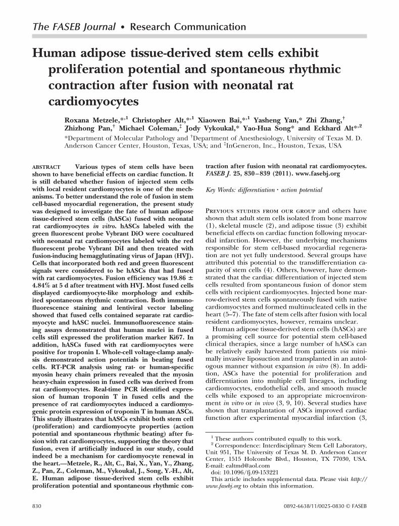
To establish the intrinsic differentiation potential of hASCsused in this study, the adipogenic differentiation potential ofhASCs was analyzed as described previously (15). Osteogenicdifferentiation of hASCs was performed according to Oeday-rajsingh-Varma et al. (16). The cells were then observed undera phase-contrast microscope (Fig. 1).
Isolation and culture of neonatal rat cardiomyocytes
Laboratory animal experiments were performed with requi-site approval and appropriate M. D. Anderson Cancer CenterAnimal Care and Use Committee oversight. Cardiomyocyteswere isolated from 2-d-old Sprague-Dawley rats using a com-mercial kit (Worthington, Lakewood, NJ, USA) according tothe manufacturer’s protocol. Cardiomyocytes cultured in aLeibovitz L-15 medium supplemented with 10% FBS wereused for subsequent experiments. Cardiomyocyte culturecontained cardiomyocytes and cardiac fibroblasts. To in-crease the fusion efficiency of hASCs and rat cardiomyocytes,the proliferation of cardiac fibroblasts was inhibited by treat-ment of the cells isolated from rat hearts with mitomycin C ata concentration of 3 �g/ml for 2 h on d 2 after the start of cellculture. The cardiomyocytes were identified by their expres-sion of troponin I, using immunofluorescence staining anal-ysis. Cell nuclei were stained blue with 4�-6-diamidino-2-phenylindole (DAPI). For analysis of the proliferation ofcardiac fibroblasts and the purity of cardiomyocytes in culturewith or without treatment with mitomycin C, cardiomyocytecultures in 24-well plates were imaged using a charge-coupleddevice (CCD) camera under a fluorescence microscope (CarlZeiss, Oberkochen, Germany) at different time points afterthe start of cell culture. The cell images were taken in 3
Figure 1. Characterization ofhASCs. A) Phase-contrast photomi-crograph of hASCs. hASCs exhib-ited a spindle- or triangle-like mor-phology at passage 3. B–E)Differentiation potential of hASCs.Cells were cultured in an adipogenicmedium (B), adipogenic controlmedium (C), osteogenic medium(D), or osteogenic control medium(E) for 3 wk. Adipogenesis and os-teogenesis of hASCs were con-firmed by the presence of lipid
drops stained red with Oil Red O (B) and calcium deposits stained red with Alizarin Red S (D) in hASCs cultured in inductionmedium, respectively. Scale bars � 50 �m.
831FUSION OF STEM CELLS WITH CARDIOMYOCYTES
random fields in each well for a total of 3 wells for eachtreatment per time point. Troponin I-positive cardiomyocytesand DAPI-stained cells were counted, and the ratio of thenumber of troponin I-positive red blood cells to DAPI-positiveblue cells was used to determine the relative proportion ofcardiomyocytes in the culture.
Cell labeling
Vybrant DiI and DiO fluorescent tracers
To track the fusion formation between hASCs and rat car-diomyocytes, hASCs at passage 3 and neonatal cardiomyo-cytes on d 3 after the start of the culture were labeled with twodifferent fluorescent dyes, Vybrant DiO (a fluorochromeemitting green light) and Vybrant DiI (a fluorochromeemitting red light), according to the protocol provided byInvitrogen (Carlsbad, CA, USA). The DiO-labeled hASCswere fused with cultured cardiomyocytes labeled with DiI at aratio of 1:10, as described below. Cells with both red andgreen fluorescent signals were considered to be fused orhybridized cells. Cells were then stained with DAPI. Thenumbers of green and red double-positive hASCs fused withcardiomyocytes and DAPI-positive total cells were counted in3 random fields in each well (24-well plate) for a total of 3wells on d 1 and 5 after treatment of HVJ. The fusionefficiency was analyzed by calculation of the ratio between thenumber of red and green double-positive fused cells and thenumber of DAPI-positive cells in wells at different time pointsafter treatment with HVJ, thus giving us the percentage of ratcardiomyocytes that were correctly fused with hASCs.
Lentiviral vector construction and hASC labelingwith lentiviral vector
pCAG-SH-GH was generously provided by the laboratory ofDr. Richard R. Behringer (M. D. Anderson Cancer Center).To generate a lentiviral construct (pLVX-Puro-HS-GR), H2B-EGFP-2A-mCherry-GPI (HS-GR) was cut from vector pCAG-SH-GH and then subcloned into lentiviral vector pLVX-Puro(Clontech, Mountain View, CA, USA) between the XhoI andXbaI sites by digestion with specific restriction enzyme andligation with DNA ligase. This subcloned lentiviral vector(pLVX-Puro-HS-GR) contained both a green fluorescenceprotein (GFP) reporter gene expressed in the nuclei and ared fluorescence protein (mCherry) reporter gene expressedin the membrane of transduced cells. The lentivirus produc-tion and transduction procedures were described previously(10). To track whether HVJ induced cellular cytoplasm fusionor nuclear incorporation, we transduced hASCs with thedual-fluorescent protein reporter gene contained within thelentiviral vector (pLVX-Puro-HS-GR). Transduced hASCswere maintained in the culture medium containing puromy-cin at a concentration of 5 �g/ml for 1 wk. hASCs with greenfluorescence in the nuclei and red fluorescence in the cellmembrane were then cocultured with rat cardiomyocytes andtreated with HVJ as described above. At 5 d after treatmentwith HVJ, the cells in coculture were stained with DAPI andobserved under a fluorescence microscope to analyze thedistributions and origins of nuclei in hASCs fused with ratcardiomyocytes.
Virus-mediated fusion formation between hASCsand neonatal rat cardiomyocytes
Fusion of hASCs and rat cardiomyocytes was induced bytreatment with HVJ (GenomONETM-CF; HVJ envelope cellfusion kit; Ishihara Sangyo Kaisha, Osaka, Japan), following
the manufacturer’s recommendations. The virus in the HVJ-Ekit is inactivated by UV irradiation. Using this preparation, wefound that the ability of the virus to replicate was lostcompletely, but its fusion activity was retained. Following thecell-labeling procedure, the hASCs were digested with tryp-sin/EDTA, washed, and combined with HVJ-E. These cellswere left to stand on ice for 5 min to allow absorption ofHVJ-E into the cell membrane, centrifuged at 2000 rpm for 5min, and then resuspended in ice-cooled fusion buffer. ThesehASCs were then added to cultured cardiomyocytes at a ratioof 1:10. The mixed cells were centrifuged at 1000 rpm for 5min and incubated at 37°C in a 5% CO2 environment for 15min.
Immunofluorescence staining
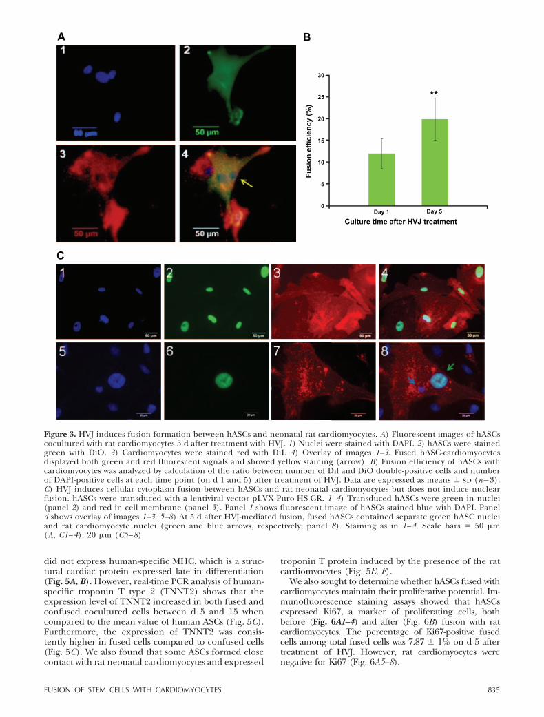
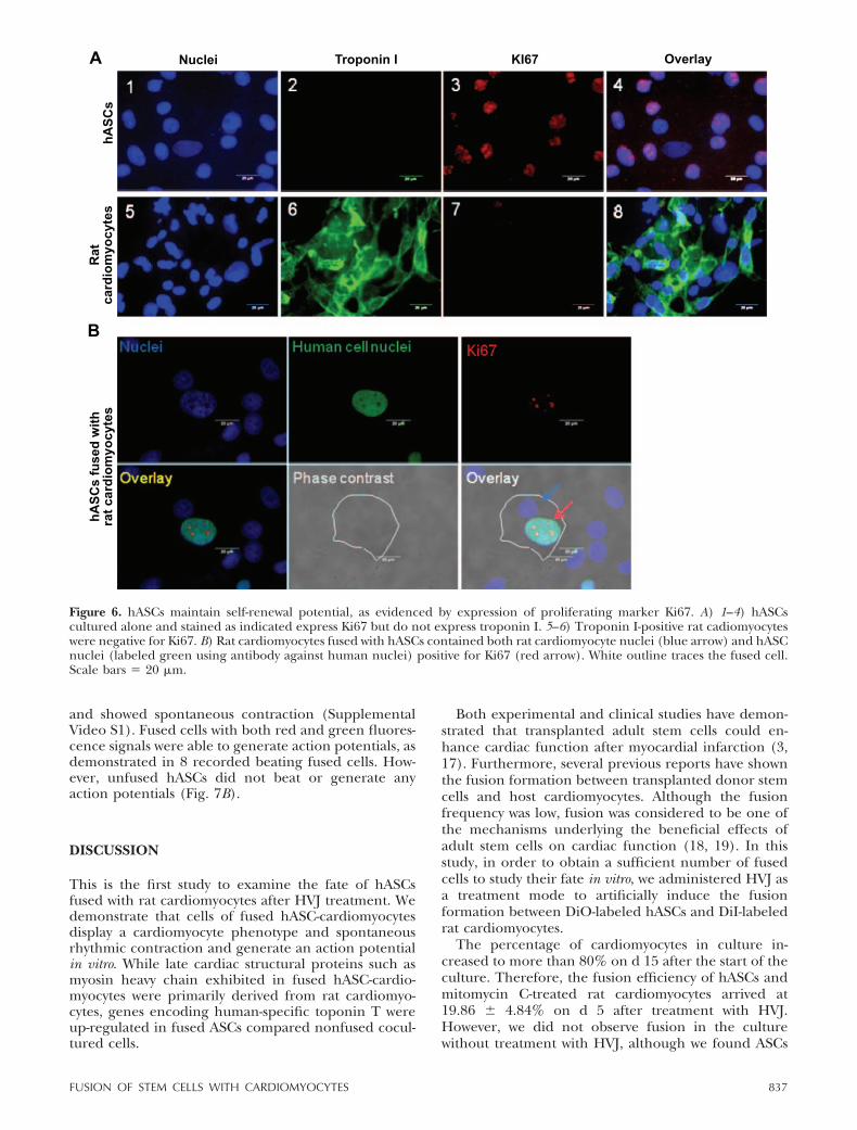
Cells cultured in the 24-well plates were washed 3 times withPBS and then fixed with 4% paraformaldehyde for 10 min atroom temperature. Cells were then washed 3 times with PBScontaining 0.3% Triton X-100 (Sigma) and then blocked with10% serum for 30 min at room temperature. Next, cells wereincubated with primary antibodies, including a mouse anti-human nuclear antibody (Chemicon, Temecula, CA, USA),rabbit anti-human and anti-rat (or specific anti-human) tro-ponin I antibody, or rabbit anti-Ki67 antibody for 1 h at 37°C.An anti-human nuclear antibody and anti-troponin I antibodywere used to track hASCs and cardiomyocytes, respectively, inthe coculture of hASCs and rat cardiomyocytes. An anti-Ki67antibody was used to track proliferation of cells. After 3washes, the cells were incubated with appropriate fluoro-phore-conjugated secondary antibodies for 1 h at roomtemperature. The nuclei were counterstained with DAPI. Thecells were then examined under a fluorescence microscope.The efficiency of Ki67-positive fused cells was calculated bythe ratio of Ki67-positive hASCs fused with cardiomyocytesagainst total fused cells from 3 fields/well for a total of 3 wells(24-well plate) 5 d after treatment with HVJ.
RT-PCR analysis of mRNA expression in hASCs fusedwith rat cardiomyocytes
Total RNA was extracted from isolated fused hASC-cardio-myocytes using an RNAqueous-Micro kit (Ambion, Austin,TX, USA). cDNA was synthesized from 1 �g of total RNAusing an iScript cDNA synthesis kit (Bio-Rad, Hercules, CA,USA) in 20 �l of reaction volume according to the manufac-turer’s instructions. cDNA samples were subjected to PCRamplification using AccuPrime SuperMix I (Invitrogen) withspecific primers. The primers and their sequences were asfollows: myosin heavy chain (MHC)-specific primers for hu-man cardiomyocytes, forward 5�-AGAAGCAGAGGAACTTC-GAC-3� and reverse 5�-CCTTGATCTGGTTGAACTCC-3�;MHC-specific primers for rat cardiomyocytes, forward 5�-GGAGACCTTCAAGCGGGAGA-3 � and reverse 5 � -AGGGCTGACTGCAGCTCCA-3�. Negative control reac-tions consisted of the PCR amplification mix describedabove with primers but no cDNA template. Commerciallyavailable human heart cDNAs (Ambion) were used as positivecontrols. PCR was performed using an Eppendorf Mastercy-cler gradient (Eppendorf, Westbury, NY, USA). The cycleswere programmed as follows: 94°C for 10 min, 35 cycles of30 s of denaturation at 94°C, 30 s at an annealing temperatureof 57°C (for a human-specific MHC primer) or 61°C (for arat-specific MHC primer), a 45-s extension step at 72°C, anda final extension step at 72°C for 5 min. Expression ofglyceraldehyde-3-phosphate dehydrogenase (GAPDH)mRNA was used as an internal control. The PCR product sizewas confirmed by loading 10 �l of PCR reaction mixtures to
832 Vol. 25 March 2011 METZELE ET AL.The FASEB Journal � www.fasebj.org
1.5% agarose gel electrophoresis. Real-time PCR was per-formed using a light cycler from Bio-Rad. Program: 95°C(10 s) and 58°C(45 s) for 50 cycles. Human troponin type2: forward 5�-GAC CTG CAG GAG AAG TTC AA-3�, reverse5�-GAG GAG CAG ATC TTT GGT GA-3�. Human GAPDH:forward 5�GAGTCAACGGATTTGGTCGT3�, reverse 5�TT-GATTTTGGAGGGATCTC3�.
Images of beating fused cells
Images of beating hASCs fused with rat cardiomyocytes (withgreen and red fluorescent signals) were recorded using aCCD camera with a phase-contrast microscope (Zeiss) 5 dafter treatment of coculture (hASCs and rat cardiomyocytes)with HVJ.
Whole-cell voltage-clamp recordings
hASCs (passage 3), rat cardiomyocytes (8 d after culture), orhASCs cocultured with rat cardiomyocytes (5 d after treat-ment with HVJ) cultured on glass coverslips in 24-well plateswere used for whole-cell voltage-clamp analysis. For recordingof action potential of cultured cells, one of the coverslips wastransferred to a chamber perfused with modified Tyrode’ssolution at a rate of 1.5 ml/min. The perfusing solution wasequilibrated in 5% CO2 and 95% O2 and maintained at roomtemperature (21–22°C). The contents of the perfusing solu-tion was as follows: 136 mM NaCl, 2 mM KCl, 1 mM MgCl2,1.8 mM CaCl2, 0.33 mM NaH2PO4, 10 mM glucose, and 10mM HEPES; the pH was adjusted to 7.3 with NaOH, and theosmolarity was 320–330 mosmol/L. Visualized whole-cellvoltage-clamp recordings were obtained using a glass pipette(resistance, 2–3 M�) filled with a solution containing: 140mM potassium gluconate, 10 mM NaCl, 2 mM MgCl2, 2 mMEGTA, 5 mM HEPES; the pH was adjusted to 7.3 with KOH, andthe osmolarity was 300–310 mosmol/L. An AxoPatch 1D ampli-fier and the AxoGraph software program (Axon Instruments,Union City, CA, USA) were used for data acquisition and onlineand offline data analyses. A seal resistance � 2 G� and an accessresistance � 10 M� were considered acceptable. Series resis-tance was optimally compensated for 80%, and it was decreasedto 60% when current oscillation was detected. The accessresistance was monitored throughout the experiment. The cellswere held at �80 mV. Voltage steps of 300 ms from �60 to �60mV in 10-mV increments were used to induce action potential.Data were digitized at 20 kHz and filtered at 2 kHz. Theexperiments were conducted at room temperature (�22°C).
Data analysis
The reported data are expressed as means sd. The statis-tical significance of the differences between groups wasdetermined using the Student’s t test. A level of P � 0.05 wasconsidered statistically significant.
RESULTS
hASCs exhibit differentiation potential
hASCs isolated from human adipose tissue exhibitedfibroblast or spindle-like morphology (Fig. 1A). Toevaluate the differentiation capacity of hASCs, hASCswere cultured in adipogenic and osteogenic mediumfor 3 wk. hASCs in an adipogenic induction mediumdisplayed characteristic multiple intracellular bright
white oil droplets. These droplets exhibited red vesicleswhen stained with Oil Red O solution (Fig. 1B). Nolipid droplets were observed in cells cultured in controlmedium (Fig. 1C). Cells cultured in osteogenic me-dium showed black regions within the monolayer,which indicated calcification deposits from differenti-ated osteoblasts. Calcification of the extracellular ma-trix (Fig. 1D) appeared red after induced hASCs werestained with Alizarin Red S dye. However, red stainingwas not observed in cells cultured in a control medium(Fig. 1E).
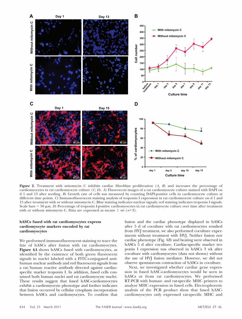
Treatment with mitomycin C inhibits the proliferationof cardiac fibroblasts and increases the percentageof cardiomyocytes in culture
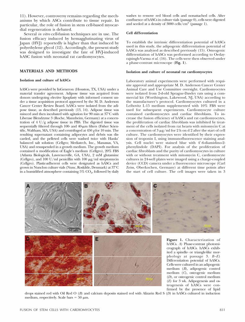
Freshly isolated cells from neonatal rat hearts con-tained both cardiomyocytes and fibroblasts in cardio-myocyte culture medium. Cell proliferation was mea-sured by counting nuclei stained blue with DAPI. Thenumber of cells isolated from rat hearts increased by3-fold within 13 d of culture, mainly because of theproliferation of cardiac fibroblasts (Fig. 2A, B). Car-diomyocytes were identified by their expression oftroponin I, using immunofluorescent staining analysiswith a rat/human reactive antibody. Because of thelimited proliferation potential of cardiomyocytes andthe strong proliferation potential of fibroblasts, therelative proportion of cardiomyocytes in culture de-creased by more than half within 15 d after culture (Fig.2C, D). To increase the proportion of cardiomyocytesand thereby maximize the fusion efficiency of hASCswith rat cardiomyocytes, the cultured cells were treatedwith mitomycin C to inhibit the proliferation of cardiacfibroblasts before treatment with HVJ. As shown in Fig.2C, D, this treatment inhibited the proliferation offibroblasts markedly and increased the proportion ofcardiomyocytes in the culture.
HVJ induces the fusion formation between hASCsand neonatal rat cardiomyocytes
hASCs (DiO green labeled) were fused with rat car-diomyocytes (DiI red labeled) after treatment with HVJ.Fused cells showed both green and red fluorescencesignals and multiple nuclei under a fluorescence mi-croscope (Fig. 3A). Most fused cells exhibited sponta-neous rhythmic contraction on d 2 after HVJ treatment.Fusion efficiency increased with culture time; fusionefficiency was 12.48 4.36% after 1 d and 19.86 4.84% 5 d after treatment with HVJ (Fig. 3B, P�0.01).To further investigate whether HVJ induced cellularcytoplasm fusion or nuclear incorporation, we trans-duced hASCs with the lentiviral vector. TransducedhASCs displayed green nuclei and red cell membranes(Fig. 3C1–4). After treatment with HVJ, fused hASC-cardiomyocytes contained both hASC nuclei and ratcardiomyocyte nuclei (Fig. 3C5–8). These results indi-cate that HVJ induced cytoplasmic fusion betweenhASCs and rat cardiomyocytes but did not induce cellnuclear fusion.
833FUSION OF STEM CELLS WITH CARDIOMYOCYTES
hASCs fused with rat cardiomyocytes expresscardiomyocyte markers encoded by ratcardiomyocytes
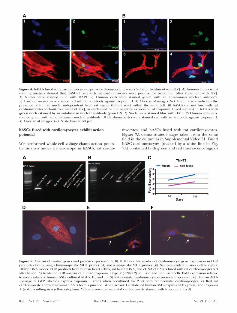
We performed immunofluorescent staining to trace thefate of hASCs after fusion with rat cardiomyocytes.Figure 4A shows hASCs fused with cardiomyocytes, asidentified by the existence of both green fluorescentsignals in nuclei labeled with a FITC-conjugated anti-human nuclear antibody and red fluorescent signals froma rat/human reactive antibody directed against cardiac-specific marker troponin I. In addition, fused cells con-tained both human nuclei and rat cardiomyocyte nuclei.These results suggest that fused hASC-cardiomyocytesexhibit a cardiomyocyte phenotype and further indicatesthat fusion occurred by cellular cytoplasm incorporationbetween hASCs and cardiomyocytes. To confirm that
fusion and the cardiac phenotype displayed in hASCsafter 5 d of coculture with rat cardiomyocytes resultedfrom HVJ treatment, we also performed coculture exper-iments without treatment with HVJ. Neither fusion norcardiac phenotype (Fig. 4B) and beating were observed inhASCs 5 d after coculture. Cardiac-specific marker tro-ponin I expression was observed in hASCs 3 wk aftercoculture with cardiomyocytes (data not shown) withoutthe use of HVJ fusion mediator. However, we did notobserve spontaneous contraction of hASCs in coculture.
Next, we investigated whether cardiac gene expres-sion in fused hASC-cardiomyocytes would be seen inhASCs or from rat cardiomyocytes. We performedRT-PCR with human- and rat-specific MHC primers toanalyze MHC expression in fused cells. Electrophoreticanalysis of the PCR product show that fused hASC-cardiomyocytes only expressed rat-specific MHC and
Figure 2. Treatment with mitomycin C inhibits cardiac fibroblast proliferation (A, B) and increases the percentage ofcardiomyocytes in rat cardiomyocyte culture (C, D). A) Fluorescent images of a rat cardiomyocyte culture stained with DAPI ond 1 and 13 after seeding. B) Growth rate of cells was measured by counting DAPI-positive cells in cardiomyocyte culture atdifferent time points. C) Immunofluorescent staining analysis of troponin I expression in rat cardiomyocyte culture on d 1 and15 after treatment with or without mitomycin C. Blue staining indicates nuclear signals; red staining indicates troponin I signals.Scale bars � 50 �m. D) Percentage of troponin I-positive cardiomyocytes in rat cardiomyocyte culture over time after treatmentwith or without mitomycin C. Data are expressed as means sd (n�3).
834 Vol. 25 March 2011 METZELE ET AL.The FASEB Journal � www.fasebj.org
did not express human-specific MHC, which is a struc-tural cardiac protein expressed late in differentiation(Fig. 5A, B). However, real-time PCR analysis of human-specific troponin T type 2 (TNNT2) shows that theexpression level of TNNT2 increased in both fused andconfused cocultured cells between d 5 and 15 whencompared to the mean value of human ASCs (Fig. 5C).Furthermore, the expression of TNNT2 was consis-tently higher in fused cells compared to confused cells(Fig. 5C). We also found that some ASCs formed closecontact with rat neonatal cardiomyocytes and expressed
troponin T protein induced by the presence of the ratcardiomyocytes (Fig. 5E, F).
We also sought to determine whether hASCs fused withcardiomyocytes maintain their proliferative potential. Im-munofluorescence staining assays showed that hASCsexpressed Ki67, a marker of proliferating cells, bothbefore (Fig. 6A1–4) and after (Fig. 6B) fusion with ratcardiomyocytes. The percentage of Ki67-positive fusedcells among total fused cells was 7.87 1% on d 5 aftertreatment of HVJ. However, rat cardiomyocytes werenegative for Ki67 (Fig. 6A5–8).
Figure 3. HVJ induces fusion formation between hASCs and neonatal rat cardiomyocytes. A) Fluorescent images of hASCscocultured with rat cardiomyocytes 5 d after treatment with HVJ. 1) Nuclei were stained with DAPI. 2) hASCs were stainedgreen with DiO. 3) Cardiomyocytes were stained red with DiI. 4) Overlay of images 1–3. Fused hASC-cardiomyocytesdisplayed both green and red fluorescent signals and showed yellow staining (arrow). B) Fusion efficiency of hASCs withcardiomyocytes was analyzed by calculation of the ratio between number of Dil and DiO double-positive cells and numberof DAPI-positive cells at each time point (on d 1 and 5) after treatment of HVJ. Data are expressed as means sd (n�3).C) HVJ induces cellular cytoplasm fusion between hASCs and rat neonatal cardiomyocytes but does not induce nuclearfusion. hASCs were transduced with a lentiviral vector pLVX-Puro-HS-GR. 1–4) Transduced hASCs were green in nuclei(panel 2) and red in cell membrane (panel 3). Panel 1 shows fluorescent image of hASCs stained blue with DAPI. Panel4 shows overlay of images 1–3. 5–8) At 5 d after HVJ-mediated fusion, fused hASCs contained separate green hASC nucleiand rat cardiomyocyte nuclei (green and blue arrows, respectively; panel 8). Staining as in 1– 4. Scale bars � 50 �m(A, C1– 4); 20 �m (C5– 8).
835FUSION OF STEM CELLS WITH CARDIOMYOCYTES
hASCs fused with cardiomyocytes exhibit actionpotential
We performed whole-cell voltage-clamp action poten-tial analysis under a microscope in hASCs, rat cardio-
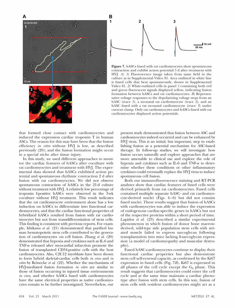
myocytes, and hASCs fused with rat cardiomyocytes.Figure 7A demonstrates images taken from the samefield in the culture as in Supplemental Video S1. FusedhASC-cardiomyocytes (tracked by a white line in Fig.7A) contained both green and red fluorescence signals
Figure 4. hASCs fused with cardiomyocytes express cardiomyocyte markers 5 d after treatment with HVJ. A) Immunofluorescentstaining analysis showed that hASCs fused with rat cardiomyocytes were positive for troponin I after treatment with HVJ.1) Nuclei were stained blue with DAPI. 2) Human cells were stained green with an anti-human nuclear antibody.3) Cardiomyocytes were stained red with an antibody against troponin I. 4) Overlay of images 1–3. Green arrow indicates thepresence of human nuclei independent from rat nuclei (blue arrow) within the same cell. B) hASCs did not fuse with ratcardiomyocytes without treatment of HVJ, as evidenced by the negative expression of troponin I (red signals) in hASCs withgreen nuclei stained by an anti-human nuclear antibody (panel 4). 1) Nuclei were stained blue with DAPI. 2) Human cells werestained green with an anti-human nuclear antibody. 3) Cardiomyocytes were stained red with an antibody against troponin I.4) Overlay of images 1–3. Scale bars � 50 �m.
Figure 5. Analysis of cardiac genes and protein expression. A, B) MHC as a late marker of cardiomyocyte gene expression in PCRproducts of cells using a human-specific MHC primer (A) and a rat-specific MHC primer (B). Samples loaded in lanes (left to right):100-bp DNA ladder, PCR products from human heart cDNA, rat heart cDNA, and cDNA of hASCs fused with rat cardiomyocytes 5 dafter fusion. C) Real-time PCR analysis of human troponin T type 2 (TNNT2) in fused and nonfused cells. Fold expression relativeto mean values of human ASCs cultured at d 5, 10, and 15. D) Rat neonatal cardiomyocyte expression troponin T. E) Human ASCs(passage 3, GFP labeled) express troponin T (red) when cocultured for 2 wk with rat neonatal cardiomyocytes. F) Red ratcardiomyocyte and yellow human ASCs form a junction. White arrows: GFP-labeled human ASCs express GFP (green) and troponinT (red), resulting in a yellow cytoplasm. Yellow arrows: rat neonatal cardiomyocyte stained with troponin T (red).
836 Vol. 25 March 2011 METZELE ET AL.The FASEB Journal � www.fasebj.org
and showed spontaneous contraction (SupplementalVideo S1). Fused cells with both red and green fluores-cence signals were able to generate action potentials, asdemonstrated in 8 recorded beating fused cells. How-ever, unfused hASCs did not beat or generate anyaction potentials (Fig. 7B).
DISCUSSION
This is the first study to examine the fate of hASCsfused with rat cardiomyocytes after HVJ treatment. Wedemonstrate that cells of fused hASC-cardiomyocytesdisplay a cardiomyocyte phenotype and spontaneousrhythmic contraction and generate an action potentialin vitro. While late cardiac structural proteins such asmyosin heavy chain exhibited in fused hASC-cardio-myocytes were primarily derived from rat cardiomyo-cytes, genes encoding human-specific toponin T wereup-regulated in fused ASCs compared nonfused cocul-tured cells.
Both experimental and clinical studies have demon-strated that transplanted adult stem cells could en-hance cardiac function after myocardial infarction (3,17). Furthermore, several previous reports have shownthe fusion formation between transplanted donor stemcells and host cardiomyocytes. Although the fusionfrequency was low, fusion was considered to be one ofthe mechanisms underlying the beneficial effects ofadult stem cells on cardiac function (18, 19). In thisstudy, in order to obtain a sufficient number of fusedcells to study their fate in vitro, we administered HVJ asa treatment mode to artificially induce the fusionformation between DiO-labeled hASCs and DiI-labeledrat cardiomyocytes.
The percentage of cardiomyocytes in culture in-creased to more than 80% on d 15 after the start of theculture. Therefore, the fusion efficiency of hASCs andmitomycin C-treated rat cardiomyocytes arrived at19.86 4.84% on d 5 after treatment with HVJ.However, we did not observe fusion in the culturewithout treatment with HVJ, although we found ASCs
Figure 6. hASCs maintain self-renewal potential, as evidenced by expression of proliferating marker Ki67. A) 1–4) hASCscultured alone and stained as indicated express Ki67 but do not express troponin I. 5–6) Troponin I-positive rat cadiomyocyteswere negative for Ki67. B) Rat cardiomyocytes fused with hASCs contained both rat cardiomyocyte nuclei (blue arrow) and hASCnuclei (labeled green using antibody against human nuclei) positive for Ki67 (red arrow). White outline traces the fused cell.Scale bars � 20 �m.
837FUSION OF STEM CELLS WITH CARDIOMYOCYTES
that formed close contact with cardiomyocytes andinduced the expression cardiac troponin T in humanASCs. The reason for this may have been that the fusionefficiency in vitro without HVJ is low, as describedpreviously (20); and the fusion formation might occurin a special niche after tissue injury.
In this study, we used different approaches to moni-tor the cardiac features of hASCs after coculture withrat cardiomyocytes and treatment with HVJ. The exper-imental data showed that hASCs exhibited action po-tential and spontaneous rhythmic contraction 2 d afterfusion with rat cardiomyocytes. We did not observespontaneous contraction of hASCs in the 21-d culturewithout treatment with HVJ. A relatively low percentage oftroponin I-positive hASCs were observed in the 3-wkcoculture without HVJ treatment. This result indicatesthat the rat cardiomyocyte environment alone has a lowinduction on hASCs to differentiate into functional car-diomyocytes, and that the cardiac functional properties ofhybridized hASCs resulted from fusion with rat cardio-myocytes but not from transdifferentiation of stem cells.This finding is consistent with previous reports. For exam-ple, Ishikawa et al. (21) demonstrated that purified hu-man hematopoietic stem cells contributed to the genera-tion of cardiomyocytes via cell fusion. Zhang et al. (20)demonstrated that hypoxia and cytokines such as IL-6 andTNF-� released after myocardial infarction promote thefusion of transplanted CD34-positive cells with the hostcardiomyocytes. Also, C2C12 myoblasts have been shownto form hybrid skeletal-cardiac cells both in vivo and invitro by Reinecke et al. (22). Whether the mechanisms ofHVJ-mediated fusion formation in vitro are similar tothose of fusion occurring in injured tissue environmentsin vivo, and whether hASCs fused with cardiomyocyteshave the same electrical properties as native cardiomyo-cytes remain to be further investigated. Nevertheless, our
present study demonstrated that fusion between ASC andcardiomyocytes indeed occurred and can be enhanced byHVJ virus. This is an initial, but important, step in estab-lishing fusion as a potential mechanism for ASC-basedtherapy. In follow-up studies, we will investigate howfusion occurs naturally and explore approaches that aremore amenable to clinical use and explore the role ofhypoxia and cytokines such as IL-6 and TNF-� to deter-mine whether these conditions or other inflammatorycytokines could eventually replace the HVJ virus to inducespontaneous cell fusion.
Both our immunofluorescence staining and RT-PCRanalyses show that cardiac features of fused cells werederived primarily from rat cardiomyocytes. Fused cellscontained multiple separate hASC- and rat cardiomyo-cyte-derived nuclei (Figs. 4–6) but did not containfused nuclei. These results suggest that fusion of hASCswith cardiomyocytes was able to induce the expressionof endogenous cardiac-specific genes in hASCs but notof the respective proteins within a short period of time.Lapidos et al. (23) described a similar experimentalphenomenon in which fusion of donor bone marrow-derived, wild-type side population stem cells with stri-ated muscle failed to express sarcoglycan followingtransplantation into mice lacking �-sarcoglycan expres-sion (a model of cardiomyopathy and muscular dystro-phy).
Fused hASC-cardiomyocytes continue to display theirfunctional cardiac properties but also demonstratestem cell self-renewal capacity, as confirmed by the Ki67expression in fused cells (Fig. 7B). Ki67 is expressed inall phases of the cell cycle except the G0 phase. Thisresult suggests that cardiomyocytes could enter the cellcycle and at the same time maintain a cardiac pheno-type after fusion with stem cells. In this way, fusion ofstem cells with resident cardiomyocytes might act as a
Figure 7. hASCs fused with rat cardiomyocytes show spontaneouscontraction and exhibit action potential 5 d after treatment withHVJ. A) 1) Fluorescence image taken from same field in theculture as in Supplemental Video S1. Area outlined in white lineis fused cells that beat spontaneously, shown in SupplementalVideo S1. 2) White-outlined cells in panel 1 containing both redand green fluorescent signals displayed yellow, indicating fusionformation between hASCs and rat cardiomyocytes. B) Represen-tative voltage responses to the depolarizing voltage steps from anhASC (trace 1), a neonatal rat cardiomyocyte (trace 2), and anhASC fused with a rat neonatal cardiomyocyte (trace 3) undercurrent clamp. Only rat cardiomyocytes and hASCs fused with ratcardiomyocytes displayed action potentials.
838 Vol. 25 March 2011 METZELE ET AL.The FASEB Journal � www.fasebj.org
rapid, intermediate repair mechanism that enablessalvage of the damaged heart en route to more durableregeneration of the myocardium.
This study demonstrates that rat cardiomyocytesfused with ASCs display both stem cell proliferatingcapacity and functional cardiomyocyte properties (ac-tion potential, and spontaneous rhythmic beating)within 5 d after fusion. Our in vitro data indicate thatstem cells are capable and have the theoretical capacityto compensate for a sudden loss of cardiomyocytesimmediately following myocardial infarction via fusionwith cardiomyocytes, supporting the concept that fu-sion, even if artificially induced in our study, is apotential mechanism for cardiomyocyte renewal in theheart.
The authors thank Sebastian Gehmert, Sanga Gehmert,Grace Wu, Martha French, and Cynthia Austin for their excel-lent technical assistance. This work was supported by the Alli-ance of Cardiovascular Researchers, grant 543102 (to E.A.).
REFERENCES
1. Dawn, B., Tiwari, S., Kucia, M. J., Zuba-Surma, E. K., Guo, Y.,Sanganalmath, S. K., Abdel-Latif, A., Hunt, G., Vincent, R. J.,Taher, H., Reed, N. J., Ratajczak, M. Z., and Bolli, R. (2008)Transplantation of bone marrow-derived very small embryonic-like stem cells attenuates left ventricular dysfunction and re-modeling after myocardial infarction. Stem Cells 26, 1646–1655
2. Farahmand, P., Lai, T. Y., Weisel, R. D., Fazel, S., Yau, T.,Menasche, P., and Li, R. K. (2008) Skeletal myoblasts preserveremote matrix architecture and global function when implantedearly or late after coronary ligation into infarcted or remotemyocardium. Circulation 118, S130–137
3. Valina, C., Pinkernell, K., Song, Y. H., Bai, X., Sadat, S.,Campeau, R. J., Le Jemtel, T. H., and Alt, E. (2007) Intracoro-nary administration of autologous adipose tissue-derived stemcells improves left ventricular function, perfusion, and remod-elling after acute myocardial infarction. Eur. Heart. J. 28, 2667–2677
4. Deb, A., Wang, S., Skelding, K. A., Miller, D., Simper, D., andCaplice, N. M. (2003) Bone marrow-derived cardiomyocytes arepresent in adult human heart: A study of gender-mismatchedbone marrow transplantation patients. Circulation 107, 1247–1249
5. Noiseux, N., Gnecchi, M., Lopez-Ilasaca, M., Zhang, L., Solo-mon, S. D., Deb, A., Dzau, V. J., and Pratt, R. E. (2006)Mesenchymal stem cells overexpressing Akt dramatically repairinfarcted myocardium and improve cardiac function despiteinfrequent cellular fusion or differentiation. Mol. Ther. 14,840–850
6. Alvarez-Dolado, M., Pardal, R., Garcia-Verdugo, J. M., Fike, J. R.,Lee, H. O., Pfeffer, K., Lois, C., Morrison, S. J., and Alvarez-Buylla, A. (2003) Fusion of bone-marrow-derived cells withPurkinje neurons, cardiomyocytes and hepatocytes. Nature 425,968–973
7. Rota, M., Kajstura, J., Hosoda, T., Bearzi, C., Vitale, S., Esposito,G., Iaffaldano, G., Padin-Iruegas, M. E., Gonzalez, A., Rizzi, R.,Small, N., Muraski, J., Alvarez, R., Chen, X., Urbanek, K., Bolli,R., Houser, S. R., Leri, A., Sussman, M. A., and Anversa, P.(2007) Bone marrow cells adopt the cardiomyogenic fate invivo. Proc. Natl. Acad. Sci. U. S. A. 104, 17783–17788
8. Strem, B. M., and Hedrick, M. H. (2005) The growing importanceof fat in regenerative medicine. Trends Biotechnol. 23, 64–66
9. Planat-Benard, V., Menard, C., Andre, M., Puceat, M., Perez, A.,Garcia-Verdugo, J. M., Penicaud, L., and Casteilla, L. (2004)Spontaneous cardiomyocyte differentiation from adipose tissuestroma cells. Circ. Res. 94, 223–229
10. Mochizuki, H., Schwartz, J. P., Tanaka, K., Brady, R. O., andReiser, J. (1998) High-titer human immunodeficiency virus type1-based vector systems for gene delivery into nondividing cells.J. Virol. 72, 8873–8883
11. Cai, L., Johnstone, B. H., Cook, T. G., Tan, J., Fishbein, M. C.,Chen, P. S., and March, K. L. (2009) IFATS series: humanadipose tissue-derived stem cells induce angiogenesis and nervesprouting following myocardial infarction, in conjunction withpotent preservation of cardiac function. Stem Cells 27, 230–237
12. Kim, J., and Okada, Y. (1987) Difference in capacities forvirion-to-virion fusion of young and aged HVJ (Sendai virus): amodel of membrane fusion. J. Membr. Biol. 97, 241–249
13. Gimble, J., and Guilak, F. (2003) Adipose-derived adult stemcells: isolation, characterization, and differentiation potential.Cytotherapy 5, 362–369
14. Bai, X., Ma, J., Pan, Z., Song, Y. H., Freyberg, S., Yan, Y.,Vykoukal, D., and Alt, E. (2007) Electrophysiological propertiesof human adipose tissue-derived stem cells. Am. J. Physiol. CellPhysiol. 293, C1539–C1550
15. Pittenger, M. F., Mackay, A. M., Beck, S. C., Jaiswal, R. K.,Douglas, R., Mosca, J. D., Moorman, M. A., Simonetti, D. W.,Craig, S., and Marshak, D. R. (1999) Multilineage potential ofadult human mesenchymal stem cells. Science 284, 143–147
16. Oedayrajsingh-Varma, M. J., van Ham, S. M., Knippenberg, M.,Helder, M. N., Klein-Nulend, J., Schouten, T. E., Ritt, M. J., andvan Milligen, F. J. (2006) Adipose tissue-derived mesenchymalstem cell yield and growth characteristics are affected by thetissue-harvesting procedure. Cytotherapy 8, 166–177
17. Stamm, C., Kleine, H. D., Westphal, B., Petzsch, M., Kittner, C.,Nienaber, C. A., Freund, M., and Steinhoff, G. (2004) CABGand bone marrow stem cell transplantation after myocardialinfarction. Thorac. Cardiovasc. Surg. 52, 152–158
18. Nygren, J. M., Jovinge, S., Breitbach, M., Sawen, P., Roll, W.,Hescheler, J., Taneera, J., Fleischmann, B. K., and Jacobsen,S. E. (2004) Bone marrow-derived hematopoietic cells generatecardiomyocytes at a low frequency through cell fusion, but nottransdifferentiation. Nat. Med. 10, 494–501
19. Oh, H., Bradfute, S. B., Gallardo, T. D., Nakamura, T., Gaussin,V., Mishina, Y., Pocius, J., Michael, L. H., Behringer, R. R.,Garry, D. J., Entman, M. L., and Schneider, M. D. (2003)Cardiac progenitor cells from adult myocardium: homing,differentiation, and fusion after infarction. Proc. Natl. Acad. Sci.U. S. A. 100, 12313–12318
20. Zhang, S., Shpall, E., Willerson, J. T., and Yeh, E. T. (2007)Fusion of human hematopoietic progenitor cells and murinecardiomyocytes is mediated by alpha 4 beta 1 integrin/vascularcell adhesion molecule-1 interaction. Circ. Res. 100, 693–702
21. Ishikawa, F., Shimazu, H., Shultz, L. D., Fukata, M., Nakamura, R.,Lyons, B., Shimoda, K., Shimoda, S., Kanemaru, T., Nakamura, K.,Ito, H., Kaji, Y., Perry, A. C., and Harada, M. (2006) Purifiedhuman hematopoietic stem cells contribute to the generation ofcardiomyocytes through cell fusion. FASEB J. 20, 950–952
22. Reinecke, H., Minami, E., Poppa, V., and Murry, C. E. (2004)Evidence for fusion between cardiac and skeletal muscle cells.Circ. Res. 94, e56–e60
23. Lapidos, K. A., Chen, Y. E., Earley, J. U., Heydemann, A., Huber,J. M., Chien, M., Ma, A., and McNally, E. M. (2004) Trans-planted hematopoietic stem cells demonstrate impaired sar-coglycan expression after engraftment into cardiac and skeletalmuscle. J. Clin. Invest. 114, 1577–1585
Received for publication December 23, 2009.Accepted for publication October 28, 2010.
839FUSION OF STEM CELLS WITH CARDIOMYOCYTES