The estrogen receptor binds tightly to its responsive element as a ligand-induced homodimer
12
Cell, Vol. 55, 145-156, October 7, 1988, Copyright 0 1988 by Cell Press The Estrogen Receptor Binds Tightly to Its Responsive Element as a Ligand-Induced Homodimer Vijay Kumar and Pierre Chambon Laboratoire de G&+tique Molgculaire des Eucaryotes du CNRS Unite 184 de Biologie Mol&ulaire et de GEnie G&&ique de I’ INSERM lnstitut de Chimie Biologique, Faculte de MBdecine 67085 Strasbourg CBdex, France Summary Extracts containing wild-type or mutant human estro- gen receptor (ER) have been used to study the binding of ER to its responsive element (ERE). Estradiol (E2) or the antiestrogen hydroxytamoxifen is required for ER binding as assayed by gel retardation. The DNA binding domain (DBD) encompasses the highly con- served region C. Both intact ER-ES complexes and ER mutants truncated for the hormone binding domain (HBD) bind as dimers to an ERE. However, an HBD- truncated ER binds less tightly to an ERE than an in- tact ER-EP complex. The DBD and the HBD contain a constitutive and a stronger ER-induced dimerization function, respectively. Thus, in addition to inducing the activation function associated with the HBD, es- trogen plays a crucial role in the formation of stable ER dimers that bind tightly to ERE. Introduction Recent cloning and sequence comparison of nuclear receptors indicate that they belong to a superfamily of nu- clear receptors whose function is dependent on the bind- ing of small hydrophobic ligands, such as steroid and thy- roid hormones, vitamin D3, and retinoids. Functional analyses have shown that these receptors act as tran- scriptional enhancer factors by interacting with inducible regulatory sequences (responsive elements; RE), which triggers a cascade of biological processes through modu- lation of transcription of specific target genes (for refer- ences and reviews, see Yamamoto, 1985; Green and Chambon, 1986; Evans and Hollenberg, 1988; Evans, 1988; Gronemeyer et al., 1988). However, the mechanism(s) by which these receptors interact with their RE and then stimulate transcription is still unknown. In the case of the estrogen receptor (ER), it has been shown that the hormone binding domain (HBD) (region E; Krust et al., 1986; Kumar et al., 1986) and the putative DNA binding domain (DBD) (region C; Krust et al., 1986), which is responsible for tight nuclear binding (Kumar et al., 1986) and specific recognition of RE of tar- get genes (Green and Chambon, 1987; Kumar et al., 1987), correspond to separable discrete elements that can function independently (Webster et al., 1988a, 1988b). The HBD also appears to contain an estradiol-inducible transcription activation function (Kumar et al., 1987; Web- ster et al., 1988b), whereas the less conserved A/B region is important for efficient activation of transcription from some promoters (Kumar et al., 1987; Tora et al., submitted). In addition to inducing the activation function present in the region containing the HBD, estrogen is required for tight nuclear binding, and it has been suggested that it in- duces the formation of receptor dimers in solution (Lind- stedt et al., 1986; Gordon and Notides, 1986). Since estrogen-responsive elements (ERE) appear to be palin- dromic sequences (Klein-Hitpap et al., 1988; Klock et al., 1987; Martinez et al., 1987, and references therein), it is tempting to speculate that the ER binds to them as a dimer, as proposed earlier by Yamamoto and Alberts (1972). We have tested this possibility by analyzing in vitro the interaction of the human ER (hER) with its RE using a gel retardation (band shift) assay to monitor the forma- tion of ER-ERE complexes. We report here that the hormone is required for the receptor to bind to ERE, and we characterize further the DNA binding domain. More important, we show that bind- ing of estradiol to the hormone binding domain induces the formation of receptor dimers, which is essential for tight binding to the palindromic ERE. Results Estradiol-Dependent Binding of hER to an ERE In Vitro A gel retardation assay was used to investigate the bind- ing of hER to the ERE of the Xenopus vitellogenin (VIT) A2 gene (Klein-Hitpap et al., 1986; wild-type ERE in Table 1A). Whole cell extracts (WCE; 0.4 M KICI) of HeLa cells transiently transfected with vectors expiressing wild-type (HEO) or mutant hERs (see Figure 2) were incubated at 0% for 15 min in the presence of poly(dl-dC) to decrease nonspecific binding, and then for 15 min al: 20% with the 32P-labeled wild-type ERE. No retarded complex was seen when HeLa cells were transfected with the parental vector pKCR2 (Figure lA, lanes 1 and 2). Either a very weak (Figure lA, lane 3) or no retarded complex (see Figures 3A, 3C, and 7A, lane 1) was observed when es- tradiol (E2) was not added to the culture medium of HeLa cells. The reason a “very weak” complex was seen in some experiments is unknown, but it may be because of the presence of residual estrogens in the culture medium. In contrast, addition of 1O-8 M E2 before harvesting the cells resulted in the formation of an intense retarded com- plex (Figure lA, lane 4) that was specific for the wild-type ERE, since no complex could be detected (lane 5) using the mutated ERE probe C (Table lC), which contains mu- tations known to be markedly deleterious in vivo (Klock et al., 1987). Western blot analysis of the retarded complexes using the monoclonal antibodies H222 and D75 (Greene et al., 1984) revealed the presence of hER at the position of the retarded band (data not shown). Similar specific E2- dependent complexes were formed using 0.4 M KCI WCE of the human breast cancer cells MCF-7 that contain the hER (Green et al., 1986; Figure lA, compare lanes 6-8). The specificity of the retarded complex was confirmed by
-
Upload
independent -
Category
Documents
-
view
1 -
download
0
Transcript of The estrogen receptor binds tightly to its responsive element as a ligand-induced homodimer
Cell, Vol. 55, 145-156, October 7, 1988, Copyright 0 1988 by Cell Press
The Estrogen Receptor B inds T ightly to Its Responsive E lement as a Ligand-Induced Homodimer
Vijay Kumar and Pierre Chambon Laboratoire de G&+tique Molgculaire des Eucaryotes du CNRS Unite 184 de Biologie Mol&ulaire et de GEnie G&&ique de I’INSERM lnstitut de Chimie Biologique, Faculte de MBdecine 67085 Strasbourg CBdex, France
Summary
Extracts containing wild-type or mutant human estro- gen receptor (ER) have been used to study the binding of ER to its responsive element (ERE). Estradiol (E2) or the antiestrogen hydroxytamoxifen is required for ER binding as assayed by gel retardation. The DNA binding domain (DBD) encompasses the highly con- served region C. Both intact ER-ES complexes and ER mutants truncated for the hormone binding domain (HBD) bind as dimers to an ERE. However, an HBD- truncated ER binds less tightly to an ERE than an in- tact ER-EP complex. The DBD and the HBD contain a constitutive and a stronger ER-induced dimerization function, respectively. Thus, in addition to inducing the activation function associated with the HBD, es- trogen plays a crucial role in the formation of stable ER dimers that bind tightly to ERE.
Introduction
Recent cloning and sequence comparison of nuclear receptors indicate that they belong to a superfamily of nu- clear receptors whose function is dependent on the bind- ing of small hydrophobic ligands, such as steroid and thy- roid hormones, vitamin D3, and retinoids. Functional analyses have shown that these receptors act as tran- scriptional enhancer factors by interacting with inducible regulatory sequences (responsive elements; RE), which triggers a cascade of biological processes through modu- lation of transcription of specific target genes (for refer- ences and reviews, see Yamamoto, 1985; Green and Chambon, 1986; Evans and Hollenberg, 1988; Evans, 1988; Gronemeyer et al., 1988).
However, the mechanism(s) by which these receptors interact with their RE and then stimulate transcription is still unknown. In the case of the estrogen receptor (ER), it has been shown that the hormone binding domain (HBD) (region E; Krust et al., 1986; Kumar et al., 1986) and the putative DNA binding domain (DBD) (region C; Krust et al., 1986), which is responsible for tight nuclear binding (Kumar et al., 1986) and specific recognition of RE of tar- get genes (Green and Chambon, 1987; Kumar et al., 1987), correspond to separable discrete elements that can function independently (Webster et al., 1988a, 1988b). The HBD also appears to contain an estradiol-inducible transcription activation function (Kumar et al., 1987; Web- ster et al., 1988b), whereas the less conserved A/B region
is important for efficient activation of transcription from some promoters (Kumar et al., 1987; Tora et al., submitted). In addition to inducing the activation function present in the region containing the HBD, estrogen is required for tight nuclear binding, and it has been suggested that it in- duces the formation of receptor dimers in solution (Lind- stedt et al., 1986; Gordon and Notides, 1986). Since estrogen-responsive elements (ERE) appear to be palin- dromic sequences (Klein-Hitpap et al., 1988; Klock et al., 1987; Martinez et al., 1987, and references therein), it is tempting to speculate that the ER binds to them as a dimer, as proposed earlier by Yamamoto and Alberts (1972). We have tested this possibility by analyzing in vitro the interaction of the human ER (hER) with its RE using a gel retardation (band shift) assay to monitor the forma- tion of ER-ERE complexes.
We report here that the hormone is required for the receptor to bind to ERE, and we characterize further the DNA binding domain. More important, we show that bind- ing of estradiol to the hormone binding domain induces the formation of receptor dimers, which is essential for tight binding to the palindromic ERE.
Results
Estradiol-Dependent Binding of hER to an ERE In Vitro A gel retardation assay was used to investigate the bind- ing of hER to the ERE of the Xenopus vitellogenin (VIT) A2 gene (Klein-Hitpap et al., 1986; wild-type ERE in Table 1A). Whole cell extracts (WCE; 0.4 M KICI) of HeLa cells transiently transfected with vectors expiressing wild-type (HEO) or mutant hERs (see Figure 2) were incubated at 0% for 15 min in the presence of poly(dl-dC) to decrease nonspecific binding, and then for 15 min al: 20% with the 32P-labeled wild-type ERE. No retarded complex was seen when HeLa cells were transfected with the parental vector pKCR2 (Figure lA, lanes 1 and 2). Either a very weak (Figure lA, lane 3) or no retarded complex (see Figures 3A, 3C, and 7A, lane 1) was observed when es- tradiol (E2) was not added to the culture medium of HeLa cells. The reason a “very weak” complex was seen in some experiments is unknown, but it may be because of the presence of residual estrogens in the culture medium. In contrast, addition of 1O-8 M E2 before harvesting the cells resulted in the formation of an intense retarded com- plex (Figure lA, lane 4) that was specific for the wild-type ERE, since no complex could be detected (lane 5) using the mutated ERE probe C (Table lC), which contains mu- tations known to be markedly deleterious in vivo (Klock et al., 1987). Western blot analysis of the retarded complexes using the monoclonal antibodies H222 and D75 (Greene et al., 1984) revealed the presence of hER at the position of the retarded band (data not shown). Similar specific E2- dependent complexes were formed using 0.4 M KCI WCE of the human breast cancer cells MCF-7 that contain the hER (Green et al., 1986; Figure lA, compare lanes 6-8). The specificity of the retarded complex was confirmed by

Cell 146
Probe A represents the wild-type estrogen responsive element (WT-ERE) that corresponds to -306 to -342 of the promoter/up. stream element of the Xenopus vitellogenin A2 gene (Klein-Hitpa et al., 1986). The limits of the 13 bp palindromic ERE containing 5 bp arms and a 3 bp spacer region are marked by arrows. The mutants studied in vivo for their transcription activation function are indicated as probes B (Martinez et al., 1987) C (Klock et al., 1987), D (Klein- Hitpa et al., 1988) and E (Berry, Nunez, and Chambon, unpublished data). A 2-fold symmetrical GRE is shown as probe G (Klock et al., 1987). The base changes made to derive the mutant ERE and GRE se- quences from wild-type ERE are boxed.
“competition” experiments. Cold wild-type ERE probe (Fig- ure lB, lanes 1-8) was approximately five times more effi- cient at decreasing the labeling of the specific complex
than the mutant probe E (lanes g--16), which is a weaker ERE in vivo (Berry, Nunez, and Chambon, unpublished data; see also Figure 4). Moreover, no complexes were formed between HE0 and the glucocorticoid-responsive element (GRE) probe G (see Table 1G and Figure 38, lane 7).
We then investigated whether the estradiol requirement for ERE binding could also be demonstrated in vitro. Es- tradiol was added to low (50 mM KCI) and high (0.4 M KCI) salt WCE of HeLa cells that were not exposed to the hor- mone in vivo (see legend to Figure 1C). Very little or no specific complex was formed in the absence of hormone, whether the extract was made in a low (Figure IC, lanes 1 and 4; see also Figure 7H, lanes 1 and 6) or a high (lanes 5 and 8 of Figure 1C) salt buffer. In contrast, addition of E2 to the extract in vitro resulted in the appearance of the retarded complex (Figure lC, lanes 2 and 3, lanes 6 and 7; see also Figure 7H), which was stronger when the incu- bation was carried out at 25% (lanes 3 and 7of Figure 1C) rather than at 0% (lanes 2 and 6 of Figure 1C). It is impor- tant to stress that in all of these experiments (as in those described below), the absence of a retarded complex was not due to variations in receptor content as evidenced by Western blot analysis, both before and after incubation in vitro (data not shown; see also Experimental Procedures and Kumar et al., 1986, 1987). We conclude that specific binding of hER to an ERE can be achieved in vitro and that this binding is markedly dependent on the addition of the hormone either to the transfected cells or to their extracts
1234 5 6 78 12345.578 910111213141516 f 234 5678 ,234 / 1 1
Estradiol added to cells Estradiol added to extracts in vivo in vitro
Figure 1. Specific and Ligand-Dependent Binding of the Human ER to Its Cognate Response Element In Vitro (A) WCE of HeLa cells transfected with either the parent expression vector (pKCR2) or wild-type hER (HEO), and WCE of MCF-7 cells were pre- pared and incubated (see Experimental Procedures) with the radiolabeled probes A and C (see Table 1) as indicated. The cells were treated in vivo either without (-) or with (+) lo-* M oestradiol (E2). The free and bound oligonucleotides were resolved in a low ionic strength nondenatur- ing “retardation” 5% polyacrylamide gel and visualized by autoradiography (see Experimental Procedures). (8) Extracts from HeLa cells transfected with HE0 and treated with estradiol were incubated with a subsaturating concentration of radiolabeled probe A (lo4 cpm) and increasing amounts of unlabeled probes A or E (as indicated), as in (A). For quantitative analysis, the complexes were cut out from the gel and counted in a liquid scintillation spectrometer. (C) HEO-transfected HeLa cells not treated with estradiol were extracted in buffer A containing 50 mM (low salt) or 400 mM (high salt) KCI. After preincubation with poly(dl.dC), both extracts were incubated with probe A under four different conditions: a, no treatment, sample left at 0°C; b, incubation with 10-s M estradiol for 90 min at 0°C; c, incubation with 10-s M estradiol at 0°C for 1 hr followed by incubation at 25% for 30 min; d, incubation first at 25% without ligand for 30 min, followed by incubation with 10-s M estradiol for 1 hr at OOC. The final concentration of the different buffer constituents in the reaction mixture were: 10 mM Tris-HCI (pH 7.5) 1 mM DTT, 10% (v/v) glycerol, and either 100 mM KCi (high salt extract) or 25 mM KCI (low salt extract). The resulting protein-DNA complexes were analyzed as in (A). (D) Heta cells transfected with HE0 were treated 1 hr before harvesting with either IO-* M estradiol, 10e7 M hydroxytamoxifen (OHT) or 10-s M estradiol + 10-c M OHT, as indicated. “High salt” WCE were incubated with probe A as in (A).

Binding of ER to Its Responsive Element 147
Figure 2. Characteristics of the Human Estrogen Receptor (hER) and Its Mutants The division of the 595 amino acid-long hER into six regions (A-F) is shown at the top of the figure together with the percentage homolo- gies for each region between the human and chicken (cER) ERs (see Krust et al., 1986, for details). The extremities of the core region C are indicated below box C. The various deletion mutants and one substi- tution mutant (see Experimental Procedures) are shown schematical- ly below the hER/cER homology box. HE0 represents the wild-type human ER, as indicated by a solid line from amino acids 1 to 595, while a gap in the mutants corresponds to the deleted portion in the recep tor (see also Experimental Procedures). The table at the right-hand side shows the positions of deleted amino acids in the mutants, along with their functional analysis for estradiol binding, in vitro ERE bind- ing, and in vivo activation of transcription using the reporter gene vit- tk-CAT (Kumar et al., 1987). (+) indicates wild-type activity (loo%), whereas (-) means no detectable activity. Values that fall between these two extremes are shown as percentages of wild-type activity. *, determined by using the pS2-CAT reporter plasmid (Kumar et al., 1987).
in vitro. Note that very little complex was formed with low salt extracts prepared from HeLa cells exposed to the hor- mone in vivo, which is in agreement with the nuclear tight binding of the occupied ER (for references, see Kumar et al., 1986; data not shown).
Efficient complex formation was observed in the pres- ence of the estrogen antagonist hydroxy-tamoxifen (OHT), which is known to cause tight nuclear binding of the ER in vivo (Geier et al., 1987; Figure lD, compare lanes 1 to 4; an identical complex was obtained with tamoxifen; data not shown). However, the ERE-hER-OHT complex (lanes 3 and 4) migrated reproducibly 10% slower than the com- plex formed in the presence of E2 (lane 2). Interesting- ly, the nonsteroidal estrogen agonist diethyl-stilbestrol yielded a complex migrating as the “OHT complex” (data not shown).
Region C Is Indispensable for ERE Binding, and Constitutive Binding Is Observed when the Hormone Binding Domain Is Deleted The 66 amino acid-long region that is highly conserved among the various members of the nuclear receptor fam- ily (core of region C, see top of Figure 2) is responsible for
specific target gene activation and may belong to the DNA binding domain (Green and Chambon, 1987; Kumar et al., 1987). A series of hER deletion mutants (Figure 2) was used to map the domain responsible for the formation of the hER-ERE complex. Complex formatio~n was not al- tered by deletions that affect the A/B region (only (HE2 and HElO, Figure 3A, lanes 3 and 8; Figure 3C, lanes 3 to 8), whereas it was abolished by deletion or point mutations that are either within (HE11 and H27, lanes 9 to 10 of Fig- ure 3A) or overlap (HE3 and HE5, lanes 4 and 5 of Figure 3A) region C. It is interesting that all mutations that affect E2 binding (HE5 and HE9 in lanes 5 and 6 of Figure 3A; HE6, HE7, and HE8, HE47, and HE48; Kumar et al., 1986, 1987; data not shown) also abolish complex formation. In contrast, mutations within the hinge region D (HE12, lane 11) or the removal of region F (HE13, lane 7) did not impair hER-ERE complex formation, and no complex was ob- served with HE14 (lane 12), which corresponds essentially to the HBD (Kumar et al., 1986).
The role of region C in ERE binding was further sup- ported by using chimeric receptors. A human glucocorti- coid receptor (hGR)-retarded complex was formed when 0.6 M KCI extracts of Cos-1 cells transfected with a vector expressing hGR (HGl) (in the presence of dexametha- sone [Dex]) were incubated with the GRE probe G (Table 1G; compare lanes 1 and 2 in Figure 3B), but not with an ERE (lanes 12 and its control lane 13 in Figure 3B). Re- placing the core of region C of the hER with that of hGR (ER-GR.CASl, Kumar et al., 1987) resulted in the forma- tion of a complex with GRE (lanes 4 and 8 in Figure 38) and the converse chimera GR-ERCASl (Kumar et al., 1987) formed a complex with an ERE (lane lo), but not with GRE (lane 5).
In agreement with our previous suggestion (Kumar et al., 1987), hER truncated for the HBD did bind constitu- tively to an ERE (Figure 3C, HE21, HE38, and HE15, see Figure 2; the fact that these complexes ha.ve a migration very similar to that of HE0 is probably due to a tight globu- lar structure of hydrophobic domain E; Green et al., 1986). A hGR truncated for the hormone domain (HG3) also formed a complex with a GRE, but not witlh an ERE (Fig- ure 3B, lanes 3 and 11). Mutants truncated for both A/B and E/D regions were used to delineate further the mini- mal domain required for ERE binding. Complexes were observed with HE70 and HE72, but nat with HE16 and HE20 (see Figure 2; Figure 3C, lanes 1%1,6; note that as a result of the lack of a suitable antibody, we do not know whether HE20 was efficiently expressed in HeLa cells; see also Kumar et al., 1987). Thus region C, with perhaps some sequence immediately flanking it (amino acids 179 to 282), is necessary and sufficient for bincling to an ERE.
Wild-type and mutant EREs (Table 1) wlere used to in- vestigate whether the truncated mutants exhibit the same binding specificity as wild-type hER (Figure 4). In all cases a complex was formed with wild-type ERE (probe A, solid triangles), but not with probes C, D, and F [(bands present in these lanes correspond to nonspecific complexes) or the GRE probe G (data not shown). However, specific complexes were seen with probes B and E. Note, however, that these complexes were weaker with HE15, HE70, and

Cdl 148
0
12345678 9 10 111213141516
Figure 3. Localization of the Regions Responsible for DNA Binding
(A) WCE from HeLa cells transfected with either HE3, HE5, HE9, HElO, HE1 1, HE12, HE13, HE14, or HE27 (as indicated) and treated before harvesting with (+) 10-s M estradiol (E2) were incubated with radiolabeled probe A. The reaction products were separated in a 5% polyacryla- mide gel. HEO-WCE from cells treated without (-) or with (+) hormone were run as controls (lanes 1 and 2). (5) WCE from Cos-I cells transfected with the parent expression vector (pKCR2), ERE-binders (HEO, HE19, and GR-ERCASI), or GRE-binders (HGI, HG3, and ER-GR.CASl) and treated before harvesting with either 10-s M estradiol (E2) or 10m7 M dexamethasone (Dex; as indicated) were incubated with either ERE probe A or GRE probe G. The resulting receptor-DNA complexes were analyzed on a 4% polyacrylamide gel. (C) WCE from HeLa cells transfected with either HEO, HE15, HE16, HE19, HE20, HE21, HE38, HE49, HE50, HE68, HE69, HE70, or HE72 and treated before harvesting without (-) or with (+) 10-s M estradiol were incubated with probe A. The receptor-ERE complexes were resolved as in (A).
HE72. Dimethyl sulfate (DMS) interference assays (Figure 5) were also carried out to demonstrate that methylation of the same G residues on both strands of the ERE (in- dicated on both sides of Figure 5) prevented binding of wild-type hER and its truncated mutants HE69 (A/B trun- cation), HE15 (F/E/D truncation), and HE72 (A/B/E/F trun- cation) (see Figure 2). It is interesting that methylation of the same G residues also prevented interaction between wild-type ERE and hER-OHT complexes (Figure 5, com- pare lanes 3-6 with lanes 7-10).
HEOt+EB) HEW-EZ) HE70(-E2) HE72(-E2) HElSL+E*)
Robe ‘ABCDE?‘ABCDEF”ABCDEF’ ‘ABCDEF”ABCDEF’
123456 7 8 9 10 ,112 1314 1516 17 1s 192021222324 252627282930
Figure 4. ERE Binding Specificity of Wild-Type and Mutant hERs WCE from HeLa cells transfected with wild-type (HEO) and mutant hERs (HE15, HE70, HE72, and HE19) and treated before harvesting with (+) or without (-) 10-s M estradiol (E2) were incubated with probes A-F. The receptor-probe complexes were resolved on a 5% polyacryla- mide gel. For quantitative analysis, the receptor-bound complexes were cut out from the gel and counted in a liquid scintillation spectrometer. Filled triangles and arrows, see text.
The Estrogen Receptor Binds as a Dimer The possibility that the ER binds as a dimer was first in- vestigated by SDS-polyacrylamide gel electrophoresis
12 34 5 6 7 8910111213141516171819202122
Figure 5. Methylation Interference Analysis of the hER-ERE Complex Preparative gel retardation assays were performed with partially meth- ylated probe A 5’ end-labeled on either the upper (U) or lower (L) strand, and WCE from HeLa cells transfected with either HE0 or mu- tant receptors (HE15 HE89, HE70, and HE72 as indicated). Whether HeLa cells were treated before harvesting with or without hormone (+ or -) is indicated in parentheses (E2, oestradiol; OHT, ri-hydroxy- tamoxifen). The bound “retarded” (6) and free (F) probe bands were electroeluted, treated with piperidine, and analyzed by electrophoresis on a 15% sequencing gel. The control lanes 1 and 2 represent G lad- ders of the upper and lower strands of the probe directly after partial methylation. G residues whose methylation interferes with the recep- tor-ERE complex formation are encircled.

Binding of ER to Its Responsive Element 149
-68
HE19 -
,I 23, ,456, I - + I
UV-Irradiation Figure 6. Formation of hER Dimers by UV Crosslinking
WCE of HeLa ceils transfected with HE0 and HE19 and treated with estradiol before harvesting were incubated with a bromo-deoxyuridine (U) substituted wild-type ERE SATTAGGUCACAGUGACCTGCW, After
3’TAATCCAGUGTCAC”GGACGG-5’ UV irradiation for 1 hr, the samples were electrophoresed on a 10% SDS-polyacrylamide gel and processed for Western blot analysis as described in Experimental Procedures. Lane 1, untreated WCE (10 ul) containing HEO; lanes 2-3, IO and 5 ul of the proceding extract in- cubated with the above probe, but not UV-irradiated; lanes 4-6, WCE containing HE0 and HE19 incubated with the substituted probe either separately (20 ul) or together (10 ul each) as indicated and then UV-ir- radiated. Arrowheads: 1, homodimers of HEO; 2, HEO-HE19 heterodi- men 3, homodimers of HE19. Molecular weight markers were: 8-galac- tosidase (116 kd), phosphorylase b (94 kd), bovine serum albumin (68 kd), and ovalbumin (43 kd).
(SDS-PAGE) of UV irradiated complexes formed between “HE0 extracts” and a 32P-BrdU-substituted wild-type ERE probe. Two retarded specific 32P-labeled complexes could be detected after UV irradiation and SDS-PAGE, but the slower migrating one was always faint even after prolonged UV irradiation (data not shown). Western blot analysis showed that the faster cross-l inked complex migrated at the same position as the receptor (m66 kd), whereas the slower one, which could be also revealed with hER anti- bodies, migrated as a protein of approximately 130 kd (ar- row 1; Figure 6, compare lanes l-4). Moreover, two cross- linked complexes were also obtained with HE19 truncated for region A/B, but their relative migration was faster than for those seen with HEO. Western blot analysis indicated that they migrated at positions corresponding to HE19 monomer and dimer for the faster and the slower (arrow 3) complex, respectively (Figure 6, lane 6). These results strongly suggested that the faster and slower migrating complexes correspond to the UV cross-linking of one and two molecules of the hER to the ERE, respectively.
Because the efficiency of UV cross-linking was appar- ently low, the binding properties of hER to the palindromic ERE were then extensively investigated using an ap- proach similar to the one which enabled Hope and Struhl (1987) to establish that the yeast trans-activator GCN4 binds as a homodimer to its cognate RE. Extracts of HeLa
cells transfected with HE0 were mixed with extracts from cells transfected with either one of the A/B region-trun- cated mutants HE49, HE68, HE18, HESO, HE69, or HE19 (Figure 2) and incubated with wild-type EFlE (Figure 7A; E2 was added 1 hr before harvesting the cells). Retarded complexes with intermediate migration characteristics were seen in all cases, indicating that one molecule each of HE0 and truncated mutant can bind concomitantly to a single ERE. Furthermore, mixing a constant amount of “H19 extract” with increasing amounts of “HE0 extract” resulted in some increase of the intermediary complex, whereas the HE19 complex decreased, but did not disap- pear completely (Figure 78). The presence of both HE0 and HE19 in the intermediary complex was also supported by excision of the corresponding band, flollowed by SDS- PAGE and Western blot analysis (data not shown). A faint band corresponding to the position of the intermediary complex could also be seen after UV cross-linking (see Figure 6, lane 5, arrow 2).
Intermediary, heterologous complexes (hereafter called “heterodimers”) were also formed when extracts contain- ing hERs truncated for the HBD, HE72, and either HE15 or HE70 were mixed and then incubated with wild-type ERE (Figure 7D). Note, however, that in contrast to the results obtained above with HE0 and HEl9, mixing con- stant amounts of an HE7’Bcontaining extract with increas- ing amounts of an HE15 extract drove almost all of HE72 into an heterodimer with HE15 (Figure 7E).
We conclude that both receptor molecules truncated for or containing the HBD bind as dimers to the ERE. Further- more, high-affinity binding appears to require the pres- ence of a palindromic element, since no binding (Figure 4, probes C, D, and F) or decreased binding (Figure 4, probes B and E) was observed with probes mutated in one-half of the palindrome. Note that this decrease in binding was more pronounced in the case of the hormone binding domain-truncated mutants (HE15 HE70, and HE72 in Figure 4, probes B and E). However, HE15 and HE70 may bind weakly to half palindromic: motifs (arrows in HE15 and HE70 sections in Figure 4), whereas no simi- lar bands were observed in the case of the HE0 and HE19, which contain the HBD (Figure 4).
Estrogen Induces Formation of Receptor Dimers The observation that the intensity of the heterodimer band seen in section A of Figure 7 was always lower than that of the homodimer bands corresponding to individual re- ceptors, and the fact that not all of HE69 could be driven into an heterodimer complex in the presence of increasing amounts of HE0 (Figure 7, section B), suggested that the addition of E2 before harvesting the cells may result in the formation of homodimers that may be relatively stable in solution even in the absence of the ERIE. Interestingly, transfecting HE0 together with either HE49, HE68, HE18, HE50, HE69, or HE19 in HeLa cells (treated with E2 1 hr before harvesting, Figure 7, section C) resulted in a pre- dominant heterodimer band, as expected if heterodimers would be freely formed in the cells on the addition of E2. That the theoretically expected 1:2:1 rat.io between the three bands was not observed may indicate that the

Cell 150
Figure 7. Formation of Receptor “Heterodi- mers” and Localization of the Regions Involved in “Dime?’ Formation
(A) WCE from hormone-treated HeLa cells ex- pressing various hER N-terminal deletion mu- tants (HE49, HE68, HE18, HE50, HE69, and HE19) were incubated either separately with end-labeled probe A or mixed first with WCE from estradiol-treated HEO-transfected HeLa cells (15 min at 0%) and then incubated with the probe. Electrophoresis was on a 5% poly- acrylamide gel. WCE from HEO-transfected HeLa cells treated or not (-E2) with estradiol (lanes 1 and 2) were run as control. (6) An increasing amount of HEO-extract from estradiol-treated HeLacells(as in A) was mixed with a fixed amount of HE69 extract from hormone-treated HeLa cells (as in [A)) and in- cubated with probe A. Electrophoresis as in (A). (C) HeLa cells were transfected with the region A/B deletion mutants HE49, HE68, HE18, HE50, HE69, and HE19, either separately or to- gether with HE0 as indicated. The cells were treated with estradiol before harvesting and WCE were analyzed for ERE binding (probe A) as described in (A). (D) WCE containing the hER mutants HE15, HE70, and HE72 prepared from cells not treated with estradiol were incubated either separately with probe A, or two of them mixed together (as indicated) and then incubated with the same probe. The receptor-bound and free probes were separated as in (A). (E) A fixed volume of HE72 extract was mixed with an increasing amount of HE15 extract and further analysis was performed as in (D). (F) HeLa WCE containing the truncated hER mutants HE15, HE70, and HE72 (as in [Cl) were incubated either separately, or mixed with HE0 extracts from estradiol-treated HeLa cells and then incubated with probe A. The resulting receptor-DNA complexes were analyzed as in (A). (G) WCE from estradiol-treated HeLa cells transfected with the ER mutant HE69 was mixed with the extracts of cells similarly transfected with either HEO, HE3, HE4, HE5, HE9, HEIO, HEII, HE27, HE12, HE13, or HE14. After incubation with probe A, the samples were analyzed by electrophoresis as in (A). (-E2), extracts from HeLa cells not treated with E2 before harvesting. (H) HE0 and HEIS-transfected HeLa cells not treated with estradiol were homogenized in buffer A containing 50 mM KCI and centrifuged at 105,000 x g. Aliquots of the supernatants were incubated with or without IO-* M estradiol at 0% for 1 hr and then at 25% for 30 min under various condi-
tions (see below), the samples were incubated with labeled probe A, and the bound complexes were separated as in (A). Lanes 1 and 6, HE0 and HE19 extracts, respectively, not treated with the hormone; lanes 2 and 5, HE0 and HE19 extracts treated separately with estradiol; lane 3, HE0 and HE19 extracts mixed together and then treated with the hormone; lane 4 HE0 and HE19 extracts were treated separately with the hormone and then mixed and immediately bound to probe A.
receptors were not actually synthesized in a 1:l ratio into the cells.
The role of estradiol in the formation of receptor dimers was further supported by mixing in vitro extracts of HeLa cells transfected with either HE0 or HE19 in the absence of E2 (Figure 7, section H). In agreement with the results described above, binding of either HE0 or HE19 was de- pendent on addition of E2 in vitro (compare lanes 1 and 2, and lanes 5 and 6). Preincubating HE0 and HE19 to- gether in the presence of E2 before addition of the ERE resulted in a predominant heterodimer complex (lane 3) whereas this complex was much less efficiently formed when HE0 and HE19 were preincubated separately with the hormone (lane 4). Thus, no stable receptor dimers ap- pear to exist in solution in the absence of estrogen, and the formation of “relatively stable” dimers is induced by E2 in the absence of ERE. That such dimers are not, however, “fully stable” was further supported by first exposing
separately HE0 and HE19 extracts to the hormone and then incubating them together overnight at 4% before adding the labeled ERE. Under these conditions the het- erodimer complex was stronger (data not shown).
The crucial role of the HBD in the formation of “relatively stable” dimers is illustrated in section F of Figure 7, which shows that no heterodimer complexes were formed with extracts prepared from cells exposed to E2 in vivo, when HE0 was incubated with either HE15, HE70, or HE72, which are all truncated for the HBD. That no “stable” dimers are formed in solution between receptors trun- cated for the HBD is also supported by the results dis- played in sections D and E of Figure 7, which indicate that the heterodimer complex was the predominant one, even though HE16 HE72, and HE70 were transfected sepa- rately in HeLa cells (similar results were observed when HE0 was replaced by HE19 in an experiment identical to that shown in section F; data not shown). On the other

Binding of ER to Its Responsive Element 151
@ 03 0 Figure 8. An Occupied Hormone Binding Do- main Increases the Stability of Binding of El3 to
competng Probe A ol NrnOkSl ,p
8 *+ p
ERE I E p E 0’” i E
008 0 +** ; $
,,,nt?:ezo ,-,s&H %-?:I FY F
‘y&y”,” [a a’tl b cc~h b c cla (A) HeLa cell extracts containing either the oc- cupied hER (HEO) or the C-lerminal truncated mutant HE15 were first bound to labeled probe A as described in Experimental Procedures. An increasing amount of unlabeled probe A (as indicated) was then added to aliquots (10 fd) of the ERE-bound receptors, and incubation was continued at 20°C for 15 min. The bound and free probes were then resol,@ed on a 5% poly- acrylamide gel. (8) and (C) WCE from h#Drmone-treated HeLa cells transfected with HE0 or IHE or extracts
,12345678, L910111213141516 , 1234 12345678910111* HEO-WT ERE tw-WT ERE of HeLa cells transfected with HE15, HE70, or
HE72 were incubated as foll’ows: (a), individual extracts were incubated separately with poly (dl.dC) at 0% for 15 min, followed by incubation with labeled probe A at 20°C for 15 min; (b), two receptor extracts were mixed and then incubated with poly(dl.dC) and probe A as in condition (a); (c), individual receptor extracts were first incubated as mentioned in condition (a), and then mixed and further incubated at 20°C for 15 min. The receptor-bound probe was separated as in (A).
hand, the N-terminal A/B region does not appear to be im- portant for “relatively stable’ dimer formation, since the heterodimer complex was not the predominant species when HE0 extracts prepared from cells exposed to E2 were incubated with extracts containing either HE19 (Fig- ure 7A, lane 14) or HE69 (Figures 7A, lane 12; and 7G, lane 4) both of which are severely truncated in this region (see also HE69 plus HE10 (both bearing a deletion in region A/B] in section 6, lane 9). Similarly, the hinge region D and the C-terminal region F do not appear to be required for the formation of “relatively stable” receptor dimers in the presence of E2 (panel G of Figure 7, lanes 10 and 11, where extracts of the A/B truncated receptor HE69 ex- posed to E2 in vivo was mixed with either HE12 [deleted in region D] or HE13 [deleted for region F]). Furthermore, HE12 and HE13 did not form any heterodimer complex with HE72 (data not shown). Note that no intermediate complexes could be seen when HE69 was incubated with receptors altered in the DBD (HE3, HE4, HEll, HE14, and HE27, section G, lanes 5-8 and 13) or in the HBD (HE5, lane 12 and HE9; data not shown).
The Presence of the Hormone Binding Domain increases the Stability of the hER-ERE Complex The estrogen-dependent formation of “relatively stable” dimers in solution suggests that hER-ERE complexes formed in the presence of the HBD may be more stable than those formed in its absence. This was demonstrated by studying the decay of complexes formed between the ERE and either the wild-type HE0 or the HBD-truncated mutant HE15 (Figure 8A). HEO-ERE complexes appear to be at least 30 times more stable at 20% than HEISERE complexes. HE19 and HE69 gave results similar to those obtained with HEO, whereas HE21, HE38, HE70, and HE72 behave like HE15 (data not shown), indicating in par- ticular that the presence of the A/B region has no effect on the stability of the ER-ERE complex. In agreement with these results, no heterodimers were formed with HE0 and HE19, which both contain the HBD, when their respec- tive extracts were first separately incubated with the ERE and then mixed (Figure 8B, compare lanes 2 and 3). In
contrast, heterodimers were efficiently formed (Figure 8C) under identical conditions between HE72 and either HE15 or HE70, which are all truncated for the HBD (compare lanes 3 and 4 with lanes 5 and 6, and Iam% 8 and 9 with lanes 10 and 11). Thus the existence of a “relatively stable” dimer in solution markedly increases the stability of the binding of two hER molecules to an ERE.
Discussion
The ER Shows the Same Hormone Requirements for Specific DNA Binding In Vitro as Yn Viva Whether steroid hormone receptors can bind to their cog- nate RE in vitro in the absence of the horlmone has been a controversial matter (for review and references, see Green and Chambon, 1986; Willman and Beato, 1986; Dams et al., 1988). The present data establish that es- tradiol is required for efficient binding of hER in vitro, whether it is added in vivo to HeLa cells transfected with a vector expressing the receptor or in vitlro to extracts of cells transfected in the absence of the hormone. Using cell extracts, Denis et al. (1988) have also demonstrated that glucocorticoids are required for binding of GR in vitro to the GREs of the MMTV LTR. It is interesting that hydroxy-tamoxifen (OHT) can mimic the effect of estradiol, although the complex formed in the presence of this an- tiestrogen may have an altered structure (but not an al- tered stability; data not shown), as suggested by its slightly different electrophoretic behavior (see also Martin et al., 1988).
The requirements for receptor binding in vitro appear to mimic faithfully those for recognition of the ERE by the receptor in vivo. Webster et al. (1988b) have shown that, in HeLa cells, OHT can induce the binding (to its cognate RE) of a chimeric receptor containing the HBD of the ER attached to the DBD of the yeast trans-activator GAL4. The in vitro binding pattern of the ER to various mutants of the canonical palindromic ERE also parallels the enhancer activity of these ERE in vivo. Complexes were most effi- ciently formed in vitro with the perfect palindromic se- quence of the Xenopus vitellogenin A2 gene, whereas no
Cell 152
binding was observed with a GRE or the ERE mutants C and D (Table i), which cannot mediate the ER effect in vivo (Klock et al., 1987; Klein-Hitpap et al., 1988). Mutations in one-half of the palindrome resulted either in a complete loss or in a decrease in ER binding (Table 1 and Figure 4). Again, there is an excellent correlation between the in vivo and in vitro effects of such mutations, since the muta- tions present in the ERE probe E (Table l), which result in vitro in an approximately 5-fold decrease in ER affinity (Figure 1, panel B), cause an approximately 4-fold drop in transcriptional activation in vivo (Berry, Nunez, and Chambon, unpublished data).
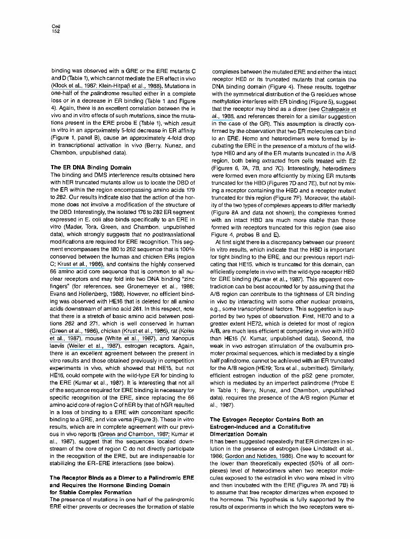
The ER DNA Binding Domain The binding and DMS interference results obtained here with hER truncated mutants allow us to locate the DBD of the ER within the region encompassing amino acids 179 to 282. Our results indicate also that the action of the hor- mone does not involve a modification of the structure of the DBD. Interestingly, the isolated 176 to 282 ER segment expressed in E. coli also binds specifically to an ERE in vitro (Mader, Tora, Green, and Chambon, unpublished data), which strongly suggests that no posttranslational modifications are required for ERE recognition. This seg- ment encompasses the 180 to 262 sequence that is 100% conserved between the human and chicken ERs (region C; Krust et al., 1986), and contains the highly conserved 66 amino acid core sequence that is common to all nu- clear receptors and may fold into two DNA binding “zinc fingers” (for references, see Gronemeyer et al., 1988; Evans and Hollenberg, 1988). However, no efficient bind- ing was observed with HE16 that is deleted for all amino acids downstream of amino acid 261. In this respect, note that there is a stretch of basic amino acid between posi- tions 262 and 271, which is well conserved in human (Green et al., 1986) chicken (Krust et al., 1986) rat (Koike et al., 1987) mouse (White et al., 1987), and Xenopus laevis (Weiler et al., 1987), estrogen receptors. Again, there is an excellent agreement between the present in vitro results and those obtained previously in competition experiments in vivo, which showed that HE15, but not HE16, could compete with the wild-type ER for binding to the ERE (Kumar et al., 1987). It is interesting that not all of the sequence required for ERE binding is necessary for specific recognition of the ERE, since replacing the 66 amino acid core of region C of hER by that of hGR resulted in a loss of binding to a ERE with concomitant specific binding to a GRE, and vice versa (Figure 3). These in vitro results, which are in complete agreement with our previ- ous in vivo reports (Green and Chambon, 1987; Kumar et al., 1987) suggest that the sequences located down- stream of the core of region C do not directly participate in the recognition of the ERE, but are indispensable for stabilizing the ER-ERE interactions (see below).
The Receptor Binds as a Dimer to a Palindromic ERE and Requires the Hormone Binding Domain for Stable Complex Formation The presence of mutations in one half of the palindromic ERE either prevents or decreases the formation of stable
complexes between the mutated ERE and either the intact receptor HE0 or its truncated mutants that contain the DNA binding domain (Figure 4). These results, together with the symmetrical distribution of the G residues whose methylation interferes with ER binding (Figure 5), suggest that the receptor may bind as a dimer (see Chalepakis et al., 1988, and references therein for a similar suggestion in the case of the GR). This assumption is directly con- firmed by the observation that two ER molecules can bind to an ERE. Homo and heterodimers were formed by in- cubating the ERE in the presence of a mixture of the wild- type HE0 and any of the ER mutants truncated in the A/B region, both being extracted from cells treated with E2 (Figures 6, 7A, 7B, and 7C). Interestingly, heterodimers were formed even more efficiently by mixing ER mutants truncated for the HBD (Figures 7D and 7E), but not by mix- ing a receptor containing the HBD and a receptor mutant truncated for this region (Figure 7F). Moreover, the stabil- ity of the two types of complexes appears to differ markedly (Figure 8A and data not shown); the complexes formed with an intact HBD are much more stable than those formed with receptors truncated for this region (see aiso Figure 4, probes B and E).
At first sight there is a discrepancy between our present in vitro results, which indicate that the HBD is important for tight binding to the ERE, and our previous report indi- cating that HE15, which is truncated for this domain, can efficiently complete in vivo with the wild-type receptor HE0 for ERE binding (Kumar et al., 1987). This apparent con- tradiction can be best accounted for by assuming that the A/B region can contribute to the tightness of ER binding in vivo by interacting with some other nuclear proteins, e.g., some transcriptional factors. This suggestion is sup- ported by two types of observation. First, HE70 and to a greater extent HE72, which is deleted for most of region A/B, are much less efficient at competing in vivo with HE0 than HE15 (V. Kumar, unpublished data). Second, the weak in vivo estrogen stimulation of the ovalbumin pro- moter proximal sequences, which is mediated by a single half palindrome, cannot be achieved with an ER truncated for the A/B region (HE19; Tora et al., submitted). Similarly, efficient estrogen induction of the pS2 gene promoter, which is mediated by an imperfect palindrome (Probe E in Table 1; Berry, Nunez, and Chambon, unpublished data), requires the presence of the A/B region (Kumar et al., 1987).
The Estrogen Receptor Contains Both an Estrogen-Induced and a Constitutive Dimerization Domain It has been suggested repeatedly that ER dimerizes in so- lution in the presence of estrogen (see Lindstedt et al., 1986; Gordon and Notides, 1986). One way to account for the lower than theoretically expected (50% of all com- plexes) level of heterodimers when two receptor mole- cules exposed to the estradiol in vivo were mixed in vitro and then incubated with the ERE (Figures 7A and 7B) is to assume that free receptor dimerizes when exposed to the hormone. This hypothesis is fully supported by the results of experiments in which the two receptors were ei-

Binding of ER to Its Responsive Element 153
ther cosynthesized in vivo in the presence of E2 (Figure 7C) or synthesized separately in vivo, but in the absence of E2 (Figure 7H). It is particularly striking that in the latter case, mixing the two receptors before addition of E2 resulted in the formation of a predominant heterodimer, whereas mixing them after addition of E2 yielded very lit- tle of such a complex. Thus, there is little doubt that the formation of “relatively stable” ER dimers is strongly fa- vored by E2 binding and, therefore, that the HBD contains an efficient estrogen-inducible dimerization domain.
The results obtained by mixing receptor molecules trun- cated for the HBD under various conditions (Figures 7D, 7E, and 8C) indicate that there is no such strong dimeriza- tion domain within or in the vicinity of the DBD. Never- theless, these truncated receptors formed heterodimers and bind very poorly to half palindromic motifs (see Figure 4). Thus dimers might also be formed in solution in the ab- sence of the HBD, albeit their stability must be too low to be detected with the present assay. Since these dimer- DNA complexes were formed irrespective of the presence of the A/B region, there must then be a weak constitutive dimerization domain within the minimal region that is re- quired for DNA binding and encompasses the highly con- served region C (see above).
Implications for Receptor Function In binding as a dimer to its palindromic responsive ele- ment, the ER conforms with the paradigm of prokaryotic regulatory DNA binding proteins (for reviews, see Takeda et al., 1983; Pabo and Sauer, 1984; Ptashne, 1986). The analogy is particularly striking with the lambda repressor, as it has been demonstrated that its N-terminal domain, which binds to DNA as a dimer, contains a weak dimeriza- tion domain that is not sufficient for formation of stable dimers in solution. However, the C-terminal domain con- tains a stronger dimerization domain, which allows the repressor to form stable dimers in solution, which in turn increases the binding of repressor to operator DNA (Pabo et al., 1979; for reviews, see Pabo and Sauer, 1984; Lewis et al., 1985). Our results strongly suggest that the DBD of the ER (most likely the conserved amino acid sequence from positions 180 to 271) binds similarly as a dimer to the palindromic ERE, making the same ERE contacts as in- tact receptor, although it binds less tightly. When bound to the ligand, the HBD allows the receptor to form stable dimers, which results in a tighter binding of the receptor to the ERE. The fact that the ER (and most likely, by anal- ogy, other members of the nuclear receptor superfamily) binds as a dimer to a palindromic ERE explains readily how this enhancer factor can bidirectionally activate initia- tion of transcription. Note that a variety of structures ap- pear to allow a protein to bind as a dimer to a palindromic sequence, since the DNA binding domain of the nuclear receptor superfamily does not contain a “prokaryotic” helix-turn-helix motif, but rather DNA binding “zinc fingers” (Miller et al., 1985; Berg, 1986; Evans and Hollen- berg, 1988; Green et al., 1988), whereas the yeast GCN4 frans-activator protein does not contain either of these DNA binding protein motifs (Hope and Struhl, 1987).
The presence of the hormone is required for tight nu-
clear binding of steroid hormone receptors in vivo and for binding of intact receptors to their responsive element in crude extracts in vitro, whereas receptors truncated for their HBD can bind to their RE in vivo anld in vitro. These observations have previously suggested that the DBD may be masked by the HBD in the absencls of hormone (for references, see Green and Chambon, 1986; Grone- meyer et al., 1988; Godowski et al., 1987; Hollenberg et al., 1987; Webster et al., 1988b; Pratt et al., 1988). That recep- tors are found associated with the 90 kd haat shock pro- tein (hsp90) in a large 8s complex in cells extracts in the absence of hormone has further suggestecl that this pro- tein may be bound to the HBD, thus masking the DBD (for references, see Joab et al., 1984; Groyer et al., 1987; Pratt et al., 1988; Denis et al., 1988). Our present results sug- gest alternative explanations to account for the hormone requirement in receptor binding to DNA. First, the finding of hsp90 bound to the receptor may be an in vitro artifact, and the hormone may just be required to induce a confor- mational modification of the HBD necessary to “induce” its dimerization domain, and thus tight binding to the DNA. However, since no binding of the intact receptor is ob- served in the absence of hormone, whereas a receptor truncated for the HBD can bind, one would have to further postulate that the presence of the unoccupied HBD destabilizes the dimerization mediated by the DBD (see above). Such a possibility would also provide an explana- tion to the otherwise puzzling observations that no inter- nal deletion within the HBD of the ER results in constitu- tive binding to the ERE either in vitro (the present study) or in vivo (Kumar et al., 1987). Alternatively, the hsp90 pro- tein may actually be bound in vivo to the dimerization do- main of the HBD in the absence of hormoiie, and the ef- fect of the hormone would be to induce a conformational change, which would in turn result in hsp90 release and exposure of the dimerization domain. Neither of these two possibilities involves masking of the DBD. We have re- cently reported that ER produced in yeast also requires estrogen (or antiestrogen) to bind efficiently to an ERE in vitro (Metzger et al., 1988). The use of yeast genetics should be of great help in solving the question of whether the dimerization region present in the HBD is “preformed” and masked (possibly by the yeast hsp9O; see Lindquist, 1986), or rather “induced” by the hormone.
We have previously shown that estrogen is required for inducing the transcriptional activation function present in the HBD (Webster et al., 1988b). We clemonstrate here that the hormone is also required for dimerization of the receptor, which is in turn crucial for tight binding of the receptor to the ERE. Whether a third function can be ascribed to the hormone, as is the case for the glucocorti- coid receptor (Picard and Yamamoto, 1987), i.e., induction of a nuclear localization signal located in the HBD, re- mains to be investigated.
The finding that efficient binding of the ER (and presum- ably of the other members of the nuclear receptor super- family) to its RE requires the estrogen-induced formation of a dimer has broad biological implications. It has been previously reported that the binding of estradiol Ito recep- tor dimers is a cooperative event (Gordon and Notides,

Cell 154
1986). Thus the actual level of “active” receptor dimers in a given cell may be very sensitive to small variations of both receptor and estrogen concentrations. Since, in addition, not all responsive elements of target genes are perfect palindromes (Berry et al., unpublished data), and therefore may bind receptor dimers with various affinities (see Fig- ures 1 and 4), it is easy to envisage how variations in the concentrations of circulating estrogen and of intracellular receptor, as well as the use of different EREs, may allow the organism to turn on or off the promoters of various tar- get genes indifferent cells at different times during devel- opment and adult life.
Experimental Procedures
Recombinants HE0 (Green et al., 1988) HE2 to HE5 and HE9 to HE14 (Kumar et al., 1986) HE27 (Green and Chambon, 1987) HE15 HE16, HE18 to HE20, HE38, HGI, HG3, ER-GRCASl, and GR-ERCASI (Kumar et al., 1987) have been described. The new N-terminal hER deletion mutants, HE49, HE50, HE68, and HE69 were constructed by cleaving at the unique Narl, FnuDII, Smal and Bgll sites of the cDNA open reading frame, respectively(see Figure 2 for position of the deletions) and ligat- ing oligonucleotides containing an in-frame Kozak’s 5’ consensus leader sequence (5”CCACC&G-3’; Kozak, 1986). Similarly, HE70 was constructed from HE15, and HE72 from HE39 (Kumar et al., 1987) by using the unique restriction sites Smal and Bgll, respectively, and ligating the Kozak’s 5’ leader sequence. In all the above cases, hER cDNA was cloned at the EcoRl site of the eukaryotic expression vector pKCR2 (Breathnach and Harris, 1983).
DNA Transfection and Cell Extract Preparation HeLa and Cos-1 cells at 30%-50% confluence in 90 mm culture plates were transiently transfected with 10 ug of plasmid DNA of receptor ex- pression vectors by using the calcium phosphate coprecipitation tech- nique (Banerji et al., 1981). The cells were grown for 48 hr in the ab- sence of hormone (Kumar et al., 1986) and unless otherwise specified in the figure legends, were incubated for 1 hr prior to harvesting with either 10-s M estradiol (for ER and ER-GRCASI) or low7 M dex- amethasome (for GR and GR-ER.CASl). Cells were washed with chilled phosphate-buffered saline (PBS), collected and homogenized (all glass homogenizer) in the binding buffer (buffer A) (20 mM Tris-HCI [pH 7.51, 2 mM DTT and 20% [v/v] glycerol) containing either 0.4 M KCI (for ERE binders) or 0.6 M KCI (for GRE binders; 0.1 ml per plate). After centrifugation at 105,000 x g for 1 hr at 2°C the whole cell extract supernatant (WCE) was collected and protein concentra- tion was determined by Bradford assay. Alternatively, WCE was also prepared by freezing the cells at -80°C suspended in KCI-supple- mented buffer A (0.1 ml per plate), thawing them over ice, and cen- trifugating at 10,000 x g for 15 min at 0°C in a Sigma centrifuge. WCE prepared by either procedure showed no apparent loss in ERE bind- ing, even after several freezings (at -8OOC) and thawings.
Low salt WCE of cells not treated with estradiol were obtained as de- scribed above, but using 50 mM KCI in buffer A.
Gel Retardation Assay WCE containing either individual receptor forms or a mixture of two of them (8-12 ug protein) were incubated with 2 ug of poly (dl,dC) (Phar- macia) at O°C for 15 min. The binding reaction, initiated by adding a [s*P]5’ end-labeled synthetic oligonucleotide probes (-5 fmol, 4 x lo4 cpm; Table 1) was incubated at 20°C for 15 min. The final concen- tration of buffer components in a 16 ul binding reaction was as follows: 10 mM Tris-HCI (pH 7.5) 1 mM DTT, 100 mM (high salt extract) or 25 mM (low salt extract) KCI and 10% (v/v) glycerol. A pre-electrophoresed (10 mA for 30 min) 4% or 5% polyacrylamide gel (acrylamide to bis- acrylamide ratio of 3O:l) containing 6.7 mM Tris-HCI (pH 7.5), 3.3 mM solium acetate, and 1 mM EDTA was used to seperate the recep- tor-DNA complexes at 30 mA with buffer recirculation (Singh et al., 1986). After suitable separation was achieved, the gel was vacuum- dried for autoradiography. For the gel retardation competition assay
displayed in Figure lC, different concentrations of unlabeled competi- tor oligonucleotide were mixed with the labeled probe (lo4 cpm) be- fore adding them to the binding reaction.
Methylation Interference Assay To identify the guanine (G) residues of the ERE that made contact with the receptor, the double-stranded probe A (Table i), 3*P-labeled at one 5’ end, was partially methylated with dimethyl sulfate (DMS) as de- scribed by Maxam and Gilbert (1980). The methylated DNA probe was incubated with wild-type and mutant receptor extracts as above except that all the components of the reaction mixture were scaled up by P-fold. After electrophoretic separation as above and brief autoradiog raphy at 4°C the receptor-bound and free oligonucleotide probes were cut from the gel and recovered by electroelution. After cleavage with piperidine (Maxam and Gilbert, 1980), approximately equal amounts of radioactivity for each sample were electrophoresed on a 15% acryl- amide-8.3 M urea gel and autoradiographed.
UV Cross-Linking and Western Blot Analysis WCE of HeLa cells transfected with HE0 or HE19 (as indicated in the legend to Figure 6) were incubated in a 40 ul final volume with 4 ug of poly (dl.dC) and 20 fmol of 5’ end-labeled or unlabeled bromode- oxyuridine (BrdU-substituted ERE probe; see legend to Figure 6) as described for gel retardation assays. The samples were transferred into separate wells of a microtiter plate (96-well), which was placed in- verted with an ice cover on a Vilber Lourmat fluorescent table and ir- radiated (maximum emission wavelength, 312 nm; maximum intensity, 8000 uW/cms) for 1 hr (Chodosh et al., 1986). SDS (sodium dodecyl sulfate) was added to the samples (0.1% final), and electrophoresis was performed in a discontinuous SDS-IO% polyacrylamide gel. After soaking the gel in a 5 mM Tris-39 mM glycine mix for 15 min, proteins were transferred onto a nitrocellulose filter (0.4 nm pore size) using the above mix and Hoefer electroblotter operated at 0.3 mA for 3 hr at 4OC. The nitrocellulose membrane was blocked with fat-free milk proteins in PBS for 30 min and incubated sequentially with anti-ER monoclonal antibodies H222 and D75 (Greene et al., 1984) and “sl-protein A (Amersham Corp.), as described by Metzger et al. (1988) and visual- ized by autoradiography. The protein markers on the SDS gel were rap- idly visualized by copper staining (Lee et al., 1987).
Acknowledgments
We are grateful to S Green for the kind gifts of HEO, HE9, HE18, HE19, HE16, HE20, and HE27 and for his help in constructing HE3 and HE4, and to 6. L. Greene (University of Chicago) and L. S. Miller (Abbott Laboratories) for the monoclonal antibodies D75 and H222, respec- tively. We thank S. Mader, L. Tora, and M. Berry for sharing unpub- iished observations. We also thank A. Weisz, N. J. G. Webster, J. H. White, and S. Green for helpful discussions. We thank N. J. G. Webster for a critical reading of the manuscript, C. Werle, and B. Boulay for help in preparing and reproducing the figures and the secretarial staff for typing the manuscript. We are grateful to the cell culture group for tech- nical assistance and to A. Staub and F. Ruffenach for preparing the oligonucleotides. V. Kumar was supported by a fellowship from the University Louis Pasteur. This work was supported by grants from the CNRS, the INSERM (CNAMTS), the Association pour la Recherche sur le Cancer, the Fondation pour la Recherche Medicale, and the Ligue Nationale Francaise contre le Cancer.
The costs of publication of this article were defrayed in part by the payment of page charges. This article must therefore be hereby marked “advertisement” in accordance with 18 U.S.C. Section 1734 solely to indicate this fact.
Received June 21, 1988; revised August 4, 1988
References
Banerji, J., Rusconi, S., and Schaffner, W. (1981). Expression of a b-globin gene is enhanced by remote SV40 DNA sequences. Cell 27, 299-308. Berg, J. M. (1986). More metal-binding fingers. Nature 379, 264-265. Breathnach, R., and Harris, B. A. (1983). Plasmids for the cloning and

Binding of ER to Its Responsive Element 155
expression of full-length double-stranded cDNAs under control of the SV40 early or late gene promoter. Nucl. Acids Res. 71, 7119-7136.
Chalepakis, G., Arnemann, J., Slater, E., Bruller, H.-J., Gross, B., and Beato, M. (1988). Differential gene activation by glucocorticoids and progestins through the hormone regulatory element of mouse mam- mary tumor virus. Cell 53, 371-382.
Chodosh, L. A., Carthew, Ft. W., and Sharp, F! A. (1986). A single poly- peptide possesses the binding and transcription activities of the adenovirus major late transcription factor. Mol. Cell. Biol. 6,4723-4733.
Denis, M., Poellinger, L., Wikstrbm, A. C., and Gustafsson, J. A. (1988). Requirement of hormone for thermal conversion of the gluco- corticoid receptor to a DNA-binding state. Nature 333, 686-688.
Evans, R. M. (1988). The steroid and thyroid hormone receptor super- family. Science 240, 889-895.
Evans, R. M ., and Hollenberg, S. M. (1988). Zinc fingers: gilt byassoci- ation. Cell 52, l-3.
Geier, A., Beery, R., Haimsohn, M., and Lunenfeld, 6. (1987). Analysis of the 4-hydroxytamoxifen (4-OHTAM) bound nuclear estrogen recep- tor from MCF-7 cells by limited proteolysis. J. Steroid Biochem. 28, 471-478.
Godowski, P J., Rusconi, S., Miesfield, R., and Yamamoto, K. R. (1987). Glucocorticoid receptor mutants that are constitutive activators of tran- scriptional enhancement. Nature 325, 365-368.
Gordon, M. S., and Notides, A. C. (1986). Computer modeling of es- tradiol interactions with the estrogen receptor. J. Steroid Biochem. 25, 177-181.
Green, S., and Chambon, P. (1986). A superfamily of potentially onco- genie hormone receptors. Nature 324, 615-617.
Green, S., and Chambon, P (1987). Oestradiol induction of a glucocor- ticoid-responsive gene by a chimaeric receptor. Nature 325, 7.5-78.
Green, S., Walter, P., Kumar, V., Krust, A., Bornert, J. M., Argos, P, and Chambon, P (1986). Human oestrogen receptor cDNA: sequence, ex- pression and homology to verb-A. Nature 320, 134-139.
Green, S., Kumar, V., Theulaz, I., Wahli, W., and Chambon, P. (1988). The N-terminal DNA-binding “zinc-finger” of the oestrogen and gluco- corticoid receptors determines target gene specificity. EMBO J., in press.
Greene, G. L., Sobel, N. B., King, W. J., and Jensen, E. V. (1984). Im- munochemical studies of estrogen receptors. J. Steroid Biochem. 20, 51-56.
Gronemeyer, H., Green, S., Kumar, V., Jeltsch, J. M., and Chambon, P (1988). Structure and function of the oestrogen receptor and other members of the nuclear receptor family. In Steroid Receptors and Dis- ease: Cancer, Boneand Circulatory Disorders, P J. Sheridan, K. Blum, and M. C. hachtenberg, eds. (New York: Marcel Dekker), pp. 153-187.
Grayer, A., Schweizer-Groyer, G., Cadepond, F., Mariller, M., and Baulieu, E. E. (1987). Antiglucocorticosteroid effects suggest why ste- roid hormone is required for receptors to bind DNA in viva but not in vitro. Nature 328, 624-626.
Hollenberg, S. M., Giguere, V., Segui, P., and Evans, R. M. (1987). Colocalization of DNA-binding and transcriptional activation functions in the human glucorticoid receptor. Cell 49, 39-46.
Hope, I. A., and Struhl, K. (1987). GCN4, a eukaryotic transcriptional activator protein, binds as a dimer to target DNA. EMBO J. 6, 2781-2784.
Joab. I., Radanyi, C., Renoir, M., Buchou, T., Catelli, M. G., Binart, N., Mester, J., and Badlieu, E. E. (1984). Common non-hormone binding component in non-transformed chick oviduct receptor of four steroid hormones. Nature 308, 850-853.
Klein-Hitpap, L., Schorpp, M., Wagner, U., and Ryffel, G. U. (1986). An estrogen-responsive element derived from the 5’flanking region of the )benOplJS vitellogenin A2 gene functions in transfected human cells. Cell 46, 1053-1061.
Klein-Hitpap, L., Ryffel, G. U., Heitlinger, E., and Cato, A. C. 8. (1988). A 13 bp palindrome is a functional estrogen responsive element and interacts specifically with estrogen receptor. Nucl. Acids. Res. 16, 647-663.
Klock, G., Strahle, U., and Schutz, G. (1987). Oestrogen and glucocor-
ticoid responsive elements are closely related but distinct. Nature 329, 734-736.
Koike, S., Sakai, M., and Muramatsu, M. (1987). Molescular cloning and characterization of rat estrogen receptor cDNA. Nucl. Acids Res. 75, 2499-2513.
Kozak, M. (1986). Point mutations define a sequence flanking the AUG initiator codon that modulates translation by eukaryotic ribosomes. Cell 44, 283-292.
Krust, A., Green, S., Argos, P, Kumar, V., Walter, F!, Bornert, J. M., and Chambon, P. (1986). The chicken oestrogen receptor sequence: ho- mology with v-erbA and the human oestrogen and glucocorticoid receptors. EMBO J. 5, 891-897.
Kumar, V., Green, S., Staub, A., and Chambon, P” (1986). Localisation of the oestradiol-binding and putative DNA-binding domains of the hu- man oestrogen receptor. EMBO J. 5, 2231-2236.
Kumar, V., Green, S., Stack, G., Berry, M., Jin, J.-R., and Chambon, P. (1987). Functional domains of the human estrogen receptor. Cell 57, 941-951.
Lee, C., Levin, A., and Branton, D. (1987). Copper staining: a five- minute protein stain for sodium dodecyl sulfate-polyacrylamide gels. Anal. Biochem. 166, 308-312.
Lewis, M., Wang, J., and Pabo, C. (1985). Structure of the operator binding domain of lambda repressor. In Biological Macromolecules and Assemblies, Volume 2, Nucleic Acids and Interactive Proteins, F. A. Jurnak and A. MacPherson, eds. (New York: John Wiley&Sons), pp. 266-287.
Lindquist, S. (1986). The heat-shock response. Annu. Rev. Biochem. 55, 1151-1191.
Linstedt, A. D., West, N. B., and Brenner, R. M. (1986). Analysis of monomeric-dimeric states of the estrogen receptor with monoclonal antiestrophilins. J. Steroid Biochem. 24, 677-686.
Martin, P. M., Berthois, Y., and Jensen, E. V. (1988). Binding of anties- trogens exposes an occult antigenic determinant in the human estro- gen receptor. Proc. Natl. Acad. Sci. USA 85, 2533-2537.
Martinez, E., Givel, F., and Wahli, W. (1987). The estrogen-responsive element as an inducible enhancer: DNA sequence requirements and conversion to a glucocorticoid-responsive element. EMBO J. 6, 371 g-3727. Maxam, A., and Gilbert, W. (1980). Sequencing end.,labeled DNA with base-specific chemical cleavages. Meth. Ezymol. 65, 499-560.
Metzger, D., White, J. H., and Chambon, P (1988). The human oestro- gen receptor functions in yeast. Nature 334, 31-36.
Miller, J., McLachlan, A. D., and Klug, A. (1985). Repetitive zinc-binding domains in the protein transcription factor IIIA from Xenopus oocytes. EMBO J. 4, 1609-1614.
Pabo, C. O., and Sauer, R. T. (1984). Protein-DNA recognition. Annu. Rev. Biochem. 53, 293-321.
Pabo, C. O., Sauer, R. T., Sturtevant, J. M., and Ptashne, M. (1979). The h repressor contains two domains. Proc. Natl. Acad. Sci. USA 76, 1608-1612.
Picard, D., and Yamamoto, K. R. (1987). Two signals rnediate hormone- dependent nuclear localization of the glucocorticoid receptor, EMBO J. 6, 3333-3340.
Pratt, W. B., Jolly, D. J., Pratt, D. V., Hollenberg, S. M., Giguere, V., Cadepond, F. M., Schweizer-Grayer. G., Catelli, M.-G., Evans, R. M., and Baulieu, E. E. (1988). A region in the steroid binding domain deter- mines formation of the non-DNA-binding, 9s glucocorticoid receptor complex. J. Biol. Chem. 263, 267-273.
Ptashne, M. (1986). A Genetic Switch: Gene Control and Phage h (San Francisco and Cambridge, MA: Blackwell Scientific Publications and Cell Press).
Singh, H., Sen, R., Baltimore, D., and Sharp, P. A. (1986). A nuclear factor that binds to a conserved sequence motif in transcriptional con- trol elements of immunoglobulin genes. Nature 319, 154-158.
Takeda, Y., Ohlendorf, D. H., Anderson, W. F., and Matthews, B. W. (1983). DNA-binding proteins. Science 221, 1020-1026.
Webster, N. J. G., Jin, J. R., Green, S., Hollis, M., and Chambon, P (1988a). The yeast UASo is a transcriptional enhancer in human HeLa
Cdl 156
cells in the presence of the GAL4 frans-activator. Cell 52, 169-178.
Webster, N. J. G., Green, S., Jin, J. R., and Chambon, P (1988b). The hormone-binding domains of the estrogen and glucocorticoid recep- tors contain an inducible transcription activation function. Cell 54, 199-207.
Weiler, I. J., Lew, D., and Shapiro, D. J. (1987). TheXenopus laevis estro- gen receptor: sequence homology with human and avian receptors and identification of multiple estrogen receptor messenger ribonucleic acids. Mol. Endo. 7, 355-362.
White, Ft., Less, J. A., Needham, M., Ham, J., and Parker, M. (1987). Structural organization and expression of the mouse estrogen recep- tor. Mol. Endo. 7, 735-744.
Wil lmann, T., and Beato, M. (1986). Steroid-free glucocorticoid recep- tor binds specifically to mouse mammary tumour virus DNA. Nature 324, 688-691. Yamamoto, K. Pt. (1985). Steroid receptor regulated transcription of specific genes and gene networks. Annu. Rev. Genet. 19, 209-252.
Yamamoto, K. R., and Alberts, B. (1972). In vitro conversion of estradiol- receptor protein to its nuclear form: dependence on hormone and DNA. Proc. Natl. Acad. Sci. USA 69, 2105-2109.
Note Added in Proof
Material cited as Tora et al., submitted, is now in press: Tora, L., Gaub, M. P, Mader, S., Dierich, A., Bellard, M., and Chambon, P (1988). EMBC J.