
An avian influenza H5N1 virus that binds to a human-type receptor
24
1 An avian influenza H5N1 virus that binds to human-type receptor Prasert Auewarakul*, Ornpreya Suptawiwat, Alita Kongchanagul, Chak Sangma 5 , Yasuo Suzuki 6 , Kumnuan Ungchusak 3 , Suda Louisirirotchanakul, Hatairat Lerdsamran, Phisanu Pooruk, Arunee Thitithanyanont 1 , Chakrarat Pittayawonganon 3 , Chao-Tan Guo 6 , Hiroaki Hiramatsu 6 , Wipawee Jampangern 2 , Supamit Chunsutthiwat 4 , Pilaipan Puthavathana Departments of Microbiology, Faculty of Medicine Siriraj Hospital, 1 Faculty of Science, and 2 Faculty of Tropical Medicine, Mahidol University; 3 Bureau of Epidemiology and 4 Department of Disease Control; 5 Faculty of Science, Kasetsart University; Thailand; and 6 College of Life and Health Sciences, Chubu University, Japan Running title: H5N1 virus that binds human-type receptor *Corresponding author: Prasert Auewarakul, MD, Dr med mailing address: Department of Microbiology, Faculty of Medicine Siriraj Hospital, Mahidol University, Bangkok 10700, Thailand Fax: +662 4184148 E-mail: [email protected] Word count of abstract: 177 Word count in the manuscript: ACCEPTED Copyright © 2007, American Society for Microbiology and/or the Listed Authors/Institutions. All Rights Reserved. J. Virol. doi:10.1128/JVI.00468-07 JVI Accepts, published online ahead of print on 11 July 2007
-
Upload
independent -
Category
Documents
-
view
0 -
download
0
Transcript of An avian influenza H5N1 virus that binds to a human-type receptor

1
An avian influenza H5N1 virus that binds to human-type receptor
Prasert Auewarakul*, Ornpreya Suptawiwat, Alita Kongchanagul,
Chak Sangma5, Yasuo Suzuki6, Kumnuan Ungchusak3, Suda Louisirirotchanakul,
Hatairat Lerdsamran, Phisanu Pooruk, Arunee Thitithanyanont1, Chakrarat
Pittayawonganon3, Chao-Tan Guo6, Hiroaki Hiramatsu6, Wipawee Jampangern2,
Supamit Chunsutthiwat4, Pilaipan Puthavathana
Departments of Microbiology, Faculty of Medicine Siriraj Hospital, 1Faculty of
Science, and 2Faculty of Tropical Medicine, Mahidol University; 3Bureau of
Epidemiology and 4Department of Disease Control; 5Faculty of Science, Kasetsart
University; Thailand; and 6College of Life and Health Sciences, Chubu University,
Japan
Running title: H5N1 virus that binds human-type receptor
*Corresponding author: Prasert Auewarakul, MD, Dr med
mailing address: Department of Microbiology, Faculty of Medicine Siriraj Hospital,
Mahidol University, Bangkok 10700, Thailand
Fax: +662 4184148
E-mail: [email protected]
Word count of abstract: 177
Word count in the manuscript:
ACCEPTED
Copyright © 2007, American Society for Microbiology and/or the Listed Authors/Institutions. All Rights Reserved.J. Virol. doi:10.1128/JVI.00468-07 JVI Accepts, published online ahead of print on 11 July 2007
2
Abstract
Avian influenza viruses preferentially recognize sialosugar chains terminating
in sialic acid-α2,3-galactose (SAα2,3Gal), whereas human influenza viruses
preferentially recognize SAα2,6Gal. A conversion to SAα2,6Gal specificity is
believed to be one of the changes required for the introduction of new hemagglutinin
(HA) subtypes to human population, which can lead to pandemic. H5N1 avian
influenza virus is a major threat for emergence of a pandemic virus. As of June 12th,
2007, the virus has been reported in 45 countries, and 312 human cases with 190
deaths have been confirmed. We describe here substitutions at position 129 and 134
identified in a virus isolated from a fatal human case that could change the receptor-
binding preference of HA of H5N1 virus from SAα2,3Gal to both SAα2,3Gal and
SAα2,6Gal. Molecular modeling demonstrated that the mutation may stabilize
SAα2,6Gal in its optimal cis conformation in the binding pocket. The mutation was
found in approximately half of the viral sequences directly amplified from a
respiratory specimen of the patient. Our data confirm the presence of H5N1 virus with
the ability to bind to human-type receptor in this patient and suggest the selection and
expansion of the mutant with human-type receptor specificity in human host
environment.
ACCEPTED

3
Introduction
In contrast to most avian influenza viruses, which do not readily infect human,
highly pathogenic H5N1 avian influenza strains can transmit directly from avian
species to human and cause severe diseases. Despite the ability to infect and cause
severe disease in human, most H5N1 viruses do not bind SAα2,6Gal receptor with
high affinity (17). It is believed that this receptor binding property is the major factor
preventing the H5N1 virus from efficiently transmitting from person to person and
causing a pandemic (17). Receptor binding preference of H5N1 viruses can be altered
by only a few amino acid substitutions in the HA protein. Mutations that change the
receptor binding preference from avian- to human-type could potentially enable the
virus to transmit efficiently in human population and cause a catastrophic pandemic.
Monitoring of the viral changes is therefore extremely important in the current
situation that H5N1 viruses are spreading progressively.
A previous study showed that mutations at positions 226 and 228 (H3
numbering) (Q226L, G228S), which are the adaptive mutations for H2 and H3 (5,
19), could reduce the binding affinity to SAα2,3Gal of a 1997 H5N1 (5, 19). Human
2003 H5N1 isolates from Hong Kong, which contain a mutation at the position 227
(S227N) (H3 numbering), were shown to have a reduced binding affinity to
SAα2,3Gal and an increased affinity toward SAα2,6Gal (1). Another report
demonstrated that neither these mutations nor the mutations that could adapt H1
viruses to the human receptor (E190D and G225D) (H3 numbering) could completely
convert a Vietnam 2004 H5N1 virus to the 2,6-type receptor specificity (16). Except
for the S227N, these mutations have not been found in H5N1 viruses isolated from
human or animals. Recently N182K and Q192R mutations were shown to enhance
binding of a Vietnam 2004 H5N1 HA to SAα2,6Gal receptor (20). Although, it was
ACCEPTED
4
described that the N182K mutation was found in Kan-1 HA (20), the original Kan-1
virus as well as the HA sequence of Kan-1 in the GenBank Database does not contain
this mutation. It is not clear where this mutation was derived from. The Q192R
mutation was found in a clone that was present as a minor population in a viral isolate
and was identified after plaque-purification. In contrast, here we show a naturally
occurring mutant with an ability to bind SAα2,6Gal that was directly identified in a
human nasopharyngeal specimen.
ACCEPTED
5
Materials and methods
Cloning and generation of mutants
A fragment of HA gene covering receptor binding site (nucleotide 413 - 905)
was amplified from RNA extracted from the nasopharyngeal specimen using the high
fidelity enzyme Pfu and the primers HHAf2 (GGTCCAGTCATGAAGCCTCA) and
HA-H5r12 (TTTATCGCCCCCATTGGAGT). The PCR product was cloned into
pGEM T-Easy. One hundred clones were picked up and sequenced. Two clones were
pooled for each sequencing reaction. A clone with L129V and A134V mutations were
used in a spliced overlapping extension reaction, which joined 2 PCR fragments with
overlapping sequences. The reaction swapped the sequence with the mutations into
HA gene of Kan-1 virus in the reverse genetic plasmid pHw2000, in which the
cleavage site had been modified to a low pathogenic sequence. Wild type or the
mutant pHw2000 HA were transfected into Vero cells together with the other 7
genomic segments of A/PR/8/34 to generate the viruses.
Hemagglutination assay
10% suspension of goose (Anser cygnoides) red blood cell (GRBC) was
prepared in phosphate-buffered saline (PBS) and a 50-µl aliquot was treated with 1.25
U of α2,3-sialidase cloned from S. thyphimurium LT2 (7) (Takara, Japan) for 1 hour
at 37ºC. Untreated and treated red blood cells were used in hemagglutination assay.
HA-Receptor binding assay
The receptor binding preference was analyzed by a solid-phase direct binding
assay as previously described (20) using the sialylglycopolymer which contains N-
acetylneuraminic acid linked to galactose through either an α-2,3 or α-2,6 bond
(Neu5Acα2,3LacNAcb-pAP and Neu5Acα2,6LacNAcb-pAP) (18). Serial dilutions
of each sialylglycopolymer were prepared in PBS and 100 µl was added to the wells
ACCEPTED

6
of 96-well microtiter plates (Polystyrene Universal-Bind Microplate, Corning, USA)
and allowed to attach overnight at 4oC. The plate containing sialylglycopolymer was
irradiated under UV light at 254 nm for 10 minutes then washed 5 times with 200 µl
PBS. The plate was blocked for 8 hours at 4oC with PBS containing 2% skim milk
powder. After washing 5 times with 200 µl PBS containing 0.1% Tween 20, virus
culture supernatant containing 128 HA unit was allowed to attach onto the plate on ice
overnight. After the incubation, the plate was washed 5 times with ice-cold PBS-0.1%
Tween 20 and 50 µl of anti-HA goat hyper-immune serum at dilution of 1:2000 was
added to each well and was allowed to incubate on ice for 2 hours. The plate was then
washed again with ice-cold PBS-0.1% Tween 20 and 50 µl of the polyclonal rabbit
anti-goat immunoglobulins/HRP conjugate (DakoCytomation) at dilution of 1:2000
was added to each well. After incubation on ice for 2 h, the wells were extensively
washed with PBS-0.1% Tween 20, and 100 µl per well of premixed
tetramethylbenzidine/H2O2 substrate was added. After incubation at room temperature
for 15 min, the reaction was stopped with 50 µl of 1M H2SO4 and the absorbance at
450/630 nm was read.
Molecular dynamic simulation
The crystal structures of HA from A/Duck/Singapore/3/97 (H5N1) (4) were
used as templates for the simulations of HA binding to SAα2,3Gal (PDB entry 1JSN)
and SAα2,6Gal (PDB entry 1JSO). In the 1JSO structural template, the sialic residue
has no galactose unit connected therefore we added it with the torsion angle of 55˚.
Both glycosides in the two structures were terminated with methoxy group and used
as the input for molecular dynamics simulations. In homology modeling, the wild type
and mutant HA (L129V/A134V) were three-dimensional aligned via the SWISS-
MODEL server (12) on 1JSN and 1JSO and used as the initial input structures for
ACCEPTED
7
molecular dynamics simulations. In order to provide a control of classical SAα2,6Gal-
tropic HA, we ran a human influenza virus H1 HA structure (PDB entry 1RVZ) (2) in
a similar simulation.
All structures were solvated using TIP5P water model (10) and performed
energy minimization to relieve bad contacts caused by unreasonable distances in the
initial structures then equilibrated for 100 ps before 3 ns productive run at 300K using
SANDER module in AMBER9 program (University of California, San Francisco)
with Glycam04 parameter (http://glycam.ccrc.uga.edu). Xmgrace (http://plasma-
gate.weizmann.ac.il/Grace/), VMD (8) and AMBER tools running on UNIX were
exploited to visualize and manipulate all figures.
ACCEPTED
8
Results
We screened for H5N1 isolates with altered receptor-binding preference by a
hemagglutination assay using goose red blood cells that were treated with a
SAα2,3Gal-specific sialidase (7). Theoretically, the sialidase digestion should abolish
hemagglutination by SAα2,3Gal-specific viruses, whereas viruses that can bind to
SAα2,6Gal should maintain hemagglutination activity with the treated red blood
cells. The sialidase treatment did not affect hemagglutination titer of human influenza
viruses. An example of H1N1 virus (A/Thailand/Siriraj-12/06), of which the
hemagglutination titer was not affected by the treatment, is shown in Fig.1a. We have
tested 4 human H5N1 isolates (A/Thailand/1(KAN-1)/2004, A/Thailand/3(SP-
83)/2004, A/Thailand/5(KK-494)/2004, A/Thailand/676/2005). The sialidase
treatment completely abolished hemagglutination activity of all the viruses with more
than 256 folds reduction in hemagglutination titer except for A/Thailand/676/2005
(Th676), which partially maintained its hemagglutination activity in sialidase-treated
red blood cells with some reduction in hemagglutination titer (Fig. 1a). This suggested
an enhanced binding to SAα2,6Gal of this virus. The virus was isolated from a 5 year-
old boy in Thailand. The patient had a progressive viral pneumonia that led to
respiratory failure and death by 12 days after onset of illness.
Sequence of the HA gene of Th676 (accession number: DQ360835) revealed
two substitutions at the position 129 (leucine to valine, L129V) and position 134
(alanine to valine, A134V). These two substitutions were particularly interesting
because they are located close to the 130 loop of the receptor binding domain (14).
Direct sequencing of viral RNA from culture showed double peaks at both positions
indicating mixture of wild type and mutant viruses. The mutant peaks at both
positions were 2-3 folds larger than the wild type peaks (Fig. 1b). We have also
ACCEPTED
9
amplified the viral sequence directly from nasopharyngeal specimen. Cloning and
sequencing of the amplification products showed the substitution L129V in 54 out of
92 clones, and the substitution A134V in 52 out of 92 clones. Among these mutant
clones, 42 had both substitutions (45.6% of total clones). In order to test whether
these two substitutions were responsible for the alteration of receptor binding
preference, we generated reverse genetic viruses with wild type HA or mutated HA
carrying these two substitutions individually or simultaneously by the reverse genetics
method (6). However, the mutated HA carrying A134V alone did not yield viable
virus probably because the mutation was not compatible with the genetic background
of the reverse genetic virus. The wild type HA clone was derived from A/Thailand/1
(KAN-1)/2004 (H5N1) (Kan-1). Other genomic segments of the reverse genetic
viruses were from A/PR/8/34 (H1N1). In contrast to the Kan-1 HA described recently
to bind SAα2,6Gal by Yamada et al (20), our Kan-1 HA clone (accession number:
EF107522) as well as the Kan-1 viral isolate (accession number: AY555150) does not
contain the N182K mutation, which was described to enhance binding to SAα2,6Gal
(20). Hemagglutination pattern of the L129V/A134V reverse genetic virus indicated
an enhanced SAα2,6Gal binding, whereas that of the L129V virus did not (Fig. 1a).
The lesser degree of reduction of hemagglutination titer of the L129V/A134V reverse
genetic virus (4-fold) as compared to that of the Th676 (8-fold) might reflect the fact
that the Th676 was a mixture of wild type and mutant viruses. We also used a recently
described direct binding assay using sialylglycopolymers (3) and confirmed the effect
of the L129V/A134V mutation on the receptor binding preference. The reverse
genetic virus carrying wild type Kan-1 HA showed SAα2,3Gal-specificity and did not
bind significantly to SAα2,6Gal, whereas a human H1N1 virus, A/Thailand/Siriraj-
12/06, bound only to SAα2,6Gal (Fig. 2a and b). And while the L129V alone did not
ACCEPTED

10
change the receptor binding preference, the HA containing both L129V and A134V
mutations bound equally well to both SAα2,3Gal and SAα2,6Gal (Fig. 2c and d)
In HA binding pocket, the SAα2,3Gal and SAα2,6Gal receptors were shown
to have specific conformation, either cis or trans (4). Torsion angle (Φ) is the angle
between two planes containing O6, C1 of the SA unit and O3 (or O6), C3 of Gal unit
(Fig. 3a). The angle indicates whether the glycoside is in cis (Φ = 56˚) or trans
conformation (Φ = -55˚) (Fig. 3a). Within its bound state to H5 in an x-ray co-crystal
structure (4), SAα2,3Gal was found in trans conformation. The trans conformation of
SAα2,3Gal allows 4-OH (O4) and the glycosidic oxygen (O3) of the Gal unit to
interact optimally to Gln222 of H5 (4). In contrast, if bound in trans conformation,
the 4-OH in SAα2,6Gal would be too far away from the Gln222. And if bound in cis
conformation, the glycosidic oxygen (O6) in SAα2,6Gal would be too far away from
the Gln222. The lack of interactions between 4-OH and Gln222 in trans conformation
and between O6 and Gln222 in cis conformation makes the binding of H5 to
SAα2,6Gal unstable in both conformations. Conformation of SAα2,6Gal in solution
was shown to have cis-to-trans ratio of 9:1 (11), which suggests that the optimal
binding conformation for SAα2,6Gal is cis because no additional energy would be
required for the conformational change. Furthermore, a recent molecular modeling
has predicted SAα2,6Gal in H5 binding to be in cis conformation (9). In our
simulation, a human H1 (PDB entry 1RVZ) showed an average SAα2,6Gal Φ angle
of 63°, which indicated a cis conformation (Fig. 3b). This provides a positive control
for SAα2,6Gal binding in our simulation and indicates that the Φ angle can be used as
indicator for the receptor preference. In order to have a structural insight in the altered
receptor specificity of the mutant HA, we performed molecular dynamics simulations
using two sialic acid-H5 co-crystals as reference structures for the two types of
ACCEPTED
11
glycosidic linkage, SAα2,3Gal (PDB entry 1JSN) and SAα2,6Gal (PDB entry 1JSO)
(4). In our modeling, both the wild type and the mutant L129V/A134V HAs shared
similar binding pattern within the sialic binding pocket as previously reported for both
SAα2,3Gal and SAα2,6Gal binding (14, 19). However, it was observed that, over the
period of simulations, hydrophobicity and spatial constraint changes at residues 129
and 134 due to the different alkyl side chains, especially the A134V mutation which
happened near the glycosidic linkage, caused some changes in glycoside binding
patterns. The Φ angle indicated that SAα2,6Gal in the L129V/A134V HA spent most
of the time in the low energy cis conformation, whereas those in the wild type Kan-1
HA and the template H5 was forced to change from their cis conformation in the
initial input structures to be in trans conformation (Fig. 3c). Although SAα2,6Gal in
both the wild type Kan-1 HA and the template HA was in trans conformation at the
end of the simulation period, the SAα2,6Gal in Kan-1 HA stayed longer in cis
conformation than that in the template HA. This suggested that the wild type Kan-1
HA might have a slightly increased affinity to SAα2,6Gal as compared to the
template HA of A/Duck/Singapore/3/97. The increased stability in cis conformation
of the binding to L129V/A134V HA was due to an alternative interaction between
Gly221 and 4-OH or 3-OH (O3) of Gal caused by a displacement of Gly221 by the
A134V mutation (Fig. 3d). In the simulations of SAα2,3Gal binding to
L129V/A134V HA, the Φ angle significantly moved away from the preferred angle
(from -55˚ to -30˚) (Fig. 3c). Although the SAα2,3Gal was still in trans conformation,
the A134V mutation pushed Gln222 slightly away from the optimal binding
condition. Widening of the gap between Gln222 and the glycosidic oxygen (O3)
caused by larger hydrophobic side chain of Val134 might reduce the binding affinity
of the mutant HA to SAα2,3Gal.
ACCEPTED

12
Discussion
In human, the SAα2,6Gal receptor is expressed mainly in upper airway, while
the SAα2,3Gal receptor is expressed in alveoli and terminal bronchiole (13). A virus
with good affinity to both SAα2,3Gal and SAα2,6Gal receptors may be a very
dangerous one, which could both infect efficiently via its binding to SAα2,6Gal in the
upper airway and cause severe infection in the lung via its binding to SAα2,3Gal.
This hypothesis is supported by the fact that one of the two well-characterized HA
genes from the H1N1 1918 pandemic virus binds efficiently to both SAα2,3Gal and
SAα2,6Gal (3, 15).
Although receptor binding preference is a major factor determining host
species tropism, we do not know whether the alteration in receptor binding property is
sufficient to enable the virus to transmit from person to person efficiently.
Nevertheless, our finding indicated that an adaptation of H5N1 virus to human host by
receptor binding site modification could and did indeed happen. Since the
identification of this patient, there have been 3 confirmed human cases in the country,
but none of the viral isolates from these cases contained the L129V and A134V
substitutions. It is likely that this particular mutant has been eliminated by the
infection control measures.
Our report demonstrates that the avian influenza H5N1 virus could be
naturally adapted to the human-type receptor. We need to intensify our effort to detect
such viruses as early as possible. Our data also provide a genetic marker that can be
used to screen for an H5N1 virus with pandemic potential. H5N1 viruses may have
several ways to adapt their receptor binding property, but mutations that were found
directly in patients, such as the L129V/A134V, are more likely than those artificially
generated and tested mutations to be the mutation that could cause a pandemic.
ACCEPTED
13
Acknowledgements
This work was supported by a research grant from the National Research
Council, Ellison Foundation and the National Center for Genetic Engineering and
Biotechnology of Thailand. The activity was a part of the newly established Thailand
Avian Influenza Monitoring Network (TAIM Net). We would like to thank Dr.
Atthapon Eiamudomkan, Dr. Prasongsak Nakhornkwang, Mrs. Manee Phonpasee,
Mr. Sanya Kittisoontaropas, Mr. Vivat Pvongpasert, Mrs. Wannasatre Rattanalum,
Mrs. Chansuda Sukbumrung, and Dr. Sompong Boonsuepchat of the NakhonNayok
Provincial Public Health Office. The reverse genetic plasmids pHw2000 with
A/PR/8/34 genomic segments were kindly provided by RG Webster and E Hoffmann.
All molecular modeling was conducted on the PSIPHI computer cluster at the
National Center for Genetic Engineering and Biotecnology (BIOTEC) and financially
supported by Thailand Research Fund (TRF) and Kasetsart University Research and
Development Institute (KURDI).
References
1. Gambaryan, A., A. Tuzikov, G. Pazynina, N. Bovin, A. Balish, and A.
Klimov. 2006. Evolution of the receptor binding phenotype of influenza A
(H5) viruses. Virology 344:432-8.
2. Gamblin, S. J., L. F. Haire, R. J. Russell, D. J. Stevens, B. Xiao, Y. Ha, N.
Vasisht, D. A. Steinhauer, R. S. Daniels, A. Elliot, D. C. Wiley, and J. J.
Skehel. 2004. The structure and receptor binding properties of the 1918
influenza hemagglutinin. Science 303:1838-42.
3. Glaser, L., J. Stevens, D. Zamarin, I. A. Wilson, A. Garcia-Sastre, T. M.
Tumpey, C. F. Basler, J. K. Taubenberger, and P. Palese. 2005. A single
ACCEPTED
14
amino acid substitution in 1918 influenza virus hemagglutinin changes
receptor binding specificity. J Virol 79:11533-6.
4. Ha, Y., D. J. Stevens, J. J. Skehel, and D. C. Wiley. 2001. X-ray structures
of H5 avian and H9 swine influenza virus hemagglutinins bound to avian and
human receptor analogs. Proc Natl Acad Sci U S A 98:11181-6.
5. Harvey, R., A. C. Martin, M. Zambon, and W. S. Barclay. 2004.
Restrictions to the adaptation of influenza a virus h5 hemagglutinin to the
human host. J Virol 78:502-7.
6. Hoffmann, E., G. Neumann, Y. Kawaoka, G. Hobom, and R. G. Webster.
2000. A DNA transfection system for generation of influenza A virus from
eight plasmids. Proc Natl Acad Sci U S A 97:6108-13.
7. Hoyer, L. L., P. Roggentin, R. Schauer, and E. R. Vimr. 1991. Purification
and properties of cloned Salmonella typhimurium LT2 sialidase with virus-
typical kinetic preference for sialyl alpha 2----3 linkages. J Biochem (Tokyo)
110:462-7.
8. Humphrey, W., A. Dalke, and K. Schulten. 1996. VMD: visual molecular
dynamics. J Mol Graph 14:33-8, 27-8.
9. Li, M., and B. Wang. 2006. Computational studies of H5N1 hemagglutinin
binding with SA-alpha-2, 3-Gal and SA-alpha-2, 6-Gal. Biochem Biophys Res
Commun 347:662-8.
10. Mahoney, M. W., and W. L. Jorgensen. 2000. A five-site model for liquid
water and the reproduction of the density anomaly by rigid, nonpolarizable
potential functions. Journal of Chemical Physics 112:8910-8922.
ACCEPTED

15
11. Poppe, L., R. Stuike-Prill, B. Meyer, and H. van Halbeek. 1992. The
solution conformation of sialyl-alpha (2----6)-lactose studied by modern NMR
techniques and Monte Carlo simulations. J Biomol NMR 2:109-36.
12. Schwede, T., J. Kopp, N. Guex, and M. C. Peitsch. 2003. SWISS-MODEL:
An automated protein homology-modeling server. Nucleic Acids Res
31:3381-5.
13. Shinya, K., M. Ebina, S. Yamada, M. Ono, N. Kasai, and Y. Kawaoka.
2006. Avian flu: influenza virus receptors in the human airway. Nature
440:435-6.
14. Skehel, J. J., and D. C. Wiley. 2000. Receptor binding and membrane fusion
in virus entry: the influenza hemagglutinin. Annu Rev Biochem 69:531-69.
15. Stevens, J., O. Blixt, L. Glaser, J. K. Taubenberger, P. Palese, J. C.
Paulson, and I. A. Wilson. 2006. Glycan microarray analysis of the
hemagglutinins from modern and pandemic influenza viruses reveals different
receptor specificities. J Mol Biol 355:1143-55.
16. Stevens, J., O. Blixt, T. M. Tumpey, J. K. Taubenberger, J. C. Paulson,
and I. A. Wilson. 2006. Structure and Receptor Specificity of the
Hemagglutinin from an H5N1 Influenza Virus. Science.
17. Suzuki, Y. 2005. Sialobiology of influenza: molecular mechanism of host
range variation of influenza viruses. Biol Pharm Bull 28:399-408.
18. Totani, K., T. Kubota, T. Kuroda, T. Murata, K. I. Hidari, T. Suzuki, Y.
Suzuki, K. Kobayashi, H. Ashida, K. Yamamoto, and T. Usui. 2003.
Chemoenzymatic synthesis and application of glycopolymers containing
multivalent sialyloligosaccharides with a poly(L-glutamic acid) backbone for
inhibition of infection by influenza viruses. Glycobiology 13:315-26.
ACCEPTED
16
19. Vines, A., K. Wells, M. Matrosovich, M. R. Castrucci, T. Ito, and Y.
Kawaoka. 1998. The role of influenza A virus hemagglutinin residues 226
and 228 in receptor specificity and host range restriction. J Virol 72:7626-31.
20. Yamada, S., Y. Suzuki, T. Suzuki, M. Q. Le, C. A. Nidom, Y. Sakai-
Tagawa, Y. Muramoto, M. Ito, M. Kiso, T. Horimoto, K. Shinya, T.
Sawada, T. Usui, T. Murata, Y. Lin, A. Hay, L. F. Haire, D. J. Stevens, R.
J. Russell, S. J. Gamblin, J. J. Skehel, and Y. Kawaoka. 2006.
Haemagglutinin mutations responsible for the binding of H5N1 influenza A
viruses to human-type receptors. Nature 444:378-82.
ACCEPTED
17
Fig.1 Abolition of hemagglutination titers in SAα2,3Gal-specific sialidase-treated red
blood cells as compared to those in untreated cells indicated SAα2,3Gal mono-
specificity for H5N1 viruses except for Th676 and the L129V/A134V reverse genetic
virus (RV), of which the titers were only modestly affected suggesting an enhanced
binding to SAα2,6Gal (a). Electropherogram of sequencing reaction of the Th676
virus shows T to G mutation changing amino acid from leucine to valine at the
position 129, and C to T mutation changing the amino acid from alanine to valine at
the position 134 (arrow heads) (b).
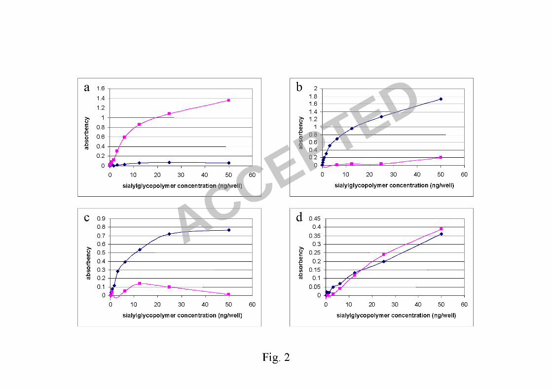
Fig.2 Direct binding assay using sialylglycopolymers: Human H1N1 virus
(A/Thailand/Siriraj-12/06) showed receptor preference for SAα2,6Gal (a), whereas
the reverse genetic virus with Kan-1 wild type HA showed receptor preference for
SAα2,3Gal (b). The reverse genetic virus carrying HA with L129V mutation showed
similar receptor preference to the wild type virus (c), whereas the virus carrying HA
with L129V/A134V mutations showed receptor preference for both SAα2,3Gal and
SAα2,6Gal (d). Diamonds represent absorbencies of SAα2,3Gal binding and squares
represent absorbencies of SAα2,6Gal binding.
Fig. 3. The two glycoside backbone conformations, SAα2,6Gal and SAα2,3Gal cut
from the x-ray co-crystal 1JSI and 1JSIN (4) with different value of torsion (Φ) angles
that were used as the cis or trans conformation characteristics (a). For a human H1
virus (1RVZ), SAα2,6Gal showed an average Φ angle of 63° indicating a cis
conformation in the molecular dynamics simulation (b). The amount of time each
receptor analog spent having particular Φ angle for SAα2,3Gal (upper panel) and
SAα2,6Gal (lower panel) in the binding sites of the template H5
ACCEPTED
18
(A/Duck/Singapore/3/97) (black), wild type Kan-1 (green), and L129V/A134V
mutant (red) as the results of molecular dynamics simulations indicated altered
binding conformation in the L129V/A134V mutant (c). The modeling showed that
SAα2,6Gal in cis conformation in the wild type Kan-1 HA pockets had long distances
between O6 and Gln222 (3.47 Å), and between 4-OH and Gly221 (7.72 Å) (upper
panel). However, in the L129V/A134V mutant, the glycoside was stabilized with
Gly221 and Gal interaction as observed by shorter distance between 4-0H and Gln221
(2.02 Å) (lower panel) (d).
ACCEPTED

ACCEPTED
ACCEPTED
ACCEPTED

ACCEPTED

ACCEPTED

ACCEPTED