Characterization of Highly Pathogenic H5N1 Avian Influenza A Viruses Isolated from South Korea
11
JOURNAL OF VIROLOGY, Mar. 2005, p. 3692–3702 Vol. 79, No. 6 0022-538X/05/$08.000 doi:10.1128/JVI.79.6.3692–3702.2005 Copyright © 2005, American Society for Microbiology. All Rights Reserved. Characterization of Highly Pathogenic H5N1 Avian Influenza A Viruses Isolated from South Korea Chang-Won Lee, 1 David L. Suarez, 1 Terrence M. Tumpey, 2 Haan-Woo Sung, 3 Yong-Kuk Kwon, 3 Youn-Jeong Lee, 3 Jun-Gu Choi, 3 Seong-Joon Joh, 3 Min-Chul Kim, 3 Eun-Kyoung Lee, 3 Jong-Myung Park, 3 Xiuhua Lu, 2 Jacqueline M. Katz, 2 Erica Spackman, 1 David E. Swayne, 1 * and Jae-Hong Kim 3 Southeast Poultry Research Laboratory, Agricultural Research Service, U.S. Department of Agriculture, Athens, 1 and Centers for Disease Control and Prevention, Atlanta, 2 Georgia, and National Veterinary Research and Quarantine Service, Anyang, Korea 3 Received 4 August 2004/Accepted 13 October 2004 An unprecedented outbreak of H5N1 highly pathogenic avian influenza (HPAI) has been reported for poultry in eight different Asian countries, including South Korea, since December 2003. A phylogenetic analysis of the eight viral genes showed that the H5N1 poultry isolates from South Korea were of avian origin and contained the hemagglutinin and neuraminidase genes of the A/goose/Guangdong/1/96 (Gs/Gd) lineage. The current H5N1 strains in Asia, including the Korean isolates, share a gene constellation similar to that of the Penfold Park, Hong Kong, isolates from late 2002 and contain some molecular markers that seem to have been fixed in the Gs/Gd lineage virus since 2001. However, despite genetic similarities among recent H5N1 isolates, the topology of the phylogenetic tree clearly differentiates the Korean isolates from the Vietnamese and Thai isolates which have been reported to infect humans. A representative Korean isolate was inoculated into mice, with no mortality and no virus being isolated from the brain, although high titers of virus were observed in the lungs. The same isolate, however, caused systemic infections in chickens and quail and killed all of the birds within 2 and 4 days of intranasal inoculation, respectively. This isolate also replicated in multiple organs and tissues of ducks and caused some mortality. However, lower virus titers were observed in all corresponding tissues of ducks than in chicken and quail tissues, and the histological lesions were restricted to the respiratory tract. This study characterizes the molecular and biological properties of the H5N1 HPAI viruses from South Korea and emphasizes the need for comparative analyses of the H5N1 isolates from different countries to help elucidate the risk of a human pandemic from the strains of H5N1 HPAI currently circulating in Asia. Since mid-December 2003, nine Asian countries have con- firmed outbreaks of highly pathogenic avian influenza (HPAI) caused by the H5N1 strain of influenza virus (World Organi- zation for Animal Health, or OIE [http://www.oie.int]). The outbreak has also resulted in at least 29 fatal cases out of 40 confirmed human infections in Vietnam and Thailand as of 9 September 2004, according to the World Health Organization (http://www.who.int/en/). South Korea reported an outbreak of the disease among chickens and ducks on 10 December 2003, which was the first official report of disease for this outbreak of H5N1 in Asia and was the first HPAI outbreak in South Ko- rea’s history. Because transmission to humans is a concern, workers involved in the eradication program and people living in areas near the infected poultry farms were monitored for infection; no human cases were identified (Y. T. Kim, Korea Center for Disease Control and Prevention, Seoul, Korea, per- sonal communication). Avian influenza (AI) virus infection is not usually considered a zoonotic infection, but under certain circumstances, the virus poses a serious public health threat. The first documented cases of direct transmission of H5N1 AI virus from poultry to humans occurred in Hong Kong in 1997, when 6 of 18 infected people died (3, 36). The viruses from Hong Kong all had avian virus-like gene segments and were believed to be reassortants with a hemagglutinin (HA) gene that was A/goose/Guangdong/ 1/96 (Gs/Gd) (H5N1)-like, a neuraminidase (NA) gene that was A/teal/Hong Kong/W312/97 (W312) (H6N1)-like, and in- ternal genes from A/quail/Hong Kong/G1/97 (H9N2)-like and W312-like viruses (11, 14, 49). With the recognition that the human infections were likely the result of exposure to infected poultry, all of the poultry in Hong Kong was killed, and no H5N1 viruses with the same constellation of viral genes have been isolated since. However, a wide variety of influenza vi- ruses with genetically related gene segments continued to cir- culate in domestic poultry in Hong Kong and southern China during 1999–2000 (1, 10, 47). Furthermore, these viruses have undergone reassortment with other AI viruses, and multiple genotypes (designated A to E) of H5N1 viruses containing HA and NA genes of the Gs/Gd lineage were present in chickens and quail in live bird markets in Hong Kong between February and May 2001 (9). A second depopulation of poultry in Hong Kong was conducted in May 2001 to eradicate the viruses from the markets. In 2002, novel genotypes (designated X, Y, Z, and Z ) of H5N1 viruses were isolated from poultry in Hong Kong and also from wild birds that resulted in the deaths of several species of waterfowl and shorebirds in Hong Kong city parks (8, 34). This was an unusual observation, since wild bird species infected with AI virus normally do not show clinical signs (38). * Corresponding author. Mailing address: Southeast Poultry Re- search Laboratory, USDA-ARS, 934 College Station Rd., Athens, GA 30605. Phone: (706) 546-3433. Fax: (706) 546-3161. E-mail: dswayne @seprl.usda.gov. 3692 on October 7, 2016 by guest http://jvi.asm.org/ Downloaded from
Transcript of Characterization of Highly Pathogenic H5N1 Avian Influenza A Viruses Isolated from South Korea

JOURNAL OF VIROLOGY, Mar. 2005, p. 3692–3702 Vol. 79, No. 60022-538X/05/$08.00�0 doi:10.1128/JVI.79.6.3692–3702.2005Copyright © 2005, American Society for Microbiology. All Rights Reserved.
Characterization of Highly Pathogenic H5N1 Avian Influenza AViruses Isolated from South Korea
Chang-Won Lee,1 David L. Suarez,1 Terrence M. Tumpey,2 Haan-Woo Sung,3 Yong-Kuk Kwon,3Youn-Jeong Lee,3 Jun-Gu Choi,3 Seong-Joon Joh,3 Min-Chul Kim,3 Eun-Kyoung Lee,3
Jong-Myung Park,3 Xiuhua Lu,2 Jacqueline M. Katz,2 Erica Spackman,1David E. Swayne,1* and Jae-Hong Kim3
Southeast Poultry Research Laboratory, Agricultural Research Service, U.S. Department of Agriculture, Athens,1
and Centers for Disease Control and Prevention, Atlanta,2 Georgia, and National Veterinary Researchand Quarantine Service, Anyang, Korea3
Received 4 August 2004/Accepted 13 October 2004
An unprecedented outbreak of H5N1 highly pathogenic avian influenza (HPAI) has been reported for poultryin eight different Asian countries, including South Korea, since December 2003. A phylogenetic analysis of theeight viral genes showed that the H5N1 poultry isolates from South Korea were of avian origin and containedthe hemagglutinin and neuraminidase genes of the A/goose/Guangdong/1/96 (Gs/Gd) lineage. The currentH5N1 strains in Asia, including the Korean isolates, share a gene constellation similar to that of the PenfoldPark, Hong Kong, isolates from late 2002 and contain some molecular markers that seem to have been fixedin the Gs/Gd lineage virus since 2001. However, despite genetic similarities among recent H5N1 isolates, thetopology of the phylogenetic tree clearly differentiates the Korean isolates from the Vietnamese and Thaiisolates which have been reported to infect humans. A representative Korean isolate was inoculated into mice,with no mortality and no virus being isolated from the brain, although high titers of virus were observed in thelungs. The same isolate, however, caused systemic infections in chickens and quail and killed all of the birdswithin 2 and 4 days of intranasal inoculation, respectively. This isolate also replicated in multiple organs andtissues of ducks and caused some mortality. However, lower virus titers were observed in all correspondingtissues of ducks than in chicken and quail tissues, and the histological lesions were restricted to the respiratorytract. This study characterizes the molecular and biological properties of the H5N1 HPAI viruses from SouthKorea and emphasizes the need for comparative analyses of the H5N1 isolates from different countries to helpelucidate the risk of a human pandemic from the strains of H5N1 HPAI currently circulating in Asia.
Since mid-December 2003, nine Asian countries have con-firmed outbreaks of highly pathogenic avian influenza (HPAI)caused by the H5N1 strain of influenza virus (World Organi-zation for Animal Health, or OIE [http://www.oie.int]). Theoutbreak has also resulted in at least 29 fatal cases out of 40confirmed human infections in Vietnam and Thailand as of 9September 2004, according to the World Health Organization(http://www.who.int/en/). South Korea reported an outbreak ofthe disease among chickens and ducks on 10 December 2003,which was the first official report of disease for this outbreak ofH5N1 in Asia and was the first HPAI outbreak in South Ko-rea’s history. Because transmission to humans is a concern,workers involved in the eradication program and people livingin areas near the infected poultry farms were monitored forinfection; no human cases were identified (Y. T. Kim, KoreaCenter for Disease Control and Prevention, Seoul, Korea, per-sonal communication).
Avian influenza (AI) virus infection is not usually considereda zoonotic infection, but under certain circumstances, the virusposes a serious public health threat. The first documentedcases of direct transmission of H5N1 AI virus from poultry tohumans occurred in Hong Kong in 1997, when 6 of 18 infected
people died (3, 36). The viruses from Hong Kong all had avianvirus-like gene segments and were believed to be reassortantswith a hemagglutinin (HA) gene that was A/goose/Guangdong/1/96 (Gs/Gd) (H5N1)-like, a neuraminidase (NA) gene thatwas A/teal/Hong Kong/W312/97 (W312) (H6N1)-like, and in-ternal genes from A/quail/Hong Kong/G1/97 (H9N2)-like andW312-like viruses (11, 14, 49). With the recognition that thehuman infections were likely the result of exposure to infectedpoultry, all of the poultry in Hong Kong was killed, and noH5N1 viruses with the same constellation of viral genes havebeen isolated since. However, a wide variety of influenza vi-ruses with genetically related gene segments continued to cir-culate in domestic poultry in Hong Kong and southern Chinaduring 1999–2000 (1, 10, 47). Furthermore, these viruses haveundergone reassortment with other AI viruses, and multiplegenotypes (designated A to E) of H5N1 viruses containing HAand NA genes of the Gs/Gd lineage were present in chickensand quail in live bird markets in Hong Kong between Februaryand May 2001 (9). A second depopulation of poultry in HongKong was conducted in May 2001 to eradicate the viruses fromthe markets. In 2002, novel genotypes (designated X, Y, Z, andZ�) of H5N1 viruses were isolated from poultry in Hong Kongand also from wild birds that resulted in the deaths of severalspecies of waterfowl and shorebirds in Hong Kong city parks(8, 34). This was an unusual observation, since wild bird speciesinfected with AI virus normally do not show clinical signs (38).
* Corresponding author. Mailing address: Southeast Poultry Re-search Laboratory, USDA-ARS, 934 College Station Rd., Athens, GA30605. Phone: (706) 546-3433. Fax: (706) 546-3161. E-mail: [email protected].
3692
on October 7, 2016 by guest
http://jvi.asm.org/
Dow
nloaded from

In February 2003, the H5N1 AI virus again crossed the speciesbarrier from birds to a 33-year-old man and his 5-year-old sonand eventually caused the death of the father (22). This dem-onstrated that some strains of H5N1 subtype AI virus arecapable of being transmitted directly from poultry to humans,although the efficiency of human-to-human transmission ap-pears to be low.
For AI viruses in gallinaceous birds, including chickens andturkeys, the presence of multiple basic amino acids at the HAcleavage site correlates well with increased pathogenicity dueto viral replication not only in the respiratory and gastrointes-tinal tracts of birds, but also in multiple organs and tissues (38,48). However, even in the humans that died from infectionswith the Hong Kong viruses in 1997 (H5N1/97), there was noevidence of virus replication outside of the respiratory tract,which suggests that the HA cleavage site sequence may not bethe primary genetic determinant of virulence for humans (42,50). Furthermore, in a mouse model of influenza, clear differ-ences in pathogenicity were observed among the 17 virusesisolated from humans, which were all highly pathogenic inchickens (5, 7, 15, 17). Thus, it has been assumed that theunique internal gene constellation of the H5N1/97 virus wascritical for its pathogenicity in mammals. Hatta et al. (13)showed that a lysine at residue 627 in the PB2 protein wascrucial for the high level of virulence in mice infected with theH5N1/97 virus. In a pig model of influenza, the pathogenicityof influenza virus was related to the nonstructural (NS) gene ofthe H5N1/97 virus, which was believed to have conferred moreresistance to the antiviral effects of interferon and tumor ne-crosis factor alpha (30). Recent studies also showed that patho-genesis in humans may be related to aberrant immune re-sponses, which may be related to NS or other internal genes(22).
Continuing H5N1 HPAI outbreaks in Asia emphasize theimportance of full genetic characterizations of different virusesfrom different countries and of pathogenesis studies using dif-ferent animal species. The present study provides molecularand biological properties of the Korean H5N1 HPAI isolates.The information in the present study will be valuable for un-derstanding HPAI isolates from different countries affected bythe present HPAI outbreak in Asia.
MATERIALS AND METHODS
Viruses. The virus isolates (A/chicken/Korea/ES/03 and A/duck/Korea/ESD1/03) analyzed in this study were isolated from birds from the first two infectedpremises in South Korea. Viruses were detected by virus isolation in embryo-nating chicken eggs (ECEs). The isolates were typed as influenza A H5N1 virusesby hemagglutinin inhibition (HI) and neuraminidase inhibition tests with a panelof reference antisera (39, 45).
Additional H5N1 viruses used for comparative sequence analysis in this studywere A/duck/HK/821/02 and A/egret/HK/757.2/02 (received courtesy of TrevorEllis, Department of Agriculture, Fisheries and Conservation, Hong Kong). Aworking stock was produced by passage in 10-day-old ECEs. The allantoic fluidfrom infected eggs was harvested, divided into aliquots, and stored at �70°Cuntil it was used for experiments. All manipulations of live viruses were per-formed in a biosafety level 3 agriculture (BSL-3AG) containment facility, and allpersonnel were required to use respiratory protection when working with liveviruses or experimentally infected animals.
Sequencing and phylogenetic analysis of influenza virus genes. Viral RNAswere extracted by the use of Trizol LS reagent (Life Technologies, Rockville,Md.) from infectious allantoic fluid from ECEs. Standard reverse transcription-PCR was performed by use of a One-Step RT-PCR kit (QIAGEN, Valencia,Calif.) with primers specific for influenza virus. The primer sequences and am-
plification conditions used are available upon request. The PCR products wereseparated in an agarose gel by electrophoresis, and amplicons of the appropriatesizes were subsequently excised from the gel and extracted by use of a QIAGENgel extraction kit.
Sequencing was performed with a PRISM Ready Reaction DyeDeoxy Termi-nator cycle sequencing kit (Perkin-Elmer, Foster City, Calif.) run on a 3730automated sequencer (Perkin-Elmer). The nucleotide sequences were comparedinitially with the Megalign program (DNASTAR, Madison, Wis.), using theClustal V alignment algorithm. Pair-wise sequence alignments were also per-formed with the Megalign program to determine nucleotide and amino acidsequence similarities. Phylogenetic comparisons of the aligned sequences foreach gene segment were generated by the maximum parsimony method by aheuristic search with PAUP 4.0b10 software (Sinauer Associates, Inc., Sunder-land, Mass.) (41). The complete genome sequence of A/duck/China/E319-2/03(AY518360 to AY518367) and partial sequences of A/Vietnam/1196/04(AY526745 to AY526752) and several Thailand isolates (AY535022, AY535023,and AY555150 to AY555153) were obtained from GenBank.
Biological assays. Standard procedures were used for determinations of hem-agglutination titers with chicken erythrocytes as well as of virus titers in chickenembryo fibroblast (CEF) cells, ECEs, and Madin-Darby canine kidney (MDCK)cells (39). Virus titration end points were calculated by the method of Reed andMuench (26).
Chicken intravenous pathogenicity test. Pathogenicity tests were performed inaccordance with instructions in the OIE manual. Briefly, eight 4-week-old spe-cific-pathogen-free White Plymouth Rock chickens (Gallus gallus domesticus)were used for intravenous pathogenicity studies (0.2 ml of a 1:10 dilution ofbacterium-free allantoic fluid containing 107.4 50% egg infective doses [EID50] ofA/chicken/Korea/ES/03 [H5N1] virus) to establish the virulence of the virusstrain for international regulatory purposes (44; http://www.oie.int/eng/normes/en_mmanual.htm). The chickens were housed in stainless steel isolation cabinetsthat were ventilated under negative pressure with HEPA-filtered air, and carewas provided as required by the Institutional Animal Care and Use Committeebased on the Guide for the Care and Use of Agricultural Animals in AgriculturalResearch and Teaching. Animals had ad libitum access to food and water.
Pathogenesis studies with four bird species. An intranasal inoculation studywas conducted to determine the pathogenesis of virus infection in four bird types.Eight 4-week-old White Plymouth Rock and eight 4-week-old White Leghorn(WL) chickens (G. gallus domesticus), eight 5-week-old Japanese quail (Coturnixcoturnix japonica), and 10 2-week-old Pekin ducks (Anas platyrhyncos) wereinoculated intranasally (i.n.) with 105.7 to 105.9 EID50 of A/chicken/Korea/ES/03(H5N1) virus. For the quail and duck groups, one sick quail and two clinicallynormal ducks were euthanized on day 3 postinoculation (p.i.) with sodiumpentobarbital (100 mg/kg of body weight) given intravenously. In addition, fourand three birds that died in the chicken and quail groups, respectively, and twoducks that died were evaluated for gross lesions, and their tissues were collectedfor virus isolation, histopathology, and immunohistochemistry (IHC). Portions ofthe brain, lung, and heart tissues plus oropharyngeal and cloacal swabs in 1.5 mlof brain heart infusion medium were stored frozen at �70°C, and titers ofinfectious virus were subsequently determined as previously described (39).Briefly, tissues were weighed and homogenized in brain heart infusion medium,and clarified homogenates were titrated for virus infectivity in ECEs. For swabsamples, the swab medium was clarified before inoculation into ECEs.
Duck infection and transmission studies. A group of 20 2-week-old Pekinducks were inoculated intravenously (i.v.) and i.n. with 106.5 EID50 of A/chicken/Korea/ES/03 virus. At 4 h p.i., 10 uninfected ducks were placed in direct contactwith the inoculated birds. All birds were observed daily for 3 weeks. Oropha-ryngeal and cloacal swabs were collected at 2, 4, and 7 days p.i. and tested for thepresence of influenza virus. Sera were collected at 1 and 2 weeks p.i. and also atthe end of the experiment (3 weeks p.i.).
Mouse experiments. Female 6- to 8-week-old BALB/c mice (eight per group)were infected i.n. with 10-fold serial dilutions of A/chicken/Korea/ES/03 viruscontaining doses ranging from 100 to 107 EID50. Three mice per group weresacrificed on day 3 p.i., and whole lungs were collected to determine the 50%mouse infective dose (MID50). The remaining mice in each group were moni-tored for survival over a 14-day period to determine the 50% lethal dose (LD50).An additional three mice were infected with 106 EID50 of virus and sacrificed onday 3 p.i., and whole lungs and brains were collected and homogenized in 1 mlof cold phosphate-buffered saline. The solid debris was removed by a briefcentrifugation before the homogenates were titrated for virus infectivity in ECEsfrom initial dilutions of 1:10 (lungs) or 1:2 (brain). The limit of virus detectionwas 101.2 EID50/ml for lung and 100.8 EID50/ml for brain tissues. Two mice wereeuthanized on day 4 p.i., and their lungs, brains, spleens, livers, kidneys, andhearts were collected for histopathology and IHC.
VOL. 79, 2005 CHARACTERIZATION OF HPAI H5N1 VIRUSES FROM KOREA 3693
on October 7, 2016 by guest
http://jvi.asm.org/
Dow
nloaded from

Histopathology and IHC. Procedures for histopathology and IHC were doneaccording to previously described methods (25, 40). Briefly, nasal cavities, tra-cheas, lungs, air sacs, cloacal bursas, kidneys, adrenal glands, thymuses, thyroids,gonads, brains, livers, hearts, pancreases, intestines, spleens, and thigh muscletissues were collected, fixed in 10% neutral buffered formalin solution, embed-ded in paraffin, sectioned, and stained with hematoxylin and eosin. Duplicatesections were stained by IHC methods to determine the influenza viral antigendistribution in individual tissues. A monoclonal antibody against the influenza Avirus nucleoprotein (P13C11), developed at the Southeast Poultry ResearchLaboratory, was used as the primary antibody for a streptavidin-biotin-alkalinephosphatase complex-based IHC method as previously described (25, 40).
Nucleotide sequence accession numbers. The sequences reported in this paperhave been deposited in the GenBank database under accession no. AY676021 toAY676052.
RESULTS
Epidemiology. The National Veterinary Research and Quar-antine Service in South Korea confirmed a case of AI subtypeH5N1 in chickens on 15 December 2003. The first case wasdetected in early December on a parent stock farm for broilers(broiler breeders) in the Eumsung district, Ch’ungch’ong-bukdo province, in the central part of the country, 80 km (50miles) southeast of Seoul. The affected flock contained 24,000chickens. Of the 24,000 birds, there were 19,000 confirmeddeaths, and the remaining 5,000 birds were destroyed. The AIvirus isolated, A/chicken/Korea/ES/03 (H5N1), was confirmedto be an HPAI virus by intravenous pathogenicity testing inchickens.
A second case of AI was diagnosed in ducks at a farmlocated approximately 0.35 km from the first case. Decreases inegg production and feed consumption rates without clinicalsigns or mortality were reported. The isolated AI virus,A/duck/Korea/ESD1/03 (H5N1), was confirmed to be an HPAIvirus by intravenous pathogenicity testing in chickens. Al-though the first virus isolation was made from the chickenfarm, the duck farm likely was infected before the chicken farmbased on a serological investigation. There was no knownmovement of birds between the two farms. An epidemiologicalstudy later identified the parent duck farm as a possible initialsource of virus since this farm also provided ducklings to asecond farm that also became infected. After the apparentinitial success of controlling the spread of the disease, H5N1AI virus-infected poultry were identified on 17 additionalfarms by 20 March 2004, the day when the last case was re-ported. Control measures included the depopulation of farmswith infected poultry as well as the depopulation of all poultryfarms within a 3-km-radius protection zone (stamping out),
quarantines, thorough disinfection, and zoning; this includedmovement restrictions within a 10-km-radius surveillance zonearound the affected farm. Approximately 5 million chickensand ducks have been slaughtered and buried since the nationfirst reported the HPAI outbreak. Vaccination for HPAI iscurrently prohibited in South Korea and was not considered acontrol option.
Phylogenetic analysis. The sequences of all eight gene seg-ments from two isolates from Korean poultry and from twoadditional strains that were isolated from a dead egret and aduck in Hong Kong city parks in 2002 were determined andcompared to other H5N1 sequences that were available inGenBank. A phylogenetic analysis of the eight gene segmentsshowed that the Korean isolates were of avian origin and, ingeneral, showed the highest sequence similarity to A/duck/HK/821/02 and A/duck/China/E319-2/03 isolates (Table 1).
H5. The HA gene of the Korean isolates belonged to theGs/Gd lineage (Fig. 1). The Gs/Gd lineage includes theH5N1/97 viruses, viruses isolated or associated with geese andducks from Hong Kong in 1999 and 2000, a virus that wasisolated in South Korea from imported duck meat from China(A/duck/Anyang/AVL-1/01), multiple genotypes of H5N1 vi-ruses isolated from terrestrial and aquatic poultry in HongKong in 2001 and 2002, wild bird isolates from Hong Kong in2002, human isolates from early 2003, and all of the currentAsian H5N1 isolates. From the topology of the tree, it wasclear that the Gs/Gd lineage viruses are continuously evolving,and a chronological assortment of the isolates was observed.The recent isolates from Vietnam and Thailand were closelyrelated to each other and clustered with both wild bird andhuman isolates collected in Hong Kong in 2002 and 2003. TheSouth Korean isolates roughly clustered with a Chinese (A/duck/China/E319-2/03) isolate that was isolated from ducksbeing smuggled into Taiwan from China (ProMED, 01/01/2004[http://www.promedmail.org]).
The South Korean isolates were almost identical to eachother (99.8% nucleotide identity) and had high nucleotidesequence identity (97.9%) with the A/duck/China/E319-2/03virus. Based on the amino acid sequence at the HA1-HA2connecting peptide, the Korean isolates had a one-basic-amino-acid deletion (KRKKR/G) compared to the majority of theGs/Gd lineage isolates, which had the HA cleavage site se-quence RRRKKR/G (Table 2). The isolate from China andanother duck isolate, A/duck/HK/573.4/01, also had a one-amino-acid deletion, with the sequences RRRKR/G and
TABLE 1. Genetic similarity among eight gene segments of CK/Korea/ES/03 and other influenza isolates
Gene Region ofcomparison (nt)
% Nucleotide (amino acid) similarity with influenza virus CK/Korea/ES/03
DK/Korea/ESD1/03 DK/HK/821/02 DK/China/E319-2/03 Thailand/1(KAN-1)/04a
Hemagglutinin H5 29–1736 99.9 (99.6) 97.5 (96.8) 97.9 (97.5) 96.7 (95.8)Neuraminidase N1 21–1373 99.8 (99.8) 97.9 (97.8) 97.0 (96.4) 95.9 (94.4)NS (NS1, NS2) 27–862 99.6 (99.1, 99.2) 98.5 (97.3, 97.5) 98.8 (97.8, 98.3) —M (M1, M2) 26–1014 99.9 (100, 100) 99.2 (100, 99.0) 98.8 (100, 100) —NP 46–1542 99.7 (99.6) 99.1 (99.8) 99.2 (99.8) —PA 25–2175 99.9 (99.9) 93.5 (97.2) 92.9 (96.5) —PB1 25–2298 100 (100) 99.1 (99.5) 98.5 (99.3) —PB2 28–2307 99.8 (99.3) 99.0 (99.5) 98.8 (99.1) —
a —, sequence not available.
3694 LEE ET AL. J. VIROL.
on October 7, 2016 by guest
http://jvi.asm.org/
Dow
nloaded from

RRKKR/G, respectively. Previous reports did not addresswhether these HA cleavage site sequence differences had aneffect on pathogenicity, and all viruses with similar sequencesare equally regarded as HPAI viruses for chickens.
All Gs/Gd lineage isolates, including the human isolatesfrom Thailand and Vietnam (16), had a glutamine residue atposition 222 (position 226 in H3 numbering) and a glycine
residue at position 224 (position 228 in H3 numbering), whichare related to the preferential binding of sialic acids joined tothe sugar chain through an �-2,3 linkage which is typical forinfluenza viruses of birds (Table 2).
N1. A phylogenetic analysis of the N1 gene showed that theNA genes of Korean isolates also belong to the Gs/Gd lineage(Fig. 2). However, the general topology of the N1 tree is
FIG. 1. Phylogenetic tree based on nucleotide sequences of H5 genes from Gs/Gd lineage isolates. The tree was generated by the maximumparsimony method with the PAUP4.0b10 program, using bootstrap replication (100 bootstraps) and a heuristic search method. The tree is rootedto A/goose/Guangdong/96. Abbreviations: CK, chicken; DK, duck; GS, goose; HK, Hong Kong.
VOL. 79, 2005 CHARACTERIZATION OF HPAI H5N1 VIRUSES FROM KOREA 3695
on October 7, 2016 by guest
http://jvi.asm.org/
Dow
nloaded from
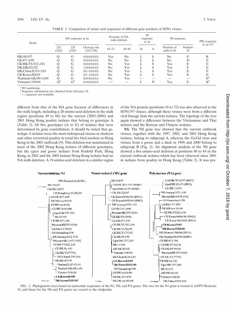
different from that of the HA gene because of differences inthe stalk length, including a 20-amino-acid deletion in the stalkregion (positions 49 to 68) for the current (2003–2004) and2001 Hong Kong poultry isolates that belong to genotype A(Table 2). Of five genotypes (A to E) of isolates that weredetermined by gene constellation, it should be noted that ge-notype A isolates were the most widespread viruses in chickensand other terrestrial poultry in retail live bird markets in HongKong in the 2001 outbreak (9). This deletion was maintained inmost of the 2002 Hong Kong isolates of different genotypes,but the egret and goose isolates from Penfold Park, HongKong, in 2002 and the 2003 human Hong Kong isolates had noNA stalk deletion. A 19-amino-acid deletion in a similar region
of the NA protein (positions 54 to 72) was also observed in theH5N1/97 viruses, although these viruses were from a differentviral lineage than the current isolates. The topology of the treeagain showed a difference between the Vietnamese and Thaiisolates and the Korean and Chinese isolates.
NS. The NS gene tree showed that the current outbreakviruses, together with the 1997, 2002, and 2003 Hong Kongisolates, belong to subgroup A, whereas the Gs/Gd virus andviruses from a goose and a duck in 1999 and 2000 belong tosubgroup B (Fig. 2). An alignment analysis of the NS geneshowed a five-amino-acid deletion at positions 80 to 84 in thecurrent outbreak isolates which has been observed since 2001in isolates from poultry in Hong Kong (Table 2). It was pre-
TABLE 2. Comparison of amino acid sequences of different gene products of H5N1 viruses
Strain
HA sequence at aa Presence of NAstalk deletion
Msequence
at aaNS sequence
PB2 sequenceat aa 627
222(226)a
224(228)a
Cleavage site(323–330) 54–72 49–68 26 31 Deletion of
aa80 to 84Position
92
HK/483/97 Q G RERRRKKR Yes No L S No E KGS/437-4/99 Q G RERRRKKR No No L S No D ECK/HK/YU822.2/01 Q G RERRRKKR No Yes L S Yes E EDK/HK/821/02 Q G IERRRKKR No Yes L S Yes E EDK/China/E319-2/03 Q G RE-RRRKR No Yes L S Yes E ECK/Korea/ES/03 Q G RE-KRKKR No Yes L S Yes E EThailand/1(KAN-1)/04 Q G RERRRKKR No Yes — — — — Eb
Vietnam/1196/04 Qb Gb RERRRKKR —c — I N Yes E Kb
a H3 numbering.b Sequence information was obtained from reference 18.c —, sequence not available.
FIG. 2. Phylogenetic trees based on nucleotide sequences of the N1, NS, and PA genes. The tree for the N1 gene is rooted to A/FPV/Rostock/34, and those for the NS and PA genes are rooted to the midpoints.
3696 LEE ET AL. J. VIROL.
on October 7, 2016 by guest
http://jvi.asm.org/
Dow
nloaded from

viously reported that the presence of glutamic acid at position92 of the NS1 molecule was related to the ability of H5N1/97viruses to escape host antiviral cytokine responses (30). Be-cause of the five-amino-acid deletion, the glutamic acid atposition 97 was shifted to position 92.
Internal protein genes. A phylogenetic analysis of five of theinternal protein genes showed a close relationship among allrecent isolates (2002 wild bird isolates, a 2003 human isolate,and current outbreak viruses), although the Korean isolateswere clearly distinguishable from the Vietnamese isolate. ThePA gene of the Korean isolates was unique and did not clusterclosely with those of any other influenza viruses (Fig. 2), and itshighest nucleotide sequence identity (96.7%) was with theA/duck/Shantou/1588/00 (H9N1) virus, which was isolatedfrom southern China. None of the internal protein genes of thecurrent outbreak isolates were related to either Gs/Gd-likeviruses or the G1 (H9N2) and W312 (H6N1) viruses, which arebelieved to have been the donor strains of the internal proteingenes of 1997 Hong Kong viruses. The Korean and Chineseisolates had no E-to-K mutation at position 627 of the PB2protein (Table 2), which was responsible for the high virulenceof A/Hong Kong/483/97 in mice (13). It was recently reportedthat three of four human isolates from Vietnam had this mu-tation but that the human virus from Thailand did not (16). Ineach tree of the different internal protein genes, the Koreanand other recent isolates roughly clustered together. However,the topology of the tree and the branch length indicated somegenetic difference between the Korean and Vietnamese iso-lates. H9N2 viruses isolated from chickens in Korea in 1996and 1999 were also compared, and no relationship was foundbetween these viruses and the recent Korean isolates.
Biological characteristics. The biological parameters of egg-grown stocks of Korean chicken and duck H5N1 isolates wereexamined. Both isolates replicated well and produced large,well-defined plaques in CEFs without the addition of trypsin.The infectivity titers in CEF cells (3.8 � 1010 and 1.8 � 1010
PFU/ml, respectively), MDCK cells (109.5 and 109.3 50% tissueculture infective doses/ml, respectively), and ECEs (109.1 and108.5 50% egg lethal doses/ml, respectively) were comparablefor both the chicken and duck isolates.
Virulence and pathogenesis of A/chicken/Korea/ES/03 indifferent avian species. Since the two Korean isolates hadalmost identical genome sequences and showed similar biolog-ical characteristics in cell culture, animal tests were done withonly the A/chicken/Korea/ES/03 isolate. By definition, AI vi-ruses that kill 75% or more of eight i.v. inoculated chickenswithin 10 days are classified as highly pathogenic (38), and theinoculation of 4-week-old chickens with the Korean viruscaused 100% mortality within 1 day (Table 3).
The intranasal inoculation of chickens and Japanese quailwith 105.9 EID50 of virus also caused 100% mortality, but themean death times (MDTs), i.e., 2.0 and 3.8 days, respectively,were longer than that for i.v. inoculated chickens. The MDTfor i.n. inoculated chickens was similar to that reported for theA/chicken/Hong Kong/220/97 virus, but the MDT for quail wasdelayed 1 day with the Korean isolate (24, 35). The Koreanvirus replicated and was shed from both the gastrointestinaland respiratory tracts of chickens, as shown by the relativelyhigh titers (5.8 to 6.6 log10 EID50/ml) of virus recovered fromoropharyngeal and cloacal swabs (Fig. 3). Furthermore, high
titers of AI virus were also detected in heart and brain tissuesas well as in lungs (6.8 to 9.5 log10 EID50/g of tissue). Virustiters in tissues and swabs were similar for both i.v. and i.n.inoculated chickens. Quail also supported rapid growth of theSouth Korean virus, as shown by the high titers of virus ob-served in brain, heart, and lung tissues (6.5 to 8.0 log10 EID50/gof tissue) as well as in oropharyngeal and cloacal swabs (Fig.3).
For i.v. and i.n. inoculated chickens, similar histological le-sions and antigen distributions were observed, characterized byan abundant infiltration of heterophils and histiocytes into theair-blood capillary wall and by proteinaceous fluid and a fewnecrotic cells in the air capillaries (Fig. 4a). Other commonlesions identified included severe multifocal splenic necrosis(10 of 12), multifocal necrosis of adrenal corticotrophic cells (4of 4), moderate to severe cardiac myocyte degeneration andnecrosis (10 of 12), and multifocal nonsuppurative encephalitis(12 of 12). Abundant AI viral antigen was identified in bloodvessel endothelial cells in most tissues (Fig. 4b), tissue macro-
TABLE 3. Pathogenicity of A/CK/Korea/ES/03 virus in differentavian species
Species(inoculation route) Mortalitya MDT
(days)Dose
(ELD50)b
White Rocks (i.v.) 8/8 1.0 107.4
White Rocks (i.n.) 8/8 1.9 105.9
White Leghorns (i.n.) 8/8 2.0 105.9
Japanese quail (i.n.) 7/7 3.8 105.9
Pekin ducks (i.n.) 2/8 4.0 105.7
a Mortality is reported as follows: number of birds that died/number of birdsinoculated.
b The titer given was determined by back titer determination of the inoculum.ELD50, 50% egg lethal dose.
FIG. 3. Comparison of mean titers of A/chicken/Korea/ES/03 virusrecovered from different avian species. Tissues and swabs from tra-cheas and cloacae were collected from four individual birds in eachgroup. For the chicken group, samples were collected from four birdsthat died on day 2 p.i. For the quail and duck groups, one sick quail andtwo clinically normal ducks were euthanized on day 3 p.i. In addition,samples were collected from two ducks that died on day 4 p.i. and alsofrom three quail that died on day 3 p.i. (one bird) and day 4 p.i. (twobirds). Virus titers were determined in eggs and expressed as EID50per gram for tissue samples and EID50 per milliliter for tracheal andcloacal swabs. The limits of virus detection (horizontal dotted lines) fortissues and swabs were 101.9 and 100.9 EID50, respectively.
VOL. 79, 2005 CHARACTERIZATION OF HPAI H5N1 VIRUSES FROM KOREA 3697
on October 7, 2016 by guest
http://jvi.asm.org/
Dow
nloaded from

FIG. 4. Experimental studies of chickens, quail, ducks, and mice that were intranasally inoculated with A/chicken/Korea/ES/03 virus. Pho-tomicrographs of hematoxylin-and-eosin-stained tissue sections (a, c-e, i, and k) or sections stained by IHC to demonstrate AI virus (b, f-h, j, andl). (a) Heterophilic to histiocytic interstitial pneumonia in a 4-week-old chicken that died 2 days after inoculation. Bar � 30 �m. (b) AI viral antigenin macrophages, heterophils, and air capillary and blood vessel endothelium in the lung of the same chicken. Bar � 50 �m. (c) Large focus ofnecrosis adjacent to pancreatic acinar cells undergoing individual cell necrosis in a 5-week-old quail that died 4 days after inoculation. Bar � 50�m. (d) Neuronal chromatolysis and necrosis in medulla of the same quail. Bar � 30 �m. (e) Large area of necrosis in corticotrophic cells inadrenal gland of the same quail. Bar � 50 �m. (f) AI viral antigen in necrotic pancreatic acinar cells of the same quail. Bar � 10 �m. (g) AI viralantigen in neurons in medulla of the same quail. Bar � 10 �m. (h) AI viral antigen in autonomic ganglial neurons and necrotic corticotrophic cells
3698 LEE ET AL. J. VIROL.
on October 7, 2016 by guest
http://jvi.asm.org/
Dow
nloaded from

phages and heterophils (Fig. 4b), cardiac myocytes, and adre-nal corticotrophic cells. The staining was a mixture of nuclearand cytoplasmic. The nuclear staining tended to be homoge-nous while the cytoplasmic staining was granular or homoge-nous. The cytoplasmic staining was most homogenous in en-dothelial and adrenal corticotrophic cells. The quail had severemultifocal pancreatic necrosis (4 of 4) (Fig. 4c), widespreadneuronal necrosis in the brain (3 of 4) (Fig. 4d), moderate focalmyocyte degeneration (4 of 4), multifocal necrosis in the go-nads (4 of 4), and severe multifocal to diffuse adrenal necrosis(2 of 3) (Fig. 4e). Nuclear and cytoplasmic staining for viralantigens was most frequent in necrotic pancreatic acinar cells(Fig. 4f), smooth muscle cells in intestinal and oviduct walls,most brain neurons (Fig. 4g), and adrenal corticotrophic cells(Fig. 4 h), slightly less frequent in cardiac myocytes, seminif-erous tubules, ovarian follicles, and the respiratory epitheliumof the nasal cavity, and infrequent in the blood vessel endo-thelium and tissue macrophages.
In contrast, i.n. inoculation of the Korean virus into Pekinducks resulted in a mortality rate of only 25%, with an MDT of4.0 days for affected birds (Table 3). This mortality was inter-mediate compared to the high mortality (100%) observed forducks infected with the 2002 wild bird virus (34) and the lackof morbidity and mortality observed for ducks inoculated withsome of the H5N1/97 and 1999 goose viruses and also withA/duck/Anyang/AVL-1/01 viruses (24, 43). Virus titers werelower from all corresponding tissues (2.1 to 4.1 log10 EID50/g)and swabs (1.6 to 1.7 log10 EID50/ml) than those for chickensand quail (Fig. 3). The two ducks that were euthanized on day3 p.i. lacked histological lesions, and AI viral antigens wereonly observed by IHC in a few respiratory epithelial cells. Twoducks that died had much less severe histological lesions andless antigen than those seen in chickens or quail. The mostconsistent lesions were unilateral severe heterophilic sinusitisand rhinitis (2 of 2) and moderately severe fibrinous air sac-culitis (2 of 2) (Fig. 4i). AI viral antigen was demonstrated inthe nasal sinus and air sac epithelium, in fibrinocellular debris(Fig. 4j), and in a few pancreatic acinar cells and cardiacmyocytes. The last two cell types had more prominent nuclearstaining than cytoplasmic staining.
Duck infection and transmission. In a separate experiment,the transmissibility of the A/chicken/Korea/ES/03 virus to con-tact control ducks was determined. Twenty juvenile ducks wereinoculated either i.v. or i.n., and 10 uninfected ducks wereplaced in direct contact with the inoculated birds at 4 h p.i.Seven of the i.v. inoculated ducks died by 4 (three birds) and 7(four birds) days p.i. However, none of the i.n. inoculated birdsshowed morbidity or mortality during the observation period.Small amounts of infectious virus (�1.0 log10 50% tissue cul-ture infective dose/ml) were detected in tracheal and cloacalswabs from many of the inoculated and contact control birds at2, 4, and 7 days p.i. Although we cannot rule out the possibilitythat contact control birds were infected by a residual inoculumin the birds or in their environment, it is unlikely, especially forthe i.v. inoculated group, and the high HI antibody titers foundin all contact birds indicate the horizontal transmission ofviruses among birds (Table 4).
Pathogenicity in mice. Previously, we demonstrated the highlevel of pathogenicity of H5N1/97 viruses in mice compared toother HPAI H5 viruses (5). To determine the pathogenicity ofthe A/chicken/Korea/ES/03 virus in a mammalian host, weinoculated BALB/c mice i.n. and determined the virus repli-cation rate, the morbidity (measured by weight loss) of themice, and the mortality rate. The MID50 of the isolate was102.8/ml. The infection of mice resulted in high titers of virus inthe lungs on day 3 p.i. (mean titer of 105.8 EID50/ml), but thevirus was not detected (�100.8 EID50/ml) in brain tissues. Nomortality was observed throughout the experiment, althoughinfected mice displayed a slight weight reduction on day 6 p.i.Previous studies established that viruses with LD50 values of�106.5 were considered to have low levels of pathogenicitywhile viruses with LD50 values of �103.0 were consideredhighly pathogenic in mouse models (15). The LD50 of theA/chicken/Korea/03 virus was �107.0 EID50, and therefore thisvirus has a low-pathogenicity phenotype.
The two mice that were euthanized on day 4 p.i. had mild tomoderate multifocal necrotizing bronchitis and adjacent alve-olitis characterized by increased numbers of alveolar macro-phages and an infiltration of neutrophils (Fig. 4k). The hilarand ventral areas of the lung lobes had a more moderate
in adrenal gland of the same quail. Bar � 50 �m. (i) Fibrinous air sacculitis of the cervical air sac in a 2-week-old duck that died 2 days afterinoculation. Bar � 100 �m. (j) AI viral antigen within the fibrinocellular debris on the surface of a cervical air sac. Bar � 50 �m. (k) Necrotizingbronchitis with luminal cellular debris and moderately severe adjacent alveolitis, with predominately alveolar macrophages but some neutrophils.Bar � 30 �m. (l) AI viral antigen in nuclei and cytoplasm of bronchial epithelial cells (arrowheads) and alveolar macrophage (arrow). Bar � 25�m.
TABLE 4. Virus transmission study with A/CK/Korea/ES/03 in ducks
Expt groupVirus isolation on indicated day p.i.a
MortalitybAntibody titer on indicated day p.i.c
2 4 7 7 14 21
i.v. inoculated ducks 11/20 (19/20) 2/17 (10/17) 0/13 (4/13) 7/20c 6.5 0.9 7.7 1.1 8.3 0.8Contact controls 5/10 (9/10) 0/10 (5/10) 0/10 (6/10) 0/10 3.6 3.0 6.0 1.1 7.1 1.3i.n. inoculated ducks 10/20 (7/20) 4/20 (2/20) 1/20 (6/20) 0/20 6.6 0.9 7.4 0.9 7.0 1.0Contact controls 0/10 (3/10) 3/10 (1/10) 4/10 (4/10) 0/10 3.1 3.7 6.8 1.2 7.9 1.0
a Data are presented as follows: number of ducks with positive orophayngeal samples/number of ducks sampled (number with positive cloacal samples/number ofducks sampled).
b Presented as number of dead ducks/number of inoculated ducks.c Presented as Log2 HI titer standard deviation. Antibody titers were determined by an HI test in which the test serum had been diluted twofold.
VOL. 79, 2005 CHARACTERIZATION OF HPAI H5N1 VIRUSES FROM KOREA 3699
on October 7, 2016 by guest
http://jvi.asm.org/
Dow
nloaded from

diffuse alveolitis, while the dorsal areas of the lung lobes lackedlesions. The AI viral antigen was common in the nuclei andcytoplasm of the respiratory epithelium and in luminal cellulardebris of the bronchi and bronchioles (Fig. 4l). Lung areas withalveolitis had scattered AI viral antigen staining in alveolarmacrophages and occasionally in neutrophils (Fig. 4l). Histo-logical lesions and the AI viral antigen were absent from in-ternal organs and the brain. This localized infection of theH5N1 HPAI virus in experimentally infected mice withoutmortality was similar to results seen with the A/Hong Kong/437/99 (H5N1), A/chicken/Queretero/7653-20/95 (H5N2), andA/chicken/Scotland/59 (H5N1) HPAI viruses (1, 5). In con-trast, experimental infections with the A/Hong Kong/156/97(H5N1) HPAI virus caused high mortality rates and moresevere lesions in the respiratory tract (5). Some of the 1997human strains caused systemic infections, with lesions and AIvirus replication in the brain (7, 17).
DISCUSSION
The first reported isolation of the Gs/Gd-like lineage viruses,which have multiple basic amino acids at the HA cleavage siteand are highly pathogenic for chickens, was obtained from agoose in China in 1996 (49). Since then, H5N1 AI viruses fromAsia have shown considerable variation in their internal genesthrough reassortment with other AI viruses and continuousevolution of the HA and NA genes. A phylogenetic analysisshowed that the Korean isolates and recent H5N1 viruses fromdifferent countries are also reassortants that contain the HAand NA genes of the Gs/Gd lineage and the internal genes ofother avian viruses. Although the Korean isolates had a uniquePA gene (Fig. 2), it is interesting that the current H5N1 strainsin Asia share a similar gene constellation to the virus identifiedin Penfold Park, Hong Kong, in 2002 (34). This is in contrast tothe presence of multiple genotypes of the H5N1 virus in HongKong in 2001–2002 (8, 9). The 2002 wild bird isolates werereported to replicate more successfully than usual in ducks andinfected a wide range of wild birds (34). Thus, it is reasonableto speculate that a virus with a gene constellation similar tothat of the 2002 isolates may be a dominant H5N1 virus in wildbirds and therefore may have a higher chance of spreading todifferent countries in Asia. However, no H5N1 AI viruses havebeen isolated from migratory birds outside the outbreak areas.
There are also molecular markers that seem to have beenfixed in the Gs/Gd lineage since 2001. The 2001 Hong Kongisolates that belonged to genotype A were the most widespreadviruses in chickens and other terrestrial poultry in retail livebird markets in Hong Kong during the 2001 outbreak, and thisgenotype of virus has a 20-amino-acid deletion in the NA stalk(9). This deletion was also observed in most of the 2002 HongKong isolates and all of the current isolates for which se-quences are available. Five-amino-acid deletions in the NS1protein are also unique to H5N1 viruses isolated after 2001,which belong to NS subtype (group) A (Fig. 2). It is not clearwhether this marker is associated with interspecies transmis-sion. However, the deletions in the NS gene result in thepresence of glutamic acid instead of aspartic acid at position92, which is known to confer some resistance to the antiviraleffects of interferon and tumor necrosis factor produced by thehost (30). It will be interesting to study if there is any relation-
ship between the stalk deletion in NA, which is a commoncharacteristic of chicken-adapted influenza viruses; the uniquedeletion of the NS gene, together with an optimal gene con-stellation of recent H5N1 viruses; the high prevalence of theseviruses in Asia; and the extraordinary pathogenicity of theseviruses for gallinaceous birds, especially chickens.
Regarding human health concerns, the Korean H5N1 iso-lates appear to have low pandemic potential. Although 19infected premises have been identified from different regionsin South Korea, no human infections have been reported. Todate, only two countries, Vietnam and Thailand, have reportedlaboratory-confirmed cases of H5N1 virus infections in hu-mans. A phylogenetic analysis also showed genetic differencesbetween the Korean and Vietnamese isolates in all eight genesegments and also between these isolates and Thai isolates inat least the HA and NA sequences. In contrast to the Vietnam(16) and Thai isolates, the Korean isolates had no mutations inthe M2 protein (Table 2), which are commonly related toresistance to antiviral drugs that are commonly used for influ-enza (37). However, it is not clear whether a particularly lethalstrain for humans has evolved in Vietnam or Thailand. Thegenetic differences among isolates may have resulted fromseparate introductions of HPAI viruses into different coun-tries. However, the similar gene constellation of recent H5N1viruses indicates a progressive divergence of HPAI virus froma single origin in geographically distinct bird populations.
Most of the recent outbreaks of pathogenic H5N1 infectionin Asia have occurred in domestic chickens. Although Koreahad nine H5N1 cases in duck farms, in general, no mortalitywas involved in the field. This coincided with the low (25%) orno mortality observed in our two separate experiments withducks that were infected i.n. with high titers of the virus (Ta-bles 3 and 4). Previous studies have shown that H5N1 virusisolates from Hong Kong and southern China did not replicateefficiently in ducks and induced only mild histological lesions ingeneral (24, 35). A striking exception was the induction ofsevere disease and death in ducks with the 2002 wild birdviruses that were isolated in Hong Kong (34). Phylogenetically,the Vietnamese and Thai isolates are closer to 2002 wild birdisolates than to the Korean or Chinese isolates, and furthercomparative studies are needed to see if their genetic differ-ences coincide with their high levels of pathogenicity in ducks.
The low level of pathogenicity of the Korean isolate in do-mestic ducks may be a concern for control efforts for this H5N1virus, since the virus could become widespread in this hostwithout detection. Our experiments also reinforced the possi-bility that the virus was able to be transmitted to contactcontrol ducks, although shedding of the virus was low (Table4).
In contrast to the high levels of pathogenicity and neuro-virulence in mice observed for some H5N1 viruses isolatedfrom poultry and humans in Hong Kong in 1997 (5, 7, 17), theKorean virus had a low level of pathogenicity in mice, and novirus was detected in the brain. The low level of pathogenicityof the Korean virus in mice also contrasts with the results ofChen et al. (2), who demonstrated that recent H5N1 isolatesfrom ducks in mainland China are becoming increasingly vir-ulent for mice. However, there are limitations to the directextrapolation of results from mouse experiments to assess-ments of the pathogenic potential for humans. Mice have been
3700 LEE ET AL. J. VIROL.
on October 7, 2016 by guest
http://jvi.asm.org/
Dow
nloaded from

used as a convenient small animal model with which to betterunderstand general determinants of virulence for mammalianspecies rather than the absolute ability to infect mammals.
A major determinant of host range is the affinity of the viralHA protein for the host cell sialic acid receptor (4, 19, 28, 32).The Korean isolates and human isolates from Thailand andVietnam (18) had no mutations in the receptor binding site(glutamine to leucine at position 226 or glycine to serine atposition 228) in HA that would preferentially bind to sialic acidin human cells (20, 29, 46). The Korean isolates and humanisolates from Thailand and Vietnam also did not have theserine-to-asparagine mutation at position 227 that was foundin A/Hong Kong/213/03 and was shown to alter the virulence ofone human H5N1/97 virus in mice (8, 13). However, a recentstudy showed that ciliated cells in the respiratory epithelium ofhumans express �-2,3-linked sialic acid receptors, which allowthe entry and replication of AI viruses (18). Thus, in rare cases,such as those in Thailand and Vietnam, it is possible for AIviruses to infect humans and even to cause a fatal outcome.Nevertheless, ciliated cells seem less than optimal for influenzavirus replication since epidemic human influenza viruses typi-cally target nonciliated cells in vitro (18). However, it is alwaysdifficult to interpret and relate in vitro data to in vivo out-comes, and there is in vivo evidence of infection of ciliated cellsfor humans and nonhuman primates (21, 27). Furthermore, itshould be noted that the 1918 pandemic human H1 virus couldspread successfully through human populations while retainingthe receptor binding site amino acids that are characteristic ofan avian precursor HA (6, 33). Thus, it is also possible that theH5N1 viruses from Thailand and Vietnam may have acquiredstructural conformations that allowed binding to human cellswhile retaining avian-like residues, although no evidence ofhuman-to-human transmission in the current H5N1 outbreakshas been observed thus far.
The big questions that remain regarding the recent AsianHPAI outbreak are where the virus originated and how thisvirus spread to at least eight different countries. SouthernChina, where various species of poultry, including domesticducks, and pigs are raised alongside each other in high-densitysmall farms, has been considered a likely geographic reservoirof new viruses with pandemic potential (31). Both H5 and H9AI viruses that resulted in human infections have been isolatedfrom this region (8, 12, 23, 36). Furthermore, H5N1 AI viruseswere isolated from geese in Guangdong in 1996 (49), fromdomestic ducks between 1999 and 2002 (2), and from geeseimported into Hong Kong from Guangdong during 1999–2001(1, 47), all of which support the potential role of southernChina as the reservoir of H5N1 HPAI viruses. The currentwide prevalence of H5N1 viruses throughout East Asia sug-gests that viruses may have spread to different countries by thelegal and illegal introduction of infected poultry or poultryproducts. In May 2001, an H5N1 virus was isolated from duckmeat that had been imported to South Korea from China (43).The H5N1 virus was also detected in Japan in imported duckmeat from China in May 2003 (ProMED, 12/05/2003 [http://www.promedmail.org]). Additionally, a recent isolation of anH5N1 virus in Taiwan was traced to smuggled ducks fromChina (ProMED, 01/01/2004 [http://www.promedmail.org]),which further reinforces the possibility of H5N1 AI virusspread. It is also possible that wild birds may have played a role
in introducing the viruses into different countries. However,the detection of H5N1 HPAI viruses has only been successfulfor local feral or captive wild birds either in parks or in areasassociated with HPAI outbreaks in poultry, and no direct ev-idence of infection in wild birds outside these areas with in-fected poultry has been reported. In Korea, surveillance wasconducted on different wild bird species after the HPAI out-break, with 5,460 fecal samples being collected and tested from62 different migratory bird habitats and 112 samples beingcollected directly from different wild birds. Although eightdifferent HA subtypes of AI viruses were identified in thatsurveillance, no H5 subtype virus has been isolated so far (J. H.Kim, unpublished data). Recently, H5N1 viruses were isolatedfrom 3 of 259 magpies near the two different farms whereH5N1 HPAI was confirmed (Kim, unpublished data). How-ever, since magpies are a nonmigratory common bird in Koreaand typically reside within 3.5 km of nest areas, it is likely thatthe magpies were infected from exposure to infected chickensand not vice versa.
Currently, genetic analyses of all H5N1 viruses isolated fromdifferent farms and different species in South Korea are ongo-ing, and preliminary data show little genetic variation amongKorean isolates, in contrast to the heterogeneity of the isolatesobserved in other countries. Those differences may be relatedto the prevalence of viruses before initial detection and thespeed of control measure implementation. South Korea hastaken all necessary measures for the detection of virus, report-ing to OIE, control of the virus, and prevention of the spreadof the virus in accordance with OIE guidelines. Swift controlmeasures that included stamping out of infected and neighbor-ing farms were completed in every case in South Korea within3 weeks. Considering the wide prevalence of H5N1 viruses, allAsian countries must exert multilateral efforts of increasedsurveillance, transparency in reporting the disease, and imple-mentation of swift control measures to prevent H5N1 virusesfrom becoming endemic to this region.
This study provides a characterization of recent H5N1 vi-ruses isolated from poultry in Korea that caused the first HPAIoutbreak in South Korea’s history, which has a relationshipwith the current H5N1 outbreak in Asia. To avoid future out-breaks, we need a clear understanding of how this unprece-dented epidemic began in Asia. Therefore, further character-ization of the H5N1 viruses from different countries is urgentlyneeded.
ACKNOWLEDGMENTS
This work was partially supported by USDA ARS CRIS project6612-32000-039.
We thank Joan Beck, Suzanne DeBlois, and the SAA sequencingfacility for technical assistance and Roger Brock for animal care as-sistance.
REFERENCES
1. Cauthen, A. N., D. E. Swayne, S. Schultz-Cherry, M. L. Perdue, and D. L.Suarez. 2000. Continued circulation in China of highly pathogenic avianinfluenza viruses encoding the hemagglutinin gene associated with the 1997H5N1 outbreak in poultry and humans. J. Virol. 74:6592–6599.
2. Chen, H., G. Deng, Z. Li, G. Tian, Y. Li, P. Jiao, L. Zhang, Z. Liu, R. G.Webster, and K. Yu. 2004. The evolution of H5N1 influenza viruses in ducksin southern China. Proc. Natl. Acad. Sci. USA 101:10452–10457.
3. Claas, E. C., A. D. Osterhaus, R. van Beek, J. C. De Jong, G. F. Rimmel-zwaan, D. A. Senne, S. Krauss, K. F. Shortridge, and R. G. Webster. 1998.Human influenza A H5N1 virus related to a highly pathogenic avian influ-enza virus. Lancet 351:472–477.
VOL. 79, 2005 CHARACTERIZATION OF HPAI H5N1 VIRUSES FROM KOREA 3701
on October 7, 2016 by guest
http://jvi.asm.org/
Dow
nloaded from

4. Connor, R. J., Y. Kawaoka, R. G. Webster, and J. C. Paulson. 1994. Receptorspecificity in human, avian, and equine H2 and H3 influenza virus isolates.Virology 205:17–23.
5. Dybing, J. K., S. Schultz-Cherry, D. E. Swayne, D. L. Suarez, and M. L.Perdue. 2000. Distinct pathogenesis of Hong Kong-origin H5N1 viruses inmice compared to that of other highly pathogenic H5 avian influenza viruses.J. Virol. 74:1443–1450.
6. Gamblin, S. J., L. F. Haire, R. J. Russell, D. J. Stevens, B. Xiao, Y. Ha, N.Vasisht, D. A. Steinhauer, R. S. Daniels, A. Elliot, D. C. Wiley, and J. J.Skehel. 2004. The structure and receptor binding properties of the 1918influenza hemagglutinin. Science 303:1838–1842.
7. Gao, P., S. Watanabe, T. Ito, H. Goto, K. Wells, M. McGregor, A. J. Cooley,and Y. Kawaoka. 1999. Biological heterogeneity, including systemic replica-tion in mice, of H5N1 influenza A virus isolates from humans in Hong Kong.J. Virol. 73:3184–3189.
8. Guan, Y., L. L. Poon, C. Y. Cheung, T. M. Ellis, W. Lim, A. S. Lipatov, K. H.Chan, K. M. Sturm-Ramirez, C. L. Cheung, Y. H. Leung, K. Y. Yuen, R. G.Webster, and J. S. Peiris. 2004. H5N1 influenza: a protean pandemic threat.Proc. Natl. Acad. Sci. USA 101:8156–8161.
9. Guan, Y., J. S. Peiris, A. S. Lipatov, T. M. Ellis, K. C. Dyrting, S. Krauss,L. J. Zhang, R. G. Webster, and K. F. Shortridge. 2002. Emergence ofmultiple genotypes of H5N1 avian influenza viruses in Hong Kong SAR.Proc. Natl. Acad. Sci. USA 99:8950–8955.
10. Guan, Y., M. Peiris, K. F. Kong, K. C. Dyrting, T. M. Ellis, T. Sit, L. J.Zhang, and K. F. Shortridge. 2002. H5N1 influenza viruses isolated fromgeese in Southeastern China: evidence for genetic reassortment and inter-species transmission to ducks. Virology 292:16–23.
11. Guan, Y., K. F. Shortridge, S. Krauss, and R. G. Webster. 1999. Molecularcharacterization of H9N2 influenza viruses: were they the donors of the“internal” genes of H5N1 viruses in Hong Kong? Proc. Natl. Acad. Sci. USA96:9363–9367.
12. Guo, Y. J., S. Krauss, D. A. Senne, I. P. Mo, K. S. Lo, X. P. Xiong, M.Norwood, K. F. Shortridge, R. G. Webster, and Y. Guan. 2000. Character-ization of the pathogenicity of members of the newly established H9N2influenza virus lineages in Asia. Virology 267:279–288.
13. Hatta, M., P. Gao, P. Halfmann, and Y. Kawaoka. 2001. Molecular basis forhigh virulence of Hong Kong H5N1 influenza A viruses. Science 293:1840–1842.
14. Hoffmann, E., J. Stech, I. Leneva, S. Krauss, C. Scholtissek, P. S. Chin, M.Peiris, K. F. Shortridge, and R. G. Webster. 2000. Characterization of theinfluenza A virus gene pool in avian species in southern China: was H6N1 aderivative or a precursor of H5N1? J. Virol. 74:6309–6315.
15. Katz, J. M., X. Lu, T. M. Tumpey, C. B. Smith, M. W. Shaw, and K.Subbarao. 2000. Molecular correlates of influenza A H5N1 virus pathogen-esis in mice. J. Virol. 74:10807–10810.
16. Li, K. S., Y. Guan, J. Wang, G. J. Smith, K. M. Xu, L. Duan, A. P. Rahardjo,P. Puthavathana, C. Buranathai, T. D. Nguyen, A. T. Estoepangestie, A.Chaisingh, P. Auewarakul, H. T. Long, N. T. Hanh, R. J. Webby, L. L. Poon,H. Chen, K. F. Shortridge, K. Y. Yuen, R. G. Webster, and J. S. Peiris. 2004.Genesis of a highly pathogenic and potentially pandemic H5N1 influenzavirus in eastern Asia. Nature 430:209–213.
17. Lu, X., T. M. Tumpey, T. Morken, S. R. Zaki, N. J. Cox, and J. M. Katz. 1999.A mouse model for the evaluation of pathogenesis and immunity to influ-enza A (H5N1) viruses isolated from humans. J. Virol. 73:5903–5911.
18. Matrosovich, M. N., T. Y. Matrosovich, T. Gray, N. A. Roberts, and H. D.Klenk. 2004. Human and avian influenza viruses target different cell types incultures of human airway epithelium. Proc. Natl. Acad. Sci. USA 101:4620–4624.
19. Matrosovich, M. N., S. Krauss, and R. G. Webster. 2001. H9N2 influenza Aviruses from poultry in Asia have human virus-like receptor specificity. Vi-rology 281:156–162.
20. Matrosovich, M., A. Tuzikov, N. Bovin, A. Gambaryan, A. Klimov, M. R.Castrucci, I. Donatelli, and Y. Kawaoka. 2000. Early alterations of thereceptor-binding properties of H1, H2, and H3 avian influenza virus hem-agglutinins after their introduction into mammals. J. Virol. 74:8502–8512.
21. Mulder, J., and J. F. Hers. 1979. Influenza. Wolters-Noordhoff, Groningen,The Netherlands.
22. Peiris, J. S., W. C. Yu, C. W. Leung, C. Y. Cheung, W. F. Ng, J. M. Nicholls,T. K. Ng, K. H. Chan, S. T. Lai, W. L. Lim, K. Y. Yuen, and Y. Guan. 2004.Re-emergence of fatal human influenza A subtype H5N1 disease. Lancet363:617–619.
23. Peiris, M., K. Y. Yuen, C. W. Leung, K. H. Chan, P. L. Ip, R. W. Lai, W. K.Orr, and K. F. Shortridge. 1999. Human infection with influenza H9N2.Lancet 354:916–917.
24. Perkins, L. E., and D. E. Swayne. 2003. Comparative susceptibility of se-lected avian and mammalian species to a Hong Kong-origin H5N1 high-pathogenicity avian influenza virus. Avian Dis. 47:956–967.
25. Perkins, L. E., and D. E. Swayne. 2002. Pathogenicity of a Hong Kong-originH5N1 highly pathogenic avian influenza virus for emus, geese, ducks, andpigeons. Avian Dis. 46:53–63.
26. Reed, L. J., and H. Muench. 1938. A simple method for estimating fiftypercent endpoints. Am. J. Hyg. 27:493–497.
27. Rimmelzwaan, G. F., T. Kuiken, G. van Amerongen, T. M. Bestebroer, R. A.Fouchier, and A. D. Osterhaus. 2001. Pathogenesis of influenza A (H5N1)virus infection in a primate model. J. Virol. 75:6687–6691.
28. Rogers, G. N., and B. L. D’Souza. 1989. Receptor binding properties ofhuman and animal H1 influenza virus isolates. Virology 173:317–322.
29. Rogers, G. N., J. C. Paulson, R. S. Daniels, J. J. Skehel, I. A. Wilson, andD. C. Wiley. 1983. Single amino acid substitutions in influenza haemagglu-tinin change receptor binding specificity. Nature 304:76–78.
30. Seo, S. H., E. Hoffmann, and R. G. Webster. 2002. Lethal H5N1 influenzaviruses escape host anti-viral cytokine responses. Nat. Med. 8:950–954.
31. Shortridge, K. F., and C. H. Stuart-Harris. 1982. An influenza epicentre?Lancet 2:812–813.
32. Skehel, J. J., and D. C. Wiley. 2000. Receptor binding and membrane fusionin virus entry: the influenza hemagglutinin. Annu. Rev. Biochem. 69:531–569.
33. Stevens, J., A. L. Corper, C. F. Basler, J. K. Taubenberger, P. Palese, andI. A. Wilson. 2004. Structure of the uncleaved human H1 hemagglutinin fromthe extinct 1918 influenza virus. Science 303:1866–1870.
34. Sturm-Ramirez, K. M., T. Ellis, B. Bousfield, L. Bissett, K. Dyrting, J. E.Rehg, L. Poon, Y. Guan, M. Peiris, and R. G. Webster. 2004. ReemergingH5N1 influenza viruses in Hong Kong in 2002 are highly pathogenic toducks. J. Virol. 78:4892–4901.
35. Suarez, D. L., M. L. Perdue, N. Cox, T. Rowe, C. Bender, J. Huang, and D. E.Swayne. 1998. Comparisons of highly virulent H5N1 influenza A virusesisolated from humans and chickens from Hong Kong. J. Virol. 72:6678–6688.
36. Subbarao, K., A. Klimov, J. Katz, H. Regnery, W. Lim, H. Hall, M. Perdue,D. Swayne, C. Bender, J. Huang, M. Hemphill, T. Rowe, M. Shaw, X. Xu, K.Fukuda, and N. Cox. 1998. Characterization of an avian influenza A (H5N1)virus isolated from a child with a fatal respiratory illness. Science 279:393–396.
37. Suzuki, H., R. Saito, H. Masuda, H. Oshitani, M. Sato, and I. Sato. 2003.Emergence of amantadine-resistant influenza A viruses: epidemiologicalstudy. J. Infect. Chemother. 9:195–200.
38. Swayne, D. E., and D. A. Halvorson. 2003. Influenza, p. 135–160. In Y. M.Saif, H. J. Barnes, J. R. Glisson, A. M. Fadly, L. R. McDougald, and D. E.Swayne (ed.), Diseases of poultry. Iowa State University Press, Ames, Iowa.
39. Swayne, D. E., D. A. Senne, and C. W. Beard. 1998. Avian influenza, p.150–155. In D. E. Swayne (ed.), A laboratory manual for the isolation andidentification of avian pathogens. American Association of Avian Patholo-gists, Kennett Square, Pa.
40. Swayne, D. E. 1997. Pathobiology of H5N2 Mexican avian influenza virusesfor chickens. Vet. Pathol. 34:557–567.
41. Swofford, D. L. 1998. PAUP*. Phylogenetic analysis using parsimony (*andother methods), v. 4. Sinauer Associates, Sunderland, Mass.
42. To, K. F., P. K. Chan, K. F. Chan, W. K. Lee, W. Y. Lam, K. F. Wong, N. L.Tang, D. N. Tsang, R. Y. Sung, T. A. Buckley, J. S. Tam, and A. F. Cheng.2001. Pathology of fatal human infection associated with avian influenza AH5N1 virus. J. Med. Virol. 63:242–246.
43. Tumpey, T. M., D. L. Suarez, L. E. Perkins, D. A. Senne, J. G. Lee, Y. J. Lee,I. P. Mo, H. W. Sung, and D. E. Swayne. 2002. Characterization of a highlypathogenic H5N1 avian influenza A virus isolated from duck meat. J. Virol.76:6344–6355.
44. USAHA. 1994. Report of the Committee on Transmissible Diseases of Poul-try and Other Avian Species. Criteria for determining that an AI virusisolation causing an outbreak must be considered for eradication, p. 522. InProceedings of the 98th Annual Meeting of the U.S. Animal Health Asso-ciation. USAHA, Grand Rapids, Mich.
45. Van Deusen, R. A., V. S. Hinshaw, D. A. Senne, and D. Pellacani. 1983.Microneuraminidase-inhibition assay for classification of influenza A virusneuraminidases. Avian Dis. 27:745–750.
46. Vines, A., K. Wells, M. Matrosovich, M. R. Castrucci, T. Ito, and Y.Kawaoka. 1998. The role of influenza A virus hemagglutinin residues 226and 228 in receptor specificity and host range restriction. J. Virol. 72:7626–7631.
47. Webster, R. G., Y. Guan, M. Peiris, D. Walker, S. Krauss, N. N. Zhou, E. A.Govorkova, T. M. Ellis, K. C. Dyrting, T. Sit, D. R. Perez, and K. F.Shortridge. 2002. Characterization of H5N1 influenza viruses that continueto circulate in geese in southeastern China. J. Virol. 76:118–126.
48. Webster, R. G., and R. Rott. 1987. Influenza virus A pathogenicity: thepivotal role of hemagglutinin. Cell 50:665–666.
49. Xu, X., K. Subbarao, N. J. Cox, and Y. Guo. 1997. Genetic characterizationof the pathogenic influenza A/Goose/Guangdong/1/96 (H5N1) virus: simi-larity of its hemagglutinin gene to those of H5N1 viruses from the 1997outbreaks in Hong Kong. Virology 261:15–19.
50. Yuen, K. Y., P. K. Chan, M. Peiris, D. N. Tsang, T. L. Que, K. F. Shortridge,P. T. Cheung, W. K. To, E. T. Ho, R. Sung, and A. F. Cheng. 1998. Clinicalfeatures and rapid viral diagnosis of human disease associated with avianinfluenza A H5N1 virus. Lancet 351:467–471.
3702 LEE ET AL. J. VIROL.
on October 7, 2016 by guest
http://jvi.asm.org/
Dow
nloaded from