Viruses of hyperthermophilic archaea: entry and egress from ...
Upload
khangminh22Category
view
1download
0
Structure of virusesLecture 4
Biology W3310/4310 Virology
Spring 2015
In order to create something that func2ons properly -‐ a container, a chair, a house -‐ its essence has to be explored, for it should serve its purpose to perfec2on, i.e., it should be durable, inexpensive, and beau2ful. -‐ WALTER GROPIUS

Func/ons of structural proteins
Protec'on of the genome
-‐ Assembly of a stable, protecJve protein shell
-‐ Specific recogniJon and packaging of the nucleic acid genome
-‐ InteracJon with host cell membranes to form the envelope
©Principles of Virology, ASM Press

Func/ons of structural proteins
Delivery of the genome
-‐ Bind host cell receptors
-‐ UncoaJng of the genome
-‐ Fusion with cell membranes
-‐ Transport of genome to the appropriate site
©Principles of Virology, ASM Press
ScEYEnce StudiosPrinciples of Virology, 4eFig. APP01-349-28-14
nsP1
P1234
nsP2 nsP3nsP4
Golgi
2
3
4
5
6
7
8
9
10
11
12
13
1
ER
Cytoplasm Nucleus
6KPE2 E1Capsid
5' c
5' c
5' c
5' c
5' c
5' c
5' c

• Subunit
-‐ Single folded polypepJde chain
• Structural unit (protomer, asymmetric unit)
-‐ Unit from which capsids or nucleocapsids are built; one or more subunits
• Capsid (capsa = LaJn, box)
-‐ Protein shell surrounding genome
• Nucleocapsid (core)
-‐ Nucleic acid -‐ protein assembly within virion
• Envelope (viral membrane)
-‐ Host cell-‐derived lipid bilayer
• Virion
-‐ InfecJous virus parJcle
Defini/ons
©Principles of Virology, ASM Press

Pu6ng virus par/cles into perspec/ve
• Nanometer: 10-‐9 meters
• Alpha helix in protein: 1 nm diameter
• DNA: 2 nm diameter
• Ribosome: 20 nm diameter
• Poliovirus: 30 nm
• Pandoravirus: 1000 nm

Virus par/cles are metastable
• Must protect the genome (stable)
• Must come apart on infecJon (unstable)
©Principles of Virology, ASM Press

• Virus parJcles have not a_ained minimum free energy conformaJon
• Unfavorable energy barrier must be surmounted
• Energy put into virus parJcle during assembly (spring loaded)
• PotenJal energy used for disassembly if cell provides proper signal
Virions are metastable

How is metastability achieved?
• Stable structure
-‐ Created by symmetrical arrangement of many idenJcal proteins to provide maximal contact
• Unstable structure
-‐ Structure is not usually permanently bonded together
-‐ Can be taken apart or loosened on infecJon to release or expose genome

Go to:
m.socraJve.com room number: virus
1
Viral capsids are metastable because:
1. They must protect the viral genome outside of the cell 2. They must come apart and release the genome into a cell 3. They have not obtained a minimum free energy
conformaJon 4. They are spring-‐loaded 5. All of the above
The tools of viral structural biology
• Electron microscopy
• X-‐ray crystallography
• Electron cryomicroscopy (cryoEM) & tomography
• Nuclear magneJc resonance spectroscopy (NMR)
Flint volume I, chapter 3, pp 85-‐88
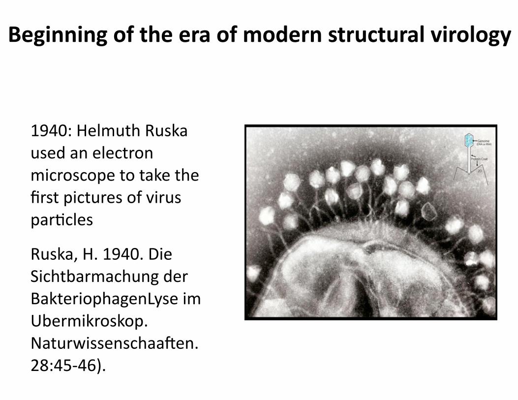
1940: Helmuth Ruska used an electron microscope to take the first pictures of virus parJcles
Ruska, H. 1940. Die Sichtbarmachung der BakteriophagenLyse im Ubermikroskop. Naturwissenschaaeen. 28:45-‐46).
Beginning of the era of modern structural virology

Electron microscopy
• Biological materials have li_le inherent contrast: need to be stained
• NegaJve staining with electron-‐dense material (uranyl acetate, phosphotungstate), sca_er electrons (1959)
• ResoluJon 50-‐75 Å (alpha helix 10 Å dia; 1 Å = 0.1 nm)
• Detailed structural interpretaJon impossible
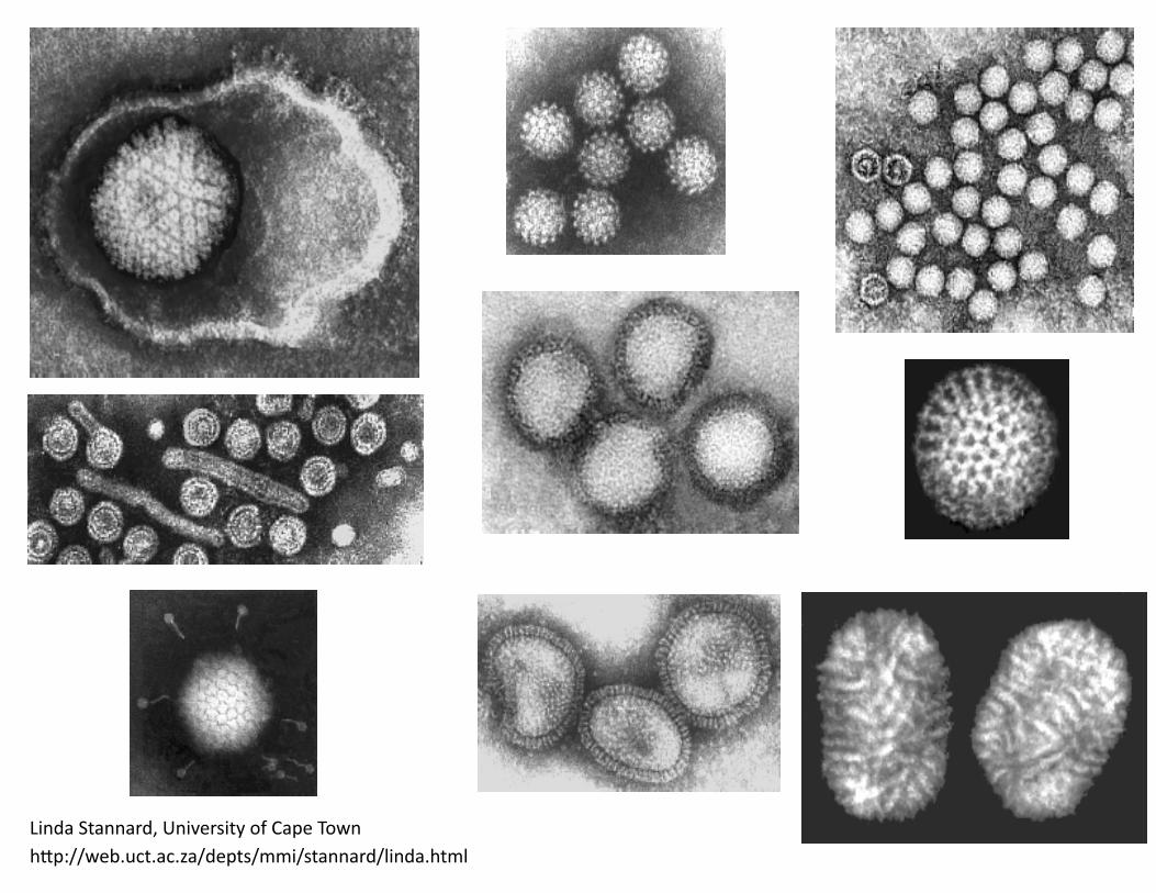
Linda Stannard, University of Cape Townh_p://web.uct.ac.za/depts/mmi/stannard/linda.html

©Principles of Virology, ASM Press

Poliovirus + CD155

X-‐ray crystallography (2-‐3 Å for viruses)
©Principles of Virology, ASM Press
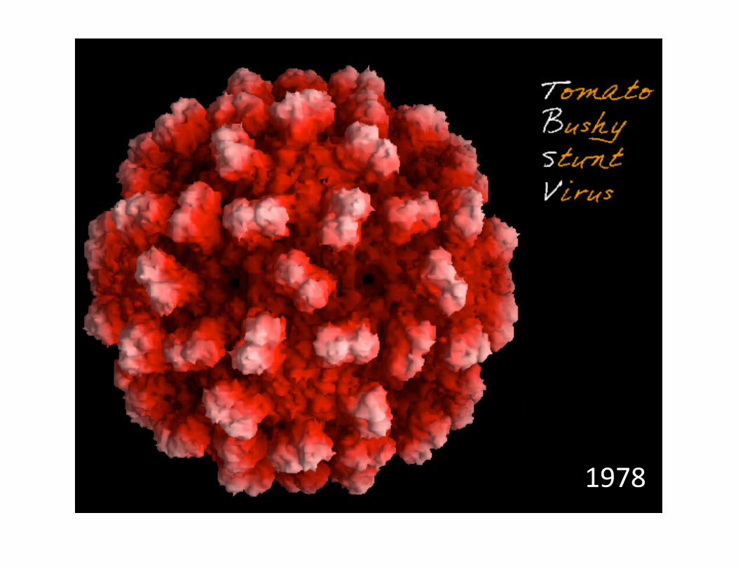
1978

Poliovirus, 1985

C. roenbergensis virus
Chuan Xiao h_p://utminers.utep.edu/cxiao/#4300 nm, >15,000 capsid proteins

Building virus par/cles: Symmetry is key
• Watson and Crick did more than discover DNA structure
• Their seminal contribuJon to virology:
-‐ IdenJcal protein subunits are distributed with helical symmetry for rod-‐shaped viruses
-‐ Platonic polyhedra symmetry for round viruses

The symmetry rules are elegant in their simplicity They provide rules for “self-‐assembly”
• Rule 1: Each subunit has ‘idenJcal’ bonding contacts with its neighbors
-‐ Repeated interacJon of chemically complementary surfaces at the subunit interfaces naturally leads to a symmetric arrangement
• Rule 2: These bonding contacts are usually non-‐covalent
-‐ Reversible; error-‐free assembly

Symmetry and self-‐assembly
• Many capsid proteins can self assemble into ‘virus-‐like parJcles’ (VLPs)
• The HBV and HPV vaccines are VLPs made in yeast

Helical symmetryCoat protein molecules engage in idenJcal, equivalent interacJons with one another and with the viral genome to allow construcJon of a large, stable structure from a single protein subunit
©Principles of Virology, ASM Press


Helical symmetry
©Principles of Virology, ASM Press

©Principles of Virology, ASM Press

Enveloped RNA viruses with (-‐) ssRNA and helical capsids
• Paramyxoviridae (measles virus, mumps virus)
• Rhabdoviridae (rabies virus)
• Orthomyxoviridae (influenza virus)
• Filoviridae (Ebola virus)
• The nucleocapsid is the nucleic acid-‐protein assembly that is packaged within the virion
©Principles of Virology, ASM Press

Go to:
m.socraJve.com room number: virus
2
Which of the following describe virus symmetry and self assembly?
1. The bonding contacts are usually covalent 2. Each subunit always has idenJcal bonding contacts with its
neighbors 3. The bonding contacts of subunits are usually non-‐covalent 4. Each subunit has different bonding contacts with its
neighbors 5. None of the above

How can you make a round capsid from proteins with irregular shapes?
• Clue 1: All round capsids have precise numbers of proteins; mulJples of 60 are common (60, 180, 240, 960)
• Clue 2: Spherical viruses come in many sizes, but capsid proteins are 20-‐60 kDa average

Caspar & Klug’s 1962 solu/on
• They knew from Watson & Crick’s work that round capsids are icosahedrons -‐ no other Platonic solids were used
• Capsid subunits tended to be arranged as hexamers and pentamers

Icosahedral symmetry
• Icosahedron: solid with 20 faces, each an equilateral triangle
• Allows formaJon of a closed shell with smallest number (60) of idenJcal subunits
©Principles of Virology, ASM Press

• Made of 60 idenJcal protein subunits
• The protein subunit is the structural unit
• InteracJons of all molecules with their neighbors are idenJcal (head-‐to-‐head, tail-‐to-‐tail)
Simple icosahedral capsids
©Principles of Virology, ASM Press

Adeno-‐associated virus 2 (parvovirus) 25 nm T=1 60 copies of a single capsid protein
©Principles of Virology, ASM PressScEYEnce StudiosPrinciples of Virology, 4eFig. APP01-199-28-14
5' c
5' c
p5 TRTR
p19 p40
AnAOH3’
AnAOH3’
AnAOH3’
AnAOH3’
rep ORFcap ORF
AnAOH3’
VP1
VP2
VP3
2.3 kb
Rep 78
mRNAs
Rep 68
Rep 52
Rep 40
4.2 kb
3.9 kb
3.6 kb
3.3 kb
A
B
AAP
5' c
5' c
5' c
ssDNA

• Three modes of subunit packing (orange, yellow, purple)
• Pentamers & hexamers
• Bonding interacJons are quasiequivalent: all engage tail-‐to-‐tail and head-‐to-‐head
180 idenJcal protein subunits©Principles of Virology, ASM Press
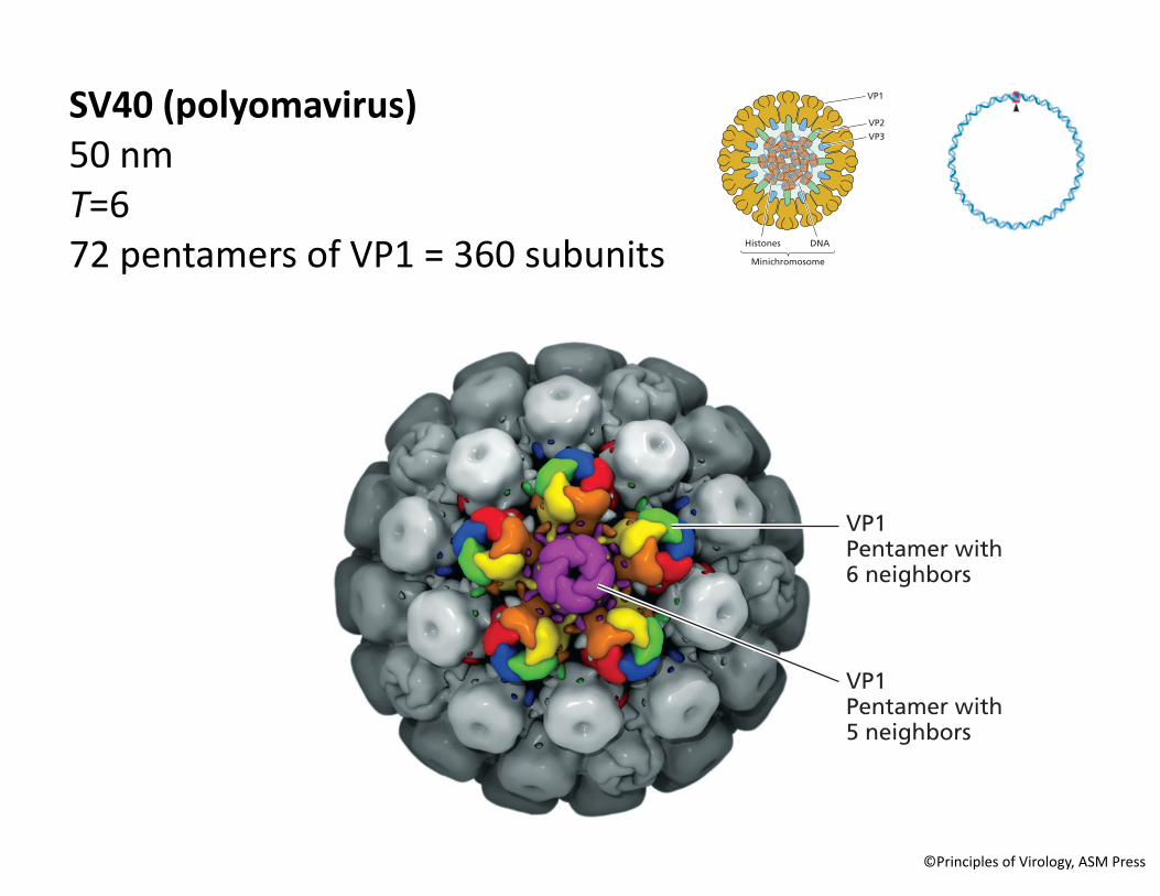
How are larger virus par/cles built? By adding more subunits

Quasiequivalence
• When a capsid contains more than 60 subunits, each occupies a quasiequivalent posiJon
• The noncovalent binding properJes of subunits in different structural environments are similar, but not idenJcal
ScEYEnce StudiosPrinciples of Virology, 4eVolume 01Fig. 04.139-21-14
BA
N
VP1Pentamer with 6 neighbors
VP1Pentamer with 5 neighbors
C
αC' αC
SV40 (polyomavirus) 50 nm T=6 72 pentamers of VP1 = 360 subunits
VP1
VP2VP3
DNAHistones
Minichromosome
©Principles of Virology, ASM Press

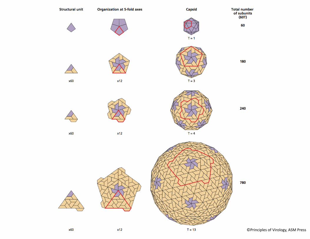
Triangula/on number, T
• The number of facets per triangular face of an icosahedron
• Combining several triangular facets allows assembly of larger face from same structural unit
Capsids with T>1 have a 6-fold axis of
symmetry
©Principles of Virology, ASM Press
©Principles of Virology, ASM Press

Go to:
m.socraJve.com room number: virus
3
Which of the following are characteris/cs of icosahedral symmetry in viral capsids?
1. Produces a solid with 20 faces, each an equilateral triangle 2. Allows formaJon of a closed shell with 60 idenJcal subunits 3. Fivefold, threefold, and twofold axes of symmetry 4. The T number describes the number of facets per icosahedral
face 5. All of the above

Adenovirus • 150 nm
• T=25 capsid, 720 copies viral protein II + 60 copies of protein III
• Fibers at 12 verJces ©Principles of Virology, ASM Press
Large complex capsids
• DisJnct components with different symmetries
• Presence of proteins devoted to specialized roles

VP7 trimers, T=13 VP3 monomers, T=2Reoviruses •T=13 •70 -‐ 90 nm •two concentric shells
Complex capsids with two icosahedral protein layers
©Principles of Virology, ASM Press
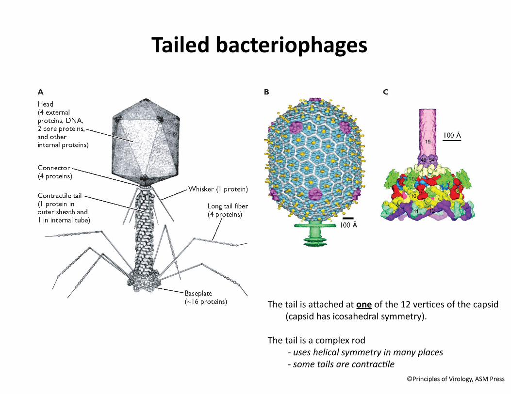
The tail is a_ached at one of the 12 verJces of the capsid (capsid has icosahedral symmetry).
The tail is a complex rod -‐ uses helical symmetry in many places -‐ some tails are contrac2le
©Principles of Virology, ASM Press
Tailed bacteriophages

An iron loaded spike
©Principles of Virology, ASM Press
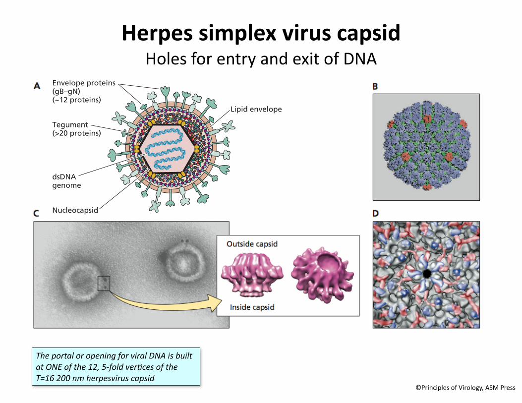
Herpes simplex virus capsid Holes for entry and exit of DNA
The portal or opening for viral DNA is built at ONE of the 12, 5-‐fold ver2ces of the T=16 200 nm herpesvirus capsid
©Principles of Virology, ASM Press
ScEYEnce StudiosPrinciples of Virology, 4eFig. APP01-139-27-14
A
B
Long region(126 kb)
Short region(26 kb)
Lipid envelope
Envelope proteins(gB–gN)(~12 proteins)
Nucleocapsid
dsDNAgenome
Tegument(>20 proteins)
TRL UL
OriL OriS
IRL IRS TRSUS
OriS
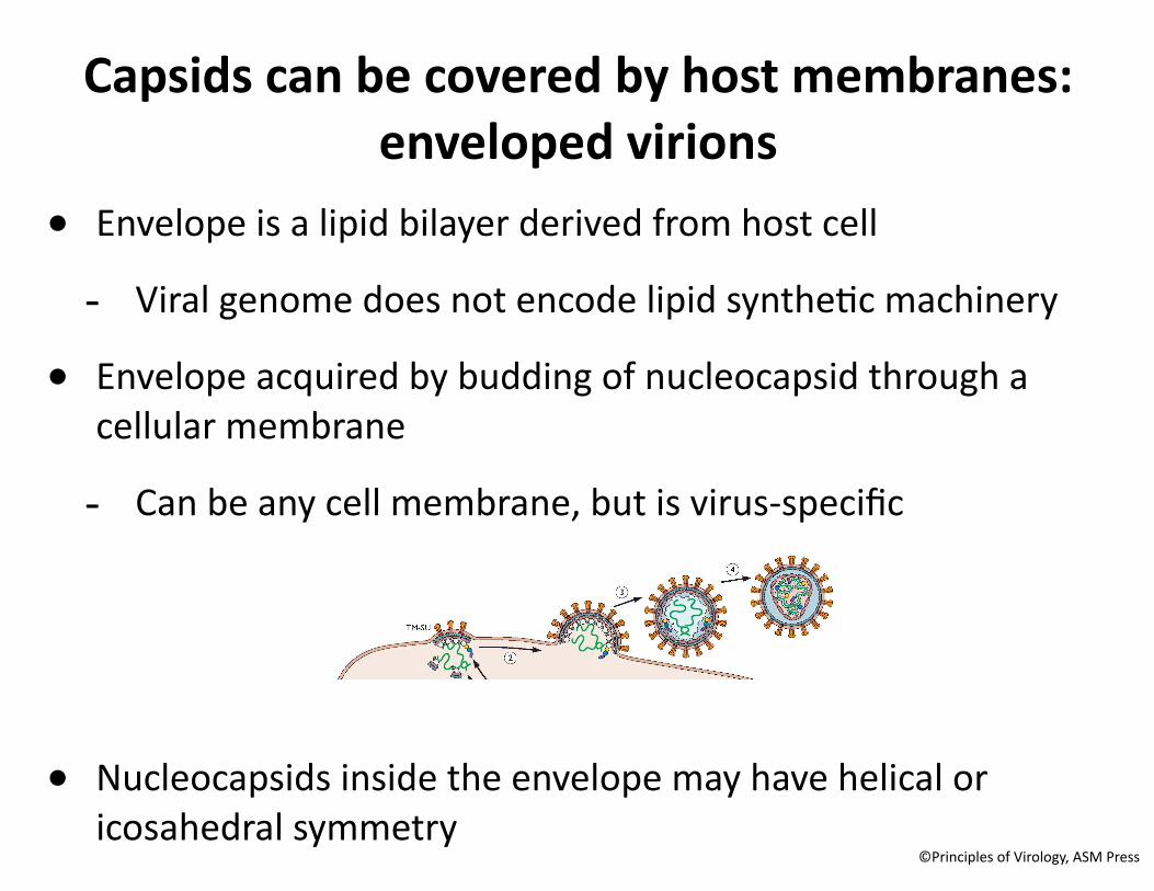
Capsids can be covered by host membranes: enveloped virions
• Envelope is a lipid bilayer derived from host cell
-‐ Viral genome does not encode lipid syntheJc machinery
• Envelope acquired by budding of nucleocapsid through a cellular membrane
-‐ Can be any cell membrane, but is virus-‐specific
• Nucleocapsids inside the envelope may have helical or icosahedral symmetry
©Principles of Virology, ASM Press

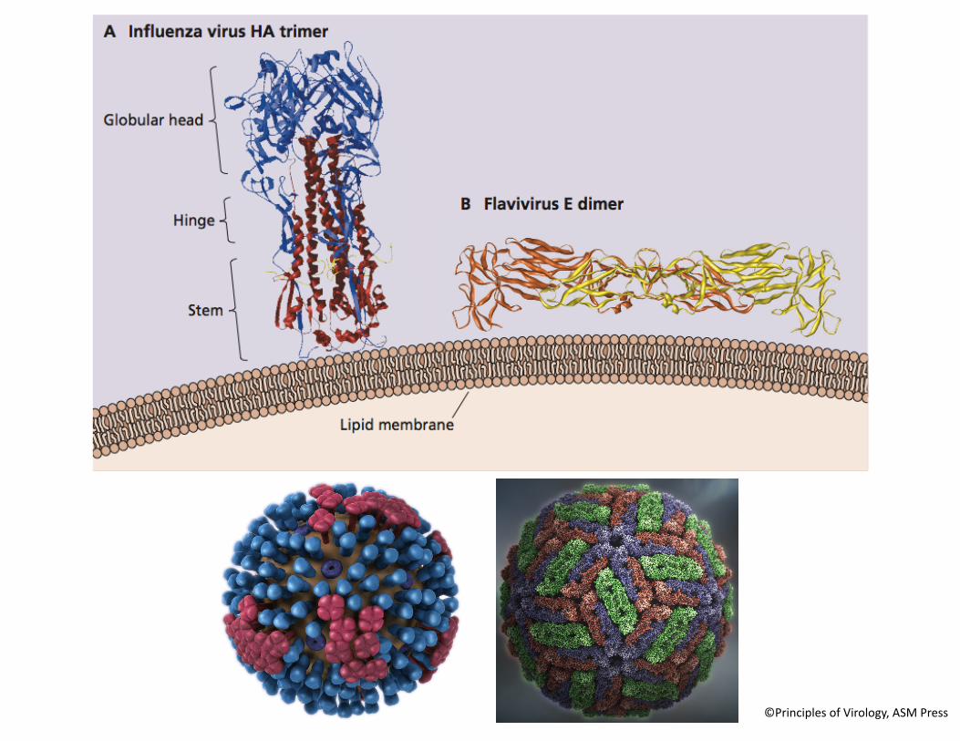
Viral envelope glycoproteins
• Integral membrane glycoproteins
• Ectodomain: a_achment, anJgenic sites, fusion
• Internal domain: assembly
• Oligomeric: spikes
©Principles of Virology, ASM Press
©Principles of Virology, ASM Press

©Principles of Virology, ASM Press
Helical nucleocapsids -‐ unstructured envelopes
Icosahedral nucleocapsids -‐ structured envelopes
Membrane protein (M)
Envelope (E) dimer
Capsid (C)
RNA
5' c
ScEYEnce StudiosPrinciples of Virology, 4eFig. APP01-119-28-14
A
5'5'
3'
2.1 kb
2.4 kb
3.4 kb
0.7kb
DR1
Pre-C
ORF C
ORF X
ORF P
ORF S
Pre-S2Pre-S1
DR2
t
AnA
OH
3'AnA
OH
3'AnA
OH
3'AnA
OH
3'
B
(–)
(+)
5' c
c 5'
c 5'5'
c
PregenomeRNA
3'
42 nm
Viral particleViral DNA
Capsid
Large (L)
Medium (M)
Small (S)
Polymerase(P)
15–25 nm
20 ! 20–200 nm
Incomplete particles
Polymerase (L)
Genomic RNA coatedwith nucleoproteins(N, VP30)
Polymerase associatedprotein (VP35)
Matrix (VP40)
Glycoprotein (GP)
VP24
Hemagglutinin (HA)Neuraminidase (NA)
Ion channel (M2)
Matrix protein (M1)
Lipid bilayerNuclear export protein
(NEP/NS2)Segmented (–) strand RNA coatedwith nucleocapsid protein (NP)
RNA polymerase(PB1, PB2, PA)
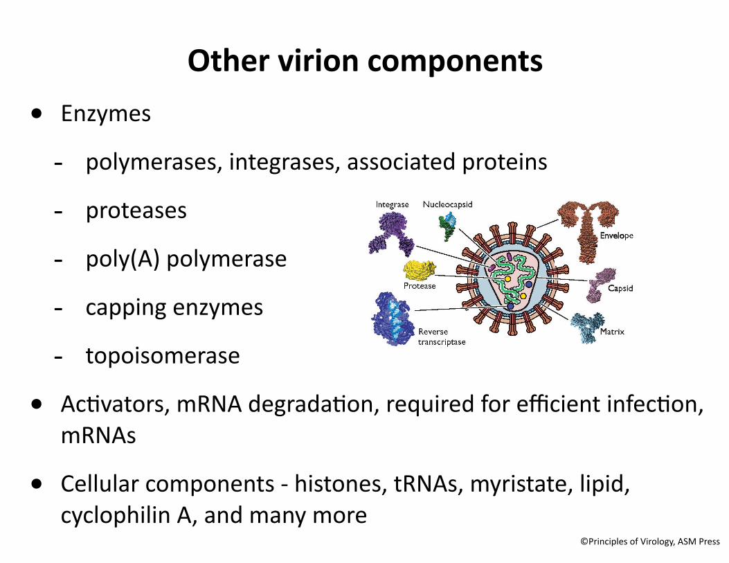
Other virion components• Enzymes
-‐ polymerases, integrases, associated proteins
-‐ proteases
-‐ poly(A) polymerase
-‐ capping enzymes
-‐ topoisomerase
• AcJvators, mRNA degradaJon, required for efficient infecJon, mRNAs
• Cellular components -‐ histones, tRNAs, myristate, lipid, cyclophilin A, and many more
©Principles of Virology, ASM Press
Copyright © 2022 FDOKUMEN