Reversion of CTL escape–variant immunodeficiency viruses in vivo
7
ARTICLES NATURE MEDICINE VOLUME 10 | NUMBER 3 | MARCH 2004 275 Early attempts to design HIV vaccines focused on antibody responses, but the variability of the envelope proteins encoded by circulating virus strains has been a major obstacle to the development of broadly protective antibodies 1–4 . Recent vaccine approaches have focused instead on eliciting CTL responses 5–7 . CTLs are believed to have a major role in modulating the course of HIV disease. The appearance of CTL responses is temporally associated with the resolution of pri- mary viremia in HIV-infected humans 8,9 , and depletion of CD8 + cells results in substantial increases in viral load in SIV-infected macaques 10–12 . It is now well established that CTLs select for viral escape variants that are resistant to immune recognition 13–24 , but the fate of these escape mutations after transmission to new hosts is unclear. Escape from CTL responses can be associated with a loss of immune control of viral replication, in naive and vaccinated hosts 14,16,17,25 . One group has shown that escape-variant viruses can be transmitted from moth- ers to infants who shared the restricting human leukocyte antigen (HLA) element HLA-B27 (ref. 26). However, the majority of sexually transmitted HIV infections occur between individuals who express different HLA class I alleles. No study thus far has shown stable trans- mission of CTL escape viruses between HLA-disparate individuals, although some indirect evidence of such transmission has been reported 24,26 . If CTL escape mutations can be preserved after trans- mission of HIV between HLA class I–disparate individuals, it is likely that HIV escape variants are currently being propagated in popula- tions. Over time, epitopes targeted by CTL-based vaccines could be lost from circulating virus strains, rendering vaccines based on single or consensus strains ineffective. To determine the fate of CTL escape mutations after transmission of HIV, we modeled viral transmission between HLA-similar and HLA- disparate individuals using rhesus macaques infected with CTL escape–variant SIV. We first observed that CTL escape mutations were lost from a heterogeneous SIV isolate upon passage to animals lacking the major histocompatibility complex (MHC) class I determinants to recognize the mutant epitopes. These observations suggested that selection might favor reversion of CTL escape mutations in MHC- disparate hosts. However, it was also possible that we merely detected the stochastic outgrowth of different quasispecies, with no significant selective pressure exerted by MHC class I molecules. To test our hypothesis more rigorously, we challenged macaques with a clonal virus that harbored escape mutations in frequently recognized CTL epitopes, reasoning that any observed evolution of mutant epitope sequences would result from de novo mutations rather than the out- growth of particular quasispecies present in the inoculum. We also compared the fitness of the engineered virus to that of wild-type SIV in vitro, using competitive coculture assays. Our results suggest that there is a previously unappreciated cost to viral fitness associated with escape from CTL responses. 1 Wisconsin National Primate Research Center, Madison, Wisconsin 53715, USA. 2 Department of Pathology and Laboratory Medicine, University of Wisconsin Medical School, Madison, Wisconsin 53706, USA. 3 New England Primate Research Center, Southborough, Massachusetts 01772, USA. 4 California State University, San Marcos, San Marcos, California 92096, USA. 5 La Jolla Institute of Allergy and Immunology, San Diego, California 92121, USA. 6 Northwestern University Medical School, Chicago, Illinois 60611, USA. 7 AIDS Vaccine Program, National Cancer Institute, Frederick, Maryland 21702, USA. 8 Department of Biological Sciences, University of South Carolina, Columbia, South Carolina 29208, USA. Correspondence should be addressed to D.I.W. ([email protected]). Published online 15 February 2004; doi:10.1038/nm998 Reversion of CTL escape–variant immunodeficiency viruses in vivo Thomas C Friedrich 1 , Elizabeth J Dodds 2 , Levi J Yant 2 , Lara Vojnov 2 , Richard Rudersdorf 1 , Candice Cullen 1 , David T Evans 3 , Ronald C Desrosiers 3 , Bianca R Mothé 4 , John Sidney 5 , Alessandro Sette 5 , Kevin Kunstman 6 , Steven Wolinsky 6 , Michael Piatak 7 , Jeffrey Lifson 7 , Austin L Hughes 8 , Nancy Wilson 1 , David H O’Connor 2 & David I Watkins 1,2 Engendering cytotoxic T-lymphocyte (CTL) responses is likely to be an important goal of HIV vaccines. However, CTLs select for viral variants that escape immune detection. Maintenance of such escape variants in human populations could pose an obstacle to HIV vaccine development. We first observed that escape mutations in a heterogeneous simian immunodeficiency virus (SIV) isolate were lost upon passage to new animals. We therefore infected macaques with a cloned SIV bearing escape mutations in three immunodominant CTL epitopes, and followed viral evolution after infection. Here we show that each mutant epitope sequence continued to evolve in vivo, often re-establishing the original, CTL-susceptible sequence. We conclude that escape from CTL responses may exact a cost to viral fitness. In the absence of selective pressure upon transmission to new hosts, these original escape mutations can be lost. This suggests that some HIV CTL epitopes will be maintained in human populations. © 2004 Nature Publishing Group http://www.nature.com/naturemedicine
Transcript of Reversion of CTL escape–variant immunodeficiency viruses in vivo

A R T I C L E S
NATURE MEDICINE VOLUME 10 | NUMBER 3 | MARCH 2004 275
Early attempts to design HIV vaccines focused on antibody responses,but the variability of the envelope proteins encoded by circulatingvirus strains has been a major obstacle to the development of broadlyprotective antibodies1–4. Recent vaccine approaches have focusedinstead on eliciting CTL responses5–7. CTLs are believed to have amajor role in modulating the course of HIV disease. The appearanceof CTL responses is temporally associated with the resolution of pri-mary viremia in HIV-infected humans8,9, and depletion of CD8+ cellsresults in substantial increases in viral load in SIV-infectedmacaques10–12.
It is now well established that CTLs select for viral escape variantsthat are resistant to immune recognition13–24, but the fate of theseescape mutations after transmission to new hosts is unclear. Escapefrom CTL responses can be associated with a loss of immune controlof viral replication, in naive and vaccinated hosts14,16,17,25. One grouphas shown that escape-variant viruses can be transmitted from moth-ers to infants who shared the restricting human leukocyte antigen(HLA) element HLA-B27 (ref. 26). However, the majority of sexuallytransmitted HIV infections occur between individuals who expressdifferent HLA class I alleles. No study thus far has shown stable trans-mission of CTL escape viruses between HLA-disparate individuals,although some indirect evidence of such transmission has beenreported24,26. If CTL escape mutations can be preserved after trans-mission of HIV between HLA class I–disparate individuals, it is likely
that HIV escape variants are currently being propagated in popula-tions. Over time, epitopes targeted by CTL-based vaccines could belost from circulating virus strains, rendering vaccines based on singleor consensus strains ineffective.
To determine the fate of CTL escape mutations after transmission ofHIV, we modeled viral transmission between HLA-similar and HLA-disparate individuals using rhesus macaques infected with CTLescape–variant SIV. We first observed that CTL escape mutations werelost from a heterogeneous SIV isolate upon passage to animals lackingthe major histocompatibility complex (MHC) class I determinants torecognize the mutant epitopes. These observations suggested thatselection might favor reversion of CTL escape mutations in MHC-disparate hosts. However, it was also possible that we merely detectedthe stochastic outgrowth of different quasispecies, with no significantselective pressure exerted by MHC class I molecules. To test ourhypothesis more rigorously, we challenged macaques with a clonalvirus that harbored escape mutations in frequently recognized CTLepitopes, reasoning that any observed evolution of mutant epitopesequences would result from de novo mutations rather than the out-growth of particular quasispecies present in the inoculum. We alsocompared the fitness of the engineered virus to that of wild-type SIVin vitro, using competitive coculture assays. Our results suggest thatthere is a previously unappreciated cost to viral fitness associated withescape from CTL responses.
1Wisconsin National Primate Research Center, Madison, Wisconsin 53715, USA. 2Department of Pathology and Laboratory Medicine, University of Wisconsin MedicalSchool, Madison, Wisconsin 53706, USA. 3New England Primate Research Center, Southborough, Massachusetts 01772, USA. 4California State University, SanMarcos, San Marcos, California 92096, USA. 5La Jolla Institute of Allergy and Immunology, San Diego, California 92121, USA. 6Northwestern University MedicalSchool, Chicago, Illinois 60611, USA. 7AIDS Vaccine Program, National Cancer Institute, Frederick, Maryland 21702, USA. 8Department of Biological Sciences,University of South Carolina, Columbia, South Carolina 29208, USA. Correspondence should be addressed to D.I.W. ([email protected]).
Published online 15 February 2004; doi:10.1038/nm998
Reversion of CTL escape–variant immunodeficiencyviruses in vivoThomas C Friedrich1, Elizabeth J Dodds2, Levi J Yant2, Lara Vojnov2, Richard Rudersdorf1, Candice Cullen1,David T Evans3, Ronald C Desrosiers3, Bianca R Mothé4, John Sidney5, Alessandro Sette5, Kevin Kunstman6,Steven Wolinsky6, Michael Piatak7, Jeffrey Lifson7, Austin L Hughes8, Nancy Wilson1, David H O’Connor2 &David I Watkins1,2
Engendering cytotoxic T-lymphocyte (CTL) responses is likely to be an important goal of HIV vaccines. However, CTLs select forviral variants that escape immune detection. Maintenance of such escape variants in human populations could pose an obstacleto HIV vaccine development. We first observed that escape mutations in a heterogeneous simian immunodeficiency virus (SIV)isolate were lost upon passage to new animals. We therefore infected macaques with a cloned SIV bearing escape mutations inthree immunodominant CTL epitopes, and followed viral evolution after infection. Here we show that each mutant epitopesequence continued to evolve in vivo, often re-establishing the original, CTL-susceptible sequence. We conclude that escapefrom CTL responses may exact a cost to viral fitness. In the absence of selective pressure upon transmission to new hosts, theseoriginal escape mutations can be lost. This suggests that some HIV CTL epitopes will be maintained in human populations.
©20
04 N
atur
e P
ublis
hing
Gro
up
http
://w
ww
.nat
ure.
com
/nat
urem
edic
ine

A R T I C L E S
276 VOLUME 10 | NUMBER 3 | MARCH 2004 NATURE MEDICINE
RESULTSEscape mutations are lost from heterogeneous SIV in vivoTo determine the fate of CTL escape mutations in transmitted virusvariants, we examined the evolution of variant epitope sequences infive animals infected with a heterogeneous biological isolate, SIVppm(ref. 19), that is derived from the well-characterized pathogenic clone,SIVmac239 (ref. 27 and Supplementary Fig. 1 online). The SIVppminoculum sequence encodes amino acid substitutions in CTL epitopesrestricted by Mamu-A*01 (Tat28–35SL8 (abbreviated Tat SL8); aminoacid sequence STPESANL), Mamu-A*02 (Nef159–167YY9 (Nef YY9);YTSGPGIRY) and Mamu-B*17 (Nef165–173IW9 (Nef IW9); IRYPKT-
FGW). Two of the animals infected with this isolate were Mamu-B*17-positive, one was Mamu-A*01-positive, and none expressed Mamu-A*02.
We first sequenced the overlapping Mamu-A*02-restricted (NefYY9) and Mamu-B*17-restricted (Nef IW9) epitopes. In the two ani-mals that expressed neither of these restricting elements, the epitopesequences of both Nef YY9 and Nef IW9 evolved toward the SIVmac239wild-type sequence (Fig. 1a). In contrast, in the two animals thatexpressed Mamu-B*17 but not Mamu-A*02, further escape mutationwas seen in the B*17-restricted Nef IW9 epitope, whereas the A*02-restricted Nef YY9 epitope again evolved toward wild type (Fig. 1b). We
Figure 1 Sequence variation in macaques challenged with SIVppm, abiological isolate with amino acid replacements in CTL epitopesrestricted by common macaque MHC class I molecules Mamu-A*01 (TatSL8), Mamu-A*02 (Nef YY9) and Mamu-B*17 (Nef IW9). (a) In animalslacking both Mamu-A*02 and Mamu-B*17, epitope sequences of bothNef YY9 (green shading) and Nef IW9 (blue letters) evolve toward wildtype. p.i., post infection. (b) In animals expressing Mamu-B*17 but notMamu-A*02, only the region of Nef YY9 that does not overlap the B*17-restricted Nef IW9 epitope evolves toward wild type. Variants in theB*17-restricted epitope continue to evolve. (c) Oscillation occurs in tatcodon 35 (C-terminal anchor of Tat SL8) in Mamu-A*01-negativeanimals infected with SIVppm. In contrast, there is further evolution awayfrom SIVmac239 epitope sequence in Mamu-A*01-positive animal(95024) infected with SIVppm. Tat SL8 sequence is shaded in yellow.
©20
04 N
atur
e P
ublis
hing
Gro
up
http
://w
ww
.nat
ure.
com
/nat
urem
edic
ine

A R T I C L E S
NATURE MEDICINE VOLUME 10 | NUMBER 3 | MARCH 2004 277
next examined the sequences of Tat SL8. The sequence of tat in theinoculum was not available, but it is likely that the SIVppm inoculumcontained variation in the Mamu-A*01-restricted Tat SL8 epitope, asall sequences of this region isolated from a Mamu-A*01-negative ani-mal 3 weeks after infection differed from those of SIVmac239 within theepitope. As SIVppm infection progressed in Mamu-A*01-negative ani-mals, we detected oscillation in tat codon 35, the C-terminal anchor ofthe epitope, but there was no reversion to wild-type sequences (Fig. 1c).We observed three different residues (glutamine, arginine and trypto-phan) in tat codon 35 in Mamu-A*01-negative macaques, none ofwhich is predicted to allow epitope peptide binding to Mamu-A*01.However, Tat SL8 sequences evolved even further away from SIVmac239in Mamu-A*01-positive animal 95024, with leucine and histidinereplacing proline and the wild-type serine by late chronic infection.Together, these observations raise the possibility that escape mutantscontinue to evolve in MHC-disparate hosts and that, under certain circumstances, wild-type epitope sequences can be re-established.
Construction of a cloned CTL escape virusTo study the transmission and evolution of viral escape variants in vivowith greater rigor, we constructed a molecularly cloned mutant SIVthat differed from SIVmac239 at only three defined epitopes recognizedby high-frequency CTLs. The molecularly cloned virus enabled us tostudy the effects of CTL escape mutations in a viral genetic back-ground free of other variations that might affect viral replicationand/or pathogenicity in vivo. Moreover, because infections were initi-ated with a clonal sequence, any variation we detected would have toarise de novo within the infected animal, rather than reflecting the out-growth of one or more members of a complex population of quasi-species present during initial infection.
We analyzed epitopes bound by two common Indian rhesusmacaque MHC class I molecules, Mamu-A*01 and Mamu-B*17. Wehave previously shown that macaques that express both Mamu-A*01and B*17 are predisposed to unusual control of SIVmac239 replication,and produce three high-frequency (immunodominant) CTLresponses during acute infection28. The epitopes recognized by theseCTLs are derived from structural (Gag) and nonstructural (Tat and
Nef) proteins, and accumulate escape mutations with varying kinetics.CTLs specific for Gag181–189CM9 (abbreviated Gag CM9; sequenceCTPYDINQM) and Tat SL8 are immunodominant during acute infec-tion of Mamu-A*01-positive macaques29. Gag CM9–specific CTLsselect for escape variants intermittently and only during chronic infec-tion23,30, whereas Tat SL8–specific CTLs select for rapid viral escapewithin 4 weeks of infection20. CTLs recognizing Nef IW9 are immun-odominant in Mamu-B*17-positive macaques and select for escapevariants with intermediate kinetics, ∼ 16 weeks after infection23.
The consensus escape mutation in the Gag CM9 epitope encoded athreonine-to-alanine substitution occurring at the secondary anchorposition of the epitope (T182A), and reduced peptide binding to theMamu-A*01 molecule by tenfold (Table 1). In late-stage virussequences, the mutation encoding T182A was also associated with twonucleotide changes outside the epitope, each of which encoded aisoleucine-to-valine substitution (I161V and I206V). We have shownthat these compensatory substitutions probably restore fitness to theT182A escape virus, and we therefore included them in our mutagene-sis strategy30,31.
The consensus escape sequence for Tat SL8 had substitutions inpositions 1 and 8 (S28P and L35Q). Leu35 is the C-terminal anchor ofthe Tat SL8 peptide, and consequently this substitution essentiallyabrogates the peptide’s binding to Mamu-A*01 (Table 1). Consensusescape substitutions for Nef IW9 were mapped to positions 1 and 6(I165T and T170I); this sequence was also observed in a previousstudy19. These substitutions reduced binding to Mamu-B*17 by >90%(Table 1), but because position 6 is thought to interact with the T-cellreceptor, this variant may also affect T-cell recognition.
Table 1 Binding of wild-type and variant peptides to MHC class I
Epitope Sequence MHC class I IC50 (nM) % reduction
Gag CM9 Mamu-A*01 22 —
Mutant Mamu-A*01 354 94
Tat SL8 Mamu-A*01 43 —
Mutant Mamu-A*01 25,535 99.9
Nef IW9 Mamu-B*17 7.6 —
Mutant Mamu-B*17 339 98
Peptide binding to Mamu-A*01 or Mamu-B*17 molecules was assessed using epitopepeptides or variants in competition assays with radiolabeled reference peptides, asdescribed previously37,38. Reported are the concentration of epitope peptide at whichbound radioactivity is reduced by 50% (IC50) and, for escape variants, percentreduction in binding with respect to wild-type peptide. Residues that differ betweenwild-type ans 3x SIV are underlined.
Figure 2 Reduced fitness of 3x SIV in vitro. (a) 3x SIV lags behind wild-type(wt) virus in early replication kinetics. Results shown are from three separatetrials with each virus inoculum. (b) Wild-type virus outgrows 3x SIV wheninoculated in equal amounts. Wild-type vRNA is expressed as percentage oftotal copies of vRNA in each sample. Results are representative ofindependent triplicate experiments. Three independent quantitative RT-PCR/SSP reactions were performed for each experiment.
©20
04 N
atur
e P
ublis
hing
Gro
up
http
://w
ww
.nat
ure.
com
/nat
urem
edic
ine
A R T I C L E S
278 VOLUME 10 | NUMBER 3 | MARCH 2004 NATURE MEDICINE
We sequenced the complete genomes of viruses isolated fromplasma at the time of necropsy in 35 animals (D.H.O., K. Krebs,E.J.D. and D.I.W., unpublished observations). In doing so, we did notdetect mutations outside these epitopes occurring in associationwith intraepitopic variation, except for those mutations that wereassociated with escape in Gag CM9 as discussed above. We thereforeconclude that it is unlikely that we could not detect other compensa-tory substitutions that might enhance the fitness of viral escape vari-ants in vivo.
Triple-mutant virus shows reduced fitness in vitroWe next produced a clonal virus encoding escape mutations in allthree epitopes (3x SIV) and characterized its in vitro growth prop-erties. When the same dose of wild-type or 3x SIV was used toinfect macaque peripheral blood mononuclear cells (PBMCs), 3xSIV replication lagged behind that of wild-type SIV in the first 96 h. After 5 d in culture, however, the two viruses attained similartiters (Fig. 2a). These results suggest that 3x SIV replication is lessefficient than that of the wild-type virus. To compare the fitnesslevels of the two viruses more directly, we devised a direct viralcompetition assay in which macaque PBMCs were infected withmixtures of the two virus stocks, totaling 10 ng of p27. Replicationof each virus species in the culture was detected using a quantita-tive RT-PCR technique with sequence-specific primers (SSP) thatspecifically targeted the wild-type nef epitope sequence. Becausethe quantitative RT-PCR/SSP reaction was inherently less efficientthan the nonselective reaction, we could not effectively discrimi-nate between wild-type and 3x SIV in the first 48 h of infection,when total virus concentrations were low. However, the direct com-
petition assays showed that after 5 d in culture, wild-type SIV hadcompletely outgrown the mutant when the inocula consisted ofequal amounts of the two viruses (Fig. 2b). In contrast, 3x SIVremained predominant throughout the culture period when theinoculum ratios favored it, indicating that 3x SIV can outgrowwild-type virus, but only when inoculated in excess. We thereforeconcluded that growth of 3x SIV is impaired compared with wild-type SIVmac239.
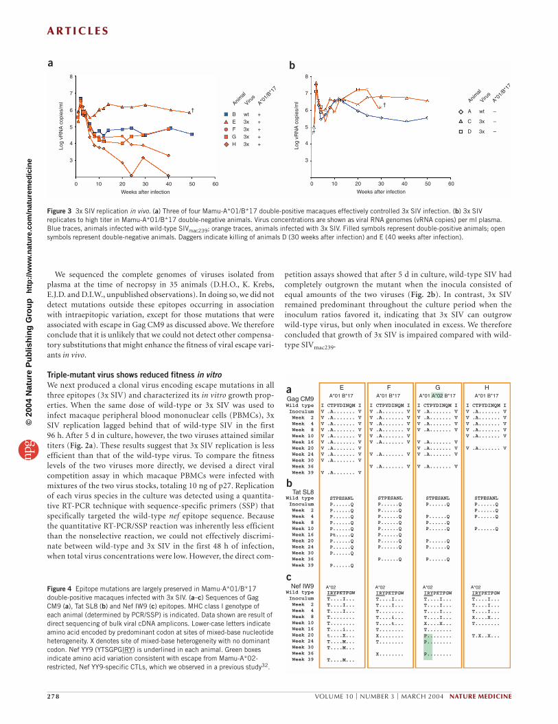
Figure 3 3x SIV replication in vivo. (a) Three of four Mamu-A*01/B*17 double-positive macaques effectively controlled 3x SIV infection. (b) 3x SIVreplicates to high titer in Mamu-A*01/B*17 double-negative animals. Virus concentrations are shown as viral RNA genomes (vRNA copies) per ml plasma. Blue traces, animals infected with wild-type SIVmac239; orange traces, animals infected with 3x SIV. Filled symbols represent double-positive animals; opensymbols represent double-negative animals. Daggers indicate killing of animals D (30 weeks after infection) and E (40 weeks after infection).
Figure 4 Epitope mutations are largely preserved in Mamu-A*01/B*17double-positive macaques infected with 3x SIV. (a–c) Sequences of GagCM9 (a), Tat SL8 (b) and Nef IW9 (c) epitopes. MHC class I genotype ofeach animal (determined by PCR/SSP) is indicated. Data shown are result ofdirect sequencing of bulk viral cDNA amplicons. Lower-case letters indicateamino acid encoded by predominant codon at sites of mixed-base nucleotideheterogeneity. X denotes site of mixed-base heterogeneity with no dominantcodon. Nef YY9 (YTSGPGIRY) is underlined in each animal. Green boxesindicate amino acid variation consistent with escape from Mamu-A*02-restricted, Nef YY9-specific CTLs, which we observed in a previous study32.
©20
04 N
atur
e P
ublis
hing
Gro
up
http
://w
ww
.nat
ure.
com
/nat
urem
edic
ine

A R T I C L E S
NATURE MEDICINE VOLUME 10 | NUMBER 3 | MARCH 2004 279
3x SIV mutations are preserved in MHC-similar animalsTo assess the in vivo replicative potential of 3x SIV and model the fateof epitope mutations in HLA-similar hosts, we infected four Mamu-A*01/B*17 double-positive macaques with 3x SIV. As controls, we alsoinfected one Mamu-A*01/B*17 double-negative animal (A) and onedouble-positive animal (B) with the same dose of wild-type SIVmac239.Both viruses reached titers greater than 106 viral RNA (vRNA) copiesper ml plasma in every animal. Notably, three of four double-positivemacaques maintained 3x SIV viral loads below 100,000 vRNA copiesper ml in the chronic phase (Fig. 3a), in contrast to double-negativeanimals, whose chronic-phase virus loads were at least tenfold higher(Fig. 3b). However, CTL responses directed against the mutant epi-topes were barely detectable in these animals throughout infection(data not shown). Furthermore, the engineered escape mutations in 3xSIV Gag and Tat were preserved throughout our study period in alldouble-positive animals (Fig. 4a,b). In contrast, the mutant Nef IW9sequence continued to evolve in each of these animals (Fig. 4c). Thisresult agrees with previously reported observations of maternal-to-fetal transmission of an escape variant of HIV-1 (ref. 26). In that study,HLA-B27-positive infants did not recognize a variant HLA-B27-restricted epitope present in the virus transmitted by their B27-positive mothers, and the transmitted escape variant sequence per-sisted for several years.
Reversion of 3x SIV epitopes in MHC-disparate animalsBecause HIV is most often transmitted between HLA class I–disparateindividuals, we next inoculated two Mamu-A*01/B*17 double-negativemacaques (animals C and D) with 3x SIV. Peak 3x SIV viremia in theseanimals was greater than 107 copies per ml plasma, similar to thatobserved in the Mamu-A*01/B*17-double-negative animal (A)infected with wild-type SIVmac239 (Fig. 3b). Animals A and C had clini-cal courses typical of macaques previously infected with SIVmac239 inour laboratory. In contrast, animal D’s course was similar to that ofrapid progressors, and showed severe thrombocytopenia and a CD4+
cell count <500/µl when killed 30 weeks after infection. However,although animal C survived much longer, 3x SIV caused persistentlyhigh viremia (>106 copies/ml) during the chronic phase of infection inboth double-negative macaques challenged with this stock.
3x SIV epitope sequences evolved rapidly in the double-negativeanimals. The wild-type Gag CM9 epitope sequence was completely re-established in animals C and D by 16 weeks after infection (Fig. 5a).The sequence of the Nef IW9 epitope also evolved toward wild typeduring the first 16 weeks of infection in each double-negative macaque(Fig. 5c). Coincident escape from a Mamu-A*02-restricted CTLresponse to an overlapping epitope (Nef YY9; ref. 32) also resulted innew substitutions in this region (Fig. 5b). Thus, there seems to be astrong selective advantage favoring the wild-type sequences in boththe Nef IW9 and Gag CM9 epitopes in vivo, reflecting the reduced fit-ness of 3x SIV we observed in vitro. Indeed, our attempts to produceviruses harboring the single mutation encoding T182A (CAPYD-INQM) were unsuccessful, suggesting that without the compensatorysubstitutions flanking the epitope, this mutation exacts a catastrophiccost to viral replication. Recent experimental evidence from our labo-
ratory30 and others31,33,34 supports this conclusion. In contrast, muta-tions introduced in the rapidly escaping Tat SL8 epitope were pre-served in animal D throughout the study period (Fig. 5c), and acomplex population of sequences was detected in this epitope in ani-mal C beginning 30 weeks after infection. The composition of Tat SL8sequences in animal C late in infection was reminiscent of that foundin Mamu-A*01-negative animals infected with SIVppm (Fig. 1c).Escape mutations in Tat SL8 may also reduce fitness in vivo, but arelikely to be better tolerated than mutations affecting the Nef IW9 orGag CM9 epitopes. Together, our results suggest that mutations inCTL epitopes can have moderate to severe negative effects on viralreplicative fitness.
DISCUSSIONWe have shown that SIV CTL escape variants can revert to wild-typesequences after infecting new hosts possessing disparate MHC class Ialleles. We also observed an apparent cost to replicative fitness in aSIVmac239 variant engineered to contain CTL escape mutations. Thesefindings led us to hypothesize that CTL pressure often selects forescape mutations in infected individuals, but some of these escapevariant sequences will not be stably propagated as viruses circulate inHLA-diverse populations. In our study, the heterogeneous SIVppm iso-late harbored mutations in Mamu-A*02- and B*17-restricted CTLepitopes that evolved toward wild-type SIVmac239 sequences inmacaques that did not express these molecules. To rigorously test thehypothesis that CTL escape mutations can reduce viral fitness, weengineered a clonal virus encoding three CTL escape variants.Replication of this cloned escape mutant virus, 3x SIV, was reduced invitro with respect to the wild-type virus, indicating that incorporation
Figure 5 Two of three 3x SIV epitope sequences evolve toward wild type inMamu-A*01/B*17 double-negative macaques. (a–c) Sequences of Gag CM9(a), Tat SL8 (b) and Nef IW9 (c) epitopes. Black boxes around animal namesindicate Mamu-A*01/B*17 double-negative. Data are presented as in Figure 4. Colored borders indicate strain of SIV used to challenge each animal(blue, wild-type SIV; orange, 3x SIV). Replacements consistent with escapefrom Mamu-A*02-restricted epitope that overlaps Nef IW9 are boxed in green.
©20
04 N
atur
e P
ublis
hing
Gro
up
http
://w
ww
.nat
ure.
com
/nat
urem
edic
ine

A R T I C L E S
280 VOLUME 10 | NUMBER 3 | MARCH 2004 NATURE MEDICINE
of one or more epitope mutations into the SIVmac239 sequence reducedviral fitness. Indeed, the mutation in Gag CM9 in particular seems tohave a profound negative effect on viral fitness30,31,33,34. Accordingly,wild-type Gag CM9 and Nef IW9 epitope sequences were re-established in Mamu-A*01- and B*17-negative animals within 16 weeks of infection with the molecularly cloned 3x SIV. In contrast,the mutant Tat SL8 epitope sequence of 3x SIV did not revert to wildtype in the absence of immune selection, but instead showed somesequence oscillation, suggesting that variation is better tolerated in thisepitope. Escape mutations in Tat SL8 underwent similar oscillations inMamu-A*01-negative animals infected with SIVppm, in agreementwith this hypothesis.
It is likely that escape variants will only outgrow the wild-type viruswhen the benefit of escape from a particular CTL response outweighsthe loss of fitness. However, it is likely that multiple interacting deter-minants affect the selection of CTL escape viruses. We have previouslyobserved that rapidly escaping CTL epitopes tend to be the targets ofparticularly high-avidity CTLs23. These epitopes also cluster in regula-tory and accessory proteins. Variation in these sequences may there-fore incur lower fitness costs than variations in epitopes derived fromstructurally or functionally conserved regions. Furthermore, escapefrom CTLs that target particularly conserved regions may involve sev-eral substitutions, including extraepitopic substitutions that compen-sate for the loss of fitness resulting from escape. The outgrowth of CTLescape variants therefore probably represents the additive effects ofseveral virological and immunological parameters, including CTL fre-quency and efficacy, the number of nucleotide substitutions requiredfor effective escape, and the costs of these substitutions to fitness andviral replication. We expect this constellation of factors to be differentfor each CTL epitope; their differential effects will determine whetherescape mutations will be lost or preserved upon transmission to newhosts.
It is heartening to believe that epitopes targeted by effective CTLsmay not be entirely lost from circulating virus sequences as the HIVepidemic progresses. Successful vaccines for HIV will probably need tostimulate CTL responses. Unfortunately, the propensity of CTLs toselect for escape variant viruses, and the possibility that these variantscan be propagated in human populations, represent enormous poten-tial obstacles to the efficacy of CTL-based vaccines. Our results showthat escape mutations can revert to CTL-susceptible sequences whenescape variant viruses are transmitted among MHC-disparate individ-uals, but we must be prudent in extending these results by analogy tohumans infected with HIV. Set-point viremia with SIVmac239 in Indianrhesus macaques is typically tenfold higher than that seen in HIV-infected humans, which may affect the rate of viral evolution in vivo.Moreover, macaques expressing Mamu-A*01, especially in associationwith Mamu-B*17, seem to be predisposed to unusually effective con-trol of SIVmac239 infection28. As a result, analogous studies in humanswill be required to determine the extent to which HIV CTL epitopesequences will be preserved in circulating viruses.
Our results may also proffer criteria by which the efficacy of particu-lar CTL responses can be assessed. Transmitted CTL escape mutationsmay evolve at different rates in the absence of immune selection innew hosts, as the regions from which they are derived are under differ-ent constraints. CTL-based vaccines may thus be most effective if theyinclude epitopes in which escape mutations revert rapidly after trans-mission into new hosts. This strategy offers at least two potential ben-efits over current approaches. First, rapid re-establishment ofCTL-susceptible sequences suggests that targeted epitopes will persistin circulating virus sequences. Second, such rapid evolution alsoimplies a particularly large fitness cost associated with those escape
mutations. Viral escape from CTL targeting of such regions may there-fore involve particularly drastic reductions in fitness, ‘crippling’ thereplicating virus population and facilitating control of infection.Vaccine-induced CTL responses that drive the evolution of viral vari-ants with low replicative fitness could offer hope for immune controland amelioration of HIV disease.
METHODSAnimals. Indian rhesus macaques (Macaca mulatta) from the WisconsinNational Primate Research Center colony were typed for MHC class I allelesMamu-A*01, A*02, A*08, A*11, B*01, B*03, B*04, B*17 and B*29 by sequence-specific PCR35. Animals were cared for according to regulations of the Guide forthe Care and Use of Laboratory Animals of the National Research Council, asapproved by the University of Wisconsin Institutional Animal Care and UseCommittee.
Viruses and infections. Animals were infected with the heterogeneous isolateSIVppm in a previous study19. The passage history of SIVppm has not been wellcharacterized, but sequencing and phylogenetic analyses suggest that it is theresult of a low number of passages of cloned SIVmac239 through macaquesexpressing common MHC class I molecules (Supplementary Fig. 1 online).Wild-type and 3x SIVmac239 were produced by transfection of Vero cells withplasmid DNA encoding proviral sequences. CEMx174 cells were added to theVero cultures 1 d after transfection and removed to flasks 3 d after transfection.Viruses were then amplified on CEMx174 cells, and cell-free supernatant wascollected 2 d after peak syncytium formation. Direct sequencing of RT-PCRamplicons representing all 3x SIV open reading frames (as described previ-ously23) confirmed that this stock harbored only the engineered nucleotide sub-stitutions and was otherwise identical to SIVmac239 (data not shown). Animalswere challenged intravenously with 100 TCID50 of either virus stock. Plasmavirus load was determined by the branched-chain DNA assay (BayerDiagnostics).
Viral competition assay. Two million phytohemagglutinin-stimulated rhesusmacaque PBMCs were infected in triplicate, as described above, with 10 ng p27total virus containing wild-type SIVmac239 or 3x SIV alone or together. Cultureswere maintained for 5 d, and supernatants were sampled every 24 h for theduration of the assay. Results shown in Figure 2b are representative of two sep-arate assays using two independently prepared stocks of 3x SIV. We determinedthe proportion of wild-type vRNA in each sample using a modification of apublished method36. Total vRNA in supernatants was measured using a nonse-lective quantitative PCR assay with primers 9700-F (5′-TAAAA-GAAAAGGGGGGACTG-3′) and nef-R (5′-CCTCATCCTCCTGTGC-3′). Forselective amplification of wild-type SIVmac239 vRNA in the presence of 3x SIVvRNA, we used primer 9700-F with a sequence-specific primer, SIV 9845-R (5′-AAATGTCTTTGGGTATCTAA-3′), targeting the two-nucleotide polymor-phisms in the nef coding region. Reverse transcription and amplification weredone in a single reaction using the Roche SYBR Green RNA Master Probes kit.Reaction conditions were optimized for primer concentration, MnOAc con-centration and annealing temperature. The sequence-specific primer 9845-Rwould not anneal to 3x SIV vRNA above 59 °C at any concentration of primeror MnOAc tested. Reactions were done in the Roche Lightcycler, using the fol-lowing cycling parameters: 61 °C for 25 min (at which temperature wild-typevRNA is selectively reverse transcribed with primer 9845-R), 95 °C for 30 s, and45 cycles of 95 °C for 1 s, 58 °C for 10 s, 72 °C for 10 s and 77 °C for 2 s.Fluorescence was measured at 71 °C at the end of every PCR cycle. Melt curveswere obtained for every sample after PCR. The number of cycles required toreach threshold fluorescence was determined, and the number of vRNA copiesinitially present was calculated by extrapolation onto a standard curve preparedfrom synthetic dilutions of wild-type vRNA. All reactions were done in tripli-cate, with the mean of the values used for calculations. Our threshold of detec-tion with this method was 0.08% wild-type vRNA in 2 million copies of 3x SIVvRNA (data not shown).
Viral sequence analysis. Amplification of viral cDNA by RT-PCR was done asdescribed previously23, using viral genomic RNA isolated from animal plasmaor tissue culture supernatants. We used three previously described primer
280 VOLUME 10 | NUMBER 3 | MARCH 2004 NATURE MEDICINE
©20
04 N
atur
e P
ublis
hing
Gro
up
http
://w
ww
.nat
ure.
com
/nat
urem
edic
ine
A R T I C L E S
NATURE MEDICINE VOLUME 10 | NUMBER 3 | MARCH 2004 281
pairs23 to amplify regions surrounding the sequences encoding Gag CM9, TatSL8 and Nef IW9. PCR products were directly sequenced on an ABI 377 or ABI3730 automated sequencer. Mixed bases and nucleotide replacements weredetected in sequence data using Sequencher and MacVector 7.0 software.
Note: Supplementary information is available on the Nature Medicine website.
ACKNOWLEDGMENTSWe gratefully acknowledge J. Helgeland, J. Mitchen, C. Bunger, K. Vielhuber and E. Rakasz; I. Bolton and the veterinarians and technicians of the WNPRC; and T. Jacoby and W. Rehrauer for their work in support of this study. We also thank W. Rehrauer for critical review of this manuscript. This work was supported byNational Institutes of Health grants RO1-AI-46366, RO1-AI-49120 and RO1-AI-52056 to D.I.W., and P51 RR001676-43 to the WNPRC. D.I.W. is an ElizabethGlaser Scientist. A.S. is supported by NIH-NIAID contract NO1-AI-95362.
COMPETING INTERESTS STATEMENTThe authors declare that they have no competing financial interests.
Received 30 October 2003; accepted 26 January 2004Published online at http://www.nature.com/naturemedicine/
1. Burton, D.R. Antibodies, viruses and vaccines. Nat. Rev. Immunol. 2, 706–713(2002).
2. Ho, D.D. & Huang, Y. The HIV-1 vaccine race. Cell 110, 135–138 (2002).3. Richman, D.D., Wrin, T., Little, S.J. & Petropoulos, C.J. Rapid evolution of the neu-
tralizing antibody response to HIV type 1 infection. Proc. Natl. Acad. Sci. USA 100,4144–4149 (2003).
4. Wei, X. et al. Antibody neutralization and escape by HIV-1. Nature 422, 307–312(2003).
5. McMichael, A.J. & Rowland-Jones, S.L. Cellular immune responses to HIV. Nature410, 980–987 (2001).
6. Walker, B.D. & Korber, B.T. Immune control of HIV: the obstacles of HLA and viraldiversity. Nat. Immunol. 2, 473–475 (2001).
7. McMichael, A.J. & Hanke, T. HIV vaccines 1983-2003. Nat. Med. 9, 874–880(2003).
8. Borrow, P., Lewicki, H., Hahn, B.H., Shaw, G.M. & Oldstone, M.B. Virus-specificCD8+ cytotoxic T-lymphocyte activity associated with control of viremia in primaryhuman immunodeficiency virus type 1 infection. J. Virol. 68, 6103–6110 (1994).
9. Koup, R.A. et al. Temporal association of cellular immune responses with the initialcontrol of viremia in primary human immunodeficiency virus type 1 syndrome. J. Virol. 68, 4650–4655 (1994).
10. Jin, X. et al. Dramatic rise in plasma viremia after CD8+ T cell depletion in simianimmunodeficiency virus-infected macaques. J. Exp. Med. 189, 991–998 (1999).
11. Matano, T. et al. Administration of an anti-CD8 monoclonal antibody interferes withthe clearance of chimeric simian/human immunodeficiency virus during primaryinfections of rhesus macaques. J. Virol. 72, 164–169 (1998).
12. Schmitz, J.E. et al. Control of viremia in simian immunodeficiency virus infection byCD8+ lymphocytes. Science 283, 857–860 (1999).
13. Phillips, R.E. et al. Human immunodeficiency virus genetic variation that canescape cytotoxic T cell recognition. Nature 354, 453–459 (1991).
14. Koenig, S. et al. Transfer of HIV-1-specific cytotoxic T lymphocytes to an AIDSpatient leads to selection for mutant HIV variants and subsequent disease progres-sion. Nat. Med. 1, 330–336 (1995).
15. Wolinsky, S.M. et al. Adaptive evolution of human immunodeficiency virus-type 1during the natural course of infection. Science 272, 537–542 (1996).
16. Borrow, P. et al. Antiviral pressure exerted by HIV-1-specific cytotoxic T lymphocytes
(CTLs) during primary infection demonstrated by rapid selection of CTL escapevirus. Nat. Med. 3, 205–211 (1997).
17. Goulder, P.J. et al. Late escape from an immunodominant cytotoxic T-lymphocyteresponse associated with progression to AIDS. Nat. Med. 3, 212–217 (1997).
18. Price, D.A. et al. Positive selection of HIV-1 cytotoxic T lymphocyte escape variantsduring primary infection. Proc. Natl. Acad. Sci. USA 94, 1890–1895 (1997).
19. Evans, D.T. et al. Virus-specific cytotoxic T-lymphocyte responses select for amino-acid variation in simian immunodeficiency virus env and Nef. Nat. Med. 5,1270–1276 (1999).
20. Allen, T.M. et al. Tat-specific cytotoxic T lymphocytes select for SIV escape variantsduring resolution of primary viraemia. Nature 407, 386–390 (2000).
21. Kelleher, A.D. et al. Clustered mutations in HIV-1 gag are consistently required forescape from HLA-B27-restricted cytotoxic T lymphocyte responses. J. Exp. Med.193, 375–386 (2001).
22. McMichael, A. T cell responses and viral escape. Cell 93, 673–676 (1998).23. O’Connor, D.H. et al. Acute phase cytotoxic T lymphocyte escape is a hallmark of
simian immunodeficiency virus infection. Nat. Med. 8, 493–499 (2002).24. Moore, C.B. et al. Evidence of HIV-1 adaptation to HLA-restricted immune
responses at a population level. Science 296, 1439–1443 (2002).25. Barouch, D.H. et al. Eventual AIDS vaccine failure in a rhesus monkey by viral
escape from cytotoxic T lymphocytes. Nature 415, 335–339 (2002).26. Goulder, P.J. et al. Evolution and transmission of stable CTL escape mutations in
HIV infection. Nature 412, 334–338 (2001).27. Regier, D.A. & Desrosiers, R.C. The complete nucleotide sequence of a pathogenic
molecular clone of simian immunodeficiency virus. AIDS Res. Hum. Retroviruses 6,1221–1231 (1990).
28. O’Connor, D.H. et al. Major histocompatibility complex class I alleles associatedwith slow simian immunodeficiency virus disease progression bind epitopes recog-nized by dominant acute-phase cytotoxic-T-lymphocyte responses. J. Virol. 77,9029–9040 (2003).
29. Mothe, B.R. et al. Dominance of CD8 responses specific for epitopes bound by a sin-gle major histocompatibility complex class I molecule during the acute phase of viralinfection. J. Virol. 76, 875–884 (2002).
30. Friedrich, T.C. et al. Extra-epitopic compensatory substitutions restore fitness tosimian immunodeficiency virus variants that escape from an immunodominant cyto-toxic T-lymphocyte response. J. Virol. (in the press).
31. Peyerl, F.W. et al. Simian-human immunodeficiency virus escape from cytotoxic T-lymphocyte recognition at a structurally constrained epitope. J. Virol. 77,12572–12578 (2003).
32. Vogel, T.U. et al. Escape in one of two cytotoxic T-lymphocyte epitopes bound by ahigh-frequency major histocompatibility complex class I molecule, Mamu-A*02: aparadigm for virus evolution and persistence? J. Virol. 76, 11623–11636 (2002).
33. Rue, S.M., Roos, J.W., Amzel, L.M., Clements, J.E. & Barber, S.A. Hydrogen bond-ing at a conserved threonine in lentivirus capsid is required for virus replication. J. Virol. 77, 8009–8018 (2003).
34. von Schwedler, U.K., Stray, K.M., Garrus, J.E. & Sundquist, W.I. Functional surfacesof the human immunodeficiency virus type 1 capsid protein. J. Virol. 77,5439–5450 (2003).
35. Knapp, L.A., Lehmann, E., Hennes, L., Eberle, M.E. & Watkins, D.I. High-resolutionHLA-DRB typing using denaturing gradient gel electrophoresis and direct sequenc-ing. Tissue Antigens 50, 170–177 (1997).
36. Hance, A.J. et al. Changes in human immunodeficiency virus type 1 populationsafter treatment interruption in patients failing antiretroviral therapy. J. Virol. 75,6410–6417 (2001).
37. Sidney, J. et al. Definition of the Mamu A*01 peptide binding specificity: applica-tion to the identification of wild-type and optimized ligands from simian immunode-ficiency virus regulatory proteins. J. Immunol. 165, 6387–6399 (2000).
38. Mothe, B.R. et al. Characterization of the peptide-binding specificity of Mamu-B*17and identification of Mamu-B*17-restricted epitopes derived from simian immunod-eficiency virus proteins. J. Immunol. 169, 210–219 (2002).
NATURE MEDICINE VOLUME 10 | NUMBER 3 | MARCH 2004 281
©20
04 N
atur
e P
ublis
hing
Gro
up
http
://w
ww
.nat
ure.
com
/nat
urem
edic
ine