Antagonism of the prostaglandin D receptors DP and CRTH2 ...
Upload
independentCategory
view
6download
0
Convergent Evolution of Escape from HepaciviralAntagonism in PrimatesMaulik R. Patel1, Yueh-Ming Loo2., Stacy M. Horner2., Michael Gale Jr.2, Harmit S. Malik1,3*
1 Division of Basic Sciences, Fred Hutchinson Cancer Research Center, Seattle, Washington, United States of America, 2 Department of Immunology, University of
Washington School of Medicine, Seattle, Washington, United States of America, 3 Howard Hughes Medical Institute, Fred Hutchinson Cancer Research Center, Seattle,
Washington, United States of America
Abstract
The ability to mount an interferon response on sensing viral infection is a critical component of mammalian innateimmunity. Several viruses directly antagonize viral sensing pathways to block activation of the host immune response. Here,we show that recurrent viral antagonism has shaped the evolution of the host protein MAVS—a crucial component of theviral-sensing pathway in primates. From sequencing and phylogenetic analyses of MAVS from 21 simian primates, we foundthat MAVS has evolved under strong positive selection. We focused on how this positive selection has shaped MAVS’susceptibility to Hepatitis C virus (HCV). We functionally tested MAVS proteins from diverse primate species for their abilityto resist antagonism by HCV, which uses its protease NS3/4A to cleave human MAVS. We found that MAVS from multipleprimates are resistant to inhibition by the HCV protease. This resistance maps to single changes within the proteasecleavage site in MAVS, which protect MAVS from getting cleaved by the HCV protease. Remarkably, most of these changeshave been independently acquired at a single residue 506 that evolved under positive selection. We show that ‘‘escape’’mutations lower affinity of the NS3 protease for MAVS and allow it to better restrict HCV replication. We further show thatNS3 proteases from all other primate hepaciviruses, including the highly divergent GBV-A and GBV-C viruses, arefunctionally similar to HCV. We conclude that convergent evolution at residue 506 in multiple primates has resulted inescape from antagonism by hepaciviruses. Our study provides a model whereby insights into the ancient history of viralinfections in primates can be gained using extant host and virus genes. Our analyses also provide a means by whichprimates might clear infections by extant hepaciviruses like HCV.
Citation: Patel MR, Loo Y-M, Horner SM, Gale M Jr, Malik HS (2012) Convergent Evolution of Escape from Hepaciviral Antagonism in Primates. PLoS Biol 10(3):e1001282. doi:10.1371/journal.pbio.1001282
Academic Editor: Laurence D. Hurst, University of Bath, United Kingdom
Received September 27, 2011; Accepted January 30, 2012; Published March 13, 2012
Copyright: � 2012 Patel et al. This is an open-access article distributed under the terms of the Creative Commons Attribution License, which permitsunrestricted use, distribution, and reproduction in any medium, provided the original author and source are credited.
Funding: The authors are supported by a Helen Hay Whitney Foundation postdoctoral fellowship (MRP), an Irvington Institute Fellowship of the Cancer ResearchInstitute (SMH), US National Institutes of Health grants AI060389, AI88778, and AI083019 (MG), and a National Science Foundation CAREER award (HSM). HSM is anEarly Career Scientist of the Howard Hughes Medical Institute. The funders had no role in study design, data collection and analysis, decision to publish, orpreparation of the manuscript.
Competing Interests: The authors have declared that no competing interests exist.
Abbreviations: BEB, Bayes Empirical Bayes; BVDV, bovine viral diarrhea virus; CARDIF, CARD adapter inducing interferon-beta; dN, nonsynonymous; dS,synonymous; HCV, Hepatitis C Virus; IPS-1, interferon beta promoter stimulator protein 1; MAVS, mitochondrial antiviral signaling protein; NS3, non-structuralprotein 3; NS4A, non-structural protein 4A; OWM, Old World monkeys; PKR, Protein Kinase R; PPr, posterior probabilities; REL, random effects likelihood; VISA,virus-induced-signaling adapter; YFV, yellow fever virus
* E-mail: [email protected]
. These authors contributed equally to this work.
Introduction
Among the myriad of antiviral mechanisms employed by
mammalian cells, the ability to sense viral RNA has emerged as a
critical component of innate immunity. Viral RNA is detected in the
cytoplasm by sensor proteins RIG-I and MDA-5 [1–3]. Both these
sensors act through a common downstream effector Mitochondrial
antiviral signaling (MAVS) (also known as IPS-1, VISA, and
Cardif), which in turn mounts an interferon response (schematic of
MAVS pathway in Figure S1) [4–7]. Given the importance of the
RNA virus-sensing pathway in the antiviral response, it is not
surprising that several highly diverse classes of viruses have evolved
ways to inhibit multiple steps of the viral-sensing pathway.
In particular, several viruses encode antagonists of MAVS
function [5,8–13]. Hepatitis C virus (HCV) encodes a protease
NS3, which in concert with its cofactor NS4A cleaves human
MAVS [5,8]. HCV is a single-stranded positive sense RNA virus
that belongs to the Flaviviridae class of viruses that causes chronic
liver disease and is estimated to infect 170 million people globally
(about 3% of the world’s population) [14,15]. HCV is known to
naturally infect only humans, although chimpanzees can also be
experimentally infected with it. GB viruses GBV-A, GBV-B, and
GBV-C are other HCV-related viruses belonging to the Flaviviridae
class that infect primates, although it is not clear whether they are
pathogenic to their hosts. We refer to all GB viruses as
hepaciviruses in this article, although some GB viruses have also
previously been referred to as Pegiviruses (the International
Committee on Taxonomy of Viruses has not yet formally assigned
GB viruses to any genus) [15]. Recently, HCV-like viruses have
also been found in non-primate mammalian species, specifically in
bats and dogs [16,17].
We investigated the functional consequences of MAVS
evolution, putatively driven by antagonism from ancient viruses
(paleoviruses). Viral antagonism can impose persistent selective
PLoS Biology | www.plosbiology.org 1 March 2012 | Volume 10 | Issue 3 | e1001282

pressure on host antiviral factors like MAVS, resulting in positive
selection (i.e., accumulation of an excess of nonsynonymous
changes relative to synonymous changes over evolutionary time).
Positive selection has been seen previously in many antiviral
factors characterized in primate genomes [18–24]. Positive
selection largely reflects adaptations to past viral infections, with
adaptive changes providing beneficial consequences for the host to
overcome viral antagonism. However, the resulting adaptive
changes can also influence resistance or susceptibility to present-
day viruses. For example, adaptive changes at key residues in the
antiviral gene Protein Kinase R (PKR), likely driven by ancient
viruses, are important determinants of PKR’s ability to resist
antagonism by present-day poxviruses [18,25]. Thus, antiviral
genes evolving under positive selection are good candidates to be
genetic determinants of resistance or susceptibility to present-day
viruses.
Here, we sought to determine whether MAVS has adaptively
evolved during primate history and whether this positive selection
has consequences for its resistance against HCV antagonism. We
found that MAVS has evolved under strong positive selection in
primates. We further found that one consequence of this selection
is that MAVS from multiple primate species has independently
become resistant to HCV antagonism. Remarkably, most of these
primate MAVS variants are resistant due to changes at the same
residue, and we dissect the functional consequences of this change
using a combination of biochemistry and virology. Finally, based
on functional analysis of other extant HCV-like viruses, we infer
that these adaptive ‘‘escape’’ changes in MAVS were likely driven
by ancient hepaciviruses, potentially providing paleoviral insights
into hepaciviral infections over the course of primate evolution.
Results
We sequenced and analyzed MAVS cDNA from a total of 21
simian primate species representing nearly 40 million years of
simian primate evolution (Figure 1A) [26]. The phylogeny
constructed with MAVS sequences was in agreement with the
recently published well-supported phylogeny of primates [26]. We
found that many residues in MAVS were highly conserved
throughout the course of 35 million years of primate evolution,
reflecting the constraint to maintain MAVS function via purifying
selection. However, using maximum-likelihood analyses [27,28],
we found that MAVS has evolved under strong positive selection
in primates. Specifically, models that allow the rate of nonsynon-
ymous (dN) changes to be greater than that for synonymous (dS)
changes are statistically a much better fit to the data than models
that disallow positive selection (p,0.001, Table 1). Although there
appears to be considerable variation in dN/dS ratios across
numerous branches of the MAVS phylogeny (Figure 1A),
according to branch-site random effects likelihood (REL) analysis,
no single branch has statistically significant evidence of positive
Author Summary
Hepatitis C virus (HCV) causes chronic liver disease and isestimated to infect 170 million people worldwide. HCV isable to establish a persistent infection in part by inhibitingthe innate immune response. It does so by using itsprotease, NS3, to cleave the host’s antiviral factor MAVS,which normally activates the interferon response. Using anassay that measures MAVS activity, we found that multipleprimate species contain a version of MAVS that is resistantto HCV antagonism. Surprisingly, most of these primateshave independently converged on changes in the sameamino acid residue of MAVS to escape cleavage by theHCV protease. We found that the HCV protease has lowerbinding affinity for these resistant MAVS variants, whichconsequently are more effective at restricting HCVinfection. Using a combination of phylogenetic andfunctional analyses of proteases from other HCV-relatedviruses, we infer that ancestral primates were likelyexposed to and adapted to HCV-like viruses. Oneconsequence of this adaptation is that changes that havegiven rise to extant MAVS variants may now provideprotection from modern-day viruses.
Figure 1. MAVS has evolved under positive selection inprimates. (A) dN/dS ratios for the MAVS gene, calculated using free-ratio model in PAML, are shown on a phylogeny [26] along withcommon and scientific names of the primate species represented.Branches with dN/dS ratios greater than 1 are highlighted in red. Forbranches that did not experience any synonymous (S) changes, numberof nonsynonymous (N) and S changes are shown in parentheses. (B)Residues identified as being under recurrent positive selection areindicated on a schematic of the MAVS protein, including the CARD,Proline-rich, and transmembrane (TM) domains. CodeML program ofPAML software was used to identify sites under positive selection bycomparing M7 (beta) versus M8 (beta & v) models, assuming codonfrequencies according to the F61 model. Web-based implementation ofHyPhy package was used to perform REL analysis. We present onlythose sites identified with a posterior probability higher than 0.9 in bothBayes empirical Bayes (BEB) and random effects likelihood (REL)methods.doi:10.1371/journal.pbio.1001282.g001
Evolution of Escape of MAVS from Hepaciviruses
PLoS Biology | www.plosbiology.org 2 March 2012 | Volume 10 | Issue 3 | e1001282
selection (p.0.05) [29]. Indeed, models that allow dN/dS to be
variable along branches do not have a statistically higher
likelihood than models that account for one common dN/dS
ratio across the entire phylogeny (p.0.05, Table 1) [27,28],
suggesting that positive selection is not confined to a few primate
lineages but is rather pervasive throughout the primate phylogeny.
In addition to inferences about adaptive evolution across the entire
MAVS protein, we can infer positive selection at the single amino
acid resolution based on recurrent nonsynonymous mutations at a
single position across multiple lineages (dN/dS.1 at a single
residue). According to maximum likelihood analysis using PAML
[27,28], 10 residues have high posterior probabilities of having
dN/dS greater than 1 (.0.9). Nine of these 10 residues are also
supported by an independent random effects likelihood (REL)
analysis (Figure 1B, Table S1) [30]. These findings of positive
selection are consistent with the hypothesis that MAVS has
encountered and adapted to viral antagonists throughout primate
history.
Given the rapid divergence of primate MAVS via positive
selection, we investigated whether MAVS alleles from different
primate species harbored functional variation in terms of their
susceptibility to antagonism by extant viruses. For instance, if
primates have encountered and adapted to escape antagonists
similar to HCV protease, then MAVS from some of these primate
species might be resistant to inhibition by HCV NS3/4A. We
tested this possibility using a facile assay to measure HCV NS3/4A
inhibition of MAVS activity. Over-expression of human MAVS in
cultured cells is sufficient to activate the interferon response, which
can be assayed by the IFN-b promoter-driven firefly luciferase
reporter. We cloned MAVS from 20 different primate species into
expression vectors with a strong CMV promoter and transiently
transfected them into human-derived 293T cells. We found that
MAVS from all primate species was capable of inducing IFN-b-
driven luciferase expression, despite relying on the signaling
machinery of human cells (Figure 2A, Figure S2). Consistent with
previous observations [5,8], we observed that co-expression of
HCV NS3/4A protease robustly inhibited human MAVS
signaling to the IFN-b promoter (Figure 2A). This inhibition is
specific to the HCV protease-mediated proteolysis at residue 508,
since introducing a C508R mutation in human MAVS blocks this
inhibition, as shown previously (Figure 2A) [8]. We found that
HCV NS3/4A also inhibited signaling driven by MAVS from
most primates. However, MAVS proteins from four species—olive
baboon, rhesus macaque, spider monkey, and dusky titi monkey—
were capable of significant IFN-b induction even in the presence
of the HCV protease (Figure 2A). Except for olive baboon and
rhesus macaque, all the ‘‘resistant’’ species are phylogenetically
well separated from each other by ‘‘susceptible’’ species
(Figure 1A). Thus, resistance to inhibition by HCV protease
appears to have been independently acquired at least three
different times.
We next investigated the molecular basis of MAVS resistance to
HCV NS3/4A inhibition. We found that MAVS from olive
baboon has the same cysteine to arginine change at position 508 as
in our positive control MAVS with C508R mutation (Figure 2B),
disrupting the protease cleavage site and thus providing an
explanation for its escape from HCV protease inhibition.
However, the cysteine at 508 is conserved in other ‘‘resistant’’
MAVS’ (Figure 2B), suggesting that genetic changes at different
residue(s) must be responsible for their resistance to HCV
protease. By examining the MAVS protein sequences from rhesus
macaque, spider monkey, and dusky titi monkey, we found that
changes at only a single residue, 506, can parsimoniously explain
MAVS escape from HCV protease antagonism in these variants
(Figure 2B). While most primate MAVS proteins have an ancestral
valine residue at position 506, the three HCV protease ‘‘resistant’’
MAVS’ have all independently altered this residue to either
glycine or alanine. We therefore considered the remarkable
possibility that MAVS has converged on the same escape strategy
multiple times in primate evolution. Consistent with escape from
viral antagonism being responsible for this convergent evolution,
residue 506 is one of nine MAVS residues with high statistical
support of having evolved under positive selection (Figure 1B,
Table 1).
To assess whether changes at position 506 are causal for
resistance to HCV NS3/4A protease, we swapped the evolved
glycine and alanine residues in rhesus macaque and spider monkey
MAVS, respectively, back to the ancestral valine (G506V and
A506V, Figure 3A). We found that both of these swapped MAVS
variants were capable of IFN-b induction and were now
susceptible to HCV NS3/4A inhibition (Figure 3A). Conversely,
changing the ancestral valine in human and talapoin MAVS to
glycine (V506G) and to alanine (V506A) in tamarin monkey
MAVS confers significant resistance to NS3/4A protease antag-
onism (Figure 3A). Taken together, these data demonstrate that
changes at residue 506 in MAVS are necessary and sufficient to
explain most of the variation in resistance and susceptibility
to the HCV NS3/4A protease among primate MAVS proteins.
However, not all changes appear to be equally protective. For
instance, Allen Swamp Monkey has a methionine instead of the
Table 1. Pervasive adaptive evolution of MAVS in primates.
Selection Models Codon Frequency Model Degrees of Freedom 22 X lnL l p Value% Sites with dN/dS.1(Average dN/dS)
M7 (beta) versus M8 (beta & v) F61 2 42.5 p,0.0001 7.7 (4.4)
F3X4 2 39.9 p,0.0001 7.8 (4.5)
M8a (beta & vs) versus M8 (beta & v) F61 1 41.3 p,0.0001 7.7 (4.4)
F3X4 1 39.3 p,0.0001 7.8 (4.5)
M0 (fixed v) versus M1 (variable v) F61 38 26.2 0.93 N/A
F3X4 38 30.7 0.80 N/A
M7 is a null model in which dN/dS (v).1 is not allowed. M7 assumes 0,v,1 is beta-distributed among sites, while M8a assumes 0, v#1. These null models arecompared to selection model M8 with an extra category that allows sites with v.1 in likelihood ratio tests. These comparisons support evolution of MAVS underpositive selection in primates. Models M0 and M1 are free-ratio models that are compared to test for branch-specific positive selection. M0 model allows all branches tohave the same common v, while M1 allows each branch to have its own v. This comparison does not support episodic selection of MAVS in primates. All analyses weredone using codeml program in PAML software.doi:10.1371/journal.pbio.1001282.t001
Evolution of Escape of MAVS from Hepaciviruses
PLoS Biology | www.plosbiology.org 3 March 2012 | Volume 10 | Issue 3 | e1001282

ancestral valine at position 506 but is still susceptible to inhibition
in the luciferase assay (Figure 2B, Figure 3A) (but see below).
Residue 506 occupies the P3 position of the NS3/4A protease
cleavage site, only two residues away from where the protease
cleaves MAVS (Figure 2). Previous studies have demonstrated
that this position is an important determinant of HCV
polyprotein cleavage by NS3/4A protease, with valine being
the preferred residue at P3 [31,32]. However, functional effects of
variation at the P3 position for MAVS cleavage were previously
unknown. We investigated whether ‘‘resistant’’ changes at residue
506 protect MAVS from cleavage by the viral protease
(Figure 3B). We found that ‘‘resistant’’ rhesus macaque and
spider monkey MAVS’ are not cleaved by the NS3/4A protease
(Figure 3B). However, single amino acid substitutions back to
ancestral valine render both rhesus macaque (G506V) and spider
monkey (A506V) MAVS’ susceptible to cleavage. Conversely,
V506G changes in human and talapoin MAVS and a V506A
change in tamarin monkey MAVS render these previously
susceptible variants to now become resistant to NS3/4A protease
cleavage (Figure 3B). Intriguingly, we also found that Allen’s
swamp monkey MAVS, which possesses a methionine at position
506, becomes more susceptible to cleavage when the methionine
is changed back to the ancestral valine (M506V) (Figure 2B,
Figure 3B), suggesting that methionine at 506 does offer
protection even though this was less evident from the IFN-binduction assay (Figure 2A, Figure 3A). Thus, cleavage
susceptibility to HCV NS3/4A protease explains the variation
we saw in the IFN-b induction assay (Figure 2A, Figure 3A).
Figure 2. MAVS from multiple primate species is resistant to antagonism by HCV protease NS3/4A. (A) Induction of IFN-b promoter, asmeasured by luciferase firefly activity, upon expression of MAVS cDNA from corresponding species coexpressed with (+) or without (2) HCV NS3/4A.Primates with MAVS capable of significant IFN-b induction even in the presence of HCV protease are highlighted in bold. Human (C508R) refers tosubstitution of Cysteine (C) at position 508 with Arginine (R) in human MAVS. Firefly luciferase activity is normalized as being 100% for MAVS fromeach species in absence of HCV NS3/4A. All experiments are done in triplicates, and error bars indicate standard deviation. (B) Amino acid sequence ofMAVS (residues 503–514) from corresponding primates in (A). The scissor icon indicates HCV NS3/4A cleavage site. The P positions for the HCVprotease are indicated at the bottom of the alignment. Arginine (R) at residue 508 in olive baboon, believed to prevent MAVS cleavage, is boxed andthe C508R change in human MAVS is underlined. Position 506, highlighted in bold, correlates with the ability of primate MAVS to induce IFN-b evenin the presence of HCV NS3/4A protease as seen in (A). Changes away from the ancestral valine (V) at position 506 are indicated in red.doi:10.1371/journal.pbio.1001282.g002
Evolution of Escape of MAVS from Hepaciviruses
PLoS Biology | www.plosbiology.org 4 March 2012 | Volume 10 | Issue 3 | e1001282
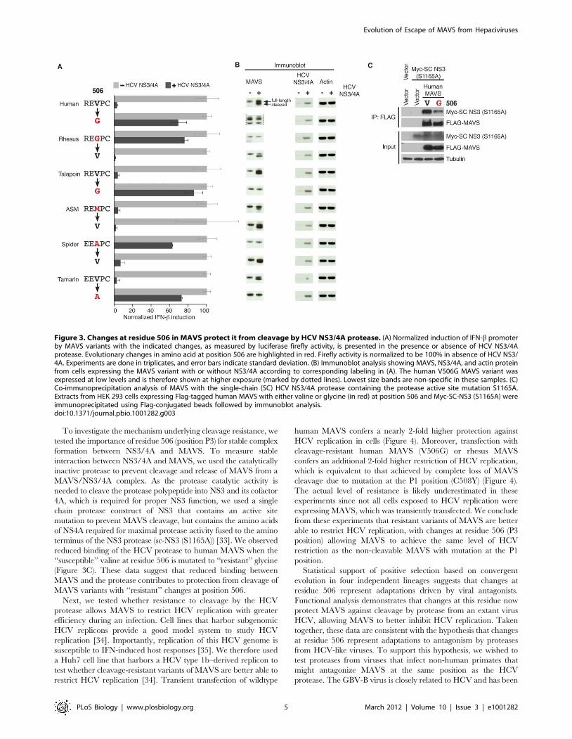
To investigate the mechanism underlying cleavage resistance, we
tested the importance of residue 506 (position P3) for stable complex
formation between NS3/4A and MAVS. To measure stable
interaction between NS3/4A and MAVS, we used the catalytically
inactive protease to prevent cleavage and release of MAVS from a
MAVS/NS3/4A complex. As the protease catalytic activity is
needed to cleave the protease polypeptide into NS3 and its cofactor
4A, which is required for proper NS3 function, we used a single
chain protease construct of NS3 that contains an active site
mutation to prevent MAVS cleavage, but contains the amino acids
of NS4A required for maximal protease activity fused to the amino
terminus of the NS3 protease (sc-NS3 (S1165A)) [33]. We observed
reduced binding of the HCV protease to human MAVS when the
‘‘susceptible’’ valine at residue 506 is mutated to ‘‘resistant’’ glycine
(Figure 3C). These data suggest that reduced binding between
MAVS and the protease contributes to protection from cleavage of
MAVS variants with ‘‘resistant’’ changes at position 506.
Next, we tested whether resistance to cleavage by the HCV
protease allows MAVS to restrict HCV replication with greater
efficiency during an infection. Cell lines that harbor subgenomic
HCV replicons provide a good model system to study HCV
replication [34]. Importantly, replication of this HCV genome is
susceptible to IFN-induced host responses [35]. We therefore used
a Huh7 cell line that harbors a HCV type 1b–derived replicon to
test whether cleavage-resistant variants of MAVS are better able to
restrict HCV replication [34]. Transient transfection of wildtype
human MAVS confers a nearly 2-fold higher protection against
HCV replication in cells (Figure 4). Moreover, transfection with
cleavage-resistant human MAVS (V506G) or rhesus MAVS
confers an additional 2-fold higher restriction of HCV replication,
which is equivalent to that achieved by complete loss of MAVS
cleavage due to mutation at the P1 position (C508Y) (Figure 4).
The actual level of resistance is likely underestimated in these
experiments since not all cells exposed to HCV replication were
expressing MAVS, which was transiently transfected. We conclude
from these experiments that resistant variants of MAVS are better
able to restrict HCV replication, with changes at residue 506 (P3
position) allowing MAVS to achieve the same level of HCV
restriction as the non-cleavable MAVS with mutation at the P1
position.
Statistical support of positive selection based on convergent
evolution in four independent lineages suggests that changes at
residue 506 represent adaptations driven by viral antagonists.
Functional analysis demonstrates that changes at this residue now
protect MAVS against cleavage by protease from an extant virus
HCV, allowing MAVS to better inhibit HCV replication. Taken
together, these data are consistent with the hypothesis that changes
at residue 506 represent adaptations to antagonism by proteases
from HCV-like viruses. To support this hypothesis, we wished to
test proteases from viruses that infect non-human primates that
might antagonize MAVS at the same position as the HCV
protease. The GBV-B virus is closely related to HCV and has been
Figure 3. Changes at residue 506 in MAVS protect it from cleavage by HCV NS3/4A protease. (A) Normalized induction of IFN-b promoterby MAVS variants with the indicated changes, as measured by luciferase firefly activity, is presented in the presence or absence of HCV NS3/4Aprotease. Evolutionary changes in amino acid at position 506 are highlighted in red. Firefly activity is normalized to be 100% in absence of HCV NS3/4A. Experiments are done in triplicates, and error bars indicate standard deviation. (B) Immunoblot analysis showing MAVS, NS3/4A, and actin proteinfrom cells expressing the MAVS variant with or without NS3/4A according to corresponding labeling in (A). The human V506G MAVS variant wasexpressed at low levels and is therefore shown at higher exposure (marked by dotted lines). Lowest size bands are non-specific in these samples. (C)Co-immunoprecipitation analysis of MAVS with the single-chain (SC) HCV NS3/4A protease containing the protease active site mutation S1165A.Extracts from HEK 293 cells expressing Flag-tagged human MAVS with either valine or glycine (in red) at position 506 and Myc-SC-NS3 (S1165A) wereimmunoprecipitated using Flag-conjugated beads followed by immunoblot analysis.doi:10.1371/journal.pbio.1001282.g003
Evolution of Escape of MAVS from Hepaciviruses
PLoS Biology | www.plosbiology.org 5 March 2012 | Volume 10 | Issue 3 | e1001282

tentatively assigned to the Hepacivirus genus. And although its
natural host is unknown, it has been shown to infect tamarins and
marmosets [36,37]. More distantly related to HCV are the GBV-
A and GBV-C (also known as hepatitis G) viruses, which are
known to naturally infect primates. GBV-A infects New World
monkeys while GBV-C is found in humans and chimps. These two
viruses have tentatively been assigned to a new Pegivirus genus, as
a sister genus to the Hepacivirus genus within the Flaviviridae
family (although this designation has not yet been formally
accepted by the International Committee on Taxonomy of
Viruses) [37–42]. We refer to the Hepacivirus/Pegivirus genera
as hepaciviruses here, for convenience and because of their shared
properties we observed (Figure 5). All GB viruses encode the NS3
protease. We therefore tested the ability of NS3 proteases from
these viruses to antagonize human MAVS. Consistent with our
hypothesis, we found that the NS3 protease from both GBV-A and
GBV-C strongly inhibited signaling by human MAVS (Figure 5B).
Consistent with previous reports, NS3 protease from GBV-B also
inhibited human MAVS, while proteases from the more distantly
related pestivirus bovine viral diarrhea virus (BVDV) and flavivirus
yellow fever virus (YFV) did not antagonize MAVS (Figure 5) [43].
Thus, the property of antagonizing MAVS is shared and exclusive
to the group of viruses encompassing the HCV and GB viruses.
We next tested the effect of positively selected changes at
residue 506 on the ability of the GB viruses to inhibit MAVS.
Remarkably, changes at residue 506 away from ancestral valine
in MAVS allow it to signal in the presence of NS3 proteases from
all GB viruses (Figure 6, Figure S3). However, there are some
species- and virus-specific differences. For example, methionine
at residue 506 in Allen’s swamp monkey MAVS appears to
provide significant protection against protease from GBV-B virus
compared to other GB viruses and HCV (Figure 2A, Figure 6).
Also, alanine at residue 506 in red-mustached tamarin monkey
did not provide a much higher protection against antagonism by
GB viruses (compared to HCV, Figure 2A) than the ancestral
valine. These idiosyncratic differences likely reflect slightly
different properties of the HCV and GB virus proteases.
However, our overall data suggest that the ability of NS3
proteases to antagonize MAVS, and to be impeded by the same
adaptive changes in MAVS, is a phylogenetically discrete
characteristic exclusive to HCV and the three GBV viruses,
Figure 4. Protease-resistant MAVS better restricts HCV repli-cation. Immunoblot showing HCV NS5A protein from lysates of Huh7-K2040 cells transiently transfected with control plasmid (vector alone),human, or rhesus MAVS variants with indicated mutations. The amountof HCV restriction achieved by indicated MAVS variant is quantifiedbelow the Western blot.doi:10.1371/journal.pbio.1001282.g004
Figure 5. NS3 proteases from distantly related GB viruses inhibit human MAVS. (A) Phylogram made using NS3 sequence from theindicated viruses. Bootstrap values are indicated for each branch. Scale represents 0.1 amino acid substitutions per site. Bovine viral diarrheal virus(BVDV) and Yellow fever virus (YFV) are representative pesti- and flaviviruses, respectively. We refer to GBV-C and GBV-A viruses as hepaciviruses,although they have not been formally assigned to a particular genus within the Flaviviridae family. GBV-C is also known as Hepatitis G virus (HGV).CHV stands for canine hepacivirus [17]. Corresponding hosts that can be naturally infected by each virus are indicated on the right. (B) IFN-binduction, as determined by luciferase firefly activity upon expression of human MAVS in the presence of NS3 protease from the corresponding virusin (A). IFN-b induction is normalized as being 100% in absence of any protease. Experiments were performed in triplicate with error bars indicatingstandard deviation.doi:10.1371/journal.pbio.1001282.g005
Evolution of Escape of MAVS from Hepaciviruses
PLoS Biology | www.plosbiology.org 6 March 2012 | Volume 10 | Issue 3 | e1001282

and likely inherited from a common ancestor. Importantly, these
data demonstrate that non-human primates are susceptible to
infection by viruses that antagonize MAVS in a manner similar to
HCV.
To infer the minimum age of viruses that drove the evolution at
residue 506, we wanted to date these adaptive changes. We took
advantage of the phylogenetically well-characterized macaque
lineage, which includes rhesus macaque with the ‘‘resistant’’
change at position 506 [44]. We sequenced the C-terminus of
MAVS from six different Macaque species and found that all of
them, including the most ancestral species, the barbary macaque
M. sylvanus, possess the ‘‘resistant’’ glycine instead of the ancestral
valine at position 506 (Figure 7). Furthermore, we found no
polymorphisms at this position within 20 rhesus macaque samples,
suggesting that the change to glycine is likely a fixed change within
the species. Since the macaque lineage separated 5–8 million years
ago from that of baboons [44], which retain the ancestral valine at
position 506, our data suggest that the progenitor macaque lineage
encountered and adapted to a virus that antagonized MAVS at
least 5–8 million years ago.
Discussion
In this study, we have shown that positive selection in MAVS of
multiple primate species has independently resulted in its escape
from antagonism by hepaciviral proteases. Including baboons that
have acquired a change at residue 508, we found that 5 out of the
20 primates that we characterized possess MAVS resistant to
hepaciviral antagonism. Remarkably, MAVS resistance in four of
these species is a product of independently acquired changes at the
same single residue 506, which allow escape from hepaciviral NS3
protease recognition and cleavage.
MAVS as a Genetic Determinant of Hepaciviral ClearanceHCV cleaves MAVS in order to block the interferon response.
The importance of this strategy employed by HCV is highlighted
by the fact that interferon treatment is often clinically used to
successfully cure HCV infection. Furthermore, a polymorphism
near a type III interferon gene is associated with spontaneous and
treatment-induced clearance of HCV [45,46]. Thus, given the
importance of the interferon response, we hypothesize that
primates that possess MAVS resistant to cleavage by NS3 protease
should be better at clearing hepaciviral infections than species with
susceptible MAVS. Consistent with this idea, we found that
resistant variants of MAVS are better able to restrict HCV
replication in cell culture. Furthermore, although sampling of
hepaciviruses from primates has been sparse, thus far hepaci-
viruses have only been found in primates with ‘‘susceptible’’
MAVS, such as human, chimp, common marmoset, red-chested
mustached tamarin, and owl monkey [38]. In contrast, hepaci-
viruses were assayed for but not found in spider monkeys, which
have ‘‘resistant’’ MAVS [38]. Our study provides functional
evidence that implicates host genetics in determining the outcome
of diverse hepaciviral infections even between closely related
primate species.
Ancient Hepaciviruses as Drivers of MAVS EvolutionThe remarkable case of convergent evolution at residue 506 is
indicative of adaptive evolution, likely driven by ancient viruses
(paleoviruses). Based on the phylogenetic and the functional
evidence using extant viruses as surrogates, we can attempt to infer
the nature of these paleoviruses, although our ‘‘indirect paleovir-
Figure 6. Protective effect of changes at residue 506 onantagonism by GB virus proteases. IFN-b induction resulting fromexpression of MAVS from indicated species, as determined by luciferasefirefly activity, is presented for either the ancestral ‘‘susceptible’’ valine(light grey) or derived ‘‘resistant’’ variants (dark grey) at position 506 asshown. Luciferase firefly activity is normalized as being 100% in absenceof the protease for each MAVS variant. Susceptibility to each of the GB-virus protease is shown. x- and y-axes are the same for all graphs. Allexperiments were performed in triplicate with error bars indicatingstandard deviation.doi:10.1371/journal.pbio.1001282.g006
Figure 7. Mapping the adaptive change at site 506 in theMacaque lineage. Accepted Macaque phylogeny. Both species namesand common names are shown. Amino acid at position 506 in MAVS isshown on the right. The escape variant glycine (G) is shown in red. Notethat the outgroup Olive baboon has valine (V) at position 506. Thisindicates that the common ancestor of macaques underwent theadaptive change 5–8 million years ago. MYA, million years ago.doi:10.1371/journal.pbio.1001282.g007
Evolution of Escape of MAVS from Hepaciviruses
PLoS Biology | www.plosbiology.org 7 March 2012 | Volume 10 | Issue 3 | e1001282
ology’’ is limited in its ability to formally prove the existence of
such paleoviruses. Functional evidence that changes at residue 506
provide protection from antagonism by HCV is consistent with the
hypothesis of ancient hepaciviruses being the causative agents of
the evolution at residue 506. Since HCV per se is a human-specific
virus, it is unlikely to have been responsible for MAVS evolution in
non-human primates. Instead, we sought to find viruses with
similar antagonistic properties as HCV. We found that NS3
proteases from GBV-A and GBV-C viruses, which naturally infect
non-human primates, not only share the ability to antagonize
MAVS but are also impeded by the same evolutionary changes at
residue 506 (GBV-B protease also behaves in a similar manner to
the other GB virus proteases and while it can experimentally infect
some New World primates, its natural host is not known). These
data suggest that despite the high degree of divergence, all
hepaciviruses are capable of antagonizing MAVS. This shared
property must have been present in their common ancestor and
subsequently inherited by all extinct and present-day hepaci-
viruses. Furthermore, this common ancestor must have been
present tens of millions of years ago. GBV-C viruses that infect
humans and chimps are thought to have co-diverged with their
ancestors 7 million years ago [47]. Similarly, GBV-A viruses are
thought to have co-speciated with their hosts over the course of last
20 million years [47,48]. Divergence of GBV-C from GBV-A and
from their common ancestor from HCV presumably occurred
even earlier. Taken together, these data suggest that hepaciviruses
capable of antagonizing MAVS are an extremely ancient group of
viruses that are old enough and distributed widely enough to be
responsible for driving MAVS evolution at residue 506. Consistent
with the ancient presence of hepaciviruses, we dated the change at
residue 506 in the macaque lineage to 5–8 million years ago. Our
findings imply that HCV and GB viruses represent the latest in a
continuum of hepaciviral infections that have plagued primates for
millions of years. Finally, it is interesting to note that MAVS in
three Old World monkeys (OWM) has independently evolved
resistance to hepaciviral antagonism despite the fact that
hepaciviruses have not yet been detected in OWM.
Multiple lines of evidence outlined above support the hypothesis
that ancient hepaciviruses were responsible for driving evolution in
MAVS. However, it is also formally possible that a different class
of viral antagonists unrelated to the hepaciviral NS3 proteases
drove the evolution at residue 506. Although it is not formally
possible to rule out such an alternative, there is little theoretical or
empirical evidence in support of this. Residue 506 is not an
‘‘Achilles’ heel’’ of MAVS that would make it an especially
attractive target for antagonism by other viruses. There does not
appear to be anything special about residue 506, except that it sits
within a stretch of residues that corresponds to the cleavage site for
hepaciviral protease NS3. Consistent with this idea, all other
known viral antagonists of MAVS, including proteases from other
viruses, interact with different regions of MAVS [9–12].
Furthermore, other residues under positive selection in MAVS
are spread throughout the length of the protein, suggesting that
MAVS can be antagonized along multiple surfaces and not just at
or near residue 506. Taken together, these reasons make it unlikely
that other viral antagonists besides hepaciviral proteases have
converged to drive the evolution at residue 506.
Inferring Paleovirology from Adaptive Evolution of MAVSWe have previously proposed that virus-driven adaptive
evolution of host antiviral factors can be used as a tool in
paleovirology, the study of ancient viruses and their impact of host
evolution [49,50]. The reasoning behind this idea is that positive
selection in host antiviral factors essentially represents viral
‘‘footprints’’ that can reveal the action of ancient viruses. An
inherent limitation of our ‘‘indirect’’ paleovirology approach is
that it cannot formally rule out the possibility that either an
unrelated virus or another selective pressure altogether was
responsible for the observed evolution. Thus, in the case of
evolution at residue 506 in MAVS, we cannot formally rule out
alternate selection scenarios. The discovery of endogenized
hepaciviral ‘‘fossils’’ in primate genomes might be an important
confirmation of ancient hepaciviruses and their ability to infect
primates. While such viral ‘‘fossils’’ would provide direct evidence
for existence of paleoviruses, they would not necessarily reveal the
functional consequences of these viruses on host evolution. Thus,
‘‘fossil’’-based and ‘‘indirect’’ paleovirology should be viewed as
complementary approaches, both of which are important to
determine the identity of paleoviruses and their impact on host
genomes.
One limitation of using adaptive evolution in host factors to
study paleovirology that has been previously discussed [50] is the
difficulty of deciphering the history of a single type of virus. For
example, protein kinase R (PKR) is a broad-spectrum antiviral
factor that is antagonized by a multitude of diverse viruses
including ssRNA and dsDNA viruses, and often at the same
domain. This is reflected in the large number of residues in PKR
that are evolving under positive selection [18]. Such overlapping
antagonism might obscure the history of individual viruses.
Moreover, every subsequent round of adaptation might overwrite
the paleoviral record of all previous infections. In contrast, despite
MAVS acting against a broad panel of RNA viruses, there are only
nine residues evolving under positive selection over primate
evolution. This suggests that only a few viruses directly antagonize
MAVS to drive its positive selection. This limited antagonism
makes it less likely that more than one virus will converge on the
same residue. Thus, studying proteins such as MAVS with sparse
positive selection widely distributed along its length offers the
opportunity to make paleoviral inferences with greater specificity.
Our study shows that the nature of the viral antagonist also
contributes to the specificity of the paleoviral record. For example,
it is surprising that the diverse hepaciviruses we tested have not
dramatically altered the substrate specificity of their NS3 proteases
over a long period of evolution. One explanation—supported by
recent data from genomic viral ‘‘fossils’’ [51]—is that long-term
virus evolution is much slower than expected due to functional
constraint [50]. Indeed, the NS3 proteases are highly constrained
because besides cleaving MAVS, they are also responsible for
cleaving the viral polypeptide at multiple locations. Thus, given
that paleovirology relies on the use of extant viruses as surrogates
of their ancient counterparts to test the function of positively
selected changes in host factors, viral ‘‘footprints’’ left by
functionally constrained antagonists like proteases are particularly
well suited for paleovirology [11,12].
Functional dissection of the interaction between residue 506 in
MAVS and HCV provides a model to study paleovirology using
extant viruses and host immune factors. This model can be applied
to the study of other residues in MAVS that have evolved under
positive selection in primates, which we suspect also reflect a
history of antagonism by other viruses. For example, coxsackie-
virus uses its 3C protease to cleave human MAVS between
residues 148 and 149 (Figure S4) [12]. Remarkably, residue 149
has evolved under one of the strongest signatures of positive
selection in MAVS (Figure 1B, Table S1). It is also interesting to
note that residue 423, which forms the P5 position within the
cleavage site for Hepatitis A virus protease, is also evolving under
positive selection (Figure 1B, Table S1, Figure S4). Future
experiments will be needed to establish the functional importance
Evolution of Escape of MAVS from Hepaciviruses
PLoS Biology | www.plosbiology.org 8 March 2012 | Volume 10 | Issue 3 | e1001282

of residues 149 and 423. Nevertheless, the fact that the cleavage
sites for all known protease antagonists of MAVS contain residues
evolving under positive selection suggests that the correlation is
unlikely to be simply a coincidence (Figure S4).
A particularly intriguing case of positive selection at residue 60
in MAVS also promises to reveal paleoviral insights (Figure 1B).
This residue has independently acquired changes in six primates.
In every instance, the ancestral asparagine has changed to serine
(Figure S5). This selective change likely reflects the pressure to
escape from some viral antagonism by evolving away from the
ancestral asparagine, while continuing to maintain MAVS
function, which might be particularly sensitive to changes at
residue 60. Residue 60 occurs within the highly conserved CARD
domain, which is utilized to directly interact with viral RNA
sensors RIG-I and MDA-5 [6]. Interestingly, MAVS in colobus
monkey is polymorphic for both asparagine and serine, suggesting
that the selective pressure driving the change might be very recent.
It will be interesting to functionally test whether serine at residue
60 provides protection from one of the known viral antagonists of
MAVS. Alternatively, the dramatic convergent evolution at
residue 60 might help discover a new viral antagonist of MAVS.
Materials and Methods
Sequencing MAVS cDNAIsolation of total RNA was done as described previously [18].
Primate MAVS cDNAs were isolated by RT-PCR using 50 ng of
total RNA as template and TA-cloned into pCR2.1 (Invitrogen).
Primer MP-11, which sits upstream of the start site, and primer
MP-12, which is downstream of the stop codon, were used for RT-
PCR (Table S2). MP-34, along with MP-11 and MP-12, was used
for sequencing the cDNAs. MAVS cDNA sequences obtained in
this study are in the process of being submitted to GenBank.
Common marmoset sequence was obtained from the UCSC
Genome Browser.
Plasmid ConstructionPrimate MAVS cDNAs were prepared using 250 ng total
RNA as template and oligo dT primer (Superscript III,
Invitrogen). cDNAs were subsequently used as a template for
PCR (Phusion, NEB). MAVS cDNAs were cloned into pFLAG-
CMV-2 between HindIII and BglII downstream of and in frame
with FLAG tag.
Single amino acid changing point mutations were introduced in
human MAVS as follows: MP-51, a forward primer upstream of
the MAVS cDNA in pFLAG-CMV-2, was used in combination
with a reverse primer that included the desired change. A forward
primer with the desired change was then used with MP-52, a
reverse primer downstream of MAVS cDNA in pFLAG-CMV-2.
The resulting products from these two PCRs were used as a
template for stitch PCR with MP-51 and MP-52 primers. The
resulting PCR product, which included the desired change in
MAVS, was ligated into pFLAG-CMV-2 between HindIII and
BglII in frame with FLAG tag. The remaining single amino acid
changes in MAVS were introduced using similar stitching strategy,
but desired amino acid changes were achieved by stitching
together N- and C-terminal MAVS of different but closely related
species.
NS3/4A from GBV-B, GBV-C, HCV, and GBV-A was cloned
into pcDNA3.1/Hygro(2) between XbaI and HindIII down-
stream of and in frame with HA tag (cloned between NheI and
XbaI).
GBV-A NS3/4A was synthesized (Genscript Inc., Piscataway,
New Jersey) using strain A-lab sequence.
Other plasmids used in this study were: pJC453–IFN-bpromoter luciferase firefly reporter construct (kindly provided by
Zhijian Chen), pRL-TK–renilla transfection control construct
(Promega Inc.), pcDNA6-BVDVpro (kindly provided by Kui Li),
pcDNA6-YFVpro (kindly provided by Kui Li), Human MAVS
(C508Y) (previously described [52]), and pcDNA-Myc-sc-NS3
(S1165A) (previously described [33]).
Evolutionary AnalysesAmino acid sequence alignment was performed using ClustalW.
This amino acid alignment was used as input for DNA sequence
alignment in PAL2NAL. Aligned DNA sequences were subse-
quently used for all analyses done in PAML software. The well-
supported primate phylogeny (Figure 1A) [26] was used for
analyses done in PAML [28]. Maximum-likelihood analysis to
determine whether MAVS is evolving under positive selection and
to determine which residues are evolving under positive selection
was done using the sites models in codeml program of PAML [28].
Sites models allow the dN/dS ratio to vary among residues.
Branch models, which allow the dN/dS ratio to vary among
different branches of the phylogeny, were used to determine dN/
dS values for different lineages. Two-ratio tests to detect episodic
selection were performed by comparing a likelihood model that
allows dN/dS to vary across phylogeny to model with dN/dS fixed
at 1. REL and branch-site REL analysis were performed using a
web-based implementation of HyPhy package (www.datamonkey.
org). Branch-site REL is based on likelihood ratio tests that identify
all lineages with a proportion of sites that are evolving with dN/
dS.1, without making a priori assumptions about which lineages
these are in the phylogeny [29].
Luciferase Assays56104 HEK293 cells were plated per well in 96-well plates and
transfected on the next day with LT1 (Mirus) transfection reagent.
The following plasmids were co-transfected in triplicates: MAVS
plasmid (2.5 ng), luciferase reporter construct pCJ453 (50 ng),
transfection control pRL-TK construct (10 ng), none or one of the
viral protease encoding plasmids (pJC1461—2.5 ng, pMP125—
2.5 ng, pMP122—10 ng, pMP123—2.5 ng, pMP126—10 ng),
and pFLAG-CMV-2 (enough to bring final DNA concentration
equal to 150 ng). One triplicate set of wells was transfected without
MAVS plasmid and one set without any DNA. 24 h after
transfection, Dual-Glo Luciferase Assay System (Promega) was
used to lyse the cells and measure luciferase firefly and renilla
activity with a luminometer. Amount of firefly or renilla activity in
wells transfected without any DNA was subtracted as background.
Firefly activity was divided by the renilla activity in the same well
to control for transfection efficiency. This firefly/renilla ratio from
cells expressing MAVS was divided by the firefly/renilla ratio from
wells without MAVS to calculate IFN-b fold induction, which was
then averaged across the set of three wells.
Immunoblot Analysis to Assay CleavageIt was not possible to get enough lysate from a single well of a
96-well plate for immunoblot analysis. We therefore scaled the
entire transfection process by 4-fold in 24-well plates. Therefore,
206105 cells were plated per well in 96-well plates and transfected
with 4 times as much LT1 transfection reagent. Concentration of
all the plasmids was also increased 4-fold. 24 h after transfection,
cells were resuspended in 36 ml NTE buffer (10 mM Tris—pH 8,
1 mM EDTA, 50 mM NaCl) containing 0.18 mg protease
inhibitor cocktail Complete Mini (Roche). Cells were then lysed
on ice for 10 min in 44 ml of NP-40 buffer (1% NP-40, 0.2%
sodium-deoxycholate, 0.12 M NaCl, 20 mM Tris—pH 8) con-
Evolution of Escape of MAVS from Hepaciviruses
PLoS Biology | www.plosbiology.org 9 March 2012 | Volume 10 | Issue 3 | e1001282

taining 0.22 mg protease inhibitor cocktail, 2.461026 mol DTT,
and 1.261026 mol PMSF. Lysate was spun at maximum speed on
tabletop centrifuge for 10 min. 75 ml of supernatant was mixed
with 19 ml of 56 SDS sample buffer with 10% b-ME. Samples
were boiled for 10 min and then 5–10 ml was used for Western
blot analysis.
Mouse monoclonal anti-FLAG M2 antibody (Sigma-Aldrich)
was used to detect FLAG-tagged MAVS, and mouse monoclonal
anti-HA.11 antibody (Covance) was used to detect HA-tagged
HCV NS3/4A. Rabbit polyclonal anti-beta Actin antibody
(Abcam) was used to detect actin. HRP-linked anti-mouse and
anti-rabbit IgG antibodies (GE Healthcare) were used as
secondary anti-bodies. Primary anti-bodies were used at 1:1,000
dilution at 4u overnight and secondary anti-bodies were used at
1:10,000 dilution for 1 h at room temperature. SuperSignal West
Dura ECL substrate (Thermo Scientific) was used for detecting
HRP.
Co-Immunoprecipitation AssayFor co-immunoprecipitation analysis, HEK 293 cells were
transfected with empty vector or pcDNA-Myc-sc-NS3 (S1165A)
protease [33] that contains aa 21–32 of HCV NS4A fused to aa
1026–1206 of the HCV NS3 protease with the protease active site
mutation S1165A, as well as empty vector or MAVS-expressing
constructs. Cells were lysed in 1% Triton X-100, 150 mm NaCl,
and 25 mM Tris-Cl pH 7.5. Coimmunoprecipitation was per-
formed using FLAG M2-agarose beads (Sigma-Aldrich) followed
by immunoblot analysis using the following antibodies: anti-myc
(Abcam), anti-Flag M2-Peroxidase (Sigma-Aldrich), and anti-
tubulin (Sigma-Aldrich).
HCV Restriction AssayHuh7-K2040 is a Huh7-based human hepatocyte cell line that
contains the self-replicating subgenomic replicon from HCV 1b
[34]. Huh7-K2040 is transfected with control vector or plasmid
expressing the indicated MAVS construct for 36 h. Cell lysates
were analyzed by immunoblot with antiserum specific to HCV
NS5A [53], tubulin (Sigma-Aldrich), and FLAG M2 (Sigma-
Aldrich) for detection of the various MAVS constructs. HCV
protein was further detected using the hyperimmune serum from
an HCV-infected patient that recognizes HCV proteins NS3,
NS4B, and NS5A [35]. Protein densitometry was determined
using the NIH ImageJ program. To derive fold restriction, NS5A
(rabbit polyclonal)/tubulin ratio for each sample was divided by
the NS5A(rabbit polyclonal)/tubulin ratio from the vector alone
control.
Supporting Information
Figure S1 Schematic of the RIG-I/MDA-5 pathway. Solid
arrows indicate direct interaction. Through a signaling cascade,
MAVS activates IRF-3 and NFk-B, which bind to and activate
IFN-b promoter.
(TIF)
Figure S2 Ability of MAVS from multiple primate species to
induce IFN-b activity. Same data as in Figure 2, but fold IFN-binduction is not normalized. Induction of IFN-b promoter, as
measured by luciferase firefly activity, upon expression of MAVS
cDNA from corresponding species coexpressed with (+) or without
(2) HCV NS3/4A. Primates with MAVS capable of significant
IFN-b induction even in presence of HCV protease are
highlighted in bold. Human (C508R) refers to substitution of
Cysteine (C) at position 508 with Arginine (R) in human MAVS.
All experiments are done in triplicates, and error bars indicate
standard deviation.
(TIF)
Figure S3 Residue 506 provides protection against NS3/4A
from all hepaciviruses. Same data as in Figure 6 but includes data
from control groups (IFN-b induction in absence of any protease).
IFN-b induction due to expression of MAVS from indicated
species, as determined by luciferase firefly activity, is presented for
either the ancestral ‘‘susceptible’’ valine or derived ‘‘resistant’’
variants (in red) at position 506 as shown. Luciferase firefly activity
is normalized as being 100% in absence of the protease.
Susceptibility to each of the GB-virus protease is shown. y-axis is
the same for all graphs. All experiments done in triplicates. Error
bars indicate standard deviation.
(TIF)
Figure S4 Positive selection within the viral protease cleavage
sites. Cleavage sites in MAVS of three viral proteases have been
mapped so far (indicated by the scissors). Residues between which
proteases cleave MAVS are indicated. Note the presence of
residues under positive selection (triangles) within or proximal to
the cleavage sites of all three proteases.
(TIF)
Figure S5 MAVS protein alignment. Residues evolving under
positive selection are highlighted in yellow.
( )
Table S1 Statistical evidence for sites evolving under positive
selection in MAVS. Likelihood ratio tests for positive selection in
MAVS were done using NSsites model M7 versus M8 comparison
assuming F61 model of codon frequency in codeml program in
PAML software. Likelihood ratio tests were also performed using
random effects likelihood (REL), implemented in web-based
HyPhy package. Residues (highlighted in bold) with Bayes
Empirical Bayes (BEB) and REL calculated posterior probabilities
(PPr) equal to or greater than 0.9 for which dN/dS.1 are shown
in Figure 1B. NEB, Naive Empirical Bayes.
(TIF)
Table S2 List of primers used in the study. Primers are in 59 to
39 orientation.
(TIF)
Acknowledgments
We thank J. Bloom, M. Daugherty, N. Elde, M. Emerman, M. Klassen,
M. Levine, E. Lim, and N. Phadnis for comments on the manuscript. We
would like to thank Hae Soo Park for technical assistance. pJC453 and
pJC1461 plasmids were kindly provided by Zhijian Chen. pcDNA-6-
GBpro, pcDNA6-BVDVpro, and pcDNA6-YFVpro plasmids were kindly
provided by Kui Li. pGAL-HGV NS3/4A plasmid was kindly provided by
Woo Jin Park. Rhesus RNA samples were kindly provided by Welkin
Johnson and Nels Elde.
Author Contributions
The author(s) have made the following declarations about their
contributions: Conceived and designed the experiments: MRP HSM.
Performed the experiments: MRP Y-ML SMH. Analyzed the data: MRP
Y-ML SMH MG HSM. Contributed reagents/materials/analysis tools: Y-
ML SMH MG. Wrote the paper: MRP HSM. Edited the manuscript:
MRP Y-ML SMH MG HSM.
Evolution of Escape of MAVS from Hepaciviruses
PLoS Biology | www.plosbiology.org 10 March 2012 | Volume 10 | Issue 3 | e1001282

References
1. Yoneyama M, Kikuchi M, Natsukawa T, Shinobu N, Imaizumi T, et al. (2004)
The RNA helicase RIG-I has an essential function in double-stranded RNA-
induced innate antiviral responses. Nat Immunol 5: 730–737.
2. Kang DC, Gopalkrishnan RV, Wu Q, Jankowsky E, Pyle AM, et al. (2002) mda-
5: an interferon-inducible putative RNA helicase with double-stranded RNA-
dependent ATPase activity and melanoma growth-suppressive properties. Proc
Natl Acad Sci U S A 99: 637–642.
3. Andrejeva J, Childs KS, Young DF, Carlos TS, Stock N, et al. (2004) The V
proteins of paramyxoviruses bind the IFN-inducible RNA helicase, mda-5, and
inhibit its activation of the IFN-beta promoter. Proc Natl Acad Sci U S A 101:
17264–17269.
4. Seth RB, Sun L, Ea CK, Chen ZJ (2005) Identification and characterization of
MAVS, a mitochondrial antiviral signaling protein that activates NF-kappaB
and IRF 3. Cell 122: 669–682.
5. Meylan E, Curran J, Hofmann K, Moradpour D, Binder M, et al. (2005) Cardif
is an adaptor protein in the RIG-I antiviral pathway and is targeted by hepatitis
C virus. Nature 437: 1167–1172.
6. Kawai T, Takahashi K, Sato S, Coban C, Kumar H, et al. (2005) IPS-1, an
adaptor triggering RIG-I- and Mda5-mediated type I interferon induction. Nat
Immunol 6: 981–988.
7. Xu LG, Wang YY, Han KJ, Li LY, Zhai Z, et al. (2005) VISA is an adapter
protein required for virus-triggered IFN-beta signaling. Mol Cell 19: 727–740.
8. Li XD, Sun L, Seth RB, Pineda G, Chen ZJ (2005) Hepatitis C virus protease
NS3/4A cleaves mitochondrial antiviral signaling protein off the mitochondria
to evade innate immunity. Proc Natl Acad Sci U S A 102: 17717–17722.
9. Graef KM, Vreede FT, Lau YF, McCall AW, Carr SM, et al. (2010) The PB2
subunit of the influenza virus RNA polymerase affects virulence by interacting
with the mitochondrial antiviral signaling protein and inhibiting expression of
beta interferon. J Virol 84: 8433–8445.
10. Iwai A, Shiozaki T, Kawai T, Akira S, Kawaoka Y, et al. (2010) Influenza A
virus polymerase inhibits type I interferon induction by binding to interferon
beta promoter stimulator 1. J Biol Chem 285: 32064–32074.
11. Yang Y, Liang Y, Qu L, Chen Z, Yi M, et al. (2007) Disruption of innate
immunity due to mitochondrial targeting of a picornaviral protease precursor.
Proc Natl Acad Sci U S A 104: 7253–7258.
12. Mukherjee A, Morosky SA, Delorme-Axford E, Dybdahl-Sissoko N,
Oberste MS, et al. (2011) The coxsackievirus B 3C protease cleaves MAVS
and TRIF to attenuate host type I interferon and apoptotic signaling. PLoS
Pathog 7: e1001311. doi:10.1371/journal.ppat.1001311.
13. Paulmann D, Magulski T, Schwarz R, Heitmann L, Flehmig B, et al. (2008)
Hepatitis A virus protein 2B suppresses beta interferon (IFN) gene transcription
by interfering with IFN regulatory factor 3 activation. J Gen Virol 89:
1593–1604.
14. Simmonds P (2004) Genetic diversity and evolution of hepatitis C virus–15 years
on. J Gen Virol 85: 3173–3188.
15. Fields BN, Knipe DM, Howley PM (2007) Fields’ virology. Philadelphia:
Wolters Kluwer Health/Lippincott Williams & Wilkins, 2 v. (xix, 3091, 3086
pp.).
16. Epstein JH, Quan PL, Briese T, Street C, Jabado O, et al. (2010) Identification
of GBV-D, a novel GB-like flavivirus from old world frugivorous bats (Pteropus
giganteus) in Bangladesh. PLoS Pathog 6: e1000972. doi:10.1371/journal.
ppat.1000972.
17. Kapoor A, Simmonds P, Gerold G, Qaisar N, Jain K, et al. (2011)
Characterization of a canine homolog of hepatitis C virus. Proc Natl Acad
Sci U S A.
18. Elde NC, Child SJ, Geballe AP, Malik HS (2009) Protein kinase R reveals an
evolutionary model for defeating viral mimicry. Nature 457: 485–489.
19. Sawyer SL, Emerman M, Malik HS (2004) Ancient adaptive evolution of the
primate antiviral DNA-editing enzyme APOBEC3G. PLoS Biol 2: E275.
doi:10.1371/journal.pbio.0020275.
20. Sawyer SL, Wu LI, Emerman M, Malik HS (2005) Positive selection of primate
TRIM5alpha identifies a critical species-specific retroviral restriction domain.
Proc Natl Acad Sci U S A 102: 2832–2837.
21. Ortiz M, Guex N, Patin E, Martin O, Xenarios I, et al. (2009) Evolutionary
trajectories of primate genes involved in HIV pathogenesis. Mol Biol Evol 26:
2865–2875.
22. McNatt MW, Zang T, Hatziioannou T, Bartlett M, Fofana IB, et al. (2009)
Species-specific activity of HIV-1 Vpu and positive selection of tetherin
transmembrane domain variants. PLoS Pathog 5: e1000300. doi:10.1371/
journal.ppat.1000300.
23. Zhang J, Webb DM (2004) Rapid evolution of primate antiviral enzyme
APOBEC3G. Hum Mol Genet 13: 1785–1791.
24. Song B, Gold B, O’Huigin C, Javanbakht H, Li X, et al. (2005) The
B30.2(SPRY) domain of the retroviral restriction factor TRIM5alpha exhibits
lineage-specific length and sequence variation in primates. J Virol 79:
6111–6121.
25. Rothenburg S, Seo EJ, Gibbs JS, Dever TE, Dittmar K (2009) Rapid evolutionof protein kinase PKR alters sensitivity to viral inhibitors. Nat Struct Mol Biol
16: 63–70.26. Perelman P, Johnson WE, Roos C, Seuanez HN, Horvath JE, et al. (2011) A
molecular phylogeny of living primates. PLoS Genet 7: e1001342. doi:10.1371/journal.pgen.1001342.
27. Yang Z (1997) PAML: a program package for phylogenetic analysis by
maximum likelihood. Comput Appl Biosci 13: 555–556.28. Yang Z (2007) PAML 4: phylogenetic analysis by maximum likelihood. Mol Biol
Evol 24: 1586–1591.29. Kosakovsky Pond SL, Murrell B, Fourment M, Frost SD, Delport W, et al.
(2011) A random effects branch-site model for detecting episodic diversifying
selection. Mol Biol Evol.30. Pond SK, Muse SV (2005) Site-to-site variation of synonymous substitution
rates. Mol Biol Evol 22: 2375–2385.31. Zhang R, Durkin J, Windsor WT, McNemar C, Ramanathan L, et al. (1997)
Probing the substrate specificity of hepatitis C virus NS3 serine protease by using
synthetic peptides. J Virol 71: 6208–6213.32. Kim SY, Park KW, Lee YJ, Back SH, Goo JH, et al. (2000) In vivo
determination of substrate specificity of hepatitis C virus NS3 protease: geneticassay for site-specific proteolysis. Anal Biochem 284: 42–48.
33. Johnson CL, Owen DM, Gale M, Jr. (2007) Functional and therapeutic analysisof hepatitis C virus NS3.4A protease control of antiviral immune defense. J Biol
Chem 282: 10792–10803.
34. Sumpter R, Jr., Wang C, Foy E, Loo YM, Gale M, Jr. (2004) Viral evolutionand interferon resistance of hepatitis C virus RNA replication in a cell culture
model. J Virol 78: 11591–11604.35. Wang C, Pflugheber J, Sumpter R, Jr., Sodora DL, Hui D, et al. (2003) Alpha
interferon induces distinct translational control programs to suppress hepatitis C
virus RNA replication. J Virol 77: 3898–3912.36. Weatherford T, Chavez D, Brasky KM, Lanford RE (2009) The marmoset
model of GB virus B infections: adaptation to host phenotypic variation. J Virol83: 5806–5814.
37. Leary TP, Desai SM, Yamaguchi J, Chalmers ML, Schlauder GG, et al. (1996)Species-specific variants of GB virus A in captive monkeys. J Virol 70:
9028–9030.
38. Bukh J, Apgar CL (1997) Five new or recently discovered (GBV-A) virus speciesare indigenous to New World monkeys and may constitute a separate genus of
the Flaviviridae. Virology 229: 429–436.39. Adams NJ, Prescott LE, Jarvis LM, Lewis JC, McClure MO, et al. (1998)
Detection in chimpanzees of a novel flavivirus related to GB virus-C/hepatitis G
virus. J Gen Virol 79(Pt 8): 1871–1877.40. Birkenmeyer LG, Desai SM, Muerhoff AS, Leary TP, Simons JN, et al. (1998)
Isolation of a GB virus-related genome from a chimpanzee. J Med Virol 56:44–51.
41. Linnen J, Wages J, Jr., Zhang-Keck ZY, Fry KE, Krawczynski KZ, et al. (1996)Molecular cloning and disease association of hepatitis G virus: a transfusion-
transmissible agent. Science 271: 505–508.
42. Simons JN, Leary TP, Dawson GJ, Pilot-Matias TJ, Muerhoff AS, et al. (1995)Isolation of novel virus-like sequences associated with human hepatitis. Nat Med
1: 564–569.43. Chen Z, Benureau Y, Rijnbrand R, Yi J, Wang T, et al. (2007) GB virus B
disrupts RIG-I signaling by NS3/4A-mediated cleavage of the adaptor protein
MAVS. J Virol 81: 964–976.44. Li J, Han K, Xing J, Kim HS, Rogers J, et al. (2009) Phylogeny of the macaques
(Cercopithecidae: Macaca) based on Alu elements. Gene 448: 242–249.45. Thomas DL, Thio CL, Martin MP, Qi Y, Ge D, et al. (2009) Genetic variation
in IL28B and spontaneous clearance of hepatitis C virus. Nature 461: 798–801.46. Ge D, Fellay J, Thompson AJ, Simon JS, Shianna KV, et al. (2009) Genetic
variation in IL28B predicts hepatitis C treatment-induced viral clearance.
Nature 461: 399–401.47. Charrel RN, De Micco P, de Lamballerie X (1999) Phylogenetic analysis of GB
viruses A and C: evidence for cospeciation between virus isolates and theirprimate hosts. J Gen Virol 80(Pt 9): 2329–2335.
48. Sharp PM, Simmonds P (2011) Evaluating the evidence for virus/host co-
evolution. Curr Opin Virol 1: 436–441.49. Emerman M, Malik HS (2010) Paleovirology–modern consequences of ancient
viruses. PLoS Biol 8: e1000301. doi:10.1371/journal.pbio.1000301.50. Patel MR, Emerman M, Malik HS (2011) Paleovirology—ghosts and gifts of
viruses past. Curr Opin Virol 1: 304–309.
51. Gilbert C, Feschotte C (2010) Genomic fossils calibrate the long-term evolutionof hepadnaviruses. PLoS Biol 8: doi:10.1371/journal.pbio.1000495.
52. Loo YM, Owen DM, Li K, Erickson AK, Johnson CL, et al. (2006) Viral andtherapeutic control of IFN-beta promoter stimulator 1 during hepatitis C virus
infection. Proc Natl Acad Sci U S A 103: 6001–6006.53. Wang C, Gale M, Jr., Keller BC, Huang H, Brown MS, et al. (2005)
Identification of FBL2 as a geranylgeranylated cellular protein required for
hepatitis C virus RNA replication. Mol Cell 18: 425–434.
Evolution of Escape of MAVS from Hepaciviruses
PLoS Biology | www.plosbiology.org 11 March 2012 | Volume 10 | Issue 3 | e1001282
Copyright © 2022 FDOKUMEN