Genetic characteristics of field and attenuated rabies viruses ...
116
Genetic characteristics of field and attenuated rabies viruses and molecular epidemiology of rabies in Finland and Russia Artem Metlin Evira Research Reports 2/2008
-
Upload
khangminh22 -
Category
Documents
-
view
3 -
download
0
Transcript of Genetic characteristics of field and attenuated rabies viruses ...

Genetic characteristics of field and attenuated rabies viruses and molecular epidemiology of rabies in Finland and Russia
Artem Metlin
Evira Research Reports 2/2008

Finnish Food Safety Authority Evira, Helsinki, Finland
Department of Basic Veterinary Sciences, Faculty of Veterinary
Medicine,
University of Helsinki, Helsinki, Finland
Genetic characteristics of field and attenuated rabies viruses and molecular epidemiology of rabies in Finland and Russia
Artem Metl in
ACADEMIC DISSERTATION
To be presented with the permission of the Faculty of Veterinary Medicine, University of Helsinki,
for public examination in the Walter Hall, EE-building, Viikki on Monday 28th of January 2008 at 12 o’clock
HELSINKI 2008

Supervised by Anita Huovilainen, PhD, Docent Virology Unit, Department of Animal Diseases and Food Safety, Finnish Food Safety Authority Evira, Helsinki, Finland Olli Vapalahti, MD, PhD, Professor, Haartman Institute, Faculty of Medicine, University of Helsinki, Faculty of Veterinary Medicine, University of Helsinki, Finland
Supervising professor Antti Sukura, DVM, PhD, Professor, Department of Basic Veterinary Sciences,
Faculty of Veterinary Medicine, University of Helsinki, Finland
Reviewed by Antti Vaheri, MD, PhD, Professor Haartman Institute, University of Helsinki, Finland Thomas Mueller, DVM, PhD, Institute for Epidemiology, WHO Collaborating Centre for Rabies Surveillance and Research, OIE Reference Laboratory for Rabies, Friedrich-Loeffler-Institute – Federal Research Institute for Animal Health, Wusterhausen, Germany Opponent Noel Tordo, DVM, PhD, Pasteur Institute, France

3
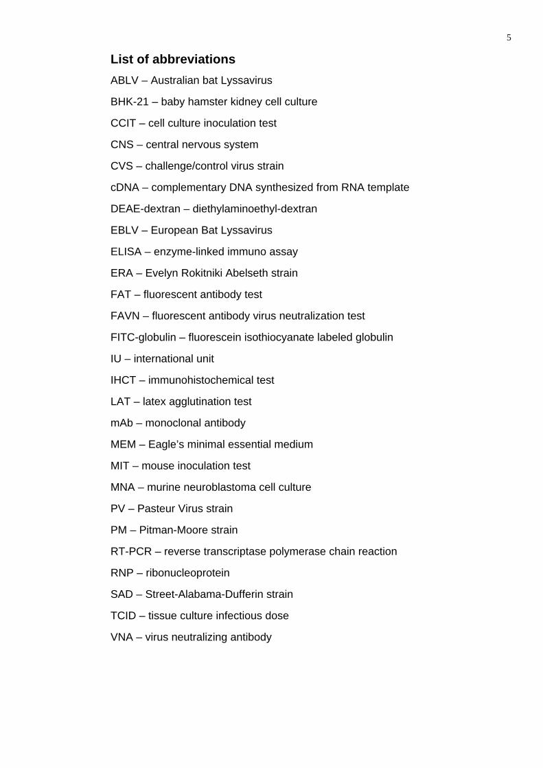
Content List of abbreviations ....................................................................................................... 5
Abstract............................................................................................................................ 6
List of original publications............................................................................................ 8
1. Literature review........................................................................................................ 9
1.2. Rabies virus characteristics ................................................................................ 9
1.2.1. Classification ................................................................................................... 9
1.2.2. Structure of virion, genome, and proteins of rabies virus............................... 10
1.3. Virus-host interaction ........................................................................................ 12
1.3.1. Pathogenesis................................................................................................. 12
1.3.2. Clinical signs.................................................................................................. 13
1.3.3. Pathological changes .................................................................................... 15
1.4. Laboratory diagnosis of rabies .......................................................................... 17
1.4.1. Detection of viral antigen ............................................................................... 17
1.4.2. Detection of viral genome.............................................................................. 18
1.4.3. Virus isolation ................................................................................................ 19
1.4.4. Detection of virus neutralizing antibodies ...................................................... 19
1.5. Rabies epidemiology......................................................................................... 21
1.5.1. Rabies situation world-wide........................................................................... 21
1.5.2. Rabies situation in Europe and in Russia ...................................................... 22
1.5.3. Models of rabies epidemics ........................................................................... 24
1.6. Fixed strains of rabies virus .............................................................................. 25
1.6.1. Laboratory strains.......................................................................................... 25
1.6.2. Vaccine strains and anti-rabies vaccines....................................................... 25
1.7. Molecular biology of rabies viruses ................................................................... 30
1.7.1. Antigenic characterization with monoclonal antibodies.................................. 30
1.7.2. Genetic characterization of rabies virus......................................................... 31
1.7.3. Entire genome sequencing of vaccine strains ............................................... 33
2. Aims of the study .................................................................................................... 35
3. Materials and methods............................................................................................ 36
3.1. Field rabies virus isolates.................................................................................. 36
3.2. Vaccine and laboratory strains.......................................................................... 36
3.3. Cell cultures ...................................................................................................... 36
3.4. Monoclonal antibodies ...................................................................................... 37

4
3.5. Laboratory techniques....................................................................................... 37
3.5.1. Fluorescent antibody test .............................................................................. 37
3.5.2. Cell culture inoculation test............................................................................ 37
3.5.3. Reverse-transcriptase polymerase chain reaction......................................... 38
3.5.4. Nucleotide sequencing .................................................................................. 39
3.5.5. Entire genome sequencing of the vaccine strain RV-97 ................................ 39
3.6. Phylogenetic analysis........................................................................................ 40
4. Results ..................................................................................................................... 42
4.1. FAT and cell culture inoculation test ................................................................. 42
4.2. Antigenic characteristics of field rabies viruses (papers I, II and III).................. 42
4.3. Development of RT-PCR tests (papers II and III).............................................. 43
4.4. Phylogenetic analysis and molecular epidemiology (papers III and IV)............. 44
4.5. Entire genome sequencing and characterization of RV-97 strain (paper V)...... 47
5. Discussion ............................................................................................................... 50
6. Concluding remarks................................................................................................ 59
7. Заключение (concluding remarks in Russian) .................................................... 60
8. Acknowledgments................................................................................................... 62
9. References ............................................................................................................... 63
5
List of abbreviations ABLV – Australian bat Lyssavirus
BHK-21 – baby hamster kidney cell culture
CCIT – cell culture inoculation test
CNS – central nervous system
CVS – challenge/control virus strain
cDNA – complementary DNA synthesized from RNA template
DEAE-dextran – diethylaminoethyl-dextran
EBLV – European Bat Lyssavirus
ELISA – enzyme-linked immuno assay
ERA – Evelyn Rokitniki Abelseth strain
FAT – fluorescent antibody test
FAVN – fluorescent antibody virus neutralization test
FITC-globulin – fluorescein isothiocyanate labeled globulin
IU – international unit
IHCT – immunohistochemical test
LAT – latex agglutination test
mAb – monoclonal antibody
MEM – Eagle’s minimal essential medium
MIT – mouse inoculation test
MNA – murine neuroblastoma cell culture
PV – Pasteur Virus strain
PM – Pitman-Moore strain
RT-PCR – reverse transcriptase polymerase chain reaction
RNP – ribonucleoprotein
SAD – Street-Alabama-Dufferin strain
TCID – tissue culture infectious dose
VNA – virus neutralizing antibody

6
Abstract Rabies is a fatal disease that affects the central nervous system of all warm-
blooded mammals. The rabies virus belongs to the order Mononegavirales, family
Rhabdoviridae, genus Lyssavirus. This virus has a negative single-stranded RNA
genome and the virions are bullet-shaped.
Rabies is reported in many countries throughout the world and has been
registered in all continents except Australia, where only the bat Lyssaviruses have been
found, and in Antarctica where the main vectors of rabies are absent. Russia and most of
the bordering countries are affected by rabies. Finland was a rabies-free country from
1959 to 1988, when a sylvatic rabies epidemic appeared with raccoon dogs as the main
host and vector of infection. That epidemic was eradicated by the oral vaccination of wild
carnivores and the parenteral immunization of dogs and cats; and Finland has been
rabies-free since 1991. However, this status is constantly under threat because rabies is
endemic in Russia and Estonia. In June 2003, a horse imported to Finland from Estonia
was clinically and laboratory diagnosed as rabies positive. The close relationship of the
isolated equine virus strain with the current Estonian strains was verified during
subsequent molecular epidemiological studies. Because the case was imported, it did not
affect Finland’s rabies-free status. Also in 2007 another 2 imported cases of rabies were
recorded: one in a human being from Philippines and the other in a dog from India.
Five different antigenic variants of the rabies virus were identified among rabies
positive field samples from Russia, Finland, and Estonia by using antinucleocapsid
monoclonal antibodies. Two rabies virus field isolates showed a different reaction pattern
that was similar to that of the vaccine strains of the SAD group, which might suggest a
new antigen variant or reverted vaccine strain. Nevertheless, the sequence analysis
showed that the vaccine strains RV-97 and SAD B19 included in the oral anti-rabies
vaccine “Sinrab” (Russia) and “Fuchsoral” (Germany), respectively, differ considerably
from all the field strains.
Field rabies viruses collected in recent years from different regions of the Russian
Federation were chosen on the basis of mAb studies and geographical origin for
molecular epidemiological studies to characterize their genetic heterogeneity and to study
their molecular epidemiology. In addition to the Russian viruses, archival samples from
Estonia and Finland and Russian vaccine strains were also included in this study. Among
the field viruses studied, two main phylogenetic groups were found, and designated as
the Pan-Eurasian and Caucasian based on their geographical origin. The Pan-Eurasian

7
group including some reference viruses from Europe was further divided into four
subgroups. All the vaccine strains were clearly different from the field strains. No
recombination between the field and vaccine virus strains was observed. The critical
roles of geographical isolation, the limitation of the genetic clustering, and the evolution of
the rabies virus were shown during this study.
The rabies virus vaccine strain RV-97 is widely used in Russia as a component of
the oral anti-rabies vaccine “Sinrab”. To characterize the molecular properties of this
strain, entire genome sequencing was conducted. A simple technique was developed to
obtain this sequence, including the 3’- and 5’- ends. The entire genome sequence and
deduced amino-acid sequences of the major viral proteins were compared with the
sequences of other known fixed rabies viruses. The strain RV-97 formed a separate
phylogenetic branch and seems to be more related to the group of Japanese strains. The
field strains from the Caucasian group seem to be phylogenetically the nearest group to
the RV-97 strain.
The data shown herein makes it possible to develop molecular methods for
distinguishing between the field rabies viruses from the vaccine strains for the rapid
recognition of the vaccine strains that are unstable or have reverted back to their
pathogenic form. The wide genetic heterogeneity verified in this study indicates that it is
important to remain on permanent alert for the appearance of rabies.

8
List of original publications
I. Metlin A.E., Cox J., Rybakov S.S., Huovilainen A., Grouzdev K.,
Neuvonen E., 2004. Monoclonal antibody characterization of rabies virus
isolates from Russia, Finland and Estonia. Journal of Veterinary Medicine
Series B 51:94-96.
II. Metlin A.E., Rybakov S.S., Gruzdev K.N., Neuvonen E., Cox J.,
Huovilainen A., 2006. Antigenic and molecular characterization of field and
vaccine rabies virus strains in the Russian Federation. Developments in
Biologicals 125:33-37.
III. Metlin A.E., Holopainen R., Tuura S., Ek-Kommonen C., Huovilainen
A., 2006. Imported case of equine rabies in Finland: clinical course of the
disease and the antigenic and genetic characterization of the virus. Journal
of Equine Veterinary Science 26:584-587.
IV. Metlin A., Rybakov S., Gruzdev K., Neuvonen E., Huovilainen A.,
2007. Genetic heterogeneity of Russian, Estonian and Finnish field rabies
viruses. Archives of Virology 152:1645-1654.
V. Metlin A., Paulin L., Suomalainen S., Neuvonen E., Rybakov S., 2007.
Mikhalishin V., Huovilainen A. Characterization of Russian rabies virus
vaccine strain RV-97. Virus Research, doi:10.1016/j.virusres.2007.11.016

9
1. Literature review
1.1. Definition and history of rabies
Rabies is a fatal viral zoonosis which causes encephalitis in many warm-blooded
animals and humans. There have been indications relative to the occurrence of rabies
since the time of Homer (eighth century B.C.) onwards. The first archival appearance of
the disease was in the fourth century B.C., but precise diagnosis was not possible before
the first century B.C. (Blancou, 1994, 2004; Neville, 2004). The first human rabies
vaccine was developed in 1885 by Louis Pasteur and since then, significant
developments have been made in this field including progress in laboratory diagnosis,
vaccination and rabies control in wild, domestic and farm animals (King et al., 2004).
1.2. Rabies virus characteristics
1.2.1. Classification
The rabies virus belongs to the order Mononegavirales, family Rhabdoviridae,
which includes at least three genera: Lyssavirus, Ephemerovirus, and Vesiculovirus
(Virus Taxonomy, 2005). The Lyssavirus genus is further divided into seven genotypes.
The genotype 1 is known to be the most widespread and comprises the classical field
rabies viruses, and the laboratory and vaccine strains. The rabies-related viruses isolated
in the African continent belong to genotypes 2, 3, and 4, with prototypes of the Lagos bat
virus, Mokola virus, and Duvenhage virus, respectively. The viruses isolated from bats in
Europe represent genotypes 5 and 6 (EBLV1 and EBLV2). The Australian bat Lyssavirus
(ABLV) represents the seventh genotype (Gould et al., 1998; Guyatt et al., 2003).
It has been found that some new rabies virus genotypes exist among viruses
isolated from the territory of the former Soviet Union. It was proposed that the Aravan
virus isolated from a bat in Kyrgyzstan should be classified as the eighth genotype (Arai
et al., 1997). The Khujand virus isolated in Tajikistan can also be classified as a separate
genotype of Lyssavirus (Kuzmin et al., 2003). The existence of two additional Lyssavirus
genotypes among strains isolated from bats (Irkut virus and West Caucasian Bat Virus) in
the Russian Federation was also recently proposed (Botvinkin et al., 2003; Kuzmin et al.,
2005).

10
1.2.2. Structure of virion, genome, and proteins of rabies virus
The virions of the rabies virus have a bullet-shaped structure (Fig. 1) with a
diameter of 75 nm and length of 100-300 nm (Matsumoto 1962; Tordo and Poch, 1988).
The virion contains two major subunits. The first subunit is the ribonucleoprotein (RNP) of
cylindrical and compact form located in the central part of the virion, the second subunit
is a thin surrounding envelope covered with spiky projections (Tordo, 1996).
Figure 1. Rabies virus virions (http://www.wadsworth.org/databank/rabies.html).
The genome of the rabies virus is a non-segmented negative-sense single-
stranded RNA of approximately 12000 nucleotides (nt) in length (Fig. 2). The virus RNA
codes 5 major proteins: the nucleoprotein (N protein), phosphoprotein (P protein), matrix
protein (M protein), glycoprotein (G protein), and the RNA-dependent RNA-polymerase (L
protein). The RNP complex is formed by the N, P, and L proteins associated with the viral
RNA. The RNP is surrounded by a lipid bilayer associated with the G and M proteins
(Goto et al., 2000). The genome also contains several non-coding regions including a G-
L intergenic region called the “pseudogene” or Ψ-region (Fig. 2), which became
suppressed and degenerated during the evolution of the virus (Tordo et al., 1986).
Figure 2. Schematic structure of the rabies virus genome (adapted from
http://www.cdc.gov/ncidod/dvrd/rabies/the_virus/virus.htm).

11
The nucleoprotein consists of 450 amino acids and participates in the transition
from RNA transcription to replication. It encapsidates the positive-strand leader RNA and
prevents further transcription of the genomic RNA (Wunner, 1991; Yang et al., 1998) and
contains several antigenic and immunodominant sites. The antigenic sites I and III
include stretches of amino acids at positions 374-383 and 313-337, respectively (Tordo,
1996). One of the immunodominant sites is known to be located at position 404-418
(Dietzschold et al., 1987; Ertl et al., 1989). Recently, the crystal structure of the rabies
virus nucleoprotein-RNA complex was determined (Albertini et al., 2006).
The P protein is a phosphorylated protein of 297 amino acids, associated with the
L protein to function as a noncatalytic cofactor for the RNA polymerization, and with the N
protein to support adequate RNA encapsidation (Chenik et al., 1994, 1998; Jacob et al.,
2001). Four additional proteins derived from the phosphoprotein gene of the rabies virus
were also found to be present in infected cells, cells transfected with a plasmid encoding
the wild-type P protein, and in the purified virus. It was shown that these proteins are
initiated from secondary downstream in-frame AUG initiation codons. The P-gene is the
only gene shown to encode more than one protein (Chenic et al., 1995). Two antigenic
sites were found to be located at positions 75-90 (Tordo, 1996). The domain 83-172 was
shown to contain the major antigenic determinants (Raux et al., 1997). An
immunodominant site was also mapped in phosphoprotein at positions 191-206 (Larson
et al., 1991), and was identified as the responsible alpha/beta interferon antagonist
(Brozka et al., 2005).
The matrix protein is a 202 amino acid polypeptide which plays a key role in virus
assembly and binding. It covers the ribonucleoprotein (RNP) coil to keep it in a
condensed form and was found to interact specifically with the glycoprotein (Mebatsion et
al., 1999). The major antigenic site is located between the amino acid residues 1 and 72
(Hiramatsu et al., 1992).
The glycoprotein is a 524 amino acid protein and is the most studied protein of the
rabies virus. It has a trimeric structure (Gaudin et al., 1992), with the first 19 amino acids
representing the signal domain which is found only in nascent protein. The glycoprotein
contains several antigenic sites and epitopes. The antigenic epitope I is represented by a
single amino acid at position 231 of the mature glycoprotein. The site II is known to be
discontinuous and involves two separate stretches between positions 34-42 and 198-200.
The antigenic site III is located at position 330-338, epitope VI at position 264, “a” – 342-
343 (Lafon et al., 1983, 1984; Seif et al., 1985; Prehaud et al., 1988; Bunschoten et

12
al., 1989; Benmansour et al., 1991).
The major immunodominant site of the glycoprotein is located between amino
acids 222 and 332 (Johnson et al., 2002b). The amino acid at position 333 of the mature
glycoprotein was found to be associated with viral pathogenicity (Seif et al., 1985;
Badrane et al., 2001). It was shown that either arginine or lysine at position 333 of the
ERA and CVS fixed rabies virus strains is necessary for rabies virulence in adult mice
(Tuffereau et al., 1989).
The L protein is the largest protein of the rabies virus and L gene occupies more
than half of the virus genome. It has been the least studied rabies protein at biochemical
and immunological levels, but the best analyzed theoretically (Tordo, 1996). The L-
protein is associated with the RNP, RNA-dependent RNA polymerase. It participates in
transcription by making five individual mRNAs, one for each viral protein.
1.3. Virus-host interaction
1.3.1. Pathogenesis
Rabies is a central nervous system (CNS) disease that is almost invariably fatal
(Dietzschold et al., 2005), except for few rare reported cases (Miah et al., 2005). The site
of rabies virus entry into the host is usually at the skin or mucosal membrane where the
virus is introduced into the deeper layers of the skin or into the muscular tissue through
biting, licking, or scratching by a rabies-infected mammal, usually a carnivore or a bat
(Charlton, 1994). The transmission of rabies can also occur under unusual
circumstances: by inhalation of large amounts of aerosolized rabies virus, and through
organ transplantation from rabies infected patients (Gode and Bhide, 1988; Krebs et al.,
1995; Hellenbrand et al., 2005; Smith et al., 2006). Rabies-infected animals have in their
salivary glands high titers of the rabies virus, which can be even greater than in the brain
(Charey and McLean, 1983). There are marked differences between the different strains
of virus and their ability to infect, spread within the body, and produce disease (Kaplan,
1996; Dietzschold et al., 2005). It has been suggested that the attenuated rabies viruses
activate the host’s innate immune and antiviral responses, while these responses are
evaded by the pathogenic rabies viruses (Wang et al., 2005). The course of the disease
can be different between animal species: for example, foxes have a comparatively
shorter morbility period than skunks (Sikes, 1962).
After the bite (Fig. 3), the virus particles “travel” to the nearby nerves and then
along the nerve fibers to the brain at a speed of a few millimeters per day (Jackson,

13
1991). It was suggested that the virus is propagated from the entry point to the CNS due
to the interaction between the P protein of the rabies virus and the dynein light chain LC8
(Poisson et al., 2001).
A bite on the head or neck will usually cause symptoms more quickly than a bite
on the hind leg. However, when the virus has entered the nerve endings, it advances
relentlessly up the nerve bodies until it reaches the spinal cord and eventually the brain.
From the brain, the virus can spread to other tissues - the salivary glands, respiratory
system, and the digestive tract (Krebs et al., 1995).
Figure 3. Spread of the rabies virus from the bite site to the CNS (by Bacon &
Macdonald, 1980).
1.3.2. Clinical signs
The clinical signs of rabies are known since the ancient times (Blancou, 1994).
The duration from bite to the appearance of clinical signs varies significantly, ranging
from a few days to a several months. The clinical signs may appear only after the
involvement of numerous neurons, and death may occur as a result from the involvement
of vital nerve centers (Schneider, 1975). There are three phases described in the clinical
course of rabies:

14
Prodromal period – the first 1 to 3 days after the rabies virus reaches the brain.
Vague neurological signs that progress rapidly - some animals may appear tamer,
some will demonstrate intense drooling. Death usually follows within 10 days, due
to paralysis.
Excitative stage – the next 2 to 3 days. This is the "furious rabies" stage - tame
animals suddenly become vicious, attacking humans and other animals as they
roam and wander. Some animals will chew and eat odd objects (rocks, sticks,
etc.). Paralysis then begins, and loss of the ability to swallow will cause frothing at
the mouth of the affected animal.
Paralytic stage - follows the excitative stage, or is the main clinical presentation
for some animals. The throat and chewing muscles are paralyzed, and the animal
is unable to swallow, causing excessive drool, the lower jaw is often dropped. This
is a dangerous period for human contact with domestic animals such as cows and
horses; "choke" (foreign body within the throat) can be a misdiagnosis of rabies,
causing humans to be potentially exposed as they investigate the problem. Similar
situations occur in dogs that appear to be choking (drooling and dropped jaw).
This is also the period when wild animals may seem tame to humans and
nocturnal animals are seen in the daylight. The paralysis progresses from the neck
and jaw to all areas of the body, the animal falls into a coma, and dies within a few
hours.
In bats, the clinical signs of a Lyssavirus-infection include loss of body mass, lack
of coordination, muscular spasms, agitation, increased vocalization, and overt aggression
(Brass, 1994; Whitby et al., 2000; Shankar et al., 2004), but in many cases, rabies in bats
can be clinically silent and left unnoticed before dead animals are found and laboratory
tests are performed (Ronsholt et al., 1998). When bats were found alive, the clinical signs
were generally described as paralysis, unprovoked vocalization, and aggression (biting)
during handling (Rabies bulletin Europe, 1989). However, almost all bats will bite when
handled (Vos et al., 2007).
Because dogs are often responsible for the transmission of rabies to humans,
clinical signs in this species are more elaborately described, studied, understood, and
include: drooping jaw, abnormal sounds when barking, dry drooping tongue, licking of it’s

15
own urine, abnormal liking of water, regurgitation, altered behavior, the biting and eating
of abnormal objects, aggression, biting without provocation, running without no apparent
reason, stiffness upon running or walking, restlessness, biting during quarantine, sleepy
appearance, gait imbalance, and frequent demonstration of the “Dog sitting” position
(Tepsumethanon et al., 2005).
1.3.3. Pathological changes
The pathology of rabies infection is typically characterized by encephalitis and
myelitis. When brain tissue from rabies virus-infected animals is observed with a
histological stain, such as hematoxylin-and-eosin, evidence of encephalomyelitis may be
recognized by light microscopy (Fig. 4 A&B). A perivascular non-suppurative infiltration is
the most frequently noted histological alteration in rabies (Perl, 1975). The cytoplasmic
eosinophilic inclusion bodies (Negri bodies) can often be found in rabies-infected neurons
(Fig. 4 C&D), especially in the pyramidal cells of the hippocampus and the Purkinje cells
of the cerebellum (Negri, 1903). These Negri bodies may vary in size from 0.25 to 27 µm,
and these inclusions have been defined as areas of active viral replication by the
identification of rabies viral antigen (Schneider et al., 1975).

16
A.
B.
C.
D.
Figure 4. A&B. Perivascular cuffing or inflammation around a blood vessel. Perivascular
inflammatory cell infiltrates in hematoxylin-and-eosin stained brain tissue. C&D. Negri
bodies stained with hematoxylin and eosin (C) and Sellers stain (D). All pictures were
taken from http://www.cdc.gov/ncidod/dvrd/rabies/diagnosis/images.

17
1.4. Laboratory diagnosis of rabies
The diagnosis of rabies has to be quick and reliable in order to evaluate the risk of
infection to the exposed individual (Zimmer et al., 1990), and it is also important for health
authorities responsible for the surveillance and control of the epidemics and epizootics
(Perrin et al., 1986). The techniques for the diagnosis of rabies are standardized
internationally, and several tests are available presently. The detection of Negri bodies in
brain smears and the histological examination were the first methods for diagnosing
rabies, but these are not currently used widely because of their low sensitivity and due to
the availability of highly sensitive and specific modern techniques which can be
subdivided into three main groups:
• Detection of viral antigen – fluorescent antibody test (FAT), enzyme-
linked immunoassay (ELISA), immunohistochemical test (IHCT), and latex
agglutination test (LAT);
• Detection and identification of viral genome – reverse transcriptase
polymerase chain reaction (RT-PCR) with subsequent nucleotide
sequencing;
• Virus isolation – mouse inoculation test (MIT) and cell culture inoculation
test (CCIT).
The above mentioned methods will be discussed in the specific subchapters
below.
1.4.1. Detection of viral antigen
Fluorescent antibody test. In 1958, Goldwasser and Kissling reported that the
fluorescent antibody technique could be used to demonstrate rabies virus antigens in
brain tissues of experimentally infected mice. Further studies have shown this method to
be an efficient tool for the diagnosis of rabies, and it later became the reference method
for the diagnosis of this infection (Beauregard et al., 1965). This technique implies the
preparation of smears, impressions or cryosections from brain tissues (Ammon’s horn,
cerebellum, cerebral cortex, and the brain stem), tissue fixation, mostly in cold acetone,
and staining with fluorescein isothiocyanate-labeled polyclonal or monoclonal anti-rabies
antibodies (Kissling, 1975; Dean et al., 1996; OIE, 2004). The FAT allows specific and
highly sensitive detection of the rabies virus antigens in brain smears, salivary gland
sections, and infected cell cultures. It can be used for the intravitam rabies diagnosis in

18
the skin biopsies (Bryceson et al., 1975; Warrell et al., 1988) and to stain rabies virus
antigens in the salivary glands (Goldwasser et al., 1959).
It is also possible to perform the FAT with formalin-fixed paraffin-embedded brain
sections using digestion with proteases, such as pepsin or trypsin (Johnson et al., 1980;
Umoh and Blenden, 1981; Barnard and Voges, 1982; Reid et al., 1983; Metlin et al.,
2007).
Enzyme-linked immunoassay test is a rapid technique that facilitates the
evaluation of a large number of samples simultaneously, which is performed on
microplates previously sensitized with anti-rabies immunoglobulin. Suspensions of
homogenized material are placed on the wells of microplate for specific binding which
can be revealed by the use of a peroxidase conjugate (Perrin et al., 1986). Additionally, a
quantitative ELISA (N-ELISA) for rabies virus detection based on the quantitation of
nucleoprotein (N) in rabies virions captured by rabies-virus-specific polyclonal antibodies
on an ELISA plate can be used for the quantitative detection of both infective and
defective interfering particles of rabies virus (Katayama et al., 1999).
Immunohistochemical testing is predominantly used for research purposes and
allows the perfect identification and localization of rabies virus antigens, and is ideal for
retrospective diagnosis (Johnson et al., 1980; Fekadu et al., 1982; Torres-Anjel et al.,
1984; Palmer et al., 1985; Shin et al., 2004). This method is usually used to stain
histological paraffin sections after deparaffinization, rehydration, and digestion with a
proteolytic enzyme (proteinase K etc.). For the specific staining of rabies virus antigen by
immunohistochemical testing, the primary anti-rabies serum, anti-species serum, and the
peroxidase complex are used (Bourgon and Charlton, 1987; Sinchaisri et al., 1992).
Latex agglutination test (LAT) is a simple and rapid technique, which may be used
more widely in the laboratory diagnosis of rabies in the future. It has been used to detect
rabies virus antigens in the saliva of dogs with 99% specificity and 95% sensitivity. The
essence of the LAT is inducing agglutination on a glass slide using polystyrene latex
beads coated with anti rabies IgG (Kasempimolporn et al., 2000, 2007).
1.4.2. Detection of viral genome
The reverse transcriptase polymerase chain reaction (RT-PCR) with subsequent
nucleotide sequencing permits the diagnosis of rabies, typing, and molecular
epidemiological studies. Since the rabies genome is RNA, the amplification procedure
consists of the reverse transcription of the target RNA strain into complimentary DNA

19
(cDNA), followed by the amplification of the cDNA by PCR (Tordo et al., 1995, 1996).
The RT-PCR procedure consists of the following steps: total RNA extraction,
cDNA synthesis with random or specific primers, amplification of the cDNA with specific
primers, and visualization of the results with horizontal electrophoresis in agarose gel
containing ethidium bromide observed under UV light (Heaton et al., 1999).
The RT-PCR is widely used for rabies diagnosis, and different parts of the genome
can be targeted for this reason, but in most cases the N gene is utilized (Kulonen and
Boldina, 1993; Bourhy et al., 1999; Ito et al., 2003; Losa-Rubio et al., 2005). A rapid RT-
PCR technique was developed for the detection of the classical rabies virus (genotype 1)
and the rabies related EBLVs (genotypes 5 and 6), and also to distinguish between the
six established rabies and rabies-related virus genotypes (Black et al., 2000, 2002). The
PCR can also be applied to detect the rabies virus genome in formalin-fixed paraffin-
embedded brain tissue (Kulonen et al., 1999) and for the intravitam diagnosis of rabies in
humans by testing saliva and cerebrospinal fluid (Crepin et al., 1998). The Real-time
PCR is a quantitative technique which allows the detection of an increase in the amount
of DNA (cDNA) during amplification. It is currently used for the ante- and post-mortem
diagnosis of rabies and the discrimination of the Lyssavirus genotypes (Wakeley et al.,
2005; Nagaraj et al., 2006; Saengseesom et al., 2007).
1.4.3. Virus isolation
The mouse inoculation test (MIT) was one of the first diagnostic tests for rabies.
Laboratory mice are inoculated intracerebrally or subcutaneously with the supernatant of
the sample suspension. The inoculated mice must be observed for up to 30 days after
inoculation. Death during the first 48 hours after inoculation must be considered as non-
specific; all the animals dead after this period must be dissected and brain samples must
be tested for rabies by the FAT (Koprowski, 1996). This method can be used for testing
the brain and salivary gland suspensions, as well as the saliva samples, for the presence
of live rabies virus (Adeiga and Audu, 1996; Delpietro et al., 2001).
The cell culture inoculation test has already replaced the MIT in many countries
and implies the isolation of rabies virus in a cell culture monolayer with visualization by
the FAT. A number of cell lines have been selected and tested: cow brain cells (CB3),
cerebral and cerebellar grey matter cells of mice (MBC-2, MBC-3), chicken embryo
fibroblasts (CEF), murine, feline and human glial cultures, human monocytic U937 and
THP-1 cells, murine macrophage IC-21, murine monocytic WEHI-3BD- and PU5-1R

20
cells, murine monocytic P388D1 and J774A.1 cells, kidney epithelial cells derived from
African green monkey (Vero), and McCoy cells (Smith et al., 1978; Clark, 1980; Celer et
al., 1991; Ray et al., 1995, 1997; Nogueira, 2004). Tollis et al., (1988) compared the
sensitivity of the murine neuroblastoma (MNA), baby hamster kidney (BHK-21), and the
canine fibrosarcoma A-72 cells, and confirmed that the MNA cells are the most sensitive
to infection with the wild strains of the rabies virus. MNA cells are currently widely used
for field virus isolation and a method employing this cell culture is recommended by the
WHO and the OIE (Webster et al., 1996, OIE, 2004).
1.4.4. Detection of virus neutralizing antibodies
The detection of anti-rabies virus neutralizing antibodies (VNA) is widely used to
evaluate the potency of anti-rabies vaccines because the minimal level of the VNA
needed to protect animals against rabies has been determined as ≥ 0.5 IU/ml (OIE,
2004).
Virus neutralization assay has also been found useful for the monitoring of
Lyssaviruses among bats (Arguin et al., 2002; Lumlertdacha et al., 2005).
Virus neutralization in mice or cell cultures. The determination of the VNA was
previously conducted by virus neutralization in mice (Atanasiu, 1973) and subsequently
replaced by the fluorescent antibody virus neutralization (FAVN) or rapid fluorescent
focus inhibition testing (RFFIT) (Thomas, 1975; Zalan et al., 1979; Smith et al., 1996;
Cliquet et al., 1998). This method enables the determination of antibody levels by the
neutralization of a known dose of the rabies virus (commonly the CVS strain). Serum
samples are tested and compared with the neutralization of reference standard serum
with an antibody level of 0.5 or 1.0 IU/ml. This test can be conducted on microplates and
the results viewed with fluorescence microscopy. The registration of the results can be
automated by various means: by using an inverted fluorescence microscope coupled with
a video camera and color image analysis software computer system (Peharpre et al.,
1999); a peroxidase conjugate can be used instead of the fluorescent conjugate, and in
this case an automatic multi-channel spectrophotometer can be used for the registration
of the results (Hostnik, 2000a); flow cytometry method for calculating anti-rabies VNA has
also automated results registration (Bordignon et al., 2002).
ELISA can also be used to determine antibody levels in serum samples
(Kasempimolporn et al., 2007).
21
This method can be used for assessing the efficacy of oral fox vaccination campaigns
and it was demonstrated that by using commercial ready-to-use microplates sensitized
with rabies virus glycoprotein, a simple and rapid ELISA technique enables the obtaining
of a rabies antibody quantization in field fox serum samples (Mebatsion et al., 1989;
Esterhuysen et al., 1995; Cliquet et al., 2000, 2003, 2004, 2007; Servat et al., 2007).
The latex agglutination test is also used to detect rabies-specific antibodies. Latex
beads are sensitized by coating them with purified rabies glycoprotein to detect anti-
glycoprotein antibodies in serum samples. The visible agglutination is observed in the
positive sera with a titer ≥ 2 IU/ml within 3–5 min after mixing, while serum samples
containing less than 2 IU/ml do not agglutinate (Madhusudana and Saraswati, 2003).
1.5. Rabies epidemiology
1.5.1. Rabies situation world-wide
Rabies is widely distributed throughout the world and is present in all continents
except Australia, where only bat Lyssavirus has been found, and in Antarctica.
Worldwide, it has been estimated that approximately 55000 persons die annually due to
rabies; 99% of human rabies deaths have occurred in the developing countries. The total
global expenditure on rabies prevention is well over US$ 1 billion annually (Warrel et al.,
1995; WHO Expert Consultation on Rabies, 2005).
Different animal species can be responsible for viral circulation and rabies
transmission in different continents and countries. Canids have been determined to be
the main hosts of the rabies virus in Africa; in most cases they are also responsible for
the transmission of the virus to humans. In addition to canids (domestic and wild dogs,
jackals, and wolves), mongooses, and bats are involved in rabies epidemics, as occurs in
Africa (Adeiga et al., 1996; Bingham, 2005). Dogs are also the primary reservoir for
rabies in Thailand (Tepsumethanon et al., 2005). In the USA, several species are
involved in rabies epidemics but the main reservoirs are raccoons and skunks (Krebs et
al., 2003). An epizootic of raccoon rabies, begun in the USA in the late 1970s, and
developed into one of the largest and most extensive in the history of wildlife rabies
(Childs et al., 2000). Rabies has been detected in rodents and lagomorphs, mostly in
woodchucks (Childs et al., 1997) and also in arctic foxes (Ballard et al., 2001). In
addition, bats are sometimes responsible for the transmission of rabies to humans (Miah,
2005).

22
1.5.2. Rabies situation in Europe and in Russia
Foxes and raccoon dogs are considered to be the most susceptible species in
Europe (Artois, 1992; Kihm et al., 1992; Gylys et al., 1998). According to rabies data for
the years 2005 and 2006, there were 13 rabies-free countries in Europe: Belgium, the
Czech Republic, Cyprus, Finland, Greece, Iceland, Italy, Ireland, Luxembourg, Norway,
Portugal, Sweden, Switzerland, and Lichtenstein. With 9,830 cases of rabies (including 9
human and 35 bat cases) in 2005, the total number of cases of rabies in Europe
(European countries and European parts of Russia) has increased by 80% when
compared with 2004 to approximately similar level as that of 2003. This increase is based
on a 2 to 2.3-fold higher reporting of rabies cases from Russia and Ukraine (Rabies
Bulletin Europe, 2005). A similar tendency was observed in Turkey, where dog-mediated
rabies is the main problem (Rabies Bulletin Europe, 2005; Johnson et al., 2006; Kilic et
al., 2006). Finland is a rabies-free country but three imported rabies cases were recorded
recently. In 2003 rabies was found in a horse imported from Estonia (Metlin et al., 2006);
in 2007a human case was recorded in a Philippine male working on a cruise ship (Kallio-
Kokko H., personal communication); later in the same year, another rabies case occurred
in a pappy imported from India (communication of the Ministry of Agriculture and Forestry
of Finland).
Wild animals, manly red fox, are still the main rabies hosts in Europe. In 2006,
9172 rabies cases were reported in Europe, 6152 of these occurred in wild animals, 2984
in domestic animals, and only 34 in bats. In addition, 2 human rabies cases were
reported (Rabies Bulletin Europe, 2006).
Rabies is endemic in the Baltic countries; in 2006, 114 animal rabies cases were
recorded in Estonia, 472 in Latvia, and 2232 cases in Lithuania. The number of rabies
cases in animals from Estonia decreased in 2006 by 57% when compared with 2005.
This may be due to the oral wildlife vaccination started in Estonia during 2005 (Niin et al.,
2007). In Latvia and Lithuania, an increase in the number of animal rabies cases was
recorded in 2006 compared with 2005 (an increase of about 10% in Latvia and
approximately 26% in Lithuania (Rabies Bulletin Europe, 2006).
Rabies is a very serious veterinary and public health problem in the Russian
Federation. The disease is widespread throughout the country (Fig. 5) and affects
different animal species: farm animals (mainly cattle, pigs, sheep, goats, and horses),
domestic pet animals (predominantly dogs and cats), and wild animals (red foxes,

23
raccoon dogs, badgers, wolves, lynx, etc.). The disease is enzootic in Russia and there is
an increasing tendency in the number of rabies affected territories (Dudnikov, 2003b);
also almost 10 human rabies cases are reported annually.
Figure 5. Map of the Russian Federation and bordering countries. The Russian
regions affected by rabies (according to data recorded in 2006) are colored dark grey.
The largest outbreaks of rabies have occurred in the south-western regions of the
Russian Federation and rabies is spreading further to the northern and eastern directions
(Makarov and Vorob’ev, 2004). The number of animal rabies cases increased annually
from 2000 to 2005, except for 2004, when a decrease in rabies cases was recorded (Fig.
6). In 2006, the number of animal rabies cases decreased substantially (approximately
57%).
Figure 6. Dynamics of the number of animal rabies cases (total for all species) in
Russia during the last 8 years.

24
It is well established that the main reservoirs of the rabies virus in the Russian
Federation are wild carnivores. These animals are very susceptible to rabies, intensively
excrete virus with saliva, are inclined to long and distant migrations, are aggressive and,
in addition, have a high population density. These factors in combination with the
elevated rate of generation change and relatively long incubation period assure the
continuous epidemic process despite the fatality of infection. Foxes are considered to be
the main wild vector of rabies in Russia, followed by raccoon dogs (Sidorov et al., 2004).
Raccoon dogs have been introduced to the European part of the former Soviet Union in
the 1930-1940, and became an important vector for rabies in Russian. The first cases of
rabies in these animals were recorded during the winter of 1931-1932 and the first case
of human rabies after being bitten by a raccoon dog occurred in 1951 (Botvinkin et al.,
1981).
The existing situation with regards to rabies diagnosis within the regions of the
Russian Federation does not allow any conclusions to be drawn relative to the
dissemination of rabies, and therefore systematic monitoring is needed (Dudnikov,
2003a).
1.5.3. Models of rabies epidemics
Understanding the interaction between ecological dynamics, spatial spread, and
evolutionary changes in infectious diseases is important, and will help in the interpretation
of epidemiological patterns and provide a basis for constructing a predictive theory of
disease emergence (Grenfell et al., 2004). The Kendall’s threshold theorem (Kendall,
1957) states that an epidemic will occur if the contact between infective and susceptible
individuals is as such that each infected individual, on average, meets and infects more
than one susceptible individual. If this happens, the disease frequency begins to increase
exponentially, if not – it declines exponentially. In reality, the exponential changes do not
persist for a long period because the number of susceptible individuals becomes limited
and there are consequent changes in the rate of contact (Bacon and Macdonald, 1980).
To predict local and spatial dynamics of the epizootics of rabies, mathematical
models have been developed and are now widely used in rabies epidemiology. These
models are based on population density, recorded data of rabies cases, landscape
particularities, etc. (Moore, 1999; Childs et al., 2000; Smith et al., 2002; Russell et al.,
2005). A simulation model was used by Thulke et al. (1999) to study the spatio-temporal

25
dynamics of a potential rabies outbreak in an immunized fox population after the
termination of a long-term, large-scale vaccination programme with two campaigns per
year: one in spring and the other in autumn. The use of integrated approaches, such as
the geographical analysis of sequence variants, coupled with information on spatial
dynamics is an indispensable aid to understand the patterns of disease emergence (Real
et al., 2005).
1.6. Fixed strains of rabies virus
1.6.1. Laboratory strains
Two laboratory strains, the CVS (Challenge/Control Virus Strain) and PMPV
(Pitman-Moore Pasteur Virus), are widely used. Both strains are considered to be
derivates of the original Pasteur virus strain (Smith et al., 1993). The CVS strain has two
stable variants: CVS-B2c and CVS-N2c, these differ in pathogenicity for healthy mice and
in the capacity to affect neurons (Morimoto et al., 1999). These laboratory strains are
used in different diagnostic tests such as the virus neutralization and focus inhibition
tests, as well as for the potency testing of anti-rabies vaccines in laboratory animals.
1.6.2. Vaccine strains and anti-rabies vaccines
Since the first rabies vaccination in 1885 by Louis Pasteur (Pasteur, 1885),
significant progress has been made in improving the pre- and post-exposure treatment of
human rabies (Dietzschold et al., 2003). There are several types of vaccines: live
attenuated, inactivated (killed), DNA-based, and vector vaccines. For the production of
anti-rabies vaccines, a number of attenuated vaccine strains are employed: the Pasteur
Virus (PV), Evelyn Rokitniki Abelseth (ERA), Street-Alabama-Dufferin (SAD), 3aG,
Fuenzalida S-51 and S-91, Ni-Ce, SRV9, PM, Nishigahara, RC-HL, Kelev, Flury,
“Shelkovo-51”, “O-73 Uz-VGNKI”, “RV-71”, “Krasnopresnenskii-85”, and the RV-97 strain
(Steck et al., 1982; Fodor et al., 1994; Gruzdev and Nedosekov, 2001; Ito et al., 2001b;
Borisov et al., 2002). The PV is one of the first vaccine strains; it was isolated from a
rabid cow in 1882 and attenuated by multiple passages in rabbits. The SAD strain was
isolated from a rabid dog in Alabama (USA) in 1935 and adapted for cultivation in the
mouse brain and in the baby hamster kidney cell culture. It has two main derivates: ERA
and Vnukovo-32. Several variants of the SAD strain exist: SAD-Berne, SAD B19, SAD-
P5/88 etc., and also non-virulent mutants SAG-1 and SAG-2. The vaccine strains
belonging to the SAD group are widely used throughout the world. One of the most
26
widely used oral anti-rabies vaccines is prepared from the SAD B19 strain, the high
immunogenicity and relative safety of this strain has been demonstrated experimentally
(Vos et al., 2000; Neubert et al., 2001).
Live attenuated vaccines are still in use in some developing countries for
parenteral vaccination of animals and humans. These contain live attenuated rabies virus
which has been developed in cell cultures or in live animals such as sheep. In the
developed world, live attenuated vaccines are only used for the oral immunization of wild
animals. Oral vaccines are widely used and several vaccine strains are used for the
production of such vaccines: the SAD B19 and other SAD-strains, SAG1 and SAG2 –
apathogenic deletion mutants, Vnukovo-32, and the VRG strain (Brohier et al., 1991; Vos
et al., 2000). The vaccine strain RV-97 is used in Russia for producing the oral anti-rabies
vaccine “Sinrab”. This strain was obtained in the FGI “Federal Centre for Animal Health”
(Vladimir, Russia) from strain RB-71. The ancestor to these two strains is the strain
“Sheep”, derived from the PV strain. The strain “Moscow” is also believed to be a
derivate of the PV strain (Gruzdev and Nedosekov, 2001), and was used in the former
USSR for producing anti-rabies vaccine. The strain RV-97 is adapted for cultivation in cell
culture BHK-21 (Borisov et al., 2002).
Inactivated vaccines. Complete inactivated rabies virus particles are highly
immunogenic. The vaccines based on this principle are used for the pre- and post-
exposure immunization of humans and domestic animals (Dietzschold et al., 2003). The
inactivated chicken embryo vaccines and vaccines based on virus cultivated in cell
cultures are used for veterinary and medical purposes (Sihvonen et al., 1993, 1994,
1995; Briggs et al., 1996; Benjavongkulchai et al., 1997). Modern medical vaccines can
be administered by the intradermal route (WHO, 1995; Dressen, 1997; WHO, 1997).
DNA vaccines are based on plasmid vectors expressing rabies virus glycoprotein.
These vaccines have been tested for their efficiency in several animal species (mice,
dogs and nonhuman primates), and it has been found that the DNA vaccine develops
VNA levels and offers protection comparable with those obtained with the inactivated
vaccines (Ray et al., 1997; Wang et al., 1998; Bahloul et al., 1998; Perrin et al., 1999;
Lodmell, 1999; Osorio et al., 1999; Lodmell et al., 2000, 2001, 2002). On the basis of the
results of the study conducted in mice, a single administration of the rabies DNA vaccine
may be as effective as at least five injections of the cell-culture-derived vaccine (Bahloul
et al., 2003).

27
Vector vaccines are based on recombinant viruses, and several viruses have been
tested for these purposes. The VRG vaccine was designed on the basis of poxvirus
(vaccinia virus) expressing SAD strain glycoprotein and used for oral immunization of
wildlife (Wiktor et al., 1984; Brochier et al., 1990, 1991; Winkler et al., 1992; Meslin et al.,
1994). The Adrab.gp - vaccine is based on the adenovirus expressing the ERA strain
glycoprotein and was found capable of inducing an immune response in dogs (Tims et
al., 2000). The canine herpesvirus (CHV) expressing the glycoprotein of rabies virus has
also been used successfully as an anti-rabies vaccine (Xuan et al., 1998). A raccoon
poxvirus (RCNV) recombinant vaccine for the immunization against feline panleukopenia
and rabies has been developed and tested in cats (Hu et al., 1997). A recombinant rabies
virus vaccine carrying two identical glycoprotein (G) genes (SPBNGA-GA) has also been
constructed (Faber et al., 2002).
The rabies virus vaccine strain based on vectors have shown great promise as
vaccines against other viral diseases such as human immunodeficiency virus type 1
(HIV-1) infection and hepatitis C, but a low residual pathogenicity remains a concern for
their usage (McGettigan et al., 2003).
Plant-derived antigens can also be used for the immunization against rabies. The
coat protein of alfalfa mosaic virus has been used as a carrier molecule to express the
antigenic peptides from rabies virus. The in vitro transcripts of the recombinant virus with
sequences encoding the antigenic peptides have been synthesized from DNA constructs
and used to inoculate tobacco plants. The plant-produced protein (virus particles) has
been purified and used for the immunization of mice, and specific anti-rabies virus-
neutralizing antibodies in immunized mice have been found (Yusibov et al., 1997;
Modelska et al., 1998); spinach has also been used for this purpose (Koprowski, 2002).
The transgenic maize expressing the G protein of the Vnukovo strain has also been
obtained and tested in mice. It was shown that the mice developed virus neutralizing
antibodies which were able to provide protection of 100% against the challenge of a
vampire bat strain (Loza-Rubio et al., 2007).
Oral vaccination of wildlife against rabies. Before the era of oral vaccines, the only
feasible measure for controlling rabies in wildlife was the depopulation of reservoir
species (Aubert, 1994); but currently rabies is the only zoonosis that can be controlled by
the oral vaccination of wildlife. The idea of conducting active immunization of wildlife
appeared in the last century (Baer, 1975), but many difficulties, such as the form of the
vaccine, methods of distribution and uptake control, and possible residual pathogenicity

28
have to be surpassed. Since then, several laboratory and field trials have been
conducted (Wandeler et al., 1988), and different delivery methods including vaccine traps
and wool getters were designed (Winkler and Bogel, 1992; Matter et al., 1998). Initially,
plastic vessels containing the vaccine were attached to chicken heads (Steck et al.,
1982), but recently different types of modern vaccine baits and different meal mixtures for
producing these were developed and tested (Linhart et al., 1997). The vaccine based on
the strain SAD B19 is one of the most widely used in Europe: 70 million vaccine baits
were used between 1983 and 1988 (Vos et al., 2000). Studies on the immunogenicity
and efficacy of the SAD B19 attenuated rabies virus vaccine in foxes were conducted
under laboratory conditions (Neubert et al., 2001).
Vos et al. (1999) studied the safety of the SAD B19 vaccine in 16 animal species
by different administration routes; a low residual pathogenicity was observed only for
certain rodent species, but transmission of the vaccine virus to control animals was not
demonstrable, since no vaccine virus was detected in the saliva of the six mammal
species examined. Furthermore, the genetic stability of the SAD B19 vaccine was shown
through passage in neural tissue of dogs, foxes, and mice. From those results presented
here on the innocuity and stability, it can be concluded that the SAD B19 rabies vaccine
is suitable for the oral vaccination campaigns of carnivores against rabies (Vos et al.,
1999). Nevertheless, several rabies cases have been caused by live attenuated viruses
(Pastoret et al., 1999; Wandeler, 2000), so the development of new, safer vaccine strains
is a very important issue.
Two mutant vaccine strains were obtained by directed mutagenesis of the strain
SAD. The SAG-1 contains one nucleotide substitution, while the SAG-2 has two
substitutions at amino-acid position 333 of the rabies virus glycoprotein (Follmann et al.,
1996). These vaccine strains are apathogenic for adult mice inoculated by the
intracerebral route (Flamand et al., 1993). The SAG-2 based vaccine was demonstrated
as a safe and effective vaccine for the oral immunization of canines (Fekadu et al., 1996;
Masson et al., 1996; Bingham et al., 1997, 1999; Lambot et al., 2001).
The vector-based VRG vaccine is another candidate for the oral application to
immunize wild carnivores. The pathogenicity of a vaccinia recombinant virus expressing
the rabies glycoprotein was tested with the red fox, wild boar, Eurasian badger, different
species of mice and voles, common buzzard, kestrel, carrion crow, magpie, and jay.
During the observation period, the 107 animals given the vaccine orally did not show any
clinical signs (Brochier et al., 1988, 1989). Experiments have demonstrated the efficacy

29
of a vaccinia-rabies recombinant virus administered by the oral route in foxes. Because of
its safety and heat-stability, this recombinant virus could be an excellent alternative to the
attenuated strains of rabies virus currently used in the field (Brochier et al., 1990, 1991,
1996; Desmettre et al., 1990; Lambot et al., 2001). The high thermo stability of the
commercially produced Raboral VRG bait allows its use during the summer for
emergency vaccination campaigns (Masson et al., 1999), and is being used for the
vaccination of wild raccoons in the USA (Olson et al., 2000). The VRG vaccine has also
been tested as an oral vaccine in vampire bats and significant protection was observed in
animals vaccinated 18-30 days before challenge (Setien et al., 1998).
Oral vaccination of wild animals has been successfully conducted in many
countries: such as Austria, Croatia, Switzerland, Italy, Germany, Slovenia, Czech
Republic, Slovakia, Israel, USA, Canada, Belgium, France, etc. (Steck et al., 1982;
Westerling, 1989; Gram, 1996; Separovic, 1996; Schluter, 1996; Svrcek et al., 1996;
Matouch, 1996; Mutinelli, 1996; Linhart et al., 1997; Olson et al., 1999, 2000; Hostnik,
2000b; MacInnes et al., 2001). The sylvatic rabies epidemic of 1988-1989 was
successfully eradicated in Finland by the oral immunization of wild carnivores (Nyberg et
al., 1992), and was also used in some areas of Russia. One of these vaccination areas is
located at the Finnish-Russian border within the Leningrad region and the Republic of
Karelia. The oral vaccination was organized in 2003 within the framework of the
international Finnish-Russian collaboration program for controlling rabies in wildlife, and
financially supported by the EU and the Finnish government (Metlin et al., in press).
The most common strategy for conducting oral vaccination campaigns is to use
vaccine baits at a density of approximately 25 baits per square km, twice a year, during
the spring and autumn, to avoid the negative influence of temperature on vaccine baits
and to reach adult foxes (in spring) and juvenile foxes before they disperse (in autumn)
(Aubert et al., 1994; Vos, 2003). However, further studies on the population dynamics of
the red fox, the onset and progress of the reproductive season, and maternal immunity
and the immune response of fox cubs to oral vaccination have shown that it was
necessary to optimize the old strategy and conduct two spring vaccinations: first in
March, and then before the end of May to cover young foxes (Muller et al., 1999, 2001;
Vos et al., 2001).

30
There are two ways of distributing vaccine baits in nature: manually and by air
(helicopters, airplanes). Presently, aerial distribution is widely used and special computer
models have been developed to plan the distribution of vaccine baits taking into account
many factors including landscape and terrain details (Thulke et al., 2004).
1.7. Molecular biology of rabies viruses
1.7.1. Antigenic characterization with monoclonal antibodies
The first mAbs of the rabies virus were obtained in 1978 (Wiktor et al., 1978).
Study of rabies virus isolates with mAb directed against particular viral proteins allows
antigenic characterization of the virus being evaluated and, in many cases, strain and
serotype differentiation (Mebatsion et al., 1992; Delpietro et al., 1997; Nadin-Davis et al.,
2000; Okoh, 2000).
With the help of mAbs directed against the different viral proteins, their antigenic
and functional properties have been determined. Luo et al. (1998) studied the rabies virus
glycoprotein with a panel of 35 mAbs and concluded that the G protein forms a complete
antigenic structure with conformational-depended antigenic sites and epitopes. Goto et
al. (2000) have used mAbs to map antigenic epitopes and to analyze the structure of the
nucleoprotein antigenic sites.
Monoclonal antibodies were also used in Finland during the last sylvatic rabies
outbreak. All the viruses isolated were studied using a panel of three mAbs (W-239, W-
187.5, and P-41), and all induced positive reactions indicating the persistence of the
Arctic antigenic variant of the rabies virus. Later, using the same panel of mAbs, 24
rabies samples collected from Estonia between 1989 and 1992 were studied; two
different Arctic variants were found, one of these having the same characteristics as the
Finnish isolates, the other demonstrated a unique (W-187.5 – negative) reaction
(Kulonen et al., 1993).
The three mAbs mentioned above have been included in more extensive panels
(up to 20 mAbs) and were used for the characterization of rabies viruses isolated in
Europe (Cox et al., 1992) and different parts of the African continent (Umoh et al., 1990;
Mebatsion et al., 1992). A wide range of different groups of rabies viruses was found in
these studies.
Several panels of mAbs, of different origins, have been applied during the last two
decades in Russia. In 1983, Selimov et al. have studied 39 rabies virus strains using a
panel of 4 monoclonal antibodies; later on this work was continued and 271 field rabies

31
virus isolates were studied with a mAb P-41 (Selimov et al. 1983, 1994). Gribencha et al.
(1989) developed a panel of 7 anti-nucleocapsid and 3 anti-glycoprotein mAbs, and these
have demonstrated that individual variants of the rabies virus can be detected using this
panel. Botvinkin et al. (1990) applied a panel of 39 mAbs to characterize 98 rabies
viruses, which resulted in several diverse reaction patterns indicating that different
antigen variants were found during those studies. In 1991, Gribencha et al. have
published research studies aimed at obtaining a panel of mAbs to characterize
antigenically the vaccine strain Vnukovo-32 and to compare its antigenic properties with
the field rabies viruses (Gribencha et al., 1991a, b). Botvinkin et al. (2006) applied a wide
panel of 74 mAbs and compared the results obtained with phylogenetic data, where it
was demonstrated that the results of the antigenic studies are often in concordance to
those of genetic studies.
1.7.2. Genetic characterization of rabies virus
According to the WHO Expert Consultation on Rabies (2005) it is important to
conduct molecular characterization of the new field isolates of the rabies virus. Several
phylogenetic and molecular-epidemiological studies on rabies have been performed
during the past 10 years (Smith et al., 1992; Kissi et al., 1995; Bourhy et al., 1999, Nadin-
Davis et al., 1999; Holmes et al., 2002; Kuzmin et al., 2004, Real et al., 2005). These
studies have shown that the rabies viruses can be divided into two major groups, one
comprising viruses isolated from terrestrial mammals and the other containing viruses
isolated from bats or spillover infections from bats.
Additionally, there is a viral lineage that is closely related to the bat rabies virus but
which circulates independently in raccoons and skunks, suggesting that it might
represent a secondary transmission from bats. It was also found that among terrestrial
mammals, rabies viruses cluster more by geographical origin than by host species, and in
this case, closely related viruses infect a variety of species (Davis et al., 2006). The
phylogenetic reconstruction strongly supports the hypothesis that host switching has
occurred in the history of the Lyssaviruses. It has been proven that the Lyssaviruses
have evolved in chiropters long before the emergence of the carnivoran rabies, probably
due to spillovers from bats (Badrane and Tordo, 2001). The rabies virus is an ancient
virus but it has been suggested that the current diversities in the genotype 1 of the
Lyssaviruses from diverse geographical locations and different species may have started
only within the last 500 years (Holmes et al., 2002).

32
The RT-PCR amplification, nucleotide sequencing of the different genome regions,
and the subsequent genetic and phylogenetic analysis allows the determination of the
genetic groups and the differentiation of the field and vaccine strains of the rabies virus.
Different genome regions can be and have been used in molecular-epidemiological
studies of the rabies virus. The G gene and the G-L intergenic region (pseudogene, ψ-
region) are much more variable than the N gene and evolutionary pressure on individual
protein coding genes within the genome varies considerably (Johnson et al., 2002a). The
pseudogene region has been used for the genetic characterization of rabies viruses in
several studies (Bourhy et al., 1999; Hyun et al., 2005; Nel et al., 2005). The analysis of
the sequence of the nucleoprotein is considered to be adequate for reliable phylogenetic
study of the rabies virus, and the additional glycoprotein gene sequence analysis is
important for the characterization of antigenic and immunogenic properties of the virus
(Kasempimolporn et al., 2004; Kuzmin et al., 2004).
During the last century, rabies virus was spreading to the West and South of
Europe but natural barriers, such as the Vistula River in Poland, were able to limit its
dissemination (Bourhy et al., 1999). Bourhy et al., (1999) have classified rabies viruses of
European origin into four main groups: the NEE-group (North-East Europe), the EE-group
(East Europe), the WE-group (Western Europe), and the CE-group (Central Europe).
Recently, Kuzmin et al. (2004) studied a wide range of rabies viruses isolated from the
territory of the former Soviet Union and classified these into five genogroups (A, B, C, D,
and E). The data from that study have shown that the viruses with the same geographical
origin often group together during phylogenetic analyses. The number of rabies virus
variants is co-circulating in Europe, and are often associated with the red fox; also there
are dog-associated rabies and the role of raccoon dogs in maintaining rabies in the Baltic
countries is increasing (McElhinney et al., 2006). Mansfield et al. (2006) have
demonstrated the existence of two groups within the general Arctic group: “Arctic 1” and
“Arctic 2”, the latter having two subgroups and a separate “Arctic-like” group. The study of
isolates from countries in the Middle East has shown the existence of few closely related
groups which are different from the viruses of European and African origin; no host-
dependent relations were found in this study (David et al., 2000). A molecular study of
Brazilian rabies viruses has shown the presence of three main host-specific groups,
especially among bat viruses and victims of vampire bats (Bernardi et al., 2005). Some
authors have combined the use of mAbs and molecular methods to study the
characteristics of rabies viruses (Mebatsion et al., 1993; De Mattos et al., 1996, 1999;

33
Nadin-Davis et al., 2001, 2003; Favoretto et al., 2002).
Most of the rabies cases in terrestrial animals and human beings are due to the
genotype 1 viruses, but some cases are caused by viruses from other genotypes. Spill-
over of the EBLV1 virus into the stone marten (Martes foina) under natural conditions has
been recorded in Germany (Muller et al., 2004). Also in 1998 cases of rabies in sheep
that were shown to have been infected with the EBLV-1a possibly derived from
insectivorous bats were observed in Denmark (Stougaard and Ammendrup, 1998), and in
2002, a second occurrence of the EBLV-type 1 in sheep was reported (Ronsholt, 2002).
Also, several human cases caused by EBLVs and ABLV strains have been reported in
some European countries and in Australia (Lumio et al., 1986; McColl et al., 2000; Fooks
et al., 2003; Spooner, 2003).
One of the most important issues in the field of rabies research is the identification
and characterization of new genotypes. Due to PCR and nucleotide sequencing during
the last ten years, one new genotype has been identified and four additional were
proposed. Fraser et al. (1996) isolated a new Lyssavirus from bats in Australia, which was
later identified by nucleotide sequencing as the new seventh genotype: Australian bat
Lyssavirus or ABLV (Gould et al., 1998). The phylogenetic analysis of the ABLV viruses
has shown that they form a monophyletic group distinct from the other Lyssaviruses, and
two antigenic variants of ABLV were described (Guyatt et al., 2003). Arai et al., (2003)
studied rabies virus isolated from the Lesser Mouse-eared Bat (Myotis blythi) in
Kyrgyzstan and have suggested that it belonged to a new genotype of the rabies virus
(Aravan virus). Furthermore, the Khujand virus isolated from northern Tajikistan in 2001
can be classified as a separate genotype of Lyssavirus (Kuzmin et al., 2003). Two unique
viruses (Irkut and West Caucasian) have been isolated from bats in Russia and based on
genetic studies it has been suggested that they belong to new genotypes (Botvinkin et al.,
2003; Kuzmin et al., 2005).
1.7.3. Entire genome sequencing of vaccine strains
The entire genome sequencing of the vaccine strains provides important data
relative to the rabies virus genome and allows its antigenic, genetic, and immunogenic
properties to be predicted and analyzed. To conduct complete sequencing of the rabies
virus genome, several techniques can be employed. Tordo et al. (1986, 1988) used
cloning in plasmid vector pBR322 and sequence determination by the chain-terminating
inhibitor method after inserting endonuclease restriction fragments of the cDNA inserts

34
into the M13 vectors. Conzelmann et al. (1990) employed the ligation of the synthetic
oligonucleotide to the genomic RNA of the SAD B19 vaccine strain, after which the
cDNAs obtained were ligated again with the EcoRI adaptor, and cloned into the λgt 10
phages. Ito et al. (2001b) sequenced the RT-PCR products of 13 fragments, covering
almost the full-length of the viral genome to obtain the entire genome sequence of the
vaccine strain RC-HL. The ligation of the synthetic SSON adaptor with T4 RNA ligase to
the ends of the genomic and antigenomic RNAs was employed to obtain the 3’- and 5’-
terminal noncoding regions. The PCR-amplified DNAs were then used for cloning.
The entire genome sequences of all the rabies virus vaccine strains, which are
more or less used worldwide, have been determined and published in international
databases. Nevertheless, the entire genome sequences of the field rabies viruses are not
as common as those of vaccine strains; very few of these being currently available in
public databases.

35
2. Aims of the study
The scope of the present study covers the genetic characterization of the field and
attenuated rabies viruses and the molecular-epidemiological study of rabies in Finland
and Russia. To accomplish this, the following goals had to be attained:
1. The collection of field rabies viruses from different regions of the
Russian Federation, especially in the North-Western regions;
2. The development of an RT-PCR technique for rabies diagnosis and
scientific purposes;
3. The antigenic and genetic characterization of the field rabies viruses
from Russia, Finland (archival samples), and of the vaccine strains that
are used in Russia and Finland;
4. The phylogenetic and molecular epidemiological analyses of the
obtained data;
5. The entire genome sequencing, and antigenic and genetic
characterization of the Russian vaccine strain RV-97 used for the oral
immunization of wildlife against rabies.

36
3. Materials and methods
3.1. Field rabies virus isolates
Brain samples of rabid, wild, domestic/pet, and farm animals were collected from
the different administrative regions of the Russian Federation (including the North-
Western, Western, Southern, Caucasian, Central, and Siberian regions), Finland (archive
viruses and one imported case), and Estonia (archival viruses) (papers I, II, III and IV).
Initially, 113 brain samples from rabid animals of different species were collected
from the 11 regions of the Russian Federation (paper I). This study was later continued,
and collectively 233 rabies brain samples from the 17 Russian regions, Finland, and
Estonia were analyzed (paper II, unpublished results). The Russian regions close to
Finland and those regions with an elevated number of recorded rabies cases were given
preference during the collection of isolates. Six archival samples collected from Finland
during 1988-1989 and in 2003 (imported case, paper III), and 5 samples collected from
Estonia in 1991 (paper I) were included in this study.
3.2. Vaccine and laboratory strains
Two Russian vaccine strains were included in this study (RV-97 and “Sheep”): the
strain RV-97 has been propagated in the baby hamster kidney cells. This strain is widely
used in Russia for the production of the commercial live attenuated oral anti-rabies
vaccine intended for the immunization of wild carnivores, known as “Sinrab”, FGI
“Federal Centre for Animal Health”, Vladimir, and Russia (FGI “ARRIAH”). The Strain RV-
97 is known to be a derivate of the strain “Sheep”, which was previously used for
producing brain anti-rabies vaccines (paper V).
The rabies virus strain CVS-11 (ATCC VR 959) was used in the monoclonal
antibody (mAb) characterization tests and in virus neutralization tests as a positive
control.
3.3. Cell cultures
The murine neuroblastoma cell culture (MNA) was used for the isolation of the
field rabies viruses (papers I, II, III, and IV), and the baby hamster kidney cell culture
clone C13 BHK-21 was used for the cultivation of the vaccine strains (paper V).

37
3.4. Monoclonal antibodies
A panel of five murine anti-nucleocapsid mAbs (W-239.17, W-187.5, W-187.11.2,
MW-187.6.1, and P-41) produced in the Federal Research Centre for Virus Diseases of
Animals, Tuebingen, Germany, was used to study the antigenic characteristics of the field
rabies viruses and vaccine strains (Cox et al., 1992) (papers I and II).
3.5. Laboratory techniques
3.5.1. Fluorescent antibody test
The protocols of standard techniques were used to perform the direct and indirect
FAT (Dean et al., 1996; Smith and King, 1996). The fluorescent antibody and cell culture
inoculation tests were performed using the rabies anti-nucleocapsid conjugate (BioRad,
France), polyclonal rabbit anti-rabies FITC-immunoglobulin “ARRIAH” (FGI “ARRIAH”),
and the monoclonal FITC-conjugate Centocor (Centocor Inc, USA). The rabbit anti-
mouse FITC-conjugated antibodies (DAKO, Denmark) were used as secondary
antibodies during the indirect FAT.
To prepare smears, glass slides and wooden sticks were used. After fixation in
acetone at
-20°C for at least 1h, the smears were stained with the diagnostic FITC-conjugates or
mAbs in a humid chamber for 30 minutes at 37°C, washed for 10 minutes in phosphate
buffered saline (PBS Dulbecco A, OXOID), pH 7.3, and then during 5 minutes in distilled
water. The slides stained with mAbs were dried, stained with anti-mouse FITC-
immunoglobulin, and rewashed as described above. After drying at room temperature,
one drop of 50% glycerol in PBS was added to each smear and the slides were covered
with cover-slips. The smears stained only with the anti-mouse FITC-conjugate were used
as negative controls.
3.5.2. Cell culture inoculation test
The cell culture inoculation test was performed according to the OIE Manual
(2004) with slight modifications (Kulonen et al., 1993). After preparation of 10% (w/v)
brain suspension and centrifugation at 1000 g, the supernatants were filtered using
disposable plastic syringe filters (“Schleicher & Schull” FP 30/0.45 CA-S). For virus
isolation, 1.0 ml of the filtered supernatant was mixed with 1.0 ml MNA cell suspension at
the concentration of 106 cells/ml in Eagle’s minimum essential medium (MEM) with 10%
fetal bovine serum. Diethylaminoethyl-dextran (DEAE-dextran, Sigma®, mol. wt. 500 000)

38
was added to all mixtures and these were then incubated at 37°C for 30 minutes, with
shaking every 10 minutes and centrifuged. The cell pellet obtained was resuspended in
4.0 ml of MEM and mixed with 9.0 ml of the same medium. From the volume obtained,
6.0 ml were divided into 6 wells of a 24-well tissue culture plate (Nunclon®, Denmark) and
7.0 ml were placed in 25-cm2 plastic culture flasks (Nunclon®, Denmark).
The plates and flasks were incubated at 37°C in 5% CO2 atmosphere for 4 days.
After incubation, the flasks were frozen, thawed to destroy the cells, and the liquid
obtained was used for the next passages. At least two passages were made before
staining, but in some instances more (up to 6) were done. The plates were stained with
polyclonal FITC-conjugates ARRIAH and mAbs. A similar suspension made from the
brain of a healthy dog was used as the negative control.
For the staining of the cell-culture plates, the culture medium was removed from
the wells and 1.0 ml of 80% acetone was added to each well. The plates were incubated
at 4°C for 30 minutes, the acetone was then removed and 0.2 ml of the FITC-conjugate
or mAbs was added to the appropriate wells. The plates were incubated at 37°C for 30
minutes, washed with PBS for 10 minutes and then with distilled water for another 5
minutes. The FITC-conjugated rabbit anti-mouse immunoglobulin was added to the wells
stained with mAbs, and incubation and washing were carried out as described above.
3.5.3. Reverse-transcriptase polymerase chain reaction
The total RNA for RT-PCR was isolated from cell-culture or brain suspensions
using the “RNeasy Mini Kit” (QIAGEN®, Great Britain). The cDNA synthesis, using 5 µl of
purified total RNA as a template, was carried out with the MuLV-reverse transcriptase
and the 5 pmol of Random Hexamers primer (Applied Biosystems) at 37°C for 90 minutes
in a volume of 20 µl.
Two primer pairs were designed for more detailed estimation of molecular-
biological characteristics by PCR: the first pair (N-primers) amplifies the N-gene segment
of 380 base pairs (nt 626-986), and the second pair (G-primers) 280 nucleotides (4835-
5095) covering the end of the G-gene and partially the G-L intergenic region (paper II).
The numbering of the nucleotides is according to the SAD B19 sequence (Conzelmann et
al., 1990).
All PCR reactions were carried out in a volume of 50 µl. The mixtures included 5 µl
of 10-fold buffer for DNA polymerase, 5 pmol of each primer, 1 U Dynazyme II of DNA
polymerase (Finnzymes), and 1 µl 10 mM of dNTP’s. Thirty-five cycles of template

39
denaturation at 94°C for 1 minute, primer annealing at 55°C for 1 minute, and
polymerization at 72°C for 1 minute were done. The products were run in 2% agarose gel
and results were visualized using ethidium bromide (papers II, III, and IV).
3.5.4. Nucleotide sequencing
Before sequencing, the PCR products were purified by using the “MicroSpin S-
400HR” columns (Amersham Pharmacia Biotech, USA). The BigDye Terminator Cycle
Sequencing kit was used according to the manufacturer’s instructions; the sequencing
run was carried out using the 16-capillary sequencer ABI3100 Genetic Analyzer (Applied
Biosystems, Foster City, CA, USA) at the Institute of Biotechnology, University of
Helsinki, and at the Evira Virology Unit. The sequencing results were edited by using the
Staden Package v.2003.0-beta (Staden et al., 2000).
3.5.5. Entire genome sequencing of the vaccine strain RV-97
The RV-97 strain used herein was obtained from the live, attenuated, oral anti-
rabies vaccine intended for the immunization of wild carnivores (“Sinrab”, FGI “ARRIAH”).
After 1 passage in BHK-21 cell culture (clone C13), the strain was used for further
genetic studies. The entire genome sequence of the vaccine strain RV-97 was obtained
by joining 16 fragments (Fig. 7). The 3’- and 5’- termini of the genome were also obtained
using the universal primer END_Oligo (paper V).
Figure 7. Schematic location of the primer pairs within the viral genome.
Designations on the figure: N – nucleoprotein; P – phosphoprotein; M – matrix
protein; G – glycoprotein; L – RNA-dependent RNA polymerase; Ψ – G-L
intergenic region (pseudogene).

40
The total RNA was purified from the virus-infected cell culture suspension by using
the “RNeasy Mini Kit” (QIAGEN®, cat no. 74104). The cDNA synthesis and RT-PCR were
performed, as described in paper II, with R-primers listed in table 1 or with Random
Hexamers primer (Applied Biosystems). The only difference is that polymerisation was
carried out for 1.5 min. To amplify the 3’- and 5’- terminal non-coding regions, the primers
1R and 12F were used to obtain the cDNAs. Further, the RT-PCR was conducted with
the primers 1R and 12F and an “END_Oligo” synthetic adaptor.
The technique for obtaining the 3’- and 5’- terminal non-coding regions is based on
the reverse complementarity of the first 11 nucleotide bases at both ends of the rabies
virus genome (Fauquet et al., 2005). The use of the END_oligo primer does not make it
possible to evaluate the sequence of 11 nucleotide 3’- and 5’- terminals, which are
considered to be conserved even between rabies and rabies-related viruses (Bourhy et
al., 1989). The first 11 nucleotides at the 3’- and 5’- terminals were also conserved in all
the sequences used for the phylogenetic analysis during this study, and it was presumed
that the RV-97 strain contains the same sequences.
The RT-PCR products obtained were purified by using the “MicroSpin S-400HR”
columns (Amersham Pharmacia Biotech, USA). The purified products were sequenced
with the same primers that were used for the PCR (Table 1 in paper V). A BigDye
Terminator Cycle Sequencing kit was used for the sequence reactions according to the
manufacturer’s instructions; the sequencing was carried out using the 16-capillary
sequencer ABI3100 Genetic Analyser (Applied Biosystems, Foster City, CA, USA). The
sequences were assembled by using the Staden Package v. 2003.0-beta by joining 16
fragments including the non-coding 3’- and 5’- ends of genome (Staden et al., 2000).
3.6. Phylogenetic analysis
The phylogenetic analyses were performed by using the MEGA Version 3.1
program (Kumar et al., 2004). For the field rabies viruses, the minimum evolution
phylogenetic trees were constructed and bootstrapped with 1000 replicates. The
neighbor-joining phylogenetic trees were also built using the Kimura-2 parameter. In
addition to the field strains, six rabies vaccine strains were included in the phylogenetic
analysis. The sequences obtained in this study were compared with a wide variety of
sequences published in the Gene Bank database, after which representatives of the
genogroups created were used to construct the final phylogenetic trees. The rabies virus
isolated from a bat in Colorado was used as the out-group (papers III and IV).

41
The entire genome sequence of the Russian vaccine strain RV-97 was aligned
with 10 entire genome sequences of vaccine the strains obtained from the Gene Bank
database. For phylogenetic analysis, the nucleotide sequences of the 5 viral genes of
each strain were translated into protein sequences and joined to one sequence in the
original order. The bat virus SHBRV-18 was taken as an out-group and the minimum
evolution phylogenetic tree was rooted to this virus. The phylogenetic tree was
bootstrapped with 1000 replicates (paper V).

42
4. Results 4.1. FAT and cell culture inoculation test
All samples tested by the diagnostic FAT were rabies positive. The samples were
further analyzed with indirect FAT on glass slides (field viruses from the Russian
Federation) or in cell cultures (archival viruses from Finland and Estonia and vaccine
strains). In some cases, cell culture inoculation tests and RT-PCR and nucleotide
sequencing were conducted; these results were also positive.
4.2. Antigenic characteristics of field rabies viruses (papers I, II and III)
Five antigenic variants were found among the 233 virus samples tested by means
of the reaction pattern with the panel of mAbs (Table 1).
Table 1. Antigenic variants of rabies virus isolates using mAbs Reaction with Mab Antigenic
variant/Region Genotype W-239.17 W-187.5 W-187.11.2 MW-
187.6.1 P-41
The results of this study I. Bryansk, Estonia, Finland, Kursk, Pskov, Tver
+ + + + +
II. Estonia + - + + + III. Bashkiriya, Belgorod, Bryansk, Krasnodar, Kursk, Mordoviya, Novosibirsk, N.Novgorod, Orel, Penza, Tver, Vladimir, Voronezh
+ + + + -
IV. Bashkiriya, Krasnodar,Mordoviya, Moscow, N.Novgorod, Ryazan, Tver,Tula, Vladimir
+ - + + -
V. Tver, Moscow
1
+ - + - - Results from the literature
Arctic1 1 + + + + + European fox2 1 + + + + - West-European2 1 + - + + - Logos bat3, 4 2 + - - + - Mocola3, 4 3 + - - + - Duvenhage3, 4 4 + - - + - EBL12 5 + - - - - EBL21 6 + - - + - 1 Schneider et al., 1985; 2 Cox et al., 1992; 3 Umoh et al., 1990; 4 Mebatsion et al., 1992.

43
The antigenic variant I seem to correspond to the Arctic antigenic variant, while the
antigenic variants III and IV corresponded to the European fox and West-European
antigenic variants, respectively (Cox et al., 1992). The antigenic variants II and V did not
correspond to any of the previously found variants.
The antigenic variant I circulates mainly in the North-Western regions of Russia,
Finland, and Estonia (see map, paper I). The antigenic variant II was isolated only from
the Estonian islands (Kulonen et al., 1993), and the antigenic variants III and IV from the
Central and South-Western regions of Russia. The antigenic variant V was isolated from
the Tver and Moscow regions of the Russian Federation (papers I and II).
The fixed rabies virus strains can be divided into two groups according to their
reaction pattern with these five mAbs: SAD B19 belongs to the antigenic group V and
RV-97, «Sheep» and CVS-11 strains belong to the antigenic group III.
4.3. Development of RT-PCR tests (papers II and III)
The RT-PCR that was developed is based on the amplification of two distant
regions of rabies virus genome: a fragment of the nucleoprotein gene and the G-L
intergenic region. Field rabies viruses selected on the basis of the reaction patterns with
mAbs and the vaccine strains SAD B19, CVS-11, RV-97, and «Sheep» were successfully
amplified with the N- and G-primer pairs, producing bands of the expected sizes (Fig 8).
The RT-PCR products were suitable for further automated nucleotide sequencing.
Figure 8. RT-PCR electrophoresis picture. Field rabies virus Tver-96 (line 1 N and
G) and vaccine strain RV-97 (line 2 N and G). k- - negative control, М - marker.

44
4.4. Phylogenetic analysis and molecular epidemiology (papers III and IV)
Among the field viruses, two major phylogenetic groups, the Pan-Eurasian and
Caucasian, were observed (Fig. 9). The Pan-Eurasian group was further divided into four
separate subgroups. Three of them: the Eurasian, North European, and Central Russian
subgroups were observed during this study. The fourth subgroup includes some
European viruses classified as members of the West Europe (WE) and Central Europe
(CE) groups by Bourhy et al. (1999). According to the N-gene sequence analysis, the
nucleotide differences within these groups were approximately 2% and 4-7% between
subgroups.
Figure 9. Minimum evolution phylogenetic tree. Phylogenetic relations of the two
major genogroups and 7 vaccine strains (Metlin et al. in press). Reference sequences
included: Tordo et al., 1986; Conzelmann et al., 1990, Bourhy et al., 1999; Ito et al.,
2001b; Nadin-Davis et al., 2003 ; Kuzmin et al., 2004.

45
The “Eurasian” group comprises viruses from the European, Central, Asian, and
Siberian parts of Russia, as well as from Kazakhstan (Fig. 10, paper IV). The “North-
European” group contains viruses from Finland and Estonia, as well as from the Pskov,
Bryansk, and the Leningrad regions of Russia. The viruses from the geographically close
Moscow, Vladimir, Tver, and Tula regions constitute a separate “Central-Russian” group.
A similar grouping was found based on the G-L intergenic region sequences. Additionally,
the phylogenetical groups in most cases corresponded to antigenic variants as revealed
by the mAbs.
Figure 10. Geographical distribution of the rabies viruses groups observed during
this study. N-E – North-European Group (includes archival viruses from Finland, Estonia, and Leningrad region), Eurasian – Eurasian group, C-R – Central-Russian group, Cau – Caucasian group (framed). Lakes, rivers, gulfs, seas, and oceans are light blue. Arm – Armenia; Azr – Azerbaijan; Czh – Czech Republic; Est – Estonia; Geo – Georgia; Lth – Lithuania; Lva – Latvia; Mld – Moldova; Rom – Romania; Svk – Slovakia; Uzb – Uzbekistan.
The viruses isolated from the Southern part of the Krasnodar region near the
Georgian border and Black Sea coast comprise the “Caucasian” group. They are closely
related to the viruses from Georgia and Iran (paper IV). The viruses from the Caucasian

46
group differ from those observed in the Pan-Eurasian group by 6-7% according to the N-
gene sequences, and by approximately 9% based on the G-L-intergenic region
sequences.
According to both the N- and G-L-intergenic region sequences, all the vaccine
strains evaluated (paper IV) clearly differed from the field strains and the Russian vaccine
strains “Sheep” and RV-97 grouped together with the vaccine strains of Japanese origin
Ni-Ce, RC_HL, and Nishigahara. The viruses from the Caucasian phylogroup seem to be
the closest wild ancestor of this group (Fig. 9).
A wide range of classical rabies viruses was included in the phylogenetic analysis
when the origin of the equine strain RV1904 isolated in Finland during 2003 in a horse
imported from Estonia was investigated, this virus was found to be similar to the viruses
previously isolated from Russia and the Baltic region. The clinical course of the disease
was also described (paper III).
To summarize the phylogenetic grouping, we put together and compared the
classifications by Bourhy et al. (1999) and Kuzmin et al. (2004), and the classification
used during this study (paper IV) (Table 2).
Table 2. Comparison of three different phylogenetic classifications for rabies
viruses isolated in the Eurasian continent
Classifications Origin of viruses Bourhy et al., (1999) Kuzmin et al.,
(2004) This study
Yakutia, Tuva, Krasnoyarsk (Russia) --- A ---
Far East of Russia --- B --- Kazakhstan, Central, Siberian and South parts of Russia
--- C Eurasian
Central and Western regions of Russia
9215HON, Hungary, ungrouped D Central-
Russian North-Western and Western Russia, Finland, Estonia, Slovakia, Poland
North-Eastern Europe (NEE) E North-
European
Eurasian phylogroup
Western Eastern and Central Europe
Western Europe (WE), Eastern Europe (EE) and Central Europe (CE)
--- ---
Caucuses of Russia, Georgia, Iran --- UG Caucasian
*The overlapping groupings are shadowed.

47
It was obvious that the phylogroups proposed by different research groups
overlapped in many cases, but alternatively, they supplemented each other. Some
ungrouped viruses may further be classified as members of a distinct group during further
research.
4.5. Entire genome sequencing and characterization of RV-97 strain
(paper V)
The entire genome sequence of the RV-97 strain obtained was 11932 nt in length.
Among vaccine strains, 4 different phylogroups can be found and this division is
supported by the high bootstrap values (96-100). The Russian vaccine strain RV-97
forms a separate branch and seem to be more related to the Japanese group of vaccine
strains (Nishigahara etc.) (Fig. 11), since it shares 92-93% nucleotide sequence identity
and 95% amino-acid sequence identity with the Japanese group. The identity of the
nucleotide and amino-acid sequences of the RV-97 strain with two other groups of
vaccine strains (SAD/PV and American strains) is 89-90% and 94%, respectively; with
the bat strain SHBRV-18 (out-group), the nucleotide and amino-acid identities were 80%
and 91%, respectively.
Figure 11. Minimum evolution phylogenetic tree. The phylogenetic relations of 11
vaccine strains. GeneBank accession numbers: Ni-Ce - AB128149, Nishigahara - AB044824, RC-HL - AB009663, PV - M13215, SAD B19 - M31046, SRV9 - AF499686, ERA - EF206707, Hep-Flury - AB085828, Lep-Flury - DQ099524, PM1503 - DQ099525, RV-97 - EF542830. The tree was rooted to the out-group strain SHBRV-18 - AY705373.

48
The vaccine strains RV-97 and SAD B19 were further compared at the amino acid
level with other vaccine strains (Table 3).
Table 3. Comparison of some antigenic/immunodominant sites/epitopes of 11
vaccine strains
Strains* Amino acid sequence structure
Nucleoprotein, AG site I (374-383). RV-97, Ni_CE, Nishigahara, RC_HL, LEP_Flury, HEP_Flury, SAD B19, SRV9, ERA, PV, LTKTDVALAD
PM1503 LTKTEMALAD Nucleoprotein, antigenic site III (313-337)
RV-97, SAD B19, SRV9, PV, PM1503 LEP_Flury, HEP_Flury, ERA HFVGCYMGQVRSLNATVIAACAPHE
Ni_CE, Nishigahara, RC_HL HFVGCYMGQIRSLNATVIAACAPHE Nucleoprotein, Immunodominant 404_418
RV-97, Ni_CE, Nishigahara, RC_HL AVYARIMMNGGRLKR SAD B19, SRV9, ERA, LEP_Flury, HEP_Flury, PM1503 AVYTRIMMNGGRLKR PV AVYTRIIMNGGRLKR
Two antigenic sites of phosphoprotein in position 75-90 RV-97, PV, ERA, SRV9, SAD B19 GKYREDFQMDEGEDPS LEP_Flury GKYREDFQMDEGEDPN HEP_Flury GKYREDFQMNEGEDPN PM1503 GKYREDFQMGEGEDPN Ni_CE GRHQEDPQMDEGEDPS Nishigahara, RC_HL GRHQEDFQMDEGEDPS
Immunodominant cytotoxic T epitope of phosphoprotein in position 191-206 RV-97 EEDDLSAEAEITHQIA LEP_Flury, SAD B19, SRV9, PV, PM1503, Ni_CE, Nishigahara, RC_HL EEDDLSVEAEIAHQIA
HEP_Flury GEDDLSVEAEIAHQIA ERA EKDDLSVEAEIAHQIA
Structure of antigenic epitope I of Glycoprotein in position 231 RV-97, LEP_Flury, HEP_Flury, PM1503, Ni_CE, Nishigahara, RC_HL, ERA, SADB19, SRV9, PV L
Antigenic site II of Glycoprotein in position 34-42 and 198-200 RV-97, HEP_Flury, ERA, Ni_CE, Nishigahara, PV, SAD B19 GCTNLSGFS KRA
LEP_Flury, PM1503 GCTNLSEFS KRA RC_HL GCTNLSGFS KRV SRV9 ECTNLSGFS KRA
Antigenic site III of Glycoprotein in position 330-338 RV-97, LEP_Flury, PM1503, Ni_CE, Nishigahara, RC_HL, ERA, SAD B19, PV KSVRTWNEI
HEP_Flury KSVQTWNEI SRV9 KSVSTWNEV
Antigenic epitope VI of Glycoprotein in position 264 RV-97, LEP_Flury H HEP_Flury, PM1503, Ni_CE, Nishigahara, RC_HL, ERA, SAD B19, SRV9, PV R
Antigenic site “a” of Glycoprotein at position 342-343 RV-97, LEP_Flury, HEP_Flury, PM1503, Ni_CE, Nishigahara, RC_HL, ERA, SAD B19, SRV9, PV KG

49
The analysis of the structure of the immunodominant 404-418 epitope of the
nucleoprotein shows that the RV-97 strain is different by 1 amino acid from the strain
SAD B19. The structure of immunodominant cytotoxic T epitope of the phosphoprotein at
position 191-206 of RV-97 seems to be unique and different from the structures of the
other vaccine strains by at least two amino acids.
Because the glycoprotein is the major antigen of the rabies virus, we analyzed the
major immunodominant site mapped at position 222-332 of the mature glycoprotein
(Johnson et al., 2002b). The structure of the antigenic epitope I within the framework of
the major immunodominant site is conserved among the vaccine strains (paper V). The
antigenic epitope VI is different from the RV-97 strain and most of the other vaccine
strains. Furthermore, part of the antigenic site III seems to be conserved among the
vaccine strains, only two of them; SRV9 and HEP_Flury, having different amino acids at
position 333, serine and glutamine, respectively. All the other strains have arginine at this
pathogenicity-dependent position. There are three sites of complete differences between
the strain RV-97 and other strains: serine at position 222 in RV-97 and alanine at all other
strains, serine in RV-97 at position 253 and proline in all the others, and isoleucine at
position 318 in RV-97 and phenylalanine in all the others.
50
5. Discussion Rabies is a serious veterinary and public health problem in Russia and in some
bordering countries. Finland was declared rabies-free in 1991. Since the last outbreak of
rabies in 1988-1989, the spread of this disease has been prevented by vaccinating cats
and dogs, and by the oral immunization of wild carnivores covering the region of
Finland’s south-eastern border with Russia (Nyberg et al., 1992). Since then, three
imported rabies cases were recorded in this country (horse, in 2003, human and dog in
2007 (paper III; Kallio-Kokko H., personal communication; communication of the Ministry
of Agriculture and Forestry of Finland). Because rabies is an important problem for both
Finland and Russia, the Finnish-Russian collaboration on rabies control was started and
all the studies described herein were conducted within frames of this collaboration.
The monoclonal antibodies allow the detection of the antigenic variants and, in
many cases, the typing of rabies virus strains. Over the last 20 years, several panels of
mAbs have been used in Russia (Selimov et al., 1983; Gribencha et al., 1989; Botvinkin
et al., 1990). This study covers several regions of Russia, as well as Finland and Estonia.
More than 200 rabies virus isolates were collected for typing by using a panel of 5
antinucleocapsid mAbs. The Russian regions, adjacent to Finland and regions having a
high number of recorded rabies cases were given preference for the collection of isolates.
The monoclonal antibodies used in our experiment have been used over the last decade
for the antigenic characterization of Lyssaviruses of European and African origin
(Schneider et al., 1985; Umoh et al., 1990; Cox et al., 1992; Mebatsion et al., 1992).
Five different antigenic variants of rabies virus isolates have been identified. The
isolates reacted in the same manner as the Arctic antigenic variants isolated within the
Russian Federation, Finland, Estonia, and Norway (Schneider et al., 1985; Kulonen and
Boldina, 1993). The variant “II”, originally isolated by Kulonen and Boldina (1993), has
been tested with a more extensive panel of antinucleocapsid mAbs. A previously
unknown rabies virus type was isolated from foxes in the Tver Region (Staritsky district)
and in the Moscow region of the Russian Federation (papers I and II and unpublished
results). Some fixed rabies viruses such as the SAD, ERA etc. show the same reaction
pattern (Cox et al., 1992).
The antigenic variant IV was found to be prevalent in the central regions of Russia
and the antigenic variant III was predominant within the South-Western regions. The
antigenic variant I was found mostly in the North-Western regions of Russia as well as in

51
Estonia and Finland, but also in the Kursk and Bryansk regions located in the Western
part of Russia. The unique antigenic variant V was only found in the Tver and Moscow
regions, located in the central part of Russia, where several rabies virus types were
isolated.
A positive reaction with the P-41 mAb was considered to be the main
characteristic of the Arctic antigenic variant. During the last sylvatic rabies epidemic in
Finland, only the Arctic antigenic variant was found. The same virus type exists in
continental Estonia as well as in the Pskov, Tver, Kursk, and Bryansk regions of the
Russian Federation. The Pskov and Tver regions are adjacent to Estonia, but a very
pronounced mixture of different rabies virus types was found only in the Tver Region.
Selimov et al. (1994) have identified P-41 positive isolates collected from Estonia,
Belarus (the Minsk region), the Leningrad, Pskov, Kaliningrad regions, and the Kola
Peninsula (the Murmansk region) during 1969-1991.
It was found, that the Russian vaccine strain RV-97 demonstrated similar reaction
pattern to the mAbs to the antigenic variant III, while the SAD B19 strain reacted similarly
to the antigenic variant V.
Unfortunately, the panel of mAbs used in this study could not be used to
differentiate between the field viruses belonging to the antigenic variants III and V from
the vaccine strains RV-97 and SAD B19; other techniques would have had to be
employed for this purpose.
At present, the PCR and nucleotide sequencing are more widely used for the
molecular studying of rabies viruses than monoclonal antibodies. The PCR developed in
this study is in use at Evira for diagnostic purpose, and also a laboratorial ante mortem
diagnosis was made with the aid of this technique (human case imported from Philippines
to Finland in 2007). In this study, the groupings obtained with the monoclonal antibodies
in many cases correlated with the phylogenetic grouping. The nucleotide sequences
obtained during this study have been deposited in the public database Gene Bank.
Finland was declared rabies-free since 1991, but in 2003 a case of rabies in an
imported horse was recorded. There has been a considerable amount of exportation and
importation of horses between Estonia and Finland, and between Russia and Finland, but
cases of imported rabies have not been previously diagnosed in Finland. The incubation
period of the rabies virus can vary considerably from a few days to several months or
even years (Baer 1991). Rabies in horses must always be differentiated from other
infectious diseases, such as pseudo rabies (Aujeszky’s disease), listeriosis etc., as well

52
as from non-infectious diseases (e.g., colic and intoxication). The occurrence of this
disease in an animal imported from a rabies-affected country is highly suggestive of an
imported case of rabies. The monoclonal antibody characterization of the equine virus
RV1904 showed a reaction pattern typical of the Arctic viruses. The Arctic viruses are
very common in Estonia and some Western regions of Russia, where rabies is endemic
in wild and domestic animals. The virus isolated from this horse was genetically
characterized and was found to be a either member of the North-East European group
(NEE) (Bourhy et al., 1999) or the E group (Kuzmin et al., 2004), which are clearly
overlapping. Presently, the Finnish Ministry of Agriculture and Forestry recommends that
horses traveling to and from a rabies-affected area should be vaccinated. Imported dogs
and cats have to be certified to show that they have developed ≥0.5 IU/ml anti-rabies
virus-neutralizing antibodies (Regulation No 998/2003/EC, OIE, 2004). This antibody
level is considered to be adequate to protect the animal against rabies infection. Thus,
quarantine of animals with an unknown vaccination status would be a valuable measure
in preventing the introduction of rabies.
The molecular epidemiological studies help to localize the origin of a virus and
make it possible to develop rabies control programs, and prevent the introduction of the
rabies infection into rabies-free areas. Two main phylogenetic groups were established
among the field viruses studied: Pan-Eurasian and Caucasian. The general Pan-Eurasian
group comprised four subgroups, designated as the Eurasian, North-European, and
Central-Russian subgroups, which corresponded to the phylogenetic groups C, D, and E
published by Kuzmin et al. (2004), but the WE/CE group of Bourhy et al. (1999) did not
overlap with any of these groups. In our studies we prefer the usage of geographical
nomenclature as it is more informative and gives key features of basic geographical
locations of the phylogenetic group (Table 2).
The Eurasian subgroup comprises viruses from Siberia (Novosibirsk) to the
Western (Belgorod) and South-Western (Krasnodar) regions of the Russian Federation.
The viruses from Omsk, Altai (Russia), and Kazakhstan, previously classified by Kuzmin
et al. (2004) as members of group C, also belong to this group. This group seems to be
the most widespread in Russia (Fig. 10, paper IV).
The viruses from the North-European group were positive for the P-41 mAb,
indicating the Arctic antigenic group origin. The North-European group includes viruses
from Finland and Estonia, in addition to the Pskov, Bryansk, and Leningrad regions. It is

53
clear that viruses circulating in Estonia and the Pskov region of Russia are very similar.
Previous antigenic and genetic studies have confirmed that the isolates
responsible for the Finnish outbreak (1988-1989) were related, but not identical to the
viruses isolated from the Baltic region (Kulonen et al., 1993; Bourhy et al., 1999; Johnson
and Fooks, 2005). It may be suggested that rabies has gradually moved from the
Russian/Estonian territories after prolonged circulation among local hosts before being
detected. The causative agents included in this study belong to the North-European
group, which corresponds to the group E of Kuzmin et al. (2004) and to the group NEE of
Bourhy et al. (1999).
Several explanations, such as animal-to-animal transfer, human-mediated transfer
(e.g., by railways), or the movement of infected animals across the frozen sea have been
suggested (Johnson and Fooks, 2005). We report a close phylogenetic relationship
among viruses isolated from Finland during the period 1988-1989 and in 2003, Estonia
between 1989 and 1990, the Leningrad region (Russia) in 1990, and from the Pskov
region (Russia) in 2002. Two ways of transmission seem to be more probable: gradual
animal-to-animal terrestrial movement via/from the Leningrad region or across the frozen
surface of the Gulf of Finland directly from the Estonian territory. The last explanation
seems to be more probable because most cases were found on the Western side of the
Kymi River and one case was found on an island on the southern rim of the coastal ice
(Nyberg et al., 1992). Rabies could still be reintroduced into Finland by imported animals,
as occurred in 2003 and in 2007, but due to the annual oral vaccination campaigns on
both sides of Russia’s south-eastern border with Finland, the development of major
sylvatic epidemic in south-eastern Finland seems unlikely.
The Central-Russian group comprises viruses mostly from the Central part of
Russia and seems to overlap with group D of Kuzmin et al. (2004). The viruses from
these regions clearly differ even from those isolated from the bordering regions, such as
the N. Novgorod region. The Oka and Volga rivers are considered as limiting factors for
virus circulation, by separating Moscow and Vladimir and parts of the Ryazan and Tver
regions from the other regions. Rivers significantly limit the extensive movement of wild
carnivores and allow the independent evolution of viruses in distinct regions. During the
last century, some barriers, such as the Vistula River in Poland, were able to restrict the
spread of rabies from Eastern to Western and Southern Europe (Bourhy et al., 1999).
However, this barrier is not absolute and animals can easily cross rivers in several ways
(across the ice in winter when the water is frozen, over bridges, or by swimming).

54
Nevertheless, the major rivers remain an important limiting factor for the development of
rabies epidemics and this must be taken into account when anti-rabies programs,
especially oral-vaccination campaigns are being developed.
The mountains are also capable of limiting the circulation of the rabies virus. In our
study, viruses from the north-western part of the Krasnodar region were clearly different
from those viruses isolated in the area located on the other side of the Caucasian
Mountains, near the border of Georgia. This area is separated from the rest of the
territory in the Krasnodar region by the Caucasian Mountain Range. Some viruses from
Iran (Nadin-Davis et al., 2003) and from Georgia (Kuzmin et al., 2004) belong to this
phylogroup.
All the field viruses studied herein clearly differed from the vaccine strains, and
also from the 96_2002 virus that was isolated in the Tver region and had a mAb reaction
pattern similar to some vaccine strains of the SAD group. The oral vaccination of wild
carnivores with different types of live-attenuated vaccines has been conducted in that
region so that the recombination between field and vaccine strains can be theoretically
possible. Although the recombination rate of the Lyssavirus is low, there are data
suggesting that this may occur (Chare et al., 2003). The Russian, Estonian, and Finnish
field rabies viruses and the Russian vaccine virus strains were analyzed phylogenetically
on the basis of two distant genome regions, and basically the grouping of viruses was
similar in both regions; these results provide no evidence to support the idea that the
recombination between field viruses and vaccine strains occurred. However, these
findings indicate that the vaccines used in the region were stable and did not revert back
to their native pathogenic form.
A specific host for the phylogenetic groups could not be clearly defined during this
study because the virus members of the phylogroups that were determined were found in
both wild and domestic/farm animals. However, the viruses from the North-European
group are mostly maintained in red foxes and raccoon dogs (Nyberg et al., 1992; Bourhy
et al., 1999; Johnson and Fooks, 2005), and are transmitted to domestic and farm
animals. The Caucasian phylogenetic group may also be associated with dogs, but
further studies have to be conducted to support this statement.
The entire genome sequence analysis allows characterization of the genetic
properties and phylogenetic relations at a very reliable level. It is especially important for
the strains used for the production of oral anti-rabies live attenuated vaccines, because
several rabies cases caused by such vaccines in wildlife have been reported (Pastoret

55
and Brochier, 1999). The vaccine strain SAD B19 is widely used for the production of live
attenuated vaccines in Europe for the immunization of wild carnivores against rabies
(Artois, 1992; Vos et al., 2000; Neubert et al., 2001). In Russia, the vaccine strain RV-97
is used for the production of anti-rabies vaccine for wild animals (Borisov et al., 2002).
Several techniques have been previously used by different research groups for the
entire sequencing of rabies virus genome: cloning into plasmid vector (PV strain, Tordo et
al., 1986), cloning in λgt 10 phages (SAD B19 strain, Conzelmann et al., 1990),
sequencing RT-PCR products, and ligation of synthetic adaptor to obtain 3’- and 5’-
terminal non-coding regions (RC-HL strain, Ito et al., 2001b). In this study, we used a
technique similar to the methods described by Ito et al. (2001b) and by Bouhy et al.
(1989). Sixteen overlapping fragments of the genome were obtained with RT-PCR, and
subjected to automated sequencing. The technique for obtaining the 3’- and 5’- terminal
non-coding regions is proposes using the universal synthetic adaptor “END_Oligo” for
both ends in combination with the first reverse and last forward primers using the RT-
PCR technique to obtain the 3’- and 5’-termini. The inverse complementarity of the first
10 nucleotide bases at both ends (Virus Taxonomy, 2005) of the rabies virus genome
allows this simple and fast technique to be used. Unfortunately, the use of the END_oligo
primer does not make it possible to evaluate the sequence of 11 nucleotides at the 3’-
and 5’- terminals, which are considered to be conserved even between rabies and rabies-
related viruses (Bourhy et al., 1989). The first 11 nucleotides at the 3’- and 5’- terminals
are also conserved in all the sequences used for phylogenetic analysis during this study,
and it was presumed that the RV-97 strain contains the same sequences.
The vaccine strain RV-97 was found not to be a member of well-known
phylogenetic groups, such as the SAD/PV (SAD B19, PV, etc.), American (Hep/Lep-
Flury, PM) or the Japanese (Nishigahara, RC-HL and Ni_Ce) groups, but it was more
closely related to the last one of these. It was assumed that the Russian rabies virus
vaccine strain RV-97 originates from the PV-strain (PV→ “Sheep” → RB-71 → RV-97;
Gruzdev and Nedosekov, 2001), but this seems unlikely on the basis of data obtained
from this study. Also another derivate of the PV strain, – “Moscow”, was used in the
former USSR for the production of anti-rabies vaccines. The analysis of additional
Russian strains could be of value when searching for the nearest common ancestor of
the Japanese vaccine strains and the RV-97 strain.
Most of the amino acid differences found in this study are located outside the
mapped antigenic and immunodominant regions. However, six of the eight known

56
antigenic sites (epitopes) of the G protein are conformational, and any amino acid
changes within close proximity to these epitopes could potentially affect the folding of the
protein (Tordo, 1996).
Two amino acid differences were found at positions 197 (alanine in RV-97, valine
in the SAD B19) and 202 (threonine in RV-97, alanine in SAD B19) inside the
immunodominant cytotoxic T epitope of the P protein. The structure of the two antigenic
sites of the P protein is identical for the RV-97 and SAD B19 strains, but the RV-97 strain
has a unique structure of the immunodominant cytotoxic T epitope of the P protein at
position 191-206. Two amino acid differences were found at positions 197 (alanine in RV-
97, valine in SAD B19) and 202 (threonine in RV-97, alanine in SAD B19). Alanine and
valine are neutral, nonpolar, and hydrophobic and we assume that this difference does
not have a major influence on the immunogenic properties of the strains. However, valine
has a larger volume (105) and surface area (160), which may be important. The change
from alanine to threonine at position 202 may be more important, because of the
differences in the properties of these two amino acids, as was discussed for the N
protein.
The structure of the antigenic site of the M protein at position 1-72 is different in
the RV-97 and SAD B19 strains, and 11 amino acid changes were found during this
study. The amino acid changes at positions 7, 22, and 44 of the M protein can be
considered homologous, because the amino acids changed at the positions mentioned
have similar chemical properties. Other changes may be relevant for differences in the
antigenic properties of M protein.
Nevertheless, only the N and G proteins are generally considered to be the most
important for immunogenicity. The RV-97 strain has alanine and the SAD B19 strain
possesses threonine located at position 407 of the nucleoprotein. Both of these amino
acids are neutral but have major differences in volume, surface area, and polarity
(alanine; volume 67, surface area 113, nonpolar; threonine: volume 93, surface area 146,
polar).
The antigenic properties of the glycoprotein were mostly compared between the
RV-97 and SAD B19 strains, because both of them are used for the production of live
attenuated oral vaccines. Fifty-four aa changes (almost 10% divergence) took place in
the glycoprotein of the RV-97 strain when compared to the SAD B19 strain. Eight of
these alterations are located within the major immunodominant epitope mapped at
position 222-332 of the mature glycoprotein (Tordo, 1996). The first two changes at

57
positions 222 and 242, from alanine to serine and back, may be important, particularly for
the specificity of antibodies than for the immunogenicity of the glycoprotein: both of these
amino acids are neutral, but serine is a polar amino acid and alanine is non-polar, and
consequently alanine is 5 times more soluble. However, these changes are reciprocal
within twenty amino acids and the immunogenicity of this part of the sequence can be
similar. The mutation of serine to proline at position 253 is considered important because
they have similar properties, but proline may alter the rigidity of the aa chain. The
mutation of proline to serine at position 253, and of lysine to glutamine at position 256
may, be very important because proline is more hydrophobic than serine and lysine
compared with glutamine, and is a very soluble amino acid. Moreover, the basic amino
acid residues, such as lysine, are known to be essential for immunogenicity (Yi et al.,
2005). The mutations of arginine to histidine at position 264, arginine to lysine at position
278, and of phenylalanine to isoleucine at position 318, seem to be conservative because
these amino acids have similar properties. All of the vaccine strains analyzed during this
study contain leucine at position 231, which represents the antigenic epitope 1 of the
glycoprotein. The RV-97 and SAD B19 strains also contain identical structures at the
antigenic site 2 (position 34-42 and 198-200), antigenic site 3 (position 330-338) and “a”
(position 342-343). But the antigenic epitope VI at position 264 changes from histidine in
RV-97 to arginine in SAD B19, both of these amino acids are basic but arginine has a
greater volume and surface area.
Because amino acid changes can potentially affect the folding of proteins which
can have an influence on the conformational antigenic sites (epitopes) (Tordo, 1996;
Moore et al., 2005), the differences determined can theoretically influence the
immunological characteristics of the RV-97 strain. Further studies need to be conducted
to support that because immunogenicity and pathogenicity of rabies virus have complex
nature and is not fully understood yet.
To differentiate between the field rabies viruses and vaccine strains, several
techniques can be employed: sequencing of RT-PCR products, the cleavage of RT-PCR
products with restriction enzymes, and real-time PCR. It was shown that the RT-PCR
products of the G-L intergenic region of most of the vaccine strains are longer than those
of the field viruses. The restriction enzyme technique and real-time PCR are now under
development within the framework of the present project and the results of this study will
soon be published. The preliminary studies have shown that these techniques to be fast,
simple, and informative.

58
The data included in this thesis can be used when planning the utilization of
vaccines in different geographical regions. The wide genetic heterogeneity of field rabies
virus strains was also verified in this study and indicates that we should remain alert to
the potential spread of rabies and that international rabies-control programs must be
established in rabies-affected areas. Data from this study has shown that it is necessary
to continue investigating the genetic characteristics of the field and vaccine strains of
rabies virus in order to determine their similarities and differences.
59
6. Concluding remarks Studies on rabies are considered to be very important by the international scientific
community. Recently, several new genotypes were posited among Lyssaviruses, and
further studies in this field are acknowledged. Genetic studies also help in the
development of effective rabies-control programs, especially in wildlife.
A diagnostic RT-PCR was developed during this study and is now in use as a
supplementary method at Evira to support rabies diagnosis by FAT and cell-culture
inoculation tests. With the help of a panel of mAbs, we found a great antigenic variability
of the field rabies viruses in the Russian Federation. Further, genetic studies have helped
to identify two major genetic groups. The Pan-Eurasian group comprises at least 4
subgroups with viruses from different parts of Europe, Russia, and Kazakhstan. The
Caucasian genetic group has not been previously defined. It comprises viruses from the
Krasnodar region, an isolated region near the Georgian border, and also previously
ungrouped viruses from Georgia and viruses from Iran. The distribution of the viruses
from this group must be studied in the future, because other countries could be included
in the area of circulation of the Caucasian strains. In the future, the Caucasian may be
renamed the Trans-Caucasian group if viruses from other Caucasian countries are
included.
Currently, all the most widely used rabies vaccine strains have been sequenced
and these sequences are available at the Gene Bank. In the present study we obtained
the complete genome sequence of the vaccine strain RV-97 used in the Russian
Federation for the production of the oral anti-rabies vaccine used to immunize wild
carnivores. We also analyzed its antigenic and genetic properties in comparison with
other rabies virus vaccine strains and compared the in vivo immunogenicity of the RV-97
and SAD B19 in caged silver foxes. The differentiation between the field and vaccine
strains of the rabies virus seem to be very important and several methods can be used to
differentiate between these strains. In our study we used nucleotide sequencing for this
purpose as it is very reliable, but laborious and very expensive. A more modern method,
such as the restriction enzymes testing and real-time PCR would be a valuable asset for
this purpose in the future.
The data presented in this study can be used when planning rabies control
programs in different geographical regions. The viral sequences obtained during this
study are available to the public and can be used by other research groups in molecular-
epidemiological studies.

60
7. Заключение (concluding remarks in Russian)
Научно-исследовательская работа в области диагностики, профилактики и
борьбы с бешенством признана особо значимой международным сообществом. За
последние годы было открыто несколько новых генотипов лиссавирусов и
дальнейшие исследования в этой области имеют большое значение. Кроме того,
результаты молекулярно-биологических исследований используются при
разработке программ по борьбе с бешенством, особенно среди диких животных.
В рамках данной работы был разработан метод диагностики бешенства
основанный на обратно-транскриптазной полимеразной цепной реакции (ОТ-ПЦР).
Данный метод используется в настоящее время в Финском Агентстве по
Безопасности Продуктов Питания (Evira) в качестве дополнительного
диагностического метода наряду с реакцией иммунофлюоресценции и выделением
вируса бешенства в культуре клеток. Кроме того, он также использовался в нашей
работе для получения ПЦР-продуктов с целью их последующего нуклеотидного
секвенирования.
С помощью панели моноклональных антител была изучена антигенная
вариабельность изолятов вируса бешенства выделенных на территории
Российской Федерации. Последующие генетические исследования позволили
выявить две основные генетические группы. Пан-Евроазиатская группа включает
как минимум 4 подгруппы, в которые входят вирусы из Казахстана и различных
Европейских стран, включая Россию. Доступность референтных нуклеотидных
последовательностей в международных базах данных позволила подтвердить
достоверность наших исследований. Кавказская генетическая группа ранее не
была обозначена как таковая. В неё входят вирусы выделенные в Краснодарском
крае вблизи границы с Грузией, а также раннее выделенные вирусы в Грузии и
Иране. В будущем эта группа может быть переименована в Транс-Кавказскую в
случае если вирусы, принадлежащие к данной группе будут выявлены и в других
странах кавказского региона.
С появлением в международных базах данных большого числа полных
последовательностей геномов вакцинных штаммов вируса бешенства,
общепризнанной стала обязательность секвенирования полного генома всех
используемых вакцинных штаммов. В результате проведенных нами исследований
был получен полный геном вакцинного вируса бешенства, штамм РВ-97,

61
используемого в Российской Федерации для производства оральной
антирабической вакцины. Был проведен сравнительный анализ генетических и
антигенных свойства этого штамма в сравнении с другими вакцинными штаммами
вируса бешенства.
Дифференциация полевых изолятов и вакцинных штаммов вируса
бешенства имеет большое значение. С этой целью используются несколько
лабораторных методов. Кроме общепризнанного референтного метода –
нуклеотидного секвенирования фрагментов генома, могут применяться и другие
методы, такие как, обработка ПЦР-продуктов рестриктазами и ПЦР в реальном
времени.
62
8. Acknowledgments The work for this thesis was conducted mainly at the Finnish Food Safety Authority
(Evira). I would like to thank the Director General of Evira, Jaana Husu-Kallio, Professors
Tuula Honkanen-Buzalski and Liisa Sihvonen who contributed a great deal to Finnish-
Russian collaboration on rabies control within the framework of which most of the results
of the present study were obtained. I would also like to thank all the staff of the Virology
Unit for their help in my work with special thanks to Docent Anita Huovilainen and
Professor Erkki Neuvonen, without whom this work could not have been done. Special
thanks go to DVM Cristine Ek-Kommonen for her help in case report preparation and Arja
Engblom and Tiina Peltonen for their excellent technical assistance.
I want to express my gratitude to the Dean of the Faculty of Veterinary Medicine at
the University of Helsinki Professor Antti Sukura for his supervision of my PhD work and
to Professor Olli Vapalahti (Haartman Institute) for his help in preparing the manuscripts
of several articles and this thesis. I also would like to thank Helsinki University for
granting me in this project.
I also would like to thank our collaborators from the Institute of Biotechnology
(University of Helsinki) head of the DNA sequencing laboratory Lars Paulin and Sini
Suomalainen for their help in obtaining the entire genome sequence of the vaccine strain.
The help of Dr. Evgeniy Belik, Professor Sergey Rybakov, Professor Valery
Mikhalishin, Professor Konstantine Gruzdev and Dr. Alexey Borisov is very much
appreciated. I thank them and all the staff at the FGI “Federal Centre for Animal Health”
for their help in all aspects of my work. The supervisor of the Finnish-Russian
collaboration on rabies control performed by Dr. Sergey Yakovlev from the Federal
Veterinary and Phytosanitary Service of the Russian Federation is also greatly
appreciated.
Conducting research abroad and writing this thesis often took me away from my
family for long periods of time and I want to thank my wife Mariya Metlina for her patience
with me.
This thesis was supported by the University of Helsinki, the Finnish Food Safety
Authority and the Finnish Ministry of Agriculture and Forestry.

63
9. References 1. Adeiga, A.A., Audu, R.A., 1996. Detection of rabies virus in saliva of
apparently healthy dogs. Biomed. Lett. 54, 207-212. 2. Albertini, A.A.V., Wernimont, A.K., Muziol, T., Ravelli, R.B.G., Clapier, C.R.,
Schoehn, G., Weissenhorn, W., Ruigrok, R.W.H., 2006. Crystal structure of the rabies virus nucleoprotein-RNA complex. Science. 313, 360-363.
3. Arai, Y.T., Kuzmin, I.V., Kameoka, Y., Botvinkin, A.D., 1997. Nucleoprotein gene analysis of fixed and street rabies virus variants using RT-PCR. Arch. Virol. 142, 1787-1796.
4. Arai, Y.T., Kuzmin, I.V., Kameoka, Y., Botvinkin, A.D., 2003. New Lyssavirus genotype from the lesser mouse-eared bat (Myotis blythi), Kyrghyzstan. Emerg. Infect. Dis. 9, 333-337.
5. Arguin, P.M., Murray-Lillibridge, K., Miranda, M.E., Smith, J.S., Calaor, A.B., Rupprecht, C.E., 2002. Serologic evidence of Lyssavirus infections among bats, the Philippines. Emerg. Infect. Dis. 8, 258-262.
6. Artois, M., 1992. Transmission of rabies and impact on a population of foxes (mammalian, Vulpes vulpes L.). Mesogil. 52, 82.
7. Atanasiu, P., 1973. Quantitative assay and potency test of antirabies serum and immunoglobulin. In: Kaplan, M.M., Koprowski, H., (Eds). Laboratory techniques in rabies. vol. 2, 3rd ed. Geneva: World Health Organization/WHO Monograph Series. pp. 314–318.
8. Aubert, M., 1994. Control of rabies in foxes: what are the appropriate measures? Vet Rec. 134, 55-59.
9. Aubert, M.F., Masson, E., Artois, M., Barrat, J., 1994. Oral wildlife rabies vaccination field trials in Europe, with recent emphasis on France. Curr. Top. Microbiol. Immunol. 187, 219-243.
10. Baer, G.M., Abelseth, M.K., Debbie, J.G., 1975. Oral vaccination of foxes against rabies. Am. J. Epidemiol. 93, 487-490.
11. Baer, G.M., 1991. The natural History of Rabies. Academic Press, pp. 268-269.
12. Bacon, P.J., Macdonald, D.W., 1980. To control rabies: vaccinate foxes. New Scientist. 87, 640-645.
13. Badrane, H., Bahloul, C., Perrin, P., Tordo, N., 2001. Evidence of two Lyssavirus phylogroups with distinct pathogenicity and immunogenicity. J. Virol. 75, 3268-3276.
14. Badrane, H., Tordo, N., 2001. Host Switching in Lyssavirus History from the Chiroptera to the Carnivora Orders. J. Virol. 75, 8096–8104.
15. Bahloul, C., Jacob, Y., Tordo, N., Perrin, P., 1998. DNA-based immunization for exploring the enlargement of immunological cross-reactivity against the lyssaviruses. Vaccine. 16, 417-425.
16. Bahloul, C., Ahmed, S.B., B'chir, B.I., Kharmachi, H., Hayouni, A., Dellagi, K., 2003. Post-exposure therapy in mice against experimental rabies: a single injection of DNA vaccine is as effective as five injections of cell culture-derived vaccine. Vaccine. 22, 177-184.
17. Ballard, W.B., Follmann, E.H., Ritter, D., Robards, M.D., Cronin, M.A., 2001. Rabies and canine distemper in an arctic fox population in Alaska. J. Wildlife Dis. 37, 133-137.
18. Barnard, B.J.H., Voges, S.F., 1982. A simple technique for the rapid diagnosis of rabies in formalin-preserved brain. Onderstepoort J. Vet. Res. 49, 1993-1994.
64
19. Beauregard, M., Boulanger, P., Webster, W.A., 1965. The use of
fluorescent antibody staining in the diagnosis of rabies. Can. J. Comp. Med. and Vet. Science. 29, 141-147.
20. Benmansour, A., Leblois, H., Coulon, P., Tuffereau, C., Gaudin, Y., Flamand, A., Lafay, F., 1991. Antigenicity of rabies virus glycoprotein. J. Virol. 65, 4198-4203.
21. Benjavongkulchai, M., Kositprapa, C., Limsuwun, K., Khawplod, P., Thipkong, P., Chomchey, P., Yountong, C., Naraporn, N., Ayuthya, A.B., Raksakate, S., Samranwetaya, P., Oka, T., Ohkuma, K., Hamasaki, T., Wilde, H., 1997. An immunogenicity and efficacy study of purified chick embryo cell culture rabies vaccine manufactured in Japan. Vaccine. 15, 1816-1819.
22. Bernardi, F., Nadin-Davis, S.A., Wandeler, A.I., Armstrong, J., Gomes, A.A., Lima, F.S., Nogueira, F.R., Ito, F.H., 2005. Antigenic and genetic characterization of rabies viruses isolated from domestic and wild animals of Brazil identifies the hoary fox as a rabies reservoir. J. Gen. Virol. 86, 3153-3162.
23. Bingham, J., Schumacher, C.L., Aubert, M.F.A. Hill, F.W.G., Aubert, A., 1997. Innocuty studies of SAG-2 oral rabies vaccine in various Zimbabwean wild non-target species. Vaccine. 15, 937-943.
24. Bingham, J., Schumacher, C.L., Hill, F.W.G., Aubert, A., 1999. Efficacy of SAG2 oral rabies vaccine in two species of jackal (Canis adustus and Canis mesomelas). Vaccine. 17, 551-558.
25. Bingham, J., 2005. Canine rabies ecology in southern Africa. Emerg. Infect. Dis. 11, 1337-1342.
26. Blancou, J., 1994. Early methods for the surveillance and control of rabies in animals. Rev. Sci. Tech. Off. Int. Epiz. 13, 361-372.
27. Blancou, J., 2004. Rabies in Europe and the Mediterranean Basin: from antiquity to the 19th century. In: King, A.A., Fooks, A.R., Aubert, M., Wandeler, A.I., (Eds.) Historical perspectives of Rabies in Europe and the Mediterranean Basin, OIE, pp. 15-24.
28. Black, E.M., McElhinney, L.M., Lowings, J.P., Smith, J., Johnstone, P., Heaton, P.R., 2000. Molecular methods to distinguish between classical rabies and the rabies-related European bat lyssaviruses. J. Virol. Meth. 87, 123-131.
29. Black, E.M., Lowings, J.P., Smith, J., Heaton, P.R., McElhinney, L.M., 2002. A rapid RT-PCR method to differentiate six established genotypes of rabies and rabies-related viruses using TaqMan technology. J. Virol. Meth. 105, 25-35.
30. Bordignon, J., Comin, F., Ferreira, S., Caporale, G., Lima Filho, J., Zanetti, C., 2002. Calculating rabies virus neutralizing antibodies titers by flow cytometry. Rev. Inst. Med. Trop. 44, 151-154.
31. Borisov, A.V., Toloknov, A.C., Metlin, A.E., Mikhalishin, V.V., Rybakov, S.S., 2002. Efficacy and safety testing of the viral vaccine “Sinrab” on the targeted species. Scientific notes of Vitebsk State Academy of Veterinary Medicine. Vitebsk, Belarus, 38, 15-18.
32. Botvinkin, A.D., Savitskiĭ, V.P., Sidorov, G.N., Iudin, V.G., 1981. Importance of the raccoon dog in the epidemiology and epizootiology of rabies in the Far East. Zh. Mikrobiol. Epidemiol. Immunobiol. 12, 79-82.

65
33. Botvinkin, A.D., Selimov, M.A., Kliueva, E.V., Gribanova, L., Khismatullina,
N.A., 1990. The antigenic characteristics of field strains of the rabies virus from different districts of the USSR studied using antinucleocapsid monoclonal antibodies. Zh, Mikrobiol, Epidemiol, Immunobiol. 1, 50-54.
34. Botvinkin, A.D., Poleschuk, E.M., Kuzmin, I.V., Borisova, T.I., Gazaryan, S.V., Yanger, P., Rupprecht, C.E., 2003. Novel Lyssaviruses isolated from bats in Russia. Emerg. Inf. Dis. 9. 1623-1625.
35. Botvinkin, A.D., Kuzmin, I.V., McElhinney, L.M., Johnson, N., Fooks, A.R., 2006. The diversity of rabies virus in Russia demonstrated by anti-nucleocapsid monoclonal antibody application and limited gene sequencing. Dev. Biol. 125, 79-90.
36. Bourgon, A.R., Charlton, K.M., 1987. The demonstration of rabies antigen in paraffin-embedded tissues using the peroxidase-antiperoxidase method: a comparative study. Can. J. Vet Res. 51, 117-120.
37. Bourhy, H., Tordo, N., Lafon, M., Sureau, P., 1989. Complete cloning and molecular organization of a rabies-related virus, Mokola virus. J. Gen. Virol. 70, 2063-2074.
38. Bourhy, H., Kissi, B., Audry, L., Smreczak, M., Sadkowska-Todys, M., Kulonen, K., Tordo, N., Zmudzinski, J.F., Holmes, E.C., 1999. Ecology and evolution of rabies virus in Europe. J. Gen. Virol. 80, 2545-2557.
39. Brass, D.A., 1994. Rabies in bats — natural history and public health implications. Livia Press, Ridgefield, 352.
40. Briggs, D.J., Dreesen, D.W., Morgan, P., Chin, J.E., Seedle, C.D., Cryz, L., Gluck, R., Cryz, S.J., 1996. Safety and immunogenicity of Lyssavac Berna human diploid cell rabies vaccine in healthy adults. Vaccine. 14, 1361-1365.
41. Brochier, B.M., Languet, B., Blancou, J., Kieny, M.P., Lecocq, J.P., Costy, F., Desmettre, P., Pastoret, P.P., 1988. Use of recombinant vaccinia-rabies virus for oral vaccination of fox cubs (Vulpes vulpes, L) against rabies. Vet. Microbiol. 18, 103-108.
42. Brochier, B., Blancou, J., Thomas, I., Languet, B., Artois, M., Kieny, M.P., Lecocq, J.P., Costy, F., Desmettre, P., Chappuis, G., Pastoret P., 1989. Use of recombinant vaccinia-rabies glycoprotein virus for oral vaccination of wildlife against rabies: innocuity to several non-target bait consuming species. J. Wildl. Dis. 25, 540-547.
43. Brochier, B., Thomas, I., Bauduin, B., Leveau, T., Pastoret, P.P., Languet, B., Chappuis, G., Desmettre, P., Blancou, J., Artois, M., 1990. Use of a vaccinia-rabies recombinant virus for the oral vaccination of foxes against rabies. Vaccine. 8, 101-104.
44. Brochier, B., Kieny, M.P., Costy, F., Coppens, P., Bauduin, B., Lecocq, J.P., Languet, B., Chappuis, G., Desmettre, P., Afiademanyo, K., Libois R., Pastoret P., 1991. Large-scale eradication of rabies using recombinant vaccinia-rabies vaccine. Nature. 354, 520-522.
45. Brochier, B., Aubert, M.F.A., Pastoret, P.P., Masson, E., Schon, J., Lombard, M., Chappuis, G., Languet, B., Desmettre, P., 1996. Field use of a vaccina-rabies recombinant vaccine for the control of sylvatic rabies in Europe and North America. Rev. Sci. Tech. Off. Int. Epiz. 15, 947-970.

66
46. Brozka, K., Finke, S., Conzelmann, K.K., 2005. Identification of rabies virus
alpha/beta interferon antagonist: phosphoprotein P interferes with phosphorylation of interferon regulatory factor 3. J. Virol. 79, 7673-7681.
47. Bryceson, A.D., Greenwood, B.M., Warrell, D.A., Davidson, N.M., Pope, H.M., Lawrie, J.H., Barnes, H.J., Bailie, W.E., Wilcox, G.E., 1975. Demonstration during life of rabies antigen in humans. J. Infect. Dis. 131, 71-74.
48. Bunschoten, H., Gore, M., Claassen, I.J., Uytdehaag, F.G., Dietzschold, B., Wunner, W.H., Osterhaus, A.D., 1989. Characterization of a new virus-neutralizing epitope that denotes a sequential determinant on the rabies virus glycoprotein. J. Gen. Virol. 70, 291-298.
49. Celer, V., Belay, G., Celer, V., 1991. Kinetics of rabies virus replication in cell cultures. Acta. Vet. BRNO. 60, 161-164.
50. Chare, E.R., Gould, E.A., Holmes, E.C., 2003. Phylogenetic analysis reveals a low rate of homologous recombination in negative-sense RNA viruses. J. Gen. Virol. 84, 2691-2703.
51. Charey, A.B., McLean, R.G., 1983. The ecology of rabies: evidence of co-adaptation. J. Appl. Ecol. 20, 777-800.
52. Charlton K.M., 1994. The pathogenesis of rabies and other lyssaviral infections: recent studies. In: Rupprecht, C.E., Dietzschold, B., Koprowski, H. (Eds.) Lyssaviruses. Springer-Verlang, Berlin Heidelberg New York, pp. 95-115.
53. Chenik, M., Chebli, K., Gaudin, Y., Blondel, D., 1994. In vivo interaction of rabies virus phosphoprotein (P) and nucleoprotein (N): existence of two N-binding sites on P protein. J. Gen. Virol. 75, 2889-2896.
54. Chenik, M., Chebli, K., Blondel, D., 1995. Translation initiation at alternate in-frame AUG codons in the rabies virus phosphoprotein mRNA is mediated by a ribosomal leaky scanning mechanism. J. Virol. 69, 707-712.
55. Chenik, M., Schnell, M., Conzelmann, K.K., Blondel, D., 1998. Mapping the interacting domains between the rabies virus polymerase and phosphoprotein. J. Virol. 72. 1925-1930.
56. Childs, J.E., Colby, L, Krebs, J.W., Strine, T., Feller, M., Noah, D., Drenzek, C., Smith J.S., Rupprecht, C.E., 1997. Surveillance and spatiotemporal associations of rabies in rodents and lagomorphs in the United States, 1985-1994. J. Wildl. Dis. J33, 20-27.
57. Childs, J.E., Curns, A.T., Dey, M.E., Real, L.A., Feinstein, L., Bjornstad, O.N., Krebs, J.W., 2000. Predicting the local dynamics of epizootic rabies among raccoons in the United States. Proc Natl Acad Sci USA. 97, 13666-13671.
58. Clark, H.F., 1980. Rabies serogroup viruses in neuroblastoma cells: propagation, "Autointerference", and apparently random back-mutation of attenuated viruses to the virulent state. Infect. Immun. 27, 1012-1022.
59. Cliquet, F., Aubert, M., Sagne, L., 1998. Development of a fluorescent antibody virus neutralisation test (FAVN test) for the quantitation of rabies-neutralising antibody. J. Immunol. Meth. 212, 79-87.
60. Cliquet, F., Sagne, L., Schereffer, J.L., Aubert, M.F., 2000. ELISA test for rabies antibody titration in orally vaccinated foxes sampled in the fields. Vaccine. 18, 3272-3279.

67
61. Cliquet, F., Muller, T., Mutinelli, F., Geronutti, S., Brochier, B., Selhorst, T.,
Schereffer, J.L., Krafft, N., Burow, J., Schameitat, A., Schluter, H., Aubert, M., 2003. Standartisation and establishment of a rabies ELISA test in European laboratories for assessing the efficacy of oral fox vaccination campaigns. Vaccine. 21, 2986-2993.
62. Cliquet, F., McElhinney, L.M., Servat, A., Boucher, J.M., Lowings, J.P., Goddard, T., Mansfield, K.L., Fooks, A.R., 2004. Development of a qualitative indirect ELISA for the measurement of rabies virus-specific antibodies from vaccinated dogs and cats. J. Virol. Meth. 117, 1-8.
63. Cliquet, F., Gurbuxani, J.P., Pradhan, H.K., Pattnaik, B., Patil, S.S., Regnault, A., Begouen, H., Guiot, A.L., Sood, R., Mahl, P., Singh, R., Meslin, F.X., Picard, E., Aubert, M.F., Barrat, J., 2007. The safety and efficacy of the oral rabies vaccine SAG2 in Indian stray dogs. Vaccine. 25, 3409-3418.
64. Conzelmann, K., Cox, J., Schneider, L., Thiel, H., 1990. Molecular cloning and complete nucleotide sequence of the attenuated rabies virus SAD B19. Virology. 175, 485-499.
65. Cox, J.H., Schneider, L.G., Muller, W.W., 1992. Eine antigenvariante der fuchstollwut in Europa. Tierarztl. Umschau. 47, 824-828.
66. Crepin, P., Audry, L., Rotivel, Y., Gacoin, A., Caroff, C., Bourhy, H., 1998. Intravitam diagnosis of human rabies by PCR using saliva and cerebrospinal fluid. J. Clin. Microbiol. 36, 1117-1121.
67. David, D., Yakobson, B., Smith, J.S., Stram, Y., 2000. Molecular epidemiology of rabies virus isolates from Israel and other middle- and Near-Eastern countries. J. Clin. Microbiol. 38, 755-762.
68. Davis, P.L., Bourhy, H., Holmes, E.C., 2006. The evolutionary history and dynamics of bat rabies virus. Infection, Genetics and Evolution. 6, 464–473.
69. Dean, D.J., Abelseth, M.K., Atanasiu, P., 1996. The fluorescent antibody test. In: Meslin, F.-X., Kaplan, M.M., Koprowski H., (Eds.). Laboratory Techniques in Rabies. Geneva, pp. 88-93.
70. Delpietro, H.A., Gury-Dhomen, F., Larghi, O.P., Mena-Segura, C., Abramo, L., 1997. Monoclonal antibody characterization of rabies virus strains isolated in the River Plate Basin. Zentralbl. Veterinarmed B. 44, 477-483.
71. Delpietro, H.A., Larghi, O.P., Russo, R.G., 2001. Virus isolation from saliva and salivary glands of cattle naturally infected with paralytic rabies. Preventive Vet. Med. 48, 223-228.
72. De Mattos, C.A., De Mattos, C.C., Smith, J.S., Miller, E.T., Papo, S., Utrera, A., Osburn, B.I., 1996. Genetic characterization of rabies field isolates from Venezuela. J. Clin. Microbiol. 34, 1553-1558.
73. De Mattos, C.C., De Mattos, C.A., Loza-Rubio, E., Aguilar-Setien, A., Orciari, L.A., Smith, J.S., 1999. Molecular characterization of rabies virus isolates from Mexico: implications for transmission dynamics and human risk. Am. J. Trop. Med. Hyg. 6, 587-597.
74. Desmettre, P., Languet, B., Chappuis, G., Brochier, B., Thomas, I., Lecocq, J.P., Kieny, M.P., Blancou, J., Aubert, M., Artois, M., Pastoret, P.P., 1990. Use of vaccinia rabies recombinant for oral vaccination of wildlife. Vet. Microbiol. 23, 227-236.

68
75. Dietzschold, B., Lafon, M., Wang, H., Otvos, L., Celis, E., Wunner, W.H., Koprowski, H., 1987. Localization and immunological characterization oh antigenic domains of rabies virus internal N and NS proteins. Virus res. 8, 103-125.
76. Dietzschold, B., Faber, M., Schnell, M., 2003. New approaches to the prevention and eradication of rabies. Expert Rev. Vaccines. 2, 89-96.
77. Dietzschold, B., Schnell, M., Koprowski H., 2005. Pathogenesis of rabies. Curr. Top. Microbiol. Immunol. 292, 45-56.
78. Dressen, D.W., 1997. A global review of rabies vaccines for human use. Vaccine. 15, 2-6.
79. Dudnikov, S.A., 2003a. Epizootic rabies situation in Russia (1991-2002): I. Diagnostic aspects. Proceedings of the conference in commemoration of the 45th anniversary of the ARRIAH, 103-108.
80. Dudnikov, S.A., 2003b. Epizootic rabies situation in Russia (1991-2002): II. Epizootologic aspects. Proceedings of the conference in commemoration of the 45th anniversary of the ARRIAH, 108-112.
81. Ertl, H.C., Dietzschold, B., Gore, M., Otvos, L.J., Larson, J.K., Wunner, W.H., Koprowski, H., 1989. Induction of rabies virus-specific T-helper cells by synthetic peptides that carry dominant T-helper cell epitopes of the viral ribonucleoprotein. J Virol. 63, 2885-92.
82. Esterhuysen, J., Prehaud, C., Thomos, G.R., 1995. A liquid-phase blocking ELISA for detection of antibodies to rabies virus. J. Virol. Meth. 51, 31-42.
83. Faber, M., Pulmanausahakul, R., Hodawadekar, S.S., Spitsin, S., McGettigan, J.P., Schnell, M.J., Dietzschold, B., 2002. Overexpression of the rabies virus glycoprotein results in enhancement of apoptosis and antiviral immune response. J Virol. 76, 3374-3381.
84. Favoretto, S.R., Carrier, M.L., Cunha, E.M.S., Aguiar, E.A., Silva, L.H., Sodre, M.M., Souza, M.C., Kotait, I., 2002. Antigenic typing of Brazilian rabies virus samples isolated from animals and humans, 1989-2000. Rev. Inst. Med. Trop. 44, 91-95.
85. Fekadu, M., Chandler, F.W., Harrison, A.K., 1982. Pathogenesis of rabies in dogs inoculated with Ethiopian rabies virus strain. Immunofluorescence, histological and ultrastructural studies of central nervous system. Arch. Virol. 71, 109-126.
86. Fekadu, M., Nesby, S.L., Shaddock, J.H., Schumacher, C.L., Linhart, S.B., Sanderlin, D.W., 1996. Immunogenicity, efficacy and safety of an oral rabies vaccine (SAG2) in dogs. Vaccine. 14, 465-468.
87. Flamand, A., Coulon, P., Lafay, F., Tuffereau, C., 1993. Avirulent mutants of rabies virus and their use as a live vaccine. Trends Microbiol. 1, 317.
88. Fodor, I., Grabko, V.I., Khozinski, V.V., Selimov, M.A., 1994. Nucleotide and deduced amino acid sequences of glycoprotein gene of rabies virus vaccine strain Vnukovo-32. Arch. Virol. 135, 451-459.
89. Fooks, A.R., McElhinney, L.M., Pounder, D.J., Finnegan, C.J., Mansfield, K., Johnson, N., Brookes, S.M., Parsons, G., White, K., McIntyre, P.G., Nathwani, D., 2003. Case report: isolation of a European bat lyssavirus type 2a from a fatal human case of rabies encephalitis. J. Med. Virol. 71, 281-289.
90. Follmann, E.H., Ritter, D.G., Baer, G.M., 1996. Evaluation of the safety of two attenuated oral rabies vaccines, SAG1 and SAG2, in six Arctic mammals. Vaccine. 14, 270-273.

69
91. Fraser, G.C., Hooper, P.T., Lunt, R.A., Gould, A.R., Gleeson, L.J., Hyatt, A.D., Russel, G.M., Kattenbelt, J.A., 1996. Encephalitis caused by a lyssavirus in fruit bats in Australia. Emerg. Infect. Dis. 2, 327-331.
92. Gaudin, Y., Ruigrok, R.W., Tuffereau, C., Knossow, M., Flamand, A., 1992. Rabies virus glycoprotein is a trimer. Virology. 187, 627-632.
93. Gode, G.R., Bhide, N.K., 1988. Two rabies deaths after corneal grafts from one donor. Lancet, October 1, p. 791.
94. Gould, A.R., Hyatt, A.D., Lunt, R., Kattenbelt, J.A., Hengstberger, S., Blacksell, S.D., 1998. Characterization of a novel lyssavirus isolated from Pteropid bats in Australia. Virus. Res. 54, 165-187.
95. Goldwasser, R.A., Kissling, R.E., 1958. Fluorescent antibody staining of street and fixed rabies virus antigens. Proc. Soc. Exper. Biol. Med. 98, 219-223.
96. Goldwasser, R.A., Kissling, R.E., Carski, T.R., 1959. Fluorescent antibody staining of rabies virus antigens in the salivary glands of rabid animals. Bull. WHO. 20, 579-588.
97. Goto, H., Minamoto, N., Ito, H., Ito, N., Sugiyama, M., Kinjo, T., Kawai, A., 2000. Mapping of epitopes and structural analysis of antigenic sites in the nucleoprotein of rabies virus. J. Gen. Virol. 81, 119-127.
98. Gram, G., 1996. Rabies situation and oral vaccination of foxes in Austria. WHO Conference on Oral Immunization of Foxes Against Rabies in Central and Eastern Europe. Slovenia, Portroz, 33-43.
99. Grenfell, B.T., Pybus, O.G., Gog, J.R., Wood, J.L., Daly, J.M., Mumford, J.A., Holmes, E.C., 2004. Unifying the epidemiological and evolutionary dynamics of pathogens. Science. 303, 327-332.
100. Gribencha, S.V., Gribanova, L.Y., Malkov, G.B., Barinsky, I.F., 1989. Population structure of some street rabies virus strains. Arch. Virol. 104, 347-350.
101. Gribencha, S.V., Vasilenko, O.V., Kuz'mitskaia, T.M., Furalev, V.A., Sveshnikov, P.G., Tatarov, A.G., Kolotvina, P.V., Barinskiĭ, I.F., 1991a. The interaction of monoclonal antibodies to the structural proteins of the Vnukovo-32 vaccinal virus with other viruses of the rabies group. Vopr. Virusol. 36, 399-402.
102. Gribencha, S.V., Vasilenko, O.V., Furalev, V.A., Kliushnik, S., Kuz'mitskaia, T.M., Sveshinkov, P.G., Barinskiĭ, I.F. 1991b. The isolation and characteristics of hybridomas producing monoclonal antibodies to the structural proteins of the Vnukovo-32 strain of the rabies virus. Vopr. Virusol. 36, 318-321
103. Gruzdev, K.N., Nedosekov, V.V., 2001. Rabies of Animals. Moscow, Akvarium, p. 57.
104. Guyatt, K.J., Twin, J., Davis, P., Holmes, E.C., Smith, G.A., Smith, I.L., Mackenzie, J.S., Young, P.L., 2003. A molecular epidemiological study of Australian bat lyssavirus. J. Gen. Virol. 84, 485-496.
105. Gylys, L., Chomel, B.B., Gradner, I.A., 1998. Epidemiological surveillance of rabies in Lithuania from 1986-1996. Rev. Sci. Tech. Off. Int. Epiz. 17, 691-698.
106. Heaton, P.R., McElhinney, L.M., Lowings, J.P., 1999. Detection and identification of rabies and rabies related viruses using rapid-cycle PCR. J. Virol. Meth. 81, 63-69.

70
107. Hellenbrand, W., Meyer, C., Rasch, G., Steffens, I., Ammon, A., 2005. Cases of rabies in Germany following organ transplantation. Euro. Surveill. 10, E050224.6
108. Hiramatsu, K., Mifune, K., Mannen, K., Nishizono, A., Kawano, H., Ito, Y., Kawai, A., 1992. Mapping of the antigenic determinants recognized by monoclonal antibodies against the M2 protein of rabies virus. Virology. 187, 472-479.
109. Holmes, E.C., Woelk, C.H., Kassis, R., Bourhy, H., 2002. Genetic constraints and the adaptive evolution of rabies virus in nature. Virology. 292, 247-257.
110. Hostnik, P., 2000a. The modification of fluorescent antibody virus neutralization (FAVN) test for detection of antibodies to rabies virus. J. Vet. Med. 47, 423-427.
111. Hostnik, P., 2000b. Rabies in Slovenia and its control. WHO meeting of rabies control for veterinary experts in middle and east European countries. Estonia, Tallinn, 15-20.
112. Hu, L., Ngichabe, C., Trimarchi, C.V., Esposito, J.J., Scott, F.W., 1997. Raccoon poxvirus live recombinant feline panleukopenia virus VP2 and rabies virus glycoprotein bivalent vaccine. Vaccine. 15, 1466-1472.
113. Hyun, B.H., Lee, K.K., Kim, I.J., Lee, K.W., Park, H.J., Lee, O.S., An, S.H., Lee, J.B., 2005. Molecular epidemiology of rabies virus isolates from South Korea. Virus Res. 114, 113-125.
114. Ito, M., Itou, T., Sakai, T., Santos, M.F., Arai, Y.T., Takasaki, T., Kurane, I., Ito, F.H., 2001a. Detection of rabies virus RNA isolated from several species of animals in Brazil by RT-PCR. J. Vet. Med. Sci. 63, 1309-1313.
115. Ito, N., Kakemizu, M., Ito, K., Yamamoto, A., Yoshida, Y., Sugiyama M., Minamoto N., 2001b. A comparison of complete sequences of the attenuated RC-HL strain of rabies virus used for production of animal vaccine in Japan, and the parental Nishigahara strain. Microbiol. Immunol. 45, 51-58.
116. Ito, M., Itou, T., Shoji, Y., Sakai, T., Ito, F.H., Arai, Y.T., Takasaki, T., Kurane, I. 2003. Discrimination between dog-related and vampire bat-related rabies viruses in Brazil by strain-specific reverse transcriptase-polymerase chain reaction and restriction fragment length polymorphism analysis. J. Clin. Virol. 26, 317-330.
117. Jacob, Y., Real, E., Tordo, N., 2001. Functional interaction map of lyssavirus phosphoprotein: identification of the minimal transcription domains. J. Virol. 75, 9613-9622.
118. Jackson, A.C., 1991. Biological basis of rabies virus neurovirulence in mice: comparative pathogenesis study using the immunoperoxidase technique. J. Virol. 65, 537-540.
119. Johnson, N., McElhinney, M., Smith, J., Lowings, P., Fooks, A.R., 2002a. Phylogenetic comparison of the genus Lyssavirus using distal coding sequences of glycoprotein and nucleoprotein genes. Arch. Virol. 147, 2111-2123.
120. Johnson, N., Mansfield, K.L., Fooks, A.R., 2002b. Canine vaccine recipients recognize an immunodominant region of the rabies virus glycoprotein. J. Gen. Virol. 83, 2663-2669.
121. Johnson, N., Fooks, A.R., 2005. Archival study of a Finnish isolate from the 1988/89 rabies outbreak. Arch. Virol. 150, 1407-1414.

71
122. Johnson, N., Un, H., Vos, A., Aylan, O., Fooks, A.R. 2006. Wildlife rabies in Western Turkey: the spread of rabies through the western provinces of Turkey. Epidemiol. Infect. 134, 369-375.
123. Johnson, P.K., Swoveland, P.T., Emmons, R.W., 1980. Diagnosis of rabies by immunofluorescence in trypsin-treated histologic sections. J. Am. Med. Assoc. 4, 41-43.
124. Kaplan, M.M., 1996. Safety precautions in handling rabies virus. In: Meslin, F.-X., Kaplan, M.M., Koprowski, H., (Eds.). Laboratory techniques in rabies. 4th edition, WHO, Geneva, pp. 3-7.
125. Kasempimolporn, S., Saengseesom, W., Lumlertdacha, B., Sitprija, V., 2000. Detection of rabies virus antigen in dog saliva using a latex agglutination test. J. Clin. Microbiol. 38, 3098-3099.
126. Kasempimolporn, S., Saengseesom, W., Tirawatnapong, T., Puempumpanich, S., Sitprija, V., 2004. Genetic typing of feline rabies virus isolated in greater Bangkok, Thailand. Microbiol Immunol. 48, 307-311.
127. Kasempimolporn, S., Sichanasai, B., Saengseesom, W., Puempumpanich, S., Chatraporn, S., Sitprija, V., 2007. Prevalence of rabies virus infection and rabies antibody in stray dogs: A survey in Bangkok, Thailand. Prev. Vet. Med. 78, 325-332.
128. Katayama, S., Yamanaka, M., Ota, S., Shimizu, Y., 1999. A new quantitative method for rabies virus by detection of nucleoprotein in virion using ELISA. J. Vet. Med. Sci. 61, 411-416.
129. Kendall, D.G., 1957. Discussion of `Measles periodicity and community size' by M. S. Bartlett. J. Roy. Stat. Soc. 120, 48-70.
130. Kihm, U., Flamand, A., Pastoret, P.P., Peterhans, E., 1992. Round table on epidemiology and control of fox rabies. Vet. Microbiol. 33, 297-301.
131. Kilic, B., Unal, B., Semin, S., Konakci, S.K., 2006. An important public health problem: rabies suspected bites and post-exposure prophylaxis in a health district in Turkey. Int. J. Infect. Dis. 10, 248-254.
132. King, A.A., Fooks, A.R., Aubert, M., Wandeler, A.I., 2004. Historical perspectives of Rabies in Europe and the Mediterranean Basin, OIE.
133. Kissi, B., Tordo, N., Bourhy, H., 1995. Genetic polymorphism in the rabies virus nucleoprotein gene. Virology. 209, 526–537.
134. Kissling, R.E., 1975. The fluorescent antibody test in rabies. In: Baer G.M. The natural history of rabies. Academic Press, Vol. 1, pp. 401-416.
135. Koprowski, P., 1996. The mouse inoculation test. Laboratory Techniques in Rabies. Meslin, F.-X., Kaplan, M.M., Koprowski, H. Geneva, pp. 85-93.
136. Koprowski H., 2002. Old and new prescriptions for infectious diseases and the newest recipes for biomedical products in plants. Arch. Immunol. Ther. Exp. 50, 365-369.
137. Krebs, J.W., Wilson, M.L., Childs, J.E., 1995. Rabies - epidemiology, prevention, and future research. J. Mammalogy. 76, 681-694.
138. Krebs, J.W., Wheeling, J.T., Childs, J.E., 2003. Rabies surveillance in the United States during 2002. J. Amer. Vet. Med. Assoc. 223, 1736-1748.
139. Kulonen, K., Boldina, I., 1993. Differentiation of two rabies strains in Estonia with reference to recent finish isolates. J. Wildlife Dis. 29, 209-213.
140. Kulonen, K., Fekadu, M., Whitfield, S., Warner, C.K., 1999. An evaluation of immunofluorescence and PCR methods for detection of rabies in archival carnoy-fixed, paraffin-embedded brain tissue. J. Vet. Med. 46, 151-155.

72
141. Kumar, S., Tamura, K., Nei, M., 2004. MEGA 3: Integrated software for Molecular Evolutionary Genetics Analysis and sequence alignment. Briefings in Bioinformatics. 5, 150-163.
142. Kuzmin, I.V., Orciari, L.A., Arai, Y.T., Smith, J.S., Hanlon, C.A., Kameoka, Y., Rupprecht, C.E., 2003. Bat lyssaviruses (Aravan and Khujand) from Central Asia: phylogenetic relationships according to N, P and G gene sequences. Virus Res. 97, 65-79.
143. Kuzmin, I.V., Botvinkin, A.D., McElhinney, L.M., Smith, J.S., Orciari, L.A., Hughes, G.J., Fooks, A.R., Rupprecht, C.E., 2004. Molecular epidemiology of terrestrial rabies in the former Soviet Union. J. Wildlife Dis. 40, 617-631.
144. Kuzmin, I.V., Hughes, G.J., Botvinkin, A.D., Orciari, L.A., Rupprecht, C.E., 2005. Phylogenetic relationships of Irkut and West Caucasian bat viruses within the Lyssavirus genus and suggested quantitative criteria based on the N gene sequence for lyssavirus genotype definition. Virus Res. 111, 28-43.
145. Lafon, M., Wiktor, T.J., Macfarlan, R.I., 1983. Antigenic sites on the CVS rabies virus glycoprotein: analysis with monoclonal antibodies. J. Gen. Virol. 64, 843-851.
146. Lafon, M., Ideler, J., Wunner, W.H., 1984. Investigation of the antigenic structure of rabies virus glycoprotein by monoclonal antibodies. Dev. Biol. Stand. 57, 219-25.
147. Lambot, M., Blasco, E., Barrat, J., Cliquet, F., Brochier, B., Renders, C., Krafft, N., Bailly, J., Munier, M., Aubert, M.F., Pastoret P.P., 2001. Humoral and cell-mediated immune responses of foxes (Vulpes vulpes) after experimental primary and secondary oral vaccination using SAG2 and V-RG vaccines. Vaccine. 19, 1827-1835.
148. Larson, J.K., Wunner, W.H., Otvos, L., Ertl, H., 1991. Identification of an immunodominant epitope within the phosphoprotein of rabies virus that is recognized by both class I- and Class II-restricted T cells. J. Virol. 65, 5673-5679.
149. Linhart, S.B., King, R., Zamir, S., Naveh, U., Davidson, M., Perl, S., 1997. Oral rabies vaccination of red foxes and golden jackals in Israel: preliminary bait evaluation. Rev. Sci. Tech. Off. Int. Epiz. 16, 874-880.
150. Lodmell, D.L. 1999. Rabies DNA vaccines for protection and therapeutic treatment. Expert. Opin. Investig. Drugs. 8, 115-122.
151. Lodmell, D.L., Ray, N.B., Ulrich, J.T., Ewalt, L.C., 2000. DNA vaccination of mice against rabies virus: effects of the route of vaccination and the adjuvant monophosphoril lipid A (MPL®). Vaccine. 18, 1059-1066.
152. Lodmell, D.L., Ewalt, L.C., 2001. Post-exposure DNA vaccination protects mice against rabies virus. Vaccine. 19, 2468-2473.
153. Lodmell, D.L., Parnell, M.J., Bailey, J.R., Ewalt, L.C., Hanlon, C.A., 2002. Rabies DNA vaccination of non-human primates: post-exposure studies using gene gun methodology that accelerates induction of neutralizing antibody and enhances neutralizing antibody titers. Vaccine. 20, 2221-2228.
154. Loza-Rubio, E., Rojas-Anaya, E., Banda-Ruíz, V.M., Nadin-Davis, S.A., Cortez-García, B. 2005. Detection of multiple strains of rabies virus RNA using primers designed to target Mexican vampire bat variants. Epidemiol. Infect. 133, 927-934.
73
155. Loza-Rubio, E., Rojas-Anaya, E., Gomez Nieto, L., Olivera Flores, M.T.J., Gomez, M.A., 2007. Development of and edible rabies vaccine in maize, using Vnukovo strain. Towards the elimination of Rabies in Eurasia. Joint OIE/WHO/EU International Conference. Paris, 27-30 May. Abstract Book, 71.
156. Lumio, J., Hillbom, M., Roine, R., Ketonen, L., Haltia, M., Valle, M., Neuvonen, E., Lahdevirta, J., 1986. Human rabies of bat origin in Europe. Lancet. 1, 378.
157. Lumlertdacha, B., Boongird, K., Wanghongsa, S., Wacharapluesadee, S., Chanhome, L., Khawplod, P., Hemachudha, T., Kuzmin, I., Rupprecht, C.E., 2005. Survey for bat lyssaviruses, Thailand. Emerg. Infect. Dis. 11, 232-236.
158. Luo, T.R., Minamoto, N., Hishida, M., Yamamoto, K., Fujise, T., Hiraga, S., Ito, N., Sugiyama, M., Kinjo T., 1998. Antigenic and functional analyses of glycoprotein of rabies virus using monoclonal antibodies. Microbiol. Immunol. 42, 187-193.
159. MacInnes, C.D., Smith, S.M., Tinline, R.R., Ayers, N.R., Bachmann, P., Ball, D.G., Calder, L.A., Crosgrey, S.J., Fielding, C., Hauschildt, P., Honig, J.M., Johnston, D.H., Lawson, K.F., Nunan, C.P., Pedde, M.A., Pond, B., Stewart, R.B., Voigt, D.R., 2001. Elimination of rabies from red foxes in eastern Ontario. J. Wildlife Dis. 37, 119-132.
160. Madhusudana, S.N., Saraswati, S., 2003. Development and evaluation of a latex agglutination test for rabies antibodies. J. Clin. Virol. 27, 129-135.
161. Makarov, V.V., Vorob’ev, A.A., 2004. Actual problems of rabies: natural niduses, methodology of investigation and control in the Central Russia. Veterinary Pathology. 3, 102-116.
162. Mansfield, K.L., Racloz, V., McElhinney, L.M., Marston, D.A., Johnson, N., Ronsholt, L., Christensen, L.S., Neuvonen, E., Botvinkin, A.D., Rupprecht, C.E., Fooks, A.R., 2006. Molecular epidemiological study of Arctic rabies virus isolates from Greenland and comparison with isolates from throughout the Arctic and Baltic regions. Virus Res. 116, 1-10.
163. Masson, E., Cliquet, F., Aubert, M., Barrat, J., Aubert, A., Artois, M., Schumacher, C.L., 1996. Safety study of the SAG2 rabies virus mutant in several non-target species with a view to its future use for the immunization of foxes in Europe. Vaccine. 14, 1506-1510.
164. Masson, E., Bruyere-Masson, V., Vuillaume, P., Lemoyne, S., Aubert, M., 1999. Rabies oral vaccination of foxes during the summer with the VRG vaccine bait. Vet. Res. 30, 595-605.
165. Matouch, O., 1996. Rabies control by oral vaccination in the Czech Republic. WHO Conference on Oral Immunization of Foxes Against Rabies in Central and Eastern Europe. Slovenia, Portroz, 12-15.
166. Matsumoto, S., 1962. Electron microscopy of nerve cells infected with street rabies virus. Virology. 17, 156-158.
167. Matter, H.C., Schumacher, C.L., Kharmachi, H., Hammami, S., Tlatli, A., Jemli, J., Mrabet, L., Meslin, F.X., Aubert, M.F., Neuenschwander, B.E., Hicheri, K.E., 1998. Field evaluation of two bait delivery systems for the oral immunization of dogs against rabies in Tunisia. Vaccine. 16, 657-665.
168. McColl, K.A., Tordo, N., Aguilar Setien, A., 2000. Bat Lyssavirus infections. Rev. Sci. Tech. Off. Int. Epiz. 19, 177-196.

74
169. McElhinney, L.M., Marston, D., Johnson, N., Black, C., Matouch, O., Lalosevic, D., Stankov, S., Must, K., Smreczak, M., Zmudzinski, I.F., Botvinkin, A., Aylan, O., Vanek, E., Cliquet, F., Muller, T., Fooks, A.R., 2006. Molecular epidemiology of rabies viruses in Europe. Dev. Biol. 125, 17-28.
170. McGettigan, J.P., Pomerantz, R.J., Siler, C.A., McKenna, P.M., Foley, H.D., Dietzschold, B., Schnell, M.J., 2003. Second-generation rabies virus-based vaccine vectors expressing human immunodeficiency virus type 1 gag have greatly reduced pathogenicity but are highly immunogenic. J. Virol. 77, 237-44.
171. Mebatsion, T., Frost, J.W., Krauss H., 1989. Enzyme-linked immunosorbent assay (ELISA) using staphylococcal protein A for the measurement of rabies antibody in various species. Zentralbl. Veterinarmed B. 36, 532-536.
172. Mebatsion, T., Cox, J.H., Frost, J.W., 1992. Isolation and characterization of 115 street rabies virus isolates from Ethiopia by using monoclonal antibodies: identification of 2 isolates as Mokola and Lagos bat viruses. J. Infect. Dis. 166, 972-977.
173. Mebatsion, T., Cox, J.H., Conzelmann, K.K., 1993. Molecular analysis of rabies-related viruses from Ethiopia. Onderstepoort J. Vet. Res. 60, 289-294.
174. Mebatsion, T., Weiland, F., Conzelmann, K., 1999. Matrix protein of rabies virus is responsible for the assembly and budding of bullet-shaped particles and interacts with the transmembrane spike glycoprotein. J. Virol. 73, 5673-5679.
175. Meslin, F.X., Fishbein, D.B., Matter, H.C., 1994. Rationale and prospects for rabies elimination in developing countries. In: Lyssaviruses. Rupprecht, C.E., Dietzchold, B., Koprowski, H. (Eds.) Springer, NY, USA, pp. 1-26.
176. Metlin, A.E., Holopainen, T., Tuura, S., Ek-Kommonen, C., Huovilainen, A., 2006. Imported case of equine rabies in Finland: clinical course of the disease and the antigenic and genetic characterization of the virus. J. Equine Vet. Sci. 26, 584-587.
177. Metlin, A.E., Rybakov, S.S., Gruzdev, K.N., Egorov, A.A., Chepurkin, A.V., 2007. Liquide-phase variant of the fluorescent antibody test for rabies diagnosis. Veterinary. 7, 56-59.
178. Metlin, A.E., Rybakov, S.S., Gruzdev, K.N., Mikhalishin, V.V., Huovilainen, A., Neuvonen, E. Scientific Finnish-Russian collaboration programme on rabies: the outcome of five years and future prospects. Dev. Biol. in press.
179. Miah, A., 2005. Bat rabies – the Achilles heel of a viral killer? Lancet. 366, 876-877.
180. Modelska, A., Dietzschold, B., Sleysh, N., Fu, Z.F., Steplewski, K., Hooper, D.C., Koprowski, H., Yusibov, V., 1998. Immunization against rabies with plant-derived antigen. Proc. Natl. Acad. Sci. USA. 95, 2481-2485.
181. Moore, D.A., 1999. Spatial diffusion of raccoon rabies in Pennsylvania, USA. Prev. Vet. Med. 40, 19-32.
182. Moore, S.M., Ricke, T.A., Davis, R.D., Briggs, D.J., 2005. The influence of homologous vs. heterologous challenge virus strains on the serological test results of rabies virus neutralizing assays. Biologicals. 33, 269-276.

75
183. Morimoto, K., Hooper, D.G., Spitsin, S., Koprowski, H., Dietzschold B., 1999. Pathogenicity of different rabies virus variants inversely correlates with apoptosis and rabies virus glycoprotein expression in infected primary neuron cultures. J. Virol. 73, 510-518.
184. Mutinelli, F., Tollis, M., 1996. Rabies in Italy. WHO Conference on Oral Immunization of Foxes Against Rabies in Central and Eastern Europe. Slovenia, Portroz, 47-51.
185. Muller, T., Schuster, P., Wenzel, U., Vos, A., Selhorst, T., Neubert, A.A. 1999. Maternal immunity and the immune response of fox cubs (Vulpes vulpes) to oral vaccination against rabies. Abstracts 10th Annual Rabies in the Americas Meeting. USA, San Diego, 83.
186. Muller, T.F., Schuster, P., Vos, A.C., Selhorst, T., Wenzel, U.D., Neubert, A.M., 2001. Effect of maternal immunity on the immune response to oral vaccination against rabies in young foxes. Am. J. Vet. Res. 62, 1154-1158.
187. Muller, T., Cox, J., Peter, W., Schafer, R., Johnson, N., McElhinney, L.M., Geue, J.L., Tjornehoj, K., Fooks, A.R., 2004. Spill-over of European bat lyssavirus type 1 into a stone marten (Martes foina) in Germany. J. Vet. Med. B. 51. 49-54.
188. Nadin-Davis, S.A., Sampath, M.I., Casey, G.A., Tinline, R.R., Wandeler, A.I., 1999. Phylogeographic patterns exhibited by Ontario rabies virus variants. Epidemiol. Infect. 123, 325–336.
189. Nadin-Davis, S.A., Sheen, M., Abdel-Malik, M., Elmgren, L., Armstrong, J., Wandeler, A.I., 2000. A panel of monoclonal antibodies targeting the rabies virus phosphoprotein identifies a highly variable epitope of value for sensitive strain discrimination. J. Clin. Microbiol. 38, 1397-1403.
190. Nadin-Davis, S.A., Huang, W., Armstrong, J., Casey, G.A., Bahloul, C., Tordo, N., Wandeler, A.I., 2001. Antigenic and genetic divergence of rabies viruses from bat species indigenous to Canada. Virus Res. 74, 139-156.
191. Nadin-Davis, S.A., Simani, S., Armstrong, J., Fayaz, A., Wandeler, A.I., 2003. Molecular and antigenic characterization of rabies viruses from Iran identifies variants with distinct epidemiological origins. Epidemiol. Infect. 131, 777-790.
192. Nagaraj, T., Vasanth, J.P., Desai, A., Kamat, A., Madhusudana, S.N., Ravi, V., 2006. Ante mortem diagnosis of human rabies using saliva samples: Comparison of real time and conventional RT-PCR techniques. J. Clin. Virol. 36, 17–23.
193. Negri, A., 1903. Beitrag zum Stadium der Aetiologic der Tollwut. Ztschr F. Hyg. u. Infectionskr. 14, 507.
194. Nel, L.H., Sabeta, C.T., von Teichman, B., Jaftha, J.B., Rupprecht, C.E., Bingham, J., 2005. Mongoose rabies in southern Africa: a re-evaluation based on molecular epidemiology. Virus Res. 109, 165-173.
195. Neubert, A., Shuster, P., Muller, T., Vos, A., Pommerening, E., 2001. Immunogenicity and efficacy of the oral rabies vaccine SAD B19 in foxes. J. Vet. Med. 48, 179-183.
196. Neville, J., 2004. Rabies in the ancient world. In: King, A.A., Fooks, A.R., Aubert, M., Wandeler, A.I. (Eds.). Historical perspectives of Rabies in Europe and the Mediterranean Basin, OIE, 1-13.

76
197. Niin, E., Laine, M., Partel, A., 2007. Rabies Eradication programme in Estonia by means of oral vaccination of wildlife: First results. Towards the elimination of Rabies in Eurasia. Joint OIE/WHO/EU International Conference. Paris, 27-30 May Abstract Book. 41.
198. Nogueira, Y.L., 2004. Estimate of validity of a new method for the isolation of rabies virus. Rev. Saude Publica. 38, 1-8.
199. Nyberg, M., Kulonen, K., Neuvonen, E., Ek-Kommonen, C., Nuogram, M., Westerling, B., 1992. An epidemic of sylvatic rabies in Finland – descriptive epidemiology and results of oral vaccination. Acta. Vet. Scand. 33, 43-57.
200. OIE Manual of Standards for diagnostic techniques, 2004. OIE, Geneva. 201. Okoh, A.E., 2000. Antigenic characterization of rabies virus isolates from
vaccinated dogs in plateau state, Nigeria. Vet. Res. Commun. 24, 203-211. 202. Olson, C.A., Werner, P.A., 1999. Oral rabies vaccine contact by raccoons
and nontarget species in a field trial in Florida. J. Wildlife Dis. 35, 687-695. 203. Olson, C.A., Mitchell, K.D., Werner, P.A., 2000. Bait ingestion by free-
ranging raccoons and nontarget species in an oral rabies vaccine field trial in Florida. J. Wildlife Dis. 36, 734-743.
204. Osorio, J.E., Tomlinson, C.C., Frank, R.S., Haanes, E.J., Rushlow, K., Haynes, J.R., Stinchcomb, D.T., 1999. Immunization of dogs and cats with a DNA vaccine against rabies virus. Vaccine. 17, 1109-1116.
205. Palmer, D.G., Ossent, P., Suter, M.M., Ferrari E., 1985. Demonstration of rabies viral antigen in paraffin tissue sections: Comparison of immunofluorescence technique with the unlabeled antibody enzyme method. Am. J. Vet. Res. 46, 283-286.
206. Pasteur, L., 1885. Methode pour prevenir la rage morsure. CR Acad. Sci. 101, 722-765.
207. Pastoret, P.P., Brochier, B., 1999. Epidemiology and control of fox rabies in Europe. Vaccine. 17, 1750-1754.
208. Peharpre, D., Cliquet, F., Sagne, E., Renders, C., Costy, F., Aubert, M., 1999. Comparison of visual microscopic and computer-automated fluorescence detection of rabies virus neutralizing antibodies. J. Vet. Diagn. Invest. 11, 330-333.
209. Perl, D.P., 1975. The pathology of rabies in the central nervous system. In: Baer G.M. The natural history of rabies. Academic Press. Vol. 1. pp. 235-272.
210. Perrin, P., Rollin, P.E., Sureau, P., 1986. A rapid rabies enzyme immuno-diagnosis (RREID) useful and simple technique for the routine diagnosis of rabies. J. Biol. Stand. 14, 217-222.
211. Perrin, P., Jacob, Y., Aguilar-Setien, A., Loza-Rubio, E., Jallet, C., Desmezieres, E., Aubert, M., Cliquet, F., Tordo, N., 1999. Immunization of dogs with a DNA vaccine induces protection against rabies virus. Vaccine. 18, 479-486.
212. Poisson, N., Real, E., Gaudin, Y., Vaney, M., King, S., Jacob, Y., Tordo, N., Blondel, D., 2001. Molecular basis for the interaction between rabies virus phosphoprotein P and the dynein light chain LC8: dissociation of dynein-binding properties and transcriptional functionality of P. J. Gen. Virol. 82, 2691–2696.
213. Prehaud, C., Coulon, P., LaFay, F., Thiers, C., Flamand, A., 1988. Antigenic site II of the rabies virus glycoprotein: structure and role in viral virulence. J. Virol. 62, 1-7.

77
214. Rabies Bulletin Europe. Information. Surveillance. Research. 1989. Observations on the course of a bat rabies case in the Federal Republic in Germany. 13, 11.
215. Rabies bulletin Europe. Information. Surveillance. Research. 2005. Vol. 25 – № 4. , pp. 3-4.
216. Rabies bulletin Europe. Information. Surveillance. Research. 2006. Vol. 30 – № 4. pp. 21-22.
217. Raux, H., Iseni, F., Lafay, F., Blondel, D., 1997. Mapping of monoclonal antibody epitopes of the rabies virus P protein. J. Gen. Virol. 78, 119-124.
218. Ray, N.B., Ewalt, L.C., Lodmell, D.L., 1995. Rabies virus replication in primary murine bone marrow macrophages and in human and murine macrophage-like cell lines: implications for viral persistence. J. Virol. 69, 764-72.
219. Ray, N.B., Power, C., Lynch, W.P., Ewalt, L.C., Lodmell, D.L., 1997. Rabies viruses infect primary cultures of murine, feline, and human microglia and astrocytes. Arch. Virol. 142, 1011-1019.
220. Ray, N.B., Ewalt, L.C., Lodmell, D.L., 1997. Nanogram quantities of plasmid DNA encoding the rabies virus glycoprotein protect mice against lethal rabies virus infection. Vaccine. 15, 892-895.
221. Real, L.A., Henderson, J.C., Biek, R., Snaman, J., Jack, T.L., Childs, J.E., Stahl, E., Waller, L., Tinline, R., Nadin-Davis, S., 2005. Unifying the spatial population dynamics and molecular evolution of epidemic rabies virus. Proc. Natl. Acad. Sci. USA. 102, 12107-12111.
222. Regulation (EC) No 998/2003 of the European Parliament and of the council of 26 May 2003 on the animal health requirements applicable to the non-commercial movement of pet animals and amending Council Directive 92/65/EEC.
223. Reid, F.L., Hall, N.H., Smith, J.S., Baer, G.M., 1983. Increased immunofluorescent staining of rabies-infected, formalin-fixed brain tissue after pepsin and trypsin digestion. J. Clin. Microbiol. 18, 968-971.
224. Ronsholt, L., Sorensen, K.J., Bruschke, C.J.M., Wellenberg, G.J., Oirschot, J.T., Whitby, J.E., Bourhy, H., 1998. Clinically silent rabies infection in (zoo) bats. Vet. Rec. 142, 519-520.
225. Ronsholt, L., 2002. A new case of European bat lyssavirus (EBL) infection in Danish sheep. Rab. Bull. Europe 2, 15.
226. Russell, C.A., Smith, D.L., Childs, J.E., Real, L.A., 2005, Predictive spatial dynamics and strategic planning for raccoon rabies emergence in Ohio. PLoS Biol. 3, 0382-0388.
227. Saengseesom, W., Mitmoonpitak, C., Kasempimolporn, S., Sitprija, V., 2007. Real-time PCR analysis of dog cerebrospinal fluid and saliva for ante-mortem diagnosis of rabies. Southeast Asian J. Trop. Med. Public Health. 1, 53-57.
228. Seif, I., Coulon, P., Rollin, P.E., Flamand, A., 1985. Rabies virulence: effect on pathogenicity and sequence characterization of rabies virus mutations affecting antigenic site III of the glycoprotein. J. Virol. 53, 926-34.
229. Selimov, M.A., Botvinkin, A.D., Tatarov, A.G., 1983. Identification of sylvatic and Arctic rabies strains using monoclonal antibodies. Vopr. Virusol. 2, 243-244.

78
230. Selimov, M.A., Botvinkin, A.D., Khozinskii, V.V., Gribanova, L., 1994. New data on the spread of P-41-positive strains of the rabies virus in arctic and extra-arctic regions. Zh. Mikrobiol. Epidemiol. Immunobiol. 2, 53-56.
231. Separovic, S., 1996. The current epizootiological situation and immunoprophylaxis of rabies in animals in Republic of Croatia. WHO Conference on Oral Immunization of Foxes Against Rabies in Central and Eastern Europe. Slovenia, Portroz, 44-46.
232. Servat, A., Feyssaguet, M., Blanchard, I., Morize, J.L., Schereffer, J.L., Boue, F., Cliquet, F., 2007. A quantitative indirect ELISA to monitor the effectiveness of rabies vaccination in domestic and wild carnivores. J. Immunol. Meth. 318, 1-10.
233. Setien, A.A., Brochier, B., Tordo, N., De Paz, O., Desmettre, P., Peharpre, D., Pastoret, P.P., 1998. Experimental rabies infection and oral vaccination in vampire bats (Desmodus rotundus). Vaccine. 16, 1122-1126.
234. Schluter, H., 1996. Rabies control in eastern and central Europe. Country report based on questionnaire. WHO Conference on Oral Immunization of Foxes Against Rabies in Central and Eastern Europe. Slovenia, Portroz, pp. 61-86.
235. Schneider, L.G., 1975. Spread of virus within central nervous system. In: Baer G.M. The natural history of rabies. Academic Press, Vol. 1. 199-301.
236. Schneider, L.G., Odegaard, O.A., Mueller, J., Selimov, M., 1985. Application of monoclonal antibodies for epidemiological investigations and oral vaccination studies. Arctic viruses. In: Kuwert, E., Merieux, C., Koprowski, H., Bogel, K., (Eds.). Rabies in the Tropics. Springer-Verlag, Berlin, pp. 47-59.
237. Shankar, V., Bowen, R.A., Davis, A.D., Rupprecht, C.E., O’Shea, T.J., 2004. Rabies in a colony of big brown bats (Eptesicus fuscus). J. Wildlife Dis. 40, 403–413.
238. Shin, T., Weinstock, D., Castro, M.D., Hamir, A.N., Wampler, T., Walter, M., Kim, H.Y., Acland, H., 2004. Immunohistochemical localization of endothelial and inducible nitric oxide synthase within neurons of cattle with rabies. J. Vet. Med. Sci., 66, 539-541.
239. Sidorov, G.N., Poleschuk, E.M., Sidorova, D.G., 2004. Natural focuses of rabies in Russia in the XX and beginning of XXI centuries. Veterinary Pathology. 3, 86-101.
240. Sihvonen, L., Kulonen, K., Sovery, T., Nieminen, M., 1993. Rabies antibody titres in vaccinated reindeer. Acta Vet. Scand. 34, 199-202.
241. Sihvonen, L., Kulonen, K., Neuvonen, E., 1994. Immunization of cattle against rabies using inactivated cell culture vaccines. Acta Vet. Scand. 35, 371-376.
242. Sihvonen, L., Kulonen, K., Neuvonen, E., Pekkanen, K., 1995. Rabies antibodies in vaccinated dogs. Acta. Vet. Scand. 36, 87-91.
243. Sikes, R.K., 1962. Pathogenesis of rabies in wildlife. Comparative effect of varying doses of rabies virus inoculated into foxes and skunks. Am. J. Vet. Res. 23, 1041-1051.
244. Sinchaisri, T.A., Nagata, T., Yoshikawa, Y., Kai, C., Yamanouchi, K., 1992. Immunohistochemical and histopathological study of experimental rabies infection in mice. J. Vet. Med. Sci. 54, 409-416.

79
245. Smith, A.L., Tignor, G.H., Emmons, R.W., Woodie, J.D., 1978. Isolation of field rabies virus strains in CER and murine neuroblastoma cell cultures. Intervirology. 9, 359-361.
246. Smith, D.L., Lucey, B., Waller, L.A., Childs, J.E., Real, L.A., 2002. Predicting the spatial dynamics of rabies epidemics on heterogeneous landscapes. Proc. Natl. Acad. Sci. USA. 99, 3668-3672.
247. Smith, J.S., Yager, P.A., Baer, G.M., 1996. A rapid fluorescent focus inhibition test (RFFIT) for determining rabies virus-neutralizing antibodies. In: Meslin, F.-X., Kaplan, M.M., Koprowski, H., (Eds.). Laboratory techniques in rabies, WHO, Geneva. pp. 145-155.
248. Smith, J.S., King, A.A., 1996. Monoclonal antibodies for the identification of rabies and non-rabies lyssaviruses. In: Meslin, F.-X., Kaplan, M.M., Koprowski, H., (Eds.). Laboratory techniques in rabies, WHO, Geneva. pp. 181-191.
249. Smith, J.M., McDonald, R.A., 2006. Emerging viral infections in transplantation. Pediatr Transplant. 10, 838-843.
250. Smith, J.S., Orciari, L.A., Yager, P.A., Seidel, H.D., Warner, C.K., 1992. Epidemiologic and historical relationships among 87 rabies virus isolates as determined by limited sequence analysis. J. Infect. Dis. 166, 296–307.
251. Smith, J.S., Seidel, H.D., 1993. Rabies: a new look at an old disease. Prog. Med. Virol. 40, 82-106.
252. Spooner, M.H., 2003. UK has first rabies death in a century. CMAJ. 168, 331.
253. Staden, R, Beal, K.F., Bonfield, J.K., 2000. The Staden package, 1998. Meth. Mol. Biol. 132, 115-130.
254. Steck, F., Wandeler, A., Bichsel, P., Capt, S., Schneider, L., 1982. Oral immunization of foxes against rabies. Zbl. Vet. Med. B. 29, 372-396.
255. Stougaard, E., Ammendrup, E., 1998. Rabies in individual countries: Denmark. Rab. Bull. Europe 22, 6.
256. Svrcek, S., Sokol, J., Lovas, B., Durove, A., Suliova, J., Zavadova, J., Ondrejka, R., Benisek, Z., Borsukova, O., Kubinec, J., Madar, M., 1996. Epizootological situation and control of rabies in animals in Slovak Republic. WHO Conference on Oral Immunization of Foxes Against Rabies in Central and Eastern Europe. Slovenia, Portroz, pp. 16-32.
257. Tepsumethanon, V., Wilde, H., Meslin, F.X., 2005. Six criteria for rabies diagnosis in living dogs. J. Med. Assoc. Thai. 88, 419-422.
258. Thomas, J.B., 1975. The serum neutralization, indirect fluorescent antibody, and rapid fluorescent focus inhibition test. In: Baer G.M. The natural history of rabies. Academic Press, Vol. 1, pp. 417-433.
259. Thulke, H.H., Tischendorf, L., Staubach, C., Selhorst, T., Jeltsch, F., Muller, T., Schluter, H., Wissel, C., 1999. The spatio-temporal dynamics of a post-vaccination resurgence of rabies in foxes and emergency vaccination planning. Prev. Vet. Med. 47, 1-21.
260. Thulke, H.H., Selhorst, T., Muller, T., Wyszomirski, T., Muller, U., Breitenmoser, U., 2004. Assessing anti-rabies baiting - what happens on the ground? BMC Infect. Dis. 4, 9.
261. Tims, T., Briggs, D.J., Davis, R.D., Moore, S.M., Xiang, Z., Ertl, H.C., Fu, Z.F., 2000. Adult dogs receiving a rabies booster dose with a recombinant adenovirus expressing rabies virus glycoprotein develop high titers of neutralizing antibodies. Vaccine. 18, 2804-2807.

80
262. Tollis, M., Buonavoglia, C., Trani, L., Vignolo, E., 1988. Sensitivity of different cell lines for rabies virus isolation. J. Vet. Med. B. 35, 504-508.
263. Tordo, N., Poch, O., Ermine, A., Keith, G., Rougeon F., 1986. Walking along the rabies genome: is the large G-L intergenic region a remnant gene? Proc. Natl. Acad. Sci. USA. 83, 3914-3918.
264. Tordo, N., Poch, O., Ermine, A., Keith, G., Rougeon, F., 1988. Completion of the rabies virus genome sequence determination: highly conserved domains among the L (polymerase) proteins of unsegmented negative-strand RNA viruses. Virology. 165, 565-576.
265. Tordo, N., Poch, O., 1988. Structure of rabies virus. In: Campbell, J.B., Charlton K.M. (Eds.). Rabies. Boston, 25-45.
266. Tordo, N., Bourhy, H., Sacramento, D., 1995. PCR technology for Lyssavirus diagnosis. In: Clewley J. et al. (Eds.). The polymerase chain reaction (PCR) for human viral diagnosis. London, 125-145.
267. Tordo, N., 1996. Characteristics and molecular biology of the rabies virus. In: Meslin, F.-X., Kaplan, M.M., Koprowski, H., (Eds.). Laboratory Techniques in Rabies. Geneva, pp. 28-53.
268. Tordo, N., Sacramento, D., Bourhy, H., 1996. The polymerase chain reaction (PCR) technique for diagnosis, typing and epidemiological studies of rabies. In: Meslin, F.-X., Kaplan, M.M., Koprowski, H., (Eds.). Laboratory Techniques in Rabies. Geneva, pp. 157-170.
269. Torres-Anjel, M.J., Montano-Hirose, J., Cazabon, E.P.I., Oakman, J.K., Wiktor, T.J., 1984. A new approach to the pathobiology of rabies virus as aided by immunoperoxidase staining. Am. Assn. Vet. Lab. Diagn. 27, 1-26.
270. Tuffereau, C., Leblois, M., Benejean, J., Coulon, P., Lafay, F., Flamand, A., 1989. Arginine or lysine in position 333 of ERA and CVS glycoprotein is necessary for rabies virulence in adult mice. Virology. 172, 206-212.
271. Umoh, J.U., Blenden, D.C., 1981. Immunofluorescent staining of rabies virus antigen in formalin-fixed tissue after treatment with trypsin. Bull. WHO. 59, 737-44.
272. Umoh, J.U., Cox, J.H., Schneider, L.G., 1990. Antigenic characterization of street rabies virus isolates from Nigeria using monoclonal antibodies. J. Vet. Med. 37, 222-228.
273. Virus Taxonomy. Classification and Nomenclature of Viruses: The eighth Report of the International Committee on Taxonomy of Viruses. Part II – The Negative Sense Single Stranded RNA Viruses. Fauquet, C.M., Mayo, M.A., Maniloff, J., Desselberger, U., Ball, L.A. (Eds.). Elsevier Academic Press, 2005. pp. 609-614.
274. Vos, A., Neubert, A., Aylan, O., Schuster, P., Pommerening, E., Muller, T., Chai Chivatsi, D., 1999. An update on safety studies of SAD B19 rabies virus vaccine in target and non target species. Epidemiol. Infect. 123, 165-175.
275. Vos, A., Muller, T., Schuster, P., Shluter, H., Neubert, A., 2000. Oral vaccination of foxes against rabies with SAD B19 in Europe, 1983-1998: A review. Vet. Bull. 70, 1-6.
276. Vos, A., Muller, T., Selhorst, T., Schuster, P., Neubert A., Schluter H., 2001. Optimising spring oral vaccination campaigns of foxes against rabies. Dtsch. tierarztl. Wschr. 108, 55-59.
277. Vos, A., 2003. Oral vaccination against rabies and the behavioral ecology of the red fox (Vulpes vulpes). J. Vet. Med. B. 50, 477-483.

81
278. Vos, A., Kaipf, I., Denzinger, A., Fooks, A.R., Johnson, N., Muller, T. 2007. European bat lyssaviruses — an ecological enigma. Acta Chiropterologica. 9, 283–296.
279. Wakeley, P.R., Johnson, N., McElhinney, L.M., Marston, D., Sawyer, J., Fooks, A.R., 2005. Development of a Real-Time, TaqMan Reverse Transcription-PCR Assay for Detection and Differentiation of Lyssavirus Genotypes 1, 5, and 6. J. Clin. Microbiol. 43, 2786–2792.
280. Wandeler, A.I., Capt, S., Kappeler, A., Hauser, R., 1988. Oral immunization against rabies: concept and first field experiments. Rev. Infect. Dis. 10. Suppl. 4, 649-653.
281. Wandeler, A.I., 2000. Oral immunization against rabies: afterthoughts and foresight. Swiss Arch. Vet. Med. 142, 455-462.
282. Wang, Y., Xiang, Z., Pasquini, S., Ertl, H.C., 1998. Effect of passive immunization or maternally transferred immunity on the antibody response to a genetic vaccine to rabies virus. J. Virol. 72, 1790-1796.
283. Wang, Z.W., Sarmento, L., Wang, Y., Li, X., Dhingra, V., Tseggai, T., Jiang, B., Fu, Z.F., 2005. Attenuated rabies virus activates, while pathogenic rabies virus evades, the host innate immune responses in the central nervous system J. Virol. 79, 12554–12565.
284. Warrell, M.J., Looareesuwan, S., Manatsathit, S., White, N.J., Phuapradit, P., Vejjajiva, A., Hoke, C.H., Burke, D.S., Warrell, D.A., 1988. Rapid diagnosis of rabies and post-vaccinal encephalitides. Clin. Exp. Immunol. 71, 229-234.
285. Warrel, D.A., Warrel, M.J., 1995. Human rabies: a continuing challenge in the tropical world. Schweiz. Med. Wochenschr. 125, 879-885.
286. Webster, W.A., Casey, G.A., 1996. Virus isolation in neuroblastoma cell culture. In: Meslin, F.-X., Kaplan, M.M., Koprowski, H., (Eds.). Laboratory Techniques in Rabies. Geneva, pp. 96-103.
287. Westerling, B., 1989. A Field trial on oral immunization of raccoon dogs and foxes against rabies in Finland 1988-1989. Rabies Bull. Eur. 2, 9-12.
288. Whitby, J.E., Heaton, P.R. Black, E.M. Wooldridge, M. Mcelhinney, L.M., Johnstone, P., 2000. First isolation of a rabies-related virus from a Daubenton’s bat in the United Kingdom. Vet. Rec. 147, 385–388.
289. WHO. Report of a WHO Consultation on intradermal application of human rabies vaccine. WHO, Geneva, 1995.
290. WHO recommendations on rabies post-exposure treatment and the correct technique of intradermal immunization against rabies. WHO/EMC/ZOO.96.6. 1997.
291. WHO Expert Consultation on Rabies: first report. WHO technical report series 931, 2005, Geneva, Switzerland
292. Wiktor, T.J., Koprowski, H., 1978. Monoclonal antibodies against rabies virus produced by somatic cells hybridization: detection of antigenic variants. Proc. Nat. Acad. Sci. USA. 75, 3938-3942.
293. Wiktor, T.J., Macfarlan, R.I., Reagan, K.J., Dietzschold, B., Curtis, P.J., Wunner, W.H., Kieny, M.P., Lathe, R., Lecocq, J.P., Mackett, M., Moss, B., Koprowski H., 1984. Protection from rabies by a vaccinia virus recombinant containing the rabies virus glycoprotein gene. Proc. Natl. Acad. Sci. USA. 81, 7194-7198.
294. Winkler, W.G., Bogel, K., 1992. Control of rabies in Wildlife. Scientific Am. 266, 56-62.

82
295. Wunner, W.H., 1991. The chemical composition and molecular structure of rabies viruses. In: Baer, G.M., Natural history of rabies, 2nd ed. CRC Press, Inc., Boca Raton, Fla. pp. 31-67.
296. Xuan, X., Tuchiya, K., Sato, I., Nishikawa, Y., Onoderaz, Y., Takashima, Y., Yamamoto, A., Katsumata, A., Iwata, A., Ueda, S., Mikami, T., Otsuka, H., 1998. Biological and immunogenic properties of rabies virus glycoprotein expressed by canine herpesvirus vector. Vaccine. 16, 969-976.
297. Yang, J., Hooper, D.C., Wunner, W.H., Koprowski, H., Dietzschold, B., Fu, Z.F., 1998. The specificity of rabies virus RNA encapsidation by nucleoprotein. Virology. 242, 107-117.
298. Yi, C.E., Ba, L., Zang, L., Ho, D.D., Chen, Z., 2005. Single amino acid substitutions in the severe acute respiratory syndrome coronavirus spike glycoprotein determine viral entry and immunogenicity of a major neutralizing domain. J. Virol. 79, 11638-11646.
299. Yusibov, V., Modelska, A., Steplewski, K., Agadjanyan, M., Weiner, D., Hooper, D.C., Koprowski, H., 1997. Antigens produced in plants by infection with chimeric plant viruses immunize against rabies virus and HIV-1. Proc. Natl. Acad. Sci. USA. 94, 5784-5788.
300. Zalan, E., Wilson, C., Pukitis, D., 1979. A microtest for the quantitation of rabies virus neutralizing antibodies. J. Biol. Standart. 7, 213-220.
301. Zimmer, K., Wiegand, D., Manz, D., Frost, J.W., Reinacher, M., Frese, K., 1990. Evaluation of five different methods for routine diagnosis of rabies. J. Vet. Med. 37, 392-400.

78
Paper I

National Veterinary and Food Research Institute, Helsinki, Finland
Monoclonal Antibody Characterization of Rabies Virus Isolates from Russia,
Finland and Estonia
A. E. Metlin1,4, J. Cox2, S. S. Rybakov
1, A. Huovilainen3, K. N. Grouzdev
1 and E. Neuvonen3
Addresses of authors: 1Federal Centre for Animal Health, Laboratory of Rabies and Prion Diseases, Yur’evets, Vladimir,Russia, 600900; 2Federal Research Centre for Virus Diseases of Animals, Postfach (P.O. Box) 1149, D-72001 Tubingen,Germany; 3Department of Virology, National Veterinary and Food Research Institute, P.O. Box 45 (Hameentie 57), FIN-00581,Helsinki, Finland; 4Corresponding author: Fax: +7 0922 260 753; E-mail: [email protected], [email protected]
With 1 figure and 1 table Received for publication August 7, 2003
Summary
Five different rabies virus variants were identified among
rabies virus-positive samples from Russia, Finland andEstonia, using a panel of five anti-nucleocapsid monoclonalantibodies. Two rabies virus isolates showed a different reac-
tion pattern, suggesting the presence of a new antigenic vari-ant. The results were compared with the data obtained byother research groups.
Introduction
The use of antinucleocapsid monoclonal antibodies (anti-NC
mAbs) allows rabies virus (RV) isolates to be characterized(Umoh et al., 1990). Anti-NC mAbs have been used todifferentiate Street Alabama Dufferin (SAD) vaccine virusesfrom RV field isolates, especially in oral vaccination areas
(Schneider et al., 1985). Oral vaccination of wildlife againstrabies is being conducted in some regions in Russia and insoutheastern Finland every year. However, rabies is endemic in
Russia and Estonia, and one imported case was recentlyrecorded in Finland (Rabies Bull. Eur., 2003). This reportdescribes the characterization of RV isolates from Russia,
Finland and Estonia using anti-NC mAbs, which representsthe first extensive investigation covering these three countries.Regions in Russia bordering Finland and Estonia and regions
with an enormous number of recorded rabies cases were givenpreference in collecting isolates.
Materials and Methods
Rabies virus-positive brain samples from 113 individualmammals of several species were randomly collected in Russia,Finland and Estonia (donated by Dr Boldina, Republic
Veterinary Laboratory, Tallinn, Estonia) in 2001–2002,1988–1989 and 1991, respectively. The polyclonal anti-RVfluorescent conjugate (fluorescein isothiocyanate) (FITC con-
jugate) �ARRIAH� (ARRIAH, Vladimir, Russia) and mono-clonal FITC conjugate �Centocor� (Centocor Inc., Malvern,PA, USA) were used to carry out the direct fluorescent
antibody test (FAT). The RV strain Challenge Virus Standard(CVS-11, American Type Culture Collection VR959) was usedas a positive control. A panel of five murine anti-NC mAbs(W-239.17, W-187.5, W-187.11.2, MW-187.6.1 and P-41)
produced in the Federal Research Centre for Virus Diseasesof Animals, Tubingen, Germany was used to characterize the
RV isolates (Cox et al., 1992). Protocols of standard tech-niques modified for our experiment were used to carry outdirect and indirect FATs (Dean et al., 1996; Smith and King,
1996).
Results
All the brain samples examined were positive for RV in directFAT, using both diagnostic FITC conjugates. The mAbreaction pattern of the RV isolates with anti-NC mAbs ispresented in Table 1.
Five RV variants could be recognized among the isolatestested (Table 1), three of which were found in both wildlife anddomestic animals (variants I, III and IV). Variants II and V
were found in wildlife. Most isolates tested belonged to RVvariant III.
Discussion
It is particularly important to compare the mAb characteri-zation data obtained in Europe and Russia. However, it is very
difficult to compare the existing data, because the experimentsconducted in Russia by Botvinkin et al. (1990) and Selimovet al. (1994) were fairly limited and were performed acomparatively long time ago.
Most of the RV variants identified here have shown thesame reaction pattern as previous RV isolates from Europe(Schneider et al., 1985; Cox et al., 1992). Variant I showed the
same reaction pattern as the Arctic viruses isolated in Norway,Finland and Estonia. A positive reaction with all five mAbs,especially P-41, is considered to be the main characteristic of
the Arctic strains (Schneider et al., 1985; Kulonen andBoldina, 1993). The Arctic viruses were found in wild canidsand domestic and farm animals. Variant II, originally isolated
by Kulonen and Boldina (1993), was tested with a moreextensive panel of anti-NC mAbs. It shows a positive reactionwith P-41, but is negative with W-187.5 and represents aseparate antigenic variant. The results of this study give
evidence that variant II is rare and may exist only in limitedareas in the Baltic countries. RV variants III and IV showed areaction pattern similar to that of a European fox rabies strain
and its eastern variant (Cox et al., 1992). No animal speciespreference was found in antigenic variant III but antigenicvariant IV was found mostly in wild canids. Two isolates from
the Tver Region, originating from fox and racoon dog,
U. S. Copyright Clearance Center Code Statement: 0931–1793/2004/5102–0094$15.00/0 www.blackwell-synergy.com
J. Vet. Med. B 51, 94–96 (2004)
� 2004 Blackwell Verlag, Berlin
ISSN 0931–1793

displayed different reaction patterns (variant V). Some fixedRVs such as SAD, Evelyn–Rockitinicki–Abelseth (ERA) etc.show the same reaction pattern (Cox et al., 1992). Oralvaccination of wildlife has been conducted in the Tver Region
and several types of oral-RV vaccines have been employedhere. The future goal is to characterize variant V withnucleotide sequencing to determine whether this variant is a
reversed fixed vaccine strain or a new antigenic variant of RV.The geographical distribution of the RV variants identified
is shown in Fig. 1. Variant IV is prevalent in the central
regions of Russia and variant III in the southwestern regions.Variant V has only been found in the Tver Region, whereseveral other RV variants have also been isolated. Antigenicvariant I, showing a reaction similar to that of previously
isolated Arctic strains (Schneider et al., 1985; Cox et al., 1992)was found mostly in the northwestern regions of Russia.However four isolates, originating from one fox and three
cows from the Kursk Region in the southwestern part ofRussia, were ascribed to this variant. The limited distributionof variant I in the Kursk Region suggests that it is a sporadic
incursion from neighbouring regions or countries. In previousstudies conducted by Selimov et al. (1983, 1994), Arcticisolates were also found in Belarus and the central and Far
Eastern parts of Russia at considerable distances from thePolar regions. It was also recently shown that RV isolatesbelonging to Arctic antigenic variants can be found as farsouth as in Iran (Nadin-Davis et al., 2003).
Recently a European bat lyssa virus 1, a genotype 5 rabiesvirus, was isolated from a stone marten in Germany (Mulleret al., 2004). All characterized virus isolates from Russia,
Finland and Estonia were genotype I rabies viruses.To expand the level of knowledge of RV variants circulating
in Eastern Europe, more regions in Russia and the European
countries should be involved and other molecular tools shouldbe employed.
Acknowledgements
The authors are very grateful to Drs J. Husu-Kallio, I. Boldina,N.A. Yaremenko, and S.S. Yakovlev and Mrs L.K. Frymov-ich for their assistance in conducting this study and preparing
the manuscript for publication.
References
Botvinkin, A. D., M. A. Selimov, E. V. Kliueva, L. Gribanova, and
N. A. Khismatullina, 1990: The antigenic characteristics of field
strains of the rabies virus from different districts of the USSR
studied using antinucleocapsid monoclonal antibodies. Zh. Micro-
biol. Epidemiol. Immunobiol. 1, 50–54.
Cox, J. H., L. G. Schneider, and W. W. Muller, 1992: Eine Antigen-
variante der Fuchstollwut in Europa. Tierarztl. Umschau 47, 824–
828.
Dean, D. J., M. K. Abelseth, and P. Atanasiu, 1996: The fluorescent
antibody test. In: Meslin, F.-X., M. M. Kaplan, and H. Koprowski
(eds), Laboratory Techniques in Rabies, pp. 88–93. WHO, Geneva.
Kulonen, K., and I. Boldina, 1993: Differentiation of two rabies strains
in Estonia with reference to recent Finnish isolates. J. Wildlife Dis.
29, 209–213.
Table 1. The reaction pattern of rabies virus isolates with antiNC mAbs
Variant Country or region
Reaction pattern with mAbsIsolates
per variantW-239.17 W-187.5 W-187.11.2 MW-187.6.1 P-41
I Finland, Estonia, Pskov, Kursk, Tver + + + + + 24II Estonia +* )* + + +* 2III Belgorod, Kursk, N. Novgorod, Orel,
Penza, Voronezh, Tver+ + + + ) 64
IV Tver, Tula, Ruazan, Vladimir + ) + + ) 21V Tver + ) + ) ) 2Total 113
*Kulonen et al. (1993).
Fig. 1. Map of the European part of Russia and Eastern Europe.Geographical localisation of the RV variants revealed. (*S. Pb. – S.Petersburg, Iv. – Ivanovo Region, Vlad. – Vladimir Region, Belg. –Belgorod Region, Mord. – R. Mordovia, N. Novg. – N. NovgorodRegion, FIN – Finland, EST – Estonia, LVA – Latvia, LTU –Lithuania, BYE – Belarus, UKR – Ukraine).
Monoclonal Antibody Characterization of Rabies Virus Isolates 95

Muller, T., J. Cox, W. Peter, R. Schafer, N. Johnson, L. M. McEl-
hinney, J. L. Geue, K. Tjørnehøj, and A. R. Fooks, 2004: Spill-over
of European Bat Lyssavirus type 1 into a stone marten (Martes
foina) in Germany. J. Vet. Med. B 51, 2.
Nadin-Davis, S. A., S. Simani, J. Armstrong, A. Fayaz, and A. I.
Wandeler, 2003: Molecular and antigenic characterization of rabies
viruses from Iran identifies variants with distinct epidemiological
origins. Epidemiol. Infect. 131, 777–790.
Rabies Bulletin Europe, 2003: Information, Surveillance, Research. In:
Potzsch, C. (ed.), Vol. 27, No. 2, pp. 9–18.
Schneider, L. G., O. A. Odegaard, J. Mueller, and M. Selimov, 1985:
Application of monoclonal antibodies for epidemiological investi-
gations and oral vaccination studies. Arctic viruses. In: Kuwert, E.,
C. Merieux, H. Koprowski, and K. Bogel (eds), Rabies in the
Tropics, pp. 47–59. Springer-Verlag, Berlin.
Selimov, M. A., A. D. Botvinkin, and A. G. Tatarov, 1983: Identifi-
cation of silvatic and Arctic rabies strains using monoclonal anti-
bodies. Vopr. Virusol. 2, 243–244.
Selimov, M. A., A. D. Botvinkin, V. V. Khozinskii, and L. Gribanova,
1994: New data on the spread of P-41-positive strains of the rabies
virus in Arctic and extra-Arctic regions. Zh. Mikrobiol. Epidemiol.
Immunobiol. 2, 53–56.
Smith, J. S., and A. A. King, 1996: Monoclonal antibodies for the
identification of rabies and non-rabies lyssaviruses. In: Meslin
F.-X., M. M. Kaplan, and H. Koprowski (eds), Laboratory Tech-
niques in Rabies, pp. 145–155. WHO, Geneva.
Umoh, J. U., J. H. Cox, and L. G. Schneider, 1990: Antigenic char-
acterisation of street rabies virus isolates from Nigeria using
monoclonal antibodies. J. Vet. Med. B 37, 222–228.
96 A. E. Metlin et al.

Paper II

33
Dodet B, Schudel A, Pastoret PP, Lombard M (eds) : Rabies in Europe.Dev Biol (Basel). Basel, Karger, 2006, vol 125, p 33-37.
Antigenic and Molecular Characterizationof Field and Vaccine Rabies Virus Strainsin the Russian FederationA.E. Metlin1, S.S. Rybakov1, K.N. Gruzdev1, E. Neuvonen2,J. Cox3, A. Huovilainen2
1 Federal Centre for Animal Health, Yur’evets, Vladimir, Russia2 National Veterinary and Food Research Institute, Helsinki, Finland3 Federal Research Centre for Virus Diseases of Animals, Wusterhausen, Germany
INTRODUCTION
Different panels of monoclonal antibodies (mAbs) have been successfully usedworldwide to study the antigenic characteristics of field rabies viruses and vaccinestrains [1-6]. The combination of mAb studies and nucleotide sequencing of differentviral genes (mainly nucleoprotein gene [N-gene]) has also successfully been used forthis purpose [7,8]. In this case, the glycoprotein gene and the so-called ψ-region(hypervariable intergenic region) are significantly more variable than N-gene andtherefore they are suitable targets for molecular epidemiological studies [9].
The study of nucleotide and amino acid sequences of attenuated rabies virusstrains used for vaccine production is most important. Such investigations allow forthe detection of conserved and variable genome regions and the development ofreliable methods for the differentiation of vaccine strains and field isolates [10].
The main objective of this work is to characterize the antigenic and genetic prop-erties of the field rabies viruses isolated recently in the Russian Federation. Attenu-ated vaccine strains were also studied and correlated with the field strains. Further-more, a reverse transcriptase-polymerase chain reaction (RT-PCR) test suitable forthe diagnostic and molecular epidemiological study of rabies virus was developed.
MATERIALS AND METHODS
Brain samples from diagnosed or suspected rabid animals were obtained from regional veterinary labo-ratories in Russia. Both field and vaccine strains were isolated in MNA and/or BHK-21 cell culture using pre-viously published methods [11]. Antigenic characteristics of 154 field viruses isolated in cell culture were
125•Mise globale 23.2.2006 9:58 Page 33

studied by indirect immunofluorescence test (IFAT) using a panel of five mouse anti-nucleocapside mAbs :W-239.17, W-187.5, W-187.11.2, MW-187.6.1 and P-41 (J. Cox, Tübingen, Germany). In addition to the fieldstrains, three Russian vaccine strains (PB-97, “ARRIAH” and “Sheep”) were studied with this panel.
The total RNA for RT-PCR was isolated from cell culture suspension using “RNeasy Mini Kit” (QIAGEN®,GB). The cDNA synthesis with 5µl of purified total RNA as a template was carried out with MuLV-reversetranscriptase and 5 pmols of Random Hexamers primer (Applied Biosystems) at 37° C for 90 minutes in avolume of 20 µl.
Two pairs of primers were designed : the first pair (N-primers) amplified an N-gene segment of 384 basepairs (bp) (nt 626-1010) and the second pair (G-primers) amplified 280 bp (nt 4835 – 5115) covering the endof the G-gene and a part of the pseudogene ψ region. The numbering of the nucleotides is according to theSAD B19 sequence [12].
All PCR reactions were carried out in a volume of 50 µl. Mixtures included 5 µl of 10-fold buffer forDNA polymerase, 5 pmol of each primer, 1 U Dynazyme II of DNA polymerase (Finnzymes) and 1 µl 10 mMof dNTP’s. Thirty-five cycles of template denaturation at 94° C for one minute, primer annealing at 55° C forone minute and polymerization at 72° C for one minute were run. The products were run on 2 % agarose geland the results were visualized by ethidium bromide staining under UV.
Before sequencing, the PCR products were purified using “MicroSpin S-400HR” columns (AmershamPharmacia Biotech, USA). The sequencing was carried out in 16 channel automatic sequencer ABI 3100(Applied Biosystems) at the Institute of Biotechnology, University of Helsinki. The nucleotide identities werecalculated with the program WATER of the EMBOSS (The European Molecular Biology Open Source Soft-ware Suite) Package, version 2.9.0.
RESULTS AND DISCUSSIONMonoclonal antibody studies
On the basis of the mAb results (Table 1), antigenic variant I seems to corre-spond to the Arctic antigenic variant, while antigenic variants III and IV correspondto European fox and West European antigenic variants, respectively. Antigenicvariants II and V do not correspond to any of the previously recovered variants.
It has been shown previously that antigenic variant I circulates mainly in theNorthwestern regions of Russia, in Finland and in Estonia. Antigenic variant II hasbeen isolated only in the Estonian islands [3], and antigenic variants III and IV inthe Central and Southwest regions of Russia. Antigenic variant V was isolated inthe Tver region [6]. In addition, antigenic variant III circulates in the Novosibirsk,Vladimir and Mordovia regions and variant IV in Nizhni Novgorod and Mordoviaregions. The results of the determination of antigenic variants of field rabies virusisolates using mAbs are presented in Table 1.
Fixed rabies virus strains can be divided into two groups according to their reac-tion pattern with these five mAbs: SAD B19 belongs to antigenic group V and PB-97,and “ARRIAH”, “Sheep” and CVS strains belong to the antigenic group III.
RT-PCR, sequencing and the sequence analysis
About 50 field rabies viruses and the vaccine strains PB-97, “Sheep” and“ARRIAH ” were successfully amplified with N- and G-primer pairs producingbands of expected sizes. Examples are shown in Figure 1.
In addition to the “Sheep” and the PB-97 vaccine strains, over 20 Russian fieldrabies virus isolates originating in different geographical areas and isolated fromdifferent animal species were partially sequenced, covering the genome regionsamplified by PCR. The two vaccine strains turned out to be identical at the G/pseu-dogene ψ region and shared 99.7 % nucleotide identity at the N-gene region. The
34 METLIN/RYBAKOV/GRUZDEV/NEUVONEN/COX/HUOVILAINEN
125•Mise globale 23.2.2006 9:58 Page 34

FIELD AND VACCINE RABIES STRAINS IN RUSSIA 35
Table 1 : Determination of antigenic variants of rabies virus isolates using mAbs.
Antigenic Genotype Reaction with mAbvariant/Region W-239.17 W-187.5 W-187.11.2 MW-187.6.1 P-41
Results of present investigation
I. Finland1, Estonia1, 1 + + + + +Pskov1, Kursk1, Tver1
II. Estonia1 1 + - + + +
III. Belgorod1, Kursk1, 1 + + + + -N. Novgorod1, Orel1,Penza1, Voronezh1,Tver1, Vladimir,Mordovia, Novosibirsk
IV. Tver1, Tula1, 1 + - + + -Ryazan1, Vladimir1,N. Novgorod, Mordovia
V. Tver1 1 + - + - -
Results in the literature
Arctic2 1 + + + + +
European fox3 1 + + + + -
West European3 1 + - + + -
Logos bat4, 5 2 + - - + -
Mocola4, 5 3 + - - + -
Duvenhage4, 5 4 + - - + -
EBL13 5 + - - - -
EBL2 6 + - - + -
1 Metlin et al [6] ; 2Schneider et al [13] ; 3Cox et al [2] ; 4Umoh et al [14] ; 5Mebatsion et al [15].
vaccine strains differed clearly from the field strains, sharing approximately 93 %and 84 % nucleotide identities with the field isolates on the N-gene and the G/pseu-dogene ψ region respectively.
CONCLUSION
Based on our mAb data, the majority of Russian rabies virus field isolates can beeasily differentiated with this panel from the vaccine strain SAD B19. On the otherhand, the strain PB-97 used widely for oral vaccination in Russia is impossible todistinguish from the field strains belonging to antigenic group III. For this purposethe extended panel of mAbs should be used.
125•Mise globale 23.2.2006 9:58 Page 35

The primary sequence analysis shows that the vaccine strain PB-97 included inthe oral antirabic vaccine “Sinrab” in many regions (Tver, Novosibirsk, Belgorodand other Russian regions) differs considerably from the field strains. This makesthe future development of reliable methods for the differentiation of Russian vac-cine and field strains possible.
REFERENCES
1 Botvinkin AD, Selimov MA, Kluyeva EV, et al : Antigenic characterization of rabies virus field isolatesfrom different USSR regions using anti-nucleocapside monoclonal antibodies. J Microbiol EpidemiolImmunobiol 1990 ; 1 : 50-54.
2 Cox JH, Schneider LG, Muller WW: Eine antigenvariante der fuchstollwut in Europa. Tierartztl.Umschau 1992 ; 47 : 824-828.
3 Kulonen K, Boldina I : Differentiation of two rabies strains in Estonia with reference to recent finish iso-lates. J Wildl Dis 1993 ; 29 (2) : 209-213.
4 Kuzmin IV : An Arctic fox rabies virus strain as the cause of human rabies in Russian Siberia. Arch Virol1999 ; 144 : 627-629.
5 Goto H, Minamoto N, Ito H, Ito N, et al : Mapping of epitopes and structural analysis of antigenic sites inthe nucleoprotein of rabies virus. J Gen Virol 2000 ; 81 : 119-127.
6 Metlin AE, Cox J, Rybakov SS, Huovilainen A, Grouzdev KN, Neuvonen E : Monoclonal antibody char-acterization of rabies virus isolates from Russia, Finland and Estonia. J Vet Med B Infect Dis Vet PublicHealth 2004 ; 51 : 94-96.
7 Favoretto SR, Carrier ML, Cunha EMS, et al : Antigenic typing of Brazilian rabies virus samples isolatedfrom animals and humans, 1989-2000. Rev Inst Med Trop Sao Paulo 2002 ; 44 (2) : 91-95.
8 Nadin-Davis SA, Simani S, Armstrong J et al : Molecular and antigenic characterization of rabies virusesfrom Iran identifies variants with distinct epidemiological origins. Epidemiol Infect 2003 ; 131 : 777-7790.
9 Johnson N, McElhinney M, Smith J, et al : Phylogenetic comparison of the genus Lyssavirus using distalcoding sequences of glycoprotein and nucleoprotein genes. Arch Virol 2002 ; 147 : 2111-2123.
10 Fodor I, Grabko VI, Khozinski VV, Selimov MA: Nucleotide and deduced amino acid sequences of gly-coprotein gene of rabies virus vaccine strain Vnucovo-32. Arch Virol 1994 ; 135 : 451-459.
36 METLIN/RYBAKOV/GRUZDEV/NEUVONEN/COX/HUOVILAINEN
A B
Fig. 1 : Amplification of vaccine and field rabies virus strains with RT-PCR. A. Isolate no 96 (tracks 1,N and G) and vaccine strain PB – 97 (tracks 2, N and G). M – marker. The numbers along the trackindicate the size of PCR-product. k – negative control. B. Isolates no 1 (tracks 1, N and G), 13 (tracks2, N and G), 18 (tracks 3, N and G) and 85 (tracks 4, N and G).
125•Mise globale 23.2.2006 9:58 Page 36

11 Manual of standards for diagnostic techniques, 2004, OIE, Geneva.
12 Conzelmann KK, Cox JH, Schneider LG, Thiel HJ : Molecular cloning and complete nucleotidesequence of the attenuated rabies virus SAD B19. Virology 1990 ; 175 (2) : 485-499.
13 Schneider LG, Odegaard OA, Mueller J, Selimov M: Application of monoclonal antibodies for epidemi-ological investigations and oral vaccination studies, in Kuwert E, Merieux C, Koprowski H, Bogel K(eds) : Rabies in the Tropics. Berlin, 1985, pp 47-59.
14 Umoh JU, Cox JH, Schneider LG: Antigenic characterization of street rabies virus isolates from Nigeriausing monoclonal antibodies. Zentralbl Veterinarmed B 1990 ; 37 : 222-228.
15 Mebatsion T, Cox JH, Frost JW: Isolation and Characterization of 115 street rabies virus isolates fromEthiopia by using monoclonal antibodies : identification of 2 isolates as Mokola and Logos bat viruses.J Infect Dis 1992 ; 166 : 972-977.
Dr. A.E. Metlin, Federal Centre for Animal Health, 600901, Yur’evets, Vladimir, Russia.E-mail : [email protected]
FIELD AND VACCINE RABIES STRAINS IN RUSSIA 37
125•Mise globale 23.2.2006 9:58 Page 37

Paper III

584 Journal of Equine Veterinary Science December 2006
CASE REPORT REFEREED
ABSTRACT
In June 2003, a horse imported to Finland from Estoniashowed neurological signs suggestive of rabies. The ra-bies diagnosis was confirmed with a direct fluorescentantibody test, by isolating the virus in cell culture, by re-verse transcription polymerase chain reaction (RT-PCR), and partial sequencing of the viral genome. Theclose relationship of the equine virus strain to the cur-rent Estonian strains was verified in subsequent molec-ular epidemiologic studies. Because the case was clearlyimported, it did not affect Finland’s rabies-free status.
Keywords: Rabies; Import; Equine; MolecularEpidemiology; RT-PCR
From the Finnish Food Safety Authority EVIRA, Department of Animal Diseasesand Food Safety Research, Virology, Helsinki, Finlanda; Federal Centre for AnimalHealth, Yur’evets, Vladimir, Russiab; and Municipal Veterinarian, Lehtimäki,Finland.c
GeneBank/EMBL Accession numbers: AY705415 and DQ269940Reprint requests: Artem Metlin, Finnish Food Safety Authority EVIRA,Department of Animal Diseases and Food Safety Research, Virology,Mustialankatu 3, FI-00790 Helsinki, Finland0737-0806/$ - see front matter© 2006 Elsevier Inc. All rights reserved.doi:10.1016/j.jevs.2006.11.001
Imported Case of Equine Rabies in Finland: ClinicalCourse of the Disease and the Antigenic andGenetic Characterization of the VirusA. E. Metlin, DVM,a,b R. Holopainen, MSc,a S. Tuura,c C. Ek-Kommonen, DVM,aand A. Huovilainen, PhDa
The main domestic hosts for the rabies virus are dogs,cats, cattle, and horses. The most common wildlife hostsin Europe and the Baltic region are red foxes (Vulpesvulpes) and raccoon dogs (Nyctereutes procyonoides).Finland has been free of rabies since 1991.4 During thelast outbreak in Finland in 1988–1989, 66 cases wererecorded, 73% of them in raccoon dogs. The circulatingviruses reacted positively with the monoclonal antibody(MAb) P-41,5,6 indicating that it was an Arctic strain.6
Additional antigenic and genetic studies supported thisfinding and confirmed that the isolate responsible for theoutbreak was related to the viruses isolated in the Balticregion.8-11 Since 1989, the spread of rabies has been pre-vented by vaccinating dogs and cats and by oral immu-nization of wild animals in southeastern Finland. This oralvaccination campaign was initially organized once a yearfrom 1989 until 2004, and has since been conductedtwice a year, covering the southeast border region. Since2003, the campaign has also covered the Russian side ofthe border (Leningrad region and Republic of Karelia).
The aim of the current study is to analyze the im-ported case of rabies from a clinical, antigenic, and ge-netic perspective.
MATERIALS AND METHODSPhysical and neurological examinations were performedon the horse before euthanasia. To sedate the horse,which at this point was fractious, 30 mg detomidinhy-drochloride (Domosedan, Orion, Finland) was adminis-tered intramuscularly by shooting and 20 mg intra-venously. Additionally, 7.5 g sodium pentothal was givenintravenously to bring the animal to the ground for safeintravenous administration of 100 ml T61 (Intervet,Boxmeer, The Netherlands) for euthanasia.
Laboratory diagnosis was performed according to theOIE Manual of Standards for Diagnostic Tests andVaccines.12 The brain sample was studied using a FAT withtwo fluorescein isothiocyanate (FITC) conjugates: rabiesantinucleocapsid conjugate (BioRad, France) and the mon-oclonal FITC conjugate Centocor (Centocor Inc, Malvern,PA). The virus was isolated in murine neuroblastoma cells
INTRODUCTIONThe rabies virus belongs to the genus Lyssavirus, withinthe family Rhabdoviridae in the order Mononegavirales.This neurotropic RNA virus is transmissible to all mam-mals and causes neurologic symptoms. Diagnosis of rabiesis not possible on the basis of anamnesis and observationof clinical signs and therefore requires laboratory confir-mation using a fluorescent antibody test (FAT) on braintissue. Seven Lyssavirus genotypes are known: classical ra-bies, Lagos bat, Mokola, Duvenhage, EBL 1 and 2, andAustralian bat virus.1 Three more genotypes have beenproposed among viruses isolated recently from a bat inKyrgyzstan2 and from bats in Russia.3
584-587_YJEVS573_CaseRprt_CP 12/7/06 11:37 AM Page 584

Volume 26, Number 12 585
(MNA) and stained with both FITC conjugates. The strainisolated was named RV1904. Strain RV1511, isolated fromcattle during the Finnish outbreak in 1988–1989, was in-cluded in the study to compare the current virus with thecausative agent of that outbreak. In the antigenic character-ization of strains, a panel of five murine antinucleocapsidmonoclonal antibodies (W-239.17, W-187.5, W-187.11.2,MW-187.6.1, and P-41) was used.5 The total RNA was pu-rified from a 10% brain suspension with RNAeasy Mini Kit(Qiagen, Cat. No. 74104). RT-PCR was conducted aspublished13 using primers 5’ACT CTA ATG ACA ACTCAC AAR ATG TG 3’ (nt 626-651) and 5’AGT GAATGA GAT TGA ATA CAT GAC C 3’ (nt 989-1010). Thenumbering of nucleotides was performed in accordancewith the SAD B19 sequence.14 The sequencing of ampli-cons was performed at the DNA Synthesis and SequencingLaboratory, Institute of Biotechnology, University ofHelsinki. The partial N-gene sequences were aligned usingClustalX 1.8 software,15 analyzed using the neighbor-join-ing (NJ) method (MEGA 3 program16). The data werebootstrapped with 1000 replicates. The sequences werecompared with the following sequences obtained from theGene Bank: Germany 9212ALL, 9202ALL (U22475,U427701); France 9244FRA, 9223FRA (U42607,U43433); Yugoslavia 86111YOU, 8653YOU (U42706,U42704); Estonia 9339EST, 9342EST, 9142EST(U42707, U43432, and U22476); Russia 3678c,RV1590, RV245, RV1596, RV234, 248c, 483a,(AY352468, AY352472, AY352475, AY352474,AY352476, AY352460, and AY352487); Hungary9215HON (U43025); Japan Komatsugava (AY352494);USA 1421 (AY352500); Canada 1991 (U11734); Pakistan196p (AY352495); Pakistan 277p (AY352496); IndiaRV61 (AY352493).
RESULTS
Clinical Signs
After being imported from Estonia in 2003, a 2-year-oldgelding was kept on pasture for approximately 4 weeks to-gether with other horses. Approximately 1 month after itsarrival, the owner observed that the horse had fallen overfrom a standing position with its legs straight. Two daysbefore that, the other horses seemed to be chasing thenew horse away. After it fell down, the horse shivered andbecame ataxic. During the subsequent examination, thehorse shivered, overreacted to being touched and stag-gered around. Its body temperature was 39.7oC and itsheartbeat 48 beats/minute. When palpating the vertebralcolumn of the neck, the upper two-thirds reacted easily topain, as did the muscles of that part of the neck. Whenbending its neck, the horse staggered and nearly fell over.It had a drowsy and absent appearance. The maxillarymuscles and lips were tense and swollen and the horseclenched its teeth. The horse was, however, able to eat
slowly and to drink. A few bruises were noticed on theupper gingival and lip. When the horse was given an in-travenous injection, it unexpectedly bit the veterinarianon the arm. The abnormal behavior and symptoms de-scribed above were suggestive of central nervous system(CNS) affliction. At this point, rabies was strongly sus-pected and a decision was made for euthanasia with a sub-sequent examination for rabies. The rabies vaccinationstatus of the horse was not known. Post-exposure treat-ment of the veterinarian for rabies was started.
Laboratory Diagnosis and AntigenicCharacterizationBrain samples were tested using FAT as well as virus isola-tion in MNA cells and diagnostic reverse transcriptionpolymerase chain reaction (RT-PCR), and the results ob-tained were clearly positive. Viruses RV1904 and RV1511reacted positively with all five MAbs. This reaction patternis known to be typical for the Arctic antigenic variant.
Genetic AnalysisA wide range of classical rabies viruses was included in thephylogenetic analysis. The equine strain RV1904 is simi-lar to viruses isolated previously in Russia and the Balticregion (Fig. 1). It shares 99.4% nucleotide identity withviruses RV1596 and 9142EST isolated in Pskov (Russia)and in Estonia, respectively. Strain RV1511, isolated fromcattle during the Finnish outbreak in 1988–1989, shares98.8% nucleotide identity with RV1904. The partial N-gene sequences of both the RV1904 and RV1511 viruseshave been submitted to the Gene Bank (AY705415 andDQ269940, respectively).
DISCUSSIONFinland has been declared rabies free since 1991, but ra-bies still occurs widely in the Baltic States and Russia.17 In2003, the sporadic case of rabies in an imported horse, re-ferred to in this article, was recorded. A considerableamount of exporting and importing of horses betweenEstonia and Finland, and between Russia and Finland, hasalways occurred, but no cases of imported rabies havepreviously been recorded in Finland. Vaccinating domes-tic animals such as dogs and cats against rabies is stronglyrecommended, especially for cross-border transit. In ra-bies-affected areas of the Russian Federation, vaccinationof cattle and other ruminants also is common practice.
The incubation period for rabies can vary from a fewdays to several months or years.18 It always must be dif-ferentiated from Aujeszky’s disease, Listeriosis, and someother acute infections, colic, and intoxications. The onsetof the disease in an animal imported from a rabies-af-fected country allows for the possibility of other importedcases. Monoclonal antibody characterization of theequine virus RV1904 showed a reaction pattern typicalfor Arctic viruses. Arctic viruses are very common in
584-587_YJEVS573_CaseRprt_CP 12/7/06 11:37 AM Page 585

586 Journal of Equine Veterinary Science December 2006
Estonia and some western regions of Russia,10,13 whererabies in wild and domestic animals is endemic. TheRV1511 strain, isolated in Finland during the outbreak in1988–1989 was characterized as the Arctic virus.European rabies viruses have been divided into four mainphylogenetic groups: North-East Europe (NEE), CentralEurope (CE), East Europe (EE) and West Europe (WE).9
In addition, terrestrial rabies viruses originating from dif-ferent geographical regions of the former Soviet Unionhave been divided into five phylogenetic groups (A–E).19
It is clear that the NEE group overlaps with the E groupcomprising viruses from the western part of Russia andthe Baltic region.19,20 Viruses RV1904 and RV1511 fallinto this group, indicating an epidemiologic link betweenthem and other viruses in the group.
In addition to the requirement that an anti-rabies vac-cination should be given to imported dogs and cats, theFinnish Ministry of Agriculture and Forestry recom-
mends that horses traveling to and from a rabies-affectedarea should be vaccinated. Imported dogs and cats haveto be certified to show that they have developed �0.5IU/ml anti-rabies virus-neutralizing antibodies.12 Thisantibody level is considered to be enough for protectionof the animal against rabies infection. Quarantine of ani-mals with an unknown vaccination status would be a valu-able measure in preventing the introduction of rabies.
The bitten veterinarian has successfully passed anti-ra-bies post-exposure treatment with inactivated anti-rabiesvaccine Imovax and anti-rabies immunoglobulinImogram, according to the manufacturers’ instructions.Additionally, anti-tetanus vaccine was given.
The appearance of a case of imported rabies indicatesthat a permanent state of alert for the introduction of ra-bies is still needed, even in rabies-free countries such asFinland. Because this case was clearly imported, it had noeffect on Finland’s rabies-free status.
ACKNOWLEDGMENTS
Excellent technical assistance was provided by TiinaOkkonen, B. Eng. Biotechn., and Tiina Peltonen. We wishto thank Professor Erkki Neuvonen for his fruitful discus-sions throughout the work and Dr. James Cox for his kinddonation of a monoclonal antibody panel. The study waspartially funded by the Finnish Ministry of Agricultureand Forestry.
REFERENCES1. Bourhy H, Kissi B, Tordo N. Molecular diversity of Lyssavirus
genus. Virology 1993:194:70–81.
2. Arai YT, Kuzmin IV, Kameoka Y. Botvinkin AD. New lyssavirus
genotype from the Lesser Mouse-eared Bat (Myotis blythi),
Kyrghyzstan. Emerg Infect Dis 2003;9:333–337.
3. Botvinkin AD, Poleschuk EM, Kuzmin IV, Borisova TI, Gazaryan
SV, Yager P, et al. Novel lyssaviruses isolated from bats in Russia.
Emerg Infect Dis 2003;9:1623–1625.
4. Sihvonen L. Documenting freedom from rabies and minimising the
risk of rabies being re-introduced to Finland. Rabies Bull Eur
2003;27:5–6.
5. Cox JH, Schneider LG, Müller WW. Eine Antigenvariante der
Fuchstollwut in Europa. Tierarztliche Umschau 1992;47:824–828.
6. Schneider LG, Odegaard OA, Müller J, Selimov M. Application of
monoclonal antibodies for epidemiological investigations and oral
vaccination studies, arctic viruses. In: Kuwert E, Merieux C,
Koprowski H, Bogel K, eds. Rabies in the Tropics. Berlin: Springer-
Verlag;1985:47–59.
7. Nyberg M, Kulonen K, Neuvonen E, Ek-Kommonen C, Nuorgam
M, Westerling B. An epidemic of sylvatic rabies in Finland—de-
scriptive epidemiology and results of oral vaccination. Acta Vet
Scand 1992;33:43-57.
8. Kulonen K, Boldina I. Differentiation of two rabies strains in
Estonia with reference to recent Finnish isolates. J Wildlife Dis
1993;29:209–213.
Figure 1. Neighbor-joining phylogenetic tree cre-ated on the basis of 384 nucleotides of the nucleo-protein gene. In addition, the two Finnish strains(RV1904 and RV1511), 25 previously publishedstrains9,14,19,21,22 have also been included. Batstrapvalues greater than 70 are shown.
584-587_YJEVS573_CaseRprt_CP 12/7/06 11:37 AM Page 586

Volume 26, Number 12 587
multiple sequence alignment aided by quality analysis tools. Nucleic
Acids Res 1997;15:4876–4882.
16. Kumar S, Tamura K, Nei M. Molecular evolutionary genetics anal-
ysis software for microcomputers. Comput Appl Biosci
1994;10:189–191.
17. Rabies Bulletin Europe 2005. Available at: http://www.who-
rabies-bulletin.org/Journal/Archive/Bulletin_2005_4.pdf.
Accessed November 21,2006.
18. Baer GM. The Natural History of Rabies. Boca Raton, FL: CRC
Press; 1991:268–269.
19. Kuzmin IV, Botvinkin AD, McElhinney LM, Smith JS, Orciari LA,
Hughes GJ, et al. Molecular epidemiology of terrestrial rabies in
the former Soviet Union. J Wildl Dis 2004;40:617–631.
20. Vanaga S, van der Heide R, Joffe R, van der Poel WH. Rabies in
wildlife in Latvia. Vector Borne Zoonot Dis 2003;3:117–124.
21. Kissi B, Tordo N, Bourhy H. Genetic polymorphism in the rabies
virus nucleoprotein gene. Virology 1995;209:526–537.
22. Tordo N, Poch O, Ermine A, Keith G, Rougeon F. Walking along
the rabies genome: is the large G-L intergenic region a remnant
gene? Proc Natl Acad Sci USA 1986;83:3914–3918.
9. Bourhy H, Kissi B, Audry L, Smreczak M, Sadkowska-Todys M,
Kulonen K, et al. Ecology and evolution of rabies virus in Europe.
J Gen Virol 1999;80:2545–2557.
10. Metlin AE, Cox J, Rybakov SS, Huovilainen A, Grouzdev KN,
Neuvonen E. Monoclonal antibody characterization of rabies virus
isolates from Russia, Finland and Estonia. J Vet Med B
2004;51:94–96.
11. Johnson N, Fooks AR. Archival study of a Finnish isolate from the
1988/89 rabies outbreak. Arch Virol 2005;150:1407–1414.
12. Rabies. In: Manual of Standards for Diagnostic Tests and Vaccines.
4th ed. OIE: 2001.
13. Metlin AE, Rybakov SS, Gruzdev KN, Neuvonen E, Cox J,
Huovilainen A. Antigenic and molecular characterization of field
and vaccine rabies virus strains in the Russian Federation. Dev Biol
2006;125:33–37.
14. Conzelmann KK, Cox JH, Schneider LG, Thiel HJ. Molecular
cloning and complete nucleotide sequence of the attenuated rabies
virus SAD B19. Virology 1990;175:485–499.
15. Thompson JD, Gibson TJ, Plewniak F, Jeanmougin F, Higgins
DG. The CLUSTAL-X windows interface: flexible strategies for
584-587_YJEVS573_CaseRprt_CP 12/7/06 11:37 AM Page 587

Paper IV

Arch Virol (2007)DOI 10.1007/s00705-007-1001-6Printed in the Netherlands
Genetic heterogeneity of Russian, Estonian and Finnish field rabies viruses
A. E. Metlin1;2;3, S. Rybakov2, K. Gruzdev2, E. Neuvonen1, A. Huovilainen1
1 Finnish Food Safety Authority, EVIRA, Helsinki, Finland2 FGI Federal Centre for Animal Health, Vladimir, Russia3 Faculty of Veterinary Medicine, Department of Basic Veterinary Sciences, University of Helsinki, Helsinki, Finland
Received 19 December 2006; Accepted 27 April 2007; Published online 11 June 2007# Springer-Verlag 2007
Summary
Thirty-five field rabies virus strains were collected
in recent years in different regions of the Russian
Federation in order to characterize their genetic het-
erogeneity and to study their molecular epidemi-
ology. In addition to the Russian viruses, seven
archive samples from Estonia and Finland and two
Russian vaccine strains were also included in the
study. The viruses collected were subjected to two
different reverse transcription-polymerase chain re-
action tests, the amplicons were sequenced and the
sequences were analysed phylogenetically. Among
the field viruses studied, two main phylogenetic
groups were found and designated as Pan-Eurasian
and Caucasian according to their geographic origin.
The Pan-Eurasian group, comprising some refer-
ence viruses from Europe, was further divided into
four subgroups. All of the vaccine strains were
clearly different from the field strains. No recombi-
nation between the field and vaccine virus strains
was observed. The data obtained here show the
critical role of geographical isolation and limitation
for the genetic clustering and evolution of the
rabies virus and also help in predicting its distribu-
tion from rabies-affected areas to rabies-free areas.
Introduction
The rabies virus belongs to the genus Lyssavirus
within the family Rhabdoviridae within the order
Mononegavirales [37]. The genus Lyssavirus is fur-
ther divided into seven genotypes. Genotype 1 is
known to be the most widespread and comprises
the classical rabies viruses including the majority
of field, laboratory and vaccine strains. Rabies-
related viruses isolated in Africa belong to geno-
types 2, 3 and 4, with the prototypes Lagos bat
virus, Mokola virus and Duvenhage virus, respec-
tively. Viruses isolated in bats in Europe represent
genotypes 5 and 6 (EBLV-1 and EBLV-2). The
Australian bat Lyssavirus (ABLV) represents the
seventh genotype [9]. Four more bat viruses iso-
lated in the former USSR have yet to be included
in the existing or new genotypes [1, 4].
The rabies virus genome is a non-segmented
negative-strand RNA molecule of about 12000
nucleotides (nt), which encodes five major viral
proteins. With molecular methods, the genetic vari-
ability of rabies viruses has become much easier to
The study was carried out at the Finnish Food SafetyAuthority, EVIRA, Animal Diseases and Food SafetyResearch, Virology Unit, Helsinki, Finland.Correspondence: Artem E. Metlin, Finnish Food Safety
Authority, EVIRA, Mustialankatu 3, 00790 Helsinki, Finlande-mail: [email protected]

study. The reverse transcription-polymerase chain
reaction test (RT-PCR), subsequent nucleotide se-
quencing and phylogenetic analysis are important
tools for molecular epidemiological studies. Several
genome regions have been used for these purposes.
Although the G gene and G-L intergenic region
(pseudo-gene, c-region) are far more variable than
the N gene [14], both of these regions have been
used for the genetic characterization of rabies vi-
ruses [5, 11, 16, 20, 27].
Rabies is endemic in the Russian Federation:
5253 animal rabies cases were reported in Russia
in 2005, a 40% increase in cases compared with
2004. In Estonia, 266 animal-rabies cases were re-
ported in 2005 [30]. Finland has been a rabies-free
country since 1991, and only one imported case, in
a horse from Estonia, was reported in 2003 [25].
Since the last outbreak in 1988–1989, the spread of
rabies has been prevented by vaccinating cats and
dogs, and by oral immunization of wild carnivores
covering the Finnish–Russian southeast border re-
gion [25, 28, 33]. This oral-vaccination campaign
was initially organized annually from 1989 until
2003 and has since been conducted twice a year.
Since 2003, the campaign has also covered the
Russian side of the border, the territory of Lenin-
grad region and the Republic of Karelia.
Several phylogenetic and molecular-epidemio-
logical studies on rabies have been conducted over
the past 10 years. Bourhy et al. [5] classified rabies
viruses of European origin into four main groups:
the NEE-group (North-East Europe), the EE-group
(East Europe), the WE-group (Western Europe)
and the CE-group (Central Europe). Kuzmin et al.
[20] recently studied a wide range of rabies viruses
isolated in the territory of the former Soviet Union
and classified them into five groups (A, B, C, D
and E). The data show that viruses with the same
geographical origin often group together in phylo-
genetic analysis. Mansfield et al. [22] have dem-
onstrated the existence of two groups within the
general Arctic group: arctic 1 and 2, the latter with
two subgroups and a separate Arctic-like group.
Studying isolates from countries in the Middle
and Near East has demonstrated the existence of
a few closely related groups that are different from
the viruses of European and African origin. No
host-dependent relationships were found in this
study [8].
Recent monoclonal antibody (mAb) character-
ization of rabies viruses from Russia, Finland and
Estonia revealed the existence offive antigenic groups
[23].Most of themhad a similarmAb reaction pattern
to the European genotype 1 rabies viruses, but two
field viruses originating in the Tver region had an
identical reaction pattern with vaccine strains from
the SAD group. This led the authors to suspect that
those strains could be reverted vaccine strains be-
cause several types of the oral anti-rabies live-atten-
uated vaccines have been applied in the Tver region.
The main goal of the present study is to evaluate
the phylogeny and molecular epidemiology of ra-
bies viruses isolated in different regions of Russia,
Finland and Estonia and to discuss the cross-border
spread of the rabies virus in the Northern European
and Caucasian parts of Russia. Furthermore, we
wanted to look for correlations between geography
and natural barriers and to compare the wild-type
rabies viruses with the vaccine strains used for the
oral anti-rabies immunization of wild carnivores in
Russia and Finland. All of this work was carried out
within the framework of the Finnish-Russian collab-
oration programme to prevent the spread of rabies to
Finland and also to rabies-free areas in Russia.
Materials and methods
Field isolates and vaccine strains
Brain samples of 35 rabid animals were collected in 14 ad-ministrative regions, including northwestern, western, south-ern, Caucasian, central and Siberian parts of the RussianFederation (Table 1). Four Finnish isolates were collectedduring the outbreak in 1988–1989 [28] and three Estonianisolates in 1989–1990 [17]. They were isolated in murineneuroblastoma (MNA) cells and stored at �70 �C. TwoRussian vaccine strains were included in this study (RV-97and ‘‘Sheep’’): the RV-97 strain is now in use for the produc-tion of the live-attenuated oral anti-rabies vaccine SINRAB(FGI ‘‘Federal Centre for Animal Health’’, Vladimir, Russia)and is known to be a derivate of the RB-71 strain. The‘‘Sheep’’ strain is the parent strain for the RB-71 strain andwas used for producing brain anti-rabies vaccines in the past.
Diagnostic and molecular techniques
The fluorescent antibody test (FAT) and cell culture inocula-tion test were performed using rabies antinucleocapsid con-
A. E. Metlin et al.

Table 1. Information on isolates=strains collected for the present study
Codeof isolate
Origin Animalspecies
Phylo-group Reference GeneBank accession number
or strain N-genefragment
G-pseudogenefragment
1 2 3 4 5 6 7
3502f Russia, Altai red fox C Kuzmin et al.[20]
AY352455 –
40_2005 Russia, lynx Eurasian this study DQ462431 DQ46833641_2005 Bashkiria cattle DQ462432 DQ46833748_2005 cat DQ462433 DQ46833851_2005 dog DQ462434 DQ46833969_2005 red fox DQ462436 DQ46834123_2002 Russia, red fox AY705417 AY70267833_2002 Belgorod dog AY705419 AY70268041_2002 pig AY705420 AY70268128_2005 Russia, dog DQ462428 DQ468333
Bryansk33_2005 red fox North-European DQ462429 DQ468334RV262 red fox D Kuzmin et al. AY352457 –857r Russia,
Chabarovskraccoon dog B [20] AY352458 –
248c Russia, Chita steppe fox AY352460 –10_2005 Russia, red fox Eurasian this study DQ462422 DQ46832712_2005 Krasnodar cattle Caucasian DQ462423 DQ46832818_2005 dog DQ462424 DQ46832921_2005 dog DQ462426 DQ46833125_2005 red fox Eurasian DQ462427 DQ4683321305f red fox C Kuzmin et al.
[20]AY352461 –
RVHK Russia,Krasnoyarsk
human A AY352462 –
Leningrad_ Russia, red fox North-European this study DQ462437 DQ4683421990 Leningrad DQ462430 DQ4683354_2005 Russia, Moscow red fox Central-Russian DQ462435 DQ4683406_2005 red fox AY705416 AY70267720_2002 Russia, red fox Eurasian AY705422 AY702683623_2003 N. Novgorod dog AY705425 AY70268678_2002 cat647_2003 Russia,
Novosibirskred fox AY705423 AY702684
3687f Russia, Omsk red fox C Kuzmin et al. AY352470 –3605f red fox [20] AY352467 –18_2002 Russia, Penza red fox Eurasian this study AY705412 EF0952043_2002 Russia, Pskov raccoon dog North-European this study AY705418 AY7026797_2002 red fox AY705424 AY702685RV1596 red fox E Kuzmin et al. AY352474 –RV245 human [20] AY352475 –188_2002 Russia, Ryazan cattle Central-Russian this study EF095203 AY702673RV234 Russia, Tula dog D Kuzmin et al. AY352476 –765w Russia, Tuva wolf C [20] AY352483 –3561d dog AY352481 –
(continued)
Phylogeny of rabies viruses from Russia

Table 1 (continued)
Codeof isolate
Origin Animalspecies
Phylo-group Reference GeneBank accession number
or strain N-genefragment
G-pseudogenefragment
1 2 3 4 5 6 7
RV250 rodent A AY352480 –84_2002 Russia, Tver wolf Central-Russian this study AY705426 AY70268785_2002 raccoon dog AY705427 AY70268888_2002 raccoon dog AY705428 AY70268996_2002 red fox AY705431 AY738335189_2002 Russia, wolf Central-Russian this study AY705413 AY702674190_2002 Vladimir raccoon dog AY705414 AY70267551_2001 red fox AY705421 AY7026822_2005 red fox DQ462425 DQ4683302070f Russia,
Volgogradred fox C Kuzmin et al.
[20]AY352484 –
1_2002 Russia,Voronezh�
red fox Eurasian this study AY705407 AY702668
120_1989 Estonia red fox North-European this study AY705410 AY702671124_1989 red fox AY705411 AY702672EST_1990 red fox AY705429 EF0952059342EST raccoon dog E Bourhy et al.
[5]U43432 –
113_1988 Finland raccoon dog North-European this study AY705409 AY702670109_1988 red fox AY705408 AY702669RV1511_1989 cattle Metlin et al. DQ269940� EF0952061904_2003 horse [24, 25]� AY705415� AY7026769445FRA France red fox WE Bourhy et al. U42700 –8663FRA red fox [5] U42605 –8903FRA red fox U42606 –9213ALL Germany red fox CE U42702 –RV308 Georgia human UG Kuzmin et al.
[20]AY352497 –
V686 Iran cow 1a Nadin-Davis AY854581 –V703 sheep 1 et al. [26] AY854583 –408s Kazakhstan sheep C Kuzmin et al. AY352490 –409f red fox [20] AY352489 –86111YOU Yugoslavia red fox WE Bourhy et al.
[5]U42706 –
2054 USA, Colorado big brownbat
out-group Smith et al.[38]
AF394888 –
RV-97 Vaccine strains this study AY705430 AY702690‘Sheep’ AY705406 AY702667Nishigahara Ito et al. [12] AB044824RC_HL AB009663PV Tordo et al.
[36]M13215
SAD B19 Conzelmannet al. [7]
M31046
� For N-gene partial sequences only.
A. E. Metlin et al.

jugate (BioRad, France) and the monoclonal FITC-conjugateCentocor (Centocor Inc, Malvern. PA, USA) according toprotocols recommended by the World Organization forAnimal Health (OIE) [29]. Purification of total RNA, cDNAsynthesis, RT-PCR and sequencing of the PCR products werecarried out in principle as described earlier [24]. Two primerpairs were designed: N-forward 30-ACTCTAATGACAACTCACAARATGTG-50 and N-reverse 50-AGTGAATGAGATTGAATACATGACC-30 amplify an N-gene segment of 384base pairs (nt 626–1010), G-forward 30-GGGAAGGTCA-TATCTTCATGGG-50 and G-reverse 50-ATGAAA GCACCGTTGGTCACTG-30 amplify 280 nucleotides (4835–5115)covering the end of the G gene and part of the G-L intergenicregion. The numbering of the nucleotides is according tothe SAD B19 sequence [7]. The primer sequences werefurther cut out. The CLUSTALW [35] program was usedfor sequence analysis and multiple-sequence alignment.
Phylogenetic analyses
Phylogenetic analyses were conducted using the MEGAVersion 3.1 program [18, 19]. Minimum Evolution phyloge-netic trees were constructed and bootstrapped with 1000 rep-licates. Neighbour-Joining phylogenetic trees were also builtusing the Kimura-2 parameter. In addition to the field strains,six rabies vaccine strains were included in the phylogeneticanalysis. The sequences obtained in this study were com-pared with a wide variety of sequences published in the GeneBank database, and then representatives of the genogroupscreated were used to construct the final phylogenetic trees.The rabies virus isolated from a bat in Colorado was used asan out-group. All of the sequences used in this study arelisted in Table 1 and put in alphabetical order according totheir geographical origin.
Results
All brain samples were studied using FAT and were
found to be rabies virus positive. Viruses originat-
ing from Finland and Estonia stored as cell culture
suspensions were re-isolated in MNA cells. The
positive diagnosis was further supported by RT-
PCR and nucleotide sequencing.
Two types of tests were applied for analysing
phylogenetic relationships: the Minimal Evolution
and Neighbour-Joining methods. Because both
methods produced the same clustering of viruses,
only the Minimum Evolution tree is shown and
discussed below (Fig. 1). Trees based on both of
the genome regions produced a similar grouping.
Only the tree based on the partial N-gene se-
quences is shown.
Fig. 1. Minimum Evolution phylogenetic tree of 69 fieldrabies viruses and 6 vaccine strains according to a par-tial fragment of the N-gene. Russian vaccine strains aremarked by �, and field viruses from Russia, Estoniaand Finland obtained or this study are marked by .Referent viruses marked as follows: �Kuzmin et al. [20];��Bourhy et al. [5]; ���Nadin-Davis et al. [26]; ����Smithet al. [38]. The strain names and geographical originsare given for all field viruses. Numbers indicate the boot-strap value from 1000 replicates, only values �70% areshown
Phylogeny of rabies viruses from Russia

Twomain phylogenetic groups, Pan-Eurasian and
Caucasian were found among the field viruses. The
geographical location of the phylogenetic groups
is shown on the map (Fig. 2). According to the
N-gene tree, the Pan-Eurasian group was further
divided into four subgroups: Eurasian, North-
European, Central-Russian, and a fourth group in-
cluding some European viruses classified by Bourhy
et al. [5] as members of the Western Europe (WE)
and Central Europe (CE) groups. According to the
partial N-gene sequence analysis, the nucleotide
differences within these groups were a maximum
of about 2 and 4–7% between subgroups.
The Eurasian group comprises viruses from
European (Belgorod, Voronezh, Penza, Krasnodar),
Central (N. Novgorod), Asian (Bashkiria) and
Siberan (Novosibirsk) parts of Russia. Several
viruses (1305f – Krasnodar; 2070 – Volgograd;
3502f – Altai; 3687f and 3605f – Omsk; 765w
and 3561d – Tuva; 408s and 409f – Kazakhstan),
whose sequences were published before by Kuzmin
et al. [20], also fell into this group.
The North European group comprises viruses
from Finland and Estonia, as well as from the Pskov,
Bryansk and Leningrad regions of Russia. Archival
viruses from Finland, Estonia and the Leningrad
region (N-E) group are also indicated on the map.
The Finland and Leningrad region are nevertheless
officially rabies-free areas at present. The RV1596
and RV245 viruses from Pskov [20] and the
9342EST virus from Estonia [5] are members of
this group.
Viruses from the geographically close (Moscow,
Vladimir, Tver, and Ryazan) regions constitute a
separate Central-Russian group. Comparison of par-
tial N-gene sequences has shown that the RV234
from Tula and RV262 from Bryansk [20] viruses
also belong to this group. The nucleotide sequences
of the fox rabies virus 96_2002 from Tver and the
SAD B19 strain, which have a similar MAb pattern
[23], differ by 7.2% in the N-gene fragment and by
about 14% in the G-L intergenic region.
Viruses isolated in the southern part of the
Krasnodar region near the Georgian border and the
Black Sea coast (12_2005, 18_2005, 21_2005) make
up a separate Caucasian group. They are closely
related to each other and also to the RV308 virus
from Georgia, which remained ungrouped in earlier
studies [20]. The RV703 and RV686 viruses from
Iran (1 and 1a genetic groups, respectively, by
Nadin-Davis et al. [26]) were also included in
the Caucasian phylogenic group. Viruses from the
Caucasian group differ from the viruses which form
the Pan-Eurasian group according to N-gene se-
quences by 6–7% and according to G-L intergenic
region sequences by about 9%.
All of the vaccine strains studied clearly differed
from the field viruses. The Russian vaccine strains
‘‘Sheep’’ and RV-97 were grouped together with
the RC-HL and Nishigahara vaccine strains of
Japanese origin. No recombining of the field and
vaccine virus strains was observed.
Discussion
Rabies is still a serious veterinary and public health
problem in Russia and in the Baltic countries. Phy-
logeny and molecular epidemiological studies often
help to locate the origin of a virus. They also make
Fig. 2. Geographical distribution of rabies viruses groupsrevealed in this study. N-E North-European Group (includesarchival viruses from Finland, Estonia and Leningrad region),Eurasian Eurasian group, C-R Central-Russian group, CauCaucasian group (circled). Lakes, rivers, gulfs, seas andoceans are light grey. Arm Armenia; Azr Azerbaijan; CzhCzech Republic; Est Estonia; Geo Georgia; Lth Lithuania;Lva Latvia; Mld Moldova; Rom Romania; Svk SlovakRepublic; Uzb Uzbekistan
A. E. Metlin et al.

it possible to develop effective rabies control pro-
grammes and to prevent the introduction of the
rabies infection into rabies-free areas in the future.
In this study, two main phylogenetic groups were
established among the field viruses studied: Pan-
Eurasian and Caucasian. The general Pan-Eurasian
group comprises four subgroups: the Eurasian, North-
European and Central-Russian subgroups, which
correspond to the phylogenetic groups C, D and E
published by Kuzmin et al. [20], respectively, and
the WE=CE group of Bourhy et al. [5]. Several
nomenclatures for the grouping of rabies viruses
inside genotype 1 were offered by different re-
search groups [5, 20]. The geographical clustering
is becoming clearer and more evident because of
the fact that more material is accumulating all the
time, in this report too.
The Eurasian subgroup comprises viruses with
a wide geographical origin (Fig. 2): from Siberia
(Novosibirsk) to the western (Belgorod) and south-
western (Krasnodar) regions of the Russian Fed-
eration. Viruses from Omsk, Altai (Russia) and
Kazakhstan, previously classified by Kuzmin et al.
[20] as members of group C, also belong to this
group. This group seems to be the most widespread
in Russia.
Viruses from the North-European group have
been positive for the P-41mAb [23, 24], indicating
the Arctic antigenic group. The North-European
group includes viruses from Finland and Estonia,
plus the Pskov, Bryansk and Leningrad regions. It is
obvious that viruses circulating in Estonia and the
Pskov region of Russia are very similar.
Previous antigenic and genetic studies have con-
firmed that the isolates responsible for the Finnish
outbreak (1988–89) were related but not identical
to the viruses isolated in the Baltic region [5, 15,
17, 23], suggesting that rabies had perhaps gradu-
ally moved from the Russian=Estonian territories
after prolonged circulation among local hosts be-
fore being detected. In this study, the causative
agents (strains 113_1988, 109_1988 and RV1511,
Fig. 1) belong to the North-European group, which
corresponds to group E by Kuzmin et al. [20] and to
group NEE by Bourhy et al. [5]. Several expla-
nations, such as animal-to-animal transfer, human-
mediated transfer, for instance by railways, or the
movement of infected animals across the frozen sea
have been suggested [15]. The way in which the
virus was introduced to the rabies-free area is still
unclear, but here we report a close phylogenetic
relationship among viruses isolated in Finland in
the period 1988–89 and 2003, in Estonia in 1989–
90, in the Leningrad region (Russia) in 1990 and in
the Pskov region (Russia) in 2002. Two ways of
transmission seem to be more probable: animal-
to-animal gradual terrestrial movement via=fromthe Leningrad region or across the frozen surface
of the Gulf of Finland directly from Estonian terri-
tory. According to Nyberg et al. [28], the last expla-
nation seems to be more probable because most
cases were found on the Western side of the Kymi
river and one case was found on an island on the
southern rim of the coastal ice [28]. It is possible
that the virus was introduced in both ways. Rabies
could still be reintroduced to Finland by imported
animals, as in 2003 [25] but due to the annual oral
vaccination campaigns on both sides of the south-
eastern Finnish-Russian border, the development of
mass sylvatic epidemic in southeastern Finland
seems improbable.
The Central-Russian group comprises viruses
mostly from the Central part of Russia and seems
to overlap with viruses classified earlier as group D
[20]. Viruses from these regions clearly differ even
from those isolated from bordering regions like the
N. Novgorod region. The Oka and Volga rivers
(Fig. 2) can be regarded as factors limiting virus
circulation by separating Moscow and Vladimir
and parts of the Ryazan and Tver regions from the
other regions. Rivers significantly limit the inten-
sive movement of wild carnivores and allow the
independent evolution of viruses in separated re-
gions. During the last century, some barriers, such
as the Vistula River in Poland, were able to limit the
spread [5] of rabies from Eastern to Western and
Southern Europe. Of course, this barrier is not ab-
solute, and animals are able to cross rivers in the
wintertime, when the water is frozen, or go over
bridges or swim, but major rivers remain a consid-
erable limiting factor for the development of rabies
epidemics. This fact must be taken into account
when anti-rabies programmes, especially oral-vac-
cination campaigns are developed.
Phylogeny of rabies viruses from Russia

Besides rivers, mountain ranges were also capa-
ble of strongly limiting the circulation of the rabies
virus. In our study, viruses from the northwestern
part of the Krasnodar region (10_2005 and 25_2005)
clearly differed from viruses isolated in the area
situated on the other side of the Caucasian Moun-
tains (12_2005, 18_2005 and 21_2005), near the
Georgian border. This area is separated from the
other territory of the Krasnodar region by the Cau-
casian Mountain Range. According to the partial
N-gene sequence, the V703 and V686 viruses from
Iran [26] and the RV308 virus from Georgia [20]
belong to this phylogroup. The Ural Mountains
could also play a role in the separation of viral
circulation, but in order to verify this, further stud-
ies must be conducted.
All of the field viruses studied here clearly dif-
fered from the vaccine strains, and also from the
96_2002 strain that was isolated in the Tver region,
with a mAb reaction pattern similar to some vaccine
strains of the SAD group [23]. The oral vaccination
of wildlife with various live-attenuated vaccines has
been conducted in that region, and the recombina-
tion between field and vaccine strains would have
been theoretically possible. Although the recombi-
nation rate within the genus Lyssavirus is low, there
are some data suggesting that it might occur [6].
Russian, Estonian and Finnish field rabies viruses
and Russian vaccine virus strains were analysed
phylogenetically on the basis of two distant genome
regions. Basically, the grouping of viruses was
similar in both regions, which provides no evidence
for the idea of the recombining of field viruses and
vaccine strains.
The pathogenicity of rabies viruses seems to de-
pend, at least in part, on the presence of an arginine
or a lysine residue at position 333 of the mature
viral glycoprotein [34]. Although our study does
not cover this genome region, it is clear on the basis
of sequence data obtained that none of the strains
isolated from sick animals were attenuated vaccine
strains. This indicates that the vaccines used in the
region were stable and did not revert back to their
native pathogenic form.
A specific host for phylogenetic groups could
not be perfectly defined in this study because the
virus members of the phylogroups determined were
found in both wild and domestic=farm animals.
However, viruses from the North-European group
are mostly hosted by red foxes and raccoon dogs
[5, 15, 28], which transmit them to domestic and
farm animals. The Caucasian phylogenetic group
can also be associated with dogs, but further studies
need to be conducted to support this statement.
It is necessary to continue investigations on
the genetic characteristics of the field and vaccine
strains of the rabies virus in order to determine their
similarities and differences. These data can be used
when planning the use of vaccines in different geo-
graphical regions. The wide genetic heterogeneity
of the field rabies virus strains verified in this study
indicates that there should still be a permanent
alertness to the potential spread of rabies and that
international rabies control programmes must be
established in rabies-affected areas.
Acknowledgments
The authors would like to thank all the staff of the region-al veterinary laboratories from the Krasnodar, Vladimir,Moscow, Novosibirsk, Ryazan, Pskov, Tver, Bryansk,Belgorod, N. Novgorod, Voronezh, Penza and Leningradregions and the Republic of Bashkiria for submitting ra-bies samples for this study. We would also like to thankTiina Peltonen, (EVIRA, Finland) and Eremina Anna (FGIARRIAH, Russia) for their excellent technical assistance andDr. Inna Boldina (BIP of the Muuga Port, Estonia) for herkind donation of rabies viruses isolated in Estonia.
Wewould like to thank professor Olli Vapalahti for his crit-ical reading of the manuscript and for his valuable comments.The present study was partly funded by the Ministry of Agri-culture and Forestry in Finland and by University of Helsinki.
References
1. Arai YT, Yamada K, Kameoka Y, Horimoto T,Yamamoto K, Yabe S, Nakayama M, Tashiro M (1997)Nucleoprotein gene analysis of fixed and street rabiesvirus variants using RT-PCR. ArchVirol 142: 1787–1796
2. Badrane H, Tordo N (2001) Host switching in Lyssa-virus history from the Chiroptera to the Carnivoraorders. J Virol 75(17): 8096–8104
3. Bernardi F, Nadin-Davis SA,Wandeler AI, Armstrong J,Gomes AA, Lima FS, Nogueira FR, Ito FH (2005)Antigenic and genetic characterization of rabies virusesisolated from domestic and wild animals of Brazilidentify the hoary fox as a rabies reservoir. J Gen Virol86: 3153–3162
A. E. Metlin et al.

4. Botvinkin AD, Poleschuk EM, Kuzmin IV, Borisova TI,Gazaryan SV, Yanger P, Rupprecht CE (2003) NovelLyssaviruses isolated from bats in Russia. Emerg InfectDis 9(12): 1623–1625
5. Bourhy H, Kissi B, Audry L, Smreczak M, Sadkowska-Todys M, Kulonen K, Tordo N, Zmudzinski JF, HolmesEC (1999) Ecology and evolution of rabies virus inEurope. J Gen Virol 80: 2545–2557
6. Chare ER, Gould EA, Holmes EC (2003) Phylogeneticanalysis reveals a low rate of homologous recombi-nation in negative-sense RNA viruses. J Gen Virol 84:2691–2703
7. Conzelmann KK, Cox JH, Schneider LG, Thiel HJ(1990) Molecular cloning and complete nucleotide se-quence of the attenuated rabies virus SAD B19. Virol-ogy 175(2): 485–499
8. David D, Yakobson B, Smith JS, Stram Y (2000)Molecular epidemiology of rabies virus isolates fromIsrael and other middle- and Near-Eastern countries.J Clin Microbiol 38(2): 755–762
9. Gould AR, Hyatt AD, Lunt R, Kattenbelt JA,Hengstberger S, Blacksell SD (1998) Characterizationof a novel lyssavirus isolated from Pteropid bats inAustralia. Virus Res 54: 165–187
10. Gould AR, Kattenbelt JA, Gumley SG, Lunt RA (2002)Characterisation of an Australian bat lyssavirus variantisolated from an insectivorous bat. Virus Res 89(1):1–28
11. Hyun BH, Lee KK, Kim IJ, Lee KW, Park HJ, Lee OS,An SH, Lee JB (2005) Molecular epidemiology of ra-bies virus isolates from South Korea. Virus Res 114:113–125
12. Ito N, Kakemizu M, Ito KA, Yamamoto A, Yoshida Y,Sugiyama M, Minamoto N (2001) A comparison ofcomplete genome sequences of the attenuated RC-HLstrain of rabies virus used for production of animalvaccine in Japan, and the parental Nishigahara strain.Microbiol Immunol 45(1): 51–58
13. Ito M, Itou T, Shoji Y, Sakai T, Ito FH, Arai YT,Takasaki T, Kurane I (2003) Discrimination betweendog-related and vampire bat-related rabies viruses inBrazil by strain-specific reverse transcriptase-polymer-ase chain reaction and restriction fragment length poly-morphism analysis. J Clin Virol 26(3): 317–330
14. Johnson N, McElhinney M, Smith J, Lowings P,Fooks AR (2002) Phylogenetic comparison of the ge-nus Lyssavirus using distal coding sequences of gly-coprotein and nucleoprotein genes. Arch Virol 147:2111–2123
15. Johnson N, Fooks AR (2005) Archival study of aFinnish isolate from the 1988=89 rabies outbreak. ArchVirol 150: 1407–1414
16. Kissi B, Tordo N, Bourhy H (1995) Genetic polymor-phism in the rabies virus nucleoprotein gene. Virology209(2): 526–537
17. Kulonen K, Boldina I (1993) Differentiation of tworabies strains in Estonia with reference to recent Finnishisolates. J Wildlife Dis 29(2): 209–213
18. Kumar S, Tamura K, Nei M (1994) Molecular evolu-tionary genetics analysis software for microcomputers.Comput Appl Biosci 10: 189–191
19. Kumar S, Tamura K, Nei M (2004) MEGA 3: integratedsoftware for molecular evolutionary genetics analysisand sequence alignment. Brief Bioinform 5: 150–163
20. Kuzmin IV, Botvinkin AD, McElhinney LM, SmithJS, Orciari LA, Hughes GJ, Fooks AR, Rupprecht CE(2004) Molecular epidemiology of terrestrial rabiesin the former Soviet Union. J Wildlife Dis 40(4):617–631
21. Le Mercier P, Jacob Y, Tordo N (1997) The com-plete Mokola virus genome sequence: structure of theRNA-dependent RNA polymerase. J Gen Virol 78:1571–1576
22. Mansfield KL, Racloz V, McElhinney LM,Marston DA,Johnson N, Ronsholt L, Christensen LS, Neuvonen E,Botvinkin AD, Rupprecht CE, Fooks AR (2006) Molec-ular epidemiological study of Arctic rabies virus isolatesfrom Greenland and comparison with isolates fromthroughout the Arctic and Baltic regions. Virus Res 116:1–10
23. Metlin AE, Cox J, Rybakov SS, Huovilainen A,Grouzdev K, Neuvonen E (2004) Monoclonal antibodycharacterization of rabies virus isolates from Russia,Finland and Estonia. J Vet Med B 51: 94–96
24. Metlin AE, Rybakov SS, Gruzdev KN, Neuvonen E,Cox J, Huovilainen A (2006) Antigenic and molecularcharacterization of field and vaccine rabies virus strainsin the Russian Federation. Dev Biol 125: 33–37
25. Metlin AE, Holopainen R, Tuura S, Ek-Kommonen C,Huovilainen A (2006) Imported case of equine rabies inFinland: clinical course of the disease and the antigenicand genetic characterization of the virus. J Equine VetSci 26: 584–587
26. Nadin-Davis SA, Simani S, Armstrong J, Fayaz A,Wandeler AI (2003) Molecular and antigenic character-ization of rabies viruses from Iran identifies variantswith distinct epidemiological origins. Epidemiol Infect131: 777–790
27. Nel LH, Sabeta CT, von Teichman B, Jaftha JB,Rupprecht CE, Bingham J (2005) Mongoose rabies insouthern Africa: a re-evaluation based on molecularepidemiology. Virus Res 109(2): 165–173
28. Nyberg M, Kulonen K, Neuvonen E, Ek-Kommonen C,Nuogram M, Westerling B (1992) An epidemic ofsylvatic rabies in Finland – descriptive epidemiologyand results of oral vaccination. Acta Vet Scand 33:43–57
29. OIE (2000) Manual of standards for diagnostic tests andvaccines, fourth edn. Rabies. OIE, Paris, pp 276–291
30. Rabies Bulletin Europe (2005) 29(4), Quarter 4, pp 4–20
Phylogeny of rabies viruses from Russia

Paper V


UN
CO
RR
EC
TED
PR
OO
F
Please cite this article in press as: Metlin, A., et al., Characterization of Russian rabies virus vaccine strain RV-97, Virus Res. (2007),doi:10.1016/j.virusres.2007.11.016
ARTICLE IN PRESS+ModelVIRUS 94586 1–6
2 A. Metlin et al. / Virus Research xxx (2007) xxx–xxx
the tyrosine residue at position 303 is also related to virulence54
in adult mice (Takayama-Ito et al., 2006).55
In N protein, antigenic sites I and III were mapped at positions56
374–383 and 313–337 (Tordo, 1996). In addition, one immun-57
odominant site is situated at position 404–418 of N protein (Ertl58
et al., 1989). In P protein, an immunodominant site was mapped59
at position 191–206 (Larson et al., 1991) and two antigenic60
sites were located in a position 75–90 (Tordo, 1996). The major61
M protein antigenic site is located between residues 1 and 7262
(Hiramatsu et al., 1992). The L protein is the largest protein of63
the rabies virus including more than half of the genome (Tordo,64
1996).65
A number of attenuated vaccine strains exist for producing66
anti-rabies vaccines. The oral live attenuated anti-rabies vaccine67
“Sinrab” based on the RV-97 strain was used in several coun-68
tries of the former Soviet Union, such as Kazakhstan, Ukraine,69
Belarus and Russia. It is also used for vaccinating wildlife in the70
Russian regions bordering Finland to create an anti-rabies buffer71
zone. Strain RV-97 was obtained at the Federal Governmen-72
tal Institution “Federal Centre for Animal Health”, Vladimir,73
Russia (FGI “ARRIAH”) from strain RB-71 (a derivate of the74
attenuated “Sheep” strain with an official history going back to75
the PV strain) and was adapted for cultivation in baby hamster76
kidney cell culture (BHK-21) (Gruzdev and Nedosekov, 2001;77
Borisov et al., 2002). One of the most widely used oral anti-78
rabies vaccines is based on the SAD B19 strain (Vos et al., 79
2000). The SAD strain was obtained in the USA. It has two 80
main derivates: ERA, and Vnukovo-32 (Fodor et al., 1994), and 81
several variants: SAD-Berne, SAD B19, SAD-P5/88, SAG-1 82
and SAG-2 (Follmann et al., 1996). Pasteur Virus (PV), Evelyn 83
Rokitniki Abelseth (ERA), Pittman-Moore (PM), Nishigahara, 84
RC-HL, Flury and many others are also in use for the production 85
of anti-rabies vaccines throughout the world (Tordo et al., 1986; 86
Gruzdev and Nedosekov, 2001; Ito et al., 2001; Shimizu et al., 87
2006). Q4 88
A genetic knowledge of the vaccine strains is essential. This 89
is particularly important for the strains used for the production 90
of live attenuated vaccines intended to protect wild carnivores 91
against rabies, because several rabies cases caused by such vac- 92
cines have been reported (Pastoret and Brochier, 1999). The data 93
on viral genomes makes it possible to differentiate vaccine and 94
field strains in the event of attenuated virus reversion in field 95
conditions. A knowledge of the gene sequences, in comparison 96
with sequences of viruses circulating in the territory subjected 97
to the vaccination campaigns, would demonstrate the compati- 98
bility of these viruses, e.g. how effective the vaccine strain could 99
be against the viruses circulating locally. In some regions, where 100
two different vaccines are in use in adjacent territories, such as 101
south-eastern Finland and north-western Russia, the question 102
might arise of which strain had reverted to its pathogenic form. 103
Table 1Primers used for amplification and sequencing of the RV-97 strain Q10
Designation Sequence of primers Length Location (according to SAD B19)
1R 5′-AAC CCA GTA AAC GAT ACC A-3′ 19 804–786G0341 ACT CTA ATG ACA ACT CAC AAR ATG TG 26 626–651G0342 AGT GAA TGA GAT TGA ATA CAT GAC C 25 1,008–9862F CCT AGG CTT GAG TGG GAA G 19 943–9612R TCT CTC TGA GAA GGA GTG 18 2,008–1,99114F GAG ATT ATA TCC TAT GTC ACT G 22 1,883–1,90414R2 ACT AAC CCA ATC ATC CTA TG 20 2,773–2,75413F ATC GCT ATA CAT CTT GCT AAC 21 2,388–2,40813R CAG GTT GGT GCA TCC TTC G 19 3,487–3,4696F2# GTG CCA TTA AAC CGC TGC AT 20 3,210–3,2296R# ACT TGG AGA TAT GAT AAC G 19 3,790–3,7723F CAG ATA TGA AGA GTC TCT A 19 3,694–3,7113R CGT TGC GTA GAT TCT GGT C 19 4,791–4,773G0326 ATG AAA GCA CCG TTG GTC ACT G 22 4,665–4,687G0327 GGG AAG GTC ATA TCT TCA TGG G 22 5,543–5,5204F TGC ATC TCA GCG AAG TAT G 19 5,300–5,3184R GAA AGT CTC CCA AGG AAT G 19 6,403–6,3857F TCG CTG GGG ATC AAG TCT TG 20 6,265–6,2857R TCC AGG TGA AAT GCA TAT GTG 21 7,277–7,2568F CAT GAC AGA CAA YCT GAA C 19 7,175–7,1948R TTC TAG AAG GCG AGT GAA G 19 8,310–8,29115F CTC CTG CAT CTC CAA CGA TC 20 7,859–7,87815R CTC AGT GAG CTC TTC AGC T 19 8,505–8,5249F AGT CCT GGA TTC ACG CGT TG 20 8,225–8,2459R2 GCA TAA AGC ATC AAT GGC TG 20 9,288–9,26910F AGT CAC TAA TGT TCA TGT GG 20 8,971–8,99110R ATT CTC TGG AGA CGC CGT GA 20 9,994–9,97411F ATG AAA GAA GGC AAC AGA T 19 9,908–9,92711R ARA GTG GTC CAT CTA TAG AC 20 10,899–10,87912F GAA GTT ACT GAC ATT GCA TC 20 10,821–10,841END Oligo 5′-CGA CGT TGT AAA ACG ACG GCC AGT ACG CTT AAC AA-3′a 35 –
a End Oligo fragment of 11 nt which are inverse complimentary at both ends of the genome is bold and underlined.

UN
CO
RR
EC
TED
PR
OO
F
Please cite this article in press as: Metlin, A., et al., Characterization of Russian rabies virus vaccine strain RV-97, Virus Res. (2007),doi:10.1016/j.virusres.2007.11.016
ARTICLE IN PRESS+ModelVIRUS 94586 1–6
A. Metlin et al. / Virus Research xxx (2007) xxx–xxx 3
In Russia, the vaccine strain RV-97 has been used for 10104
years for the production of anti-rabies vaccine for wild animals105
(Borisov et al., 2002). Despite using this vaccine over a wide106
area, only partial nucleotide sequences of this strain have pre-107
viously been obtained and published (Metlin et al., 2007). The108
main goal of the present study was to obtain the entire genome109
sequence of the Russian vaccine strain RV-97, including the 3′-110
and 5′-terminal non-coding regions of the rabies virus genome.111
Several methods of obtaining the entire RNA genome sequences112
of strains used in rabies vaccines have been used before, includ-113
ing cloning in plasmid vector or sequencing RT-PCR products114
and ligation of the synthetic adaptor to the ends of the genome115
(Tordo et al., 1986; Conzelmann et al., 1990; Ito et al., 2001).116
Sequence comparison with other rabies vaccine strains and phy-117
logenetic analysis based on complete protein sequences also falls118
within the scope of this study.119
The RV-97 strain used here was obtained from the live, atten-120
uated, oral anti-rabies vaccine intended for immunization of wild121
carnivores (“Sinrab”, FGI ARRIAH). After 1 passage in BHK-122
21 cell culture (clone C13), the strain was used for further genetic123
studies. The total RNA was purified from virus-infected cell124
culture suspension using “RNeasy Mini Kit” (QIAGEN®, cat125
no. 74104). The cDNA synthesis and RT-PCR were performed126
in principle, as published earlier (Metlin et al., 2007), with R-127
primers listed in Table 1 or with Random Hexamers primer128
(Applied Biosystems). The only difference is that polymerisa-129
tion was carried out for 1.5 min. To amplify 3′- and 5′-terminal130
non-coding regions, primers 1R and 12F were used to obtain131
cDNAs. Further RT-PCR was conducted with primers 1R and132
12F and an “END Oligo” synthetic adaptor. To obtain the entire133
genome sequence we used a technique similar to the method134
described by Ito et al. (2001). The technique for obtaining 3′-135
Fig. 1. Minimum evolution phylogeny tree. Phylogenetic relations between Q9
12 rabies viruses found by analysing and comparing their complete aminoacid sequences. Bootstrap value is 1000 (shown on the tree) and phylogeneticgroups of strains are circled and named. Gene Bank accession numbers: Ni Ce,AB128149; Nishigahara, AB044824; RC-HL, AB009663; PV, M13215; SADB19, M31046; SRV9, AF499686; ERA, EF206707; Hep-Flury, AB085828; Lep-Flury, DQ099524; PM1503, DQ099525; RV-97, EF542830. The tree was rootedto the out-group strain SHBRV-18, AY705373.
and 5′-terminal non-coding regions is different and is based on 136
the reverse complementarity of the first 11 nucleotide bases at 137
both ends of the rabies virus genome (Fauquet et al., 2005). 138
This method is easy to perform and makes it possible to quickly 139
Fig. 2. Amino acid structure of the major immunodominant site and pathogenicity dependent site of mature rabies virus glycoprotein of 11 vaccine strains (222–333amino acids) and 1 bat rabies virus. Antigenic sites and epitopes (AG) inside the immunodominant site are outlined and named. Epitopes of complete difference ofRV-97 from other strains are marked by vector (↓).

UN
CO
RR
EC
TED
PR
OO
F
Please cite this article in press as: Metlin, A., et al., Characterization of Russian rabies virus vaccine strain RV-97, Virus Res. (2007),doi:10.1016/j.virusres.2007.11.016
ARTICLE IN PRESS+ModelVIRUS 94586 1–6
4 A. Metlin et al. / Virus Research xxx (2007) xxx–xxx
obtain non-coding terminal region sequences directly from RT-140
PCR products without cloning. Unfortunately the use of the141
END oligo primer does not make it possible to evaluate the142
sequence of 11 nucleotide 3′- and 5′-terminals, which are con-143
sidered to be conserved even between rabies and rabies-related144
viruses (Bourhy et al., 1989). The first 11 nucleotides in 3′- and145
5′-terminals are also conserved in all the sequences used for146
phylogenetic analysis in this study and it was presumed that the 147
RV-97 strain contains the same sequences. 148
The RT-PCR products obtained were purified using 149
“MicroSpin S-400HR” columns (Amersham Pharmacia 150
Biotech, USA). The purified products were sequenced with the 151
same primers used for the PCR (Table 1). A BigDye Terminator 152
Cycle Sequencing kit was used for performing sequencing 153
Fig. 3. Amino acid structure of the P-protein of 11 vaccine strains. Antigenic sites (AG) and immunodominant cytotoxic T site (*I-D site) are outlined and named.Epitopes of complete difference of RV-97 from other vaccine strains are marked by vector (↓).

UN
CO
RR
EC
TED
PR
OO
F
Please cite this article in press as: Metlin, A., et al., Characterization of Russian rabies virus vaccine strain RV-97, Virus Res. (2007),doi:10.1016/j.virusres.2007.11.016
ARTICLE IN PRESS+ModelVIRUS 94586 1–6
A. Metlin et al. / Virus Research xxx (2007) xxx–xxx 5
reactions according to the manufacturer’s instructions and the154
sequencing run was carried out using the 16-capillary sequencer155
ABI3100 Genetic Analyser (Applied Biosystems, Foster City,156
CA, USA). The sequences were assembled using the Staden157
Package v. 2003.0-beta by joining 16 fragments including158
non-coding 3′- and 5′-ends of genome (Staden et al., 2002).Q5159
The sequence obtained has been published in the Gene Bank160
under accession number EF542830. It is 11,932 nt in length,161
contains 5 open reading frames coding 5 major viral proteins162
N, P, M, G and L, which are 450, 297, 202, 524 and 2127 aa163
long, respectively.164
Phylogenetic analysis was conducted using MEGA version165
3.1 (Kumar et al., 2004). The entire genome sequence of RV-166
97 was aligned with 10 entire genome sequences of vaccine167
strains obtained from the Gene Bank (Fig. 1). In order to con-168
struct the phylogeny, nucleotide sequences of five viral genes of169
every strain were translated into protein sequences and joined to170
one sequence in the original order. The bat virus SHBRV-18 was171
taken as an out-group and the phylogenetic tree was rooted to the172
translated protein sequence of this virus. A minimum evolution-173
ary amino acid tree was built using a “Poisson correction” model174
and bootstrapped with 1000 replicates. Among vaccine strains,175
four different phylogroups could be found, and the grouping was176
supported by high bootstrap values (Fig. 1). The Russian vac-177
cine strain RV-97 forms the separate phylogenetic branch that178
is most related to the Japanese group of vaccine strains (Ni Ce,179
RC-HL, Nishigahara). It shares 92–93% nt identity and about180
95% aa identity with them. Identities of nucleotide and amino181
acid sequences of RV-97 and two other groups of vaccine strains182
(SAD/PV and American groups) are 89–90% and about 94%,183
correspondingly, for out-group strain SHBRV-18 nucleotide and184
amino acid identities were 80% and 91%, respectively.185
The deduced aa sequences of different RV-97 proteins were186
compared with the aa sequences of the other vaccine stains.187
N and L proteins are the most conserved viral proteins with188
the aa identities varying between 0.951–0.976 and 0.955–0.964,189
respectively. The L protein also contains long motifs completely190
conserved for all rabies vaccine strains analysed in this study.191
The P protein and G protein are the less conserved proteins with192
an aa identity varying between 0.869–0.889 and 0.875–0.901,193
respectively. Most aa differences found in this study were194
situated outside the mapped antigenic and immunodominant195
regions.196
Generally, G and N proteins are considered to be the most197
important for immunogenicity. The antigenic properties of gly-198
coprotein were mostly compared here between RV-97 and SAD199
B19, because both of them are used for the production of live,200
attenuated oral vaccines. Fifty-four aa differences (about 10%201
divergence) were found in the entire glycoprotein of RV-97 com-202
pared with SAD B19. Eight of them are situated inside the major203
immunodominant site mapped at position 222–332 (Fig. 2) of204
mature glycoprotein (Johnson et al., 2002). On the basis of the205
side groups of the aa differences, proline/serine at position 253206
and lysine/glutamine at position 256 may be important: unlike207
serine, proline is hydrophobic aa, and lysine, in contrast to glu-208
tamine, is very soluble aa. Basic aa residues such as lysine are209
also known to be essential for immunogenicity (Yi et al., 2005).210
The other differences found probably have little importance 211
because of their similar properties. 212
P-protein was found to be the very divergent protein and RV- 213
97 contains 15 unique changes of aa compared with the other 214
vaccine strains, including 2 unique changes inside the immun- 215
odominant cytotoxic T site of P protein at positions 197 (alanine 216
in RV-97, valine in SAD B19) and 202 (threonine in RV-97, 217
alanine in SAD B19) (Fig. 3). 218
There were also three unique aas at the major immunodomi- 219
nant epitope of glycoprotein (222–332 aa). As with most of the 220
vaccine strains analysed in this study, RV-97 also contains argi- 221
nine at position 333 (Fig. 2), which is known to be responsible 222
for the rabies virus pathogenicity (Seif et al., 1985; Badrane et 223
al., 2001). Most of the strains studied contained aspargine at a 224
position 194 (except HEP-Flury—histidine) found to be respon- 225
sible for the re-emergence of the pathogenic phenotype of the 226
rabies virus (Faber et al., 2005). Some divergences were found 227
among strains studied at positions 242, 255, 268, and 303 which 228
were found to be important for the pathogenicity of the RC-HL 229
strain in adult mice (Takayama-Ito et al., 2006). But the RV-97 230
strain does not contain any unique aa residues at these positions. 231
Phylogenetic analysis revealed that vaccine strain RV-97 is 232
not a member of previously known phylogenetic groups, but it 233
is more related to the Japanese group of vaccine strains. In ear- 234
lier studies we have shown that RV-97 and its ancestor “Sheep” 235
strains are closely related (Metlin et al., 2007). According to the 236
official history vaccine strain RV-97 originates in the PV strain, 237
which seems unlikely on the basis of data obtained in this study. 238
The analysis of further Russian strains could be of value when 239
trying to find the nearest common ancestor for the Japanese vac- 240
cine strains and RV-97. Nevertheless, the data obtained in this 241
study will help to develop laboratory methods for the discrim- 242
ination of field rabies viruses in vaccine strains, with special 243
emphasis on application in areas where live attenuated vaccines 244
are in use for controlling rabies in wildlife. 245
Uncited references Q6 246
Shimizu et al. (2007) and Staden et al. (2000). 247
Acknowledgements 248
We would like to thank Professor Olli Vapalahti (Helsinki 249
University) for his critical reading of the manuscript and all the 250
staff of Evira and FGI ARRIAH for their participation in the 251
technical part of this research. This work was funded by the 252
Finnish Ministry of Agriculture and Forestry and by Helsinki 253
University. 254
References 255
Arai, Y.T., Yamada, K., Kameoka, Y., Horimoto, T., Yamamoto, K., Yabe, S., 256
Nakayama, M., Tashiro, M., 1997. Nucleoprotein gene analysis of fixed and 257
street rabies virus variants using RT-PCR. Arch. Virol. 142, 1787–1796. 258
Badrane, H., Bahloul, C., Perrin, P., Tordo, N., 2001. Evidence of two Lyssavirus 259
phylogroups with distinct pathogenicity and immunogenicity. J. Virol. 75, 260
3268–3276. 261

UN
CO
RR
EC
TED
PR
OO
F
Please cite this article in press as: Metlin, A., et al., Characterization of Russian rabies virus vaccine strain RV-97, Virus Res. (2007),doi:10.1016/j.virusres.2007.11.016
ARTICLE IN PRESS+ModelVIRUS 94586 1–6
6 A. Metlin et al. / Virus Research xxx (2007) xxx–xxx
Benmansour, A., Leblois, H., Coulon, P., Tuffereau, C., Gaudin, Y., Flamand,262
A., Lafay, F., 1991. Antigenicity of rabies virus glycoprotein. J. Virol. 65,263
4198–4203.264
Borisov, A.V., Toloknov, A.C., Metlin, A.E., Mikhalishin, V.V., Rybakov, S.S.,265
2002. Efficacy and safety testing of the viral vaccine “Sinrab” on the targetedQ7266
species. Sci. Notes Vitebsk State Acad. Vet. Med. 38, 15–18.267
Botvinkin, A.D., Poleschuk, E.M., Kuzmin, I.V., Borisova, T.I., Gazaryan, S.V.,268
Yanger, P., Rupprecht, C.E., 2003. Novel Lyssaviruses isolated from bats in269
Russia. Emerg. Infect. Dis. 9, 1623–1625.270
Bourhy, H., Tordo, N., Lafon, M., Sureau, P., 1989. Complete cloning and molec-271
ular organization of a rabies-related virus, Mokola Virus. J. Gen. Virol. 70,272
2063–2074.273
Bunschoten, H., Gore, M., Claassen, I.J., Uytdehaag, F.G., Dietzschold, B.,274
Wunner, W.H., Osterhaus, A.D., 1989. Characterization of a new virus-275
neutralizing epitope that denotes a sequential determinant on the rabies virus276
glycoprotein. J. Gen. Virol. 70, 291–298.277
Conzelmann, K., Cox, J., Schneider, L., Thiel, H., 1990. Molecular cloning278
and complete nucleotide sequence of the attenuated rabies virus SAD B19.279
Virology 175, 485–499.280
Ertl, H.C., Dietzschold, B., Gore, M., Otvos Jr., L., Larson, J.K., Wunner, W.H.,281
Koprowski, H., 1989. Induction of rabies virus-specific T-helper cells by282
synthetic peptides that carry dominant T-helper cell epitopes of the viral283
ribonucleoprotein. J. Virol. 63, 2885–2892.284
Faber, M., Faber, M.L., Papaneri, A., Bette, M., Weihe, E., Dietzschold, B.,285
Schnell, M.J., 2005. A single amino acid change in rabies virus glycopro-286
tein increases virus spread and enhances virus pathogenicity. J. Virol. 79,287
14141–14148.288
Fodor, I., Grabko, V.I., Khozinski, V.V., Selimov, M.A., 1994. Nucleotide and289
deduced amino acid sequences of the glycoprotein gene of rabies virus290
vaccine strain Vnukovo-32. Arch. Virol. 135, 451–459.291
Follmann, E.H., Ritter, D.G., Baer, G.M., 1996. Evaluation of the safety of two292
attenuated oral rabies vaccines, SAG1 and SAG2, in six Arctic mammals.293
Vaccine 14, 270–273.294
Fauquet, C.M., Mayo, M.A., Maniloff, J., Desselberger, U., Ball, L.A., 2005.295
Virus Taxonomy. Classification and Nomenclature of Viruses: The EighthQ8296
Report of the International Committee on Taxonomy of Viruses. Part II—The297
Negative Sense Single Stranded RNA Viruses. Elsevier Academic Press, pp.298
609–614.299
Gruzdev, K.N., Nedosekov, V.V., 2001. Rabies of Animals [Book in Russian].300
Akvarium, Moscow.301
Hiramatsu, K., Mifune, K., Mannen, K., Nishizono, A., Kawano, H., Ito, Y.,302
Kawai, A., 1992. Mapping of the antigenic determinants recognized by303
monoclonal antibodies against the M2 protein of rabies virus. Virology 187,304
472–479.305
Ito, M., Itou, T., Sakai, T., Santos, M.F., Arai, Y.T., Takasaki, T., Kurane, I., Ito,306
F.H., 2001. Detection of rabies virus RNA isolated from several species of307
animals in Brazil by RT-PCR. J. Vet. Med. Sci. 63, 1309–1313.308
Johnson, N., Mansfield, K.L., Fooks, A.R., 2002. Canine vaccine recipients309
recognize and immunodominant region of the rabies virus glycoprotein. J.310
Gen. Virol. 83, 2663–2669.
Kumar, S., Tamura, K., Nei, M., 2004. MEGA3: Integrated software for Molec- 311
ular Evolutionary Genetics Analysis and sequence alignment. Brief. Bioinf. 312
5, 150–163. 313
Kuzmin, I.V., Hughes, G.J., Botvinkin, A.D., Orciari, L.A., Rupprecht, C.E., 314
2005. Phylogenetic relationships of Irkut and West Caucasian bat viruses 315
within the Lyssavirus genus and suggested quantitative criteria based on the 316
N gene sequence for Lyssavirus genotype definition. Virus Res. 111, 28–43. 317
Lafon, M., Ideler, J., Wunner, W.H., 1984. Investigation of the antigenic structure 318
of rabies virus glycoprotein by monoclonal antibodies. Dev. Biol. Stand. 57, 319
219–225. 320
Larson, J.K., Wunner, W.H., Otvos, L., Ertl, H., 1991. Identification of an 321
immunodominant epitope within the phosphoprotein of rabies virus that 322
is recognized by both class I- and Class II-restricted T cells. J. Virol. 65, 323
5673–5679. 324
McColl, K.A., Tordo, N., Setien, A., 2000. Bat Lyssavirus infections. Rev. Sci. 325
Tech. Off. Int. Epiz. 19, 177–196. 326
Metlin, A., Rybakov, S., Gruzdev, K., Neuvonen, E., Huovilainen, A., 2007. 327
Genetic heterogeneity of Russian, Estonian and Finnish field rabies viruses. 328
Arch. Virol. (Epub ahead of print). 329
Pastoret, P.P., Brochier, B., 1999. Epidemiology and control of fox rabies in 330
Europe. Vaccine 17, 1750–1754. 331
Prehaud, C., Coulon, P., Lafay, F., Thiers, C., Flamand, A., 1988. Antigenic site 332
II of the rabies virus glycoprote: structure and role in viral virulence. J. Virol. 333
62, 1–7. 334
Seif, I., Coulon, P., Rollin, P.E., Flamand, A., 1985. Rabies virulence: effect 335
on pathogenicity and sequence characterization of rabies virus mutations 336
affecting antigenic site III of the glycoprotein. J. Virol. 53, 926–934. 337
Shimizu, K., Ito, N., Mita, T., Yamada, K., Hosokawa-Muto, J., Sugiyama, M., 338
Minamoto, N., 2007. Involvement of nucleoprotein, phosphoprotein, and 339
matrix protein genes of rabies virus in virulence for adult mice. Virus Res. 340
123, 154–160. 341
Staden, R., Beal, K.F., Bonfield, J.K., 2000. The Staden package, 1998. Meth. 342
Mol. Biol. 132, 115–130. 343
Takayama-Ito, M., Ito, N., Yamada, K., Sugiyama, M., Minamoto, N., 2006. 344
Multiple amino acids in the glycoprotein of rabies virus are responsible for 345
pathogenicity in adult mice. Virus Res. 115, 169–175 (Epub 2005 September 346
26). 347
Tordo, N., Poch, O., Ermine, A., Keith, G., Rougeon, F., 1986. Walking along 348
the rabies genome: is the large G–L intergenic region a remnant gene? Proc. 349
Natl. Acad. Sci. U.S.A. 83, 3914–3918. 350
Tordo, N., 1996. Characteristics and molecular biology of rabies virus. In: Mes- 351
lin, F.X., Kaplan, M.M., Koprowski, H. (Eds.), Laboratory Techniques in 352
Rabies. WHO, Geneva, pp. 28–51. 353
Vos, A., Muller, T., Schuster, P., Schluter, H., Neuber, A., 2000. Oral vaccination 354
of foxes against rabies with SAD B19 in Europe, 1983–1998: a review. Vet. 355
Bull. 70, 1–6. 356
Yi, C.E., Ba, L., Zang, L., Ho, D.D., Chen, Z., 2005. Single amino acid substitu- 357
tions in the severe acute respiratory syndrome coronavirus spike glycoprotein 358
determine viral entry and immunogenicity of a major neutralizing domain. 359
J. Virol. 79, 11638–11646. 360

Finnish Food Safety Authority EviraMustialankatu 3, FI-00790 Helsinki, FinlandTel. +358 20 77 2003 • Fax +358 20 77 24350www.evira.fi
Cove
r photo
: Art
em
Metl
in
ISSN 1796-4660, ISBN 978-952-225-002-5 (print)ISSN 1797-2981, ISBN 978-952-225-003-2 (pdf)Helsinki University Printing House, 2008