Oxpholipin 11D: An Anti-Inflammatory Peptide That Binds Cholesterol and Oxidized Phospholipids
Upload
independentCategory
view
7download
0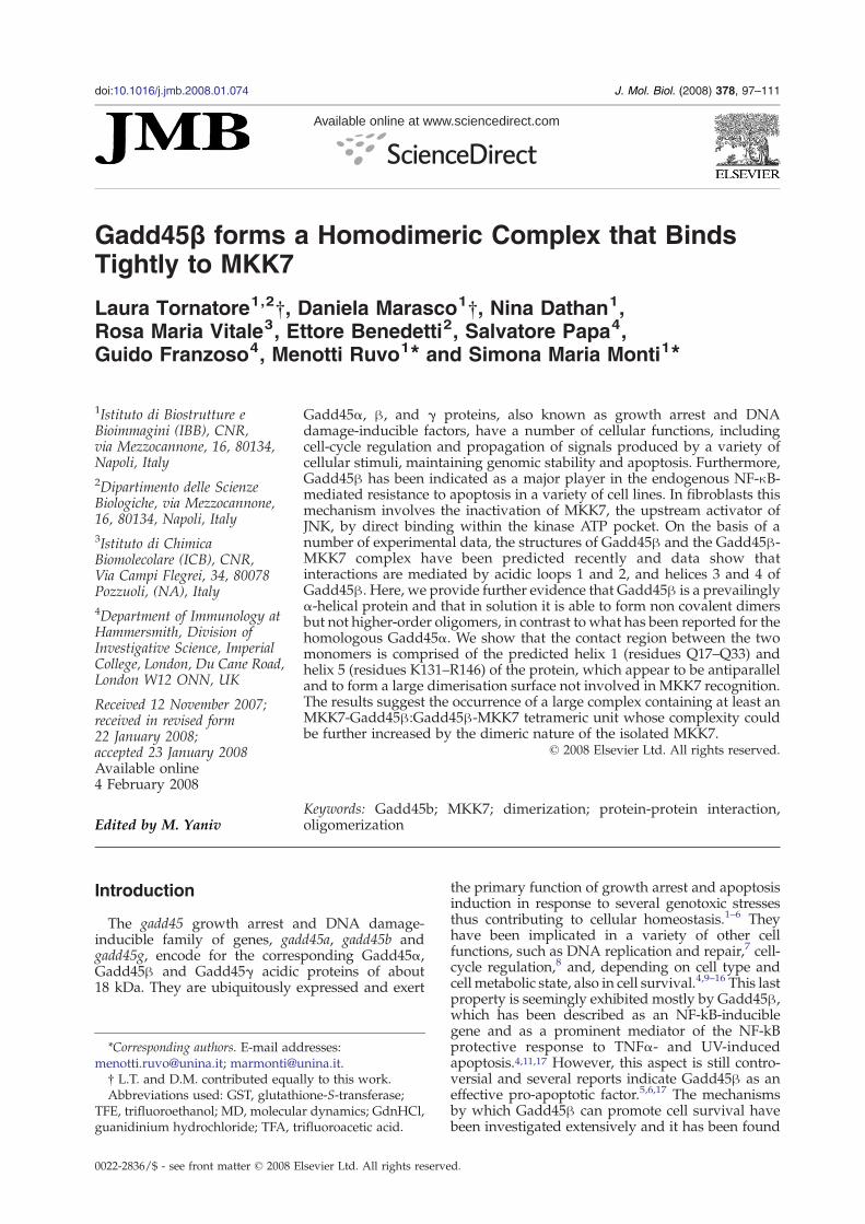
doi:10.1016/j.jmb.2008.01.074 J. Mol. Biol. (2008) 378, 97–111
Available online at www.sciencedirect.com
Gadd45β forms a Homodimeric Complex that BindsTightly to MKK7
Laura Tornatore1,2†, Daniela Marasco1†, Nina Dathan1,Rosa Maria Vitale3, Ettore Benedetti2, Salvatore Papa4,Guido Franzoso4, Menotti Ruvo1⁎ and Simona Maria Monti1⁎
1Istituto di Biostrutture eBioimmagini (IBB), CNR,via Mezzocannone, 16, 80134,Napoli, Italy2Dipartimento delle ScienzeBiologiche, via Mezzocannone,16, 80134, Napoli, Italy3Istituto di ChimicaBiomolecolare (ICB), CNR,Via Campi Flegrei, 34, 80078Pozzuoli, (NA), Italy4Department of Immunology atHammersmith, Division ofInvestigative Science, ImperialCollege, London, Du Cane Road,London W12 ONN, UK
Received 12 November 2007;received in revised form22 January 2008;accepted 23 January 2008Available online4 February 2008
*Corresponding authors. E-mail [email protected]; marmonti@u† L.T. and D.M. contributed equaAbbreviations used: GST, glutathi
TFE, trifluoroethanol; MD, moleculaguanidinium hydrochloride; TFA, tr
0022-2836/$ - see front matter © 2008 E
Gadd45α, β, and γ proteins, also known as growth arrest and DNAdamage-inducible factors, have a number of cellular functions, includingcell-cycle regulation and propagation of signals produced by a variety ofcellular stimuli, maintaining genomic stability and apoptosis. Furthermore,Gadd45β has been indicated as a major player in the endogenous NF-κB-mediated resistance to apoptosis in a variety of cell lines. In fibroblasts thismechanism involves the inactivation of MKK7, the upstream activator ofJNK, by direct binding within the kinase ATP pocket. On the basis of anumber of experimental data, the structures of Gadd45β and the Gadd45β-MKK7 complex have been predicted recently and data show thatinteractions are mediated by acidic loops 1 and 2, and helices 3 and 4 ofGadd45β. Here, we provide further evidence that Gadd45β is a prevailinglyα-helical protein and that in solution it is able to form non covalent dimersbut not higher-order oligomers, in contrast to what has been reported for thehomologous Gadd45α. We show that the contact region between the twomonomers is comprised of the predicted helix 1 (residues Q17–Q33) andhelix 5 (residues K131–R146) of the protein, which appear to be antiparalleland to form a large dimerisation surface not involved in MKK7 recognition.The results suggest the occurrence of a large complex containing at least anMKK7-Gadd45β:Gadd45β-MKK7 tetrameric unit whose complexity couldbe further increased by the dimeric nature of the isolated MKK7.
© 2008 Elsevier Ltd. All rights reserved.
Edited by M. Yaniv
Keywords: Gadd45b; MKK7; dimerization; protein-protein interaction,oligomerizationIntroduction
The gadd45 growth arrest and DNA damage-inducible family of genes, gadd45a, gadd45b andgadd45g, encode for the corresponding Gadd45α,Gadd45β and Gadd45γ acidic proteins of about18 kDa. They are ubiquitously expressed and exert
resses:nina.it.lly to this work.one-S-transferase;r dynamics; GdnHCl,ifluoroacetic acid.
lsevier Ltd. All rights reserve
the primary function of growth arrest and apoptosisinduction in response to several genotoxic stressesthus contributing to cellular homeostasis.1–6 Theyhave been implicated in a variety of other cellfunctions, such as DNA replication and repair,7 cell-cycle regulation,8 and, depending on cell type andcell metabolic state, also in cell survival.4,9–16 This lastproperty is seemingly exhibited mostly by Gadd45β,which has been described as an NF-kB-induciblegene and as a prominent mediator of the NF-kBprotective response to TNFα- and UV-inducedapoptosis.4,11,17 However, this aspect is still contro-versial and several reports indicate Gadd45β as aneffective pro-apoptotic factor.5,6,17 The mechanismsby which Gadd45β can promote cell survival havebeen investigated extensively and it has been found
d.

98 Homodimerisation of Gadd45β and Binding to MKK7
that in MEFs and other cells, upon NF-kB induction,it provides selective JNK inactivation by inhibitionof the upstream MKK7.1,10,18 In hematopoietic cells,instead, it blocks JNK activation by binding toMKK4,4,19 and in B cells it is a critical mediator of the
Fig. 1. (a) Alignment of Gadd45α, β and γ sequences. Anpresent in the region corresponding to the second acidic loop,with the other two sequences. To align the Gadd45α variant coacidic loop 1, residue Asp67 of Gadd45β is mutated to Arg inself-association are underlined. (b) A representation of the protdescribed in Ref. 1. (c) A ribbon representation of Gadd45β thrto the following scheme: hydrophobic residues, white; polar rhistidine residues, cyan; tyrosine residues, in pink.
pro-survival activity of CD40 elicited in response toFas stimulation.9
Since there is no reported enzymatic activity forthe Gadd45 proteins, it is believed that they exerttheir functions by interacting with protein partners.
identity of about 60% is observed. Most differences arewith a Gadd45γ pentapeptide stretch remaining unpairedrrectly, an eight-residue long stretch is required. Within thethe α variant and to Gly in Gadd45γ. Regions involved inein secondary structure is schematised based on the modelee-dimensional model, coloured by residue type accordingesidues, yellow; acidic residues, red; basic residues, blue;

Fig. 2. Characterisation of the oligomeric state ofGadd45β. (a) Gel-filtration analysis of the recombinantprotein on Superdex 75 10/30 under non reducingconditions. Two peaks at elution volumes compatible
99Homodimerisation of Gadd45β and Binding to MKK7
Indeed, other thanMKK7 andMKK4, highly specificinteractions with PCNA,6,20–22 cdc2,23 waf/p21,24
cdk1/cyclinB1,25 MEKK426 and CRIF127 are in-volved in Gadd45 regulation of the cell cycle andthe response to external cell stimuli. A furtherinteraction with the protein nucleophosmin hasbeen described for Gadd45α,28 and it has beenshown that this protein can work as a vehicle fornuclear import. However, it is not known whetherGadd45β and Gadd45γ share a similar mechanismof nuclear translocation. Importantly, it has beenreported that Gadd45 proteins are also able tohomo- and heterodimerise or oligomerise, andregions involved in self-association of Gadd45αhave been investigated using overlapping syntheticpeptides spanning the entire protein sequence.29
The primary sequences of Gadd45 proteins sharean overall 70% homology (about 60% identity, seeFig. 1a) and all contain six cysteines, five of which(from the second to the sixth) are located in highlyconserved positions.An unusually long stretch of glutamic and
aspartic acid residues, only partially conservedwithin the Gadd45γ variant, can be found startingfrom position 60 of Gadd45α and Gadd45β. Theseresidues have been described as having a key role ininhibition of both cdc223 and MKK7.1,10 However,the 3D structure of this important class of proteins isnot known, and so far a predicted model has beenreported1 for Gadd45β (Fig. 1b and c) and itscomplex with MKK7. These structures, supportedby various experimental evidences, present a centralfour-stranded β-sheet surrounded by five α-helicesand, as expected for a nuclear protein, all cysteineresidues are predicted to be far from each other andthus in the reduced state.1 The acidic stretch appearsto be in a large loop interacting with several basicand polar residues within the kinase active site, andit is part of the minimum region of Gadd45β neededto bind and block MKK7 activity (A60-D86 frag-ment). Other regions involving the putative helix 3(H3), a second acidic loop and part of helix 4 (H4)harbour other key residues contacting MKK7.Following an approach of protein enzymatic
fragmentation and HPLC fractionation, here weidentify the regions of Gadd45β implicated in self-association and confirm those involved in bindingwith MKK7. We show also that Gadd45β is unableto form higher-order oligomers, as only dimers aredetected using different methods. These studiesextend our knowledge of Gadd45β properties andsuggest that protein self-association can have aprimary role in regulating its biological activity.
with dimeric and tetrameric forms are detected. (b) Thesame analysis carried out after inclusion of 1 mM DTT inthe running buffer. The peak at lower elution volumesdisappears, suggesting that it is due to covalent disulfidebridges. (c) The calibration curve used to determine Mr isreported. Cytochrome c, 12.4 kDa; RNase A, 14.7 kDa;chymotrypsinogen, 25.0 kDa; carbonic anhydrase,29.0 kDa; ovalbumin, 44.0 kDa; BSA, 66.0 kDa. All mea-surements were done at least twice. (d) Non-denaturingpolyacrylamide gel analysis of Gadd45β at a concentrationof 1.8 mg/mL (100 μM).
Results
Gadd45β purification
Recombinant construct pGEX6P-GADD45β al-lowed expression of the protein as a glutathione-S-transferase (GST)-fusion product containing a highly
specific cleavage site for PreScission Proteaseupstream of the Gadd45β protein. The applied over-expression systemwas quite efficient, producingmorethan 6 mg of highly purified protein from 1 L ofinduced culture under the reported conditions.1 Therecombinant protein obtained after removal of GSThad the sequence reported inFig. 1b andwasusedonlyfor oligomerisation studies. The protein was charac-terisedbySDS-PAGEandLC-MSanalysisdetermining
100 Homodimerisation of Gadd45β and Binding to MKK7
the exact MW (MWExp/Theor, 18096.6±1.0 Da/18096.1 Da). His6-Gadd45β used in ELISA assayswas obtained similarly with high yields and purity.The value of 19196.5 amu as determined by LC-MSwas consistent with the protein sequence (MWTheor19196.52 amu). The protein was derivatised efficientlywith biotin as described in Experimental Procedures.Protein derivatisation was assessed by mass spectro-metry showing that about 90% of the proteinharboured one biotin molecule, while the remainingappeared underivatised (data not shown).
Investigation of Gadd45β oligomerisation by gelfiltration and CD studies
Since previous studies reported on the capacity ofGadd45 proteins to oligomerise,29 we investigatedthis point by carrying out a gel-filtration analysis inthe presence and/or in the absence of DTT and bynative electrophoresis experiments. Gel-filtrationanalysis of protein aliquots at a concentration of5.0 μM showed that, in the absence of DTT, two
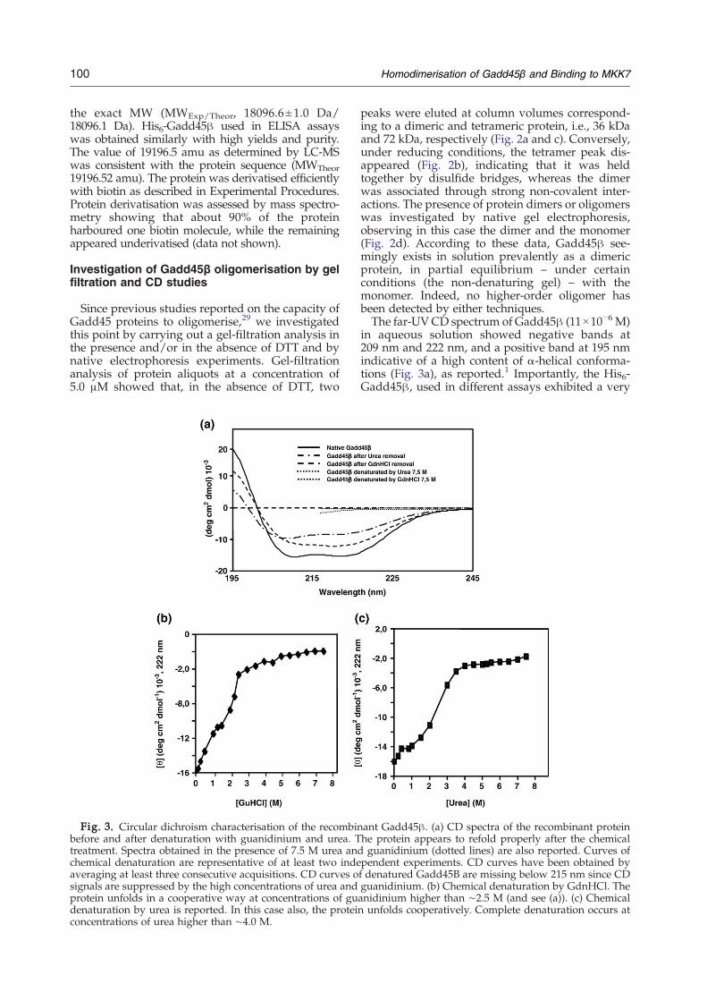
Fig. 3. Circular dichroism characterisation of the recombinbefore and after denaturation with guanidinium and urea. Ttreatment. Spectra obtained in the presence of 7.5 M urea andchemical denaturation are representative of at least two indeaveraging at least three consecutive acquisitions. CD curves osignals are suppressed by the high concentrations of urea andprotein unfolds in a cooperative way at concentrations of gudenaturation by urea is reported. In this case also, the proteinconcentrations of urea higher than ∼4.0 M.
peaks were eluted at column volumes correspond-ing to a dimeric and tetrameric protein, i.e., 36 kDaand 72 kDa, respectively (Fig. 2a and c). Conversely,under reducing conditions, the tetramer peak dis-appeared (Fig. 2b), indicating that it was heldtogether by disulfide bridges, whereas the dimerwas associated through strong non-covalent inter-actions. The presence of protein dimers or oligomerswas investigated by native gel electrophoresis,observing in this case the dimer and the monomer(Fig. 2d). According to these data, Gadd45β see-mingly exists in solution prevalently as a dimericprotein, in partial equilibrium – under certainconditions (the non-denaturing gel) – with themonomer. Indeed, no higher-order oligomer hasbeen detected by either techniques.The far-UVCD spectrum of Gadd45β (11×10−6 M)
in aqueous solution showed negative bands at209 nm and 222 nm, and a positive band at 195 nmindicative of a high content of α-helical conforma-tions (Fig. 3a), as reported.1 Importantly, the His6-Gadd45β, used in different assays exhibited a very
ant Gadd45β. (a) CD spectra of the recombinant proteinhe protein appears to refold properly after the chemicalguanidinium (dotted lines) are also reported. Curves of
pendent experiments. CD curves have been obtained byf denatured Gadd45B are missing below 215 nm since CDguanidinium. (b) Chemical denaturation by GdnHCl. Theanidinium higher than ∼2.5 M (and see (a)). (c) Chemicalunfolds cooperatively. Complete denaturation occurs at
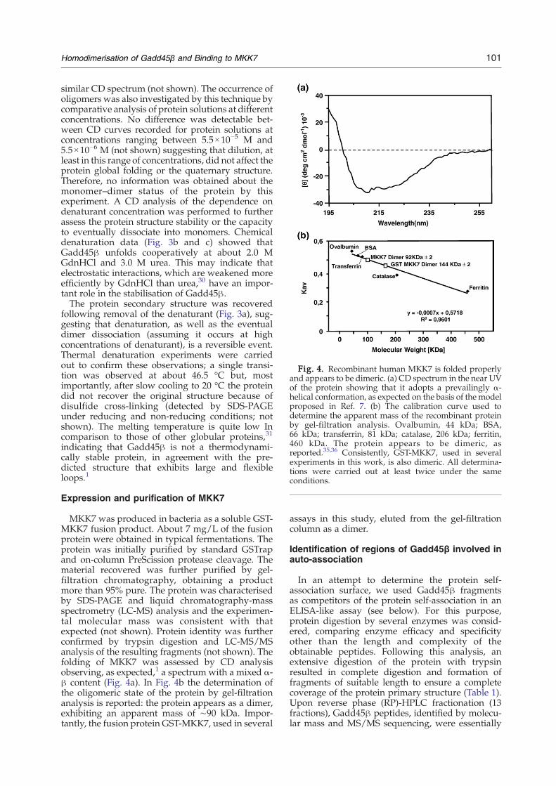
Fig. 4. Recombinant human MKK7 is folded properlyand appears to be dimeric. (a) CD spectrum in the near UVof the protein showing that it adopts a prevailingly α-helical conformation, as expected on the basis of the modelproposed in Ref. 7. (b) The calibration curve used todetermine the apparent mass of the recombinant proteinby gel-filtration analysis. Ovalbumin, 44 kDa; BSA,66 kDa; transferrin, 81 kDa; catalase, 206 kDa; ferritin,460 kDa. The protein appears to be dimeric, asreported.35,36 Consistently, GST-MKK7, used in severalexperiments in this work, is also dimeric. All determina-tions were carried out at least twice under the sameconditions.
101Homodimerisation of Gadd45β and Binding to MKK7
similar CD spectrum (not shown). The occurrence ofoligomers was also investigated by this technique bycomparative analysis of protein solutions at differentconcentrations. No difference was detectable bet-ween CD curves recorded for protein solutions atconcentrations ranging between 5.5×10⁻⁵ M and5.5×10−6 M (not shown) suggesting that dilution, atleast in this range of concentrations, did not affect theprotein global folding or the quaternary structure.Therefore, no information was obtained about themonomer–dimer status of the protein by thisexperiment. A CD analysis of the dependence ondenaturant concentration was performed to furtherassess the protein structure stability or the capacityto eventually dissociate into monomers. Chemicaldenaturation data (Fig. 3b and c) showed thatGadd45β unfolds cooperatively at about 2.0 MGdnHCl and 3.0 M urea. This may indicate thatelectrostatic interactions, which are weakened moreefficiently by GdnHCl than urea,30 have an impor-tant role in the stabilisation of Gadd45β.The protein secondary structure was recovered
following removal of the denaturant (Fig. 3a), sug-gesting that denaturation, as well as the eventualdimer dissociation (assuming it occurs at highconcentrations of denaturant), is a reversible event.Thermal denaturation experiments were carriedout to confirm these observations; a single transi-tion was observed at about 46.5 °C but, mostimportantly, after slow cooling to 20 °C the proteindid not recover the original structure because ofdisulfide cross-linking (detected by SDS-PAGEunder reducing and non-reducing conditions; notshown). The melting temperature is quite low Incomparison to those of other globular proteins,31
indicating that Gadd45β is not a thermodynami-cally stable protein, in agreement with the pre-dicted structure that exhibits large and flexibleloops.1
Expression and purification of MKK7
MKK7 was produced in bacteria as a soluble GST-MKK7 fusion product. About 7 mg/L of the fusionprotein were obtained in typical fermentations. Theprotein was initially purified by standard GSTrapand on-column PreScission protease cleavage. Thematerial recovered was further purified by gel-filtration chromatography, obtaining a productmore than 95% pure. The protein was characterisedby SDS-PAGE and liquid chromatography-massspectrometry (LC-MS) analysis and the experimen-tal molecular mass was consistent with thatexpected (not shown). Protein identity was furtherconfirmed by trypsin digestion and LC-MS/MSanalysis of the resulting fragments (not shown). Thefolding of MKK7 was assessed by CD analysisobserving, as expected,1 a spectrum with a mixed α-β content (Fig. 4a). In Fig. 4b the determination ofthe oligomeric state of the protein by gel-filtrationanalysis is reported: the protein appears as a dimer,exhibiting an apparent mass of ∼90 kDa. Impor-tantly, the fusion protein GST-MKK7, used in several
assays in this study, eluted from the gel-filtrationcolumn as a dimer.
Identification of regions of Gadd45β involved inauto-association
In an attempt to determine the protein self-association surface, we used Gadd45β fragmentsas competitors of the protein self-association in anELISA-like assay (see below). For this purpose,protein digestion by several enzymes was consid-ered, comparing enzyme efficacy and specificityother than the length and complexity of theobtainable peptides. Following this analysis, anextensive digestion of the protein with trypsinresulted in complete digestion and formation offragments of suitable length to ensure a completecoverage of the protein primary structure (Table 1).Upon reverse phase (RP)-HPLC fractionation (13fractions), Gadd45β peptides, identified by molecu-lar mass and MS/MS sequencing, were essentially
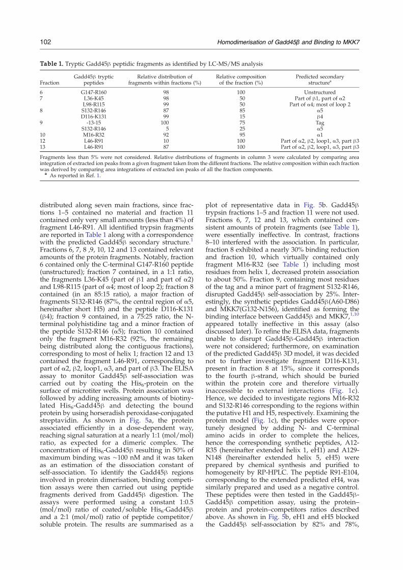
Table 1. Tryptic Gadd45β peptidic fragments as identified by LC-MS/MS analysis
FractionGadd45β tryptic
peptidesRelative distribution of
fragments within fractions (%)Relative compositionof the fraction (%)
Predicted secondarystructurea
6 G147-R160 98 100 Unstructured7 L36-K45 98 50 Part of β1, part of α2
L98-R115 99 50 Part of α4; most of loop 28 S132-R146 87 85 α5
D116-K131 99 15 β49 -13-15 100 75 Tag
S132-R146 5 25 α510 M16-R32 92 95 α112 L46-R91 10 100 Part of α2, β2, loop1, α3, part β313 L46-R91 87 100 Part of α2, β2, loop1, α3, part β3
Fragments less than 5% were not considered. Relative distributions of fragments in column 3 were calculated by comparing areaintegration of extracted ion peaks from a given fragment taken from the different fractions. The relative composition within each fractionwas derived by comparing area integrations of extracted ion peaks of all the fraction components.
a As reported in Ref. 1.
102 Homodimerisation of Gadd45β and Binding to MKK7
distributed along seven main fractions, since frac-tions 1–5 contained no material and fraction 11contained only very small amounts (less than 4%) offragment L46-R91. All identified trypsin fragmentsare reported in Table 1 along with a correspondencewith the predicted Gadd45β secondary structure.1
Fractions 6, 7, 8 ,9, 10, 12 and 13 contained relevantamounts of the protein fragments. Notably, fraction6 contained only the C-terminal G147-R160 peptide(unstructured); fraction 7 contained, in a 1:1 ratio,the fragments L36-K45 (part of β1 and part of α2)and L98-R115 (part of α4; most of loop 2); fraction 8contained (in an 85:15 ratio), a major fraction offragments S132-R146 (87%, the central region of α5,hereinafter short H5) and the peptide D116-K131(β4); fraction 9 contained, in a 75:25 ratio, the N-terminal polyhistidine tag and a minor fraction ofthe peptide S132-R146 (α5); fraction 10 containedonly the fragment M16-R32 (92%, the remainingbeing distributed along the contiguous fractions),corresponding to most of helix 1; fraction 12 and 13contained the fragment L46-R91, corresponding topart of α2, β2, loop1, α3, and part of β3. The ELISAassay to monitor Gadd45β self-association wascarried out by coating the His6-protein on thesurface of microtiter wells. Protein association wasfollowed by adding increasing amounts of biotiny-lated His6-Gadd45β and detecting the boundprotein by using horseradish peroxidase-conjugatedstreptavidin. As shown in Fig. 5a, the proteinassociated efficiently in a dose-dependent way,reaching signal saturation at a nearly 1:1 (mol/mol)ratio, as expected for a dimeric complex. Theconcentration of His6-Gadd45β resulting in 50% ofmaximum binding was ∼100 nM and it was takenas an estimation of the dissociation constant ofself-association. To identify the Gadd45β regionsinvolved in protein dimerisation, binding competi-tion assays were then carried out using peptidefragments derived from Gadd45β digestion. Theassays were performed using a constant 1:0.5(mol/mol) ratio of coated/soluble His6-Gadd45βand a 2:1 (mol/mol) ratio of peptide competitor/soluble protein. The results are summarised as a
plot of representative data in Fig. 5b. Gadd45βtrypsin fractions 1–5 and fraction 11 were not used.Fractions 6, 7, 12 and 13, which contained con-sistent amounts of protein fragments (see Table 1),were essentially ineffective. In contrast, fractions8–10 interfered with the association. In particular,fraction 8 exhibited a nearly 30% binding reductionand fraction 10, which virtually contained onlyfragment M16-R32 (see Table 1) including mostresidues from helix 1, decreased protein associationto about 50%. Fraction 9, containing most residuesof the tag and a minor part of fragment S132-R146,disrupted Gadd45β self-association by 25%. Inter-estingly, the synthetic peptides Gadd45β(A60-D86)and MKK7(G132-N156), identified as forming thebinding interface between Gadd45β and MKK7,1,10
appeared totally ineffective in this assay (alsodiscussed later). To refine the ELISA data, fragmentsunable to disrupt Gadd45β-Gadd45β interactionwere not considered; furthermore, on examinationof the predicted Gadd45β 3D model, it was decidednot to further investigate fragment D116-K131,present in fraction 8 at 15%, since it correspondsto the fourth β-strand, which should be buriedwithin the protein core and therefore virtuallyinaccessible to external interactions (Fig. 1c).Hence, we decided to investigate regions M16-R32and S132-R146 corresponding to the regions withinthe putative H1 and H5, respectively. Examining theprotein model (Fig. 1c), the peptides were oppor-tunely designed by adding N- and C-terminalamino acids in order to complete the helices,hence the corresponding synthetic peptides, A12-R35 (hereinafter extended helix 1, eH1) and A129-N148 (hereinafter extended helix 5, eH5) wereprepared by chemical synthesis and purified tohomogeneity by RP-HPLC. The peptide R91-E104,corresponding to the extended predicted eH4, wassimilarly prepared and used as a negative control.These peptides were then tested in the Gadd45β-Gadd45β competition assay, using the protein–protein and protein–competitors ratios describedabove. As shown in Fig. 5b, eH1 and eH5 blockedthe Gadd45β self-association by 82% and 78%,

Fig. 5. Gadd45β is able to self-associate, the interactionbeing disrupted by Gadd45β trypsin fragments. (a) Dose-dependent binding of biotinylated Gadd45β to the plate-adsorbed protein: Gadd45β self associates with an esti-mated KD of ∼100 nM, assumed as the concentration ofGadd45β resulting in 50% maximum binding.29 Notably,the signal reaches saturation at a 1:1 protein ratio,suggesting the formation of dimers only. (b) Competitionassay of the Gadd45β self-association by the trypsin-generated protein fragments and by synthetic peptidesdesigned to reproduce helices as predicted in the proteinmodel.1 (c) Dose-dependent inhibition of Gadd45β self-association by full-length GST-MKK7, eH4 (residues 91–104), eH5 (residues 129–148), eH1 (residues 12–35) and theshort helix 5 (residues 132–146). While the kinase and eH4are not able to block the Gadd45β self-association, peptidescorresponding to eH1 and eH5, reduce the associationmarkedly. Short helix 5, derived by trypsin cleavage, is lesseffective than the entire eH5. Data are representative of atleast three independent experiments.
103Homodimerisation of Gadd45β and Binding to MKK7
respectively, supporting the hypothesis that theseregions are strongly involved in the interaction. Incontrast, peptide R91-E104 (eH4), lying in the closeproximity of H1 in the model (Fig. 1c), as well aspeptides Gadd45β(A60-D86) and MKK7(G132-N156), and the full-length kinase did not interferewith the protein homodimerisation. A dose-depen-dent competition assay carried out with eH1, eH5,the short H5, eH4 and full-length MKK7, furtherconfirmed the properties of H1 and H5 and theinefficacy of H4 to abrogate dimerisation (See Fig.5c). As expected, the full-length kinase (fused toGST) was unable to block the Gadd45β self-association even at higher concentrations. Remark-ably, the IC50 for these competitors was 100 nM forH1, 180 nM for the eH5 and only 600 nM for theshort H5, indicating that the full H5 was more thanthree times more efficient in disrupting the proteinself-association. These results might account for therelatively weak inhibition exhibited by fractions 8(30%) and 9 (25%), which contained only short H5.To further investigate this point, the syntheticpeptides reproducing eH1, eH5, short H5 and eH4were analyzed by CD. The analyses were carriedout both in phosphate buffer and in the presence of20% (v/v) trifluoroethanol (TFE) to measure therelative propensity of peptides to adopt α-helicalconformations. As reported in Fig. 6a–d, whereaseH1, eH4 and eH5 adopted a partially foldedstructure in buffer and readily folded into α-helicesupon the addition of TFE, the short H5 remained inthe unfolded state even after addition of thestructuring solvent, suggesting an intrinsic incapa-city to form organised structures. Thus, increasingthe length at the N and C termini of the putative H5provided a fivefold increase in potency that can beascribed, in part, to the lack of any structure ofshorter variant.To further extend our knowledge of the interaction
between the two monomers, we developed an assayto monitor the binding between MKK7 andGadd45β. This interaction has been thoroughlyinvestigated in previous work by pull-down assaysusing several point-mutated variants of bothproteins.1,10 As shown in Fig. 7a, the associationbetween the two purified protein was very strong: arough estimation of the KD, deduced by theGadd45β concentration at half of the saturationsignal, was about 13 nM. Noticeably, signal satura-tion was reached at a molar ratio of ∼1:1, suggestingthat oneMKK7molecule should be sufficient to bindto one molecule of Gadd45β. The competitionexperiment carried out using 42 nM MKK7, 21 nMGadd45β and a twofold excess of competitors overthe soluble protein, is reported in Fig. 7b. As shown,peptides corresponding to the putative eH1, eH4and eH5, MKK7(G132-N156) and Gadd45β(A60-D86) were used. Consistent with the notion thatMKK7 proximity does not interfere with theGadd45β dimerisation, eH1 and eH5 do not affectthe binding, whereas peptides Gadd45β(A60-D86)and MKK7(G132-N156), believed to form part of theinterface between the two proteins, completely
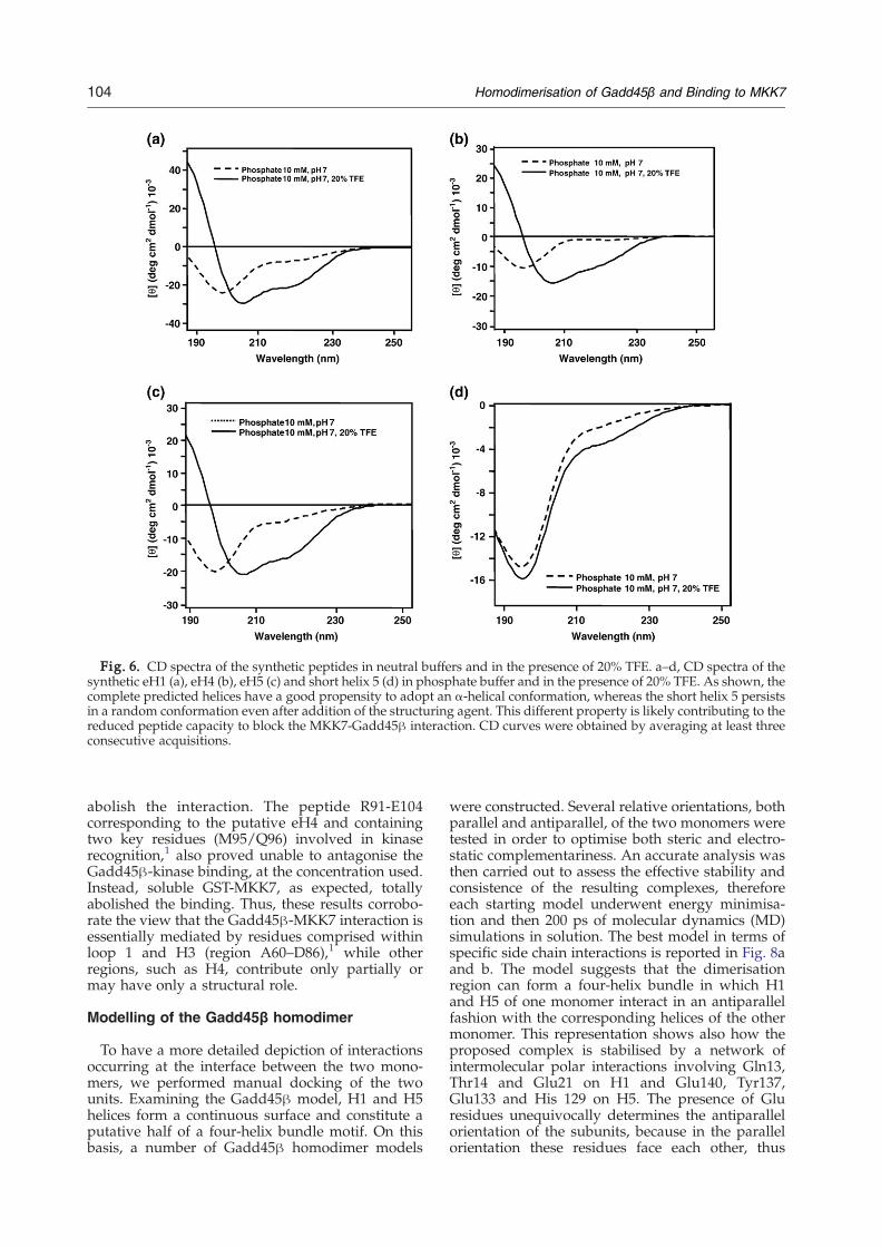
Fig. 6. CD spectra of the synthetic peptides in neutral buffers and in the presence of 20% TFE. a–d, CD spectra of thesynthetic eH1 (a), eH4 (b), eH5 (c) and short helix 5 (d) in phosphate buffer and in the presence of 20% TFE. As shown, thecomplete predicted helices have a good propensity to adopt an α-helical conformation, whereas the short helix 5 persistsin a random conformation even after addition of the structuring agent. This different property is likely contributing to thereduced peptide capacity to block the MKK7-Gadd45β interaction. CD curves were obtained by averaging at least threeconsecutive acquisitions.
104 Homodimerisation of Gadd45β and Binding to MKK7
abolish the interaction. The peptide R91-E104corresponding to the putative eH4 and containingtwo key residues (M95/Q96) involved in kinaserecognition,1 also proved unable to antagonise theGadd45β-kinase binding, at the concentration used.Instead, soluble GST-MKK7, as expected, totallyabolished the binding. Thus, these results corrobo-rate the view that the Gadd45β-MKK7 interaction isessentially mediated by residues comprised withinloop 1 and H3 (region A60–D86),1 while otherregions, such as H4, contribute only partially ormay have only a structural role.
Modelling of the Gadd45β homodimer
To have a more detailed depiction of interactionsoccurring at the interface between the two mono-mers, we performed manual docking of the twounits. Examining the Gadd45β model, H1 and H5helices form a continuous surface and constitute aputative half of a four-helix bundle motif. On thisbasis, a number of Gadd45β homodimer models
were constructed. Several relative orientations, bothparallel and antiparallel, of the two monomers weretested in order to optimise both steric and electro-static complementariness. An accurate analysis wasthen carried out to assess the effective stability andconsistence of the resulting complexes, thereforeeach starting model underwent energy minimisa-tion and then 200 ps of molecular dynamics (MD)simulations in solution. The best model in terms ofspecific side chain interactions is reported in Fig. 8aand b. The model suggests that the dimerisationregion can form a four-helix bundle in which H1and H5 of one monomer interact in an antiparallelfashion with the corresponding helices of the othermonomer. This representation shows also how theproposed complex is stabilised by a network ofintermolecular polar interactions involving Gln13,Thr14 and Glu21 on H1 and Glu140, Tyr137,Glu133 and His 129 on H5. The presence of Gluresidues unequivocally determines the antiparallelorientation of the subunits, because in the parallelorientation these residues face each other, thus
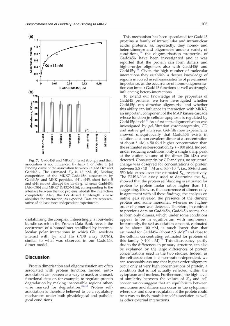
Fig. 7. Gadd45β and MKK7 interact strongly and theirassociation is not influenced by helix 1 or helix 5. (a)Binding curve of the association between GST-MKK7 andGadd45β. The estimated KD is 13 nM. (b) Bindingcompetition of the MKK7–Gadd45β association byGadd45β and MKK peptides. eH1, eH5, short helix 5and eH4 cannot disrupt the binding, whereas Gadd45β[A60-D86] and MKK7 [G132-N156], corresponding to theinterface between the two proteins, abolish the interactioncompletely. Also, the GST-fused full-length kinaseabolishes the interaction, as expected. Data are represen-tative of at least three independent experiments.
105Homodimerisation of Gadd45β and Binding to MKK7
destabilising the complex. Interestingly, a four-helixbundle search in the Protein Data Bank reveals theoccurrence of a homodimer stabilised by intermo-lecular polar interactions in which Glu residuesinteract with Tyr and His (PDB entry 1U7M),similar to what was observed in our Gadd45βdimer model.
Discussion
Protein dimerisation and oligomerisation are oftenassociated with protein function. Indeed, auto-association can be seen as a way to mask or unmaskfunctional sites or, for example, to regulate proteindegradation by making inaccessible regions other-wise marked for degradation.32,33 Protein self-association is therefore believed to be a regulatorymechanism under both physiological and patholo-gical conditions.
This mechanism has been speculated for Gadd45proteins, a family of intracellular and intranuclearacidic proteins, as, reportedly, they homo- andheterodimerise and oligomerise under a variety ofconditions;29 the oligomerisation properties ofGadd45α have been investigated and it wasreported that the protein can form dimers andhigher-order oligomers also with Gadd45β andGadd45γ.29 Given the high number of molecularinteractions they establish, a deeper knowledge ofregions involved in self-association is of pre-eminentimportance, as the occurrence of homo-oligomerisa-tion can impair Gadd45 functions as well as stronglyinfluencing hetero-interactions.To extend our knowledge of the properties of
Gadd45 proteins, we have investigated whetherGadd45β can dimerise–oligomerise and whetherthis ability can influence its interaction with MKK7,an important component of the MAP kinase cascadewhose function in cellular apoptosis is regulated byGadd45β itself.10 As a first step, oligomerisation wasinvestigated by gel-filtration chromatography, CDand native gel analyses. Gel-filtration experimentsshowed unequivocally that Gadd45β exists insolution as a non-covalent dimer at a concentrationof about 5 μM, a 50-fold higher concentration thanthe estimated self-association KD (∼100 nM). Indeed,under reducing conditions, only a single sharp peakat the elution volume of the dimer (36 kDa) wasdetected. Consistently, by CD analysis, no structuralchange was observed for concentrations of proteinbetween 5.5×10⁻⁶ M and 5.5×10⁻⁵; i.e., 55-fold and550-fold excess over the estimated KD, respectively.The ELISA-like assay used to determine the KD,showed that the protein self-binding is saturated forprotein to protein molar ratios higher than 1:1,suggesting, likewise, the occurrence of dimers only.In agreement with all these findings, the analysis onnative gels revealed the presence of the dimericprotein and some monomer, whereas no higher-order oligomer was detected. Therefore, in contrastto previous data on Gadd45α, Gadd45β seems ableto form only dimers, which, under some conditionsappear to be in equilibrium with monomers.Importantly, the self-association constant, estimatedto be about 100 nM, is much lower than thatestimated for Gadd45α (about 2.5 μM)29 and close tothe cellular concentration estimated for proteins ofthis family (∼100 nM).29 This discrepancy, partlydue to the differences in primary structure, can alsobe explained by the large differences of proteinconcentrations used in the two studies. Indeed, asthe self-association is concentration-dependent, wecan reasonably assume that higher-order oligomersoccur only at very high concentrations of protein, acondition that is not actually reflected within thecytoplasm and nucleus. Furthermore, the high levelof similarity between the values of KD and cellconcentration suggest that an equilibrium betweenmonomers and dimers can occur in the cytoplasm,where up- and down-regulation of the protein couldbe a way to finely modulate self-association as wellas other external interactions.

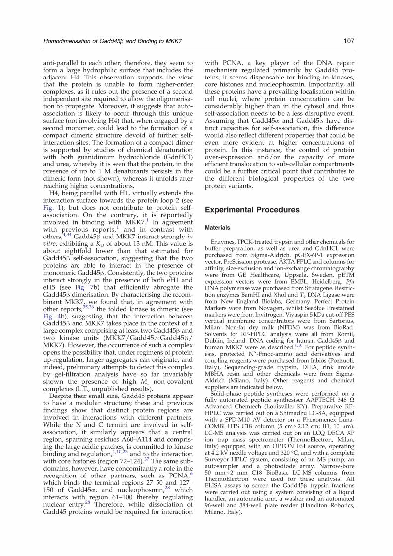
Fig. 8. Two orthogonal views of Gadd45β homodimer three-dimensional model in ribbon representation, rotatedaround the horizontal axis of the figure ((a) and (b)). One monomer is coloured pink, the other is coloured pale green.Helices H1 andH5 of eachmonomer are coloured inmagenta and dark green, respectively. Residues lying on these helicesare shown in stick representation and coloured according to the following scheme: hydrophobic residues, white; acidicresidues, red; basic residues, blue, histidine, cyan; and tyrosine, magenta.
106 Homodimerisation of Gadd45β and Binding to MKK7
Following an approach of self-interaction compe-tition with protein fragments derived from trypsindigestion, we identified two distinct Gadd45β sitesinvolved in self-association that correspond to thepredicted H1 and H5 of the protein.1 Remarkably,these findings partially agree with the previousreport on Gadd45α, for which an N-terminal and aC-terminal region have been described as beinginvolved in self-association. However, whilst the C-terminal site (129–165) is virtually overlapping thepredicted H5 of Gadd45β, the N-terminal site,identified as residues 33–61,29 does not match H1(see the sequence alignments in Fig. 1). Rather, it
overlaps with the Gadd45β sequence predicted asthe β1-α2-β2 region (Fig. 1)1 that is somewhatdownstream in the sequence. In the cited study, alarge fragment of Gadd45α (residues 20–33) exactlymatching the Gadd45βH1, has not been considered;therefore a further comparison between the twodifferent variants is not possible. However, giventhe high level of sequence homology between thetwo proteins, especially in this part of the sequences,we cannot rule out an involvement of the missingfragment in Gadd45α auto-association. Noticeably,by looking at the Gadd45β model (Fig. 1 and 8), H1and H5 are disposed contiguously and arranged
107Homodimerisation of Gadd45β and Binding to MKK7
anti-parallel to each other; therefore, they seem toform a large hydrophilic surface that includes theadjacent H4. This observation supports the viewthat the protein is unable to form higher-ordercomplexes, as it rules out the presence of a secondindependent site required to allow the oligomerisa-tion to propagate. Moreover, it suggests that auto-association is likely to occur through this uniquesurface (not involving H4) that, when engaged by asecond monomer, could lead to the formation of acompact dimeric structure devoid of further self-interaction sites. The formation of a compact dimeris supported by studies of chemical denaturationwith both guanidinium hydrochloride (GdnHCl)and urea, whereby it is seen that the protein, in thepresence of up to 1 M denaturants persists in thedimeric form (not shown), whereas it unfolds afterreaching higher concentrations.H4, being parallel with H1, virtually extends the
interaction surface towards the protein loop 2 (seeFig. 1), but does not contribute to protein self-association. On the contrary, it is reportedlyinvolved in binding with MKK7.1 In agreementwith previous reports,1 and in contrast withothers,4,34 Gadd45β and MKK7 interact strongly invitro, exhibiting a KD of about 13 nM. This value isabout eightfold lower than that estimated forGadd45β self-association, suggesting that the twoproteins are able to interact in the presence ofmonomeric Gadd45β. Consistently, the two proteinsinteract strongly in the presence of both eH1 andeH5 (see Fig. 7b) that efficiently abrogate theGadd45β dimerisation. By characterising the recom-binant MKK7, we found that, in agreement withother reports,35,36 the folded kinase is dimeric (seeFig. 4b), suggesting that the interaction betweenGadd45β and MKK7 takes place in the context of alarge complex comprising at least two Gadd45β andtwo kinase units (MKK7/Gadd45β:Gadd45β/MKK7). However, the occurrence of such a complexopens the possibility that, under regimens of proteinup-regulation, larger aggregates can originate, andindeed, preliminary attempts to detect this complexby gel-filtration analysis have so far invariablyshown the presence of high Mr non-covalentcomplexes (L.T., unpublished results).Despite their small size, Gadd45 proteins appear
to have a modular structure; these and previousfindings show that distinct protein regions areinvolved in interactions with different partners.While the N and C termini are involved in self-association, it similarly appears that a centralregion, spanning residues A60–A114 and compris-ing the large acidic patches, is committed to kinasebinding and regulation,1,10,23 and to the interactionwith core histones (region 72–124).37 The same sub-domains, however, have concomitantly a role in therecognition of other partners, such as PCNA,6
which binds the terminal regions 27–50 and 127–150 of Gadd45α, and nucleophosmin,28 whichinteracts with region 61–100 thereby regulatingnuclear entry.28 Therefore, while dissociation ofGadd45 proteins would be required for interaction
with PCNA, a key player of the DNA repairmechanism regulated primarily by Gadd45 pro-teins, it seems dispensable for binding to kinases,core histones and nucleophosmin. Importantly, allthese proteins have a prevailing localisation withincell nuclei, where protein concentration can beconsiderably higher than in the cytosol and thusself-association needs to be a less disruptive event.Assuming that Gadd45α and Gadd45β have dis-tinct capacities for self-association, this differencewould also reflect different properties that could beeven more evident at higher concentrations ofprotein. In this instance, the control of proteinover-expression and/or the capacity of moreefficient translocation to sub-cellular compartmentscould be a further critical point that contributes tothe different biological properties of the twoprotein variants.
Experimental Procedures
Materials
Enzymes, TPCK-treated trypsin and other chemicals forbuffer preparation, as well as urea and GdnHCl, werepurchased from Sigma-Aldrich. pGEX-6P-1 expressionvector, PreScission protease, ÅKTA FPLC and columns foraffinity, size-exclusion and ion-exchange chromatographywere from GE Healthcare, Uppsala, Sweden. pETMexpression vectors were from EMBL, Heidelberg. PfuDNApolymerase was purchased from Stratagene. Restric-tion enzymes BamHI and XhoI and T4 DNA Ligase werefrom New England Biolabs, Germany. Perfect ProteinMarkers were from Novagen, whilst SeeBlue Prestainedmarkers were from Invitrogen. Vivaspin 5 kDa cut-off PESvertical membrane concentrators were from Sartorius,Milan. Non-fat dry milk (NFDM) was from BioRad.Solvents for RP-HPLC analysis were all from Romil,Dublin, Ireland. DNA coding for human Gadd45β andhuman MKK7 were as described.1,10 For peptide synth-esis, protected Nα-Fmoc-amino acid derivatives andcoupling reagents were purchased from Inbios (Pozzuoli,Italy), Sequencing-grade trypsin, DIEA, rink amideMBHA resin and other chemicals were from Sigma-Aldrich (Milano, Italy). Other reagents and chemicalsuppliers are indicated below.Solid-phase peptide syntheses were performed on a
fully automated peptide synthesiser AAPTECH 348 ΩAdvanced Chemtech (Louisville, KY). Preparative RP-HPLC was carried out on a Shimadzu LC-8A, equippedwith a SPD-M10 AV detector on a Phenomenex Luna-COMBI HTS C18 column (5 cm×2.12 cm; ID, 10 μm).LC-MS analysis was carried out on an LCQ DECA XPion trap mass spectrometer (ThermoElectron, Milan,Italy) equipped with an OPTON ESI source, operatingat 4.2 kV needle voltage and 320 °C, and with a completeSurveyor HPLC system, consisting of an MS pump, anautosampler and a photodiode array. Narrow-bore50 mm×2 mm C18 BioBasic LC-MS columns fromThermoElectron were used for these analysis. AllELISA assays to screen the Gadd45β trypsin fractionswere carried out using a system consisting of a liquidhandler, an automatic arm, a washer and an automated96-well and 384-well plate reader (Hamilton Robotics,Milano, Italy).
108 Homodimerisation of Gadd45β and Binding to MKK7
Expression and purification of GADD45β inEscherichia coli
Human Gadd45β, hereinafter Gadd45β, was expressedbothaspGEX6P-GADD45βas reportedpreviously,1 andwithan N-terminal His6 tag using the vector pET28aGADD45β;10
in the latter case, recombinant protein expression wasoptimised in BL21(DE3) bacterial strain induced in thepresence of 0.1 mM isopropyl-β-D-thiogalactopyranoside(IPTG) for 16 h at 22 °C. A 200 mL pellet was re-suspendedin 20 mL of cold lysis buffer (25 mM Tris, 500 mM NaCl,10 mM Imidazole, 1 mM DTT, 0.05% (v/v) Triton X-100, pH7.5) supplemented with protease inhibitor mixture A (1 mMPMSF, 1.0 μg/mL of aprotinin, 1.0 μg/mL of leupeptin,1.0 μg/mL of pepstatin, and 1.0 mg/mL of lysozyme) andincubated at room temperature for 30 min. Cells weredisruptedby sonication on icewith 10 s on/10 s off cycles for atotal of 10 min on. After centrifugation at 15,000 rpm for30 min at 4 °C, the supernatant was purified on an ÅKTAFPLC chromatography system using a 1mLHisTrapHP. Thecolumn was washed with lysis buffer without Triton andboundproteinwas elutedusing a gradient of 10mM–500mMimidazole. Protein elution was monitored by measuringabsorbance at 280 nm and the resulting fractions wereanalyzed by SDS-15% PAGE. The eluted fractions weredialyzed against buffer A (50 mM Tris, 150 mMNaCl, 1 mMDTT, 1mMEDTA, pH7.0).MonoQ step, gel-filtration, aswellas LC-MS analysis were carried out as described.1
Analysis of the protein by gel-filtration was comparedwith a calibration obtained with marker proteins run onthe column under the same conditions. Comparison ofchromatograms in the presence and in absence of 1 mMDTT in the running buffer was carried out to show thepresence of covalent oligomers. Gel-filtration analyseswere carried out on a Superdex 75 10/300 GL column.
Cloning, expression and purification of human MKK7(1- 400), in E. coli
Human MKK7 (1-400), hereinafter MKK7, wasexpressed as a recombinant protein with an N-terminalGST-tag using the vector pGEx-6P1, allowing the expres-sion of the protein as a GST-fusion product containing ahighly specific cleavage site for PreScission Proteaseupstream of MKK7.10 The MKK7 cDNA was amplifiedby PCR using pGEX-2T-MKK7/JNKK2 as template.Recombinant protein expression was optimised in thebacterial strain BL21(DE3)TrxB, and induced in thepresence of 0.1 mM IPTG for 16 h at 22 °C. The cellsfrom a 200 mL culture were resuspended in 20 mL of coldbuffer A, protease inhibitor mixture Awas added and themixture was incubated for 30 min at room temperature.Cells were disrupted by sonication on ice and the totallysate was then centrifuged at 15,000 rpm for 30 min at4 °C. The resulting supernatant was loaded onto a 1 mLGSTrap FF column equilibrated with 50 mM Tris (pH 8.0),500 mM NaCl and washed extensively until the absor-bance at 280 nm reached baseline. The protein was theneluted with 10 mM GSH dissolved in 50 mM Tris (pH 8.0).Purity was assessed by SDS-PAGE and LC-MS analysis.When required, GST-MKK7 was digested on-column with160 U of PreScission protease per 1 mL of resin at 4 °C for 5days. Gel-filtration analysis was carried out on digestedMKK7 using a Superose 6 10/30 (GE Healthcare) columnpreviously calibrated with: ovalbumin, 44 kDa; BSA,66 kDa; transferrin, 81 kDa; catalase, 206 kDa; ferritin,460 kDa, using 50 mM Tris, 150 mM NaCl, 1 mM DTT,1 mM EDTA, pH 7.0 as the running buffer.
Digestion with trypsin and peptide fractionation
An aliquot of Gadd45β (1.0 mg; 0.052 μmol) wasdissolved in 2.0 mL of 50 mM Tris, 20 mM CaCl2, pH 8.0.TPCK-treated trypsin (Sigma) was added at a finalenzyme-substrate ratio of 1:100 and the reaction kept at37 °C with gentle agitation for 16 h. A protein sample(0.5 μg) was then analyzed by LC-MS/MS to assessprotein digestion using a BioBasic 30 mm×2 mm ID C18column. The column was equilibrated at 200 μL/min with5% CH3CN, 0.05% trifluoroacetic acid (TFA), then agradient of CH3CN from 5%–55% over 65 min wasapplied and monitored by both photodiode array and MS.TheMS analysis was conducted by alternatively recordingfull mass spectra and data-dependent mass analysis toobtain sequence information from peptide fragmentation.The remaining protein sample was finally injected on a250 mm×4.6 mm ID C18 column equilibrated at 1.5 mL/min flow-rate with 5% CH3CN, 0.1%TFA, applying a 5%–55% gradient of CH3CN over 65 min to elute the peptides;13 fractions of 5 min each were collected from time zeroto time 65 min, and were analyzed (4 μL) by LC-MS/MSas reported. Lyophilised fractions were stored at −80 °C.
Circular dichroism (CD)
CD spectra were recorded on a Jasco J-810 spectro-polarimeter (JASCO Corp, Milan, Italy) equipped with aPeltier temperature-control system according as described,1
and native Gadd45β was diluted with water to obtain afinal concentration of 5.5×10⁻⁵ M. A blank run was carriedout before every experiment and subtracted from theprotein CD spectra. Chemically induced denaturation wascarried out on native Gadd45β performing 1 deg.Cincrements every 2 min from 20 °C to 80 °C, monitoringthe CD signal at 222 nm. The temperature was returned to20 °C to investigate the refolding capacity of the thermallydenatured protein. CD spectra were again collected aftereach 2 deg.C change. To investigate the effects ofconcentration on protein oligomerisation, serially dilutedsolutions at concentrations ranging from 5.5×10⁻⁵ M to5.5×10−6 M were analyzed using cuvettes with increasingpath-lengths in order to compensate the signal loss due todilution. Chemical denaturation experiments were carriedout, evaluating the effect of urea and GdnHCl on Gadd45βdenaturation as described.30 Briefly, pH-controlled solu-tions with different concentrations of urea and GdnHCl,with protein at a constant concentration of 5.5×10⁻⁵ Mwereprepared and incubated for 16 h at 20 °C. Chemicallyinduced denaturation was monitored by recording the CDvalue at 222 nm for each sample. The reversibility of thedenaturation was controlled after removal of denaturantsfrom theunfoldedprotein sample by dialysis overnight. Thecapacity of the recovered protein to recognise MKK7 wasalso evaluated by direct ELISA binding. Correspondingblanks were always recorded and subtracted. CD spectrawere registered at 25 °C. CD analysis was also done on thesynthetic peptides used in the ELISA assays. The concen-tration of peptides was kept at 1.0×10⁻⁵ M and a 0.1 cmpath-length quartz cuvette was used. Spectra wereacquired in a 10 mM sodium phosphate buffer at pH 7.0and in the presence of increasing concentrations of TFE, upto 20% (v/v).
Non-denaturing gel electrophoresis
The Laemmli discontinuous electrophoresis systemwithout SDS was used for non-denaturing separation.
109Homodimerisation of Gadd45β and Binding to MKK7
The mobilities of proteins were determined on gels at 8%,10%, 12% and 15% (w/v) bis-acrylamide, by loading 18 μgof protein from a 1.8 mg/mL solution (100 μM). Calibra-tion markers were from Sigma-Aldrich. Calibration curveswere obtained as described,38 (and see the Sigma technicalbulletin no. MKR-137).
Peptide synthesis and purification
Peptides corresponding to different regions of Gadd45βand MKK7 were designed on the basis of a predictedmodel,1 and prepared by solid-phase peptide synthesis asC-terminally amidated and N-terminally acetylated deri-vatives following standard Fmoc chemistry protocols. ARink-amide MBHA resin (substitution 1.1 mmol/g) andamino acid derivatives with standard protections wereused in all syntheses. Cleavage from the solid support,performed by treatment with a TFA/triisopropylsilane(TIS)/water (90:5:5, by vol.) mixture for 90 min at roomtemperature, afforded the crude peptides that wereprecipitated in cold ether, dissolved in a water/acetoni-trile (1:1, v/v) mixture and lyophilised. Products werepurified by RP-HPLC using a C18 Jupiter column(50 mm×22 mm) applying a linear gradient of 5%–70%TFA (0.1% in acetonitrile) (with 95%–30% TFA (0.1% inwater)) over 30 minutes. Peptide purity and integrity wereconfirmed by LC-MS mass measurements using a Sur-veyor LC system coupled to an LCQ Deca XP massspectrometer equipped with an OPTON ESI source.Characterisation was done under standard conditions ofpeptide analysis. Peptides were designed on the basis ofthe predicted Gadd45β secondary structure described inRef. 1 and corresponded to eH1 (A12–R35, AAQKMQTV-TAAVEELLVAAQRQDR), eH4 (R91-E104, RVSGMQRLA-QLLGE), short H5 (S132-R146, SHGLVEVASYCEESR),and eH5 (A129-N148, AWKSHGLVEVASYCEESRGN).The peptide GPVWKMRFRKTGHVIAVKQMRRSGN,corresponding to fragment G132-N156 of MKK7,10 andthe fragment A60-D86 of Gadd45β (sequence: AIDEEEE-DDIALQIHFTLIQSFCCDND), corresponding to theshortest region of Gadd45β still able to bind to and blockMKK7,1 were also prepared and purified. The highly acidicpeptide Gadd45β (A60-D86) was purified by RP-HPLCusing a C18 Jupiter column (50 mm×22 mm) applying alinear gradient of 5% to 70% acetonitrile in 10 mMphosphate buffer pH 7.0, over 30 minutes.
Biotinylation of Gadd45β
Fractions of Gadd45β (1 mg/ml) were biotinylatedusing an EZ Link NHS-LC-biotin kit (Pierce) essentiallyaccording to the manufacturer's instructions but withslight modifications. One volume of 2 mg/mL NHS-LC-biotin was added to 20 volumes of protein and incubatedon ice for 1 h; the reaction was then stopped by addition ofone volume of 50 mM glycine. Biotinylated samples weredialyzed against buffer B (25 mM Tris, 150 mM NaCl1 mM EDTA, 1 mMDTT, pH 7.5) to remove excess glycineand free biotin, and stored at −80 °C. The incorporation ofone to two biotin moieties per molecule of protein wasrecorded by LC-MS analysis.
Gadd45β self-association and competition ELISAassays
An ELISA-like assay was used to monitor Gadd45βdimerisation. For this purpose, Gadd45β at concentration
of 0.52 μM in buffer B was dispensed into a 96-wellmicrotiter plate. Some wells were filled with buffer aloneand were used as blanks. After incubation for 16 h at 4 °C,the solutions were removed and the wells filled with350 μL of a 1% (w/v) solution of NFDM in PBS. The platewas incubated for 1 h at 37 °C in the dark. After washingwith buffer T-PBS (PBS with 0.004% (v/v) Tween), thewells were filled with 100 μL of biotinylated Gadd45β atconcentrations ranging between 16 μM and 2.1 μM. Eachdatum point was performed in triplicate. Followingincubation for 1 h in the dark at 37 °C the solutions wereremoved and the wells again washed with T-PBS. Then100 μL of 1:1000 horseradish peroxidase-conjugatedstreptavidin dissolved in buffer was added to each welland the plate incubated for 1 h at 37 °C in the dark. Afterremoval of the enzyme solution and washing, 100 μL ofthe chromogenic substrate o-phenylendiamine (0.4 mg/mL in 50 mM sodium phosphate-citrate buffer, containing0.4 mg/mL of urea in hydrogen peroxide) was added andthe colour was allowed to develop in the dark for 5 min.The reaction was stopped by adding 50 μL of 2.5MH2SO4.The absorbance at 490 nm was measured in all wells andthe values were averaged after subtracting the corre-sponding blanks. For the competition experiments, 100 μLaliquots of 0.52 μM His6-Gadd45β were coated on thewells of a microtiter plate and 0.26 μM biotinylated His6-Gadd45β (molar ratio 1:0.5) was used throughout (pre-saturation condition). Peptides (0.52 μM) used as compe-titors were used at a molar ratio of 1:1 to coatedunbiotinylated protein and pre-incubated with 0.26 μMbiotinylated Gadd45β for 30 min at 4 °C before addition toeach well. Peptides derived from the trypsin digestionwere used at a nominal concentration of 0.52 μM,calculated assuming a 100% trypsin cleavage and a 100%recovery from the HPLC fractionation. GST-MKK7,Gadd45β eH1, eH4, and eH5, and the synthetic peptidesMKK7(G132-N156) and Gadd45β(A60-D86), were alwaysused at a concentration of 0.52 μM. ELISA assays werecarried out at least in duplicate. Competition results arereported as (B/B0) X 100, where B is the averageabsorbance from the triplicate data points for a givenanalyte and B0 is the average absorbance determinedwithout competitor.
Dose dependent competition assay
Dose-dependent competition assays were carried out tomonitor the Gadd45β region of dimerisation. For thispurpose, 100 μl of 0.26 μM Gadd45β in buffer B wascoated on microtiter wells overnight at 4 °C. Afterblocking with NFDM for 1 h and washing with TPBS,increasing concentrations of competitors (Gadd45β (A12-R35), Gadd45β (R91-E104), Gadd45β (A129-N148), MKK7(G132-N156) and the whole kinase protein GST-MKK7ranging from 0 to 0.52 μMwere pre-incubatedwith 0.13 μMbiotinylated Gadd45β for 30 min before addition to eachwell. The subsequent steps of theELISAassayswere carriedout as described.
Gadd45β -MKK7 association
Association between Gadd45β and MKK7 was investi-gated by ELISA assays by coating the GST-fused full-length kinase for 16 h at 4 °C, at a concentration of 42 nM.After extensive washing with T-PBS buffer and blockingwith NFDM (1 h, 37 °C, 350 μL), wells were incubatedwith different solutions of biotinylated Gadd45β atconcentrations ranging from 8.4 nM to 168 nM. Bound
110 Homodimerisation of Gadd45β and Binding to MKK7
protein was then detected as described above. KD valueswere estimated as the concentration of Gadd45β able togive half of the saturation signal. Binding competitionassays were performed by coating GST-MKK7 at 42 nM asdescribed, a concentration of biotinylated Gadd45β of21 nM (pre-saturation conditions) and using the compe-titors (synthetic eH1, eH5, short H5, eH4, Gadd45β(A60-D86), MKK7(G132-N156) and GST-MKK7) at a 2:1competitor to soluble protein ratio (concentration ofcompetitors 42 nM).
Modelling of Gadd45β homodimers
The model-building procedure for Gadd45β proteinwas as described.1 To build Gadd45β homodimers, thefirst ten residues of the unstructured N-terminal regionwere removed. The resulting models obtained frommanual docking were completed by addition of allhydrogen atoms and underwent energy minimisationwith the NAMD39 package using the Charmm22 forcefield.40 Molecular dynamics simulations were run insolvent by confining the minimised complexes in rectan-gular TIP3Pwater boxes, with aminimal distance from thesolute to the box wall of 1.2 nm. Counterions (Na+) wereadded to neutralise the system. The particle mesh Ewaldmethod was applied to calculate long-range electrostaticsinteractions, setting the non-bonded cutoff to 14 Å. Thesolvated molecules were then energy minimised through1000 steps with solute atoms restrained to their startingpositions using a force constant of 1 kcal mol−1 Å−1 beforeMD simulations. After this, the molecules were submittedto 50 ps of restrained MD (1 kcal mol−1 Å−1) at constantvolume, gradual heating to 310 K, followed by 50 ps ofrestrained MD (1 kcal mol−1 Å−1) at constant pressure toadjust the system density. Production runs were carriedout for 200 ps using a time-step of 1 fs. Snapshots from theproduction run were saved every 1000 steps and analyzedwith the NAMD program. Model figures were made withMOLMOL.41
Acknowledgements
This work was supported by funds from the FIRBproject number RBNE03PX83_005 to M.R. and fromthe Centro Regionale di Competenza in Diagnosticae Farmaceutica Molecolare (CRdC-DFM). The sup-port of the National Research Council is gratefullyacknowledged.
References
1. Papa, S., Monti, S. M., Vitale, R. M., Bubici, C.,Jayawardena, S., Alvarez, K. et al. (2007). Insights intothe structural basis of the GADD45beta -mediatedinactivation of the JNK kinase, MKK7/JNKK2. J. Biol.Chem. 282, 19029–19041.
2. Abdollahi, A., Lord, K. A., Hoffmann-Liebermann, B.& Liebermann, D. A. (1991). Sequence and expressionof a cDNA encoding MyD118: a novel myeloiddifferentiation primary response gene induced bymultiple cytokines. Oncogene, 6, 165–167.
3. Amanullah, A., Azam, N., Balliet, A., Hollander, C.,Hoffman, B., Fornace, A. & Liebermann, D. (2003).
Cell signalling: cell survival and a Gadd45-factordeficiency. Nature, 424, 741.
4. Gupta, M., Gupta, S. K., Hoffman, B. & Liebermann,D. A. (2006). Gadd45a and Gadd45b protect hemato-poietic cells from UV-induced apoptosis via distinctsignaling pathways, including p38 activation and JNKinhibition. J. Biol. Chem. 281, 17552–17558.
5. Takekawa, M. & Saito, H. (1998). A family of stress-inducible GADD45-like proteins mediate activation ofthe stress-responsive MTK1/MEKK4 MAPKKK. Cell,95, 521–530.
6. Vairapandi, M., Azam, N., Balliet, A. G., Hoffman, B. &Liebermann, D. A. (2000). Characterization of MyD118,Gadd45, and proliferating cell nuclear antigen (PCNA)interacting domains. PCNA impedes MyD118 ANDGadd45-mediated negative growth control. J. Biol. Chem.275, 16810–16819.
7. Smith, M. L., Chen, I. T., Zhan, Q., Bae, I., Chen, C. Y.,Gilmer, T. M. et al. (1994). Interaction of the p53-regulated protein Gadd45 with proliferating cellnuclear antigen. Science, 266, 1376–1380.
8. Wang, X. W., Zhan, Q., Coursen, J. D., Khan, M. A.,Kontny, H. U., Yu, L. et al. (1999). GADD45 inductionof a G2/M cell cycle checkpoint. Proc. Natl Acad. Sci.USA, 96, 3706–3711.
9. Zazzeroni, F., Papa, S., Algeciras-Schimnich, A.,Alvarez, K., Melis, T., Bubici, C. et al. (2003). Gadd45beta mediates the protective effects of CD40 costi-mulation against Fas-induced apoptosis. Blood, 102,3270–3279.
10. Papa, S., Zazzeroni, F., Bubici, C., Jayawardena, S.,Alvarez, K., Matsuda, S. et al. (2004). Gadd45 betamediates the NF-kappa B suppression of JNK signal-ling by targeting MKK7/JNKK2. Nature Cell Biol. 6,146–153.
11. De Smaele, E., Zazzeroni, F., Papa, S., Nguyen, D. U.,Jin, R., Jones, J. et al. (2001). Induction of gadd45betaby NF-kappaB downregulates pro-apoptotic JNKsignalling. Nature, 414, 308–313.
12. Yang, Q., Manicone, A., Coursen, J. D., Linke, S. P.,Nagashima, M., Forgues, M. & Wang, X. W. (2000).Identification of a functional domain in a GADD45-mediated G2/M checkpoint. J. Biol. Chem. 275,36892–36898.
13. Yang, J., Zhu, H., Murphy, T. L., Ouyang, W. &Murphy, K. M. (2001). IL-18-stimulated GADD45beta required in cytokine-induced, but not TCR-induced, IFN-gamma production. Nature Immunol. 2,157–164.
14. Ijiri, K., Zerbini, L. F., Peng, H., Correa, R. G., Lu, B.,Walsh, N. et al. (2005). A novel role for GADD45betaas a mediator of MMP-13 gene expression duringchondrocyte terminal differentiation. J. Biol. Chem.280, 38544–38555.
15. Smith, G. B. & Mocarski, E. S. (2005). Contribution ofGADD45 family members to cell death suppressionby cellular Bcl-xL and cytomegalovirus vMIA. J. Virol.79, 14923–14932.
16. Fan, W., Richter, G., Cereseto, A., Beadling, C. & Smith,K. A. (1999). Cytokine response gene 6 induces p21and regulates both cell growth and arrest. Oncogene,18, 6573–6582.
17. Engelmann, A., Speidel, D., Bornkamm, G. W.,Deppert, W. & Stocking, C. (2008). Gadd45beta is apro-survival factor associated with stress-resistanttumors. Oncogene, 27, 1429–1438.
18. Papa, S., Zazzeroni, F., Pham, C. G., Bubici, C. &Franzoso, G. (2004). Linking JNK signaling to NF-kappaB: a key to survival. J. Cell Sci. 117, 5197–5208.

111Homodimerisation of Gadd45β and Binding to MKK7
19. Gupta, S. K., Gupta, M., Hoffman, B. & Liebermann,D. A. (2006). Hematopoietic cells from gadd45a-deficient and gadd45b-deficient mice exhibit im-paired stress responses to acute stimulation withcytokines,myeloablation and inflammation.Oncogene,25, 5537–5546.
20. Hall, P. A., Kearsey, J. M., Coates, P. J., Norman, D. G.,Warbrick, E. & Cox, L. S. (1995). Characterisation ofthe interaction between PCNA andGadd45.Oncogene,10, 2427–2433.
21. Smith, M. L., Chen, I. T., Zhan, Q., Bae, I., Chen, C. Y.,Gilmer, T. M. et al. (1994). Interaction of the p53-regulated protein Gadd45 with proliferating cellnuclear antigen. Science, 266, 1376–1380.
22. Azam, N., Vairapandi, M., Zhang, W., Hoffman, B. &Liebermann,D.A. (2001). Interaction ofCR6 (GADD45-gamma) with proliferating cell nuclear antgen impedesnegative growth control. J. Biol. Chem. 276, 2766–2774.
23. Jin, S., Antinore, M. J., Lung, F. D., Dong, X., Zhao, H.,Fan, F. et al. (2000). The GADD45 inhibition of Cdc2kinase correlates with GADD45-mediated growthsuppression. J. Biol. Chem. 275, 16602–16608.
24. Vairapandi, M., Balliet, A. G., Fornace, A. J., Jr,Hoffman, B. & Liebermann, D. A. (1996). The differen-tiation primary response gene MyD118, related toGADD45, encodes for a nuclear protein which inter-acts with PCNA and p21WAF1/CIP1. Oncogene, 12,2579–2594.
25. Vairapandi, M., Balliet, A. G., Hoffman, B. &Liebermann, D. A. (2002). GADD45b and GADD45gare cdc2/cyclinB1 kinase inhibitors with a role in Sand G2/M cell cycle checkpoints induced by geno-toxic stress. J. Cell Physiol. 19–2, 327–338.
26. Chi, H., Lu, B., Takekawa, M., Davis, R. J. & Flavell,R. A. (2004). GADD45beta/GADD45gamma andMEKK4 comprise a genetic pathway mediatingSTAT4-independent IFNgamma production in T cells.EMBO J. 23, 1576–1586.
27. Chung, H. K., Yi, Y.W., Jung, N. C., Kim, D., Suh, J. M.,Kim, H. et al. (2003). CR6-interacting factor 1 interactswith Gadd45 family proteins and modulates the cellcycle. J. Biol. Chem. 278, 28079–28088.
28. Gao, H., Jin, S., Song, Y., Fu, M., Wang, M., Liu, Z. et al.(2005). B23 regulates GADD45a nuclear translocationand contributes to GADD45a-induced cell cycle G2-Marrest. J. Biol. Chem. 280, 10988–10996.
29. Kovalsky, O., Lung, F. D., Roller, P. P. & Fornace,
A. J., Jr (2001). Oligomerization of human Gadd45aprotein. J. Biol. Chem. 276, 39330–39339.
30. Granata, V., Graziano, G., Ruggiero, A., Raimo, G.,Masullo, M., Arcari, P. et al. (2006). Chemicaldenaturation of the elongation factor 1alpha isolatedfrom the hyperthermophilic archaeon Sulfolobussolfataricus. Biochemistry, 45, 719–726.
31. Wang, W. (1999). Instability, stabilization, and formu-lation of liquid protein pharmaceuticals. Int. J. Pharm.185, 129–188.
32. Chung, S. H., Weiss, R. S., Frese, K. K., Prasad, B. V. &Javier, R. T. (2008). Functionally distinct monomersand trimers produced by a viral oncoprotein. Onco-gene, 27, 1412–1420.
33. D'Ambrosio, C., Talamo, F., Vitale, R. M., Amodeo, P.,Tell, G., Ferrara, L. & Scaloni, A. (2003). Probing thedimeric structure of porcine aminoacylase 1 by massspectrometric and modeling procedures. Biochemistry,42, 4430–4443.
34. Nakajima, A., Komazawa-Sakon, S., Takekawa, M.,Sasazuki, T., Yeh, W. C., Yagita, H. et al. (2006). Anantiapoptotic protein, c-FLIPL, directly binds toMKK7 and inhibits the JNK pathway. EMBO J. 25,5549–5559.
35. Cobb, M. H. & Goldsmith, E. J. (2000). Dimerization inMAP-kinase signaling. Trends Biochem. Sci. 25, 7–9.
36. Pelech, S. (2006). Dimerization in protein kinasesignaling. J. Biol. 5, 12.
37. Carrier, F., Georgel, P. T., Pourquier, P., Blake, M.,Kontny, H. U., Antinore, M. J. et al. (1999). Gadd45,a p53-responsive stress protein, modifies DNA acces-sibility on damaged chromatin. Mol. Cell Biol. 19,1673–1685.
38. Coligan, J. E., Dunn, B. M., Speicher, D. W. &Wingfild, P. T., eds. (1999). Current Protocols in ProteinScience, vol. 2, pp. 10.3.5–10.3.10, John Wiley & sonsInc., Hobochen, NJ.
39. Phillips, J. C., Braun, R., Wang, W., Gumbart, J.,Tajkhorshid, E., Villa, E. et al. (2005). Scalable moleculardynamics withNAMD. J. Comput. Chem. 26, 1781–1802.
40. MacKerell, A. D., Bashford, D., Bellott, M., Dunbrack,R. L., Evanseck, J. D., Field, M. J. et al. (1998). All-atomempirical potential for molecular modeling and dyna-mics studies of proteins. J. Phys. Chem. 102, 3586–3616.
41. Koradi, R., Billeter, M. & Wuthrich, K. (1996).MOLMOL: a program for display and analysis ofmacromolecular structures. J. Mol. Graph. 14, 51–55.
Copyright © 2022 FDOKUMEN