The effects of ultraviolet radiation on respiration and photosynthesis in two species of microalgae
10
The effects of ultraviolet radiation on respiration and photosynthesis in two species of microalgae John Beardall, Tom Berman, Stiig Markager, Rosa Martinez, and Vivian Montecino Abstract: The effects of ultraviolet (UV) radiation (310–400 nm) on respiratory activity and photosynthetic carbon assimilation of the cyanobacterium Aphanizomenon flos-aquae and the green alga Selenastrum capricornutum were examined. When exposed for 2 h to 100 or 550 μmol⋅m –2 ⋅s –1 photosynthetically active radiation (PAR) supplemented with UV-A + UV-B radiation, the two organisms differed in their responses. Photosynthetic rates were depressed by 33–81% in A. flos-aquae but never by more than 25% in S. capricornutum. Rates of oxygen uptake by A. flos-aquae following UV exposure were similar to or less than rates achieved without UV treatment. In contrast, S. capricornutum exhibited a stimulation of oxygen consumption immediately after exposure to UV + high PAR compared with treatment with PAR alone. In A. flos-aquae, dark carbon losses were completely eliminated after the cells had been exposed to UV (irrespective of the levels of PAR). This effect was less marked in S. capricornutum. Respiratory electron transport chain activity in A. flos-aquae was initially stimulated following UV + PAR exposure but was slightly impaired in S. capricornutum. The results indicate that there is considerable interspecific variation in the response of autotrophic organisms to UV radiation and that these responses are modulated by the level of concomitant exposure to PAR. Résumé : Nous avons examiné les effets du rayonnement UV (310–400 nm) sur l’activité respiratoire et l’assimilation photosynthétique du carbone chez la cyanobactérie Aphanizomenon flos-aquae et chez l’algue verte Selenastrum capricornutum. Après exposition de 2 h à un rayonnement photosynthétiquement actif (RPA) de 100 à 500 μmol⋅m –2 ⋅s –1 , avec complément de rayonnement UV-A + UV-B, les deux organismes ont réagi différemment. La production photosynthétique a baissé d’un pourcentage de 33–81% chez A. flos-aquae, mais jamais de plus de 25% chez S. capricornutum. Les taux d’assimilation de l’oxygène par A. flos-aquae après exposition aux UV étaient similaires ou inférieurs aux taux obtenus sans traitement aux UV. Par contre, on observait chez S. capricornutum une stimulation de la consommation d’oxygène tout de suite après l’exposition aux UV avec RPA élevé, par rapport au traitement par RPA seulement. Chez A. flos-aquae, les pertes de carbone en phase d’obscurité étaient complètement éliminées après exposition des cellules aux UV (quels que soient les niveaux de RPA). Cet effet était moins marqué chez S. capricornutum. L’activité de la chaîne respiratoire par transport d’électrons chez A. flos-aquae a été au départ stimulée après l’exposition aux UV+ RPA, mais a été légèrement entravée chez S. capricornutum. Les résultats indiquent qu’il existe des variations interspécifiques considérables dans la réaction des organismes autotrophes au rayonnement UV, et que ces réactions sont modulées par le niveau d’exposition concomitante au RPA. [Traduit par la Rédaction] Introduction Solar ultraviolet (UV (especially UV-B); 280–320 nm) radia- tion is recognized as having a broad range of direct and indirect effects on aquatic biota. These include a decrease in primary productivity of phytoplankton (Smith 1989; Cullen and Lesser 1991; Hader and Worrest 1991), benthic macroalgae, and sea- grasses (Larkum and Wood 1993). UV radiation is also likely to bring about changes in phytoplankton species composition by differential direct effects on the algae (Smith et al. 1992; Helbling et al. 1992) and may cause damage to herbivores, which in turn would alter grazing pressures and thereby effect changes in community structure and dynamics (Karanas et al. 1981; Bidigare 1989; Karentz et al. 1994). Although the highest levels of UV-B radiation are found in the tropics, depletion of stratospheric ozone is causing a sig- nificant rise in UV-B penetration in both Antarctic and Arctic environments (Crutzen 1992; Austin et al. 1992). The nature of solar UV radiation, its potential consequences to phyto- plankton and primary productivity, and the resistance and re- covery mechanisms found in algal cells have been thoroughly reviewed by Vincent and Roy (1993). Most studies on the effects of UV radiation on the biology and primary productivity of aquatic plants have concentrated Can. J. Fish. Aquat. Sci. 54: 687–696 (1997) Received January 25, 1995. Accepted June 12, 1996. J13529 J. Beardall. 1 Department of Ecology and Evolutionary Biology, Monash University, Clayton, Victoria 3168, Australia. T. Berman. Israel Oceanographic and Limnological Research Ltd., The Yigal Allon Kinneret Limnological Laboratory, P.O. Box 345, Tiberias 14102, Israel. S. Markager. Freshwater Biological Laboratory, University of Copenhagen, Helsingørsgade 51, DK-3400 Hillerød, Denmark. R. Martinez. Departamento de Ciencias y Téchnicas del Agua, Universidad de Cantabria E-39005, Santander, Spain. V. Montecino. Universidad de Chile, Departamento de Ciencas Ecológicas, Casilla 653, Santiago, Chile. 1 Author to whom all correspondence should be addressed. e-mail: [email protected] 687 © 1997 NRC Canada
Transcript of The effects of ultraviolet radiation on respiration and photosynthesis in two species of microalgae
The effects of ultraviolet radiation on
respiration and photosynthesis in two
species of microalgae
John Beardall, Tom Berman, Stiig Markager, Rosa Martinez, andVivian Montecino
Abstract: The effects of ultraviolet (UV) radiation (310–400 nm) on respiratory activity and photosynthetic carbon
assimilation of the cyanobacterium Aphanizomenon flos-aquae and the green alga Selenastrum capricornutum were
examined. When exposed for 2 h to 100 or 550 µmol⋅m–2⋅s–1 photosynthetically active radiation (PAR) supplemented with
UV-A + UV-B radiation, the two organisms differed in their responses. Photosynthetic rates were depressed by 33–81% in
A. flos-aquae but never by more than 25% in S. capricornutum. Rates of oxygen uptake by A. flos-aquae following
UV exposure were similar to or less than rates achieved without UV treatment. In contrast, S. capricornutum exhibited a
stimulation of oxygen consumption immediately after exposure to UV + high PAR compared with treatment with PAR alone.
In A. flos-aquae, dark carbon losses were completely eliminated after the cells had been exposed to UV (irrespective of the
levels of PAR). This effect was less marked in S. capricornutum. Respiratory electron transport chain activity in A. flos-aquae
was initially stimulated following UV + PAR exposure but was slightly impaired in S. capricornutum. The results indicate that
there is considerable interspecific variation in the response of autotrophic organisms to UV radiation and that these responses
are modulated by the level of concomitant exposure to PAR.
Résumé: Nous avons examiné les effets du rayonnement UV (310–400 nm) sur l’activité respiratoire et l’assimilation
photosynthétique du carbone chez la cyanobactérie Aphanizomenon flos-aquae et chez l’algue verte Selenastrum
capricornutum. Après exposition de 2 h à un rayonnement photosynthétiquement actif (RPA) de 100 à 500 µmol⋅m–2⋅s–1, avec
complément de rayonnement UV-A + UV-B, les deux organismes ont réagi différemment. La production photosynthétique a
baissé d’un pourcentage de 33–81% chez A. flos-aquae, mais jamais de plus de 25% chez S. capricornutum. Les taux
d’assimilation de l’oxygène par A. flos-aquae après exposition aux UV étaient similaires ou inférieurs aux taux obtenus sans
traitement aux UV. Par contre, on observait chez S. capricornutum une stimulation de la consommation d’oxygène tout de
suite après l’exposition aux UV avec RPA élevé, par rapport au traitement par RPA seulement. Chez A. flos-aquae, les pertes
de carbone en phase d’obscurité étaient complètement éliminées après exposition des cellules aux UV (quels que soient les
niveaux de RPA). Cet effet était moins marqué chez S. capricornutum. L’activité de la chaîne respiratoire par transport
d’électrons chez A. flos-aquae a été au départ stimulée après l’exposition aux UV+RPA, mais a été légèrement entravée chez
S. capricornutum. Les résultats indiquent qu’il existe des variations interspécifiques considérables dans la réaction des
organismes autotrophes au rayonnement UV, et que ces réactions sont modulées par le niveau d’exposition concomitante au RPA.
[Traduit par la Rédaction]
Introduction
Solar ultraviolet (UV (especially UV-B); 280–320 nm) radia-tion is recognized as having a broad range of direct and indirect
effects on aquatic biota. These include a decrease in primaryproductivity of phytoplankton (Smith 1989; Cullen and Lesser1991; Hader and Worrest 1991), benthic macroalgae, and sea-grasses (Larkum and Wood 1993). UV radiation is also likelyto bring about changes in phytoplankton species compositionby differential direct effects on the algae (Smith et al. 1992;Helbling et al. 1992) and may cause damage to herbivores,which in turn would alter grazing pressures and thereby effectchanges in community structure and dynamics (Karanas et al.1981; Bidigare 1989; Karentz et al. 1994).
Although the highest levels of UV-B radiation are found inthe tropics, depletion of stratospheric ozone is causing a sig-nificant rise in UV-B penetration in both Antarctic and Arcticenvironments (Crutzen 1992; Austin et al. 1992). The natureof solar UV radiation, its potential consequences to phyto-plankton and primary productivity, and the resistance and re-covery mechanisms found in algal cells have been thoroughlyreviewed by Vincent and Roy (1993).
Most studies on the effects of UV radiation on the biologyand primary productivity of aquatic plants have concentrated
Can. J. Fish. Aquat. Sci. 54: 687–696 (1997)
Received January 25, 1995. Accepted June 12, 1996.J13529
J. Beardall.1 Department of Ecology and Evolutionary Biology,Monash University, Clayton, Victoria 3168, Australia.T. Berman. Israel Oceanographic and Limnological ResearchLtd., The Yigal Allon Kinneret Limnological Laboratory, P.O.Box 345, Tiberias 14102, Israel.S. Markager. Freshwater Biological Laboratory, University ofCopenhagen, Helsingørsgade 51, DK-3400 Hillerød, Denmark.R. Martinez. Departamento de Ciencias y Téchnicas del Agua,Universidad de Cantabria E-39005, Santander, Spain.V. Montecino. Universidad de Chile, Departamento de CiencasEcológicas, Casilla 653, Santiago, Chile.
1 Author to whom all correspondence should be addressed.e-mail: [email protected]
687
© 1997 NRC Canada
on the effects upon aspects of photosynthesis. Very little at-tention has been paid to the consequences of UV radiation toplant respiration, despite the fact that UV radiation can pene-trate to biologically significant depths in clear waters (Vincentand Roy 1993) where lower levels of photosynthetically activeradiation (PAR; 400–700 nm) would mean that respiratorylosses could be a major determinant of net primary productiv-ity. Moreover, in mixed water columns the development ofphytoplankton spring blooms and areal primary productivityare sensitive to small changes in respiration rates (Smetacekand Passow 1990). Hence, overall effects of UV radiation oncarbon fixation and respiration might be of equal importancefor the ecology of phytoplankton.
Respiratory activity in algae has been most often measureddirectly as changes in dissolved oxygen (DO). Other methodshave also been utilized to estimate planktonic respiration. TheETS (electron transport system) method has been used to esti-mate potential respiratory capacity of phytoplankton (Packard1971; Owen and King 1975). This technique is based on theassumption that the activity of the respiratory ETS, as meas-ured by the transfer of electrons to a tetrazolium dye, can bedirectly related to in vivo respiration, although as yet only afew studies have investigated the relationship between thismethod and direct DO measurements (Kenner and Ahmed1975b; Packard 1971; Packard and Williams 1981; Martinez1992).
Another approach to estimate algal respiration has been tofollow the loss in the dark of 14C from cells previously radio-labelled in the light. This method has been applied both inmarine (Eppley and Sharp 1975; Smith 1977) and in freshwa-ter environments (Berman and Kaplan 1984) but does not ap-pear to have been used for evaluating respiration and releaseof dissolved organic carbon in algal cultures.
This paper describes experiments to investigate the effectof UV radiation upon both the photosynthetic and respiratoryactivities of two freshwater microalgae viz. Aphanizomenon
flos-aquae, a cyanobacterium responsible for surface bloomsin certain situations, and Selenastrum capricornutum, a greenalga. Since the consequences of exposure to UV radiation canbe modulated by the flux of PAR (Cullen and Neale 1993), westudied the effects of UV at different levels of PAR on photo-synthetic carbon uptake, dark carbon release, respiratory oxy-gen consumption, and the respiratory ETS activity in these twoorganisms.
Materials and methods
Organisms and growth conditionsSelenastrum capricornutum (Freshwater Institute culture No. 107)and Aphanizomenon flos-aquae (Utex culture No. 1648) were grownin batch culture in a medium containing 25 µM CaCl2, 150 µMMgSO4, 100 µM NaSiO3, 600 µM NaHCO3, 150 µM KCl, 4 µMHEPES and trace minerals containing trinitriloacetic acid. NaNO3
(1 mM) was added to the medium for S. capricornutum. Photon flux(PAR) for the cultures was 150 µmol⋅m–2⋅s–1 (continuous light) withno exposure to UV radiation during growth. Temperature was main-tained at 21°C. The pH was 7.6 for S. capricornutum and 7.1 forA. flos-aquae. Although the culture pH changed slightly with incuba-tion at different photon fluxes, there were no appreciable changeswith different UV exposures.
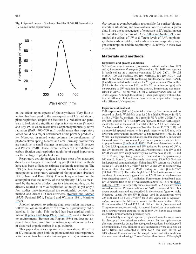
Experimental protocolCell suspensions (100 mL) were taken directly from cultures and in-cubated in plastic Whirl-Pak bags for 2 h at high (550 µmol⋅m–2⋅s–1;11 983 µW⋅cm–2), medium (350 µmol⋅m–2⋅s–1; 6536 µW⋅cm–2), orlow (100 µmol⋅m–2⋅s –1; 3268 µW⋅cm–2) photon flux of PAR, supple-mented (or not, in the case of controls) with UV-A + UV-B radiation.The lamp used for UV exposures (Toshiba Electric FL20S BLB) hada sinusoidal spectral output with a peak intensity at 352 nm, withlower and upper cutoffs at 310 and 400 nm, respectively (Fig. 1). TheWhirl-Pak bags used for incubations are transparent to UV and, underthe conditions of exposure employed, do not produce materials toxicto phytoplankton (Smith et al. 1992). PAR was determined with aLi-Cor PAR quantum sensor and UV radiation by means of UV-Aand UV-B sensors (SD 104, MACAM Photometrics). The UV-A andUV-B sensors have a high sensitivity in a narrow peak at 360 ± 10 and310 ± 10 nm, respectively, and cover bandwidths of approximately100 nm (P. Bossard, Lake Research Laboratory, EAWAG, Switzer-land, personal communication). Using these UV sensors we obtainedvalues of 1000 and 370 µW⋅cm–2 for UV-A and UV-B, respectively,from a clear sky with a PAR reading of 1760 µmol⋅m–2⋅s–1
(38 344 µW⋅cm–2). The rather high UV-B:UV-A ratio measured un-der these circumstances suggests that our UV-B sensor may have alsobeen detecting some UV-A radiation. Furthermore, broad band-passUV-A sensors tend to cut off wavelengths above 360–370 nm (Que-sada et al. 1995). Consequently our estimates of UV-A may have beenan underestimate. Precise conditions of PAR exposures differed be-tween experiments and are described in the relevant figure or table.Measured values for the UV-B fluxes used in experiments were224 ± 21 and 61.7 ± 2.5 µW⋅cm–2 for A. flos-aquae and S. capricor-nutum, respectively. Measured values for the concomitant UV-Afluxes were 444 ± 59 and 123.7 ± 4 µW⋅cm–2 for A. flos-aquae andS. capricornutum, respectively. A second, limited set of experimentson S. capricornutum exposed to the higher UV fluxes gave resultsessentially similar to those presented here.
Immediately after light exposure, replicated samples were takenfor chlorophyll determinations and cell counts. Further replicate sam-ples were taken twice during the subsequent incubations. For chlorophylldeterminations, 3-mL aliquots of cell suspensions were collected onGF/C filters and extracted at 80°C for 5 min with 10 mL of90% ethanol. Fluorescence of the sample before and after acidifica-tion was determined using a Turner Designs fluorometer. Chlorophyll
Fig. 1. Spectral output of the lamp (Toshiba FL20S BLB) used as a
UV source in the experiments.
Can. J. Fish. Aquat. Sci. Vol. 54, 1997688
© 1997 NRC Canada
concentrations (corrected for pheophytin) were calculated using theequations of Strickland and Parsons (1972).
For algal cell counts, 2- to 3-mL portions were fixed with Lugol’siodine and subsequently four replicates of each sample were countedunder a light microscope in a Neubauer haemocytometer chamber.
14C uptake in light and release in the darkFor each experiment, algal cell suspensions (24 mL) were placed insix Whirl-Pak bags to each of which 37 kBq H14CO3 was added.Immediately after addition of [14C]bicarbonate, zero time and totalactivity samples were taken. The zero time samples were filtered andprocessed immediately (see below). Total added radioactivity in eachbag was measured by adding 200 µL of the sample to 400 µL ofCarbosorb and 5 mL Aquasol scintillation fluid (DuPont NEN). Thetotal counts were used to check and correct for differences in addedcounts within the experimental bags.
The bags were exposed for 2 h to PAR with or without UV radia-tion (see above) after which duplicate, 10-mL samples were takenfrom two of the bags and the particulate fraction was collected onGF/C filters. Inorganic [14C]bicarbonate remaining on the filters wasremoved by a 1-mL rinse with 0.5 M HCl. The filters were then leftovernight before adding Aquasol and counting by scintillation to de-termine 14C incorporation into particulate material. To evaluate ex-tracellular release of 14C-dissolved organic matter, duplicate 5-mLsamples of the filtrate were placed in scintillation vials and acidifiedwith 0.5 M HCl to approximately pH 2. After gentle shaking for8–12 h in open vials to remove residual inorganic 14C, a 1-mL portionof filtrate was mixed with Aquasol and the radioactivity assayed byscintillation counting.
Dark carbon losses from respiration and extracellular release weredetermined by incubating the remaining four bags in darkness afterthe 2-h light period. Duplicate 10-mL samples were taken from eachof two bags and processed as described above after 4 and 22 h in thedark. Note that concentrations of inorganic carbon were not measuredin these samples and therefore the results of 14C incorporation experi-ments or loss are expressed as disintegrations per minute (dpm).
Details of the precise light exposures for each experiment aregiven in the appropriate figure or table captions.
Respiratory O2 consumptionAfter exposure to PAR with or without UV irradiation, subsamples(2 mL) of cell suspensions were placed in the darkened chamber of aHansatech DW1 oxygen electrode. Respiratory oxygen consumptionwas measured at various times (up to 22 h) after transfer of the cellsto darkness. The temperature in the electrode chamber during meas-urements was maintained at 21 ± 0.1°C by means of a Lauda RMS6refrigerating circulator. The electrode was calibrated using air-
saturated water (283 nmol O2⋅mL–1) and sodium bisulphite (to givezero O2).
Electron transport system activityActivity of the respiratory ETS was determined by methods based onthose of Packard (1971) and of Kenner and Ahmed (1975a). Subsam-ples (20 mL) from cultures treated with PAR and UV as previouslydescribed were collected on GF/C filters and the cells disrupted usinga Potter homogenizer. ETS activity in these extracts was determinedusing the reduction of the tetrazolium salt INT (2-(4-iodophenyl)-3,4-nitrophenyl-5-phenyltetrazolium) as described by Packard (1985).
Results
Cellular parametersChlorophyll concentrations immediately after irradiation werevery similar, within the same species (SE ±12%), for all ex-perimental treatments in this study (Table 1). In S. capricornu-tum, there was a significant drop in cellular chlorophyllcontent after 2 h exposure to light, with the lowest pigmentlevels occurring at high light conditions. The addition of UVirradiation with low or medium PAR depressed cellular chlo-rophyll contents but not with high PAR. After 22 h in darkness,S. capricornutum that had been exposed to UV irradiation to-gether with low, medium, or high fluxes of PAR showed chlo-rophyll values of 66, 75, and 84%, respectively, compared withcells not exposed to UV irradiation. The trend in A. flos-aquaewas less clear, with no evident trends immediately after theradiation treatment or subsequent to the 22-h dark incubation.
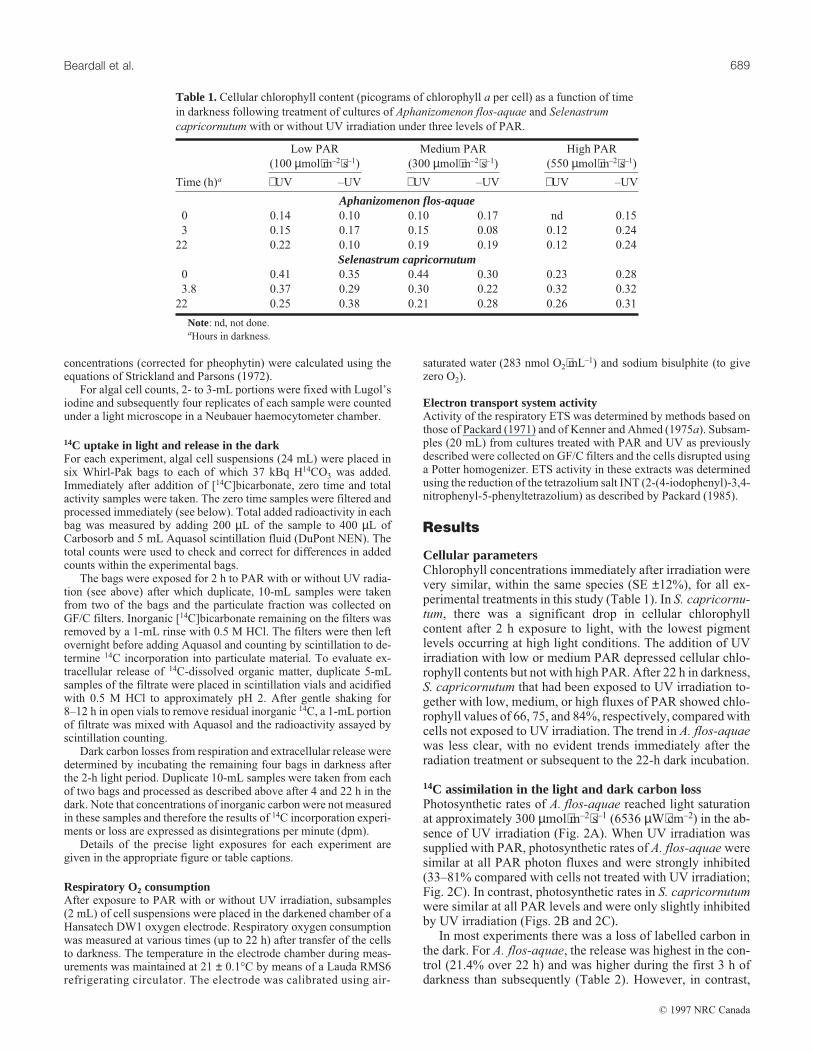
14C assimilation in the light and dark carbon lossPhotosynthetic rates of A. flos-aquae reached light saturationat approximately 300 µmol⋅m–2⋅s–1 (6536 µW⋅cm–2) in the ab-sence of UV irradiation (Fig. 2A). When UV irradiation wassupplied with PAR, photosynthetic rates of A. flos-aquae weresimilar at all PAR photon fluxes and were strongly inhibited(33–81% compared with cells not treated with UV irradiation;Fig. 2C). In contrast, photosynthetic rates in S. capricornutumwere similar at all PAR levels and were only slightly inhibitedby UV irradiation (Figs. 2B and 2C).
In most experiments there was a loss of labelled carbon inthe dark. For A. flos-aquae, the release was highest in the con-trol (21.4% over 22 h) and was higher during the first 3 h ofdarkness than subsequently (Table 2). However, in contrast,
Low PAR
(100 µmol⋅m–2⋅s–1)
Medium PAR
(300 µmol⋅m–2⋅s–1)
High PAR
(550 µmol⋅m–2⋅s–1)
Time (h)a +UV –UV +UV –UV +UV –UV
Aphanizomenon flos-aquae0 0.14 0.10 0.10 0.17 nd 0.15
3 0.15 0.17 0.15 0.08 0.12 0.24
22 0.22 0.10 0.19 0.19 0.12 0.24
Selenastrum capricornutum0 0.41 0.35 0.44 0.30 0.23 0.28
3.8 0.37 0.29 0.30 0.22 0.32 0.32
22 0.25 0.38 0.21 0.28 0.26 0.31
Note: nd, not done.aHours in darkness.
Table 1.Cellular chlorophyll content (picograms of chlorophyll a per cell) as a function of time
in darkness following treatment of cultures of Aphanizomenon flos-aquae and Selenastrum
capricornutum with or without UV irradiation under three levels of PAR.
Beardall et al. 689
© 1997 NRC Canada
the UV-treated samples of A. flos-aquae showed a slight in-crease in net 14C incorporated over 22 h, although in cellstreated with both high and low levels of PAR there were per-ceptible drops in incorporated 14C during the first 3 h of dark-ness (Table 2). With S. capricornutum, the UV-treated cellsexposed to high and medium levels of PAR lost less 14C thanthose without UV. The opposite occurred in cells exposed tolow PAR + UV (Table 2).
In all instances, the proportion of released 14C-labelled dis-solved organic material in culture filtrates was negligible incomparison with dark carbon loss when this occurred (<2and <1% of total fixed counts in A. flos-aquae and S. capri-cornutum, respectively). The levels of extracellular dissolvedorganic carbon released showed no effect of UV exposure orof different PAR fluxes.
Rates of oxygen consumptionRates of O2 consumption (measured directly by oxygen electrode)
in A. flos-aquae were dependent on the photon flux of PARgiven during the light exposure (Fig. 3) and showed a clearenhancement of postillumination respiration (EPIR; Falk-owski et al. 1985) over the first 3 h of darkness in all experi-ments. When cells were previously exposed to PAR togetherwith UV radiation, dark respiration and the EPIR effect inA. flos-aquae were decreased.
Rates of oxygen consumption by S. capricornutum alsoshowed a strong EPIR after high light irradiation (Fig. 4A),although this was markedly decreased at lower PAR expo-sures. Initial rates of respiration decreased with previous PARexposure (Fig. 4). In contrast to A. flos-aquae, EPIR in S. ca-pricornutum was slightly stimulated by the presence of UVduring exposure to PAR, and even cells exposed to low PAR(+ UV) exhibited an EPIR effect.
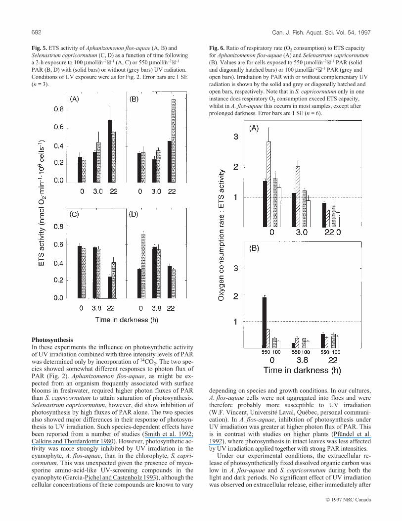
Electron transport system activityIn A. flos-aquae, cell-specific ETS activities increased duringdarkness. Exposure to UV + PAR resulted in a slight stimula-tion of ETS activity immediately after transfer of cells to dark-ness, whilst after 22 h in darkness, cells exposed to high PAR +UV had twice the ETS activity of cells exposed to PAR alone(Fig. 5).
In most cases, cell-specific ETS activity of S. capricornu-tum decreased during the 22 h in the dark, although under lowPAR (± UV) values were higher after 3.8 h than initially.There were, however, no consistent effects of UV exposure(Fig. 5).
Curiously, the initial rates of oxygen consumption inA. flos-aquae, as determined by oxygen electrode, were sig-nificantly higher than ETS capacity (Fig. 6A). This was notthe case after prolonged (22 h) exposure to darkness and didnot appear to be related to UV exposure. In the case of S. ca-pricornutum, only in the instance of cells exposed to highPAR + UV for 2 h did oxygen consumption exceed ETS ca-pacity, and that only occurred during the first 5 min of dark-ness (Fig. 6B).
Discussion
The UV fluxes applied in these experiments were lower thanthe maximum fluxes generally observed at sea level from aclear sky. Thus, the effects that we observed on both carbonfixation and respiration were likely, if anything, to underesti-mate the impacts of UV radiation in near surface waters innature, although we recognize that algae in a mixed watercolumn are only exposed to high, surface irradiance for a shortperiod of the day. Because of the characteristics of our UV-radiation sources, the algae in these samples were exposed torather low levels of UV-B, and thus our results may not bedirectly pertinent to questions concerning the potential in-crease of UV-B owing to decreasing global ozone cover. Fur-thermore, the experiments we report were conducted in thepresence of UV-A (with a UV-A:UV-B ratio of approxi-mately 2), conditions under which UV-B inhibition of growthhas been shown to be offset by repair mechanisms induced byUV-A (Quesada et al. 1995). Nevertheless, we have shownthat UV irradiation affects not only photosynthetic capacitybut also dark respiration in algae and that responses to irradia-tion may be species specific.
Fig. 2. Photosynthetic rates of (A) Aphanizomenon flos-aquae and
(B) Selenastrum capricornutum as a function of photon flux.
Photosynthetic rates are expressed as dpm⋅min–1⋅106 cells–1. Cells
were exposed to PAR alone (d) or to PAR supplemented with 224
or 61.7 µW⋅cm–2 UV-B (m) for A. flos-aquae and
S. capricornutum, respectively. Exposure of S. capricornutum to
the higher level of UV irradiation produced essentially the same
result as shown here. (C) Ratio of rates obtained with different PAR
levels of UV to that without UV for S. capricornutum (d) and A.
flos-aquae (m) (n = 3).
Can. J. Fish. Aquat. Sci. Vol. 54, 1997690
© 1997 NRC Canada

Low PAR
(100 µmol⋅m–2⋅s–1)
Medium PAR
(300 µmol⋅m–2⋅s–1)High PAR (550 µmol⋅m–2⋅s–1)
–UV +UV –UV +UV –UV +UV
Aphanizomenon flos-aquae4 h dark 14.7 (4.9) 13.1 (4.4) 13.6 (3.4) –6.2 (–1.6) 11.1 (2.8) 8.5 (2.1)
22 h dark 20.7 (0.3) –9.3(–0.5) 18.9 (1.0) –15.3 (–0.8) 21.4 (0.6) –25.8 (–1.91)
Selenastrum capricornutum4 h dark 11.4 (3.0) –1.0 (–0.3) 9.0 (2.4) 3.3 (0.9) –7.5 (–2.0) 13.9 (3.7)
22 h dark 17.1 (0.9) 30.8 (1.6) 19.5 (1.0) 15.7 (0.8) 48.4 (2.6) 14.7 (0.8)
Note: Carbon losses for the indicated times are given as the percentage decrease of initial dpm in algal cells at the
beginning of the dark period. A negative value signifies that 14C assimilation increased during the first 3 h (or 3.8 h)
darkness or the ensuing period. Values in parentheses are calculated rates given as the percentage change in the initial dpm
per hour.
Table 2.Effect of UV and PAR irradiation on dark carbon loss in Aphanizomenon flos-aquae and
Selenastrum capricornutum.
Fig. 4. Dark oxygen consumption rate (nmol O2⋅min–1⋅106 cells–1)
of Selenastrum capricornutum after a 2-h exposure to PAR with
(solid bars) or without (grey bars) UV radiation. Conditions of
exposure to PAR and UV were as for Fig. 2. Error bars are 1 SE
(n = 3).
Fig. 3. Dark oxygen consumption rate of Aphanizomenon
flos-aquae after a 2-h exposure to PAR with (solid bars) or without
(grey bars) UV radiation. Conditions of PAR and UV exposure
were as for Fig. 2. PAR fluxes were (A) 550 µmol⋅m–2⋅s–1, (B) 300
µmol⋅m–2⋅s–1, and (C) 100 µmol⋅m–2⋅s–1. Error bars are 1 SE (n = 3).
Beardall et al. 691
© 1997 NRC Canada
PhotosynthesisIn these experiments the influence on photosynthetic activityof UV irradiation combined with three intensity levels of PARwas determined only by incorporation of 14CO2. The two spe-cies showed somewhat different responses to photon flux ofPAR (Fig. 2). Aphanizomenon flos-aquae, as might be ex-pected from an organism frequently associated with surfaceblooms in freshwater, required higher photon fluxes of PARthan S. capricornutum to attain saturation of photosynthesis.Selenastrum capricornutum, however, did show inhibition ofphotosynthesis by high fluxes of PAR alone. The two speciesalso showed major differences in their response of photosyn-thesis to UV irradiation. Such species-dependent effects havebeen reported from a number of studies (Smith et al. 1992;Calkins and Thordardottir 1980). However, photosynthetic ac-tivity was more strongly inhibited by UV irradiation in thecyanophyte, A. flos-aquae, than in the chlorophyte, S. capri-cornutum. This was unexpected given the presence of myco-sporine amino-acid-like UV-screening compounds in thecyanophyte (Garcia-Pichel and Castenholz 1993), although thecellular concentrations of these compounds are known to vary
depending on species and growth conditions. In our cultures,A. flos-aquae cells were not aggregated into flocs and weretherefore probably more susceptible to UV irradiation(W.F. Vincent, Université Laval, Québec, personal communi-cation). In A. flos-aquae, inhibition of photosynthesis underUV irradiation was greater at higher photon flux of PAR. Thisis in contrast with studies on higher plants (Pfündel et al.1992), where photosynthesis in intact leaves was less affectedby UV irradiation applied together with strong PAR intensities.
Under our experimental conditions, the extracellular re-lease of photosynthetically fixed dissolved organic carbon waslow in A. flos-aquae and S. capricornutum during both thelight and dark periods. No significant effect of UV irradiationwas observed on extracellular release, either immediately after
Fig. 5. ETS activity of Aphanizomenon flos-aquae (A, B) and
Selenastrum capricornutum (C, D) as a function of time following
a 2-h exposure to 100 µmol⋅m–2⋅s–1 (A, C) or 550 µmol⋅m–2⋅s–1
PAR (B, D) with (solid bars) or without (grey bars) UV radiation.
Conditions of UV exposure were as for Fig. 2. Error bars are 1 SE
(n = 3).
Fig. 6. Ratio of respiratory rate (O2 consumption) to ETS capacity
for Aphanizomenon flos-aquae (A) and Selenastrum capricornutum
(B). Values are for cells exposed to 550 µmol⋅m–2⋅s–1 PAR (solid
and diagonally hatched bars) or 100 µmol⋅m–2⋅s–1 PAR (grey and
open bars). Irradiation by PAR with or without complementary UV
radiation is shown by the solid and grey or diagonally hatched and
open bars, respectively. Note that in S. capricornutum only in one
instance does respiratory O2 consumption exceed ETS capacity,
whilst in A. flos-aquae this occurrs in most samples, except after
prolonged darkness. Error bars are 1 SE (n = 6).
Can. J. Fish. Aquat. Sci. Vol. 54, 1997692
© 1997 NRC Canada
the irradiation period or after the subsequent dark incubation,despite the metabolic stress on the algae that might be expectedfrom this treatment.
RespirationThere have been very few studies of the effects of UV radiationon respiration of algae or other plants. Usually the determina-tion of respiration has taken a subservient role to measure-ments of photosynthesis. For example, Larkum and Wood(1993) who showed significant effects of UV radiation on phy-toplanktonic and benthic macroalgal photosynthesis simplynoted that they did not observe any effect on respiration andgave no data or the time scale for their respiration measure-ments.
We determined respiratory activity by three independentmethods: dark loss from algal cells of previously incorporated14C, direct measurement of O2 uptake, and evaluation of ETSactivity. In actuality, these methods reflect different aspects ofrespiratory metabolism and thus UV irradiation would notnecessarily be expected to have identical effects on thesemeasurements.
Dark 14C lossAs noted, extracellular release of dissolved organic carbon indarkness was negligible. It is also possible that some dissolvedorganic carbon released by the algae during the incubationperiod may have been taken up by bacteria in these samplesand subsequently respired. However, given the very predomi-nant biomass and metabolic activity of algae this would repre-sent a very small carbon flux. Thus, the bulk of the carbonlosses from the algal cells that occurred during the dark periodmust be attributed to respiration. Similar results using thistechnique were reported by Berman and Kaplan (1984).
Aphanizomenon flos-aquae cultures that were not exposedto UV irradiation exhibited a normal pattern (cf. Berman andKaplan 1984) of dark 14C loss with a decrease of about 20%of initially incorporated 14C dpm over 22 h (Table 2). At alllevels of previous irradiation with PAR, the decrease of cellu-lar 14C was relatively rapid during the first 3 h of darkness(probably because of an EPIR; Falkowski et al. 1985; Beardallet al. 1994) and much slower over the subsequent 20 h. Treat-ment with UV irradiation during the light phase not only dras-tically lowered 14C fixation but eliminated the dark drop incellular 14C. Indeed a slight rise in the cellular 14C counts wasobserved after 20 h in all treatments. Only in the low-PARtreatment was there a small dark drop in the 14C in cells duringthe first 3 h.
Selanastrum capricornutum cultures treated with mediumor low PAR (no UV) also showed normal patterns of 14C loss.Here the dark carbon loss increased with higher PAR levels(Table 2). Addition of UV irradiation either had no effect (me-dium PAR level) or accelerated the dark 14C decrease (lowPAR treatment). In the case of high PAR (without UV), aninitial 3.8-h increase of cellular 14C was observed prior to arelatively rapid 14C loss from 3.8 to 22 h. In this case, treat-ment with UV and high PAR irradiation decreased respirationrates and stabilized the cellular 14C loss after an initial 3.8-hdecline.
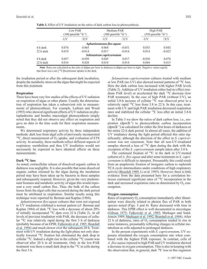
In Table 3 we show the ratios of dark carbon loss, i.e., res-piration (dpm⋅h–1) to photosynthetic carbon incorporation(dpm⋅h–1) as calculated for either the first hours of darkness orthe entire 22-h dark period. In almost all cases, the addition ofUV irradiance during the light period affected this ratio sig-nificantly, although the direction of the effect in S. capricor-nutum was not consistent. Note that all non-UV-irradiatedsamples showed a loss of 14C dpm during the dark with theexception of the S. capricornutum sample taken after 3.8 h.
The continued fixation of 14C in the dark in UV-treatedcultures of A. flos-aquae and after some treatments in S. capri-cornutum is difficult to interpret. Presumably this could resultfrom an anaplerotic fixation of inorganic carbon to replenishTCA cycle intermediates under conditions of high respiratoryactivity (Beardall 1989; Li et al. 1993). However, there is littleevidence from the data presented here for a correlation be-tween continued significant rates of 14C incorporation in thedark and increased respiration rates as determined by O2 con-sumption.
Oxygen consumptionRates of respiratory O2 consumption immediately after illumi-nation were directly related to photon flux of PAR in bothspecies tested (Figs. 3 and 4). Rates decreased with time indarkness. This EPIR effect is well documented in microlagae(Gibson 1975; Falkowski et al. 1985; Markager and Sand-Jensen 1989; Markager et al. 1992; Beardall et al. 1994). After22 h of darkness, rates of O2 consumption tended to rise insome instances, presumably reflecting changes in cellular me-tabolism as cells adjusted to prolonged darkness.
In the present experiments with S. capricornutum, UV ex-posure stimulated the oxygen consumption rate when com-bined with the highest PAR exposure. In contrast, cells ofA. flos-aquae exposed to high PAR and UV irradiation showeda decrease in oxygen consumption. This is also in keeping withthe observation that, in general, dark 14C loss in this organism
Low PAR
(100 µmol⋅m–2⋅s–1)
Medium PAR
(300 µmol⋅m–2⋅s–1)
High PAR
(550 µmol⋅m–2⋅s–1)
–UV +UV –UV +UV –UV +UV
Aphanizomenon flos-aquae4 h dark 0.074 0.065 0.068 –0.031 0.055 0.043
22 h dark 0.019 –0.014 0.017 –0.014 0.014 –0.023
Selenastrum capricornutum4 h dark 0.057 –0.050 0.045 0.017 –0.038 0.070
22 h dark 0.016 0.028 0.018 0.014 0.044 0.013
Note: Rates are calculated as the ratio of ∆dpm per hour in darkness to ∆dpm per hour in light. Negative values signify
that there was a net [14C]bicarbonate uptake in the dark.
Table 3.Effect of UV irradiation on the ratios of dark carbon loss to photosynthesis.
Beardall et al. 693
© 1997 NRC Canada
was eliminated in cells that were previously exposed to UVirradiation. These results are consistent with the proposal thatEPIR effects are associated with the provision of excess pho-tosynthate during the light, which is subsequently rapidly re-spired (Markager and Sand-Jensen 1989; Markager et al.1992; Beardall et al. 1994). Thus, in A. flos-aquae, in whichphotosynthesis was significantly inhibited by UV irradiation,EPIR was also decreased. Conversely, in S. capricornutum, inwhich UV irradiation has little effect on photosynthesis, bothUV-treated and nontreated cells showed strong EPIR effects.There was a trend towards generally higher initial oxygen con-sumption rates in UV-exposed S. capricornutum. This wouldbe consistent with the retention of regulated cellular activityafter UV exposure being dependent on increased repair pro-cesses and maintenance demands.
In conclusion, UV treatment had a negative effect on netcarbon accumulation in both species after 2 h of the light ex-posure, and these effects were still perceptible after a further22 h in darkness. It is possible to postulate that differentmechanisms are involved in the two organisms. In A. flos-aquae, carbon fixation rates were inhibited relatively strongly,which caused low accumultion of organic carbon even thoughsubsequent respiration rates were depressed. In S. capricornu-tum, carbon fixation was less affected during PAR + UV ex-posure. In this case, however, post-irradiation dark respirationrates were enhanced, a process that probably decreased the netfixation of carbon as measured over the 24-h experimentalperiod.
Electron transport system activityETS activity per cell tended to decrease in the dark in theeucaryote and increase in the procaryote (Fig. 5). In all casesexcept S. capricornutum subject to high PAR + UV, UV expo-sure led to an increase in ETS activity immediately upon dark-ening. However, at the end of the dark period, ETS activitieswere similar with or without UV pretreatment of cells or werelower for UV-irradiated samples.
In the case of A. flos-aquae, ETS activity increased withtime in the dark (Figs. 5A and 5B). Cyanobacteria have aunique association between the respiratory and the photosyn-thetic electron transport systems that share the same cyto-chrome b6/f complex (Malkin 1988). This could mean that, inthe light, the two systems compete for electrons and the respi-ratory ETS is repressed, whereas in the dark only the respira-tory ETS is operative and its activity increases. In all cases inour study the ETS activity of A. flos-aquae was higher at theend of the dark period than initially.
The addition of UV radiation caused a transient ETS in-crease, at the beginning of the dark period, compared withtreatments without UV. This might be related to the stronginhibition of photosynthesis by UV radiation on A. flos-aquaeduring the light period discussed above. If this inhibition actsto partially derepress respiration during the light period, thenthe observed transient enhancement of ETS activity upon dark-ening might be expected. The general effect of UV exposure,however, was a decrease of ETS activity, relative to non-UV-treated samples, that was particularly manifested at the end ofthe dark period in the high PAR + UV A. flos-aquae culture.The strong inhibition of photosynthesis that occurred in thesecultures (see above) might have resulted in a shortage of
photosynthetic products as substrates for respiration during thedark period (Beardall et al. 1994).
Aphanizomenon flos-aquae treated with high PAR withoutUV irradiation showed the highest ETS activity at the end ofthe dark period (0.85 nmol O2⋅106 cells–1⋅min–1), almost dou-ble that obtained with cells treated with high PAR + UV(Fig. 5B). In the absence of UV, prior exposure to high PAR(Fig. 5B) enhanced ETS activity in the dark (probably as aresult of higher photosynthesis during the light period).UV irradiation inhibited photosynthesis and, in consequence,respiratory ETS activity in the dark diminished.
We observed respiratory oxygen uptake rates exceedingETS activity in most cases for A. flos-aquae (Fig. 6). The ex-cess O2 consumption could be due to some process not meas-ured by the ETS assay. The role of the respiratory chain inprocaryotic obligate phototrophs is not completely understood.In cyanobacteria, classical inhibitors of the mitochondrial-typecytochrome b/c1 complex (i.e., antimycin) have little or no ef-fect on oxidoreductase activity of the b6/f complex. In addi-tion, it has been shown that the respiratory aa3-typecytochrome c oxidase is present in the cytoplasmic membraneof cyanobacteria. When mixed-membrane (i.e., cytoplasmicplus thylakoid) preparations are used to measure respiratoryactivity, as in our study, the capacity of the enzyme to oxidizereduced cytochrome c is lower, possibly because of the diver-sion of electron flow to other pathways (Fry and Peschek1988). This would explain the higher oxygen consumptionrates (measuring all routes of electron flow to oxygen) thanETS activity (measuring only flux through the mitochondrial-type pathway). Hence, the ETS method is probably not appro-priate for measurements of respiratory activity in phototrophicprocaryotes.
In the eucaryote S. capricornutum, all except one measure-ment of rates of oxygen uptake were within the capacity ofmeasured ETS capacity (Fig. 6). Selenastrum capricornutumshowed decreasing ETS values during the dark period. Maxi-mum activity was found upon darkening after treatment withhigh PAR without UV radiation. This is in agreement withreports of higher ETS activity co-occurring with EPIR in othereucaryotic microalgae after PAR illumination, the magnitudeof the enhancement being related to the previous radiation dos-age (Martinez 1992). However, high PAR combined with UVradiation transiently inhibited ETS activity in dark. The factthat actual oxygen consumption was enhanced after highPAR + UV suggests that there is some oxygen-consuming pro-cess occurring under these conditions that was not measuredby the ETS assay. It is possible, for instance, that the mito-chondrial cytochrome pathway (CP) was inhibited and the al-ternate respiratory pathway (AP) was stimulated under theseconditions. Weger and Dasgupta (1993) found, in Chlamydo-monas reinhardii, that AP showed a large increase relative toeither CP or total respiration in nutrient-limited versus nutri-ent-sufficient cells and in cells under temperature or osmoticstress. In the last case, CP was greatly reduced and AP main-tained respiratory electron flow. In our study, UV radiationcombined with high PAR might cause a similar effect.
It is evident that the effects of UV treatment on respiratorymetabolism were not consistent across techniques used to de-termine respiration (Table 4). This is not unexpected, sinceeach of the approaches measures different aspects of respiratorymetabolism. Nevertheless, it may be noted that for A. flos-aquae,
Can. J. Fish. Aquat. Sci. Vol. 54, 1997694
© 1997 NRC Canada
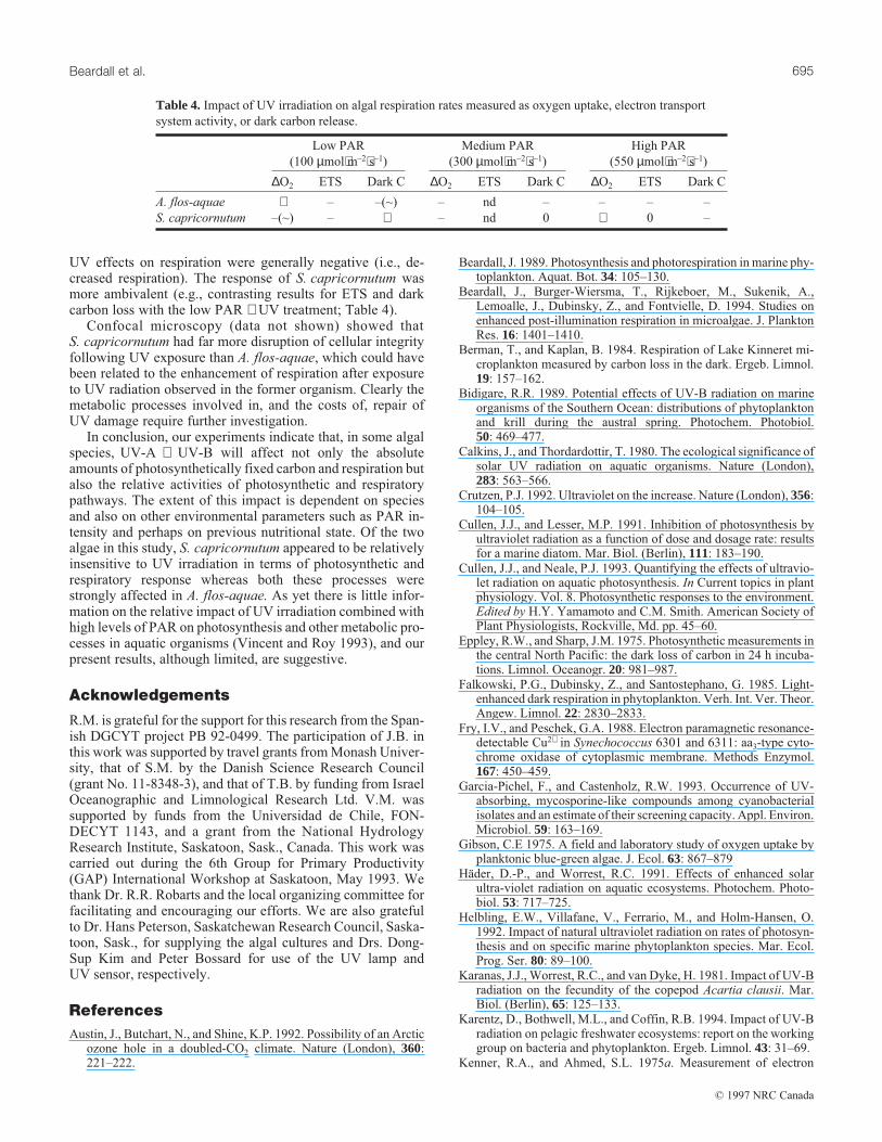
UV effects on respiration were generally negative (i.e., de-creased respiration). The response of S. capricornutum wasmore ambivalent (e.g., contrasting results for ETS and darkcarbon loss with the low PAR + UV treatment; Table 4).
Confocal microscopy (data not shown) showed thatS. capricornutum had far more disruption of cellular integrityfollowing UV exposure than A. flos-aquae, which could havebeen related to the enhancement of respiration after exposureto UV radiation observed in the former organism. Clearly themetabolic processes involved in, and the costs of, repair ofUV damage require further investigation.
In conclusion, our experiments indicate that, in some algalspecies, UV-A + UV-B will affect not only the absoluteamounts of photosynthetically fixed carbon and respiration butalso the relative activities of photosynthetic and respiratorypathways. The extent of this impact is dependent on speciesand also on other environmental parameters such as PAR in-tensity and perhaps on previous nutritional state. Of the twoalgae in this study, S. capricornutum appeared to be relativelyinsensitive to UV irradiation in terms of photosynthetic andrespiratory response whereas both these processes werestrongly affected in A. flos-aquae. As yet there is little infor-mation on the relative impact of UV irradiation combined withhigh levels of PAR on photosynthesis and other metabolic pro-cesses in aquatic organisms (Vincent and Roy 1993), and ourpresent results, although limited, are suggestive.
Acknowledgements
R.M. is grateful for the support for this research from the Span-ish DGCYT project PB 92-0499. The participation of J.B. inthis work was supported by travel grants from Monash Univer-sity, that of S.M. by the Danish Science Research Council(grant No. 11-8348-3), and that of T.B. by funding from IsraelOceanographic and Limnological Research Ltd. V.M. wassupported by funds from the Universidad de Chile, FON-DECYT 1143, and a grant from the National HydrologyResearch Institute, Saskatoon, Sask., Canada. This work wascarried out during the 6th Group for Primary Productivity(GAP) International Workshop at Saskatoon, May 1993. Wethank Dr. R.R. Robarts and the local organizing committee forfacilitating and encouraging our efforts. We are also gratefulto Dr. Hans Peterson, Saskatchewan Research Council, Saska-toon, Sask., for supplying the algal cultures and Drs. Dong-Sup Kim and Peter Bossard for use of the UV lamp andUV sensor, respectively.
References
Austin, J., Butchart, N., and Shine, K.P. 1992. Possibility of an Arcticozone hole in a doubled-CO2 climate. Nature (London), 360:221–222.
Beardall, J. 1989. Photosynthesis and photorespiration in marine phy-toplankton. Aquat. Bot. 34: 105–130.
Beardall, J., Burger-Wiersma, T., Rijkeboer, M., Sukenik, A.,Lemoalle, J., Dubinsky, Z., and Fontvielle, D. 1994. Studies onenhanced post-illumination respiration in microalgae. J. PlanktonRes. 16: 1401–1410.
Berman, T., and Kaplan, B. 1984. Respiration of Lake Kinneret mi-croplankton measured by carbon loss in the dark. Ergeb. Limnol.19: 157–162.
Bidigare, R.R. 1989. Potential effects of UV-B radiation on marineorganisms of the Southern Ocean: distributions of phytoplanktonand krill during the austral spring. Photochem. Photobiol.50: 469–477.
Calkins, J., and Thordardottir, T. 1980. The ecological significance ofsolar UV radiation on aquatic organisms. Nature (London),283: 563–566.
Crutzen, P.J. 1992. Ultraviolet on the increase. Nature (London), 356:104–105.
Cullen, J.J., and Lesser, M.P. 1991. Inhibition of photosynthesis byultraviolet radiation as a function of dose and dosage rate: resultsfor a marine diatom. Mar. Biol. (Berlin), 111: 183–190.
Cullen, J.J., and Neale, P.J. 1993. Quantifying the effects of ultravio-let radiation on aquatic photosynthesis. In Current topics in plantphysiology. Vol. 8. Photosynthetic responses to the environment.Edited by H.Y. Yamamoto and C.M. Smith. American Society ofPlant Physiologists, Rockville, Md. pp. 45–60.
Eppley, R.W., and Sharp, J.M. 1975. Photosynthetic measurements inthe central North Pacific: the dark loss of carbon in 24 h incuba-tions. Limnol. Oceanogr. 20: 981–987.
Falkowski, P.G., Dubinsky, Z., and Santostephano, G. 1985. Light-enhanced dark respiration in phytoplankton. Verh. Int. Ver. Theor.Angew. Limnol. 22: 2830–2833.
Fry, I.V., and Peschek, G.A. 1988. Electron paramagnetic resonance-detectable Cu2+ in Synechococcus 6301 and 6311: aa3-type cyto-chrome oxidase of cytoplasmic membrane. Methods Enzymol.167: 450–459.
Garcia-Pichel, F., and Castenholz, R.W. 1993. Occurrence of UV-absorbing, mycosporine-like compounds among cyanobacterialisolates and an estimate of their screening capacity. Appl. Environ.Microbiol. 59: 163–169.
Gibson, C.E 1975. A field and laboratory study of oxygen uptake byplanktonic blue-green algae. J. Ecol. 63: 867–879
Häder, D.-P., and Worrest, R.C. 1991. Effects of enhanced solarultra-violet radiation on aquatic ecosystems. Photochem. Photo-biol. 53: 717–725.
Helbling, E.W., Villafane, V., Ferrario, M., and Holm-Hansen, O.1992. Impact of natural ultraviolet radiation on rates of photosyn-thesis and on specific marine phytoplankton species. Mar. Ecol.Prog. Ser. 80: 89–100.
Karanas, J.J., Worrest, R.C., and van Dyke, H. 1981. Impact of UV-Bradiation on the fecundity of the copepod Acartia clausii. Mar.Biol. (Berlin), 65: 125–133.
Karentz, D., Bothwell, M.L., and Coffin, R.B. 1994. Impact of UV-Bradiation on pelagic freshwater ecosystems: report on the workinggroup on bacteria and phytoplankton. Ergeb. Limnol. 43: 31–69.
Kenner, R.A., and Ahmed, S.L. 1975a. Measurement of electron
Low PAR
(100 µmol⋅m–2⋅s–1)
Medium PAR
(300 µmol⋅m–2⋅s–1)
High PAR
(550 µmol⋅m–2⋅s–1)
∆O2 ETS Dark C ∆O2 ETS Dark C ∆O2 ETS Dark C
A. flos-aquae + – –(~) – nd – – – –
S. capricornutum –(~) – + – nd 0 + 0 –
Table 4. Impact of UV irradiation on algal respiration rates measured as oxygen uptake, electron transport
system activity, or dark carbon release.
Beardall et al. 695
© 1997 NRC Canada
transport activities in marine plankton. Mar. Biol. (Berlin), 33:119–128.
Kenner, R.A., and Ahmed, S.L. 1975b. Correlation between oxygenutilization and electron transport activity in marine phytoplankton.Mar. Biol. (Berlin), 33: 129–133
Larkum, A.W.D., and Wood, W.F. 1993. The effect of UV-B radia-tion on photosynthesis and respiration of phytoplankton, benthicmacroalgae and seagrasses. Photosynth. Res. 36: 17–23.
Li, W.K.D., Irwin, B.D., and Dickie, P.M. 1993. Dark fixation of 14C.Limnol. Oceanogr. 38: 483–494.
Malkin, R. 1988. Cytochrome b6/f complex: common components ofrespiratory and photosynthetic electron transport systems. Meth-ods Enzymol. 167: 341–349.
Markager, S., and Sand-Jensen, K. 1989. Patterns of night-time respi-ration in a dense phytoplankton community under a natural lightregime. J. Ecol. 77: 49–61.
Markager, S., Jespersen, A.-M., Madsen, T.V., Berdalet, E., andWeisburd, R. 1992. Diel changes in dark respiration in a planktoncommunity. Hydrobiologia, 238: 119–130.
Martinez, R. 1992. Respiration and respiratory electron transport ac-tivity in marine phytoplankton: growth rate dependence and lightenhancement. J. Plankton Res. 14: 789–797.
Owens, T., and King, F.D. 1975. The measurement of respiratory-electron-transport-system activity in marine zooplankton. Mar.Biol. 30: 27–36.
Packard, T.T. 1971. Measurement of electron transport system activ-ity in marine plankton. J. Mar. Res. 29: 235–244.
Packard, T.T. 1985. Measurement of electron transport activity ofmicroplankton. Adv. Aquat. Microbiol. 3: 207–261.
Packard, T.T., and Williams, P.leB. 1981. Rates of respiratory oxygenconsumption and electron transport in surface seawater from thenorthwest Atlantic. Oceanol. Acta, 4: 351–358.
Pfündel, E.E., Pan, R.S., and Dilley, R.A. 1992. Inhibition of violax-anthin depoxidation by ultraviolet-B radiation in isolated chloro-plasts and intact leaves. Plant Physiol. 98: 1372–1380.
Quesada, A., Mouget, J.-L., and Vincent, W.F. 1995. Growth of Ant-arctic cyanobacteria under ultraviolet radiation: UV-A counteractsUV-B inhibition. J. Phycol. 31: 242–248.
Smetacek, V., and Passow, V. 1990. Spring-bloom initiation andSverdrup’s critical depth model. Limnol. Oceanogr. 35: 228–234.
Smith, W.O. 1977. Respiration of photosynthetic carbon in theeuphotic areas of the ocean. J. Mar. Res. 35: 557–565.
Smith, R.C. 1989. Ozone, middle ultraviolet radiation and the aquaticenvironment. Photochem. Photobiol. 50: 459–468.
Smith, R.C., Prezelin, B.B., Baker, K.S., Bidigare, R.R., Boucher,N.P., Coley, T., Karentz, D., Macintyre, S., Matlick, H.A.,Menzies, D., Ondrusek, M., Wan, Z., and Waters, K.J. 1992.Ozone depletion: ultraviolet radiation and phytoplankton biologyin Antarctic waters. Science (Washington, D.C.), 255: 952–959.
Strickland, J.D.H., and Parsons, T.R. 1972. A practical handbook ofseawater analysis. 2nd ed. Bull. Fish. Res. Board Can. No. 167.
Vincent, W.F., and Roy, S. 1993. Solar ultraviolet-B radiation andaquatic primary production: damage, protection, and recovery.Environ. Rev. 1: 1–12.
Weger, H.G., and Dasgupta, R. 1993. Regulation of alternative path-way respiration in Chlamydomonas reinhardii (Chlorophyceae).J. Phycol. 29: 300–308.
Can. J. Fish. Aquat. Sci. Vol. 54, 1997696
© 1997 NRC Canada