the effects of “heidelberg scalp acupuncture” on obstructive ...
135
THE EFFECTS OF “HEIDELBERG SCALP ACUPUNCTURE” ON OBSTRUCTIVE SLEEP APNEA. -A PRELIMINAR STUDY- Maria João Rodrigues Ferreira Rocha dos Santos Dissertação de mestrado em Medicina Tradicional Chinesa 2012
-
Upload
khangminh22 -
Category
Documents
-
view
0 -
download
0
Transcript of the effects of “heidelberg scalp acupuncture” on obstructive ...
THE EFFECTS OF “HEIDELBERG SCALP
ACUPUNCTURE” ON OBSTRUCTIVE SLEEP APNEA.
-A PRELIMINAR STUDY-
Maria João Rodrigues Ferreira Rocha dos Santos
Dissertação de mestrado em Medicina Tradicional Chinesa
2012

Maria João Rodrigues Ferreira Rocha dos Santos
THE EFFECTS OF “HEIDELBERG SCALP
ACUPUNCTURE” ON OBSTRUCTIVE SLEEP APNEA.
A PRELIMINAR STUDY
Dissertação de Candidatura ao grau de Mestre
em Medicina Tradicional Chinesa submetida
ao Instituto de Ciências Biomédicas de Abel Salazar
da Universidade do Porto.
Orientador
– Prof. Doutor Henry Johannes Greten
Categoria – Professor Associado
Afiliação – Instituto de Ciências Biomédicas Abel Salazar
Co-orientador
-Prof. Doutor João Carlos Winck
Categoria – Professor Associado
Afiliação – Faculdade de Medicina da Universidade do Porto
-Mestre Dr. Nuno Correia


DEDICATION:
To my parents …
To Ana Maria Grande,
For the "philosopher stone",
For the scientific enchantment.


We are the scientific civilization ...
Knowledge is our destiny "
Jacob Bronowski
“In the history of human thinking,
the most fruitful thoughts occur, in general,
when two streams totally distinct meet together ...”
Werner Heisenberg

ACKNOWLEDGEMENTS:
To my family for education, formation and eternal devotion.
To Nuno Correia for permanent support and productive scientific restlessness.
To Professor Greten for the fulfillment of "mission" and all the teachings.
To Professor Jorge Machado for the scientific courage, by the vision that i call
integrative and progressive and for all support and motivation.
To Institute of Biomedical Sciences Abel Salazar for the opportunity and
excellence in science.
To Dr. Maria Joao Lima and Professor João Carlos Winck, for the opportunity and
availability.
To all health professionals, of the department of Pneumology of SJHC, especially
to the technician, Patricia Teles, to Dr. Anabela Marino and to Dr. João Almeida
To Resmed in the person of the therapist Sara Correia
To you, Filipa (Dantas) for your constant light on me.
To the "Family of TCM, António Moreira by fellowship, by the tireless social
gatherings; my dear" cousin "(Ana Moreira) by opening, by the scientific fascination and
Friendship; To Fernando (" Sensei ") for the continuous sharing of knowledge, for the
defense of" arca-aurea. Were two beautiful years that I will never forget. To Ana Anjos
and Susana Seca for the kindness, hospitality and support..
To the "Master" Antonio Carvalho, for the teachings, the constant encouragement
and belief in me.
To all the teachers, of the masters course, especially the mastery of Professor
Frank, by the TCM and life teachings, to Professor Mário, for be our bridge and support
To our Alma Mater, Petra, for the tireless dedication to all of us.
To all the colleagues and friends of this master and specialization with whom I
reborn and shared unforgettable moments.
To Sisters Miranda and all patients by the stimulus and belief in my practice.
To the forevermore teacher, Ana Maria Grande, by teaching me science in its true
essence.
To Professor Nuno Grande by the integrative view of science and health, by the
open mind that allowed the existence of a Master degree that besides formation on
human health, allows humanistic formation for each student.


xi
Resumo:
Introdução: O Síndrome da Apneia Obstrutiva do Sono (SAOS) é uma doença
respiratória muito comum. Recentemente o SAOS tem sido associado com o síndrome
metabólico, morbidades cardiovasculares e cerebrovasculares, entre outras condições. A
Pressão Contínua Positiva nas vias respiratórias (CPAP) permanece como tratamento
gold-standard no tratamento do SAOS. No entanto, vários estudos indicam uma grande
variabilidade na adesão ao CPAP, exigindo abordagens de tratamento mais eficientes.
Estudos anteriores fornecem dados limitados que sugerem efeitos positivos imediatos e
mediatos da acupunctura no SAOS, suportando a tese de que a acupunctura pode ser útil
neste síndrome. Neste estudo apresentam-se dez casos clínicos de SAOS tratados por
acupunctura craniana de Heidelberg (ACH).
Objectivos: Avaliar o efeito agudo da ACH no SAOS, analisando mensurações
objectivas do estudo de sono, realizado através de poligrafia cardio-respiratória e
verificando a percepção de qualidade de sono através da realização de um questionário
matinal.
Método: Indivíduos com SAOS e naïve para acupunctura serão seleccionados a
partir de um Departamento de Pneumologia. Por questões éticas, o estudo é divido em
duas fases:
• Fase 1 (estudo preliminar): Dez pacientes serão tratados com ACH, com
micro-agulhas permanentes, durante 24 horas. As medições incluem
poligrafia cardio-respiratória (PG) antes e depois da acupunctura e a
realização de um questionário matinal na manhã seguinte à segunda
poligrafia.
• Fase 2: Após uma medição basal de PG, 40 pacientes serão
randomizados em 2 grupos: a) grupo com micro-agulhas permanentes de
ACH durante 10 dias e b) um grupo controlo sujeito a acupunctura falsa.
Uma segunda medição de PG irá avaliar o efeito
Resultados:
Fase 1: 8 em 10 pacientes tratados com ACH responderam à terapia, o que é
demonstrado por uma melhoria 51.4% (p value = 0,069) no índice de Apneia-Hiponea;
51.3% (p value = 0,089) nos eventos respiratórios; 14,8% (p value = 0,226) na
dessaturação de oxigénio e de 28.8% (p value =0,88) no tempo relativo de ronco. Em
termos de percepção de qualidade de sono, nenhum paciente relatou que a qualidade de
sono tenha piorado. Sete dos dez pacientes classificaram a qualidade de sono como
igual e 3 como de melhor qualidade que no dia-a-dia.
xii
Fase 2: com base nestes resultados preliminares e em estudos prévios, é
expectável uma melhoria do índice de apneia-hipopneia, da percepção da qualidade do
sono e da qualidade de vida.
Discussão: Apesar de não terem significância estatística, os dados da fase 1
sugerem uma melhoria nos parâmetros da poligrafia e na qualidade de percepção do
sono, a favor da fase 2 do estudo. O sistema somatotópico facial correlaciona-se com as
principais projecções anatómicas dos pontos do couro cabeludo o que possivelmente
explicará os efeitos positivos nas vias aéreas. Estas relações hipotéticas podem ser
suportadas pela teoria fractal aplicada à fisiologia e ontogenia humana.
Conclusão: Se se comprovar a sua eficácia, a ACH poderá ser uma terapia
complementar no tratamento da SAOS. Os seus mecanismos de acção poderão estar
relacionados com aspectos de neurofisiologia, embriologia e ontogenia. Investigação
adicional é necessária para avaliar o feito de longo termo da ACH no SAOS.
Palavras-chave: Síndrome de apneia obstrutiva do sono; índice de apneia
Hipopneia ; acupunctura craniana; acupunctura; Medicina Tradicional Chinesa.
xiii
Abstract
Introduction: Obstructive sleep apnea (OSA) is a very common respiratory
disease. Recently OSA has been associated with metabolic syndrome, cardiovascular
and cerebrovascular morbidity, among other conditions. Continuous positive airway
pressure (CPAP) remains the “gold-standard” treatment in the management of OSA.
However, several studies indicate a great variability in compliance to CPAP demanding
more efficient treatment approaches. Previous studies provide limited data that suggests
immediate and mediate positive effects of acupuncture in OSA, supporting the thesis that
acupuncture may be useful for this syndrome. We report 10 clinical cases of OSA treated
by “Heidelberg Scalp Acupuncture” (HSA).
Objective: To evaluate the acute effect of HSA in OSA, analyzing objective
measurements of a sleep study, conducted by cardiorespiratory polygraphy and checking
the perception of quality of sleep, through the realization of a morning questionnaire.
Methods: Individuals with OSA (AHI> 5) and naïve for acupuncture will be sampled from
the Pulmonology Department. For ethical reasons, the study is divided in two phases.
• Phase 1 (preliminary study): Ten patients were treated once with HSA, applying
permanent micro-needles for 24 hours. Measurements include cardio-respiratory
polygraphy (PG) prior and after acupuncture.
• Phase 2 : After a baseline measurement of PG, 40 patients will be randomized into
two groups: a) a group with permanent HSA needles for ten days and b) a control
group subject to sham acupuncture. A second PG assessment will evaluate the
effect.
Results: Phase 1: 8 out of 10 patients treated with HSA responded to therapy, as
demonstrated by a improvement of: 51.4% (p value = 0.069) in the Apnea-Hypopnea
index; 51.3% (p value = 0.089) in respiratory events, 14.8% (p value = 0.226) in the
desaturation of oxygen and 28.8% (p value = .88) in snoring relative time. In terms of
perception of the quality of sleep, none of the patients reported that sleep quality has
worsened. Seven of the ten patients have classified sleep quality as the same and 3
classified the quality of sleep as better than in everyday life.
Phase 2: based on these preliminary results and previous studies, it is expected an
improvement of the Apnea-Hypopnea Index, perception of sleep and quality of life.
Discussion: Although without statistically significance, phase 1 data suggests an
improvement in PG parameters and sleep quality perception, in favor of phase 2 of the
study. The facial somatotopic system of HSA apparently correlates with the major
anatomical projections of the scalp points possibly explaining the positive effects on the
xiv
airways. These postulated relationships would be supported by the “fractal theory” applied
to human physiology and ontogeny.
Conclusion: If proven to be effective HSA may be a complementary therapy for
the treatment of OSA. Its mechanisms of action may be related to neurophysiological,
embryological and ontogenetic aspects. Additional research is needed to evaluate long-
term effects of HSA on OSA.
Keywords: Obstructive Sleep Apnea; Apnea Hypopnea index; Scalp acupuncture;
Acupuncture; Traditional Chinese Medicine.
xv
Contents:
CHAPTER 1: ........................................................................................................ 21
1. Introduction: .................................................................................................. 23
2. Obstrutive Sleep Apnea Syndrome .................................................................. 23
2.1 Definition: .................................................................................................... 23
2.2. Epidemiology: ............................................................................................ 25
2.2.1. Risk Factors: ....................................................................................... 25
2.2.2. Consequences of OSA ........................................................................ 28
2.3. Diagnosis: .................................................................................................. 31
2.3.1. Polysomnography................................................................................ 32
2.3.2. Polygraphy: ......................................................................................... 32
2.4. Physiopathology: ....................................................................................... 32
2.5. Treatment: ................................................................................................. 34
2.5.1. General measures: .............................................................................. 34
2.5.2. Specific measures: .............................................................................. 34
2.6 Anatomical and embryological overview ..................................................... 38
2.6.1. Pharynx embryology: ........................................................................... 38
2.7. Pharynx anatomy: ...................................................................................... 39
2.7.1. Pharinx innervation: ............................................................................. 40
2.7.2. Vessels of the Pharynx: ....................................................................... 40
Traditional Chinese Medicine Overview ............................................................... 43
3. Contemporary TCM .......................................................................................... 45
3.1.Acupuncture: ............................................................................................... 47
3.2. Relationship between the conduits and the nervous system, blood vessels
and lymphatic vessels ....................................................................................... 49
3.2.1. Relationship between the conduits and the Nervous System: ............. 49
3.2.2. Relationship between blood vessels and conduits: ............................. 49
3.2.2. Relationship between conduits and the lymphatic vessels .................. 49
3.3. Microsystem acupuncture .......................................................................... 50
3.3.1. Scalp Acupuncture .............................................................................. 51
3.4. Heidelberg model ....................................................................................... 56
3.4.1. Diagnostic process according to the Heidelberg model of TCM .......... 58
xvi
3.4.2. Pathogeny - Overview of the model of Heidelberg .............................. 61
3.4.3. Algor Laedens Theory 3rd guiding criteria ............................................ 61
3.4.4. Upper airways in Heidelberg model of TCM ........................................ 64
3.4.5. Physiopathology of OSA according to HM of Chinese medicine ......... 66
3.4.6. Treating OSA by acupuncture ............................................................. 67
CHAPTER 2:…………………………………………………………………………….81
4. CLINICAL RESEARCH PROTOCOL ............................................................... 83
CHAPTER 3:…………………………………………………………………………….93
5. RESULTS ......................................................................................................... 95
CHAPTER 4: ...................................................................................................... 107
6. DISCUSSION: ................................................................................................ 109
CHAPTER 5: ...................................................................................................... 115
7. Future Perspectives: ...................................................................................... 117
8. Conclusion: ..................................................................................................... 118
References ......................................................................................................... 119
ATTACHMENTS ................................................................................................ 127
xvii
INDEX OF FIGURES:
Figure 1: OSA anatomical changes. .................................................................................24
Figure 2. Pathophysiological components of OSA; related to cardiovascular disease mechanisms ...................................................................................................................29 Figure 3: Scheme of the physiopathology of OSA ............................................................33 Figure 4: Cranial nerves embryology ...............................................................................54 Figure 5: A, Schematic drawing of 5 week embryo showing the distribution of most of the cranial nerves, especially those supplying the pharyngeal arches. B, schematic drawing of the head and neck of an adult showing the general distribution of most of the cranial nerves. ............................................................................................................................55 Figure 6: schematic representation of an oscillatory movement. .....................................56 Figure 7 Fou Qi ............................................ 53 Figure 8: Thermodynamic system ....................................................................................57 Figure 9 Scheme of the diagnostic process. ...................................................................58 Figure 10: Four Guiding Criteria. .....................................................................................59 Figure 11Scheme of Algor Laedens Theory. ...................................................................62 Figure 12: Scheme of Physiopathology of OSA ...............................................................67 Figure 13 Throat points of Body acupuncture ..................................................................69 Figure 14: Body acupuncture points used to treat OSA. ..................................................69 Figure 15: Coordinated system of HSA ............................................................................73 Figure 16 Intersection lines to find Sk0 ............................................................................73 Figure 17 Localization of SK0 ..........................................................................................74 Figure 18 Localization of HSA points for OSA ..................................................................75 Figure 19. The cun on HSA..............................................................................................75 Figure 20: Projection lines ...............................................................................................76 Figure 21: Regens 20 Localization. .................................................................................77 Figure 22 Regens 20 Projections on the brain. ................................................................77 Figure 23: Localization of Rg 26. ....................................................................................79 Figure 24: Localization of IC20. ......................................................................................79 Figure 25: Diagram of the research protocol phases . OSA – Obstructive Sleep Apnea. HAS – Heidelberg Scalp Acupuncture. ............................................................................86 Figure 26. Flow-chart of phase 1 experimental protocol. ..................................................87 Figure 27. Flow-chart of phase 2 experimental protocol. ..................................................88 Figure 28: Polygraphy device. source: www.resmed.com ................................................88 Figure 29: How to put the device of Polygraphy ...............................................................89 Figure 30: Localization of HSA points for OSA .................................................................90 Figure 31 Scheme of the intersection points of facial somatotopy of HSA. .................... 112 Figure 32: Intersection points, inferior view .................................................................... 112 Figure 33: Innervation and vascularization structures possible involved in HSA ............. 113
xviii
INDEX OF TABLES:
Table 1 OSA Classification ................................................................................................. 31
Table 2 Benefits of CPAP .................................................................................................. 37
Table 3 Resume of Ancillary effects of CPAP. .................................................................. 37
Table 4 Body acupuncture points used to treat OSA........................................................... 68
Table 5: Resume of TCM research studies in OSA ............................................................ 70
Table 6 Results of Freire’s study ......................................................................................... 72
Table 7: Eligibility criteria: ................................................................................................. 86
Table 8: Descriptive analysis of: Apnea-hypopnea index; Respiratory events; Hypopnea index; Apnea Index – P1 vs P2 ............................................................................................ 97
Table 9:Descriptive analysis of: Oxygen saturation; dessaturation Index – P1 vs P2 ........ 97
Table 10: Descriptive analysis of; Snoring relative time – P1 vs P2 .................................. 98
Table 11: Improvement percentage ..................................................................................... 98
Table 12: Resume of Morning Questionnaire ..................................................................... 99
Table 13: Apnea Hypopnea Index – P1 vs P2 ..................................................................... 99
Table 14: Respiratory Events – P1 vs P2 .......................................................................... 100
Table 15: Apnea Index – P1 vs P2 .................................................................................... 101
Table 16: Hypopnea Index – P1 vs P2 .............................................................................. 102
Table 17: O2 saturation – P1 vs P2 ................................................................................... 103
Table 18: Oxygen dessaturation índex – P1 vs P2 ............................................................ 104
Table 19: Snoring relative time – P1vs P2 ........................................................................ 105

xix
INDEX OF GRAPHICS:
Graphic 1 Comparison of AHI at baseline (before) and after procedures. Manual acupuncture versus the control group (p=0,034). P<0,05, Mann-Whitney t test. ............... 72
Graphic 2: Apnea-Hypopnea Index – P1 vs P2 ................................................................ 100
Graphic 3: Respiratory events – P1 vs P2 ........................................................................ 101
Graphic 4: Apnea índex – P1 vs P2 .................................................................................. 102
Graphic 5: Hypopnea Index – P1 vs P2 ............................................................................ 103
Graphic 6: Average of oxygen saturation (%) – P1 vs P2 ................................................ 104
Graphic 7: Oxygen dessaturation ..................................................................................... 105
Graphic 8: Relative snoring time (%) – P1 vs P2 ............................................................. 106
Graphic 9: Quality of sleep (n=10) ................................................................................... 106
Graphic 10: Feeling in the morning (n=10) ...................................................................... 106
xx
LIST OF ABBREVIATIONS:
OSA – Obstructive Sleep Apnea
CV – Cardiovascular
NHS – National Health System
TCM – Tradicional Chinese
Medicine
AHI – Apnea-Hipopnea Index
HSA – Heidelberg Sleep Apnea
SJHC – São João Hospital Center
EPS – Epworth Scale
SDQ – Sleep Diagnosis
Questionnaire
TCS – Treatment Credibility Scale
MQ – Morning Questionnaire
VAS – Visual Analogiacal Scale
PG – Polygraphy
ABPM – Ambulatory Blood
Pressure Monitoring
BP – Blood Pressure
PSG – Polysomnography
CVD – Cardiovascular Diseases
UA – Upper Airways
PAP- Positive Airways Pressure
CPAP – Continuous Positive
Airways Pressure
BiPAP – Bilevel Positive Airway
pressure
APAP – Automatic Positive
Airway Pressure
NCC – Neural Crest Cells
CNCC – Cranial Neural Crest
Cells
CN – Cranial Nerves
AH – Apnea - Hipopnea
AH/h – Apnea – Hipopnea per
hour
SAOS –Síndrome da Apneia
Obstrutiva do Sono
AC – Acupunctura Craniana
HTN – Hypertension
ACH – Acupunctura Craniana de
Heidelberg
HTA – Hipertensão Arterial
PGC – Poligrafia
cardiorespiratoria
EAV – Escala Analogia Visual
TA – Tensão Arterial
MAPA – Monitorização
Ambulatória da Pressão Arterial
TM – Tradicional Medicine
BC – Before Christ
WHO – World Health
Organization
CNS – Central Nervous System
5-HT- Serotonine
GH – Growth Hormone
TSH – Thyroid Stimulating
Hormone
SA – Scalp Acupuncture
SA-OSA – Scalp Acupuncture for
OSA treatment
DNA – Deoxyribonucleic Acid
GD – Guiding Criteria
HM – Heidelberg Model
ALT – Algor Laedens Theory
SaO2 – Oxygen Saturation
NO – Nitric Oxide
SVE – Special Visceral Efferent
GVA – General Visceral Afferent
GSA – General Somatic Afferent
HBP – High Blood Pressure

21
CHAPTER 1:

22

23
1. Introduction:
It is generally accepted that conventional medicine (allopathic) and current health
systems have contributed decisively to advances in health.
Conventional medicine, in recent decades, has evolved exponentially, result of
technological development; of the bio-engineering, biotechnology and pharmaceutical
sciences development that has led to a deeper understanding of the human "machine".
All scientific and technical progress has brought to science and to medicine, an
increase of scientific challenge and an incessant search for answers. Today's society, as
a result of its new insights and developments, is facing new realities, new challenges and
therefore new paradigms too.
The conventional model (“western”) biomedical, contemplate the human being as
the sum of several mechanisms whose knowledge depends on the fractional
understanding of matter and living organisms.
Globalization and cultural miscegenation have made emerge a new model of
health care, so-called “integrative medicine”, whose concept assumes that the living
organism contains an intrinsic functional capacity that modulates all physiological activities
and physico-chemical properties that can be optimized from a therapeutic standpoint (1,
2).
Advocating such an integrative approach as a medical background, this research
fundamental goal is to study the validity of a new model of scalp acupuncture, hereafter
designated Heidelberg Scalp Acupuncture (HSA), as applied on the treatment of
Obstructive Sleep Apnea (OSA) and whether special somatotopic correlations can be
established.
2. Obstructive Sleep Apnea Syndrome
2.1 Definition:
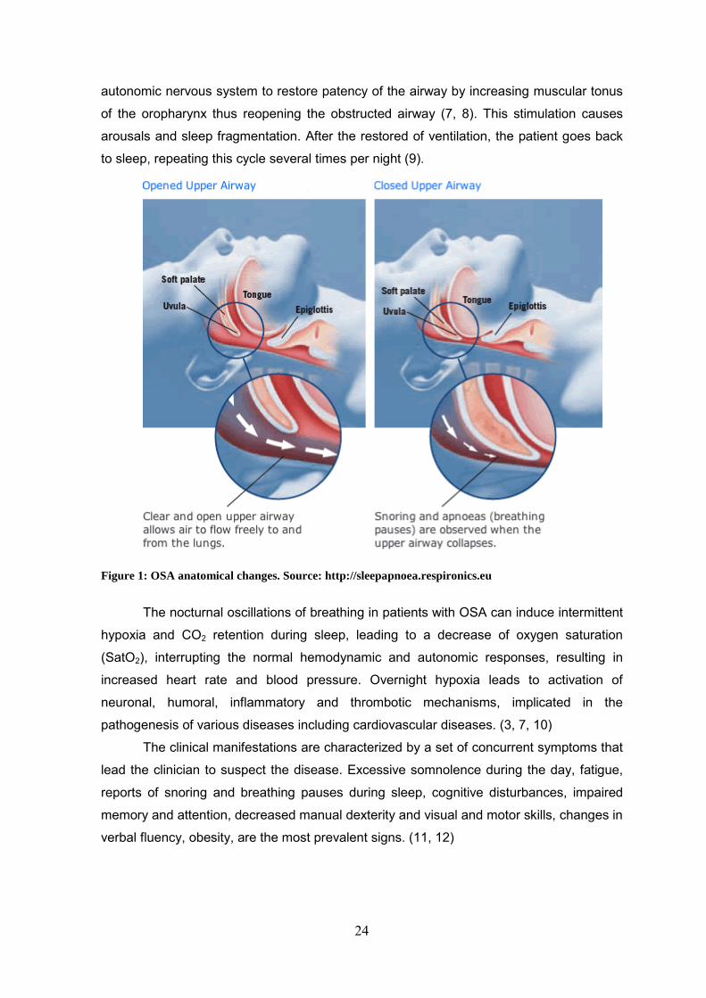
Syndrome Obstructive Sleep Apnea (OSA) is a disorder of breathing patterns
during sleep which is characterized by repeated cessations of airflow and or reductions in
airflow (apnea and or hyponea) caused by complete or partial obstruction of upper
airways by partial or complete collapse of the oropharyngeal pathway by relaxation of the
muscles of the pharynx during sleep (Fig.1) (3-6). By convention, apnea is defined as
cessation of airflow for more than 10 seconds and hypopnea is defined as the reduction of
air flow resulting in frequent awakenings during sleep or oxygen desaturation (33).
The repetitive cycle of apnea and hypopnea begins with the reduction of the airflow
by obstruction that produces hypoxia and hypercapnia, requiring stimulation of the
24
autonomic nervous system to restore patency of the airway by increasing muscular tonus
of the oropharynx thus reopening the obstructed airway (7, 8). This stimulation causes
arousals and sleep fragmentation. After the restored of ventilation, the patient goes back
to sleep, repeating this cycle several times per night (9).
Figure 1: OSA anatomical changes. Source: http://sleepapnoea.respironics.eu
The nocturnal oscillations of breathing in patients with OSA can induce intermittent
hypoxia and CO2 retention during sleep, leading to a decrease of oxygen saturation
(SatO2), interrupting the normal hemodynamic and autonomic responses, resulting in
increased heart rate and blood pressure. Overnight hypoxia leads to activation of
neuronal, humoral, inflammatory and thrombotic mechanisms, implicated in the
pathogenesis of various diseases including cardiovascular diseases. (3, 7, 10)
The clinical manifestations are characterized by a set of concurrent symptoms that
lead the clinician to suspect the disease. Excessive somnolence during the day, fatigue,
reports of snoring and breathing pauses during sleep, cognitive disturbances, impaired
memory and attention, decreased manual dexterity and visual and motor skills, changes in
verbal fluency, obesity, are the most prevalent signs. (11, 12)

25
2.2. Epidemiology:
Since the 90s, several studies indicate that OSA is more prevalent than previously
thought and is often under-diagnosed (28, 29). This disease can affect any age group, but
its prevalence is higher on middle-aged adults reaching 2 to 4%. It is estimated that OSA
prevalence will increase by 30% in 2016 in association with concurrent increase of risk
factors such as intake of alcohol and obesity (7, 16, 17).
Considering only data from polysomnography, its prevalence reaches 24% in men
and 9% in women. OSA can occur at any age, reaching a peak incidence between 40 and
50 years of age. Particularly in hypertension (HTN), studies show a prevalence of OSA in
35% to 50% of hypertensive patients, reaching 70% in cases of refractory hypertension.
Moreover, some 40% of hypertensive patients may have undiagnosed OSA. The
prevalence of OSA in coronary patients is 30%; in patients with atrial fibrillation OSA is
present at around 50% and in heart failure it may vary from 12% to 53% in published
studies. In obese individuals, the prevalence of OSA reaches 40%, while 70% have
increased weight or obesity (7).
2.2.1. Risk Factors:
OSA is a complex and chronic condition that can be initiated and develop in the
presence of multiple risk factors such as obesity, age, gender, genetic factors, craniofacial
abnormalities, excess weight, smoking, alcohol consumption, upper airway anatomy,
nasal congestion and hormonal changes during menopause, family and behavioral factors
(6, 28). Several risk factors have been identified in the development of OSA but the most
outstanding risk factor is obesity (6).
2.2.1.1. Obesity:
Obesity, especially when the deposition of adipose tissue is found in the upper part
of the body, increases the risk for OSA between 10 and 14 times, with greater effects in
middle-aged men (28).
The measure most often used to define the overweight and obesity is given by
calculating the Body Mass Index (BMI), obtained by dividing the weight in kilograms and
height in squared meters (kg/m2). (28)
Weight changes have also been associated with the progression or regression of
OSA. In subjects with no OSA or with mild OSA (AHI < 15), a 10% weight gain increases
the probability of developing moderate or worse OSA (AHI ≥ 15). Extreme obesity defined
26
as a BMI ≥ 40 kg/m2. can lead to severe OSA and in some cases can lead to daytime and
nocturnal hypoventilation, a condition known as obesity hypoventilation syndrome. The
prevalence this syndrome has been estimated to be 25% in patients with OSA who are
extremely obese. (6) The BMI measures the total body mass, but does not inform about
the location of excess adipose tissue, it is necessary to use other anthropometric
measurements to verify that the excess weight caused deposition of adipose in the upper
body, thus it is necessary to measure the external circumference of the neck. which, when
raised, is an indicator of excess deposition adipose tissue in the lateral pharyngeal areas
and consequent narrowing of the upper airways. (28)
2.2.1.2. Age
The prevalence of OSA increases with age because overweight and obesity are
more common in adulthood. Additionally, certain changes, intrinsic to the process of
biological aging, begin to manifest itself more markedly, such as a higher rigidity of the
thoracic cage and a lower muscle tone.
A high prevalence of OSA in the elderly may result from several factors including
collapsibility an increased result of anatomical aging changes of pharynx. Loss of teeth
also produces anatomical changes which can alter the dimensions and function of the
upper ai.rway (28).
2.2.1.3. Gender: OSA is more common in males than in females, with a ratio of 2:1, probably due to
higher risk factors in men. Menopause is a risk factor for sleep apnea, but the existence of
OSA in childhood, adolescence and older age means that there is no simple positive
correlation of OSA with age (16).
The increased incidence of OSA in women after menopause suggests that female
sex hormones play a protective role or male sex hormones contribute to the genesis of the
disease. These effects may result from hormonal influences on ventilation control over the
mechanical behavior of the upper airway or in patterns of body fat distribution (28).
2.2.1.3.1. Menopause
In contrast to men, women with similar degree of obesity have less collapsible
upper airway. This pathophysiologic difference in the upper airway anatomy could explain
the overall lower prevalence of OSA in premenopausal women. The increased prevalence
27
of OSA in menopausal women draws again attention to hormonal differences in the
pathogenesis of OSA. However, hormone replacement therapy has not been shown to be
an effective treatment of OSA in post-menopausal women; therefore, other factors play a
role and the optimal treatment for OSA in postmenopausal women remains continuous
positive airway pressure (CPAP) therapy (6).
2.2.1.4. Ethnicity
There are ethnic differences in the prevalence and severity of OSA. There is some
recent data suggesting that certain races or ethnic groups have a higher risk for the
disease. The Afro-americans have higher levels of apnea than elderly Caucasians. These
racial differences appear to result from anatomical risk factors that in african-americans
are more pronounced such as soft tissue augmentation on the airways, rather than from
the dimensional characteristics of the airways (6).
The prevalence of OSA in Chinese population-based studies was similar to that of
the white population; despite a lower BMI (only 5% of men had a BMI greater than 30
kg/m2). A similar prevalence of OSA in these two populations can be attributed to
cephalometric differences of White and Asian patients, such as an inferiorly positioned
hyoid bone, an extended soft palate width and reduced upper airway soft palate (6).
2.2.1.5. Craniofacial factors
Craniofacial morphology may predispose for OSA by affecting bones and soft
tissues, causing a reduction in the dimensions of the upper airway. Several structural
anomalies have been described in patients with OSA, namely: reduction of anterior-
posterior dimension of the skull base and reduction of the dimensions of the airspaces
posterior and superior, inferior displacement of the hyoid, elongation of the soft palate,
adenoid hypertrophy and / or tonsils, increased vertical facial dimension, retrognathia,
micrognathia and class II malocclusion (28).
2.2.1.6. Alcohol Intake:
Alcohol intake at bedtime can have adverse effects on nocturnal breathing and
even in the duration of apneic/hypopneic episodes. There is an association between
alcohol intake and snoring, as alcohol relaxes the muscle and therefore increases the
probability of pharyngeal collapse (28).
28
2.2.1.7. Smoking
There is a positive association between cigarette smoking and OSA. Several
mechanisms explain why smoking affects OSA, such as instability during sleep and
mucosal inflammation of the airways. The association with OSA is relatively weak, but
smoking may interact with and add to the cardiovascular risk (16, 28).
2.2.2. Consequences of OSA
Untreated OSA can contribute to the development or progression of other
disorders. If not diagnosed and properly treated, OSA can cause multiple complications,
whose effects may project negatively at personal, family and social level, contributing to a
drastic reduction in quality of life and also to an increased general rate of morbidity and
mortality associated with cardiovascular and cerebrovascular complications (16, 28).
2.2.2.1. OSA and Cardiovascular diseases:
OSA may have hemodynamic consequences such as pulmonary and systemic
hypertension, cardiac arrhythmias, ischemic heart disease, congestive heart failure and
Cheyne-Stokes breathing in patients with congestive heart failure (28).
Obstructive apneas may lead to severe intermittent hypoxemia and CO2 retention
during sleep, disrupting the normal structured autonomic and hemodynamic responses to
sleep. Apneas can occur repetitively during the night, increasing sympathetic activity to
peripheral blood vessels and consequent vasoconstriction. This level of hemodynamic
stress occurs at a time of severe hypoxemia, hypercapnia, and adrenergic activation. The
nocturnal apneas initiate pathophysiological mechanisms which may act to promote
cardiac and vascular disease (Fig. 2) (3).

29
Figure 2. Source: Sommers, 2008 - Pathophysiological components of OSA; related to cardiovascular disease mechanisms (3)
2.2.2.1.1 OSA and Hypertension:
Several reports have shown that the prevalence of HTN is greater in patients with
OSA. The association between OSA and hypertension was seen in men and women,
older and younger ages, all ethnic groups, and among normal-weight and overweight
individuals. (4)
The episodes of apnea / hypopnea during sleep induce transient increases in
blood pressure that can reach 30 mmHg or more in mean arterial pressure. During apnea
there is an increased sympathetic activity which leads to an increase in blood pressure.
There is a reduction in sympathetic activity by administering CPAP thus can contribute to
a reduction in blood pressure. (28) OSA has been proposed as an independent risk factor
for the development of essential hypertension because it can precede and predict the
onset of hypertension (3).
2.2.2.1.2. Other cardiovascular complications
Over 30% of patients with ischemic heart disease suffer from OSA. An AHI greater
than 11 is a risk factor for heart failure, independent of other risk factors. Cardiac
arrhythmias are frequently observed in patients with obstructive sleep apnea, especially
during apnea bradycardia, followed by tachycardia rewarding when the apnea ends, and
the change in heart rate caused by vagal stimulation. Other changes in heart rate can
occur, but more rarely, such as ventricular tachycardia (3, 28).

30
2.2.2.2. Cerebrovascular complications
Among subjects with stroke, the incidence of sleep-disordered breathing may be
greater than 50%. Obstructive sleep apnea is a risk but also a consequence of stroke.
(106).
Taking into account that OSA causes HTN, and that this is a major causative factor
in stroke, it is easy to understand the contribution of HTN to develop stroke in patients
with OSA. Some data provide evidence that men with increasing AHI levels experience an
increased risk of stroke. Increased risk may be through a number of pathogenic factors
influenced by intermittent hypoxemia and sympathetic stimulation that influence the
cerebral vasoregulation, atherogenesis, and atrial fibrillation. Among them are: the
reduction of cerebral blood flow, the hypercoagulability blood, increased platelet
aggregation, endothelial dysfunction, increased intracranial pressure and a change of
cerebral auto-regulation triggered by changes in blood gases that have been described in
patients with OSA (30, 106).
2.2.2.3. Psychosocial consequences
Excessive daytime drowsiness is characteristic of OSA with adverse effects on
neurocognitive function, memory, and performance of diverse tasks. In relation to
neurocognitive function impaired, patients with OSA have an increased risk for motor
vehicle accidents. OSA causes impairment in performance and is associated with an
increased risk of traffic accidents compared with the general population of drivers. Sleep
apnea, with its repeated episodes of nocturnal hypoxemia and sleep fragmentation, will
increase fatigue and sleepiness, which in turn increase lack of alertness while driving. As
a result, there are increased collision rates in OSA sufferers. During the night can occur
several manifestations such as nocturia, agitation associated with multiple awakenings
and sometimes accompanied by intense diaphoresis. Sexual impotence develops in
association with snoring. All these symptoms can contribute to a significant increase in the
frequency of divorce. Still relatively frequent are morning headaches, decreased libido,
impotence and psychological and cognitive changes. All this manifestations lead to
negative consequences in lives of patients with OSA (28, 31, 32).
31
2.3. Diagnosis:
The diagnostic criteria established for OSA are based on clinical symptoms and
signs during sleep evaluation including a physical examination and oriented sleep history,
and findings identified by sleep testing.
During a routine consultation some questions should be done to access to a
history of snoring, daytime sleepiness and an assessment of Body Mass Index,
retrognathia, or hypertension.
A comprehensive sleep history in a patient should include an evaluation for
snoring, witness apneas, breathless and or asphyxia episodes, excessive somnolence,
Epworth Sleepiness Scale (EPS) evaluation, total time of sleep, nocturia, fatigue in the
morning, morning headache, sleep insomnia or fragmentation, decreased concentration
and memory. The diagnosis of OSA requires the occurrence of 2 or more of the following
symptoms: difficulty in breathing during sleep, fatigue during the day, difficulty in
concentrating, reports of non-restorative sleep and frequent awakenings at night. The
apnea-hypopnea index (AHI), demonstrated by polysomnography or cardiorespiratory
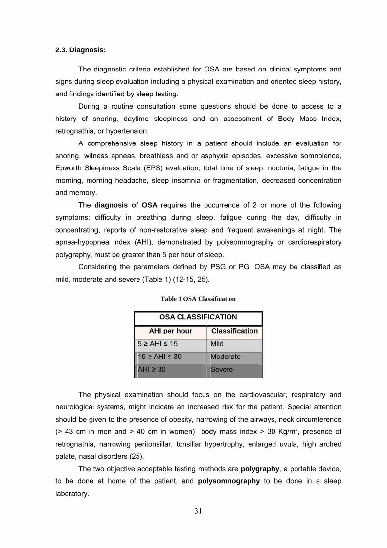
polygraphy, must be greater than 5 per hour of sleep. Considering the parameters defined by PSG or PG, OSA may be classified as
mild, moderate and severe (Table 1) (12-15, 25).
Table 1 OSA Classification
OSA CLASSIFICATION
AHI per hour Classification
5 ≥ AHI ≤ 15 Mild
15 ≥ AHI ≤ 30 Moderate
AHI ≥ 30 Severe
The physical examination should focus on the cardiovascular, respiratory and
neurological systems, might indicate an increased risk for the patient. Special attention
should be given to the presence of obesity, narrowing of the airways, neck circumference
(> 43 cm in men and > 40 cm in women) body mass index > 30 Kg/m2, presence of
retrognathia, narrowing peritonsillar, tonsillar hypertrophy, enlarged uvula, high arched
palate, nasal disorders (25).
The two objective acceptable testing methods are polygraphy , a portable device,
to be done at home of the patient, and polysomnography to be done in a sleep
laboratory.
32
2.3.1. Polysomnography
The polysomnography (PSG) is the gold standard for diagnosing OSA. It is
performed in a sleep laboratory by specialized professionals, consisting in a simultaneous/
continuous acquisition and analysis of neurophysiologic and cardiorespiratory variables
during a night sleep. This device records electroencephalogram, electro-oculograma,
electromyogram, electrocardiogram, oronasal airflow, snoring, thoraco-abdominal
breathing movements, body position, oxygen saturation and heart rate. The registration of
these variables provides important information for the sleep study: sleep staging, cardiac
abnormalities, respiratory events, respiratory effort, snoring and O2 desaturations.
However it has some disadvantages: high cost, the need for continuous attention and of
time on the part of medical staff, these disadvantages, together with the scarcity of
diagnostic resources, has required the search for alternative methods, such as
cardiorespiratory polygraphy, which does not require constant attention and is much less
costly than PSG. (26, 106)
2.3.2. Polygraphy:
Validation studies have been carried out for some of the respiratory polygraphy
systems and various groups consider them an acceptable alternative to conventional
PSG.
Polygraphy (PG) is a portable recording device more economical which allows the
Polygraphic study at home not requiring supervision technique. It allows cardiorespiratory
registration of nasal airflow, snoring, breathing movements, body position, O2 saturation
and heart rate. Although there is a smaller number of variables is considered a viable and
reliable for the diagnosis of OSA. (26, 27, 106)
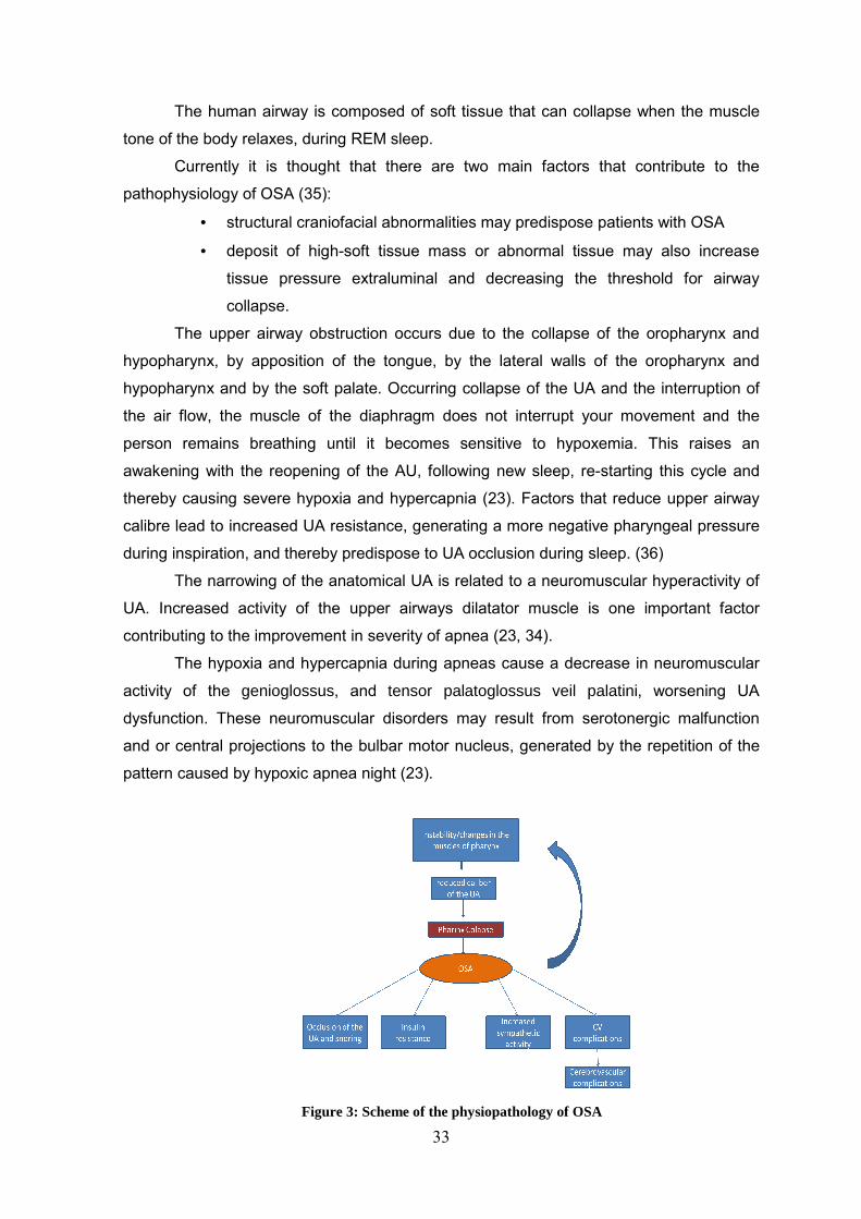
2.4. Physiopathology:
The pathophysiology of OSA is complex. There are several factors that may
contribute to the manifestation of this condition. The human upper airway is a
multifunctional structure involved in performing functional tasks. The anatomy and neural
control of the upper airways (UA) have evolved to allow these various functions. Due to
the multiple functionality of the upper airways, they have the ability to change shape and
close momentarily which is essential for speech and swallowing during wakefulness, this
capability also offers the opportunity for collapse at inopportune times, such as during
sleep (34).
33
The human airway is composed of soft tissue that can collapse when the muscle
tone of the body relaxes, during REM sleep.
Currently it is thought that there are two main factors that contribute to the
pathophysiology of OSA (35):
• structural craniofacial abnormalities may predispose patients with OSA
• deposit of high-soft tissue mass or abnormal tissue may also increase
tissue pressure extraluminal and decreasing the threshold for airway
collapse.
The upper airway obstruction occurs due to the collapse of the oropharynx and
hypopharynx, by apposition of the tongue, by the lateral walls of the oropharynx and
hypopharynx and by the soft palate. Occurring collapse of the UA and the interruption of
the air flow, the muscle of the diaphragm does not interrupt your movement and the
person remains breathing until it becomes sensitive to hypoxemia. This raises an
awakening with the reopening of the AU, following new sleep, re-starting this cycle and
thereby causing severe hypoxia and hypercapnia (23). Factors that reduce upper airway
calibre lead to increased UA resistance, generating a more negative pharyngeal pressure
during inspiration, and thereby predispose to UA occlusion during sleep. (36)
The narrowing of the anatomical UA is related to a neuromuscular hyperactivity of
UA. Increased activity of the upper airways dilatator muscle is one important factor
contributing to the improvement in severity of apnea (23, 34).
The hypoxia and hypercapnia during apneas cause a decrease in neuromuscular
activity of the genioglossus, and tensor palatoglossus veil palatini, worsening UA
dysfunction. These neuromuscular disorders may result from serotonergic malfunction
and or central projections to the bulbar motor nucleus, generated by the repetition of the
pattern caused by hypoxic apnea night (23).
Figure 3: Scheme of the physiopathology of OSA

34
2.5. Treatment:
OSA should be approached as a chronic disease and requires multidisciplinary
long term treatment. Thus, treatment of OSA must include generic and specific measures.
Ideal therapeutic approach and development of an adequate treatment plan pass through
the investigation of the etiology of OSA, the establishment of clinical severity, symptoms,
presence of comorbidities relevant expectations patient and chance to improve their
quality of life (25, 106).
2.5.1. General measures:
General measures consist in adopting measures to combat any risk factors that
enhance respiratory events. Thus, should be advised to avoid alcohol, tobacco,
benzodiazepines, barbiturates and narcotics. The diet therapy should be recommended
and weight reduction, as well as physical exercise and establishment of sleep routine,
including a scheduled with sufficient number of hours and postural conditioning. (25, 37,
38)
2.5.2. Specific measures:
In these specific measures include: pharmacological measures of positive
pressure ventilation, oral prostheses and surgical measures.
2.5.2.1. Pharmacological:
The pharmacological measures have a very questionable data, as evidence
scientific in its favor is scarce. There are no widely effective pharmacotherapies for OSA
although there are some exceptions in case of individuals with hypothyroidism or
acromegaly. However, some OSA patients, who do not tolerate CPAP and or have
symptoms sufficiently severe, justify the use of pharmacological therapy. Among the drugs
which act on the tonus of the upper airway, paroxetine, mirtazapine, physostigmine and
donepezil improved to some extent indices OSA. Recent pharmacological approaches to
the treatment of OSA, has focused on increasing the production of serotonergic neurons
and enhance cholinergic activity, because both have been shown to increase the tonus of
the upper airway (25, 38, 39).
35
In cases in which endocrine factors, such as hypothyroidism and acromegaly, are
OSA substrate, it is recommended replacement therapy, which leads to significantly
reducing the number of respiratory events (39, 106). For last say that: in medical therapies to improve nasal patency Topical nasal
corticosteroids can improve AHI in patients with OSA and concomitant rhinitis and thus
may be useful to assist the main therapies for OSA. Moreover modafinil should be use in
addition to PAP therapy for the treatment of residual excessive daytime sleepiness in OSA
(38, 106).
2.5.2.2 Surgical
A great variety of surgical procedures for the treatment of OSA have been
described. Surgery aims to reduce obstructions of the upper airways in the nose,
oropharynx, hypopharynx. It may be necessary to correct an anatomical obstruction
before prescribing an oral appliance or positive airway pressure device. Surgery may be
beneficial in selected cases as: usually patients with snoring and mild OSA, which have a
cause amenable to surgical correction and in whom treatment with CPAP is not effective
(40-42, 106).
The surgical interventions consist of the reconstruction of soft tissue and / or bone
of the AU, with the aim of increasing its diameter and unblock the UA. So are several
surgical procedures available, including: maxillofacial surgery, nasal surgery,
uvulopalatopharyngoplasty, laser-assisted uvuloplastia, somnoplastia and radiofrequency
volumetric reduction (42-45, 106).
The bariatric surgery, as adjuvant therapy is a means for achieving greater weight
loss. It is indicated for patients with BMI> or 40Kg/m2 patients with BMI> 35Kg/m2 with
higher risk factors. So, this surgery, is indicated for obese patients that by this way may
potentially condition the disappearance of the pathophysiology of OSA (25,106).
2.5.2.3 Oral appliances
Oral appliances may improve upper airway patency by enlarging the upper airway
by decreasing upper airway collapsibility. This type of device that will promote mandibular
advancement are intraoral orthodontic appliances that move the jaw forward, increasing
the diameter of the upper airways, reducing the possibility of pharyngeal collapse. These
devices have proved effective in snoring, but in OSA their effects are not so evident. Their
36
efficiency tends to be lower in obese patients because skeletal and maxillofacial factors
have less importance in the etiopathogenesis of apnea, compared with obesity (28, 46).
This therapy is commonly used in the treatment of mild and moderate OSA
patients with few symptoms, or when other treatment modalities are not tolerated or are
contraindicated. It can also be associated with CPAP or surgical measures in cases of
severe OSA (106).
2.5.2.4 Positive Pressure Ventilation
Positive pressure air is intended to keep the unobstructed upper airway during
sleep and thereby prevent the pharyngeal collapse (28). Positive air pressure aims to
keep the upper airway unobstructed during sleep and thereby prevent the pharyngeal
collapse. Applied through nasal, oral, or through an oronasal interface during sleep it is
the preferred treatment for obstructive sleep apnea. There are 3 Types of PAP treatment:
continuous PAP (CPAP), Bilevel PAP (BIPAP), and Automatic adjusting PAP (APAP) (47).
The use of such devices should be accompanied by patient education, adapting,
adjusting and ambiance to the mask (116).
2.5.2.4.1 CPAP
Continuous Positive Airway Pressure is recommended as a treatment option for
adults with moderate or severe symptomatic obstructive sleep apnea/hypopnea
syndrome. (48, 49) A continuous positive airway pressure (CPAP) is currently the
treatment form dominant and with more proven effectiveness in OSA. (106)
CPAP may provide different pressures. The optimum pressure is adjusted related
to certain individual characteristics that include data ascertained by polysomnography.
The CPAP reduces edema and congestion of the pharynx arising from microtrauma
associated with snoring, and causes significant improvement in daytime sleepiness. This
device has benefits in the pathology itself, improving the AHI and the reduction of risk
factors. Besides that it as interference in the quality of life of patients because it improves
the skills required in performing multiple daily activities, normalizes memory and cognitive
function and mood. All these benefits only occur with continued and proper use (Table 2)
(23, 28).
37
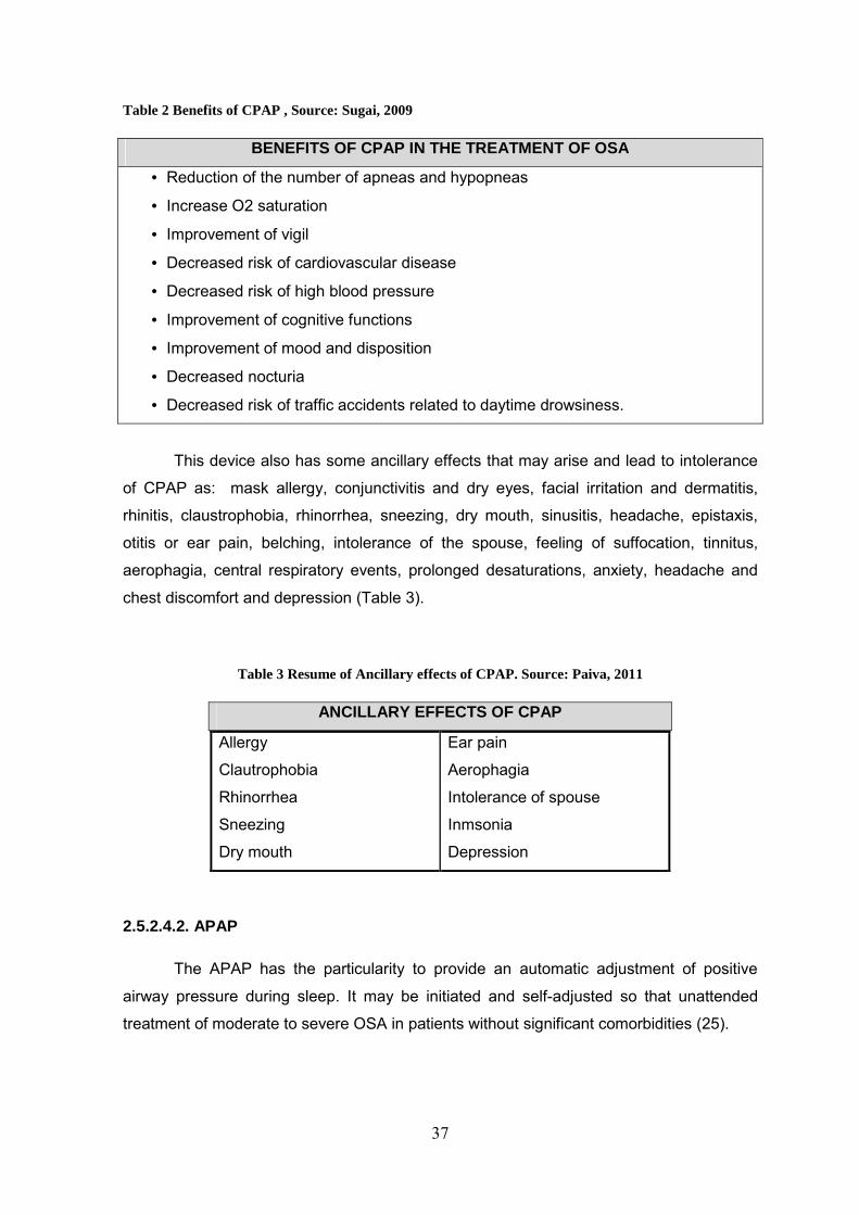
Table 2 Benefits of CPAP , Source: Sugai, 2009
BENEFITS OF CPAP IN THE TREATMENT OF OSA
• Reduction of the number of apneas and hypopneas
• Increase O2 saturation
• Improvement of vigil
• Decreased risk of cardiovascular disease
• Decreased risk of high blood pressure
• Improvement of cognitive functions
• Improvement of mood and disposition
• Decreased nocturia
• Decreased risk of traffic accidents related to daytime drowsiness.
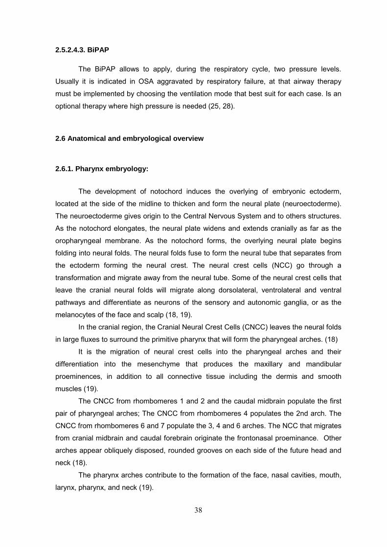
This device also has some ancillary effects that may arise and lead to intolerance
of CPAP as: mask allergy, conjunctivitis and dry eyes, facial irritation and dermatitis,
rhinitis, claustrophobia, rhinorrhea, sneezing, dry mouth, sinusitis, headache, epistaxis,
otitis or ear pain, belching, intolerance of the spouse, feeling of suffocation, tinnitus,
aerophagia, central respiratory events, prolonged desaturations, anxiety, headache and
chest discomfort and depression (Table 3).
Table 3 Resume of Ancillary effects of CPAP. Source: Paiva, 2011
ANCILLARY EFFECTS OF CPAP
Allergy
Clautrophobia
Rhinorrhea
Sneezing
Dry mouth
Ear pain
Aerophagia
Intolerance of spouse
Inmsonia
Depression
2.5.2.4.2. APAP
The APAP has the particularity to provide an automatic adjustment of positive
airway pressure during sleep. It may be initiated and self-adjusted so that unattended
treatment of moderate to severe OSA in patients without significant comorbidities (25).
38
2.5.2.4.3. BiPAP
The BiPAP allows to apply, during the respiratory cycle, two pressure levels.
Usually it is indicated in OSA aggravated by respiratory failure, at that airway therapy
must be implemented by choosing the ventilation mode that best suit for each case. Is an
optional therapy where high pressure is needed (25, 28).
2.6 Anatomical and embryological overview
2.6.1. Pharynx embryology:
The development of notochord induces the overlying of embryonic ectoderm,
located at the side of the midline to thicken and form the neural plate (neuroectoderme).
The neuroectoderme gives origin to the Central Nervous System and to others structures.
As the notochord elongates, the neural plate widens and extends cranially as far as the
oropharyngeal membrane. As the notochord forms, the overlying neural plate begins
folding into neural folds. The neural folds fuse to form the neural tube that separates from
the ectoderm forming the neural crest. The neural crest cells (NCC) go through a
transformation and migrate away from the neural tube. Some of the neural crest cells that
leave the cranial neural folds will migrate along dorsolateral, ventrolateral and ventral
pathways and differentiate as neurons of the sensory and autonomic ganglia, or as the
melanocytes of the face and scalp (18, 19).
In the cranial region, the Cranial Neural Crest Cells (CNCC) leaves the neural folds
in large fluxes to surround the primitive pharynx that will form the pharyngeal arches. (18)
It is the migration of neural crest cells into the pharyngeal arches and their
differentiation into the mesenchyme that produces the maxillary and mandibular
proeminences, in addition to all connective tissue including the dermis and smooth
muscles (19).
The CNCC from rhombomeres 1 and 2 and the caudal midbrain populate the first
pair of pharyngeal arches; The CNCC from rhombomeres 4 populates the 2nd arch. The
CNCC from rhombomeres 6 and 7 populate the 3, 4 and 6 arches. The NCC that migrates
from cranial midbrain and caudal forebrain originate the frontonasal proeminance. Other
arches appear obliquely disposed, rounded grooves on each side of the future head and
neck (18).
The pharynx arches contribute to the formation of the face, nasal cavities, mouth,
larynx, pharynx, and neck (19).
39
• Palatogenesis – It begins in the sixth week and it develops in two stages: the
development of primary palate and the development of a secondary palate. The palatal
processes, arise as bilateral extensions from the oral aspect of the maxillary processes,
reorient from a vertical position lateral to the tongue, to a horizontal position above the
tongue, making epithelial contact along the anterior–posterior length of the palate (19)
Initially, the median palate process begins to develop from the medial nasal
proeminences. The primary palate forms the anterior/midline aspect of maxilla. The
secondary palate is the primordium of the hard and soft palate. It begins to develop from
two mesenchymal projections that extend from the interior of maxillary proeminences.
During the seventh-eight week the lateral palatal processes assume a horizontal position
above the tongue (159). In the primary palate, bone gradually develops, and extends from
the maxillae and palatine bones into the palatal processes to form the hard palate. The
posterior parts of palatal processes extend posteriorly beyond the nasal septum and fuse
to form the palate, including the uvula (19).
2.7. Pharynx anatomy:
Pharynx is a fibromuscular tube that extends from the base of skull to the upper
border of upper esophageal sphincter. It can be divided into three segments:
nasopharynx, (from the base of skull to the soft palate) oropharynx (from soft palate to the
pharyngoepiglottic fold) and hypopharynx (from the pharyngoepiglottic fold to the upper
esophageal sphincter). The muscles of pharynx can be generically viewed as intrinsic and
extrinsic; the intrinsic muscles constitute the superior, middle, and inferior pharyngeal
constrictors along with thyropharyngeus and cricopharyngeus. Extrinsic muscles of the
pharynx can be categorized in 3 subgroups: (20, 21).
• Group 1 — elevators and tensors of palate (levator veli palatini, tensor veli
palatini, and palatoglossus);
• Group 2 — geniohyoid, stylohyoid, mylohyoid, thyrohyoid, diagastric,
stylopharyngeus, and palatopharyngeus, which cause superior and anterior
movement of the larynx during swallowing;
• Group 3 — aryepiglottic, thyroarytenoid, and oblique arytenoids muscles,
which close the laryngeal entry.
The 3 pharyngeal constrictor muscles, that form the outer layer, are arranged so that
the bottom constrictor muscle is overlapped by the medium constrictor muscle, which in
turn overlap the upper constrictor muscle. The constrictor muscles are not completely

40
circular. They originate from a lateral cartilage fixation, ligaments and bony points,
therefore they are not present anteriorly, so there are spaces between them or between
the superior constrictor muscle of the larynx and skull laterally. They are posteriorly they
are complete and are inserted into the larynx raphe that extends from the pharyngeal
tubercle of the occipital bone to cricopharyngeus muscle. The spaces are complemented
by the fascia of the pharynx, but allow the entrance of muscles, nerves, and arteries in the
wall of the pharynx. The muscle stylopharyngeus accompanied by glossopharyngeal and
lingual nerve passes inferiorly to the superior constrictor muscle of the larynx. The muscle
stylopharyngeus that originates medially from the region infratemporal, enters in the floor
of the mouth passing through the space between the upper and middle constrictor
muscles of the pharynx and joins the glossopharyngeal nerve on an external surface (20,
21).
The fascia of the larynx is located internally and externally to the muscle layer. The
inner layer is pharyngobasilar fascia, it is fixed to the base of the skull, provides a firm
layer which, with the help of the muscular layer, prevents distortion or collapse of the
pharyngeal wall, maintaining an open airway. The outer layer of fascia is the
buccopharyngeal fascia that is continuous with the pre-tracheal fascia. It extends to the
skull base superiorly and laterally to cover the buccinator muscle (20).
2.7.1. Pharinx innervation:
The muscles of the pharynx, except the tensor veli palatini and stilopharyngeus
muscles, receive special visceral efferent (SVE) fibers of pharyngeal plexus that is
originate in the vagus nerve, but carries fibers from the accessory cranial nerve.
Sympathetic fibers originate in the superior cervical ganglion and with the branches of
external plexus carotid and branches of the external carotid artery proceed to the pharynx.
The sensory innervation of the upper nasopharynx is made by general somatic afferent
(GSA) fibers of palatine nerve, branch of the maxillary nerve and posterior nasal
branches. The rest of this region of the pharynx, the oropharinx and the upper
hypopharynx is supplied with general visceral afferents (GVA) fibers of the
glossopharyngeal nerve. The bottom of hypopharinx is supplied with general visceral
afferent (GVA) fibers of the internal branch of the superior laryngeal nerve and recurrent
laryngeal nerve (20).
The extrinsic muscles are supplied by the branches of cranial nerves V (trigeminal),
VII (facial), IX (glossopharyngeal), X (vagus), ansa cervicalis, and XII (hypoglossal).
Pharyngeal muscles are richly innervated, with a nerve–muscle fiber innervation, which is
important for the “fine” control required for its function (21).
41
2.7.2. Vessels of the Pharynx: The blood supply of the pharynx is from branches of the ascending pharingeal
artery and from the Ascending palatine artery. The ascending pharyngeal artery arises
from the posterior wall of the proximal external carotid artery trunk, and close to the
source of the occipital artery. After a short common trunk, the ascending pharyngeal
artery divides into two major trunks: anteriorly, the pharyngeal trunk, which is extracranial;
posteriorly, the neuromeningeal trunk, which is intracranial and enters the posterior fossa
through the foramen magnum (21, 3).
The ascending palatine artery arises close to the origin of the facial artery and
passes between the styloglossus and stylopharyngeus to the side of the pharynx following
between the superior pharyngeal constrictor and the medial pterygoid muscle to near the
base of the skull. It divides near the levator veli palatini muscle into two branches: one
supplies this muscle, winding over the upper border of the superior pharyngeal constrictor,
supplies the soft palate and the palatine glands, anastomosing with the descending
palatine branch of the maxillary artery; the other crosses the superior pharyngeal
constrictor and supplies the palatine tonsil and auditory tube, anastomosing with the
tonsillar branch of the facial artery and the ascending pharyngeal artery (21, 23).

42

43
3. Traditional Chinese Medicine Overview

44
45
3. Contemporary TCM
The oldest existing therapeutic systems used by humanity for health and wellbeing
are called Traditional Medicine (TM) or complementary medicine (CM). The origin of
traditional Chinese medicine is uncertain. archaeological findings indicate that Chinese
medicine has more than 5000 years. “I Ging” is one of the oldest approaches to a
mathematical model and Huangdi Neijing, canon of Internal Medicine of the Yellow
Emperor, dating back more than 2300 years (52, 53).
Always influenced by several periods of thought and acculturation in China and in
world, Traditional Chinese Medicine (TCM) has been developing for centuries in clinical
practice. It brings together Chinese traditional natural science and social science and
combines them into an ethical and unique scientific medical system (53, 54). Since early,
this biological model system, known as TCM, was based in two philosophical theories
Taoism and Confucianism. So since the VII century BC, it was no longer regarded as
something mystical and magical. And since the II century BC it has been perceived that
the human being is a living being integrated in an environment that influences him. The
two philosophies emphasized the importance of understanding the laws of nature and it
was postulated that it was important that humans should integrates and respect these
laws rather than to resist them. Like in the ecology laws and in science in general of our
age, the human body and life was regarded as a microcosmic reflection of the macrocosm
of the universe. Thus, concepts used to explain nature, such as yin/yang and Five
Elements, became central to TCM theory The goal of the clinician was to maintain the
body‟ harmonious balance both internally and in relation to the external environment (52,
55). When practiced correctly, TCM can help protect and improve citizens’ health and
well-being (52, 53).
This vision of the human being attracted scientists and researchers from east to
west of the world and has made great contribution to health of Chinese people for
thousands years (56). In this increasingly globalized world, more than a quarter of the
world seeks traditional Chinese medicine as a significant part of their health care. Part of
this is due to the fact that conventional medicine and Chinese medicine does not compete
with each other, on the contrary they are complementary (57). For the past 50 years,
several hundreds of textbooks and monographs on TCM have been published.
Nowadays, higher education is available in a substantial number of TCM universities,
medical universities and faculties all over the world. This therapeutic system followed the
progress of sciences and Western medicine combining contemporary diagnostic methods,
such as laboratory tests and imaging, with traditional diagnostic techniques (107).
46
Lately the theory of Chinese medicine has been represented as a rational and
simple mathematical model, which shows general laws and relationships between
physiological and pathological process in human organism. As in the case of the
Heidelberg model (described below), there are also other scientists who are showing that
Chinese medicine can be put in a rational way (58).
Examples of that are Teppone and Avakyan. They theorized that Chinese
physicians described the physiology and pathophysiology of the intercellular spaces and
cavities of the body through the "Zang Fu" theory. Therefore the system of acupuncture
points is seen as part of intercellular spaces which, due to their morphological and
functional complexity, allow interactions between the interior of the body and the
surroundings. They understand the Chinese view of human wellbeing and its limitation in
a three dimensional space, as a kind of cybernetic model by which this complex system
can have common managing or controlling parameter. In this way, they tried to explain the
concept of "Qi" as a unit of measurement relatively universal. They thought “Qi” as the
parameter "X" in the equation y = f (x, z), where "z" is considered the intensity of heat
produced, which describes the predominance of endothermic or exothermic chemical
reactions in the human body. These authors consider that the Chinese diagnosis is
necessary and sufficient to determine "excess or defect" of the conditions of the conduits
y = f (x) and "excess or defect" and "hot or cold" of the pathologies of organs (zang) y = f
(x, z) (58, 59). In addition, since Gottfried Leibniz, (German scientist, mathematician and
philosopher) the Yin and Yang are considered a binary system of calculation and
comparison. This system is a simple way to describe the variability in the world. (58, 59)
For Xiaoding Cao “the regulation of yin and yang is a fundamental principle in clinical
acupuncture. Diseases result mainly from relative imbalance of yin and yang (or
deficiency) or yin excess (or deficiency). The concept of yin-yang is the basic concept of
all the oriental sciences it matches the precondition for the origin of all natural
phenomena. The yin-yang balance in all vital processes of adaptation of the living beings
and their environment is primordial (60, 61). The concept of the movements tries to explain the evolutionary processes of
nature, of the disease, of health and of the universe. it represents an analogous vision of
the different correlations of the environment with Man. It Indicate the dynamic interactions
between the functional units (61, 62).
Although it is important to emphasize the rational character of Chinese medicine it
is significantly more relevant its great heuristic value of concepts designed according to
cultural patterns of Chinese civilization. The Chinese physicians of antiquity, through
47
therapeutic and clinical observation and experimentation, developed explanations for the
results / phenomena they found (63).
3.1. Acupuncture:
In our days acupuncture more and more has been gaining attention in modern
medicine (64). The research on TCM, in modern scientific ways, has “just started”.
Scientific studies for endorsing its clinical effectiveness and benefit are mandatory.
Researches on TCM, in the last decades have been more extensive. There has been a
great effort to develop controlled clinical trials. Although with many difficulties and
sometimes limitations, these results have been published. Considering this the WHO said,
in its report on the research in TCM, that practitioners and researchers of TCM should be
encouraged to do more research. In 1997, the National Institute of Health of the United
States of America pointed out some goals for the future of Acupuncture and concludes
that more research should be conducted to clarify the physiological and biological effects
of this therapy. In 2007, the Society for Acupuncture investigation reiterated these goals
(65, 66, and 67).
Studies in neurophysiology concluded that acupuncture has the main function to
mediate the body homeostasis, acting mainly in the balance of autonomic, neurochemical
and humoral functions (13).
The functional activities of needling will promote self-regulatory systems and
regulate the balance of normal physiological activities. In cybernetics, a homomorphic
machine, (used, for example, in biological engineering) is similar to the structures of
human body. If a part of the body is slightly deviated from the balance state, the higher
and lower limits of the neural network can be restored by the interactions between the
different parts, so the body can return to its balance again. In other words the body has
self-healing capacity. However, if the imbalance is severe, it will need additional
interventions, only then, the ability to recover could be increased as well as the recovery
of the primordial balance.
The regulatory action of acupuncture is not completely clear, yet. However, today it
is accepted that three general mechanisms can explain this action. Energetic, neural and
humoral mechanisms (61, 63, 68).
• Energetic mechanism:
A growing number of scientists see the TCM as a cybernetic model. In terms of
cybernetic, the conduits (sometimes also called meridians) are information pathways that
connect whole body; upper with lower parts of the body, left with right, interior with exterior
and all of them together. They explain the conduits as a relationship among various parts
48
of the body. This corresponds to the classical conception of “Energy pathways”, the
conduits. The human body has multiple levels of neural networks that transmit information
of different kinds. When and organ or tissue or system, becomes imbalanced, information
of these disorders are transmitted through specific areas of the body. These areas are
reflex points (acupoint) or information zones. To the physiologist Guan Yuan-Jin, one of
the world’s forefront medical acupuncturists, an acupoint is actually the location on the
body surface outputting internal information and inputting treatment information from
acupuncture. He and his network, in 1976, called to the regions of body surface,
information zones (63, 68, 69).
• Neural mechanism
This mechanism made the ancient Chinese philosophical theories, known as TCM,
started to have a scientific basis. Recent research on the action of acupuncture analgesia
has brought significant benefits to the understanding of this mechanism. Generally, is
considered as neural effects due to interaction of different sensory inputs within the
Central Nervous System (CNS). The integration of the needling signals may occur at
different levels of the CNS such as spinal cord, brainstem, thalamus, caudate nucleus,
cerebral cortex to relive symptoms (68).
• Humoral mechanism
It refers to the production of substances that are secreted into the blood through
the action of acupuncture. Clinical facts indicate the role of the humoral factors in this
TCM technique. These factors include endogenous opiate-like substances and many
neurotransmitters such as: serotonine (5-HT), acetylcholine, non-epinephrine, dopamine,
among others; and include hormones like prolactin, growth hormone (GH), thyroid-
stimulating hormone, (TSH), cortisol, inter alia. Among the humoral factors, endorphins or
enkephalins, are the most significant. The action of acupuncture can occur directly
through the autonomic nervous centers and indirectly through the endocrine system to
regulate various visceral functions and secretions of the glands. The release of level of the
endocrine substances, found in blood, depends indirectly on the CNS (63, 68).
Acupuncture probably acts on related organs directly through effects on hormone
secretion and acts on target organs through the feedback control of hormone secretion.
Neural and humoral mechanisms are associated during regulatory process of
acupuncture. Sometimes neural factors are primary to humoral factors, but sometimes it is
in the other way round (68).
If the intensity and duration of needling is insufficient the neuro-humoral factors will
not be activated to inhibit to disorder effectively. On the other hand an overly long or
intense needling will became nociceptive instead of non-nociceptive (68).
49
In summary acupuncture change the bloodstream, promotes the release of
hormones, helps to increase host resistance, resistance to disease, regulates and
normalizes body functions and promotes metabolism (63)
3.2. Relationship between the conduits and the nervous system, blood vessels and lymphatic vessels
3.2.1. Relationship between the conduits and the Nervous System:
The morphological observation of 324 points revealed that 323 (99%) have
innervation of cranial and spinal nerves; 304 are related to superficial cutaneous nerves;
155 with deeper nerves and 137 with deeper and superficial nerves. Similarly, the
acupuncture points and the nerves of its correlated organs belong to the same spinal
segment or to the interior of the same nerves segment to which the organ belongs. The
conduits with internal-external relationship, also belong to the same spinal segment (64,
68).
3.2.2. Relationship between blood vessels and conduits:
Observations showed that among 309 points, 24 are on top of an artery, while 262 are
adjacent to an artery, and from the location of acupuncture points we can see that the
conduits are closely associated with blood vessels. The arterial vascularization in the area
of acupuncture points has certain ways - regular convergences, irradiation or, less regular
arrangements. Those who have regular arrangements, the points are located frequently in
the center of such arrangements (64).
3.2.2. Relationship between conduits and the lymphatic vessels
Research has found that the conduits are related to lymphatic plexus or to lymphatics
vessels and lymph nodes. The conduits Regens (Du mai), Respondence (Ren Mai) and
Zonal (Dai Mai) are related to the lymphatic plexus. The conduits, Pulmonal, Stomachal,
Cardial and Lienal, almost entirely in vascularization of lymphatic vessels, adapting deeply
or superficially, to their corresponding distribution. It showed also that the lymphatic
vessels run along the conduits yin and are fit together or in place of point copulatio trium
yin - L6 (Sanyinjiao), adapting both in distance, as in depth. (64)
Studies in bio-cybernetic stated that the conduits are a control system of the
human body and stated that there are several species of regulation and control processes

50
in the body, as body temperature, blood pressure and blood sugar. Thus, the state of
equilibrium of the conduits can aid in disease diagnosis and regulating the state of
equilibrium of the conduits will improve the treatment of disease. Therefore, it can be
considered that treatments of acupuncture, moxibustion and all techniques of Traditional
Chinese Medicine have much in common with the cybernetic theory (64, 68).
3.3. Microsystem acupuncture
A feature of modern acupuncture is the rapid development of the principle of
microsystem acupuncture. Microsystems in acupuncture correspond to reflexive areas
with particular value in diagnostics and therapeutics. The microsystem acupuncture is an
important ramification of the traditional acupuncture. It is based on particular somatotopic
fields comprising specific points of correspondence and in the idea that in each small
anatomical structures, there is a representation of the whole body or of body regions.
Currently there are several microsystems recognized by practitioners of Chinese Medicine
worldwide as: Auriculotherapy, Scalp acupuncture, Nasal therapy, Hand, Limbs and Face
acupuncture (70, 71, 72).
Microssystem acupuncture theory arises in embryology, in concepts of physics and
modern biology. In fact, microsystem acupuncture may be based on the principle of
fractalization found in nature. The number of projections of this kind of acupuncture onto
skin surface and mucous membrane is potentially unlimited. This projections are an
extrapolation not only morphological, but also functional. It is not possible to explain such
varied reflections simply by neuro-humoral connections. However, the clinical efficacy of
microsystems is irrefutable. The fractal theory, a recent influence on acupuncture, relates
the fractal field with the structure of the human body. This theory is able to explain the
appearance and structure of microsystems of acupuncture and their activity. However,
there is a need for scientific exploration and elaboration of the physiologic mechanisms
involved in these microsystems to evaluate treatment effects (71, 73).
Recently, the fractal theory has been recognized as the basic principle behind
nature’s self-organization. This theory can be potentially applicable to all disciplines of
new organizational phenomena (71, 74). There are several examples of fractalization in
human physiology, anatomy, behavior, inter alia. The heart beet sequences, respiratory
tidal volume, ion channel kinetics in cells membranes, glycolysis metabolism, DNA
sequence mapping, brain, intestine and placenta linings, airways in the lungs, arterial
system in kidneys, blood vessels in circulatory system, retinal vasculature, retinal
vasculature, neuronal growth patterns are just some examples (75-77). Applying this
model to acupuncture, it describes the conduits as the final wave cycle of an organism,
51
with projections onto the body surface at the level of the acupuncture points. According to
this concept, the main function of the conduits is to provide an information exchange
between an organism (microspace/microcosms) and its environment (macrospace /
macrocosms). The final goal of this exchange is the adaptation of an organism to the
changeable conditions of its environment. The pathology of an organ or of a system,
within the organism, or of a traumatic injury is momentarily reflected in all acupuncture
systems on the corresponding region. So, sometimes, to treat a health disorder, it is
easier to use a microsystem, where all the information about an individual is concentrated
on a limited surface (73).
3.3.1. Scalp Acupuncture
As noted above, scalp acupuncture (SA) is part of microsystems acupuncture. This
type of acupuncture is performed by punching needles into specific acupuncture points on
the scalp to treat diseases. The SA, although it was already spoken in ancient china, it
gains real highlight in the 20th century, when it makes the link between the classical
notions of TCM and neurophysiology. Thus, SA is a contemporary acupuncture technique
integrating TCM needling methods, with western medical knowledge. This kind of
acupuncture often produces results just with few needles and usually brings immediate
improvement (78-80).
3.3.1.1. Historical overview
The oldest book of Acupuncture known, Huan Di Nei Jing, described already, the
relationship between the head and body, at the level of physiology, pathology and
treatments of that time. In that book are also registered the first points of SA, besides
more, citations of acupuncture treatments in the head can be found throughout classical
literature (71, 81, 82).
The early mappings of the SA began to be drafted in 1930, through the work of
Huang Xuelong. Since 1950, with the development of neurophysiology, several doctors
have explored correlations between the brain and the human body. Then Fan's SA gives
special attention to cortial homunculus; Tang proposed the existence of two homunculi,
one in prone area and another in the supine zone; Zang and Shun formulated points of
penetration in the head, and Zhu created the concept of therapeutic lines of the head (71,
80, 82).
52
It took 20 years until it could find a central theory that incorporates brain functions
on the principles of Chinese medicine. Only in 1971, a Chinese neurosurgeon, Jiao
Shunfa, systematized the SA and its correspondences. Dr. Shunfa divided scalp areas in
motor and sensory stimulating, through the scalp, the different functional areas of the
body (71). Another physician, Toshikatsu Yamamoto, an anesthesiologist Japanese
developed a concept of SA, based on the mappings of auricular acupuncture of Nogier,
where different parts of the body are represented in the scalp. These zones are regions of
the scalp that may be more or less painful as the pathology in question (80, 82).
The SA quickly spread all over the world and in 1983, the WHO was requested the
elaboration of a standard nomenclature for the points of SA. In 1989 the WHO recognizes
the SA, which now has a standard nomenclature. Since then, the techniques and
applications of this type of acupuncture has been developing with more studies and
research (71, 81).
3.3.1.2. Scalp anatomy
The scalp is the soft tissue envelope of the cranial vault. It is divided in four regions
frontal, parietal, occipital and temporal, bilaterally. At histological level it includes five
layers of soft tissue: skin, connective tissue, galea aponeurotica, loose areolar tissue and
pericranium.
The skin of the scalp is thick and contains numerous sweat and sebaceous glands,
a large blood supply, and hair follicles. The connective tissue is a fibro-fatty layer that
connects the skin to the underlying aponeurotica and provides a passageway for the
nerves and blood vessels. Galea aponeurotica (epicranial aponeurosis) is a hard
connective tissue, fibrous tissue that runs from the frontalis muscle anteriorly to the
occipital muscle posteriorly. The loose areolar tissue connects is between the galea
aponeurotica and the periosteum. It is a zone with texture laxa and allows the three layers
of the scalp to move superficially over the periosteum (71, 80, 83).
3.3.1.2.1. Innervation of the scalp
Various nerves innervate the human scalp. Roughly speaking, the set of nerves
more significant of the scalp are the ophthalmic and maxillary branches of the trigeminal
nerve and the ventral and dorsal branches of the upper cervical nerves (80). The sensory
innervation can be separated into two components: anterior the innervation comes from
supraorbital and supratrochlear branches of the ophthalmic nerve while posteriorly comes
from occipital nerve, and third greater occipital. The last two subsequent nerves are

53
branches of C2 and C3, respectively. (20) The supratrochlear and supraorbital nerves
originate from the ophthalmic division of the trigeminal nerve. The greater occipital nerve
runs up to the vertex. The lesser occipital nerve innervates skin behind the ear. The third
occipital nerve also provides some innervation. The zygomaticotemporal nerve, arising
from the maxillary division of the trigeminal nerve, innervates the skin of the temple. The
auriculotemporal nerve, derived from the mandibular division of the trigeminal nerve,
innervates the posterior portion of the skin of the temple (84).
3.3.1.2.2. Scalp blood supply
The scalp has an abundant blood supply from branches of the internal and
external carotid arteries. There are five pairs of the scalp arteries on the left and right.
There are three pairs anterior to the ear artery supratrochlear, supraorbital artery and the
superficial temporal artery. The two pairs posterior to the ear are the posterior auricular
artery and the occipital artery. All originate directly from the external carotid artery, internal
carotid or indirectly and all go forward to the cranial vault; all arterial branches will be
anastomose forming a network that provides an abundant supply of blood to the scalp.
(81, 20)
The veins of the scalp arteries follow considerably the same name, and drain,
mostly, into external carotid veins (80).
3.3.1.2.3. Cranial nerves embryology
The neurulation in all vertebrate embryos is part of organogenesis. Until the phase
of gastrulation, develops, mainly, the outline of the digestive system. In the subsequent
stage neurulation, starts the nervous system development (Fig 4) (20).

54
Figure 4: Cranial nerves embryology; Source: www.dev.biologist.org In embrionary period which extends from the 3rd to 8th week of development, the three
germ layers, including the ectoderm, endoderm and mesoderm, catalyze the formation of
specific organs and tissues. The cells of the paraxial mesoderm (somitic) organize
themselves in blocks, on both sides of the midline, to form somitomeres which will appear
first in the cephalic region. This results initially in 42 to 44 pairs of somites that appear in
the occipital area until day 20. At week 4, there is a reorganization of the somites and the
ventral and medial somites become polymorphic and will form the sclerotomes that form
the vertebral column. Dorsolateral cells of somite comprising the dermatome, which forms
the dermis and subcutaneous tissue in that segment. A dermatome represents an area of
the skin supplied by a single nerve. According to Kemp, citing Dubuisson, the "field of C2
spinal root overlaps the C1 and C3 dermatomes, the trigeminal dermatomes, and the
territory of the lower cranial nerves that innervate the ear." The occiput, the neck and the
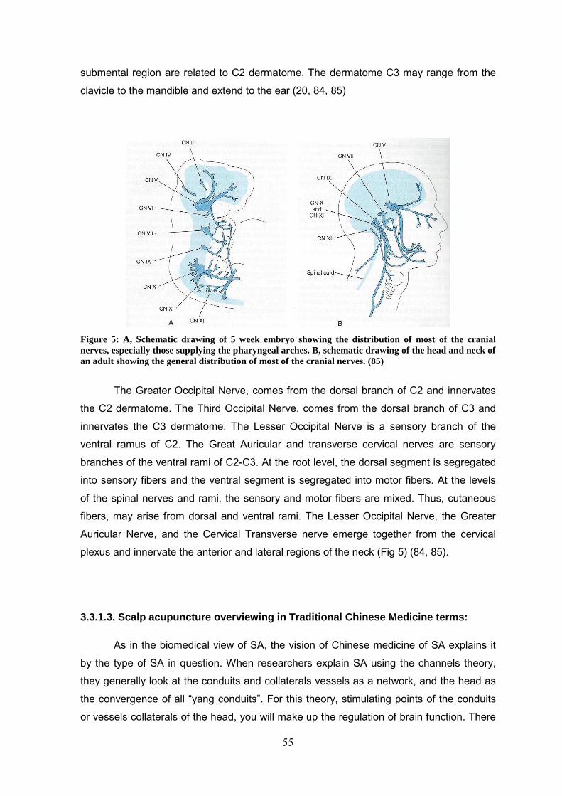
55
submental region are related to C2 dermatome. The dermatome C3 may range from the
clavicle to the mandible and extend to the ear (20, 84, 85)
Figure 5: A, Schematic drawing of 5 week embryo showing the distribution of most of the cranial nerves, especially those supplying the pharyngeal arches. B, schematic drawing of the head and neck of an adult showing the general distribution of most of the cranial nerves. (85)
The Greater Occipital Nerve, comes from the dorsal branch of C2 and innervates
the C2 dermatome. The Third Occipital Nerve, comes from the dorsal branch of C3 and
innervates the C3 dermatome. The Lesser Occipital Nerve is a sensory branch of the
ventral ramus of C2. The Great Auricular and transverse cervical nerves are sensory
branches of the ventral rami of C2-C3. At the root level, the dorsal segment is segregated
into sensory fibers and the ventral segment is segregated into motor fibers. At the levels
of the spinal nerves and rami, the sensory and motor fibers are mixed. Thus, cutaneous
fibers, may arise from dorsal and ventral rami. The Lesser Occipital Nerve, the Greater
Auricular Nerve, and the Cervical Transverse nerve emerge together from the cervical
plexus and innervate the anterior and lateral regions of the neck (Fig 5) (84, 85).
3.3.1.3. Scalp acupuncture overviewing in Traditional Chinese Medicine terms:
As in the biomedical view of SA, the vision of Chinese medicine of SA explains it
by the type of SA in question. When researchers explain SA using the channels theory,
they generally look at the conduits and collaterals vessels as a network, and the head as
the convergence of all “yang conduits”. For this theory, stimulating points of the conduits
or vessels collaterals of the head, you will make up the regulation of brain function. There
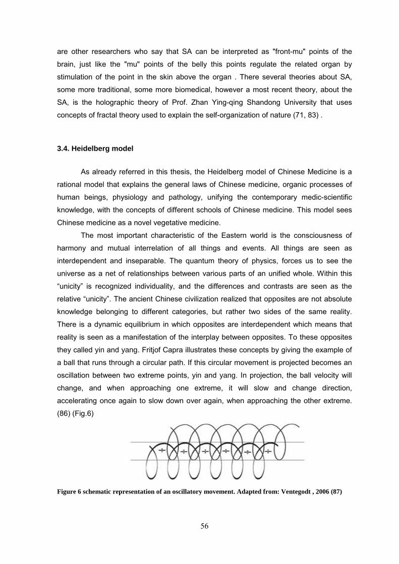
56
are other researchers who say that SA can be interpreted as "front-mu" points of the
brain, just like the "mu" points of the belly this points regulate the related organ by
stimulation of the point in the skin above the organ . There several theories about SA,
some more traditional, some more biomedical, however a most recent theory, about the
SA, is the holographic theory of Prof. Zhan Ying-qing Shandong University that uses
concepts of fractal theory used to explain the self-organization of nature (71, 83) .
3.4. Heidelberg model
As already referred in this thesis, the Heidelberg model of Chinese Medicine is a
rational model that explains the general laws of Chinese medicine, organic processes of
human beings, physiology and pathology, unifying the contemporary medic-scientific
knowledge, with the concepts of different schools of Chinese medicine. This model sees
Chinese medicine as a novel vegetative medicine.
The most important characteristic of the Eastern world is the consciousness of
harmony and mutual interrelation of all things and events. All things are seen as
interdependent and inseparable. The quantum theory of physics, forces us to see the
universe as a net of relationships between various parts of an unified whole. Within this
“unicity” is recognized individuality, and the differences and contrasts are seen as the
relative “unicity”. The ancient Chinese civilization realized that opposites are not absolute
knowledge belonging to different categories, but rather two sides of the same reality.
There is a dynamic equilibrium in which opposites are interdependent which means that
reality is seen as a manifestation of the interplay between opposites. To these opposites
they called yin and yang. Fritjof Capra illustrates these concepts by giving the example of
a ball that runs through a circular path. If this circular movement is projected becomes an
oscillation between two extreme points, yin and yang. In projection, the ball velocity will
change, and when approaching one extreme, it will slow and change direction,
accelerating once again to slow down over again, when approaching the other extreme.
(86) (Fig.6)
Figure 6 schematic representation of an oscillatory movement. Adapted from: Ventegodt , 2006 (87)
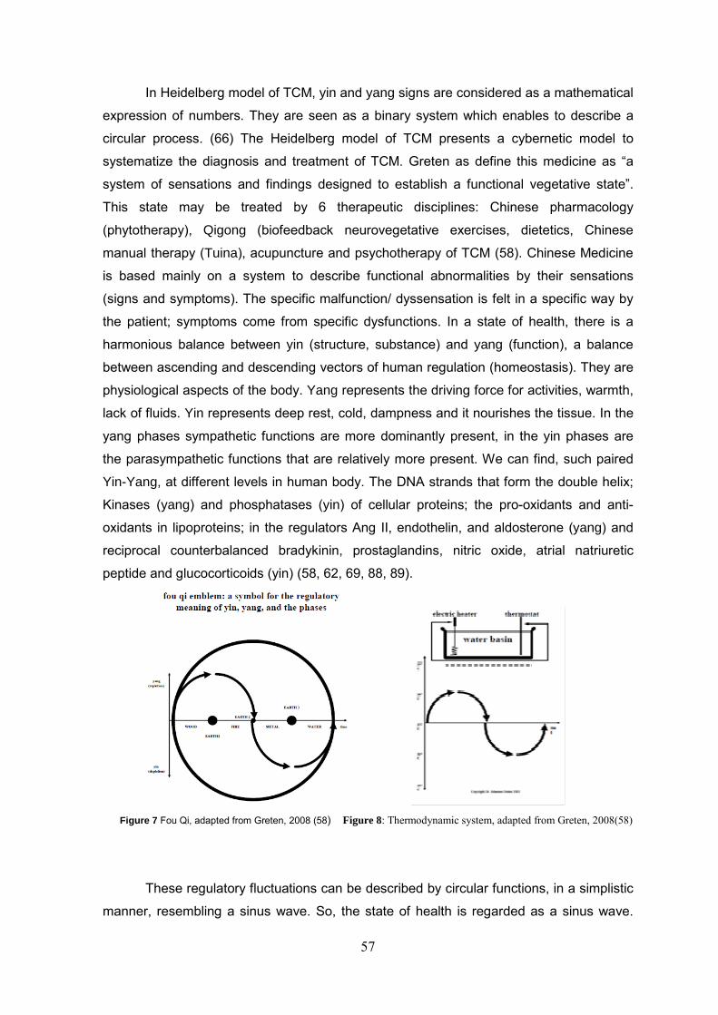
57
In Heidelberg model of TCM, yin and yang signs are considered as a mathematical
expression of numbers. They are seen as a binary system which enables to describe a
circular process. (66) The Heidelberg model of TCM presents a cybernetic model to
systematize the diagnosis and treatment of TCM. Greten as define this medicine as “a
system of sensations and findings designed to establish a functional vegetative state”.
This state may be treated by 6 therapeutic disciplines: Chinese pharmacology
(phytotherapy), Qigong (biofeedback neurovegetative exercises, dietetics, Chinese
manual therapy (Tuina), acupuncture and psychotherapy of TCM (58). Chinese Medicine
is based mainly on a system to describe functional abnormalities by their sensations
(signs and symptoms). The specific malfunction/ dyssensation is felt in a specific way by
the patient; symptoms come from specific dysfunctions. In a state of health, there is a
harmonious balance between yin (structure, substance) and yang (function), a balance
between ascending and descending vectors of human regulation (homeostasis). They are
physiological aspects of the body. Yang represents the driving force for activities, warmth,
lack of fluids. Yin represents deep rest, cold, dampness and it nourishes the tissue. In the
yang phases sympathetic functions are more dominantly present, in the yin phases are
the parasympathetic functions that are relatively more present. We can find, such paired
Yin-Yang, at different levels in human body. The DNA strands that form the double helix;
Kinases (yang) and phosphatases (yin) of cellular proteins; the pro-oxidants and anti-
oxidants in lipoproteins; in the regulators Ang II, endothelin, and aldosterone (yang) and
reciprocal counterbalanced bradykinin, prostaglandins, nitric oxide, atrial natriuretic
peptide and glucocorticoids (yin) (58, 62, 69, 88, 89).
Figure 7 Fou Qi, adapted from Greten, 2008 (58) Figure 8: Thermodynamic system, adapted from Greten, 2008(58)
These regulatory fluctuations can be described by circular functions, in a simplistic
manner, resembling a sinus wave. So, the state of health is regarded as a sinus wave.
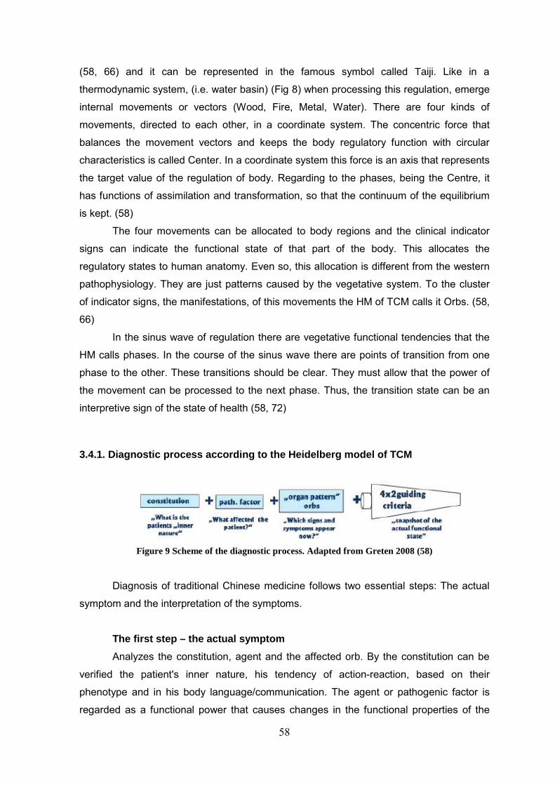
58
(58, 66) and it can be represented in the famous symbol called Taiji. Like in a
thermodynamic system, (i.e. water basin) (Fig 8) when processing this regulation, emerge
internal movements or vectors (Wood, Fire, Metal, Water). There are four kinds of
movements, directed to each other, in a coordinate system. The concentric force that
balances the movement vectors and keeps the body regulatory function with circular
characteristics is called Center. In a coordinate system this force is an axis that represents
the target value of the regulation of body. Regarding to the phases, being the Centre, it
has functions of assimilation and transformation, so that the continuum of the equilibrium
is kept. (58)
The four movements can be allocated to body regions and the clinical indicator
signs can indicate the functional state of that part of the body. This allocates the
regulatory states to human anatomy. Even so, this allocation is different from the western
pathophysiology. They are just patterns caused by the vegetative system. To the cluster
of indicator signs, the manifestations, of this movements the HM of TCM calls it Orbs. (58,
66)
In the sinus wave of regulation there are vegetative functional tendencies that the
HM calls phases. In the course of the sinus wave there are points of transition from one
phase to the other. These transitions should be clear. They must allow that the power of
the movement can be processed to the next phase. Thus, the transition state can be an
interpretive sign of the state of health (58, 72)
3.4.1. Diagnostic process according to the Heidelberg model of TCM
Figure 9 Scheme of the diagnostic process. Adapted from Greten 2008 (58)
Diagnosis of traditional Chinese medicine follows two essential steps: The actual
symptom and the interpretation of the symptoms.
The first step – the actual symptom
Analyzes the constitution, agent and the affected orb. By the constitution can be
verified the patient's inner nature, his tendency of action-reaction, based on their
phenotype and in his body language/communication. The agent or pathogenic factor is
regarded as a functional power that causes changes in the functional properties of the

59
individual and produces its own clinical signs, inducing diagnostically relevant signs
(orbs). The agent can be classified as exterior, (ventus, algor, ariditas, aestus, ardor)
interior (ira, voluptas, maeror, timor, pavor, solicitude, cogitacio) and neutral (bad nutrition,
accidents, inter alia) agents. The affected orb will indicate the clinical manifestations, the
functional state of a body region, which correlates with the functional properties of a
conduit. (58, 63, 66, 90,)
The second step - Guiding criteria
It is an Interpretative approach of body regulation, according to Heidelberg Model.
The body regulation is observed at four different levels: first at neurovegetative level
(repletion/depletion), second at humorovegetative level (Calor/Algor), third at
neuroimmunlogical level (exterior/interior); fourth at structural, cellular level (yin/yang). To
this doctrine of bodily regulation, whose components allow us to perform functional
diagnosis of the patient, the model of Heidelberg called Guiding Criteria (GC). (90)
Repletion/depletion – Quantifies and qualifies the functional capacity induced by
the neurovegetative system. It evaluates the clinical signs that are believed to be originate
primarily from “Qi” and Orbs. In general, signs of repletion indicate too much “Qi”, an
excess of vegetative stimulation. Signs of Depletion indicate lack of “Qi”, a lack of
activation, an excess of de-activation. (63, 66, 90)
Figure 10: Four Guiding Criteria. Adapted from Greten.
60
Calor/Algor – describes the activity of “xue”. It is related to microcirculation and to
humorovegetative system. It evaluates the signs that are believed to originate from the
effects of “xue”. In western terms these signs include the local interdependent
mechanisms of plasma, blood cells, endothelium and functional tissue of an organ and
also it include an activation of body fluids, involving responses in the context of fluid
distribution, supply and circulation. Calor means overactivation of “xue” (microcirculation)
and algor means lack of signs of functional microcirculation. (63, 66, 90)
Extima/Intima - evaluates the level of achievement of the effects of an exterior
agent, when it invades the body. According to Prof. Manfred Porket, from the western
point of view, it refers to clinical signs induced neuro-immunologic mechanisms.
(66,69,90) The most common pathophysiological model, underlying this mechanism is the
model of six stages of Zhang Zhongjing, called Shang Han Lun (treatise on cold induced
diseases). This model of the six stages describes the process of the agent algor
damaging the body (63, 90, 91).
Yin/Yang – allows distinguishing whether the symptoms / signs correspond to a
primary dysregulation, a functional dysregulation (yang) or if these symptoms represent a
secondary dysregulation, a structural deficit, a deficit in functional tissue (yin). Therefore,
allows to distinguish if the origin of the signs / symptoms is at the level of regulatory
function or at a structural level. If the functional tissue is in deficiency, it will be up-
regulated for achieve the proper function. However, this hyper-regulation cannot be
maintained, so, after a hyper-stimulation of the functional tissue, follows a functional
deficit, in other words, after a deficit of yin follows a yang deficit. (63, 66, 90)
The HM of TCM showed that the TCM diagnosis can be regarded as the
vegetative state of the body, which can be defined by key symptoms. According to the
HM, acupoints promote vegetative reflexes that have to match with the vegetative state of
the patient, in order to the treatment be clinically effective. One clinical scenario of
western diagnosis may, therefore, represent a multitude of diagnosis in TCM. That is why
is not easy to do research, in TCM, when the inclusion and exclusion criteria are based in
Western medicine. Usually there is a great risk that the acupuncture treatment is may be
appropriate for some patients, in a certain a vegetative state, while it may not match the
vegetative state of other patients. (63)

61
3.4.2. Pathogeny - Overview of the model of Heidelberg Health is the result of a constant equilibrium of a phase (vegetative tendency).
Normally there is continuity in the changes and alterations at which the body and psyche
are exposed, in daily constant interactions with their environment. Nevertheless,
sometimes the vegetative tendency becomes more intense and signals, the
manifestations of this tendency, become more dominant. When this happens, the
continuum of regulation is disrupted, and in the body, arises a pathological manifestation.
Thus, there are two functional states: the continuum, in which the functional
manifestations of vegetative tendency disappear, during the homeostatic process and the
state of dominance of a signal which becomes a critical symptom. (58)
• Deficient transition - The vegetative tendencies, the phases, should be clearly
processed one into to the other so that the equilibrium remains constant.
Occasionally, it may occur a lack of functional capacity of transformation in the
body. The power of transformation of a vector into the other, of the circular function
is blocked, stopping the cycle of regulation, resulting in pathological signs of the
blockage of one of the vectors (Phases) (58, 69, 90)
• Imbalance agonist / antagonist - when there is an imbalance of agonists and
antagonists, one of the phases will be in excess in relation to the other, leading to
the predominance of signals of the phase in excess. (90)
• Excess of an agent – If an agent is continuously present, the phase at which it is
related is also continuously produced leading to increase of the respective vector
(phase). (58, 69, 90)
• Yin deficiency - As already stated, the regulation model of the body can be
compared to a thermodynamic system. Thus, when a structure with less matter is
subject to variations it will manifest a more unstable variation curve, leading to
higher peaks of the variable, but the turning point will be achieved faster. In terms
of TCM, lack of yin means lack of substance, matter, this is why yin deficiency
causes an extreme course of the curve of regulation. It leads to labile “Qi”. (90)
There are four types of yin deficiency: lack of functional tissue (yin deficiency "sui
generis"); lack of body fluids; lack of microcirculation within the tissue (lack of
"Xue"); functional deficits at the cellular level (lack of "jing ")
3.4.3. Algor Laedens Theory 3 rd guiding criteria
Algor Laedens Theory (ALT) is the latin name of Shang Han Lun (treatise on cold
damage diseases) systematically introduced regular pattern of diagnosis for diseases due

62
to invasion of exogenous pathogenic factors. (92) This kind of classification of pathologies
first appeared in the writings of Zang Zhongjing, flourishing during the 2nd century of our
era. It may be said that Zang Zhongjing founded clinical medicine when he wrote this
treatise. (69)
This theory considers that an agent faces six levels of defense when invades the
body. These six levels of defense differ in vegetative functional capacity ("Qi") and "Xue".
The Stages, ALT levels, are characterized by specific clinical signs that will help to identify
on what level of defense lies the pathogenic agent, and therefore to classify the capacity
of damage on the body. The first level of defense is consists in Tenuintestinal and Vesical
conduits that depend very much on the status of "Xue". So, the lack or decrease of
microcirculation (algor) will be the prime cause of the invasion of agents. By the western
overview this theory assesses the reaction process of the body to the presence of an
agent. It reflects the absence or decrease of the microcirculation which affects first the
conduits that contain more "Xue" than "Qi” (58, 69, 90)
Figure 11Scheme of Algor Laedens Theory. Adapted from Greten (58)
The ALT allows us to interpret the third guiding criteria, extima/intimate. As the
stages I, II and III are outside the body region, and are constituted by yang conduits, their
names contains termination yang. On the other hand, the stages IV, V and VI, are directly
related to the body region, constituted by yin conduits, internal and contains the term yin.
(90)
Due to its relative importance in OSA, is important to point out the first two stages
of ALT.

63
Stage 1 – Yang major – in this stage the agent affects the Qi defensivum (Wei Qi)
that resides within the extima along the surface on the outside of the conduits. The
conduits directly related with yang major are Tenuintestinal and Vesical and indirectly
related are the coupled yin conduits Cardinal and Renal. Therefore, this stage disease will
affect the fluids and liquids saving mechanism, the separation of liquid and solid,
excretions and production and control of calor. The Yang major conduits cross the neck,
the head and shoulders; so, when an agent affects this stage the patient may show
intense pain in the head and neck, pressure between the yes, fever, shivering, no
perspiration, diffuse pain, copious urine, cold hands and feet, inter alia. (58, 69)
Stage 2 – Splendor yang – this is the stage of conduit “Qi”. When the agent algor
overcomes the first stage, it enters in the conduits. This leads to a blockade of flow of “Qi”
and “Xue”. As in the first stage there is a deficit of "Xue", so it will be needed to promote it.
As it is the "Qi" that moves the "Xue", it will be most affected, because it is being
challenged by the lack or reduction of microcirculation of the preceding stage. The main
sources of "Qi" are nutrition and breathing, thus, the Centre (responsible for the
assimilation) and the Metal phase will be affected. As the yang conduits of the Centre and
of the Metal phases are the Stomach and Crassintestinal, respectively, they will be most
affected at this stage. The Lienal conduit and the Pulmonal conduits, they are the yin
coupled conduits of Stomach and Crassintestinal; they will be indirectly affected (58, 63,
69).
The pathologies of this stage will produce disturbances in assimilation, in rhythm of
all orbs and in the exchange of “energies” between the orbs. Clinical manifestations of this
stage can be high fever, oppressive heat, desire to be cooled, profuse transpiration, thirst,
constipation, inter alia. (69)
• Main symptoms of the Stomach orb – related to the course of conduct may
arise: sinusitis or tearing pain in the face; affections of the sinuses, toothache;
breathing problems; loss of appetite; weakness and pain in the thigh and Knees.
Related to the impaired of stomach orb functions, may arise: swellings, edema on
the eye region, unclear vision, accumulation of phlegm, swellings of the face,
swellings of the mucous membranes, acute paradontosis, hoarseness and other
problems in the throat, swellings of the mamma, cough, loss of appetite, anosmia,
anhelitus. (69)
• Main symptoms of the Crassintestinal orb – related to the course of the
conduct may arise some symptoms like running nose, toothache in the maxilla, pain
in the neck, shoulders, forearm, tendency to develop tendinitis. Related to the
64
functional impairment may arise: abdominal discomfort, noisy bowel movements,
constipation due to reactive calor, diarrhoea due to algor, pain in the intestines. (69)
3.4.4. Upper airways in Heidelberg model of TCM
• Nose - In Chinese Medicine the nose can have different allocations. So, the nostrils
are the most exterior part of the respiratory tract and are said to be the diagnostic window
of the lungs. Therefore, it is primarily allocated to the pulmonary orb. As the respiratory
tract, in many cases, reacts as a functional unit, the nostrils can be said to be the
diagnostic window of the whole respiratory track.
The exterior of the nose has its own allocations: The rims of the nostrils and its small
sebaceous glands are allocated to the Centre. Thus, on the skin of the nose, on the
plateau, we see signs of the Lienal orb. In the wings of the nostrils we can see Stomach
signs
The secondary allocation of the nose is the Crassintestinal orb. The conduit of the
Crassintestinal orb runs underneath the nostrils, above the lip and touches the exterior
side of the opposite nostril. Symptoms of exterior affections of the phase metal, like
sneezing, are rather Crassintestinal.
• Sinuses – Due to the course of the conduit, the maxillary sinuses are primarily
affected by the stomach orb. Thinking in the same way, the ethmoidal sinuses or cellulae
ethmoidales and the feeling of pressure between the eyes are allocated to the Vesical orb.
The root of the nose, sometimes called “the region of pads of glasses" is allocated to the
contralateral branch of the Tenuintestinal conduit. Normally, the frontal sinuses are well
treated by this conduit, considering that they are a Yang Major problem, but if the whole
viscerocranium is affected, it is better to be treated by the Stomachal orb, considering it is
a Splendor-Yang disease. (58)
• Pharynx – in western anatomy pharynx is conventionally divided into nasopharynx,
(epipharynx) oropharynx (mesopharynx) and the laryngopharynx (hypopharynx). The
nasopharynx has the same functional relations as the sinuses. The oropharynx, as part of
the oral cavity, is allocated to the Stomachal, and the nasal cavity is allocated to the
Pulmonal and Crassintestinal. The soft palate, the uvula, can be allocated to the three
orbs (Stomachal, Pulmonal and Crassintestinal), but mostly it is related to Splendor-yang
syndrome, as it is between the two first sections, although may show clinical signs of the
three orbs. In general, all osseous structures of the body belong to the renal orb. As bone
65
structure that it is, the hard palate belongs to the renal orb, whereas the “fleshy” parts of
the oral cavity belong to the stomach orb. As oropharynx and laryngopharynx are the
prolongation of the oral cavity, most of their affections are allocated to the stomach orb.
(58, 89)
• Tongue –obviously is part of the oral cavity, so it reflects the functional properties of
the Stomachal orb. However, in Chinese medicine, the tongue is also called "the
prolonged heart" which also will point to "Shen" (constellating force originated from the
cardiac orb) deficiencies. (58)
• Uvula – Some inflammations of the uvula are so deep that the most effective
treatment for these conditions is to use yin points, therefore some authors considered it is
connected to the renal functions. This is not contradictory to the allocation to the
Splendor-yang, because when the Splendor yang is long-standing, the high Calor
produced (typical of this syndrome) will reduce the yin (the fluids) and over the time it will
lead to a yang deficiency by the long standing yin deficiency.(58)
The Centre controls all regulation. As mentioned above, the Centre is the axis of the curve
of regulation. As in the pathologies of the upper airways is required control of the surface
and a good regulation, the constant action of the Centre is required, especially of its outer
(yang) orb, the Stomach. In the same way, the metal phase which regulates the surfaces,
is required, so, the constant action of yang orb of this phase, the Crassintestinal, is
required too.
When there is a lack of microcirculation (Algor), there is a kind of depletion. All depletions
reduce the stomach functions (to move downwards), so the downward movement of
fluids, humor and pituita (phlegm) for excretion, will be reduced. This combination of
factors will lead to a blockage of Stomachal orb which, in Western terms, may be
comparable to blocked vagal functions and thereby to various manifestations, including
swelling and inflammations in the area of the face, nose, sinuses and throat.
Depletion is naturally associated to yin patterns. So, the Vesical orb, the coupled outer orb
of the Renal orb (the most representative orb of yin) may be affected. Thus, the lack of "Qi
defensivum" promotes stages I and II of the ALT. (58)

66
3.4.5. Physiopathology of OSA according to HM of Chinese medicine
As previously mentioned, OSA can occur at any age, but it mainly affects men
between 40 and 60 years and postmenopausal women. In Chinese medicine, after 35
years, humans come into yin deficiency, in structural deficiency, which derives from the
physiological reduction of the functional tissue. (23) Thus, there is a strong relationship
between the lack or progressive loss of yin, often reinforced by unruly lifestyle, and OSA
manifestation. This lack of yin will result in a homeostatic imbalance; in a disruption of the
cycle of body regulation and sinus wave. Therefore, the centripetal force of the curve, the
Centre, will be challenged. By this, the function of assimilation and transformation will be
disturbed and thereby the yang orb of the Center, the Stomachal orb, will be challenged
too.
From Chinese point of view, OSA, can be also regarded as phlegm obstructing the
orifices. (93) This means that humor and pituita (phlegm) are obstructing the upper
airways; there is not a vector sufficiently strong to lead the turbid down and to do the
transformation and assimilation function. These obstructions will cause a blockage of
Stomachal orb, which is the orb that moves and drains humor and pituita out; it is
responsible for the downwards movements for excretion. Regarding this, upper airways
obstruction can be considered a lack of yang, (function) based on a deficiency of yin
(substance). As consequence, the tissues of the upper airways will not be so well
nourished, warm and moisturized; the microcirculation, which is correlated with “Xue”, will
decrease. Knowing that “Xue” “is a form of functional capacity bound to body fluids, with
functions such as warming, moisturizing, creating “Qi” and nourishing a tissue”, (58) there
will be a stage II of ALT, therefore, will arise the instability of the upper airways which will
result, also, in the decreased activity of the dilator muscles of this region and in the
collapse of the oropharynx. In an attempt to achieve the target value, the body will react
with humorovegetative reactions, producing the so-called “reactive Calor”, which will lead
to an increased sympathetic activity plus lack of fluids, restlessness and nocturnal
awakenings, during sleep. (63, 58, 93) This will enhance the physiological activity of
Cardial Orb and consequently its coupled orb, Tenuintestinal, leading to Stage I of the
ALT. (Fig 12)
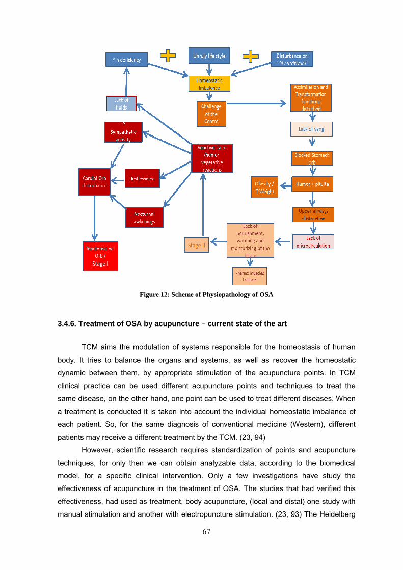
67
Figure 12: Scheme of Physiopathology of OSA
3.4.6. Treatment of OSA by acupuncture – current state of the art
TCM aims the modulation of systems responsible for the homeostasis of human
body. It tries to balance the organs and systems, as well as recover the homeostatic
dynamic between them, by appropriate stimulation of the acupuncture points. In TCM
clinical practice can be used different acupuncture points and techniques to treat the
same disease, on the other hand, one point can be used to treat different diseases. When
a treatment is conducted it is taken into account the individual homeostatic imbalance of
each patient. So, for the same diagnosis of conventional medicine (Western), different
patients may receive a different treatment by the TCM. (23, 94)
However, scientific research requires standardization of points and acupuncture
techniques, for only then we can obtain analyzable data, according to the biomedical
model, for a specific clinical intervention. Only a few investigations have study the
effectiveness of acupuncture in the treatment of OSA. The studies that had verified this
effectiveness, had used as treatment, body acupuncture, (local and distal) one study with
manual stimulation and another with electropuncture stimulation. (23, 93) The Heidelberg
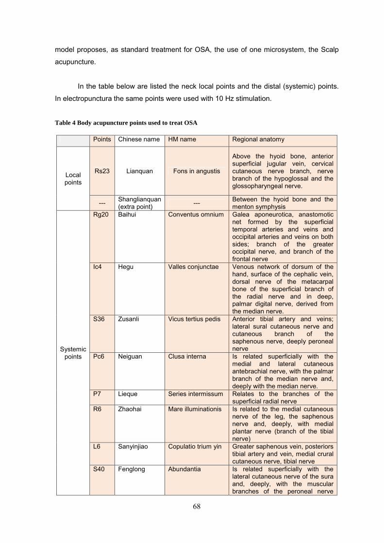
68
model proposes, as standard treatment for OSA, the use of one microsystem, the Scalp
acupuncture.
In the table below are listed the neck local points and the distal (systemic) points.
In electropunctura the same points were used with 10 Hz stimulation.
Table 4 Body acupuncture points used to treat OSA
Points Chinese name HM name Regional anatomy
Local points
Rs23 Lianquan Fons in angustis
Above the hyoid bone, anterior superficial jugular vein, cervical cutaneous nerve branch, nerve branch of the hypoglossal and the glossopharyngeal nerve.
--- Shanglianquan (extra point) --- Between the hyoid bone and the
menton symphysis
Systemic points
Rg20 Baihui Conventus omnium Galea aponeurotica, anastomotic net formed by the superficial temporal arteries and veins and occipital arteries and veins on both sides; branch of the greater occipital nerve, and branch of the frontal nerve
Ic4 Hegu Valles conjunctae Venous network of dorsum of the hand, surface of the cephalic vein, dorsal nerve of the metacarpal bone of the superficial branch of the radial nerve and in deep, palmar digital nerve, derived from the median nerve.
S36 Zusanli Vicus tertius pedis Anterior tibial artery and veins; lateral sural cutaneous nerve and cutaneous branch of the saphenous nerve, deeply peroneal nerve
Pc6 Neiguan Clusa interna Is related superficially with the medial and lateral cutaneous antebrachial nerve, with the palmar branch of the median nerve and, deeply with the median nerve.
P7 Lieque Series intermissum Relates to the branches of the superficial radial nerve
R6 Zhaohai Mare illuminationis Is related to the medial cutaneous nerve of the leg, the saphenous nerve and, deeply, with medial plantar nerve (branch of the tibial nerve)
L6 Sanyinjiao Copulatio trium yin Greater saphenous vein, posteriors tibial artery and vein, medial crural cutaneous nerve, tibial nerve
S40 Fenglong Abundantia Is related superficially with the lateral cutaneous nerve of the sura and, deeply, with the muscular branches of the peroneal nerve
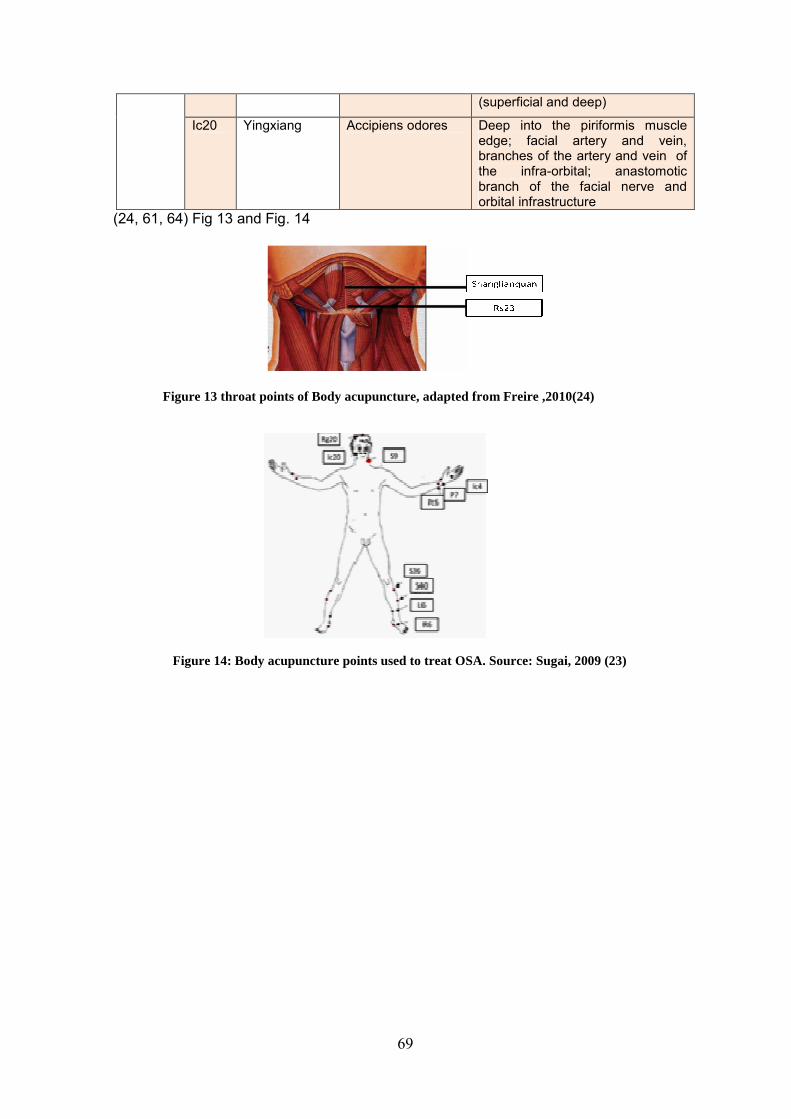
69
(superficial and deep)
Ic20 Yingxiang Accipiens odores Deep into the piriformis muscle edge; facial artery and vein, branches of the artery and vein of the infra-orbital; anastomotic branch of the facial nerve and orbital infrastructure
(24, 61, 64) Fig 13 and Fig. 14
Figure 13 throat points of Body acupuncture, adapted from Freire ,2010(24)
Figure 14: Body acupuncture points used to treat OSA. Source: Sugai, 2009 (23)
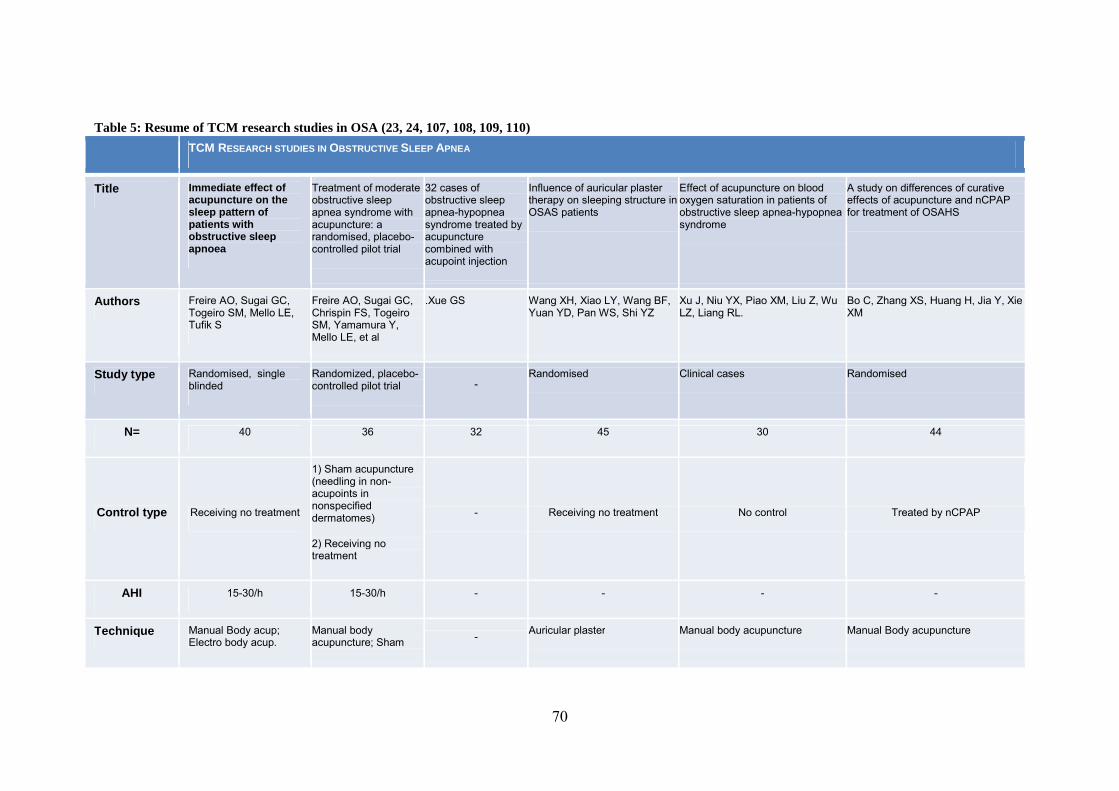
70
Table 5: Resume of TCM research studies in OSA (23, 24, 107, 108, 109, 110)
TCM RESEARCH STUDIES IN OBSTRUCTIVE SLEEP APNEA
Title Immediate effect of acupuncture on the sleep pattern of patients with obstructive sleep apnoea
Treatment of moderate obstructive sleep apnea syndrome with acupuncture: a randomised, placebo-controlled pilot trial
32 cases of obstructive sleep apnea-hypopnea syndrome treated by acupuncture combined with acupoint injection
Influence of auricular plaster therapy on sleeping structure in OSAS patients
Effect of acupuncture on blood oxygen saturation in patients of obstructive sleep apnea-hypopnea syndrome
A study on differences of curative effects of acupuncture and nCPAP for treatment of OSAHS
Authors Freire AO, Sugai GC, Togeiro SM, Mello LE, Tufik S
Freire AO, Sugai GC, Chrispin FS, Togeiro SM, Yamamura Y, Mello LE, et al
.Xue GS Wang XH, Xiao LY, Wang BF, Yuan YD, Pan WS, Shi YZ
Xu J, Niu YX, Piao XM, Liu Z, Wu LZ, Liang RL.
Bo C, Zhang XS, Huang H, Jia Y, Xie XM
Study type Randomised, single blinded
Randomized, placebo-controlled pilot trial -
Randomised Clinical cases Randomised
N= 40 36 32 45 30 44
Control type Receiving no treatment
1) Sham acupuncture (needling in non-acupoints in nonspecified dermatomes)
2) Receiving no treatment
- Receiving no treatment No control Treated by nCPAP
AHI 15-30/h 15-30/h - - - -
Technique Manual Body acup; Electro body acup.
Manual body acupuncture; Sham - Auricular plaster Manual body acupuncture Manual Body acupuncture
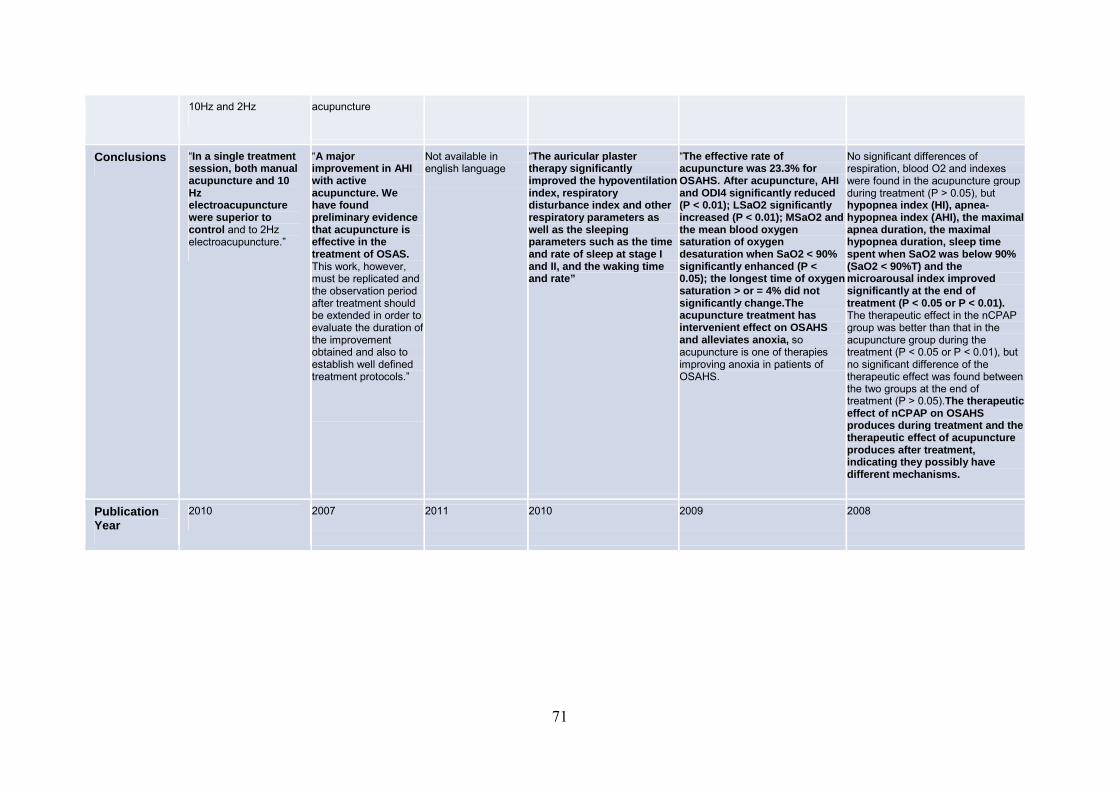
71
10Hz and 2Hz acupuncture
Conclusions “In a single treatment session, both manual acupuncture and 10 Hz electroacupuncture were superior to control and to 2Hz electroacupuncture.”
“A major improvement in AHI with active acupuncture. We have found preliminary evidence that acupuncture is effective in the treatment of OSAS. This work, however, must be replicated and the observation period after treatment should be extended in order to evaluate the duration of the improvement obtained and also to establish well defined treatment protocols.”
Not available in english language
“The auricular plaster therapy significantly improved the hypoventilation index, respiratory disturbance index and other respiratory parameters as well as the sleeping parameters such as the time and rate of sleep at stage I and II, and the waking time and rate”
“The effective rate of acupuncture was 23.3% for OSAHS. After acupuncture, AHI and ODI4 significantly reduced (P < 0.01); LSaO2 significantly increased (P < 0.01 ); MSaO2 and the mean blood oxygen saturation of oxygen desaturation when SaO2 < 90% significantly enhanced (P < 0.05); the longest time of oxygen saturation > or = 4% did not significantly change.The acupuncture treatment has intervenient effect on OSAHS and alleviates anoxia, so acupuncture is one of therapies improving anoxia in patients of OSAHS.
No significant differences of respiration, blood O2 and indexes were found in the acupuncture group during treatment (P > 0.05), but hypopnea index (HI), apnea-hypopnea index (AHI), the maximal apnea duration, the maximal hypopnea duration, sleep time spent when SaO2 was below 90% (SaO2 < 90%T) and the microarousal index improved significantly at the end of treatment (P < 0.05 or P < 0.01). The therapeutic effect in the nCPAP group was better than that in the acupuncture group during the treatment (P < 0.05 or P < 0.01), but no significant difference of the therapeutic effect was found between the two groups at the end of treatment (P > 0.05).The therapeutic effect of nCPAP on OSAHS produces during treatment and the therapeutic effect of acupuncture produces after treatment, indicating they possibly have different mechanisms.
Publication Year
2010 2007 2011 2010 2009 2008

72
As in present study, Freire's study evaluated the immediate effect of acupuncture. The results relating to the manual body acupuncture of this study were:
Table 6 results of Freire’s study
Graphic 1 Comparison of AHI at baseline (before) and after procedures. Manual acupuncture versus the control group (p=0,034). P<0,05, Mann-Whitney t test. Adapted from Freire, 2010 (24)
3.4.7 Treatment of OSA according to the Heidelberg Scalp Acupuncture model.
The Heidelberg Scalp Acupuncture (HSA) is a somatotopic system, related to a
coordinated system that correlates some head zones with anatomical regions and
concepts of TCM. (114) (Fig.15) The Heidelberg model of TCM often uses this puncture
technique to treat OSA. This technique allows to take action on stages I and II of ALT;
allows to act on the orbs and at the same time it has a direct action on the anatomical
region where the disease is manifesting.
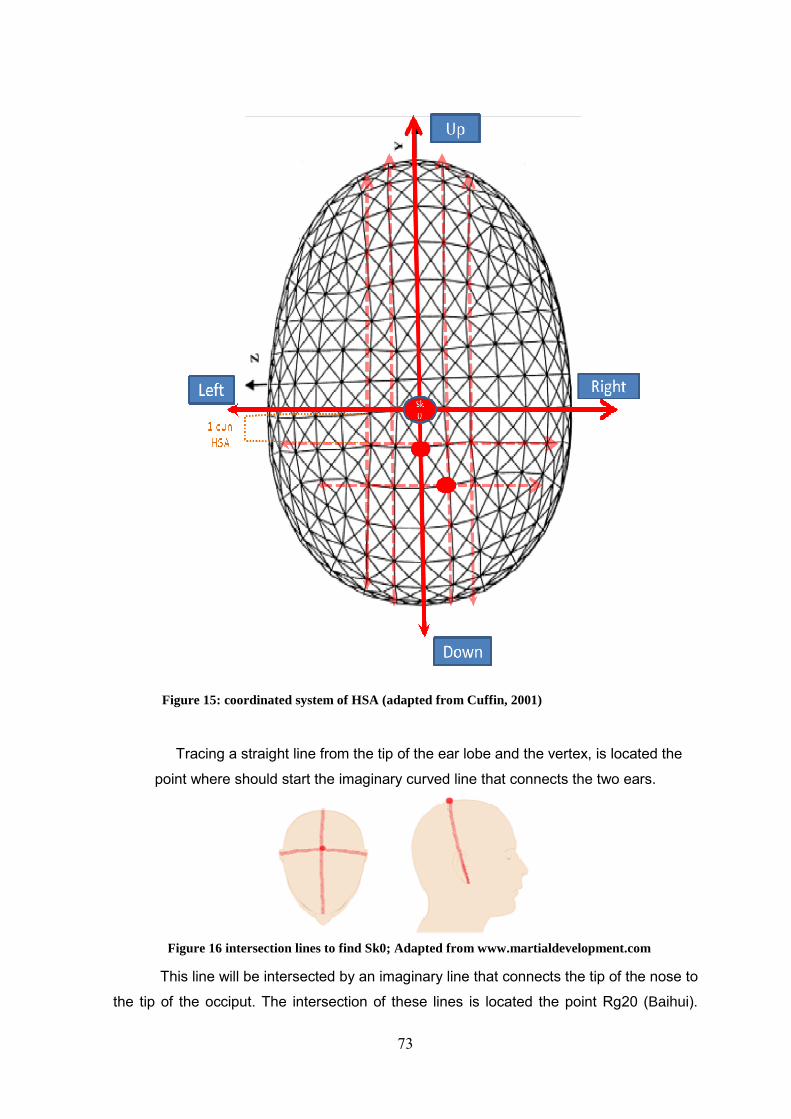
73
Tracing a straight line from the tip of the ear lobe and the vertex, is located the
point where should start the imaginary curved line that connects the two ears.
This line will be intersected by an imaginary line that connects the tip of the nose to
the tip of the occiput. The intersection of these lines is located the point Rg20 (Baihui).
Figure 15: coordinated system of HSA (adapted from Cuffin, 2001)
Figure 16 intersection lines to find Sk0; Adapted from www.martialdevelopment.com

74
This point will have the designation of Sk0, in HSA technique, it will be the reference point
to find the others. This point can be, also, a projection of the extra point Yintang,
representing the same anatomical region and the same functions. (114) (Fig 16 and Fig
17)
Figure 17 Sk0 localization; adapted from http://drgominak.com
According to this logic, the SKd1 point will correspond to point between the
eyebrows in the midline; Skd2 point will correspond to the point between the eyes and the
SKd2l1 and SKd2r1 points will correspond to the ethmoid cells. The point SKd3 match
with maxillary sinus ostium; the points Skd3l1 and SK3r1 represent the compartment of
Maxillary sinus. The SKd4 point corresponds to inferior conchae, and thus, the points
SKd4r1, Skd4r2, and Skd4l1 SKd4l2 correspond to inferior maxillary sinus zone and also
to the point of Crassintestinal, IC20. The SKd5 point corresponds to the hard palate and it
especially matches with Rg26. The SKd6 corresponds to the soft palate, The Skd7 to the
Uvula and SKd8 corresponds to the oropharynx. (Fig 18, Fig 19 and Fig.20)
Sk0
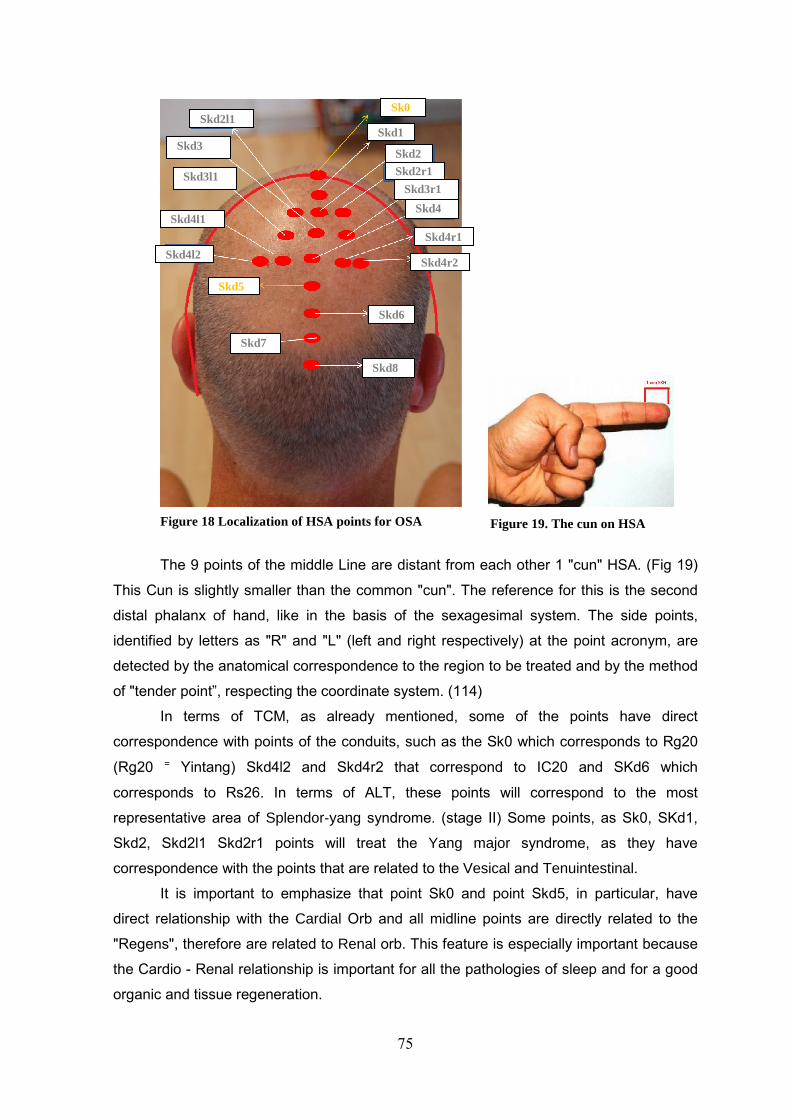
75
Figure 19. The cun on HSA
The 9 points of the middle Line are distant from each other 1 "cun" HSA. (Fig 19)
This Cun is slightly smaller than the common "cun". The reference for this is the second
distal phalanx of hand, like in the basis of the sexagesimal system. The side points,
identified by letters as "R" and "L" (left and right respectively) at the point acronym, are
detected by the anatomical correspondence to the region to be treated and by the method
of "tender point”, respecting the coordinate system. (114)
In terms of TCM, as already mentioned, some of the points have direct
correspondence with points of the conduits, such as the Sk0 which corresponds to Rg20
(Rg20 ⁼ Yintang) Skd4l2 and Skd4r2 that correspond to IC20 and SKd6 which
corresponds to Rs26. In terms of ALT, these points will correspond to the most
representative area of Splendor-yang syndrome. (stage II) Some points, as Sk0, SKd1,
Skd2, Skd2l1 Skd2r1 points will treat the Yang major syndrome, as they have
correspondence with the points that are related to the Vesical and Tenuintestinal.
It is important to emphasize that point Sk0 and point Skd5, in particular, have
direct relationship with the Cardial Orb and all midline points are directly related to the
"Regens", therefore are related to Renal orb. This feature is especially important because
the Cardio - Renal relationship is important for all the pathologies of sleep and for a good
organic and tissue regeneration.
Sk0
Skd1
Skd2
Skd2r1
Skd2l1
Skd3
Skd3r1 Skd3l1
Skd4
Skd4r1
Skd4l1
Skd65
Skd4l2 Skd4r2
Skd5
Skd7
Skd8
Fig : source Greten Figure 18 Localization of HSA points for OSA
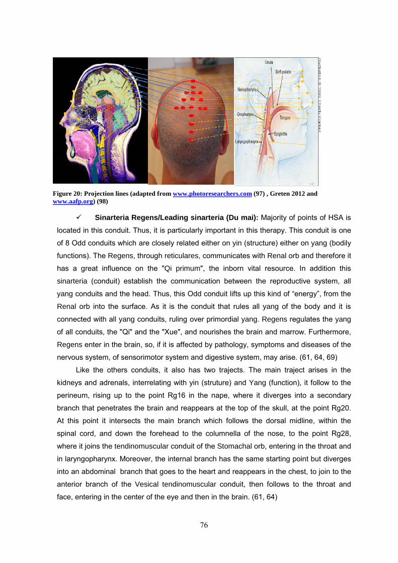
76
� Sinarteria Regens/Leading sinarteria (Du mai): Majority of points of HSA is
located in this conduit. Thus, it is particularly important in this therapy. This conduit is one
of 8 Odd conduits which are closely related either on yin (structure) either on yang (bodily
functions). The Regens, through reticulares, communicates with Renal orb and therefore it
has a great influence on the "Qi primum", the inborn vital resource. In addition this
sinarteria (conduit) establish the communication between the reproductive system, all
yang conduits and the head. Thus, this Odd conduit lifts up this kind of “energy”, from the
Renal orb into the surface. As it is the conduit that rules all yang of the body and it is
connected with all yang conduits, ruling over primordial yang. Regens regulates the yang
of all conduits, the "Qi" and the "Xue", and nourishes the brain and marrow. Furthermore,
Regens enter in the brain, so, if it is affected by pathology, symptoms and diseases of the
nervous system, of sensorimotor system and digestive system, may arise. (61, 64, 69)
Like the others conduits, it also has two trajects. The main traject arises in the
kidneys and adrenals, interrelating with yin (struture) and Yang (function), it follow to the
perineum, rising up to the point Rg16 in the nape, where it diverges into a secondary
branch that penetrates the brain and reappears at the top of the skull, at the point Rg20.
At this point it intersects the main branch which follows the dorsal midline, within the
spinal cord, and down the forehead to the columnella of the nose, to the point Rg28,
where it joins the tendinomuscular conduit of the Stomachal orb, entering in the throat and
in laryngopharynx. Moreover, the internal branch has the same starting point but diverges
into an abdominal branch that goes to the heart and reappears in the chest, to join to the
anterior branch of the Vesical tendinomuscular conduit, then follows to the throat and
face, entering in the center of the eye and then in the brain. (61, 64)
Figure 20: Projection lines (adapted from www.photoresearchers.com (97) , Greten 2012 and www.aafp.org) (98)
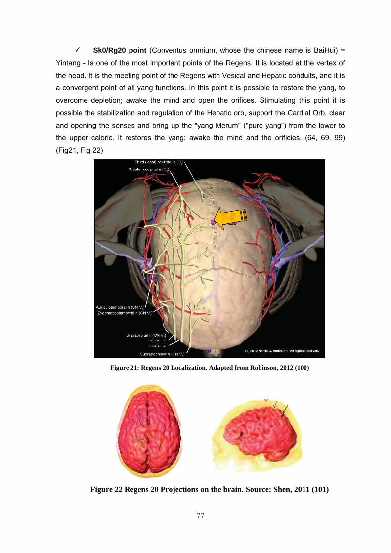
77
� Sk0/Rg20 point (Conventus omnium, whose the chinese name is BaiHui) =
Yintang - Is one of the most important points of the Regens. It is located at the vertex of
the head. It is the meeting point of the Regens with Vesical and Hepatic conduits, and it is
a convergent point of all yang functions. In this point it is possible to restore the yang, to
overcome depletion; awake the mind and open the orifices. Stimulating this point it is
possible the stabilization and regulation of the Hepatic orb, support the Cardial Orb, clear
and opening the senses and bring up the "yang Merum" ("pure yang") from the lower to
the upper caloric. It restores the yang; awake the mind and the orificies. (64, 69, 99)
(Fig21, Fig 22)
Figure 21: Regens 20 Localization. Adapted from Robinson, 2012 (100)
Figure 22 Regens 20 Projections on the brain. Source: Shen, 2011 (101)
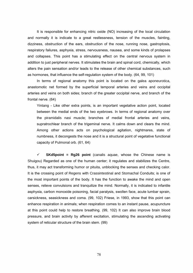
78
It is responsible for enhancing nitric oxide (NO) increasing of the local circulation
and normally it is indicate to a great restlessness, tension of the muscles, fainting,
dizziness, obstruction of the ears, obstruction of the nose, running nose, gastroptosis,
respiratory failures, asphyxia, stress, nervousness, nausea, and some kinds of prolapses
and collapses. This point has a stimulating effect on the central nervous system in
addition to just peripheral nerves. It stimulates the brain and spinal cord, chemically, which
alters the pain sensation and/or leads to the release of other chemical substances, such
as hormones, that influence the self-regulation system of the body. (64, 99, 101)
In terms of regional anatomy this point is located on the galea aponeurotica,
anastomotic net formed by the superficial temporal arteries and veins and occipital
arteries and veins on both sides; branch of the greater occipital nerve, and branch of the
frontal nerve. (64)
Yintang - Like other extra points, is an important vegetative action point, located
between the medial ends of the two eyebrows. In terms of regional anatomy over
the piramidalis nasi muscle; branches of medial frontal arteries and veins,
supratrochlear branch of the trigeminal nerve. It calms down and clears the mind.
Among other actions acts on psychological agitation, nightmares, state of
numbness, it decongests the nose and it is a structural point of vegetative functional
capacity of Pulmonal orb. (61, 64)
� SKd5point = Rg26 point (canalis aquae, whose the Chinese name is
Shuigou) Regarded as one of the human center; it regulates and stabilizes the Centre,
thus, it may act transforming humor or pituita, unblocking the senses and checking calor.
It is the crossing point of Regens with Crassintestinal and Stomachal Conduits; is one of
the most important points of the body. It has the function to awake the mind and open
senses, relieve convulsions and tranquilize the mind. Normally, it is indicated to infantile
asphyxia, carbon monoxide poisoning, facial paralysis, swollen face, acute lumbar sprain,
carsickness, seasickness and coma. (99, 102) Friese, in 1993, show that this point can
enhance respiration in animals; when respiration comes to an instant pause, acupuncture
at this point could help to restore breathing. (99, 102) It can also improve brain blood
pressure, and brain activity by afferent excitation, stimulating the ascending activating
system of reticular structure of the brain stem. (99)
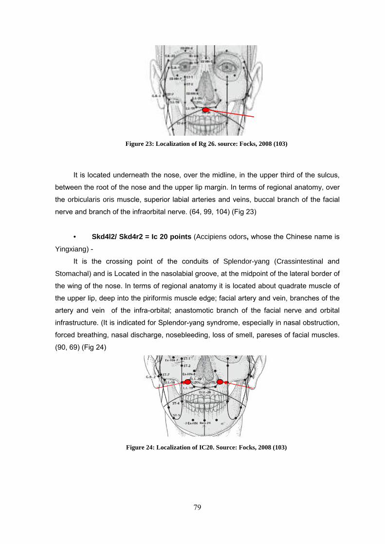
79
Figure 23: Localization of Rg 26. source: Focks, 2008 (103)
It is located underneath the nose, over the midline, in the upper third of the sulcus,
between the root of the nose and the upper lip margin. In terms of regional anatomy, over
the orbicularis oris muscle, superior labial arteries and veins, buccal branch of the facial
nerve and branch of the infraorbital nerve. (64, 99, 104) (Fig 23)
• Skd4l2/ Skd4r2 = Ic 20 points (Accipiens odors, whose the Chinese name is
Yingxiang) -
It is the crossing point of the conduits of Splendor-yang (Crassintestinal and
Stomachal) and is Located in the nasolabial groove, at the midpoint of the lateral border of
the wing of the nose. In terms of regional anatomy it is located about quadrate muscle of
the upper lip, deep into the piriformis muscle edge; facial artery and vein, branches of the
artery and vein of the infra-orbital; anastomotic branch of the facial nerve and orbital
infrastructure. (It is indicated for Splendor-yang syndrome, especially in nasal obstruction,
forced breathing, nasal discharge, nosebleeding, loss of smell, pareses of facial muscles.
(90, 69) (Fig 24)
Figure 24: Localization of IC20. Source: Focks, 2008 (103)

80

81
CHAPTER 2

82

83
4. CLINICAL RESEARCH PROTOCOL

84
4.1 Title: “Effects of Heidelberg Scalp Acupuncture on Obstructive Sleep
Apnea”
4.2. Organizational structure:
In collaboration with the master's program of the Institute Abel Salazar for
Biomedical Sciences, in Traditional Chinese Medicine, this research project was
conducted in the Consultation of Respiratory Pathology of Sleep and Sleep Laboratory of
the Department of Pulmonology of SJHC. The study had the technical support of ResMed
that has provided the equipment and consumables items. The research project did not
have any external funding.
4.3. Research team:
Main investigators:
Maria João Rodrigues Ferreira Rocha dos Santos
• Therapist of Traditional Chinese Medicine at A.R.PHARMA
• Master student of Traditional Chinese Medicine at Abel Salazar Institute for
Biomedical Sciences
Sofia Isabel Martins Milhano
• Physiotherapist at Clinica Ortopedica Nuno Alegrete
• Master student of Traditional Chinese Medicine at Abel Salazar Institute for
Biomedical Sciences
Research supervisor:
Prof. Doctor Henry Johannes Greten. Head of the Heidelberg School of Traditional
Chinese Medicine; President of the German Society of Traditional Chinese Medicine
(DGTCM), Heidelberg, Germany.
Co-supervisors:
• Prof. Dr. João Carlos Winck responsible for the consultation of pathology of sleep
and sleep laboratory of the Department of Pulmonology SJHC; associate

85
Professor in Faculty of Medicine of Oporto University; editor in chief of Portuguese
journal of pulmonology
• Dr. Nuno Correia, Specialist in Internal Medicine at the Department of Internal
Medicine - SJHC; Master in Chinese Medicine - ICBAS/UP; Board member of
Traditional Chinese Medicine Scientific Society. Professor of Physiopathology –
Nursing School of Oporto.
4.4. Research co-workers:
Dr.ª Maria João Lima, specialist in internal medicine, responsible for the
hypertension consultation at Department of Internal Medicine - SJHC; Dr.ª Anabela
Marinho, physician at consultation of pathology of sleep and sleep laboratory of the
Department of Pulmonology SJHC; Technician of Cardiopneumology Patricia Teles;
Marcia Sá physician at Family Health Unit - ACeS Grande Porto III
4.5. Study objectives :
General objective :
To evaluate HSA effects on OSA using objective measurements of standard sleep
study
Specific objectives:
• Related to OSA:
� Verify the effects of the main parameters of cardiorespiratory polygraphy:
� Agitation / movement
� Snoring
� O2 saturation
� Fatigue
� Breath threshold
� Apnea index; Hyponea index; Apnea-Hypopnea índex
• Related to HSA technique:
� Analyzing the mechanisms of action of the HSA technique
� As projections "front - mu" type of the brain region
� As sensorial stimulation and neurophysiological action
� As embryological relationship with the upper airways
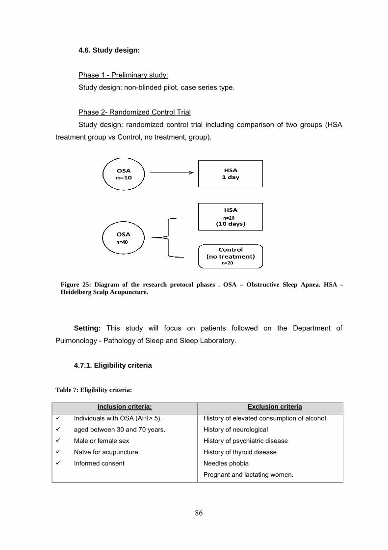
86
4.6. Study design:
Phase 1 - Preliminary study:
Study design: non-blinded pilot, case series type.
Phase 2- Randomized Control Trial
Study design: randomized control trial including comparison of two groups (HSA
treatment group vs Control, no treatment, group).
4.7 Study sample:
Setting: This study will focus on patients followed on the Department of
Pulmonology - Pathology of Sleep and Sleep Laboratory.
4.7.1. Eligibility criteria
Table 7: Eligibility criteria:
Inclusion criteria: Exclusion criteria
� Individuals with OSA (AHI> 5).
� aged between 30 and 70 years.
� Male or female sex
� Naïve for acupuncture.
� Informed consent
History of elevated consumption of alcohol
History of neurological
History of psychiatric disease
History of thyroid disease
Needles phobia
Pregnant and lactating women.
Figure 25: Diagram of the research protocol phases . OSA – Obstructive Sleep Apnea. HSA –Heidelberg Scalp Acupuncture.

87
4.9. Procedure for Data Collection:
• Phase 1 (preliminary study):
At baseline, prior to intervention, measurements included: cardio-respiratory
polygraphy (PG).
After treatment, PG was reevaluated during the sleep period of the same day of
needling.
In the morning after treatment, needles were removed and patients answered to
adapted “morning questionnaire” of Sleep Disorders Clinic, School of Sleep Medicine of
Stanford Medical Center (post-sleep study) (see attachment) that allows to check the
patient's perception of sleep (96)
Figure 26. Flow-chart of phase 1 experimental protocol.
• Phase 2:
Baseline measurements: PG; Sleep questionnaires (Sleep Disorders Questionnaire
version 1.2, Epworth scale, adapted “morning questionnaire” and the adapted
questionnaire from Treatment Credibility Scale of Borvkovec and Nau.)
During HSA, a VAS is performed to evaluate scalp point tenderness.
Needles are left in place for 10 days.
On 10th day, PG is performed on the same night during the sleep period.
On 11th day, needles are removed and patients questionnaires are repeated.
Control group: same procedures and measurements as intervention group.
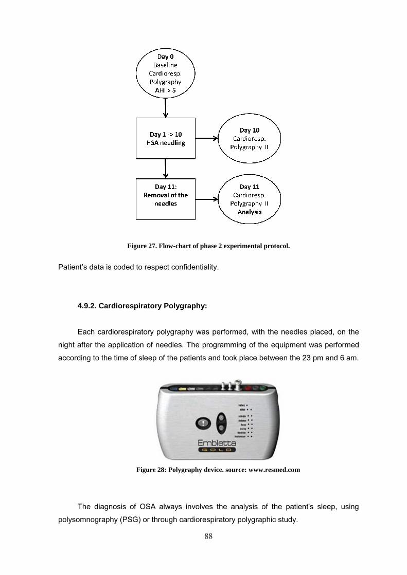
88
Figure 27. Flow-chart of phase 2 experimental protocol.
Patient’s data is coded to respect confidentiality.
4.9.2. Cardiorespiratory Polygraphy:
Each cardiorespiratory polygraphy was performed, with the needles placed, on the
night after the application of needles. The programming of the equipment was performed
according to the time of sleep of the patients and took place between the 23 pm and 6 am.
Figure 28: Polygraphy device. source: www.resmed.com
The diagnosis of OSA always involves the analysis of the patient's sleep, using
polysomnography (PSG) or through cardiorespiratory polygraphic study.
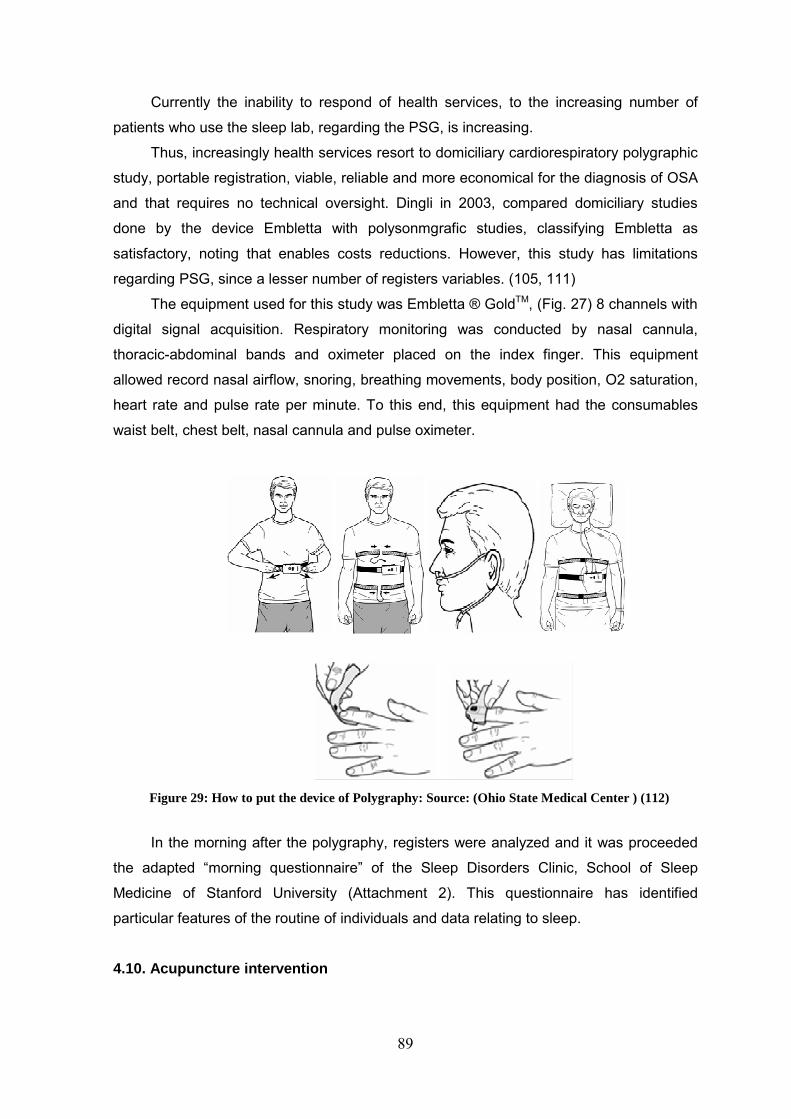
89
Currently the inability to respond of health services, to the increasing number of
patients who use the sleep lab, regarding the PSG, is increasing.
Thus, increasingly health services resort to domiciliary cardiorespiratory polygraphic
study, portable registration, viable, reliable and more economical for the diagnosis of OSA
and that requires no technical oversight. Dingli in 2003, compared domiciliary studies
done by the device Embletta with polysonmgrafic studies, classifying Embletta as
satisfactory, noting that enables costs reductions. However, this study has limitations
regarding PSG, since a lesser number of registers variables. (105, 111)
The equipment used for this study was Embletta ® GoldTM, (Fig. 27) 8 channels with
digital signal acquisition. Respiratory monitoring was conducted by nasal cannula,
thoracic-abdominal bands and oximeter placed on the index finger. This equipment
allowed record nasal airflow, snoring, breathing movements, body position, O2 saturation,
heart rate and pulse rate per minute. To this end, this equipment had the consumables
waist belt, chest belt, nasal cannula and pulse oximeter.
In the morning after the polygraphy, registers were analyzed and it was proceeded
the adapted “morning questionnaire” of the Sleep Disorders Clinic, School of Sleep
Medicine of Stanford University (Attachment 2). This questionnaire has identified
particular features of the routine of individuals and data relating to sleep.
4.10. Acupuncture intervention
Figure 29: How to put the device of Polygraphy: Source: (Ohio State Medical Center ) (112)
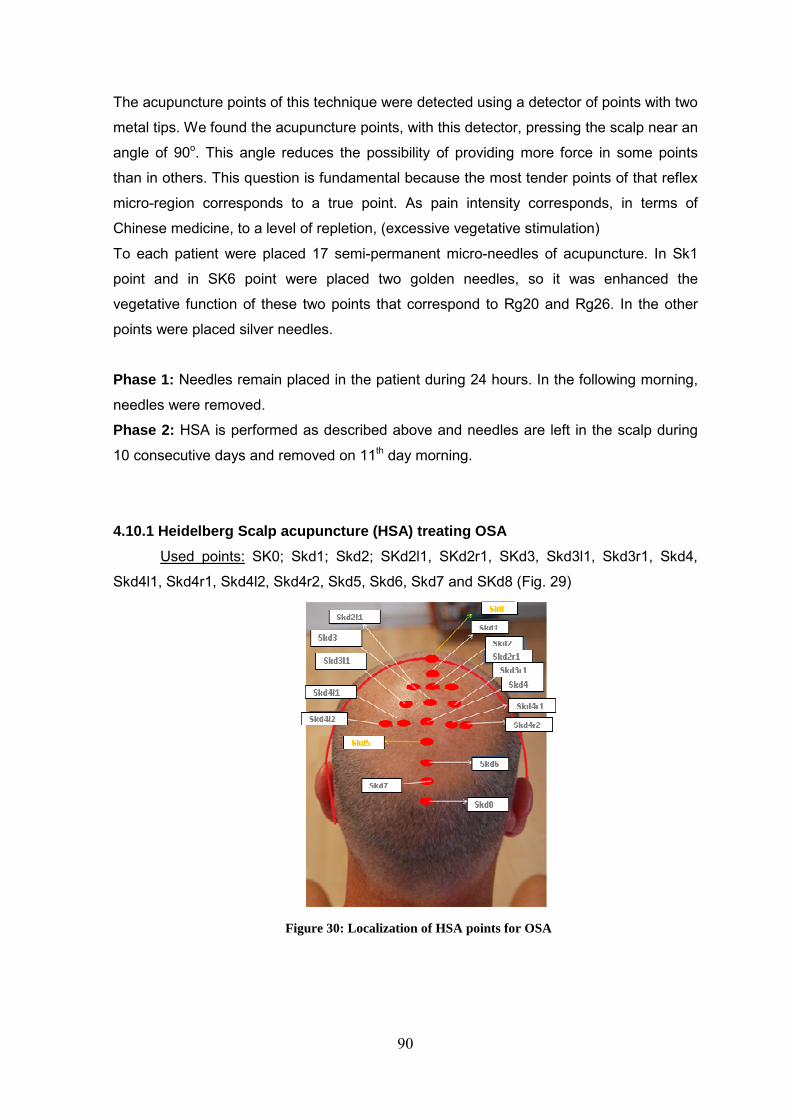
90
The acupuncture points of this technique were detected using a detector of points with two
metal tips. We found the acupuncture points, with this detector, pressing the scalp near an
angle of 90o. This angle reduces the possibility of providing more force in some points
than in others. This question is fundamental because the most tender points of that reflex
micro-region corresponds to a true point. As pain intensity corresponds, in terms of
Chinese medicine, to a level of repletion, (excessive vegetative stimulation)
To each patient were placed 17 semi-permanent micro-needles of acupuncture. In Sk1
point and in SK6 point were placed two golden needles, so it was enhanced the
vegetative function of these two points that correspond to Rg20 and Rg26. In the other
points were placed silver needles.
Phase 1: Needles remain placed in the patient during 24 hours. In the following morning,
needles were removed.
Phase 2: HSA is performed as described above and needles are left in the scalp during
10 consecutive days and removed on 11th day morning.
4.10.1 Heidelberg Scalp acupuncture (HSA) treating OSA
Used points: SK0; Skd1; Skd2; SKd2l1, SKd2r1, SKd3, Skd3l1, Skd3r1, Skd4,
Skd4l1, Skd4r1, Skd4l2, Skd4r2, Skd5, Skd6, Skd7 and SKd8 (Fig. 29)
Figure 30: Localization of HSA points for OSA

91
4.11. Statistical analyses
Data was analyzed using XL-Statistics, version 5.0., and Microsoft Excel
2010. Tests used were Spearman correlation for association of continuous
variables and Kruskal-Wallis test to compare continuous variables (Polygraphy1-
Polygraphy 2)
4.11. Ethical consideration s:
The research protocol followed the guidelines of the declaration of Helsinki of the
World Medical Association. All general ethical principles were respected according to international standards.
The participation of individuals in the study was voluntary and did not constitute any
health risk. Phase 1 lasted 3 weeks, offering a therapeutic technique (Heidelberg Scalp
Therapy) with low clinical risk, and with therapeutic potential.
All participants were informed about the purpose of the study, objectives and
procedures, acupuncture technique, side effects and possible adverse effects of
acupuncture. A written informed consent was obtained. (Attachment 5)
Data collection respects confidentially and is only used by the principal investigators
of the study. Patients do not cease their conventional medical treatment.
Prior to phase 1, approval by the Ethics Committee of the S. João Hospital Center
(SJHC) was obtained (Attachment 1).
4.12. Study finances:
The study subjects were invited to participate at no cost.
The material of acupuncture was supported by the main investigator. The
cardiorespiratory polygraphy equipment was supplied by ResMed. The study had no
significant additional cost to the patients’ usual medical approach in SJHC.
4.13. Conflicts of interest :
None of the elements of the research team are involved in activities that can
represent conflicts of interest.

92

93
CHAPTER 3

94

95
5. RESULTS

96
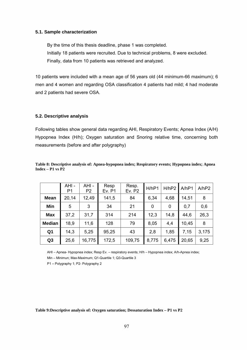
97
5.1. Sample characterization
By the time of this thesis deadline, phase 1 was completed.
Initially 18 patients were recruited. Due to technical problems, 8 were excluded.
Finally, data from 10 patients was retrieved and analyzed.
10 patients were included with a mean age of 56 years old (44 minimum-66 maximum); 6
men and 4 women and regarding OSA classification 4 patients had mild; 4 had moderate
and 2 patients had severe OSA.
5.2. Descriptive analysis
Following tables show general data regarding AHI, Respiratory Events; Apnea Index (A/H)
Hypopnea Index (H/h); Oxygen saturation and Snoring relative time, concerning both
measurements (before and after polygraphy)
Table 8: Descriptive analysis of: Apnea-hypopnea index; Respiratory events; Hypopnea index; Apnea Index – P1 vs P2
AHI -P1
AHI - P2
Resp Ev. P1
Resp. Ev. P2 H/hP1 H/hP2 A/hP1 A/hP2
Mean 20,14 12,49 141,5 84 6,34 4,68 14,51 8
Min 5 3 34 21 0 0 0,7 0,6
Max 37,2 31,7 314 214 12,3 14,8 44,6 26,3
Median 18,9 11,6 128 79 8,05 4,4 10,45 8
Q1 14,3 5,25 95,25 43 2,8 1,85 7,15 3,175
Q3 25,6 16,775 172,5 109,75 8,775 6,475 20,65 9,25
AHI – Apnea- Hypopnea index; Resp Ev. – respiratory events; H/h – Hypopnea index; A/h-Apnea index;
Min – Minimun; Max-Maximum; Q1-Quartile 1; Q3-Quartile 3
P1 – Polygraphy 1; P2- Polygraphy 2
Table 9:Descriptive analysis of: Oxygen saturation; Dessaturation Index – P1 vs P2
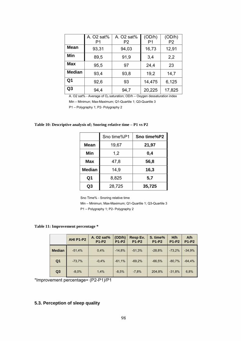
98
A. O2 sat%
P1 A. O2 sat%
P2 (OD/h)
P1 (OD/h)
P2 Mean 93,31 94,03 16,73 12,91 Min 89,5 91,9 3,4 2,2 Max 95,5 97 24,4 23 Median 93,4 93,8 19,2 14,7 Q1 92,6 93 14,475 6,125 Q3 94,4 94,7 20,225 17,825
A. O2 sat% - Average of O2 saturation; OD/h – Oxygen dessaturation index
Min – Minimun; Max-Maximum; Q1-Quartile 1; Q3-Quartile 3
P1 – Polygraphy 1; P2- Polygraphy 2
Table 10: Descriptive analysis of; Snoring relative time – P1 vs P2
Sno time%P1 Sno time%P2
Mean 19,67 21,97
Min 1,2 0,4
Max 47,8 56,8
Median 14,9 16,3
Q1 8,825 5,7
Q3 28,725 35,725
Sno Time% - Snoring relative time
Min – Minimun; Max-Maximum; Q1-Quartile 1; Q3-Quartile 3
P1 – Polygraphy 1; P2- Polygraphy 2
Table 11: Improvement percentage *
AHI P1-P2 A. O2 sat% P1-P2
(OD/h) P1-P2
Resp Ev. P1-P2
S. time% P1-P2
H/h P1-P2
A/h P1-P2
Median -51,4% 0,4% -14,8% -51,3% -28,8% -73,2% -34,9%
Q1 -73,7% -0,4% -61,1% -69,2% -66,5% -80,7% -64,4%
Q3 -8,0% 1,4% -8,5% -7,8% 204,8% -31,8% 6,8%
*Improvement percentage= (P2-P1)/P1
5.3. Perception of sleep quality

99
Table 12: Resume of Morning Questionnaire of Sleep Disorders
MORNING QUESTIONNAIRE
Patients
Evalution parameter 1 2 3 4 5 6 7 8 9 10
Time to sleep (min) 5 5 30 5 10 10 15 5 10 10
Classification of the time to sleep* AU MLU AU MLU AU AU AU MLU AU AU
Awake up times 1 2 5 3 2 4 3 3 2 1
Hours of sleep/night 9 9 7 6 7 7 8 7 7 8
Feeling on the morning A R F A A R R A R R
Quality of sleep classification AU AU BU AU AU AU AU BU BU AU MLU – Much lesser than usual; MBU - much better than usual; SU - Shorter than usual; MLU - Much longer
than usual; LU- Longer than usual; AU – As Usual; BU- Better than usual; F- Fatigue; A-Alert; R- Rested;
5.4. Inferential analyses – study outcomes Before (polygraphy 1, P1) and after (polygraphy 2, P2) intervention with HSA.
• Polygraphy Parameters
We present data from: AHI; respiratory events; apnea index; hypopnea index; O2
saturation and snoring relative time.
5.4.1 Apnea Hypopnea Index:
Table 13: Apnea Hypopnea Index – P1 vs P2 Patient AHI -P1 AHI - P2 Improv. (%)
1 20,8 4,9 -76,4%
2 14,3 19,8 38,5%
3 37,2 9,3 -75,0%
4 33,9 31,7 -6,5%
5 5 13,9 178,0%
6 14,3 3,7 -74,1%
7 17 14,9 -12,4%
8 22,9 6,3 -72,5%
9 9,5 3 -68,4%
10 26,5 17,4 -34,3%
AHI – Apnea – Hypopnea Index P1 – Polygraphy 1 P2 – Polygraphy 2 Improv. (%) – improvement percentage: (P2-P1)/P1
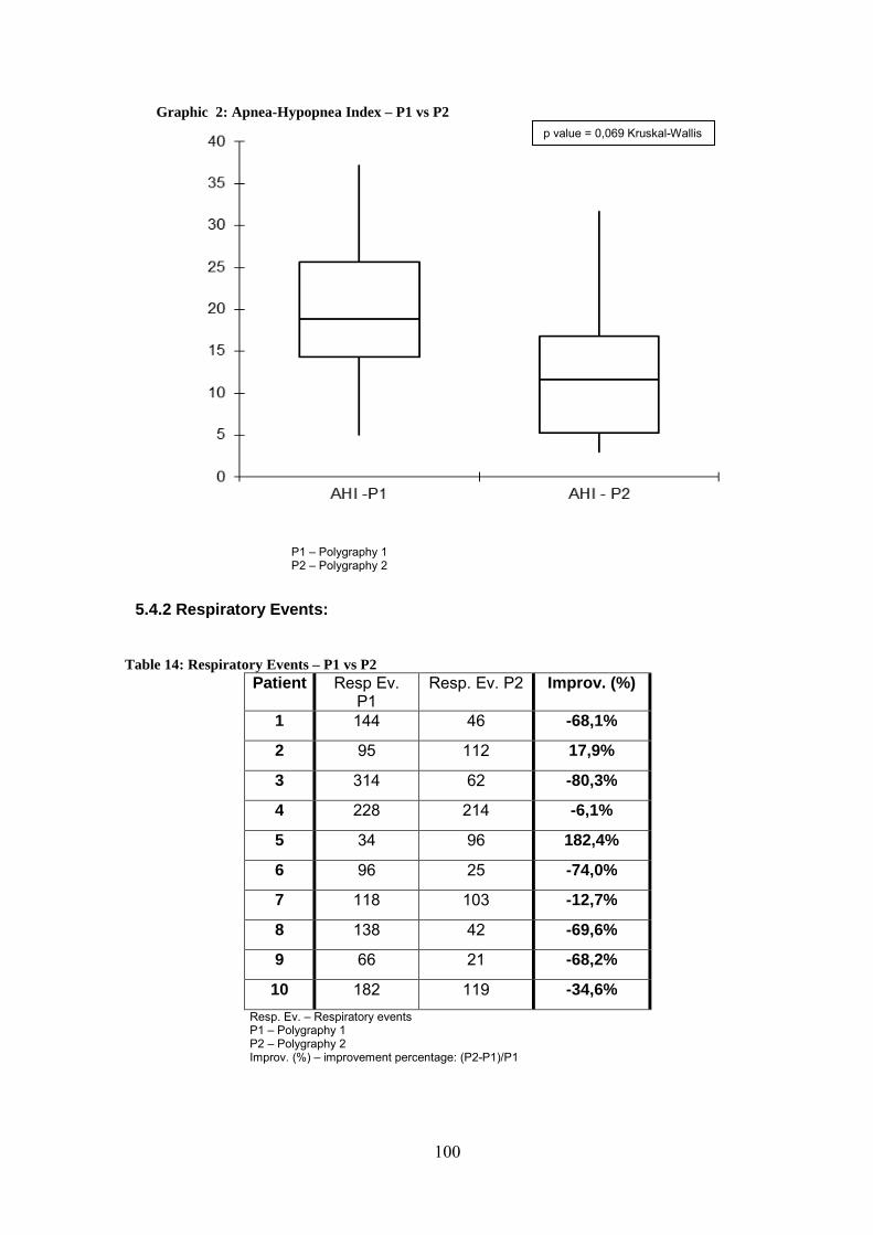
100
Graphic 2: Apnea-Hypopnea Index – P1 vs P2
P1 – Polygraphy 1 P2 – Polygraphy 2
5.4.2 Respiratory Events:
Table 14: Respiratory Events – P1 vs P2 Patient Resp Ev.
P1 Resp. Ev. P2 Improv. (%)
1 144 46 -68,1%
2 95 112 17,9%
3 314 62 -80,3%
4 228 214 -6,1%
5 34 96 182,4%
6 96 25 -74,0%
7 118 103 -12,7%
8 138 42 -69,6%
9 66 21 -68,2%
10 182 119 -34,6%
Resp. Ev. – Respiratory events P1 – Polygraphy 1 P2 – Polygraphy 2 Improv. (%) – improvement percentage: (P2-P1)/P1
p value = 0,069 Kruskal-Wallis
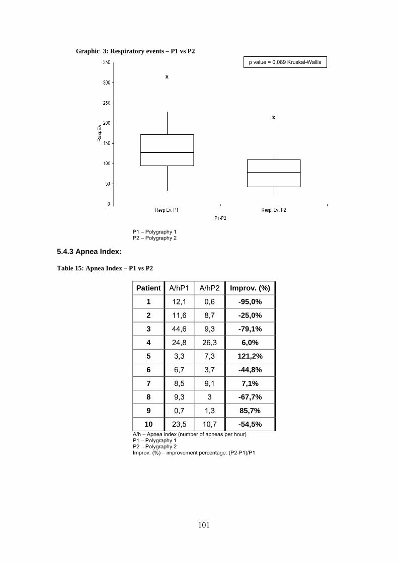
101
Graphic 3: Respiratory events – P1 vs P2
P1 – Polygraphy 1 P2 – Polygraphy 2
5.4.3 Apnea Index:
Table 15: Apnea Index – P1 vs P2
Patient A/hP1 A/hP2 Improv. (%)
1 12,1 0,6 -95,0%
2 11,6 8,7 -25,0%
3 44,6 9,3 -79,1%
4 24,8 26,3 6,0%
5 3,3 7,3 121,2%
6 6,7 3,7 -44,8%
7 8,5 9,1 7,1%
8 9,3 3 -67,7%
9 0,7 1,3 85,7%
10 23,5 10,7 -54,5% A/h – Apnea index (number of apneas per hour)
P1 – Polygraphy 1 P2 – Polygraphy 2 Improv. (%) – improvement percentage: (P2-P1)/P1
p value = 0,089 Kruskal-Wallis
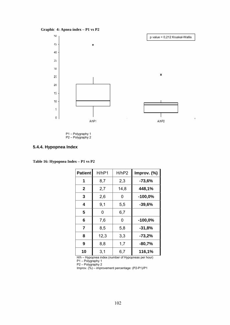
102
Graphic 4: Apnea índex – P1 vs P2
P1 – Polygraphy 1 P2 – Polygraphy 2
5.4.4. Hypopnea Index
Table 16: Hypopnea Index – P1 vs P2
Patient H/hP1 H/hP2 Improv. (%)
1 8,7 2,3 -73,6%
2 2,7 14,8 448,1%
3 2,6 0 -100,0%
4 9,1 5,5 -39,6%
5 0 6,7
6 7,6 0 -100,0%
7 8,5 5,8 -31,8%
8 12,3 3,3 -73,2%
9 8,8 1,7 -80,7%
10 3,1 6,7 116,1% H/h – Hypopnea index (number of Hypopneas per hour) P1 – Polygraphy 1 P2 – Polygraphy 2 Improv. (%) – improvement percentage: (P2-P1)/P1
p value = 0,212 Kruskal-Wallis
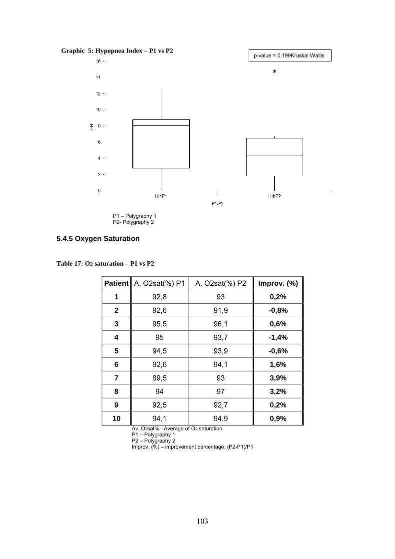
103
Graphic 5: Hypopnea Index – P1 vs P2
P1 – Polygraphy 1 P2- Polygraphy 2
5.4.5 Oxygen Saturation
Table 17: O2 saturation – P1 vs P2
Patient A. O2sat(%) P1 A. O2sat(%) P2 Improv. (%)
1 92,8 93 0,2%
2 92,6 91,9 -0,8%
3 95,5 96,1 0,6%
4 95 93,7 -1,4%
5 94,5 93,9 -0,6%
6 92,6 94,1 1,6%
7 89,5 93 3,9%
8 94 97 3,2%
9 92,5 92,7 0,2%
10 94,1 94,9 0,9% Av. O2sat% - Average of O2 saturation P1 – Polygraphy 1 P2 – Polygraphy 2 Improv. (%) – improvement percentage: (P2-P1)/P1
p-value = 0,199Kruskal-Wallis
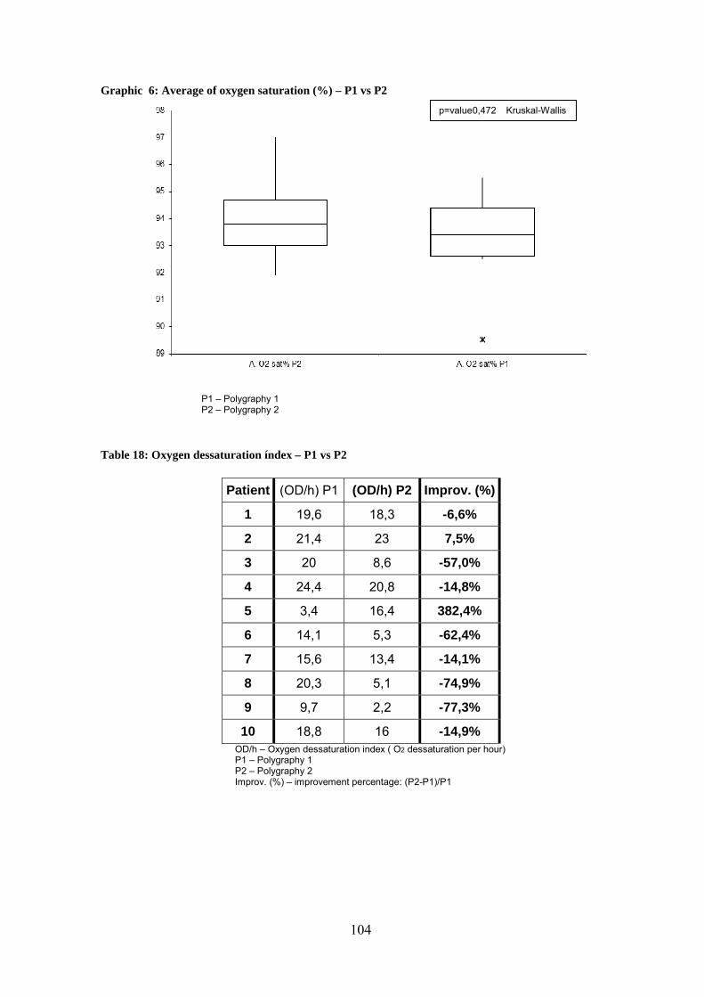
104
Graphic 6: Average of oxygen saturation (%) – P1 vs P2
P1 – Polygraphy 1 P2 – Polygraphy 2
Table 18: Oxygen dessaturation índex – P1 vs P2
Patient (OD/h) P1 (OD/h) P2 Improv. (%)
1 19,6 18,3 -6,6%
2 21,4 23 7,5%
3 20 8,6 -57,0%
4 24,4 20,8 -14,8%
5 3,4 16,4 382,4%
6 14,1 5,3 -62,4%
7 15,6 13,4 -14,1%
8 20,3 5,1 -74,9%
9 9,7 2,2 -77,3%
10 18,8 16 -14,9% OD/h – Oxygen dessaturation index ( O2 dessaturation per hour) P1 – Polygraphy 1 P2 – Polygraphy 2 Improv. (%) – improvement percentage: (P2-P1)/P1
p=value 0,472 Kruskal-Wallis

105
P1 – Polygraphy 1 P2 – Polygraphy 2
5.4.6 Snoring Relative Time:
Table 19: Snoring relative time – P1vs P2
Patient S.time%P1 S. time%P2 Improv. (%)
1 24,6 16,3 -33,7%
2 7,9 51,8 555,7%
3 11,6 2 -82,8%
4 2,7 8,4 211,1%
5 15,8 56,8 259,5%
6 47,8 16,3 -65,9%
7 14 40 185,7%
8 41 4,8 -88,3%
9 1,2 0,4 -66,7%
10 30,1 22,9 -23,9% S.time% - Snoring relative time (%) P1 – Polygraphy 1 P2 – Polygraphy 2 Improv. (%) – improvement percentage: (P2-P1)/P1
p-value = 0,226 Kruskal-Wallis
Graphic 7: Oxygen dessaturation

106
Graphic 8: Relative snoring time (%) – P1 vs P2
P1 – Polygraphy 1 P2 – Polygraphy 2
• Perception of sleep quality
5.5 Sample size estimation:
Based on the above data on the AHI, we estimated the sample size needed to achieve
statistical significance.
If the total of 66 patients would enter a two treatment parallel-design study the probability
is 80% that the study will detect a treatment difference at a two-sided 0.05 significance
level. This is based on the assumption that the standard deviation of the AHI is 10.9
Graphic 9: Quality of sleep (n=10)
Graphic 10: Feeling in the morning (n=10)
p value = 0,88 Kruskal-Wallis

107
CHAPTER4

108
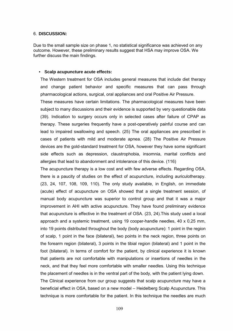
109
6. DISCUSSION: Due to the small sample size on phase 1, no statistical significance was achieved on any outcome. However, these preliminary results suggest that HSA may improve OSA. We further discuss the main findings.
• Scalp acupuncture acute effects:
The Western treatment for OSA includes general measures that include diet therapy
and change patient behavior and specific measures that can pass through
pharmacological actions, surgical, oral appliances and oral Positive Air Pressure.
These measures have certain limitations. The pharmacological measures have been
subject to many discussions and their evidence is supported by very questionable data
(39). Indication to surgery occurs only in selected cases after failure of CPAP as
therapy. These surgeries frequently have a post-operatively painful course and can
lead to impaired swallowing and speech. (25) The oral appliances are prescribed in
cases of patients with mild and moderate apnea. (28) The Positive Air Pressure
devices are the gold-standard treatment for OSA, however they have some significant
side effects such as depression, claustrophobia, insomnia, marital conflicts and
allergies that lead to abandonment and intolerance of this device. (116)
The acupuncture therapy is a low cost and with few adverse effects. Regarding OSA,
there is a paucity of studies on the effect of acupuncture, including auriculotherapy.
(23, 24, 107, 108, 109, 110). The only study available, in English, on immediate
(acute) effect of acupuncture on OSA showed that a single treatment session, of
manual body acupuncture was superior to control group and that it was a major
improvement in AHI with active acupuncture. They have found preliminary evidence
that acupuncture is effective in the treatment of OSA. (23, 24).This study used a local
approach and a systemic treatment, using 19 cooper-handle needles, 40 x 0,25 mm,
into 19 points distributed throughout the body (body acupuncture): 1 point in the region
of scalp, 1 point in the face (bilateral), two points in the neck region, three points on
the forearm region (bilateral), 3 points in the tibial region (bilateral) and 1 point in the
foot (bilateral). In terms of comfort for the patient, by clinical experience it is known
that patients are not comfortable with manipulations or insertions of needles in the
neck, and that they feel more comfortable with smaller needles. Using this technique
the placement of needles is in the ventral part of the body, with the patient lying down.
The Clinical experience from our group suggests that scalp acupuncture may have a
beneficial effect in OSA, based on a new model – Heidelberg Scalp Acupuncture. This
technique is more comfortable for the patient. In this technique the needles are much

110
smaller (micro-needles with 3mm) placed with the patient in the sitting position, with
clothes on, and without the patient notice the needles, which reduces the normal
apprehension of the puncture); it is less invasive that previous acupuncture, and so it
is more tolerable by the patients. In addition, the stimuli may be permanent, since the
needles are permanent in the selected scalp points. Therefore, we decided to conduct
a research study on the effect of scalp acupuncture in OSA in order to investigate
these field observations.
Interestingly, no previous studies on Scalp Acupuncture on Obstructive Sleep Apnea
are available in the literature.
The Heidelberg Scalp Acupuncture can present itself as an alternative or
complementary therapy for the treatment of OSA. This preliminary study, with 10
clinical cases, showed that 8 out 10 patients, with individual treatment, responded to
the therapy approach, which is demonstrated by an improvement of: 51.4% (p value =
0,069) in AHI; 51.3% (p value = 0,089) in the number of respiratory events; 14,8% (p
value = 0,226) in Oxygen Dessaturation Index and 28.8% (p value = 0,88) in Snoring
relative time. In terms of perception of sleep, no patient reported that sleep quality had
worsened; 3 of 10 patients indicated that sleep quality was better than in the regular
days. 7 patients classified the quality of sleep as the same as usual. Regarding the
feeling in the morning, 4 patients reported that they felt alert; 5 reported that they felt
rested and only one answered that he felt fatigue. All patients did not report any kind
of discomfort or complaints regarding the permanence of the needles.
In our perspective, these preliminary data supports the need for further research. A
prospective randomized control trial, in a double blinded assay (“Heidelberg assay”)
method, is currently in course.
If our research shows improvement of OSA by HSA, we may speculate of
positive effects on the other OSA associated diseases and sub-health problems,
such as cardiovascular diseases, hypertension, cerebrovascular complications,
and psychosocial consequences.
• Neurophysiological basis for HSA – a theoretical proposal
The four patients reported that the most painful points pointing to greater pathology,
were SKd5, SKd6, SKd7 SKd8 and which project the hypoglossal triangle, vagal triangle,
cranial nerve and cervical accessory nerve, respectively.
By doing line projections between points of HSA to treat OSA and the structures to
which each point corresponds we found relevant intersections on the thalamus and brain
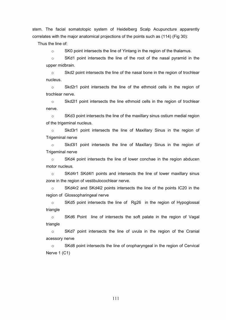
111
stem. The facial somatotopic system of Heidelberg Scalp Acupuncture apparently
correlates with the major anatomical projections of the points such as (114) (Fig 30):
Thus the line of:
o SK0 point intersects the line of Yintang in the region of the thalamus.
o SKd1 point intersects the line of the root of the nasal pyramid in the
upper midbrain.
o Skd2 point intersects the line of the nasal bone in the region of trochlear
nucleus.
o Skd2r1 point intersects the line of the ethmoid cells in the region of
trochlear nerve.
o Skd2l1 point intersects the line ethmoid cells in the region of trochlear
nerve.
o SKd3 point intersects the line of the maxillary sinus ostium medial region
of the trigeminal nucleus.
o Skd3r1 point intersects the line of Maxillary Sinus in the region of
Trigeminal nerve
o Skd3l1 point intersects the line of Maxillary Sinus in the region of
Trigeminal nerve
o SKd4 point intersects the line of lower conchae in the region abducen
motor nucleus.
o SKd4r1 SKd4l1 points and intersects the line of lower maxillary sinus
zone in the region of vestibulocochlear nerve.
o SKd4r2 and SKd4l2 points intersects the line of the points IC20 in the
region of Glossopharingeal nerve
o SKd5 point intersects the line of Rg26 in the region of Hypoglossal
triangle
o SKd6 Point line of intersects the soft palate in the region of Vagal
triangle
o SKd7 point intersects the line of uvula in the region of the Cranial
acessory nerve o SKd8 point intersects the line of oropharyngeal in the region of Cervical
Nerve 1 (C1)
112
Figure 31 Scheme of the intersection points of facial somatotopy of HSA. Adapted from www.michigan-headache-tmj-doctor.com
Figure 32: Intersection points, inferior view, adapted from Patrick J. Lynch (118)

113
In terms of innervation of the scalp, there seems to be an involvement of Supratrochlear
Nerve branches, the Great Occipital Nerve branches and branches of the Occipital Third
Nerve. (Fig.32)
In terms of vascularization was found involvement of the anastomotic net formed by the
Superficial Temporal arteries and veins, and of the occipital arteries and veins. (Fig 30) Thus, this type of projections suggests a kind of "Front-Mu" relationship with the
brainstem and with the cranial nerves related to it. (Fig. 32)
The brainstem has three main functions: serves as conduit for the ascending and
descending tracts connecting the spinal cord to the different parts of the higher centers in
the forebrain; it contains important reflex centers associated with control of respiration and
the cardiovascular system and with the control of consciousness; contains important
nuclei of cranial nerves III throught XII. (85)
The Somatic efferent cranial nerves III, IV, VI and XII have origin in cells (derived
from the basal plates) located in the somatic efferent column. Their axons are distributed
to muscles derived from head myotomes. The nerves of pharyngeal arches V, VII, IX and
X supply the embryonic pharyngeal arches. (85)
These nerves have significant importance in the innervation of the upper airways
and underlines a possible relationship to scalp stimulation by HSA. (Fig. 31)
Figure 33: Innervation and vascularization structures possible involved in HSA
114
These relationships can also verify the fractal theory applied to human physiology
and ontogeny, so, it is suggest a study to investigate and verified these embryonic and
neurophysiological relationships.
7. Limitations:
This study is the preliminary step of a research project that aims to verify the effects
of Heidelberg Scalp Acupuncture on Obstructive Sleep Apnea. In this preliminary study
we only recruit ten patients. In this phase there are obvious limitations such as the number
of patients and lack of control group. However, data suggested a positive tendency of
beneficial effects secondary to scalp needling, according to our model, supporting further
research. A randomized controlled trial is currently in course.

115
CHAPTER 5

116
117
7. Future Perspectives:
Scalp acupuncture has demonstrated beneficial effects in several diseases mostly
dependent on neurological pathology (e.g. stroke, Parkinson disease, psychiatric
diseases, speech disturbances, etc).
It is a field on Microsystems’s acupuncture with a high research potential, because
anatomically may be correlated with various structures, plus it is a simple technique to
apply, painless, and with reduced financial costs.
It is therefore necessary further investigation to have some enlightment in:
• The biological mechanisms of this technique (that can involve various areas
such as neurophysiology, embryology, endocrinology and ontogeny), particularly
in the OSA physiopathology.
• The magnitude of the therapeutical effect in different degrees of OSA (mild,
moderate and severe) and possible side effects
• The cost-benefit of the integration of this technique in the treatment approach
of OSA in order to complement conventional treatment.
• The type of permanent needles to use in OSA. Commercially available
implantable needles may be developed to be a long term complement to
CPAP treatment.
• The optimal time of permanent needling on the scalp on OSA.
118
8. Conclusion:
Despite our preliminary evidence, the Heidelberg Scalp Acupuncture (HSA) seems
to have some therapeutically benefits on OSA. Our study suggests that HSA may improve
sleep apnea as measured by polygraphy.
This technique may implicate several neurophysiological, embryological and
ontogenetic factors. More research is needed to assess the long-term effect of HAS on
OSA and to explore the mechanism of action of this treatment technique.
Further research in HSA on OSA is needed to evaluate long-term effects of scalp
acupuncture. This future research may also enlighten HSA effects on comorbidities that
benefit from OSA improvement such as: HTN, HF, cerebrovascular disease.
At the end, an integrative approach to OSA, joining conventional to complementary
therapies, may be established with a superior therapeutic benefit to the patient and
possibility avoiding higher costs and medical side effects.
119
References:
1. Ventegodt Søren. Clinical Holistic Medicine: Use and Limitations of the Biomedical
Paradigm. TheScientificWorldJOURNAL 2004; 4: 295–306
2. Graziose Rocky, et al. Merging Traditional Chinese Medicine with Modern Drug
Discovery Technologies to Find Novel Drugs and Functional Foods. Curr Drug
Discov Technol. 2010 March 1; 7(1): 2–12
3. Sommers Virend K., et al. Sleep Apnea and Cardiovascular Disease. Circulation.
2008; 118: 1080-1111
4. Wolk Robert, Shamsuzzaman Abu S.M. and Sommers Virend K.. Obesity, Sleep
Apnea, and Hypertension. Hypertension. 2003; 42:1067-1074
5. Ferreira Luciano Drager, et al. Síndrome da Apnéia Obstrutiva do Sono e sua
Relação com a Hipertensão Arterial Sistêmica. Evidências Atuais. Arq Bras
Cardiol, volume 78 (nº 5), 531-6, 2002
6. Lee Won, et al. Epidemiology of Obstructive Sleep Apnea: a Population-based
Perspective. Expert Rev Respir Med. 2008 June 1; 2(3): 349–364.
7. Domingues Nuno Manuel. Sindrome da Apneia Obstrutiva do Sono. Mestrado
integrado em medicina. Universidade da Beira Interior. 2009
8. Fogel Robert B., et al. Anatomic and Physiologic Predictors of Apnea Severity in
Morbidly Obese Subjects. SLEEP 2003;2:150-155
9. Moreira, Maria Margarida. Aderência a sistemas de pressão positiva na Síndrome
da Apneia Obstrutiva do Sono. Mestrado integrado em medicina. Faculdade de
Medicina da Universidade do Porto. 2010
10. Remsburg Stacia. Patients with obstructive sleep apnea have an abnormal
peripheral vascular response to hypoxia. J Appl Physiol 87:1148-1153, 1999
11. Vgontzas A. N. et al. Metabolic disturbances in obesity versus sleep apnoea: the
importance of visceral obesity and insulin resistance. Journal of Internal Medicine
2003; 254: 32–44
12. Drummond, Marta F. Comportamento de Mediadores Inflamatórios e Hormonais
na Síndrome de Apneia Obstrutiva do Sono - Resposta ao APAP. 2009
13. Freire, Anaflávia de Oliveira. Tratamento da síndrome de apneia hipopnéia
Obstrutiva do sono pela acupunctura. Tese de Doutoramento em Ciências.
Universidade Federal de S. Paulo, Escola Paulista de Medicina. 2004
14. Durán, Joaquin et al. Obstructive Sleep Apnea–Hypopnea and Related Clinical
Features in a Population-based Sample of Subjects Aged 30 to 70 Years. Am J
Respir Crit Care Med Vol 163. pp 685–689, 2001

120
15. Silverberg, Donald S., et al. Treating obstructive sleep apnea improves essential
hypertension and quality of life. Am Fam Physician 2002;65:229-36.
16. Jennum, P. and Riha R.L.. Epidemiology of sleep apnoea/hypopnoea syndrome
and sleep-disordered breathing. Eur Respir J 2009; 33: 907–914
17. Proposta de programa para uma intervenção sistematizada no síndrome de
apneia do sono no âmbito plano nacional de saúde 2011-2016. www.pns.dgs.pt
18. Carlson Bruce M.. Human Embryology and Developmental Biology 3rd Edition
2004
19. Moore Keith L.., T.V.N. The Developing Human- Clinically Oriented Embriology.
8th edition, 2008
20. Bogart Bruce Ian, Ort Victoria H.. Anatomia e Embriologia – Serie Elsevier de
Formação integrada. Mosby. 2007
21. Mittal RK. Motor Function of the Pharynx, Esophagus, and its Sphincters. Morgan
& Claypool Life Sciences. 2011
22. www.mc.vanderbilt.edu/HN9Pharynx03.doc
23. Sugai, Gisele C. M. Efeito Imediato e mediato da acupunctura no tratamento de
pacientes portadores com Síndrome da Apnea Obstrutiva do Sono. São Paulo.
2009. (Tese apresentada à Universidade Federal De S. Paulo-Escola Paulista de
Medicina para obtenção do título de Doutor em Ciências)
24. Freire, A. O.; et al. Immediate effect of acupuncture on the sleep pattern of
patients with obstrutive sleep apnoea. Acupunct Med 2010; 28:115-119.
25. Lawrence J. Epstein, et al. Clinical Guideline for the Evaluation,Managment and
Long-term Care of Obstrutive Sleep Apnea in Adults. Journal of Clinical Sleep
Medicine, Vol.5, Nº3,2009
26. Candela A., et al. Validation of Respiratory Polygraphy System in the Diagnosis of
sleep apnea syndrome. Arch Bronconeumol. 2005;41:71-7
27. Golp R., et al. Home Sleep Studies in the assessment of sleep hypopnea
syndrome. Chest 2002 Oct;122(4):1156-61
28. Pereira, Adão. Síndrome da Apneia Obstrutiva do Sono Fisiopatologia,
Epidemiologia, Consequências, Diagnóstico e Tratamento. ISSN 0871-3413
ArquiMed, 2007
29. Durán, Joaquin, et al. Obstructive Sleep Apnea–Hypopnea and RelatedClinical
Features in a Population-based Sample of Subjects Aged 30 to 70 Yr. Am J Respir
Crit Care Med Vol 163. pp 685–689, 2001
30. Redline, Susan; et al. Obstructive Sleep Apnea–Hypopnea and Incident Stroke -
The Sleep Heart Health Study. Am J Respir Crit Care Med Vol 182. pp 269–277,
2010
121
31. Charles F. P. George. Sleep Apnea, Alertness, and Motor Vehicle Crashes.Am J
Respir Crit Care Med Vol 176. pp 954–956, 2007
32. Sharafkhaneh Amir; et al. Association of Psychiatric Disorders and Sleep Apnea in
a Large Cohort. SLEEP, Vol. 28, No. 11, 2005
33. Fauci, A.; Braunwald, E.; Kasper, D. L.; Hauser, S. L.; Long, D. L.; Jameson, J. L.;
Loscalzo J. Harrison’s Manual of Medicine, 17th edition. Mc Graw Hill. 2009.
34. Eckert, D. J., et al. Pathophysiology of Adult Obstrutive Sleep Apnea. Proc Am
Thorac Soc Vol 5. pp 144–153, 2008
35. Ho, Matthew L.; Brass Steven D.. Obstrutive Sleep Apnea. Neurology International
2011; 3:e15
36. P.C. Deegan, W.T. McNicholas. Pathophysiology of obstructive sleep apnoea. Eur
Respir J, 1995, 8, 1161–1178
37. Peppard P, et al. Longitudinal study of moderate weight change and sleep-
disordered breathing. JAMA 2000;284(23):3015-21.
38. Veasey, Sigrid C.; et al. Medical Therapy for Obstrutive Sleep Apnea: A Review by
medical Therapy for Obstrutive Sleep Apnea Task Force of the Standards of
Practice Commitee of the American Academy of Sleep Medicine. SLEEP
2006;29(8):1036-1044.
39. Kohler, M; et al. Pharmacological approaches to the treatment of obstructive sleep
apnoea. Expert Opinion on Investigational Drugs, 18(5):647-656.
40. Kessler P, et al. Surgical management of obstructive sleep apnea. Mund Kiefer
Gesichtschir. 2007 Apr;11(2):81-8.
41. Health care Guideline: Diagnosis and treatment of Obstrutive Sleep Apnea in
adults. 6th edition. Institute for Clinical Systems Improvement. 2008
42. Won, Christine H. J.; et al. Surgical Treatment of Obstructive Sleep Apnea. Upper
Airway and Maxillomandibular Surgery. Proc Am Thorac Soc Vol 5. pp 193–199,
2008
43. Pinto, Bárbara C. P. Síndroma de Apneia Obstrutiva do Sono Diagnóstico e
terapêutica. Monografia VitalAire®; 2005.
44. Flemons, W. Obstrutive Sleep Apnea. New England Journal of Medicine
2002;347(7):498-504.
45. Ephros, H. D.;et al. Surgical treatment of snoring & obstructive sleep apnoea.
Indian J Med Res 131, February 2010, pp 267-276
46. Ferguson KA; Cartwright R; Rogers R et al. Oral Appliances for Snoring and
Obstructive Sleep Apnea: A Review. SLEEP 2006;29(2):244-262.
47. Gay P; Weaver T; Loube D et al. Evaluation of positive airway
pressure treatment for sleep related breathing disorders in adults. SLEEP

122
2006;29(3):381-401.
48. Continuous Positive Airway Pressure for treatment of Obstrutive Sleep Apnoea/
Hipopnoea syndrome. NICE technology appraisal guidance 139. National Institute
for Health and Clinical Excellence. London. 2008
49. Haentjens P.; et al. The Impact of Continuous Positive Airway Pressure on Blood
Pressure in Patients With Obstructive Sleep Apnea Syndrome. Evidence From a
Meta-analysis of Placebo-Controlled Randomized Trials. Arch Intern Med.
2007;167:757-765
50. Hormann, C. et al. Bipahsic positive airway pressure (BiPAP) – a new mode of
ventilator support. Eur J. Anaesthesiol. 1994 Jan;11(1):37-42.
51. Matos Luis Carlos. Mensurabilidade dos efeitos da prática de qigong por
termografia de infravermelhos e medição da diferença de potencial eléctrico.
Dissertação de mestrado em Medicina Tradicional Chinesa. Instituto de Ciências
Biomédicas de Abel Salazar da Universidade do Porto. 2011
52. Birch Stephen J., Felt Robert L.. Entendendo a Acupunctura. Roca, 2009
53. W.H.O. Benchmarks for training in traditional / complementary and alternative
medicine. Benchmarks for Training in Traditional Chinese Medicine. 2010
54. Weidong Wang and Yan Ma. Systematic Psychotherapy of TCM (SPT):
Concepts, Theories and Techniques. Paper conference, Shen- Interculturality and
Psycotherapy. Sigmund Freud Privat Universität. Wien 2010
55. Kaptcuk Ted J.. The Web that has no Weaver. Understanding Chinese Medicine.
2nd edition. Contemporary Books. McGraw-Hill.2000
56. Kuang HaiXue. Recent advances in Theories and practice Of chinese
medicine.www.InTechopen.com. 2011
57. Clavey Steven. Fisiologia e Patologia dos Fluídos na Medicina Tradicional
Chinesa. Roca, 2000
58. Greten Henry Johannes, Understanding TCM, scientific Chinese medicine – the
Heidelberg model. Preliminary, unrevised course version. 5th edition 2010. ISBN 3-
939087-07-06
59. Teppone Mikhail and Avakyan Romen. Modern Interpretation of Traditional
Chinese Medicine Theory. Medical Acupuncture. September 2009, Vol. 21, No. 3:
201-206
60. Van Nghi Nguyen. Medicina Tradicional Chinesa-Acupunctura-Moxibustão-
Massagens. 1ªedição, Roca. 2011
61. Yamamura Ysao. Acupunctura Tradicional - A arte de inserir. 2ª edição Roca.
2001
123
62. Hecker Hans-Ulrich, Steveling Angelica, Peuker Elmar T., Kastern Joerg. Practice
of Acupuncture. Point Location-Treatment Options-TCM Basics. Thieme. 2005
63. Anjos Ana Alexandra. Objective assessment of acupuncture effects by
microcirculation related parameters in patients with proximal femur fracture.
Porto.2011 (Dissertação de Mestrado)
64. Liang Qiu Mao. Acupunctura Chinesa e Moxibustão. 1ª edição, Roca. 2001
65. W.H.O. Acupuncture: review and analysis of reports on controlled clinical trials.
1996
66. Correia, Nuno. Effect of acupuncture in heart rate variability and non-invasive
hemodynamic parameters in patients with heart failure - a clinical research
protocol. Porto. 2010 (Dissertação de Mestrado em Medicina Tradicional Chinesa)
67. Schnyer, Rosa et.al,Society for Acupuncture Research: 2007 Conference Report:
“The Status and Future of Acupuncture Research: 10 Years Post–NIH Consensus
conference”. Journal of Alternative and Complementary Medicine Vol. 14, Number
7, 2008, pp. 859–860
68. Gan-Yuan Jin, MD; Jia-Jia X. Jin, OMD; Louis L. Jin, MS, BS. Contemporary
Medical Acupuncture-a system approach. Higher Education Press. 2006
69. Porkert Manfred; Carl-Hermann Hempen. Classical acupuncture- the standard
textbook. Phainon,1995
70. Wen, T. Sintan. Manual Terapeutico de Acupunctura. 1ªedição. Manole. 2008
71. Hao Jason J. & Hao L. Lingzhi. Chinese Scalp Acupuncture. 1st edition. Blue
Poppy Press. 2011
72. Ferreira, António Almeida. A Acupunctura na Medicina. Lidel.2010
73. Boevitch Vadim, Micro-Acupuncture Systems As Fractal Of The Human Body.
Medical Acupuncture-A Journal for Physicians by Phisicians. 2011
74. Kurakin Alexei. The Self-organization fractal theory as a universal discovery
method: the phenomenon of life. Theoretical Biology and Medical Modeling. 2011,
8:4
75. James B. Bassingthwaighte, Richard B. King, and Stephen A. Roger Fractal
Nature of Regional Myocardial Blood Flow Heterogeneity. Circ Res. 1989
September ; 65(3): 578–590
76. Sá, J.P. Marques de. Fractals in Physiology. A Biomedical Engineering
Perspective. Instituto de Engenharia BiomédicaFaculdade de Engenharia da
Universidade do Porto. 2001
77. Suki, Béla. Pulmonary Perspective - Fluctuations and Power Laws in Pulmonary
Physiology Am J Respir Crit Care Med Vol 166. pp 133–137, 2002
124
78. Hao, J. Jishun. Review of Clinical Applications of Scalp Acupuncture for Paralysis:
An expert from Chinese Scalp Acupuncture. Global Adv Health Med. 2012;
1(1):102-121
79. Ferreira, A. Almeida. Acupunctura Craniana na Prática Clinica. Lidel. 2011
80. Allam A; et al. Scalp Acupuncture Effect on Language Development in Children
with Autism: a Pilot Study. Journal of Alternative and Complementary Medicine.
2008;14(2):109-14
81. Shi-Ying, Jin; Wan-Cheng, Jin. Manual Prático de Acupunctura del Cuero
Cabelludo. Médica del Pueblo. Primera edición. 2008
82. Yamamoto, T.; Yamamoto, H.; Yamamoto, M. M. Nova Craniopunctura de
Yamamoto. 1ª edição. Roca. 2007
83. Lin Xue-jian; Wu Qi A. Universe, Human and Tai Ji – The Cerebelum: A New Zone
in Scalp Acupuncture. 1st edition. People’s Medical Publishing House. 2012
84. Kemp III, W J. et al. The innervation of the scalp: A comprehensive review
including anatomy, pathology, and neurosurgical correlates. Surg Neurol Int
2011;2:178-178
85. Snell, Richard S. Clinical Neuroanatomy. 7th Wolters Kluwer – Lippincott William &
Wilkins. 2010
86. Capra, Fritjof. O Tao da física – uma exploração dos paralelos entre a física
moderna e o misticismo oriental. 2ª edição. Editorial Preseça. 2009
87. Ventegodt et al.: Human Development VII: A Spiral Fractal Model of Fine Structure
of Physical Energy Could Explain Central Aspects of Biological Information,
Biological Organization and Biological Creativity. TheScientificWorldJOURNAL
(2006) 6, 1434–1440
88. Fan, Xiu-Juan, et al. Homeostasis and Compensatory Homeostasis: Bridging
Western Medicine and Traditional Chinese Medicine. Current Cardiology Reviews,
2011, 7, 43-46
89. Moll K.J.; Moll M. Atlas de Anatomia. 4ª edição. Lusociência. 2006
90. Seca, Susana M. F. Efeitos agudos da acupunctura na dor lombar crónica Estudo
preliminar, prospectivo, randomizado, Controlado e cego. Porto. 2011.
(Dissertação de Mestrado em Medicina Tradicional Chinesa.)
91. Xinyue, Wang. Internal Medicine of Tradicional Chinese. Higher Education Press.
2007
92. Craig m., Feng ye, Wiseman Nigel. Shang Han Lun: On Cold Damage. Churchill
Livingstone. 2000
93. Montakab, Hamid. Acupuncture for Insominia: Sleep and Dreams In Chinese
Medicine. Thieme. 2012

125
94. Stux, G.; Hammerslag R. Acupunctura Clinica: Bases Ciêntificas. 1ªedição.
Manole. 2005
95. www.apcp.com.pt
96. Ferreira, Mafalda L. Percepção do sono em indivíduos com Síndrome de Apneia
do Sono. Lisboa. 2010 (Dissertação de mestrado em ciências do sono)
97. www.photosearchers.com
98. www.aafp.org
99. Wang Qi-cai. Secondary Channels and Collaterals. 1st edition. PMPH. 2006
100. Robinson, Narda G.One Medicine, One acupuncture. Animals 2012, 2, 395-
414
101. Shen, Ein-Yiao, et al. Locating the Acupoint Baihui (GV20) Beneath the
Cerebral Cortex with MRI Reconstructed 3D Neuroimages. Hindawi Publishing
Corporation-Evidence-Based Complementary and Alternative Medicine. Vol 2011,
Article ID 362494, 6 pages
102. Draemhmphaehl, D.; Zohmann A. Acupunctura no cão e no gato: Principios
Básicos e Prática Cientifica. 1ª edição. Roca. 1997
103. Focks, Claudia. Atlas of Acupuncture. 1st edition. Churchill Livingstone, 2008
104. Focks, Claudia, März, Ulrich. Guia prático de acupunctura. Manole, 2008
105. www.resmed.com
106. Pipa, Joana A. C. Importancia do diagnóstico do Sindrome de Apneia
Obstrutiva do Sono na prevenção de doenças cardiovasculares. Porto. 2009
(Dissertação de mestrado em prevenção de doenças cardiovasculares)
107. Xue GS. [Thirty-two cases of obstructive sleep apnea-hypopnea syndrome
treated by acupuncture combined with acupoint injection]. Zhongguo Zhen Jiu.
Mar;31(3):198.
108. Wang XH, Xiao LY, Wang BF, Yuan YD, Pan WS, Shi YZ. Influence of
auricular plaster therapy on sleeping structure in OSAS patients. J Tradit Chin
Med. 2009 Mar;29(1):3-5.
109. Xu J, Niu YX, Piao XM, Liu Z, Wu LZ, Liang RL. [Effect of acupuncture on
blood oxygen saturation in patients of obstructive sleep apnea-hypopnea
syndrome]. Zhongguo Zhen Jiu. 2009 Jan;29(1):84-6.
110. Bo C, Zhang XS, Huang H, Jia Y, Xie XM. [A study on differences of curative
effects of acupuncture and nCPAP for treatment of OSAHS]. Zhongguo Zhen Jiu.
2008 Feb;28(2):79-83.
111. Dingli, K. et al. Evaluation of a portable device for diagnosing the sleep.
apnoea/hypopnoea syndrome. Eur Respir J 2003; 21: 253–259
126
112. The Ohio State University Medical Center, Sleep Center - Upon request all
patient education handouts Sleep Home Sleep Study: Embletta. 2011
113. http://sleepapnoea.respironics.eu/what_is_osa.aspx
114. Greten, Johannes. Heidelberger Schädelakupunktur. Heidelberg School
Editions 2012. ISBN 978-3-939087-17-5.
115. Cuffin, B. Neil, et al. Experimental tests of EEG source localization accuracy in
realistically shaped head models. Clinical neurology 2001 Dec;112(2):2288-2292
116. Paiva, Teresa; Penzel, Thomas. Centro de Medicina do sono - Manual
Prático. Lidel. 2009. ISBN 978-972-757-719-4.
117. http://drgominak.com
118. Patrick J. Lynch. Brain_human_normal_inferior_view.svg. Wikipedia.org

127
ATTACHMENTS

128
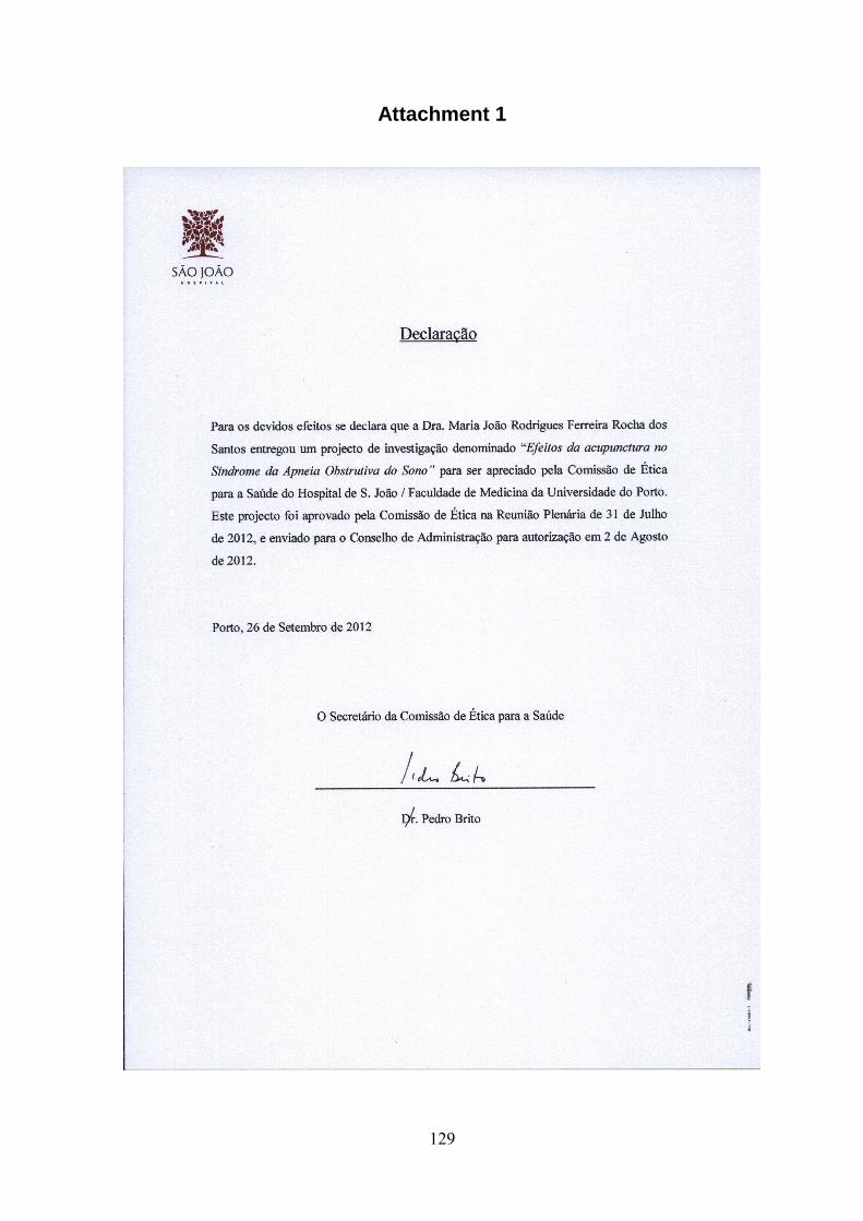
129
Attachment 1
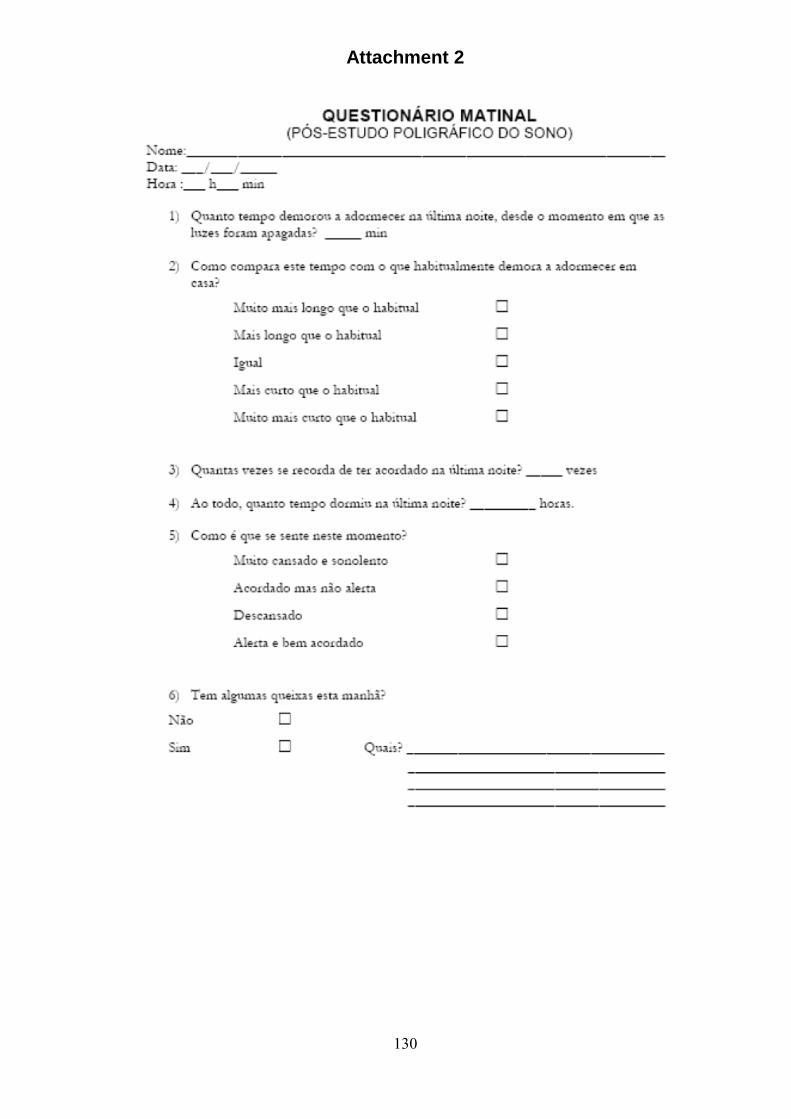
130
Attachment 2

131
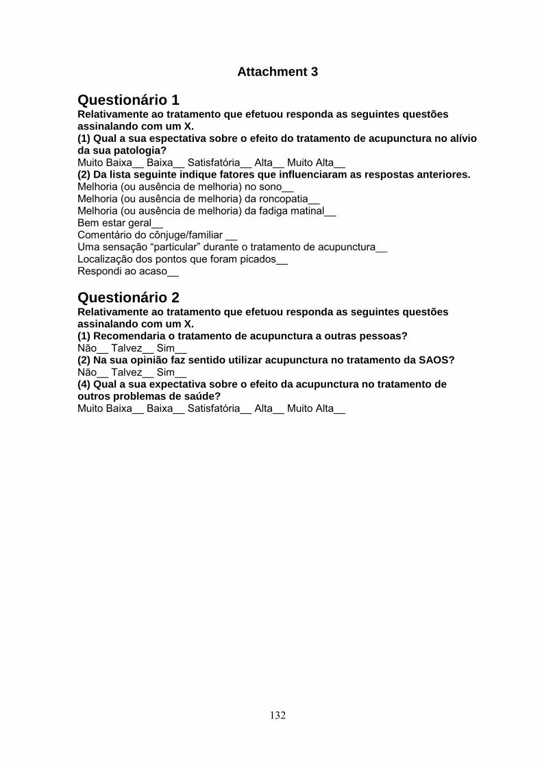
132
Attachment 3 Questionário 1 Relativamente ao tratamento que efetuou responda as seguintes questões assinalando com um X. (1) Qual a sua espectativa sobre o efeito do tratamento de acupunctura no alívio da sua patologia? Muito Baixa__ Baixa__ Satisfatória__ Alta__ Muito Alta__ (2) Da lista seguinte indique fatores que influenciaram as respostas anteriores. Melhoria (ou ausência de melhoria) no sono__ Melhoria (ou ausência de melhoria) da roncopatia__ Melhoria (ou ausência de melhoria) da fadiga matinal__ Bem estar geral__ Comentário do cônjuge/familiar __ Uma sensação “particular” durante o tratamento de acupunctura__ Localização dos pontos que foram picados__ Respondi ao acaso__ Questionário 2 Relativamente ao tratamento que efetuou responda as seguintes questões assinalando com um X. (1) Recomendaria o tratamento de acupunctura a outras pessoas? Não__ Talvez__ Sim__ (2) Na sua opinião faz sentido utilizar acupunctura no tratamento da SAOS? Não__ Talvez__ Sim__ (4) Qual a sua expectativa sobre o efeito da acupunctura no tratamento de outros problemas de saúde? Muito Baixa__ Baixa__ Satisfatória__ Alta__ Muito Alta__

133
Attachment 4: INFORMAÇÃO AO PARTICIPANTE Caro Participante, O presente estudo no qual participará designa-se “Comparação de duas estratégias de acupuntura no tratamento da apnéia do sono obstrutiva. - Estudo preliminar- ”. O objectivo do estudo é perceber se a terapia complementar da Medicina Tradicional Chinesa, a acupunctura, leva a melhoria de sintomas associados ao síndrome de apneia obstrutiva do sono e quais as alterações levadas a cabo por esta terapia quer ao nível da fisiologia, quer ao nível da qualidade de vida. No sentido de estudar os efeitos fisiológicos será sujeito a avaliação registada por poligrafia. No sentido de avaliar a qualidade de vida será pedido o preenchimento questionários: Questinário Matinal; Questionário de credibilidade do tratamento. Durante estes procedimentos estará acompanhado por médico e/ou enfermeiro, além da terapeuta de Medicina Tradicional Chinesa. A sua participação será voluntária. I Irá proceder-se à técnica de acupunctura designada por craniopunctura. Esta técnica consiste na inserção de microagulhas semipermanentes no couro cabeludo. Estas agulhas permanecerão no couro cabeludo 24 horas. Durante a colocação da agulhas será sujeito à avaliação visual da dor da punctura. Na noite após a colocação das agulhas irá repetir a poligrafia cardiorespiratória para avaliar os efeitos da técnica terapêutica. Na manhã seguinte serão realizados dois questionários simples para avaliar a sua percepção da qualidade do sono. O tratamento com acupunctura será efectuado por um profissional de saúde com formação em Medicina Chinesa Tradicional certificada por Universidade pública portuguesa. Com o tratamento de acupunctura esperamos contribuir para melhoria da sua doença , nomeadamente, melhoria do índice de apnea-hipopnea que avalia a gravidade do Síndrome da Apneia do Sono e da qualidade do sono. Note que não se espera que a acupunctura prejudique o tratamento convencional a que será sujeito nem haverá qualquer interferência no seu plano de tratamento habitual. Todas as agulhas de acupuntura são esterilizadas e descartáveis (ou seja, de uso único). Antes da inserção das agulhas, a pele será desinfectada com uma solução anti-séptica alcoólica. Os riscos associados a acupunctura são mínimos . Poderá sentir algum grau de dor ou desconforto e formigueiros no local das picadas com as agulhas de acupunctura. Mais raramente , poderá sentir tonturas, ansiedade ou náuseas. É possível que após o tratamento possam surgir ligeiros sangramentos, em particular se estiver a tomar a tomar medicamentos anti-agregantes (ex.: Aspirina, ácido acetilsalicílico) ou hipocoagulantes (ex.: Varfine ® (varfarina); Sintrom ®, acenocumarol) e/ou aparecerem ligeiros hematomas num local onde foram inseridas as agulhas que se resolverá espontaneamente. Caso esteja a tomar a medicação acima referida deverá informar a equipa de investigação. Note que durante estes procedimentos estará sempre acompanhado por Médico e ou Enfermeiro com treino em suporte avançado de vida e que o Hospital de São João está dotado de uma Equipa de Ressuscitação Interna durante 24 horas de actuação rápida, caso seja necessário, que é activada por telefone interno.
134
Sendo a sua participação voluntária terá o que tempo que necessitar para ponderar sobre a sua participação neste estudo. É livre de consultar a opinião dos seus familiares ou amigos. Caso decida aceitar, poderá posteriormente, a qualquer momento recusar continuar no estudo . Se recusar continuar neste estudo, o tratamento médico convencional não será afectado e toda a assistência habitual é-lhe garantida pelos profissionais de saúde. Ao entrar neste estudo será garantida a sua privacidade , através de confidencialidade dos dados e regras do sigilo médico. Todos os resultados obtidos serão devidamente codificados. Caso pretenda, poderá ter acesso aos resultados, mas não os poderá divulgar ou usar para fins científicos. Os dados serão apenas do conhecimento dos investigadores principais e dos orientadores do estudo e do eventual patrocinador do estudo e poderão ser posteriormente publicados em revistas científicas ou apresentados em eventos científicos. Será sempre mantido o seu anonimato. Note ainda que este estudo foi aprovado pela Comissão de Ética do Hospital de São João. Para seu conhecimento, as investigadoras principais deste estudo são as mestrandas Maria João Santos e Sofia Milhano Para qualquer esclarecimento poderá entrar em contacto com a Mestranda Maria João Santos telemóvel 91 3563504. A equipa de investigação agradece a sua participação e está ao seu dispor para qualquer esclarecimento. Hospital São João, Data:___/___/___ Com os melhores cumprimentos, __________________________ Maria João Santos Com os melhores cumprimentos, __________________________ Sofia Milhano
135
Attachment 5
Comissão de Ética para a Saúde do Hospital de S. João – EPE Modelo CES 05.A
DECLARAÇÃO DE CONSENTIMENTO Considerando a “Declaração de Helsínquia” da Associação Médica Mundial
(Helsínquia 1964; Tóquio 1975; Veneza 1983; Hong Kong 1989; Somerset West 1996 e Edimburgo 2000)
Designação do Estudo (em português): “Comparação de duas estratégias de acupuntura no tratamento da apnéia do
sono obstrutiva. -Um estudo preliminar-”.
Eu, abaixo-assinado, (nome completo do participante) -------------------------------------- -------------------------------------------------------------------------- ----, compreendi a explicação que me foi fornecida, por escrito e verbalmente, da investigação que se tenciona realizar, para qual é pedida a minha participação. Foi-me dada oportunidade de fazer as perguntas que julguei necessárias, e para todas obtive resposta satisfatória. Tomei conhecimento de que, de acordo com as recomendações da Declaração de Helsínquia, a informação que me foi prestada versou os objectivos, os métodos, os benefícios previstos, os riscos potenciais e o eventual desconforto. Além disso, foi-me afirmado que tenho o direito de decidir livremente aceitar ou recusar a todo o tempo a minha participação no estudo. Sei que se recusar não haverá qualquer prejuízo na assistência que me é prestada. Foi-me dado todo o tempo de que necessitei para reflectir sobre esta proposta de participação. Nestas circunstâncias, decido livremente aceitar participar neste projecto de investigação, tal como me foi apresentado pelo investigador(a). Data: ____ / _________________ / 20____ Assinatura do(a) participante: __________________________________________________________________ O(A) Investigador(a) responsável: Nome: _______________________________________________________________________________________ Assinatura:
_____________________________________________________________________________________