Clinical anatomy of Scalp - SRM Dental College
28
SCALP P.Ravishankar Senior Lecturer Dept.of Anatomy SRM Dental College Chennai India
-
Upload
khangminh22 -
Category
Documents
-
view
0 -
download
0
Transcript of Clinical anatomy of Scalp - SRM Dental College

SCALP
P.RavishankarSenior Lecturer
Dept.of AnatomySRM Dental College
ChennaiIndia

SCALP
• Definition
• Extent
• Layers
• Nerve supply
• Blood supply
• Clinical Anatomy

• Definition:-
Soft tissue covering the cranial vault
• Extent:-
Anteriorly:- Supra orbital margin
Posteriorly:-External occipital protuberance
& Superior nuchal line
Laterally:-Superior temporal line

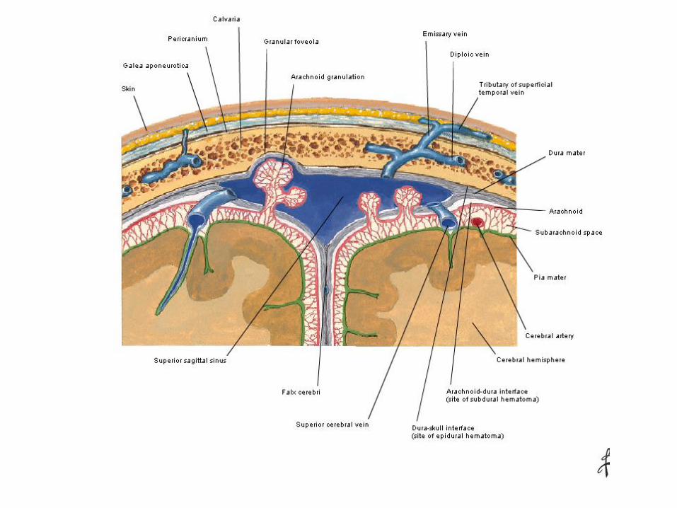
Layers of SCALP
Skin
Connective tissue
Aponeurosis
Loose areolar tissue
Pericranium


1. Skin
Thick and hair bearing and contains numerous sebaceous glands

2. Superficial fascia
• Dense fibro- fatty connective tissue.
• Firmly attached to the skin and the aponeurosis.
• Numerous arteries, veins and nerves.
• Free anastomosis between the ICA an ECA branches.


3. Epicranial Aponeurosis(Galea Aponeuritica)
• A thin tendinous sheet that unites the occipital and frontal bellies of the occipitofrontalis muscle.
• The subaponeurotic space: potantial space beneath the aponeurosis.


4.Loose areolar tissue
Extension
• Anteriorly:- Eyelids
• Posteriorly:- Highest and superior nuchal line
• Laterally:- Superior temporal line
• Dangerous area of scalp
connect the superficial veins of the scalp with the diploic veins of the skull bones and with intracranial venous sinuses

Pericranium
• Modified periosteum
• Loosely attached to bones but firmly attached to sutures


Nerve supply to scalpIn front of auricleSensory
• Supratrochelar br.of Frontal nerve
• Supraorbital br. of frontal nerve
• Zygomatiotemporal br.ofZygomatic nerve
• Auriculotemporal nerve,br.ofMandibular nerve
Motor
• Temporal br. Of Facial nerve
Behind the auricle
Sensory
• Posterior division of great auricular nerve from cervical plexus(C2,C3)
• Lesser occipital nerve from cervical plexus(C2)
• Greater occipital nerve(C2dorsal ramus)
• Third occipital nerve(C3 dorsal ramus)
Motor
• Posterior auricular br.of facial nerve


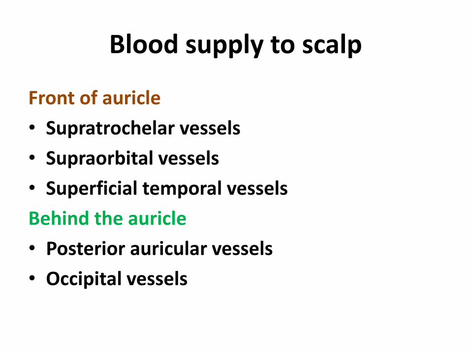
Blood supply to scalp
Front of auricle
• Supratrochelar vessels
• Supraorbital vessels
• Superficial temporal vessels
Behind the auricle
• Posterior auricular vessels
• Occipital vessels

Clinical anatomy

• Surgical layers of scalp:-first 3 layers of scalp
• Sebaceous cyst are common in skin layer
• The wounds of scalp don’t gape unless it is cut transversely – because the aponeuroticlayer is tightly attached in the anteroposterior direction of occipitio-frontalis muscle

Clinical anatomyBlack eye:-
• Black discoloration of skin around eye
• Blood and fluid collection in the upper eye lid
• Blood accumulate to upper eyelid due attachment of frontalis muscle


Black eye

Safety-valve hematoma:-
• Fracture of cranial vault in children may associate with tearing of duramater and pericranium,in such cases blood from intracranial hemorrahage communicate with subaponeurotic space of scalp through the fracture line
• The sign of cerebral compression doesn’t occurs until subaponeurotic space is filled
• Therefore blood collection in the fourth layer is called safety –valve hematoma

Dangerous layer of scalp
• Blood and pus freely tend to collect in this layer ,if pus collects in this layer the infection may travel readily along the emissary veins into the intracranial venous sinuses.

• Cephalhematoma:-
Subperiosteal collection of blood
• Caput succedaneum:-
Subcutaneous edema .it takes place during the passage of head through the birth canal due interference of venous return