The effect of the TLR9 ligand CpG-oligodeoxynucleotide on the protective immune response to...
11
RESEARCH Open Access The effect of the TLR9 ligand CpG- oligodeoxynucleotide on the protective immune response to alcelaphine herpesvirus-1-mediated malignant catarrhal fever in cattle Nevi Parameswaran 1 , George C Russell 2 , Kathryn Bartley 2 , Dawn M Grant 2 , David Deane 2 , Helen Todd 2 , Mark P Dagleish 2 and David M Haig 1* Abstract We wished to determine the effect of of CpG ODN adjuvant on the magnitude and duration of protective immunity against alcelaphine herpesvirus-1 (AlHV-1) malignant catarrhal fever (MCF), a fatal lymphoproliferative disease of cattle. Immunity was associated with a mucosal barrier of virus-neutralising antibody. The results showed that CpG ODN included either with emulsigen adjuvant and attenuated AlHV-1 (atAlHV-1) or alone with atAlHV-1 did not affect the overall protection from clinical disease or duration of immunity achieved using emulsigen and atAlHV-1. This is in contrast to other similar studies in cattle with BoHV-1 or cattle and pigs with various other immunogens. In addition to this, several other novel observations were made, not reported previously. Firstly, we were able to statistically verify that vaccine protection against MCF was associated with virus-neutralising antibodies (nAbs) in nasal secretions but was not associated with antibodies in blood plasma, nor with total virus-specific antibody (tAb) titres in either nasal secretions or blood plasma. Furthermore, CpG ODN alone as adjuvant did not support the generation of virus-neutralising antibodies. Secondly, there was a significant boost in tAb in animals with MCF comparing titres before and after challenge. This was not seen with protected animals. Finally, there was a strong IFN-γ response in animals with emulsigen and atAlHV-1 immunisation, as measured by IFN-γ secreting PBMC in culture (and a lack of IL-4) that was not affected by the inclusion of CpG ODN. This suggests that nAbs at the oro-nasal-pharyngeal region are important in protection against AlHV-1 MCF. Introduction Malignant catarrhal fever (MCF) is a usually-fatal lym- phoproliferative disease of a range of ungulates including cattle, deer, bison, water buffalo and pigs [1]. It is caused by members of the Macavirus genus of the subfamily Gammaherpesvirinae of the Herpesviridae. The best cha- racterised of these are ovine herpesvirus-2 (OvHV-2) and alcelaphine herpesvirus-1 (AlHV-1), which are carried asymptomatically by sheep and wildebeest respectively. There is no apparent disease in these reservoir hosts, but when the virus is transmitted to susceptible animals, MCF often ensues [1]. MCF has a worldwide distribution wherever reservoir and susceptible animals co-habit. MCF is currently a serious problem in several parts of the world including: Indonesia, where cattle are infected by OvHV-2, North America, in farmed bison infected by OvHV-2 and in Eastern and Southern Africa where migrating wilde- beest transmit AlHV-1 to cattle [1]. MCF in cattle caused by AlHV-1 and OvHV-2 is similar in clinical presentation with fever, inappetance, ocular opacity and oral epithelial lesions present in most cases [1-3]. Histologically, MCF is characterised by lymphocyte infiltration in multiple tissues, hyperplasia in lymph nodes and vasculitis most notably in the brain and kidneys [1-3]. The pathogenesis of MCF is an area of active study cur- rently, and T cells (predominantly CD8+ T cells) accu- mulating within the tissues are virus-infected [4,5]. These cells may be responsible for mediating the tissue damage * Correspondence: [email protected] 1 School of Veterinary Medicine and Science, University of Nottingham, Sutton Bonington, LE12 5RD Nottingham, UK Full list of author information is available at the end of the article VETERINARY RESEARCH © 2014 Parameswaran et al.; licensee BioMed Central Ltd. This is an Open Access article distributed under the terms of the Creative Commons Attribution License (http://creativecommons.org/licenses/by/2.0), which permits unrestricted use, distribution, and reproduction in any medium, provided the original work is properly credited. The Creative Commons Public Domain Dedication waiver (http://creativecommons.org/publicdomain/zero/1.0/) applies to the data made available in this article, unless otherwise stated. Parameswaran et al. Veterinary Research 2014, 45:59 http://www.veterinaryresearch.org/content/45/1/59
-
Upload
independent -
Category
Documents
-
view
1 -
download
0
Transcript of The effect of the TLR9 ligand CpG-oligodeoxynucleotide on the protective immune response to...
VETERINARY RESEARCHParameswaran et al. Veterinary Research 2014, 45:59http://www.veterinaryresearch.org/content/45/1/59
RESEARCH Open Access
The effect of the TLR9 ligand CpG-oligodeoxynucleotide on the protective immuneresponse to alcelaphine herpesvirus-1-mediatedmalignant catarrhal fever in cattleNevi Parameswaran1, George C Russell2, Kathryn Bartley2, Dawn M Grant2, David Deane2, Helen Todd2,Mark P Dagleish2 and David M Haig1*
Abstract
We wished to determine the effect of of CpG ODN adjuvant on the magnitude and duration of protectiveimmunity against alcelaphine herpesvirus-1 (AlHV-1) malignant catarrhal fever (MCF), a fatal lymphoproliferativedisease of cattle. Immunity was associated with a mucosal barrier of virus-neutralising antibody. The results showedthat CpG ODN included either with emulsigen adjuvant and attenuated AlHV-1 (atAlHV-1) or alone with atAlHV-1did not affect the overall protection from clinical disease or duration of immunity achieved using emulsigen andatAlHV-1. This is in contrast to other similar studies in cattle with BoHV-1 or cattle and pigs with various otherimmunogens. In addition to this, several other novel observations were made, not reported previously. Firstly, wewere able to statistically verify that vaccine protection against MCF was associated with virus-neutralising antibodies(nAbs) in nasal secretions but was not associated with antibodies in blood plasma, nor with total virus-specificantibody (tAb) titres in either nasal secretions or blood plasma. Furthermore, CpG ODN alone as adjuvant did notsupport the generation of virus-neutralising antibodies. Secondly, there was a significant boost in tAb in animalswith MCF comparing titres before and after challenge. This was not seen with protected animals. Finally, there wasa strong IFN-γ response in animals with emulsigen and atAlHV-1 immunisation, as measured by IFN-γ secretingPBMC in culture (and a lack of IL-4) that was not affected by the inclusion of CpG ODN. This suggests that nAbs atthe oro-nasal-pharyngeal region are important in protection against AlHV-1 MCF.
IntroductionMalignant catarrhal fever (MCF) is a usually-fatal lym-phoproliferative disease of a range of ungulates includingcattle, deer, bison, water buffalo and pigs [1]. It is causedby members of the Macavirus genus of the subfamilyGammaherpesvirinae of the Herpesviridae. The best cha-racterised of these are ovine herpesvirus-2 (OvHV-2) andalcelaphine herpesvirus-1 (AlHV-1), which are carriedasymptomatically by sheep and wildebeest respectively.There is no apparent disease in these reservoir hosts,but when the virus is transmitted to susceptible animals,MCF often ensues [1]. MCF has a worldwide distribution
* Correspondence: [email protected] of Veterinary Medicine and Science, University of Nottingham,Sutton Bonington, LE12 5RD Nottingham, UKFull list of author information is available at the end of the article
© 2014 Parameswaran et al.; licensee BioMedCreative Commons Attribution License (http:/distribution, and reproduction in any mediumDomain Dedication waiver (http://creativecomarticle, unless otherwise stated.
wherever reservoir and susceptible animals co-habit. MCFis currently a serious problem in several parts of the worldincluding: Indonesia, where cattle are infected by OvHV-2,North America, in farmed bison infected by OvHV-2 andin Eastern and Southern Africa where migrating wilde-beest transmit AlHV-1 to cattle [1].MCF in cattle caused by AlHV-1 and OvHV-2 is similar
in clinical presentation with fever, inappetance, ocularopacity and oral epithelial lesions present in most cases[1-3]. Histologically, MCF is characterised by lymphocyteinfiltration in multiple tissues, hyperplasia in lymph nodesand vasculitis most notably in the brain and kidneys [1-3].The pathogenesis of MCF is an area of active study cur-rently, and T cells (predominantly CD8+ T cells) accu-mulating within the tissues are virus-infected [4,5]. Thesecells may be responsible for mediating the tissue damage
Central Ltd. This is an Open Access article distributed under the terms of the/creativecommons.org/licenses/by/2.0), which permits unrestricted use,, provided the original work is properly credited. The Creative Commons Publicmons.org/publicdomain/zero/1.0/) applies to the data made available in this
Parameswaran et al. Veterinary Research 2014, 45:59 Page 2 of 11http://www.veterinaryresearch.org/content/45/1/59
seen in MCF although this remains to be proven. The ge-nomes of AlHV-1 and OvHV-2 have been sequenced sho-wing that they are highly-related but distinct viruses [6,7].Current control measures for MCF focus on keeping
reservoir and susceptible species apart. This is not alwayspossible, for example African pastoralists’ cattle that havebeen taken from the plains inhabited by wildebeest to up-land regions to avoid MCF are then susceptible to the ser-ious diseases of trypanosomosis and East Coast Fever[8,9]. There have been many attempts in the past to con-trol MCF by vaccination although these have either failedor not been reproducible [10-14]. However, we have deve-loped a vaccine strategy that is successful in preventingdisease in cattle challenged with AlHV-1 by inducing amucosal barrier of immunity in the oro-nasal pharyngealregion of cattle in response to intra-muscular immu-nisation in the upper neck with attenuated AlHV-1 inEmulsigen adjuvant [15]. However, the duration of im-munity is short, peaking at around two months and lastingup to six months, and a boost immunisation is required togenerate high titre virus-neutralising antibody that corre-lates with protection [15,16].The aim of this study was to improve the magnitude and
duration of protective immunity to AlHV-1 by using theTLR9 agonist unmethylated CpG oligodeoxynucleotide(CpG ODN) as an adjuvant. The TLRs are microbial pat-tern recognition receptors on or within epithelial and im-mune system cells, amongst others, that act as a first lineof defence against infection [17]. TLRs working in concertor alone can influence the quality and duration of adaptiveimmune responses. Furthermore, AlHV-1, along withmany other pathogens, has evidence of CpG suppressionwithin its genome [6]. CPG ODN has been used suc-cessfully to enhance the magnitude or quality (Th1/Th2)of protective immunity to a range of pathogens and itsduration in a variety of different species [18-20] includingcattle [21-25]. Furthermore, there are various CpG ODNformulations that have different efficacies in any given spe-cies. The CpG ODN 2007 formulation used in this studyhas been proven effective in cattle previously [21-23].Taking all this together, we have tested unmethylated
CpG ODN as an adjuvant in our MCF vaccine formu-lation and compared it to emulsigen alone or with bothadjuvants in combination. We used AlHV-1 challenge at23 weeks after boost immunisation as this had previouslyachieved only 50% protection compared to challenge at7–8 weeks (where > 90% protection has been recorded),and therefore should allow a measurement of the effect ofCpG ODN on duration of immunity.
Materials and methodsAnimals and study designClinically healthy and MCF seronegative male Friesian-Holstein calves, 6–7 months of age were used in the
experiments. All animal experiments were approved byboth the University of Nottingham and the MoredunResearch Institute’s experiments and ethics committeesand complied fully with the Home Office of Great Brit-ain and Northern Ireland “Animals (Scientific Proce-dures) Act 1986”. For the study, calves were assignedrandomly to four experimental groups and vaccinatedintramuscularly in the upper neck as described inTable 1, with atAlHV-1 plus Emulsigen (Group 1, n = 9);atAlHV-1 plus Emulsigen and CpG (Group 2, n = 9);atAlHV-1 plus CpG (Group 3, n = 5); and Emulsigenplus CpG alone (Group 4, n = 5). Numbers were chosenbased on power calculations using data from previousimmunisation experiments [15,16] (see statistical ana-lysis of data below). Intramuscular immunisation withatAlHV-1 in Emulsigen (20% v/v, MVP, Omaha, USA)was used in group 1 and group 2 calves for both primeand boost immunisations (separated by a period of onemonth) and for injection of atALHV-1 and CpGODN(group 3) and both adjuvants (group 4). Phosphoro-thioate-stabilized CpG ODN 2007 (TCGTCGTTGTCGTTTTGTCGTT) provided by Merial (Harlow, UK,purity > 90%) was included at 1 mg/injection in theimmunisation of group 2 and 3 animals as this had pre-viously been an effective adjuvant dose in cattle in vari-ous experimental vaccines [21-23].All animals were challenged with 10 mL of 103.8
TCID50/mL virulent AlHV-1 given intranasally by spray-ing 5 mL per nostril at 23 weeks after boost immunisation(week 27 of the experiment, Table 1 legend). In all expe-riments, uncoagulated blood and nasal secretions werecollected from all animals immediately prior to primaryimmunisation and weekly thereafter as described pre-viously [15,16]. After virus challenge, animal rectal tem-peratures were measured daily and clinical scoring wasperformed daily after the onset of pyrexia (defined asrectal temperature > 40 °C). Clinical scoring, based ontemperature, body condition, ocular and nasal lesions, wasused to ensure that all animals were euthanized, with anoverdose of intravenous sodium pentobarbitone, at theonset of moderate clinical signs [16].At autopsy, the following tissues were collected: brain,
buccal mucosa, rumen, reticulum, liver, kidney, lung,prescapular lymph node, mesenteric lymph node (MLN)and blood. Pieces of each tissue (except blood) werefixed in (a) 10% formal saline (b) 4% paraformaldehyde(c) zinc salts fixative [15,16] before processing routinelyand embedding in paraffin wax. Live cell suspensions ofMLN were cryopreserved under liquid nitrogen.For histological analysis, 4 μm sections of formalin-
fixed tissues were cut, mounted on glass microscopeslides and stained with haematoxylin and eosin. Inaddition, total DNA was prepared from blood buffy coatcells for PCR detection of viral DNA.
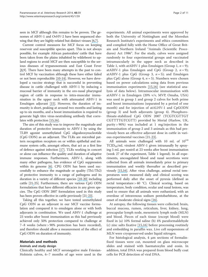
Table 1 Experimental groups: clinical/histopathology analyses and serology
Group1 Days afterchallenge2
Pathology/histology3 ViralDNA4
NS ab5 NS ab Plas ab6 Plas ab
Pre-chall After chall Pre-chall After chall
Gp 1: Virus + Em
841 99 No MCF lesions NDet 67 79 82 58
920 99 No MCF lesions NDet 24 89 44 60
221 98 No MCF lesions NDet 63 66 42 44
983 98 No MCF lesions NDet 30 50 132 248
001 91 No MCF lesions NDet 195 118 72 109
371 89 No MCF lesions NDet 151 138 79 220
675 89 No MCF lesions NDet 44 70 89 114
635 63 MCF Lesions: Lymphocytic inflammation andvasculitis. Pneumonia
32.6 35 96 47 449
101 42 Mild MCF lesions: Lymphocytic inflammation andvasculitis. Pneumonia
32.5 55 220 37 421
Gp 2: Virus + Em +CpG
105 98 No MCF lesions NDet 72 118 89 149
106 98 No MCF lesions NDet 40 66 116 198
706 97 No MCF lesions NDet 85 66 75 73
679 96 No MCF lesions NDet 29 17 81 128
236 91 No MCF lesions NDet 9 24 64 214
162 90 No MCF lesions NDet 64 97 133 218
663* 90 Mild MCF lesions. Lymphocytic inflammationand vasculitis
33.6 6 234 51 931
400678 41 Mild MCF lesions: Lymphocytic inflammationand vasculitis. Mild pneumonia
37.5 0 300 60 324
065 35 MCF Lesions: Lymphocytic inflammation andvasculitis in multiple tissues. Pneumonia
27 0 408 26 299
Gp 3: Virus + CpG
666 90 No MCF lesions NDet 0 25 46 50
021 89 No MCF lesions NDet 19 19 23 68
169 89 No MCF lesions NDet 50 64 85 139
201027 65 Mild MCF lesions. Lymphocytic inflammation andvasculitis. Pneumonia
27.6 0 40 0 0
273 36 MCF Lesions. Lymphocytic inflammation andvasculitis. Pneumonia
26.2 0 0 0 220
Gp 4: Adjuvant
658 88 No MCF lesions NDet 0 0 0 0
101027 60 Mild MCF lesions. Lymphocytic inflammation 30.3 0 0 0 0
249 36 Mild MCF lesions. Lymphocytic inflammation andvasculitis in the brain and liver only
NDet 0 90 0 104
Parameswaran et al. Veterinary Research 2014, 45:59 Page 3 of 11http://www.veterinaryresearch.org/content/45/1/59
Table 1 Experimental groups: clinical/histopathology analyses and serology (Continued)
925 36 MCF Lesions. Lymphocytic inflammation andvasculitis. Pneumonia
24.7 2 74 0 0
986 27 MCF lesions: Lymphocytic vasculitis inmultiple tissues. Pneumonia
30 0 15 0 81
1Em = emulsigen adjuvant, CpG = oligoCpG adjuvant.2Time from challenge to euthanasia/post-mortem examination.3Tissues in this analysis were: brain, lung, liver, kidney, prescapular lymph node, alimentary tract epithelium and mesenteric lymph node.4Viral DNA was assayed in terminal blood samples by qPCR to give a specific and internally-controlled diagnostic test. Data are expressed as Ct values – the cyclenumber at which specific amplification was first detected. Moderate/severe MCF usually has values in the range 21–29. NDet = specific amplification of AlHV-1DNA not detected (but control actin-specific PCR was detected). *This animal had histological evidence of infection.5Nasal secretion antibody (NS ab) titres pre-challenge and after challenge with virus.6Plasma antibody (Plas ab) titres pre-challenge and after challenge with virus.Animals in groups 1, 2 and 3 received the same dose of vaccine (1 mL of 107 TCID50 per mL) for both prime and boost, given intramuscularly and in groups 1 and2 contained 20%(v/v) Emulsigen. Control animals in group 4 received virus-free culture medium with Emulsigen prepared and administered in the same way.Animals in all groups were challenged 23 weeks after boost with 10 mL of 103.8 TCID50/mL of virulent AlHV-1 given intranasally. For mild MCF lesions, Viral DNA inthe tissues can be below detection level.
Parameswaran et al. Veterinary Research 2014, 45:59 Page 4 of 11http://www.veterinaryresearch.org/content/45/1/59
VirusThe strains of AlHV-1 used for vaccination and challengewere as described previously [15,16]. Briefly, the virulentC500 strain virus (virAlHV-1) was collected from culturesof bovine turbinate (BT) cells infected with a lymphoidcell suspension derived from pooled lymphoid tissue fromrabbits infected with AlHV-1 C500 that had developedMCF. Infected BT cell cultures were passaged onto freshBT cells by a 1:4 split four times at peak cytopathic effect(approximately weekly) after which virulent virus was har-vested from culture supernatants and cells following threerounds of freeze-thaw treatment of the cells. Cell-freevirus supernatant was stored at −80 °C as 1 mL aliquots ofpooled virus in batches and representative aliquots of eachbatch were titrated to allow calculation of the appropriatechallenge dose. Titration measured 50% tissue-culture-infectious dose (TCID50) as described previously [15,16].The attenuated AlHV-1 C500 strain (atAlHV-1) at
passage > 1000 was used as the source of virus for im-munisation [15,16]. This cell-free virus was obtainedfrom BT cell culture supernatants, clarified by centrifu-gation and stored in batches at −80 °C until required.Representative aliquots of atAlHV-1 were titrated asdescribed for virulent AlHV-1. The virus vaccine dosefor both prime and boost comprised 1 mL of 107 TCID50
per mL atAlHV-1 with adjuvant(s) as described above.
Detection of viral DNAViral DNA was assayed in pure genomic DNA samplesextracted from blood buffy coat cells by quantitative (q)PCR as described previously [15,16]. Briefly, ~50 ng oftotal DNA was assayed simultaneously for the presence ofAlHV-1 and genomic actin sequences by duplex real-timePCR analysis. Each 20 μL assay contained 900 μM of eachAlHV-1 forward and reverse primer (AlHV1-F, AlHV1-R)and 250 μM of the fluorogenic probe FAM-AlHV1;450 μM of the genomic actin primer (gACT-F: 5’-CACCTT CCA GCA GAT GTG GA-3’; gACT-R: 5’-CTAGAA GCA TTT GCG GTG GAC) and 125 μM of the
fluorogenic minor-groove binding (mgb) probe VIC-gACT(5’-VIC-AGC AAG CAG GAG TAC G-mgb) [26]; andreal-time PCR reagents containing Platinum Taq polyme-rase and ROX control dye (Invitrogen (Life Technologies),Paisley, UK). All assays were conducted using standardconditions on ABI 7000 or 7500 sequence detection sys-tems (Applied Biosystems (Life technologies), Paisley, UK).
Antibody detection by ELISA and the virus-neutralisingantibody testThe antibody ELISA was used to detect humoral virus-specific antibody titres in blood plasma samples and nasalsecretions as described previously [16]. An AlHV-1-negative serum at 1:500 dilution was also included witheach test sample series.ELISA values (difference between means of positive
and negative antigen wells for each sample dilution)were used to calculate a relative titre for each testsample, based on the standard curve produced from thepositive control serum wells on the same plate. Testsamples with ELISA values outside the range found forthe positive control serum were discarded. Standardpools of NS and plasma were used to prepare serial dilu-tions (NS 1:20–1:2560 and plasma 1:200–1:6400) thatwere used on each test plate. ELISA values from thesestandards were used to generate a best fit polynomialstandard curve (y = ax2 + bx + c) for each plate with R2
value greater than 0.98. Relative titres of unknown sam-ples were calculated from the plate standard curve usingNS samples at a dilution of 1:160 and plasma at 1:500.The virus neutralisation test was based upon 50% in-
hibition of AlHV-1-induced cytopathic effect (CPE) inBT cells by dilutions of blood plasma or nasal secretionfluid as described [15,16]. Assays were carried out in 96well tissue culture plates with BT cells at greater than80% confluence. All assays used a high titre bovine anti-AlHV-1 serum as a standard and included non-specifictoxicity-control wells containing sample and cells with-out virus.
Parameswaran et al. Veterinary Research 2014, 45:59 Page 5 of 11http://www.veterinaryresearch.org/content/45/1/59
ELISPOT and ELISA for interferon-γ and interleukin-4detectionAn ELISPOT test to quantify IFN-γ release from bovinePBMCs was carried out using Millipore Multiscreen HTSIP 0.45 μm ELISPOT plates according to the manufac-turer’s instructions (Millipore, Watford, UK). Wells werecoated with 100 μL (5 μg/mL) anti bovine-IFN-γ antibody(clone CC330, AbD Serotec, Oxford, UK) and blockedwith Iscove’s modified Dulbecco’s medium (IMDM) con-taining 10% FCS. PBMCs were added at 1 × 10⁵ per well inmedium. Viral antigen, previously prepared using 3-[(3-cholamidopropyl) dimethylammonio]-1-propanesulfonate(CHAPS) detergent from the C500 strain of AlHV1 waspurified using Amicon Ultra-0.5 μm Centrifugal FilterUnits (Millipore, Oxford, UK) and quantified using a Brad-ford protein assay (Life Science, Hemel Hemstead, UK)and added to wells in triplicate at 0.2 μg per well. A com-bination of 0.1 ng phorbol 12-myristate 13-acetate (PMA,Sigma-Aldrich, Dorset, UK) and 50 ng Ionomycin (Sigma-Aldrich, Dorset, UK) per 105 cells in each of two wellswere used to stimulate IFN-γ production as a positive con-trol. PBMCs in medium comprised the negative control.Cells were incubated for 72 h at 37 °C in a humidified 5%CO2 in air tissue culture incubator. Biotinylated anti-bovine IFN-γ (clone CC302, AbD Serotec, Oxford, UK)was used as the detection mAb at 4 μg/mL (100 μL/well).ExtrAvidin Alkaline Phosphatase and Sigma fast BCIP/NBT substrate (Sigma-Aldrich, Dorset, UK) was utilisedfor spot development. Spots were then counted using anELISPOT reader (AID GmbH, Oxford Biosystems, Oxford,UK) and averages taken from antigen exposed triplicatesand negative control triplicates, the difference gives spotforming cells (SFC).Supernatant samples (50 μL) collected after 72 h incuba-
tion of the PBMCs with viral antigen were tested usingthe Thermo Scientific Bovine IL-4 ELISA Kit (Thermo-Fisher Scientific, Loughborough, UK. Product# ESS0031).
Pathology and histopathological analysesClinical MCF was diagnosed by clinical signs and con-firmed by histopathological lesions and the detection ofvirus DNA in terminal blood samples. In clinically normalanimals, infection was deemed to have occurred if therewas histological evidence of lesions and viral DNA in thetissues. Clinical signs included fever, nasal and oculardischarge, conjunctivitis and development of corneal opa-city. The use of a clinical scoring system ensured that allanimals with MCF were euthanized at a similar stage ofthe disease. All surviving animals were euthanized around13 weeks after pathogenic virus challenge. Clinical scoresand histopathology were recorded. MCF histopathology inbrain, kidney, liver, lung, lymph nodes and alimentarytract epithelium was examined and scored for the fre-quency of lesions consistent with MCF. The pathology of
each case was summarised as MCF (tissues contained sig-nificant numbers of lesions consistent with a diagnosis ofMCF); mild (a small number of lesions, without extensiveinfiltration of lymphocytes, which may or may not be as-sociated with clinical disease); or negative (no significantlesions observed). A positive test for AlHV-1 DNA in theblood on the day of autopsy, in combination with typicalMCF histopathology, was taken as a definitive diagnosis ofeither clinical or non-clinical MCF.
Statistical analysis of dataFor the survival plot comparisons, a log rank test wasused. For the antibody titre and ELISPOT data compari-sons, the Mann Whitney test and Student’s t-test wereused. For animal group sizes, a power calculation basedon Fisher’s test was applied [27].Normally the group size is defined as that which gives
an 80% chance of detecting a difference between treat-ment groups. We assume (based on experimental data)that 93% of unvaccinated animals would get MCF (in thevirus challenge group) and that we want to detect protec-tion at a level of 50% or better. We know from previousexperiments that 6 months after immunisation boostthere will be a 50:50 ratio of survivors to cattle that suc-cumb to MCF. We estimate that there will be a 80%chance of detecting a significant difference at a two sided0.8 significance level. This equates to using at least 8 cattlefor groups 1 and 2. We used 9. For group 3 there isno published evidence in cattle that immunogen withCpGODN alone induces a significant protective responsecompared to protection positive controls (16–20), so forthis and group 4 animals group sizes were reduced to 5but still retained statistical power. Previous work hadshown that atALHV-1 alone did not induce an MCF pro-tective response in cattle after virus challenge (unpub-lished results).
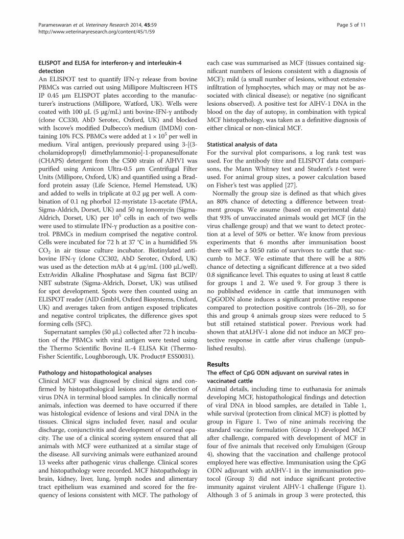
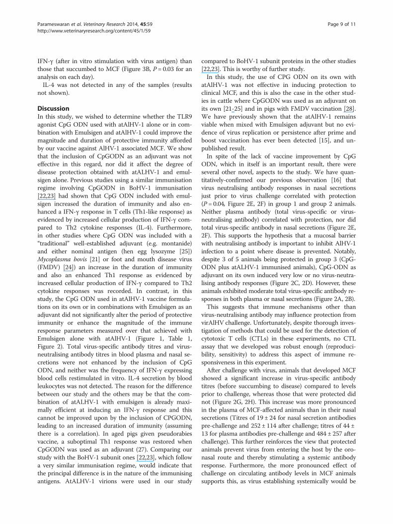
ResultsThe effect of CpG ODN adjuvant on survival rates invaccinated cattleAnimal details, including time to euthanasia for animalsdeveloping MCF, histopathological findings and detectionof viral DNA in blood samples, are detailed in Table 1,while survival (protection from clinical MCF) is plotted bygroup in Figure 1. Two of nine animals receiving thestandard vaccine formulation (Group 1) developed MCFafter challenge, compared with development of MCF infour of five animals that received only Emulsigen (Group4), showing that the vaccination and challenge protocolemployed here was effective. Immunisation using the CpGODN adjuvant with atAlHV-1 in the immunisation pro-tocol (Group 3) did not induce significant protectiveimmunity against virulent AlHV-1 challenge (Figure 1).Although 3 of 5 animals in group 3 were protected, this
0.0 2.5 5.0 7.5 10.0 12.5 15.00
20
40
60
80
100
Weeks after challenge
Per
cen
tsu
rviv
al
Gp1 virus+Em Gp2 virus+Em+CpGGp3 virus+CpG Gp4 adjuvant
Survival plot
Figure 1 Survival plot for each of the four groups of cattle.Kaplan Maier survival plot. Group 1: 9 cattle immunised and boostedwith atAlHV-1 (virus) along with Emulsigen (Em) adjuvant. Group 2: 9cattle immunised and boosted as in group 1 but including CpG ODN(CpG) as well as Emulsigen; Group 3: 5 cattle immunised and boostedwith atAlHV-1 (virus) and CpG ODN. Group 4: 5 cattle injected withadjuvant only. P = 0.02 for group 1 compared to group 4. P = 0.04 forgroup 2 compared to group 4. Groups 3 and 4 were not significantlydifferent (p = 0.24).
Parameswaran et al. Veterinary Research 2014, 45:59 Page 6 of 11http://www.veterinaryresearch.org/content/45/1/59
was not significant (P = 0.24) compared to the controlgroup 4 animals (adjuvant only group receiving virus chal-lenge). When CpG ODN was used in combination withemulsigen and AtAlHV-1 (Group 2), there was no in-crease in survival rate/protection compared to usingemulsigen alone with atAlHV-1 (Group 1). Both group 1and group 2 cattle were significantly protected (with re-spect to survival) over the duration of the experimentwhen compared to the infection control animals in group4 (P = 0.02 and P = 0.04, respectively). Postmortem ana-lysis indicated no evidence of infection in any of the pro-tected cattle except one group 2 animal (663). Table 1shows the clinical and histopathological findings of the in-dividual animals in each group. Clinical presentation forcattle with MCF was: inappetance, fever (>40 °C for twoor more consecutive days) and ocular and nasal dischargewith ocular opacity in most cases. Histologically, in MCF-affected animals the lesions consisted of interstitial and/orperivascular lymphocyte accumulations in non-lymphoidorgans (kidney, liver, lung and brain), and hyperplasia andsome architecture disruption in the lymphoid tissues. Inboth types of tissues there was evidence of vasculitis. Allof this is typical of MCF in cattle. Mild MCF presentationswere probably due to euthanasia of the animals at mild tomoderate rather than moderate to severe clinical scores inaccordance with Home Office (UK) ASPA MCF end pointcriteria. This could well have been responsible for the lowtitres of AlHV-1 DNA in the blood cells in some animals,for example in group 4 (Table 1). This qPCR analysis of
AlHV-1 DNA in terminal blood samples confirmed theclinical and histopathological diagnosis of MCF (Table 1).The survivor of clinical disease in group 2 (663), showedhistological evidence of very mild infection, and wastherefore considered infected but clinically free of disease.In animals that showed evidence of clinical MCF, lesionssuggestive of bacterial pneumonia were present also inmost of them (8 of 11, Table 1). 663 was not one of these.Of the 17 animals protected from MCF, there were nonethat presented with evidence of pneumonia at autopsy.
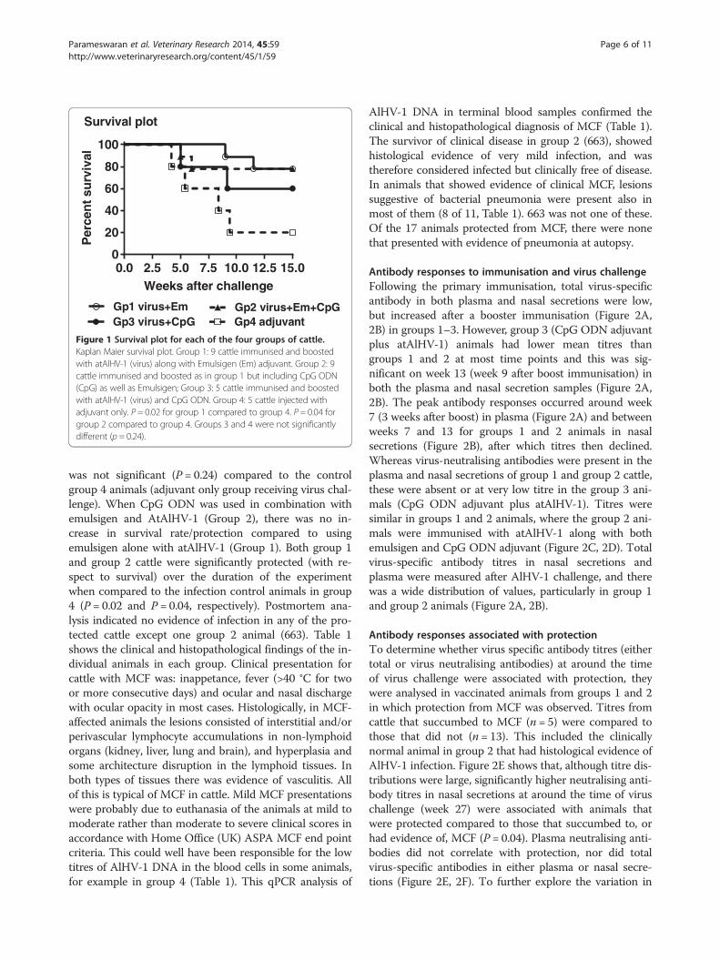
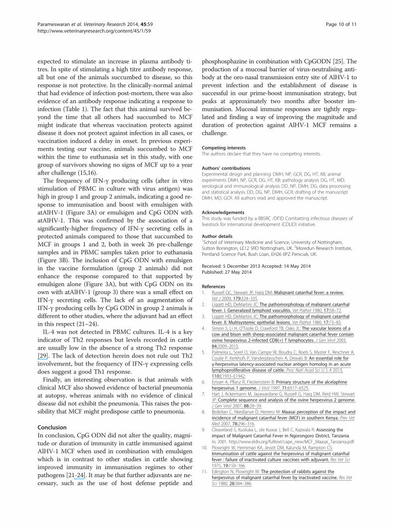
Antibody responses to immunisation and virus challengeFollowing the primary immunisation, total virus-specificantibody in both plasma and nasal secretions were low,but increased after a booster immunisation (Figure 2A,2B) in groups 1–3. However, group 3 (CpG ODN adjuvantplus atAlHV-1) animals had lower mean titres thangroups 1 and 2 at most time points and this was sig-nificant on week 13 (week 9 after boost immunisation) inboth the plasma and nasal secretion samples (Figure 2A,2B). The peak antibody responses occurred around week7 (3 weeks after boost) in plasma (Figure 2A) and betweenweeks 7 and 13 for groups 1 and 2 animals in nasalsecretions (Figure 2B), after which titres then declined.Whereas virus-neutralising antibodies were present in theplasma and nasal secretions of group 1 and group 2 cattle,these were absent or at very low titre in the group 3 ani-mals (CpG ODN adjuvant plus atAlHV-1). Titres weresimilar in groups 1 and 2 animals, where the group 2 ani-mals were immunised with atAlHV-1 along with bothemulsigen and CpG ODN adjuvant (Figure 2C, 2D). Totalvirus-specific antibody titres in nasal secretions andplasma were measured after AlHV-1 challenge, and therewas a wide distribution of values, particularly in group 1and group 2 animals (Figure 2A, 2B).
Antibody responses associated with protectionTo determine whether virus specific antibody titres (eithertotal or virus neutralising antibodies) at around the timeof virus challenge were associated with protection, theywere analysed in vaccinated animals from groups 1 and 2in which protection from MCF was observed. Titres fromcattle that succumbed to MCF (n = 5) were compared tothose that did not (n = 13). This included the clinicallynormal animal in group 2 that had histological evidence ofAlHV-1 infection. Figure 2E shows that, although titre dis-tributions were large, significantly higher neutralising anti-body titres in nasal secretions at around the time of viruschallenge (week 27) were associated with animals thatwere protected compared to those that succumbed to, orhad evidence of, MCF (P = 0.04). Plasma neutralising anti-bodies did not correlate with protection, nor did totalvirus-specific antibodies in either plasma or nasal secre-tions (Figure 2E, 2F). To further explore the variation in
0 4 8 12 16 20 24 28 32 36 400
100
200
300
400
500
Weeks
Tit
re
Gp1 virus+Em Gp2 virus+Em+CpG
Gp3 virus+CpG Gp4 adjuvant
1o 2o chall
A) Plasma ELISA
0 4 8 12 16 20 24 28 32 36 400
40
80
120
160
Weeks
Tit
re
Gp1 virus+Em Gp2 virus+Em+CpGGp3 virus+CpG Gp4 adjuvant
B) NS ELISA1o 2o chall
0 10 20 300
50
100
150
200
Weeks
Tit
re
Gp1 virus+Em Gp2 virus+Em+CpG
Gp3 virus+CpG Gp4 adjuvant
C) NS nAbs1o 2o chall
0 10 20 300
500
1000
1500
2000
2500
3000
Weeks
Tit
re
Gp1 virus+Em Gp2 virus+Em+CpG
Gp3 virus+CpG Gp4 adjuvant
1o 2o chall
D) Plasma nAbs
MCF
PROTMCF
PROT0
100
200
300
Tit
re
E) NS: ELISA nAbs
P=0.04
MCF
PROTMCF
PROT0
200
400
600
800
1000
Tit
re
F)Plasma: ELISA nAbs
Pre-c
hall
After ch
all
Pre-c
hall
After ch
all1
10
100
1000
10000
Tit
re
G) NS ELISA pre and post challenge
MCF PROTECTED
P=0.02
Pre-c
hall
After ch
all
Pre-c
hall
After ch
all0
200
400
600
800
1000
1200
Tit
re
H) Plasma ELISA pre and post challengeMCF PROTECTED
P=0.008
Figure 2 (See legend on next page.)
Parameswaran et al. Veterinary Research 2014, 45:59 Page 7 of 11http://www.veterinaryresearch.org/content/45/1/59
(See figure on previous page.)Figure 2 Virus-specific antibody responses. (A, B) Virus-specific antibody titres in each of the 4 groups of cattle detected by ELISA. Group meantitres and standard deviation of the mean (SD, bars) are shown for each time point. Arrows show timing of primary (1°) and boost (2°) immunisationsand virus challenge on week 27 (chall). (A) Blood plasma antibody; (B) antibody in nasal secretions (NS). (C, D) Virus neutralising antibody titres ineach of the 4 groups of cattle. These were performed on samples prior to immunisation, at week 7 (3 weeks after booster immunisation) and onweek 26, 1 week prior to virus challenge. Group mean titres and SD are shown for each time point; (C) Neutralising antibody titres in nasal secretions;(D) Neutralising antibody titres in blood plasma; (E, F) Comparison of total virus-specific antibody titres and virus-neutralising antibody titres in group1 and 2 at week 26, just prior to virus challenge, in cattle that subsequently developed MCF or were protected (PROT); (E) Comparison in nasalsecretions; (F) Comparison in blood plasma. P values for any significant comparisons are shown in the figures. (G, H) Comparison of total virus-specificantibody titres on week 26, just prior to virus challenge (pre-chall) and after challenge (taking the highest titre of the various time points afterchallenge) in animals that developed clinical MCF compared to those that were protected (PROT). (G) Nasal secretion antibody comparison. (H) Bloodplasma antibody comparison. P values for any significant comparisons are shown in the figures.
0 2 7 10 18 26 28 Term0
500100015002000250030003500
SF
C/1
06ce
llsVirus+EmVirus+Em+CpGVirus+CpGAdjuvant
A) IFN- ELISPOT
Weeks
w27 challboostprime
MCF
PROTMCF
PROT1
10
100
1000
10000
SF
C/1
06ce
lls
B) IFN- ELISPOT
P=0.03 P=0.03
Pre-chall after chall
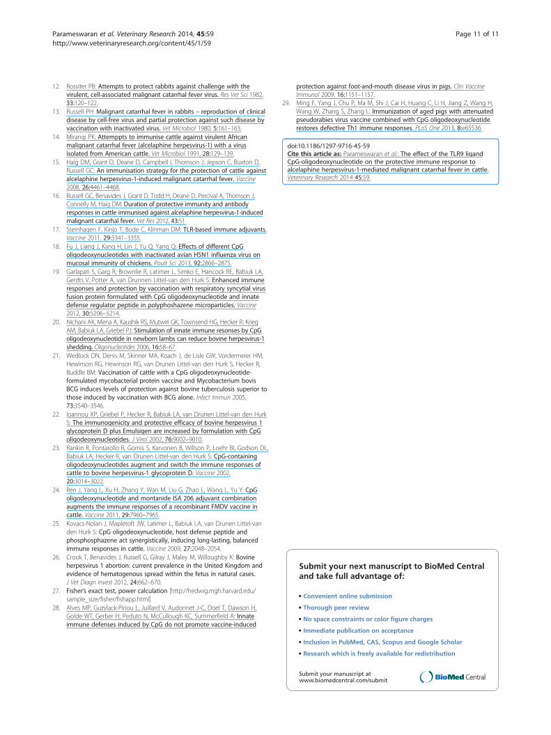
Figure 3 IFN-γ secreting PBMC frequencies. (A, B) IFN-γ-secretingcells expressed as spot-forming cells (SFC) per 106 PBMC. Blood samplesfrom weeks 0, 2, 7 (3 weeks after booster immunisation), 10, 18 and 26(1 week prior to virus challenge- Pre-chall) were taken, as well as week28 and the terminal blood sample prior to euthanasia (Term sample) inboth the MCF and protected animals. A) Analysis of all animals in eachgroup shown as group means and standard deviation of the mean (SD).(B) IFN-γ-secreting cell frequencies in individual animals (displayed as ascatter plot) comparing animals that developed MCF or were protected(PROT). Significant responses were recorded for the pre-challenge andafter challenge terminal samples only. Note there were 3 terminal bloodPBMC samples with 0 SFC in the MCF group and these do not show onthe log scale used.
Parameswaran et al. Veterinary Research 2014, 45:59 Page 8 of 11http://www.veterinaryresearch.org/content/45/1/59
total virus-specific antibody titres in animals after viruschallenge (Figure 2A, 2B), the titres were compared before(week 26) and after challenge in nasal secretions andplasma in animals with MCF or those that were protected(Figure 2G, 2H). The highest titre for each animal samplewas selected from within the various time points afterchallenge to make the comparison more robust. Therewas a significantly higher mean antibody response in thenasal secretions and plasma samples from MCF affectedanimals in groups 1 and 2 after challenge compared to be-fore challenge (Titres of 19 ± 24 for nasal secretion anti-bodies pre-challenge and 252 ± 114 after challenge; titresof 44 ± 13 for plasma antibodies pre-challenge and 484 ±257 after challenge) whereas neither nasal secretion titresnor plasma titres were different before and after challengein the protected animals.
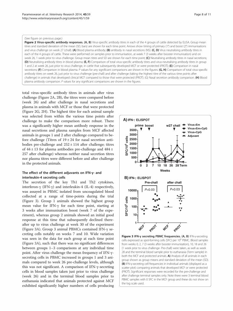
The effect of the different adjuvants on IFN-γ- andinterleukin-4 secreting cellsThe secretion of the key Th1 and Th2 cytokines,interferon-γ (IFN-γ) and interleukin-4 (IL-4) respectively,was assayed in PBMC isolated from uncoagulated bloodcollected at a range of time-points during the trial(Figure 3). Group 1 animals showed the highest groupmean value for IFN-γ for each time point, starting at3 weeks after immunisation boost (week 7 of the expe-riment), whereas group 2 animals showed an initial goodresponse at this time that subsequently declined there-after up to virus challenge at week 30 of the experiment(Figure 3A). Group 3 animal PBMCs contained IFN-γ se-creting cells notably on weeks 7 and 10. Wide variationwas seen in the data for each group at each time point(Figure 3A), such that there was no significant differencesbetween groups 1–3 comparisons at any individual timepoint. After virus challenge the mean frequency of IFN-γ-secreting cells in PBMC increased in groups 1 and 3 ani-mals compared to week 26 pre-challenge levels, althoughthis was not significant. A comparison of IFN-γ-secretingcells in blood samples taken just prior to virus challenge(week 26) and in the terminal blood samples prior toeuthanasia indicated that animals protected against MCFexhibited significantly higher numbers of cells producing
Parameswaran et al. Veterinary Research 2014, 45:59 Page 9 of 11http://www.veterinaryresearch.org/content/45/1/59
IFN-γ (after in vitro stimulation with virus antigen) thanthose that succumbed to MCF (Figure 3B, P = 0.03 for ananalysis on each day).IL-4 was not detected in any of the samples (results
not shown).
DiscussionIn this study, we wished to determine whether the TLR9agonist CpG ODN used with atAlHV-1 alone or in com-bination with Emulsigen and atAlHV-1 could improve themagnitude and duration of protective immunity affordedby our vaccine against AlHV-1 associated MCF. We showthat the inclusion of CpGODN as an adjuvant was noteffective in this regard, nor did it affect the degree ofdisease protection obtained with atALHV-1 and emul-sigen alone. Previous studies using a similar immunisationregime involving CpGODN in BoHV-1 immunisation[22,23] had shown that CpG ODN included with emul-sigen increased the duration of immunity and also en-hanced a IFN-γ response in T cells (Th1-like response) asevidenced by increased cellular production of IFN-γ com-pared to Th2 cytokine responses (IL-4). Furthermore,in other studies where CpG ODN was included with a“traditional” well-established adjuvant (e.g. montanide)and either nominal antigen (hen egg lysozyme [25])Mycoplasma bovis [21] or foot and mouth disease virus(FMDV) [24]) an increase in the duration of immunityand also an enhanced Th1 response as evidenced byincreased cellular production of IFN-γ compared to Th2cytokine responses was recorded. In contrast, in thisstudy, the CpG ODN used in atAlHV-1 vaccine formula-tions on its own or in combinations with Emulsigen as anadjuvant did not significantly alter the period of protectiveimmunity or enhance the magnitude of the immuneresponse parameters measured over that achieved withEmulsigen alone with atAlHV-1 (Figure 1, Table 1,Figure 2). Total virus-specific antibody titres and virus-neutralising antibody titres in blood plasma and nasal se-cretions were not enhanced by the inclusion of CpGODN, and neither was the frequency of IFN-γ expressingblood cells restimulated in vitro. IL-4 secretion by bloodleukocytes was not detected. The reason for the differencebetween our study and the others may be that the com-bination of atALHV-1 with emulsigen is already maxi-mally efficient at inducing an IFN-γ response and thiscannot be improved upon by the inclusion of CPGODN,leading to an increased duration of immunity (assumingthere is a correlation). In aged pigs given pseudorabiesvaccine, a suboptimal Th1 response was restored whenCpGODN was used as an adjuvant (27). Comparing ourstudy with the BoHV-1 subunit ones [22,23], which followa very similar immunisation regime, would indicate thatthe principal difference is in the nature of the immunisingantigens. AtALHV-1 virions were used in our study
compared to BoHV-1 subunit proteins in the other studies[22,23]. This is worthy of further study.In this study, the use of CPG ODN on its own with
atAlHV-1 was not effective in inducing protection toclinical MCF, and this is also the case in the other stud-ies in cattle where CpGODN was used as an adjuvant onits own [21-25] and in pigs with FMDV vaccination [28].We have previously shown that the atAlHV-1 remainsviable when mixed with Emulsigen adjuvant but no evi-dence of virus replication or persistence after prime andboost vaccination has ever been detected [15], and un-published result.In spite of the lack of vaccine improvement by CpG
ODN, which in itself is an important result, there wereseveral other novel, aspects to the study. We have quan-titatively-confirmed our previous observation [16] thatvirus neutralising antibody responses in nasal secretionsjust prior to virus challenge correlated with protection(P = 0.04, Figure 2E, 2F) in group 1 and group 2 animals.Neither plasma antibody (total virus-specific or virus-neutralising antibody) correlated with protection, nor didtotal virus-specific antibody in nasal secretions (Figure 2E,2F). This supports the hypothesis that a mucosal barrierwith neutralising antibody is important to inhibit AlHV-1infection to a point where disease is prevented. Notably,despite 3 of 5 animals being protected in group 3 (CpG-ODN plus atALHV-1 immunised animals), CpG-ODN asadjuvant on its own induced very low or no virus-neutra-lising antibody responses (Figure 2C, 2D). However, theseanimals exhibited moderate total virus-specific antibody re-sponses in both plasma or nasal secretions (Figure 2A, 2B).This suggests that immune mechanisms other than
virus-neutralising antibody may influence protection fromvirAlHV challenge. Unfortunately, despite thorough inves-tigation of methods that could be used for the detection ofcytotoxic T cells (CTLs) in these experiments, no CTLassay that we developed was robust enough (reproduci-bility, sensitivity) to address this aspect of immune re-sponsiveness in this experiment.After challenge with virus, animals that developed MCF
showed a significant increase in virus-specific antibodytitres (before succumbing to disease) compared to levelsprior to challenge, whereas those that were protected didnot (Figure 2G, 2H). This increase was more pronouncedin the plasma of MCF-affected animals than in their nasalsecretions (Titres of 19 ± 24 for nasal secretion antibodiespre-challenge and 252 ± 114 after challenge; titres of 44 ±13 for plasma antibodies pre-challenge and 484 ± 257 afterchallenge). This further reinforces the view that protectedanimals prevent virus from entering the host by the oro-nasal route and thereby stimulating a systemic antibodyresponse. Furthermore, the more pronounced effect ofchallenge on circulating antibody levels in MCF animalssupports this, as virus establishing systemically would be
Parameswaran et al. Veterinary Research 2014, 45:59 Page 10 of 11http://www.veterinaryresearch.org/content/45/1/59
expected to stimulate an increase in plasma antibody ti-tres. In spite of stimulating a high titre antibody response,all but one of the animals succumbed to disease, so thisresponse is not protective. In the clinically-normal animalthat had evidence of infection post-mortem, there was alsoevidence of an antibody response indicating a response toinfection (Table 1). The fact that this animal survived be-yond the time that all others had succumbed to MCFmight indicate that whereas vaccination protects againstdisease it does not protect against infection in all cases, orvaccination induced a delay in onset. In previous experi-ments testing our vaccine, animals succumbed to MCFwithin the time to euthanasia set in this study, with onegroup of survivors showing no signs of MCF up to a yearafter challenge (15,16).The frequency of IFN-γ producing cells (after in vitro
stimulation of PBMC in culture with virus antigen) washigh in group 1 and group 2 animals, indicating a good re-sponse to immunisation and boost with emulsigen withatAlHV-1 (Figure 3A) or emulsigen and CpG ODN withatAlHV-1. This was confirmed by the association of asignificantly-higher frequency of IFN-γ secreting cells inprotected animals compared to those that succumbed toMCF in groups 1 and 2, both in week 26 pre-challengesamples and in PBMC samples taken prior to euthanasia(Figure 3B). The inclusion of CpG ODN with emulsigenin the vaccine formulation (group 2 animals) did notenhance the response compared to that supported byemulsigen alone (Figure 3A), but with CpG ODN on itsown with atAlHV-1 (group 3) there was a small effect onIFN-γ secreting cells. The lack of an augmentation ofIFN-γ producing cells by CpG ODN in group 2 animals isdifferent to other studies, where the adjuvant had an effectin this respect (21–24).IL-4 was not detected in PBMC cultures. IL-4 is a key
indicator of Th2 responses but levels recorded in cattleare usually low in the absence of a strong Th2 response[29]. The lack of detection herein does not rule out Th2involvement, but the frequency of IFN-γ expressing cellsdoes suggest a good Th1 response.Finally, an interesting observation is that animals with
clinical MCF also showed evidence of bacterial pneumoniaat autopsy, whereas animals with no evidence of clinicaldisease did not exhibit the pneumonia. This raises the pos-sibility that MCF might predispose cattle to pneumonia.
ConclusionIn conclusion, CpG ODN did not alter the quality, magni-tude or duration of immunity in cattle immunised againstAlHV-1 MCF when used in combination with emulsigenwhich is in contrast to other studies in cattle showingimproved immunity in immunisation regimes to otherpathogens [21-24]. It may be that further adjuvants are ne-cessary, such as the use of host defense peptide and
phosphosphazine in combination with CpGODN [25]. Theproduction of a mucosal barrier of virus-neutralising anti-body at the oro-nasal transmission entry site of AlHV-1 toprevent infection and the establishment of disease issuccessful in our prime-boost immunisation strategy, butpeaks at approximately two months after booster im-munisation. Mucosal immune responses are tightly regu-lated and finding a way of improving the magnitude andduration of protection against AlHV-1 MCF remains achallenge.
Competing interestsThe authors declare that they have no competing interests.
Authors’ contributionsExperimental design and planning: DMH, NP, GCR, DG, HT, KB; animalexperiments DMH, NP, GCR, DG, HT, KB; pathology analysis DG, HT, MD;serological and immunological analysis DD, NP, DMH, DG; data processingand statistical analysis DD, DG, NP, DMH, GCR; drafting of the manuscriptDMH, MD, GCR. All authors read and approved the manuscript.
AcknowledgementsThis study was funded by a BBSRC /DFID Combatting infectious diseases oflivestock for international development (CDLID) initiative.
Author details1School of Veterinary Medicine and Science, University of Nottingham,Sutton Bonington, LE12 5RD Nottingham, UK. 2Moredun Research Institute,Pentland Science Park, Bush Loan, EH26 0PZ Penicuik, UK.
Received: 5 December 2013 Accepted: 14 May 2014Published: 27 May 2014
References1. Russell GC, Stewart JP, Haig DM: Malignant catarrhal fever: a review.
Vet J 2009, 179:324–335.2. Liggitt HD, DeMartini JC: The pathomorphology of malignant catarrhal
fever. I. Generalized lymphoid vasculitis. Vet Pathol 1980, 17:58–72.3. Liggitt HD, DeMartini JC: The pathomorphology of malignant catarrhal
fever. II. Multisystemic epithelial lesions. Vet Pathol 1980, 17:73–83.4. Simon S, Li H, O’Toole D, Crawford TB, Oaks JL: The vascular lesions of a
cow and bison with sheep-associated malignant catarrhal fever containovine herpesvirus 2-infected CD8(+) T lymphocytes. J Gen Virol 2003,84:2009–2013.
5. Palmeira L, Sorel O, Van Campe W, Boudry C, Roels S, Myster F, Reschner A,Coulie P, Kerkhofs P, Vanderplasschen A, Dewals B: An essential role forγ-herpesvirus latency-associated nuclear antigen homolog in an acutelymphoproliferative disease of cattle. Proc Natl Acad Sci U S A 2013,110:E1933–E1942.
6. Ensser A, Pflanz R, Fleckenstein B: Primary structure of the alcelaphineherpesvirus 1 genome. J Virol 1997, 71:6517–6525.
7. Hart J, Ackermann M, Jayawardane G, Russell G, Haig DM, Reid HW, StewartJP: Complete sequence and analysis of the ovine herpesvirus 2 genome.J Gen Virol 2007, 88:28–39.
8. Bedelian C, Nkedianye D, Herrero M: Maasai perception of the impact andincidence of malignant catarrhal fever (MCF) in southern Kenya. Prev VetMed 2007, 78:296–316.
9. Cleaveland S, Kusiluka L, ole Kuwai J, Bell C, Kazwala R: Assessing theimpact of Malignant Catarrhal Fever in Ngorongoro District, Tanzania.In; 2001. http://www.eldis.org/fulltext/cape_new/MCF_Maasai_Tanzania.pdf.
10. Plowright W, Herniman KA, Jessitt DM, Kalunda M, Rampton CS:Immunisation of cattle against the herpesvirus of malignant catarrhalfever : failure of inactivated culture vaccines with adjuvant. Res Vet Sci1975, 19:159–166.
11. Edington N, Plowright W: The protection of rabbits against theherpesvirus of malignant catarrhal fever by inactivated vaccine. Res VetSci 1980, 28:384–386.
Parameswaran et al. Veterinary Research 2014, 45:59 Page 11 of 11http://www.veterinaryresearch.org/content/45/1/59
12. Rossiter PB: Attempts to protect rabbits against challenge with thevirulent, cell-associated malignant catarrhal fever virus. Res Vet Sci 1982,33:120–122.
13. Russell PH: Malignant catarrhal fever in rabbits – reproduction of clinicaldisease by cell-free virus and partial protection against such disease byvaccination with inactivated virus. Vet Microbiol 1980, 5:161–163.
14. Mirangi PK: Attemppts to immunise cattle against virulent Africanmalignant catarrhal fever (alcelaphine herpesvirus-1) with a virusisolated from American cattle. Vet Microbiol 1991, 28:129–139.
15. Haig DM, Grant D, Deane D, Campbell I, Thomson J, Jepson C, Buxton D,Russell GC: An immunisation strategy for the protection of cattle againstalcelaphine herpesvirus-1-induced malignant catarrhal fever. Vaccine2008, 26:4461–4468.
16. Russell GC, Benavides J, Grant D, Todd H, Deane D, Percival A, Thomson J,Connelly M, Haig DM: Duration of protective immunity and antibodyresponses in cattle immunised against alcelaphine herpesvirus-1-inducedmalignant catarrhal fever. Vet Res 2012, 43:51.
17. Steinhagen F, Kinjo T, Bode C, Klinman DM: TLR-based immune adjuvants.Vaccine 2011, 29:3341–3355.
18. Fu J, Liang J, Kang H, Lin J, Yu Q, Yang Q: Effects of different CpGoligodeoxynucleotides with inactivated avian H5N1 influenza virus onmucosal immunity of chickens. Poult Sci 2013, 92:2866–2875.
19. Garlapati S, Garg R, Brownlie R, Latimer L, Simko E, Hancock RE, Babiuk LA,Gerdts V, Potter A, van Drunnen Littel-van den Hurk S: Enhanced immuneresponses and protection by vaccination with respiratory syncytial virusfusion protein formulated with CpG oligodeoxynucleotide and innatedefense regulator peptide in polyphoshazene microparticles. Vaccine2012, 30:5206–5214.
20. Nichani AK, Mena A, Kaushik RS, Mutwiri GK, Townsend HG, Hecker R, KriegAM, Babiuk LA, Griebel PJ: Stimulation of innate immune resonses by CpGoligodeoxynucleotide in newborn lambs can reduce bovine herpesvirus-1shedding. Oligonucleotides 2006, 16:58–67.
21. Wedlock DN, Denis M, Skinner MA, Koach J, de Lisle GW, Vordermeier HM,Hewinson RG, Hewinson RG, van Drunen Littel-van den Hurk S, Hecker R,Buddle BM: Vaccination of cattle with a CpG oligodeoxynucleotide-formulated mycobacterial protein vaccine and Mycobacterium bovisBCG induces levels of protection against bovine tuberculosis superior tothose induced by vaccination with BCG alone. Infect Immun 2005,73:3540–3546.
22. Ioannou XP, Griebel P, Hecker R, Babiuk LA, van Drunen Littel-van den HurkS: The immunogenicity and protective efficacy of bovine herpesvirus 1glycoprotein D plus Emulsigen are increased by formulation with CpGoligodeoxynucleotides. J Virol 2002, 76:9002–9010.
23. Rankin R, Pontarollo R, Gomis S, Karvonen B, Willson P, Loehr BI, Godson DL,Babiuk LA, Hecker R, van Drunen Littel-van den Hurk S: CpG-containingoligodeoxynucleotides augment and switch the immune responses ofcattle to bovine herpesvirus-1 glycoprotein D. Vaccine 2002,20:3014–3022.
24. Ren J, Yang L, Xu H, Zhang Y, Wan M, Liu G, Zhao L, Wang L, Yu Y: CpGoligodeoxynucleotide and montanide ISA 206 adjuvant combinationaugments the immune responses of a recombinant FMDV vaccine incattle. Vaccine 2011, 29:7960–7965.
25. Kovacs-Nolan J, Mapletoft JW, Latimer L, Babiuk LA, van Drunen Littel-vanden Hurk S: CpG oligodeoxynucleotide, host defense peptide andphosphosphazene act synergistically, inducing long-lasting, balancedimmune responses in cattle. Vaccine 2009, 27:2048–2054.
26. Crook T, Benavides J, Russell G, Gilray J, Maley M, Willoughby K: Bovineherpesvirus 1 abortion: current prevalence in the United Kingdom andevidence of hematogenous spread within the fetus in natural cases.J Vet Diagn Invest 2012, 24:662–670.
27. Fisher’s exact test, power calculation [http://hedwig.mgh.harvard.edu/sample_size/fisher/fishapp.html]
28. Alves MP, Guzylack-Piriou L, Juillard V, Audonnet J-C, Doel T, Dawson H,Golde WT, Gerber H, Peduto N, McCullough KC, Summerfield A: Innateimmune defenses induced by CpG do not promote vaccine-induced
protection against foot-and-mouth disease virus in pigs. Clin VaccineImmunol 2009, 16:1151–1157.
29. Ming F, Yang J, Chu P, Ma M, Shi J, Cai H, Huang C, Li H, Jiang Z, Wang H,Wang W, Zhang S, Zhang L: Immunization of aged pigs with attenuatedpseudorabies virus vaccine combined with CpG oligodeoxynucleotiderestores defective Th1 immune responses. PLoS One 2013, 8:e65536.
doi:10.1186/1297-9716-45-59Cite this article as: Parameswaran et al.: The effect of the TLR9 ligandCpG-oligodeoxynucleotide on the protective immune response toalcelaphine herpesvirus-1-mediated malignant catarrhal fever in cattle.Veterinary Research 2014 45:59.
Submit your next manuscript to BioMed Centraland take full advantage of:
• Convenient online submission
• Thorough peer review
• No space constraints or color figure charges
• Immediate publication on acceptance
• Inclusion in PubMed, CAS, Scopus and Google Scholar
• Research which is freely available for redistribution
Submit your manuscript at www.biomedcentral.com/submit