Artificial Intelligence to Extract, Analyze and Generate ...
Upload
khangminh22Category
view
1download
0
Research ArticleThe Effect of Hydroalcoholic Calendula Officinalis Extract onAndrogen-Induced Polycystic Ovary Syndrome Model inFemale Rat
Fatemeh Gharanjik ,1 Manzar Banoo Shojaeifard ,2,3 Narges Karbalaei ,4
and Marzieh Nemati 5
1Student Research Committee, Fasa University of Medical Sciences, Fasa, Iran2Physiology Department, Fasa University of Medical Sciences, Fasa, Iran3Ionizing and Non-ionizing Radiation Protection Research Center (INIRPRC), School of Paramedical Sciences, Shiraz University ofMedical Sciences, Shiraz, Iran4Histomorphometry and Stereology Research Center, Shiraz University of Medical Sciences, Shiraz, Iran5Endocrine and Metabolism Research Center, Shiraz University of Medical Sciences, Shiraz, Iran
Correspondence should be addressed to Manzar Banoo Shojaeifard; [email protected] Narges Karbalaei; [email protected]
Received 29 January 2022; Revised 12 May 2022; Accepted 4 June 2022; Published 7 July 2022
Academic Editor: Flavia Prodam
Copyright © 2022 Fatemeh Gharanjik et al. This is an open access article distributed under the Creative Commons AttributionLicense, which permits unrestricted use, distribution, and reproduction in any medium, provided the original work isproperly cited.
Background. Polycystic ovary syndrome (PCOS) is the most common hormonal disorder in women of reproductive age, and themajor cause of infertility. Today, using medicinal plants instead of chemical drugs could be an alternative treatment option forPCOS. The purpose of this study was to determine the effect of Calendula officinalis hydroalcoholic extract on PCOS in rats.Method. 60 female adult rats were randomly divided into six groups, including control, sham, PCOS group, and treated PCOSgroups receiving hydroalcoholic extract of Calendula officinalis with different dosages of 200, 500, and 1000mg/kg. PCOS wasinduced by subcutaneous injection of DHEA 6mg/100 g bw for 35 days. For two weeks, the extract was taken orally. Theserum glucose, insulin, sex hormone levels, and oxidative status were measured at the end of the experiment. The ovaries weredissected for histomorphometric and pathological analysis. Results. When compared to the control and sham groups, thePCOS group showed a significant increase in glucose, insulin, testosterone, and malondialdehyde (MDA) concentrations, cysticand atretic follicles, and thickness of the theca and tunica albuginea layers, and a significant decrease in LH concentration,total antioxidant capacity, corpus luteum, antral follicles, and oocyte diameter. The mean concentration of FSH, on the otherhand, did not change significantly. A trend of improvement was found in the treated groups with high doses of Calendulaofficinalis extract. Conclusion. In rats with PCOS and nonovulation, Calendula officinalis hydroalcoholic extract improvedoxidative stress, restored folliculogenesis, and increased ovulation.
1. Introduction
Polycystic ovary syndrome (PCOS) is a heterogeneous meta-bolic and endocrine disorder that affects 5% to 10% of womenof reproductive age and is the leading cause of infertility/sub-fertility [1]. This multifactorial disorder is distinguished byendocrine abnormalities such as hyperandrogenism, disturbed
folliculogenesis, polycystic ovaries, menstrual cycle disrup-tions, and chronic anovulation [2, 3].
The pathophysiology of PCOS is characterized by pri-mary defects in the hypothalamic-pituitary-gonad (HPG)axis, which increases the GnRH frequency and LH pulsationsecretion, resulting in a variety of metabolic disorders suchas excessive ovarian androgen production, ovarian dysfunc-
HindawiBioMed Research InternationalVolume 2022, Article ID 7402598, 16 pageshttps://doi.org/10.1155/2022/7402598

tion, and insulin resistance. In fact, PCOS is associated withirregular gonadotropin secretion, increased steroid hormonesecretion and frequency, and a high LH/FSH ratio, which leadsto an increase in androgen synthesis and prevents normal fol-licle development [4]. A lot of findings suggest that oxidativestress plays a role in the pathophysiology of PCOS, resultingin complications such as altered steroidogenesis in the ovaries,disrupted follicular development, hyperandrogenemia, infer-tility, insulin resistance, and chronic inflammation [5, 6].
Insulin resistance is common in PCOS, and it increasesthe risk of type 2 diabetes and other metabolic abnormalities.PCOS is related to a variety of diseases, including anxiety,depression, breast and endometrial cancers, neurological,and psychological disorders [4, 7].
To manage PCOS, various treatment strategies havebeen used, including a healthy lifestyle and regular exercise,ovarian laparoscopic surgery, and medication therapy withdrugs such as glucocorticoids, tamoxifen, clomiphene cit-rate, aromatase inhibitors, and metformin [8, 9]. Herbalmedications have recently received considerable attentionas a means of reducing the side effects of chemical drugs,increasing their effectiveness, and lowering their cost.
Calendula officinalis Linn (C. officinalis), a member ofthe Asteraceae family, is an annual aromatic herb with yel-low or golden-orange flowers [10]. C. officinalis is also calledpot marigold. Due to having a diverse range of biologicallyactive substances such as phenolic compounds (flavonoidsand phenolic acids) and triterpenic alcohols, steroids andsterols quinines, carotenoids, glycosidesvolatile oil, andamino acids, it has been reported that C. officinalis possessesanti-inflammatory, antioxidant, antidiabetic, antiulcer, heal-ing, antiseptic, and analgesic properties [11, 12]. It is used totreat gastrointestinal, gynaecological, and eye diseases [13].It also relieves pain caused by stomach ulcers and inflamma-tion, treats stomach ulcers and liver conjunctivitis [14], andprevents sinus infections [15]. It regulates bleeding andcauses menstruation [12, 14].
Studies have shown various pharmacological activities,such as nephroprotective, hepatoprotective, hypoglycemic,hypolipidemic, and antioxidant potential of C. officinalis inexperimental and clinical models. Due to the biologicallyactive compounds in Calendula officinalis, as well as its anti-oxidant and anti-inflammatory properties, this study aimedto investigate the effect of the C. officinalis hydroalcoholicextract on glucose and hormonal levels, oxidative stress,and histological parameters of the ovary in rats with PCOS.
2. Methods
2.1. Ethic, Animals, and Study Design. Female adult Sprague-Dawley rats weighing approximately 150-160 grams wereobtained from the stock of rats bred in the animal house ofthe Research Institute of Shiraz University of Medical Sciences(Shiraz, Iran) and housed under standard conditions (temper-ature 22 ± 2°C and relative humidity 35 ± 3%, 12h light cycle)with free access to food and water. The Animal Care and UseCommittee of the Fasa University of Medical Sciences autho-rized all experimental protocols (IR.FUMS.REC.1397.051).
Sixty female rats were randomly divided into six groupsof ten: The control group received no treatment at all, thesham group received sesame oil subcutaneously for 35 con-secutive days and distilled water for two weeks. After induc-ing PCOS with dehydroepiandrosterone (DHEA), rats weregiven distilled water for two weeks. The treated PCOSgroups were given hydroalcoholic extracts of Calendula offi-cinalis in doses of 200, 500, and 1000mg/kg body weight,respectively, for two weeks. Oral gavage was used to admin-ister distilled water and hydroalcoholic extracts of Calendulaofficinalis.
At the end of the experiment, animals were anesthetizedwith ketamine (80mg/kg) and xylazine (5mg/kg) and bloodsample was collected through the cardiac puncture; then, theserum was separated and stored in -20 for hormonal andantioxidant marker assay. Animals were then sacrificed,and their ovaries were removed and weighed for pathologi-cal and histomorphometric assessment (Figure 1).
2.2. PCOS Induction. PCOS was induced by injecting DHEA(6mg per 100 g body weight diluted in 0.2ml of sesame oil)subcutaneously for 35 days [16, 17].
Vaginal smears were taken daily to ensure polycysticovaries. Ovarian cycles were regular at the start of dailyDHEA injections into rats in the estrous stage of their repro-ductive cycle.
2.3. Vaginal Smears. The regularity of the estrous cycle wasassessed using vaginal smears. To make the smear, first,inject 0.3ml of physiological serum into the animal’s vagina,then extract some of the vaginal fluid, and deposit it over theslide. After drying, it was fixed with alcohol and diluted for20 minutes with a 1 : 20 diluted Giemsa stain. It was thenslowly rinsed with distilled water before being examinedunder an optical microscope with a magnification of 40times. Various stages of the estrous cycle (proestrous,estrous, metestrous, and deoestrous) were identified basedon the type of cells in the vaginal smear. Rats in the estrousstage of their reproductive cycle were chosen for the nextstages of the study (Figure 2).
2.4. Calendula Officinalis Hydroalcoholic Extract Preparation.The Calendula officinalis plant was approved by ShirazUniversity of Medical Sciences, Faculty of Pharmacy, with avoucher number of 1094. Calendula officinalis was cleaned,dried, and ground into a powder after being sent to the labora-tory. Afterward, 100 grams of calendula officinalis powder and500ml of ethanol 70% were poured into the percolator for 72hours to extract the contents of the plant cells. Then, the per-colator valve was opened, and the drippy liquid was pouredinto the lower container. It should be mentioned that the sol-vent (alcohol) was regularly poured into the top of the con-tainer with the help of a pipette to keep the liquid levelstable and prevent the plant powder from drying out. Next,the alcoholic solvent was removed from the resultant liquidand it was entirely concentrated using a rotary device. Theextract was converted into a blackish-brown powder using adesiccator and a vacuum pump. The amount of dry matter
2 BioMed Research International

obtained by percolation from 100g of calendula officinalishydroalcoholic extract was 18g.
2.5. Evaluation of Serum Biochemical Markers. The ratinsulin enzyme-linked immunosorbent assay kit (ELISA,Mercodia, and Uppsala, Sweden) was used to determineserum insulin concentration. The sensitivity of the insulinassay is 0.15μg/L with intra- and inter-assay coefficients ofvariations of 4.1 and 7.8%, respectively. Rat commercial hor-monal kits (ZellBio GmbH company in Germany) and theELISA method were used to measure levels of testosteronewith intra- and interassay coefficients of variations of 5.1and 9.6%, respectively; progesterone with intra- and interas-say coefficients of variations of 5.9 and 10.8%, respectively;estrogen with intra- and interassay coefficients of variationsof 6.2 and 11.1%, respectively; LH with intra- and interassaycoefficients of variations of 4.7 and 8.9%, respectively; andFSH with intra- and interassay coefficients of variations of3.8 and 7.3%, respectively. A glucose oxidase method kit
(Pars Azmoon, Tehran, Iran) was also used to measure bloodglucose levels with intra- and interassay coefficients of varia-tions of 3.4 and 6.5%, respectively. The homeostatic modelassessment of IR (HOMA-IR) index was computed as follows:½ fasting serum glucose levels ðmmol/lÞ�, ½insulin ðIU/mlÞ�/22:5.2.6. Evaluation of Oxidative Stress Markers. The serum levelof Malondialdehyde (MDA) level, as the lipid peroxidationindex, was assessed by the thiobarbituric acid reactive sub-stances (TBARS) method. The total antioxidant capacity(TAC) was quantified by commercially available assay kits(ZellBio GmbH, Ulm, Germany), using a colorimetricmethod. The TAC (mmol/L) was measured by reducingthe colorless tripiridyltriazine complex (Fe3+-TPTZ) to theferrous form complex (Fe2+-TPTZ) and detecting it spectro-photometrically at 593nm.
2.7. Histopathological Analyses. Rats were anesthetized withketamine (80mg/kg) and xylazine (5mg/kg). The ovarieswere immediately removed and weighted after a small
Induction of PCOPthrough DHEA
Normal ovary Polycystic ovaryDeveloping
primary follicle
Mature(Graafian)follicle)
Maturefollicle
Cysts
PCOS
Reduce PCOS symptoms
Improve hyper androgenism
Reduction in ovarian cysts number
Improve ovulation
Reduction in insulin resistance
Reduction in body and ovarian weight
Hyer androgenism
Anovulation
Insulin resistance
Obesity
Gavage of calendulaofficinalis extract
Figure 1: Rat model exhibiting both ovarian and characteristics of polycystic ovarian syndrome.
Proestrous Estrous Metestrous Deoestrous
Figure 2: Stages of the estrous cycles.
3BioMed Research International
ventral midline laparotomy. The right ovaries were thencleaned, dehydrated, and fixed in 10% formaldehyde (Merck,Germany). Five-micron-thick sections were prepared using amicrotome, and one section out of ten and two sections fromeach sample were chosen and stained with hematoxylin-eosin. In the final stage, the prepared tissue sections wereobserved under an optical microscope (Nikon binocularoptical microscope model E200) with a magnification of400 and different follicular groups, including primordial,unilaminar, multilaminar, antral, graafian, corpus luteum,atretic, and cystic follicles were counted. Moreover, theovum, antral follicle, and corpus luteum diameters, as wellas the thicknesses of the tonic allogena, granulose, theca,and zonaplucida, were measured [18].
2.8. Statistical Analysis. GraphPad Prism version 6 softwarewas used to analyze the data, and the results were reportedas means ± S:E:M. All data were analyzed using one-wayANOVA (post hoc: Tukey). A value of P < 0:05 was consid-ered statistically significant.
3. Results
3.1. Body and Ovaries Weight. The mean body weight andabsolute and relative ovary weights of the animals in allexperimental groups were measured. A significant increasein the mean body weight and absolute and relative ovarianweights was observed in the PCOS group compared to thecontrol and sham groups (P < 0:001) (Table 1). In all PCOSanimals receiving the hydroalcoholic extract of Calendulaofficinalis (PCOS+Cal200, PCOS+Cal500, PCOS+Cal1000),the mean body weight was lower than the PCOS group,but this difference was significant only in the PCOS+Cal1000 (P = 0:006). The absolute weight of the ovaries inall PCOS groups receiving hydroalcoholic Calendula offici-nalis extract and relative ovary weight in PCOS+Cal500and PCOS+Cal1000 showed a significant decrease comparedto the PCOS group (P < 0:01). There were no significant dif-ferences in absolute and relative weights of the ovariesamong the PCOS+Cal500, PCOS+Cal1000, control, andsham groups. However, in animals receiving the minimumdose of Calendula officinalis (PCOS+Cal200), the absoluteand relative ovary weights were significantly more than thecontrol and the sham groups (P = 0:003) (Table 1).
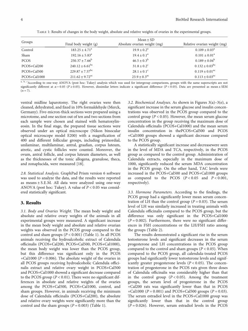
3.2. Biochemical Analyses. As shown in Figures 3(a)–3(e), asignificant increase in the serum glucose and insulin concen-trations was observed in the PCOS group compared to thecontrol group (P < 0:05). However, the mean serum glucoseconcentration in the group receiving the maximum dose ofCalendula officinalis (PCOS+Cal1000) and the mean seruminsulin concentration in thePCOS+Cal500 and PCOS+Cal1000 groups showed a significant decrease comparedto the PCOS group.
A statistically significant increase and decreasewere seenin the level of MDA and TCA, respectively, in the PCOSgroup as compared to the control group. Administration ofCalendula extracts, especially in the maximum dose of1000, significantly reduced the serum MDA concentrationin the PCOS group. On the other hand, TAC levels wereincreased in the PCOS+Cal500 and PCOS+Cal1000 groupsas compared to the PCOS (P < 0:05 and P < 0:001,respectively).
3.3. Hormone Parameters. According to the findings, thePCOS group had a significantly lower mean serum concen-tration of LH than the control group (P = 0:03). The serumlevel of LH was similarly increased in treating animals withCalendula officinalis compared to the PCOS group, but thedifference was only significant in the PCOS+Cal1000(P = 0:002). Furthermore, there were no significant differ-ences in FSH concentrations or the LH/FSH ratio amongthe groups (Table 2).
The results demonstrated a significant rise in the serumtestosterone levels and significant decreases in the serumprogesterone and LH concentrations in the PCOS groupcompared to the control and sham groups (P < 0:05). Whencompared to the PCOS group, all calendula-treated PCOSgroups had significantly lower testosterone levels and signif-icantly greater progesterone levels (P < 0:05). The concen-tration of progesterone in the PCOS rats given three dosesof Calendula officinalis was considerably higher than thatin the control group (P < 0:05). Among the treatmentgroups, the serum level of progesterone in the PCOS+Cal200 rats was significantly lower than that in PCOS+Cal1000 (P = 0:001) and PCOS+Cal500 groups (P = 0:03).The serum estradiol level in the PCOS+Cal1000 group wassignificantly lower than that in the control group(P = 0:026). However, serum estradiol levels in the PCOS
Table 1: Results of changes in the body weight, absolute and relative weights of ovaries in the experimental groups.
GroupsMean ± SD
Final body weight (g) Absolute ovarian weight (mg) Relative ovarian weight (mg)
Control 183:25 ± 4:71a 19:9 ± 0:2a 0:109 ± 0:03a
Sham 192:16 ± 5:83a 19:4 ± 0:1a 0:101 ± 0:01a
PCOS 250:37 ± 7:66b 46:5 ± 0:3b 0:189 ± 0:04b
PCOS+Cal200 240:12 ± 6:61bc 31:8 ± 0:2c 0:132 ± 0:03bc
PCOS+Cal500 229:87 ± 7:57bc 28:1 ± 0:1c 0:119 ± 0:01ac
PCOS+Cal1000 211:62 ± 9:72ac 23:9 ± 0:3ac 0:113 ± 0:03aca, b, cAccording to one-way ANOVA (post hoc: Tukey) analysis which was used for intergroup comparisons, groups with the same superscripts are notsignificantly different at α = 0:05 (P ≥ 0:05). However, dissimilar letters indicate a significant difference (P < 0:05). Data are presented as mean ± SEM(n = 7).
4 BioMed Research International
Cont
rol
Sham
PCO
S
PCO
S+Ca
l200
PCO
S+Ca
l500
PCO
S+Ca
l100
0
0
50
100
150
200Se
rum
glu
cose
conc
entr
atio
n (m
g/dl
)
a
A AB A
B
AB
AB
p = 0.018 p = 0.002
(a)
Cont
rol
Sham
PCO
S
PCO
S+Ca
l200
PCO
S+Ca
l500
PCO
S+Ca
l100
0
0.0
0.5
1.0
1.5
Seru
m in
sulin
conc
entr
atio
n (𝜇
g/l)
A AAA
B
AB
p = 0.004p = 0.003 p = 0.019 p = 0.024
(b)
Cont
rol
Sham
PCO
S
PCO
S+Ca
l200
PCO
S+Ca
l500
PCO
S+Ca
l100
0
0
5
10
15
HO
MA
-IR
A A
B
AA
A
p <
0.0
01
p <
0.0
01
p <
0.0
01
p <
0.0
01
p <
0.0
01
(c)
Cont
rol
Sham
PCO
S
PCO
S+Ca
l200
PCO
S+Ca
l500
PCO
S+Ca
l100
0
0
5
10
15
Seru
m M
DA
(𝜇m
ol/L
)
AA
B
ABCABC
AC
p =
0.0
09
p =
0.0
2
p =
0.0
2
(d)
Figure 3: Continued.
5BioMed Research International
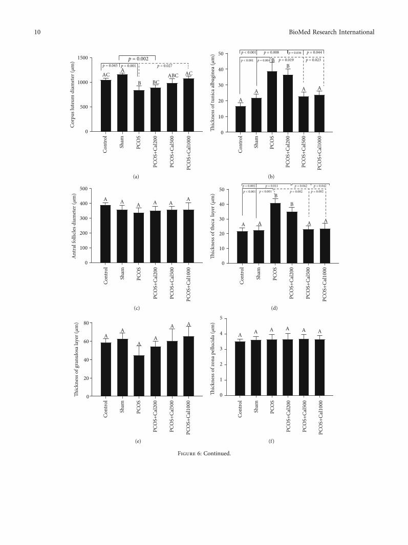
+Cal200, PCOS+Cal500, and control groups did not differsignificantly (Table 2).
3.4. Sex Cycle Changes. Sex cycle changes were assessed 8days after the end of the experiment. As shown inFigure 4, the sex cycle in the control and sham groups was100% normal. However, in the PCOS and PCOS+Cal200groups, the sex cycle was 100% irregular. In the PCOSgroups receiving moderate (500) and maximum (1000)doses of calendula officinalis, the sex cycles were37.5% and62.5% of the controls, respectively.
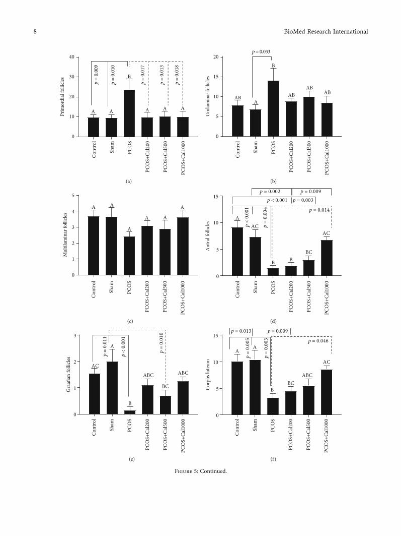
3.5. Histomorphometry and Pathological and Evaluation. Asshown in Figures 5(a)–5(h), there was no significant differ-
ence in histomorphometry characteristics between the con-trol and sham groups. When compared to the control andsham groups, the PCOS group had a substantial rise in thenumber of primordial, unilaminar, atretic, and cystic folli-cles and a significant decrease in the number of antral, graa-fian, and corpus luteum follicles. The number of primordialfollicles in all PCOS animals receiving Calendula officinalishydroalcoholic extract was the same as those in the controland sham groups, and there was no significant differencebetween them. In addition, as compared to the PCOS groupthat received the lowest dosage, a significant rise in the num-ber of antral follicles, corpus luteum and a significantdecrease in the number of atretic and cystic follicles werefound in the PCOS group that received the highest dose
Cont
rol
Sham
PCO
S
PCO
S+Ca
l200
PCO
S+Ca
l500
PCO
S+Ca
l100
0
0.0
0.5
1.0
1.5
Seru
m T
AC
(mm
ol/L
)
A A
B BC
ACDAD
p < 0.001
p <
0.0
01
p <
0.0
01
p <
0.0
01
p =
0.0
18
p < 0.001 p = 0.002
(e)
Figure 3: Fasting blood glucose (a), insulin (b), HOMA-IR (c), MDA (d), and TAC (e) levels in the experimental groups. According toone-way ANOVA (post hoc: Tukey) analysis which was used for intergroup comparisons, groups with the same letters were notsignificantly different at α = 0:05 (P ≥ 0:05). However, dissimilar letters indicate a significant difference (P < 0:05). Data are presentedas mean ± SEM(n = 7).
Table 2: Changes in sex hormones in the experimental groups.
GroupsMean ± SD
LH (mIU/ml) FSH (mIU/ml) LH/FSH Testosterone (ng/ml) Estradiol (pg/ml) Progesterone (ng/ml)
Control 0:808 ± 0:097a 1:69 ± 0:24a 0:500 ± 0:10a 2:16 ± 0:30a 339:5 ± 39:96a 8:25 ± 0:34a
Sham 0:706 ± 0:062ab 1:60 ± 0:24a 0:430 ± 0:06a 2:24 ± 0:17a 327:8 ± 79:64a 7:60 ± 0:27a
PCOS 0:681 ± 0:044b 1:59 ± 0:30a 0:458 ± 0:10a 5:70 ± 0:72b 328:6 ± 43:85a 4:37 ± 0:10b
PCOS+Cal200 0:750 ± 0:162ab 1:48 ± 0:13a 0:548 ± 0:08a 2:79 ± 0:39ac 299:7 ± 69:96a 12:38 ± 0:38c
PCOS+Cal500 0:810 ± 0:072ab 1:57 ± 0:11a 0:477 ± 0:08a 2:53 ± 0:30ac 278:3 ± 76:68a 22:86 ± 1:71d
PCOS+Cal1000 0:852 ± 0:071a 1:90 ± 0:33a 0:521 ± 0:14a 2:91 ± 0:23a 214:1 ± 58:98b 32:99 ± 1:57d
According to one-way ANOVA (post-hoc: Tukey) analysis which was used for intergroup comparisons, groups with the same letters were not significantlydifferent at α = 0:05 (P ≥ 0:05). Dissimilar letters, on the other hand, revealed a significant difference (P < 0:05). Data are presented as mean ± SEM (n = 8).
6 BioMed Research International

P
Control
E
M
Rat 1
Rat 2
Rat 3
Rat 4
Estro
us cy
cleRa
t 5Ra
t 6Ra
t 7Ra
t 8
D
01 2 3 4 5 6 7 8
P
Sham PCOS PCOS+Cal 200 PCOS+Cal 500 PCOS+Cal 1000
E
M
D
01 2 3 4 5 6 7 8
PE
M
D
01 2 3 4 5 6 7 8
PE
M
D
01 2 3 4 5 6 7 8
PE
M
D
01 2 3 4 5 6 7 8
PE
M
D
01 2 3 4 5 6 7 8
PE
M
D
01 2 3 4 5 6 7 8
PE
M
D
01 2 3 4 5 6 7 8
PE
M
D
01 2 3 4 5 6 7 8
PE
M
D
01 2 3 4 5 6 7 8
PE
M
D
01 2 3 4 5 6 7 8
PE
M
D
01 2 3 4 5 6 7 8
PE
M
D
01 2 3 4 5 6 7 8
PE
M
D
01 2 3 4 5 6 7 8
PE
M
D
01 2 3 4 5 6 7 8
PE
M
D
01 2 3 4 5 6 7 8
PE
M
D
01 2 3 4 5 6 7 8
PE
M
D
01 2 3 4 5 6 7 8
PE
M
D
01 2 3 4 5 6 7 8
PE
M
D
01 2 3 4 5 6 7 8
PE
M
D
01 2 3 4 5 6 7 8
PE
M
D
01 2 3 4 5 6 7 8
PE
M
D
01 2 3 4 5 6 7 8
PE
M
D
01 2 3 4 5 6 7 8
PE
M
D
01 2 3 4 5 6 7 8
PE
M
D
01 2 3 4 5 6 7 8
PE
M
D
01 2 3 4 5 6 7 8
PE
M
D
01 2 3 4 5 6 7 8
PE
M
D
01 2 3 4 5 6 7 8
PE
M
D
01 2 3 4 5 6 7 8
PE
M
D
01 2 3 4 5 6 7 8
PE
M
D
01 2 3 4 5 6 7 8
PE
M
D
01 2 3 4 5 6 7 8
PE
M
D
01 2 3 4 5 6 7 8
PE
M
D
01 2 3 4 5 6 7 8
PE
M
D
01 2 3 4 5 6 7 8
PE
M
D
01 2 3 4 5 6 7 8
PE
M
D
01 2 3 4 5 6 7 8
PE
M
D
01 2 3 4 5 6 7 8
PE
M
D
01 2 3 4 5 6 7 8
PE
M
D
01 2 3 4 5 6 7 8
PE
M
D
01 2 3 4 5 6 7 8
PE
M
D
01 2 3 4 5 6 7 8
PE
M
D
01 2 3 4 5 6 7 8
PE
M
D
01 2 3 4 5 6 7 8
PE
M
D
01 2 3 4 5 6 7 8
PE
M
D
01 2 3 4 5 6 7 8
PE
M
D
01 2 3 4 5 6 7 8
Days of estrous
Figure 4: Estrous cycle pattern in the control, sham, PCOS, PCOS+Cal200, PCOS+Cal500, and PCOS+Cal1000 groups (receivinghydroalcholic extract of Calendula officinalis with dosages of 200, 500, and 1000mg/kg). The results of eight representative rats fromeach group are shown. P: proestrous; E: estrous; M: metestrous; D: diestrous.
7BioMed Research International
Cont
rol
Sham
PCO
S
PCO
S+Ca
l200
PCO
S+Ca
l500
PCO
S+Ca
l100
0
0
10
20
30
40Pr
imor
dial
folli
cles
A A
B
A A A
p =
0.0
09
p =
0.0
10
p =
0.0
17
p =
0.0
13
p =
0.0
18(a)
Cont
rol
Sham
PCO
S
PCO
S+Ca
l200
PCO
S+Ca
l500
PCO
S+Ca
l100
0
0
5
10
15
20
Uni
lamin
arfo
llicle
s
ABA
B
ABAB
AB
p = 0.033
(b)
Cont
rol
Sham
PCO
S
PCO
S+Ca
l200
PCO
S+Ca
l500
PCO
S+Ca
l100
0
0
1
2
3
4
5
Mul
tilam
inar
folli
cles
A A
A
A A
A
(c)
Cont
rol
Sham
PCO
S
PCO
S+Ca
l200
PCO
S+Ca
l500
PCO
S+Ca
l100
0
0
5
10
15
Antra
lfol
licles
AAC
B BBC
AC
p <
0.0
01 p = 0.014
p < 0.001 p = 0.003p = 0.002 p = 0.009
p =
0.0
04
(d)
Cont
rol
Sham
PCO
S
PCO
S+Ca
l200
PCO
S+Ca
l500
PCO
S+Ca
l100
0
0
1
2
3
Gra
afian
folli
cles
AC
A
B
ABC
BC
ABC
p <
0.0
01
p =
0.0
11
p =
0.0
10
(e)
Cont
rol
Sham
PCO
S
PCO
S+Ca
l200
PCO
S+Ca
l500
PCO
S+Ca
l100
0
0
5
10
15
Corp
uslu
teum
AA
BBC
ABC
AC
p =
0.0
05
p = 0.009
p = 0.046
p =
0.0
03
p = 0.013
(f)
Figure 5: Continued.
8 BioMed Research International

(P < 0:05). There was no significant change in the number ofgraafian follicles between the groups receiving Calendulaofficinalis extract and the PCOS group (Figure 5(e)). Whencompared to the control and sham groups, the group receiv-ing the lowest dosage of Calendula officinalis extract showeda significant reduction in the number of antral follicles andcorpus luteum; in addition, in this group, the number ofantral follicles decreased significantly as compared to thegroup that received the highest dose (P < 0:01) (Figure 5(d)).
As shown in Figures 6(a)–6(g), a significant decrease inthe diameter of the corpus luteum and the oocyte, as wellas a significant rise in the thickness of the theca and tunicaalbuginea layers was observed in the PCOS group whencompared to the control and sham groups. Furthermore, asignificant increase in the corpus luteum and oocyte diame-ter, as well as a significant decrease in the thickness of thetheca and tunica albuginea layers, was observed in the PCOSgroups which received the maximum and average doses ofCalendula officinalis hydroalcoholic extract, compared tothe PCOS group (P < 0:05). When compared to the shamgroup, the PCOS group receiving the hydroalcoholic extractof Calendula officinalis at a dosage of 200mg/kg had a sig-nificant decrease in the thickness of the corpus luteum anda significant increase in the thickness of the theca and tunicaalbuginea layers. The findings revealed a significant differ-ence (P < 0:05) in the number of multilaminar follicles, thethickness of the granulosa and zona pellucida layers, andthe diameter of the antral follicle between the examinedgroups (Figures 5 and 6).
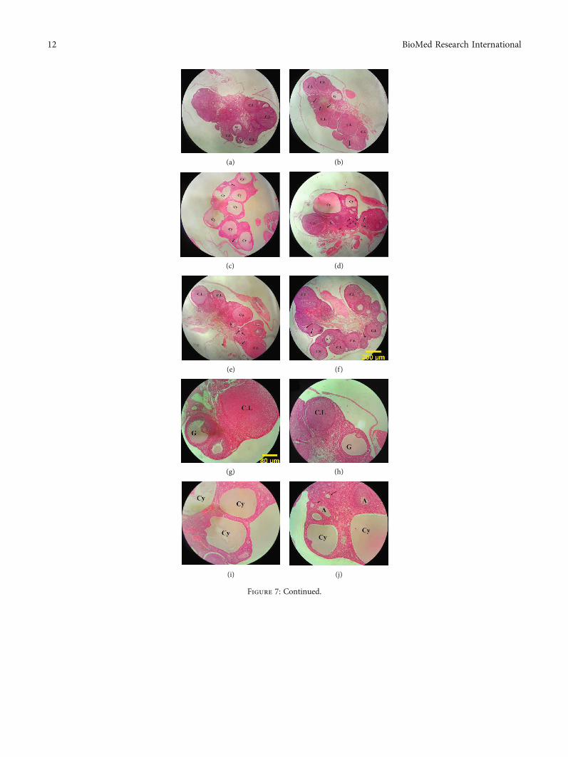
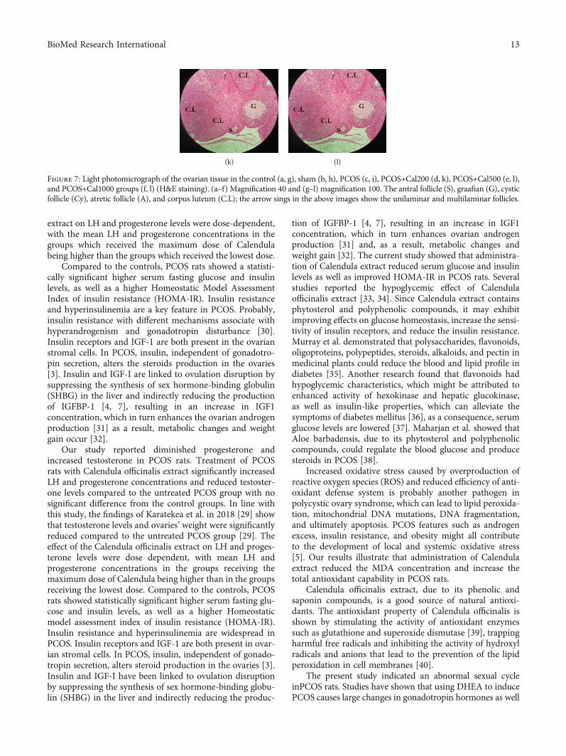
There was no pathological damage identified in the con-trol and sham groups (Figures 7(a), 7(b), 7(g), and 7(h));hence, the structure of the ovarian tissue and the processof folliculogenesis appeared to be entirely normal. Patholog-ical and morphometric abnormalities of the ovaries, includ-ing decreased diameter and number of corpus luteum,
decreased ovarian diameter, and increased ovarian volumeby cystic and atrial follicles, were found in the PCOS group(Figures 7(c) and 7(i)). Among PCOS rats treated with mari-gold extract, the animals which received the lowest doseshowed a nonsignificant change in the number of cysticand atretic follicles and corpus luteum, as well as the thick-ness of theca and tunica albuginea layers, as compared tothe PCOS group (Figures 7(d) and 7(j)). Although therewas less pathological damage to the ovaries in the PCOS+Cal500 group, there were still a number of atretic folliclesin the ovarian tissue. The number of cystic follicles in thisgroup decreased significantly when compared to the PCOSgroup, while the quantity of corpus luteum did not altermuch when compared to the PCOS group (Figures 7(e)and 7(k)). The pathological damage to the ovaries was con-siderably decreased in the PCOS+Cal1000 group, indicatingthat the existence of a high number of corpus luteum impliesa normal process of ovulation and folliculogenesis. Whencompared to the PCOS group, there was a significantdecrease in the number of cystic and atretic follicles in thePCOS+Cal1000 group. Furthermore, the thickness of thecaand tunica albuginea layers, as well as ovum diameter, wasapproximately similar to those of the control and shamgroups, indicating that ovarian tissue damage was greatlydecreased (Figures 7(f) and 7(l)).
4. Discussion
The purpose of this research was to evaluate the effects ofhydroalcoholic Calendula Officinalis extract on the serumlevels of gonadotropin and sex hormones, oxidative stress,insulin resistance, and the morphology of the ovarian tissuein female rats with androgen-induced polycystic ovary syn-drome. Results of this study confirm the subcutaneous
Cont
rol
Sham
PCO
S
PCO
S+Ca
l200
PCO
S+Ca
l500
PCO
S+Ca
l100
0
0
10
20
30A
tretic
folli
cles
A
A A
B
AB AB
p <
0.0
01
p =
0.0
08
p =
0.0
19
(g)
Cont
rol
Sham
PCO
S
PCO
S+Ca
l200
PCO
S+Ca
l500
PCO
S+Ca
l100
0
0
1
2
3
4
5
Cyst
icfo
llicle
s
A A
B
BC
ACA
p <
0.0
01
p = 0.028p = 0.005
p <
0.0
01
p <
0.0
01
p <
0.0
01
p = 0.007
(h)
Figure 5: The bar graph of the number of primordial (a), unilaminar (b), multilaminar (c), antral (d), graafian (e), corpus luteum (f), atretic(g), and cystic follicles (h). The significant differences are exhibited on each column. (A–D) According to one-way ANOVA (post hoc:Tukey) analysis which was used for intergroup comparisons, groups with same superscripts were not significantly different at α = 0:05(P ≥ 0:05). However, dissimilar letters indicate a significant difference (P < 0:05).
9BioMed Research International
Cont
rol
Sham
PCO
S
PCO
S+Ca
l200
PCO
S+Ca
l500
PCO
S+Ca
l100
0
0
500
1000
1500
Corp
us lu
teum
dia
met
er (𝜇
m)
ACA
B BCABC AC
p = 0.045p = 0.002
p = 0.027p = 0.001
(a)
Cont
rol
Sham
PCO
S
PCO
S+Ca
l200
PCO
S+Ca
l500
PCO
S+Ca
l100
0
0
10
20
30
40
50
Thic
knes
s of t
unic
a alb
ugin
ea (𝜇
m)
AA
BB
A A
p < 0.001 p = 0.004 p = 0.019 p = 0.023
p < 0.001 p = 0.008 p = 0.036 p = 0.044
(b)
Cont
rol
Sham
PCO
S
PCO
S+Ca
l200
PCO
S+Ca
l500
PCO
S+Ca
l100
0
0
100
200
300
400
500
Ant
ral f
ollic
les d
iam
eter
(𝜇m
)
A A A A AA
(c)
Cont
rol
Sham
PCO
S
PCO
S+Ca
l200
PCO
S+Ca
l500
PCO
S+Ca
l100
0
0
10
20
30
40
50
Thic
knes
s of t
heca
laye
r (𝜇
m)
A A
BB
A A
p < 0.001 p = 0.002 p = 0.002p = 0.005 p = 0.011 p = 0.042 p = 0.042
p < 0.001
(d)
Cont
rol
Sham
PCO
S
PCO
S+Ca
l200
PCO
S+Ca
l500
PCO
S+Ca
l100
0
0
20
40
60
80
Thic
knes
s of g
ranu
losa
laye
r (𝜇
m)
AA
AA
A A
(e)
Cont
rol
Sham
PCO
S
PCO
S+Ca
l200
PCO
S+Ca
l500
PCO
S+Ca
l100
0
0
1
2
3
4
5
Thic
knes
s of z
ona p
ellu
cida
(𝜇m
)
A A A A A A
(f)
Figure 6: Continued.
10 BioMed Research International

injection of DHEA-induced PCOS syndrome in rats after 21days successfully.
Our results indicated that the mean body weight; ovarianweight, the serum glucose, insulin, and MDA concentra-tions, the number of primordial, unilaminal, atretic, and cys-tic follicles, and the thickness of theca tunica layer weresignificantly increased in the PCOS group compared to thecontrol and sham groups. However, the total antioxidantcapacity, the number of antral and Graafian follicles, andcorpus luteum, as well as the thickness of the corpus luteumand oocyte in the PCOS group, were considerably lower thanthose in the control and sham groups. In addition, the sexualcycle in PCOS rats was irregular. Our findings demonstratedthat Calendula officinalis hydroalcoholic extract improvedthe oxidative stress level, insulin resistance, hormone pro-files, impaired folliculogenesis, and irregular sex cycle.
An increase in the body and ovary weights was observedin the PCOS rats. It has been demonstrated that obesityoccurs in approximately 42% of patients with polycysticovary syndrome, according to March et al. [19]. In thesepatients Probably, hormonal disturbance, inflammation,and insulin resistance make losing weight difficult. There isa relationship between PCOS and obesity or overweight;therefore, the most effective method for restoring menstrua-tion and normal ovulation is body weight reduction. [20]Calendula Officinalis hydroalcoholic extract has been shownto contain conjugated fatty acids. These active substances areuseful for the obesity treatment [21]. The deposition of adi-pose tissue, formation of follicular cysts, and accumulationof follicular fluid in the cystic follicles may all contribute tothe increased body and ovarian weights in the PCOS rats[19, 22]. In the present study, the hydroalcoholic extract ofcalendula officinalis prevented the increase of mean bodyweight in all PCOS groups, especially in the PCOS ratswhich received the maximal dose of calendula officinalisextract. Furthermore, when compared to the PCOS group,
the weight of the ovaries in all Calendula-treated PCOSgroups was significantly decreased. According to the hor-monal findings of this study, Calendula officinalis decreasedthe testosterone production, resulting in weight loss and areduction in the average weight of the ovaries, probablythrough increases in sex hormone-binding protein, whichleads to reduction of free testosterone and mitigates thePCOS symptoms [23]. Sonalika et al. discovered that blueflower extract decreases the androgen levels and, as a result,the weight of the liver, testicles, epididymis, seminal vesicles,and prostate in rats is decreased [24]. Also, the decrease inthe body and ovary weights after administration ofCalendula officinalis extract may be due to reduced fatty for-mation, decreased follicular cysts and fluid, and antioxidantcapacity. Some studies have revealed a link between obesityindexes and oxidative stress in PCOS patients [25].
In several studies, a reduction in the progesterone [26]and FSH levels and enhancement in the levels of testoster-one, estradiol, and LH [27] have been observed in bothanimals and humans with PCOS. However, in this study,following induction of PCOS, a decrease in the serum levelof the LH and no significant change in serum levels of FSHwere observed. In line with our study, Abramovich et al.found that DHEA injection reduced the LH concentrationand did not significantly alter the FSH level which may bedue to the negative feedback effect on the hypothalamus-pituitary-gonad axis [28]. Our study reported diminishedprogesterone and increased testosterone in PCOS rats.Treatment of PCOS rats with Calendula officinalis extractsignificantly increased the LH and progesterone concentra-tions and reduced the testosterone level compared to theuntreated PCOS group with no significant difference withthe control groups. In line with this study, in other studyin 2018 Atilla Karatekea et al. explain significantly testoster-one levels and ovaries’ weight were decreased compared tothe PCOS group [29]. The effects of Calendula officinalis
Cont
rol
Sham
PCO
S
PCO
S+Ca
l200
PCO
S+Ca
l500
PCO
S+Ca
l100
0
0
20
40
60
80
100
Ooc
yte d
iam
eter
(𝜇m
) A A
BAB
ABA
p = 0.027 p = 0.045p = 0.043
(g)
Figure 6: The bar graph of the corpus luteum diameter (a), thickness of tunica albugina (b), antral follicle diameter (c), thickness of thecalayer (d), granulosa layer (e), thickness of the zona pellucida (f), and oocyte diameter (g). The significant differences are shown on eachcolumn. (A–D) According to one-way ANOVA (post hoc: Tukey) analysis which was used for intergroup comparisons, groups withsame superscripts were not significantly different at α = 0:05 (P ≥ 0:05). However, dissimilar letters indicate a significant difference(P < 0:05).
11BioMed Research International
(a) (b)
(c) (d)
(e) (f)
(g) (h)
(i) (j)
Figure 7: Continued.
12 BioMed Research International
extract on LH and progesterone levels were dose-dependent,with the mean LH and progesterone concentrations in thegroups which received the maximum dose of Calendulabeing higher than the groups which received the lowest dose.
Compared to the controls, PCOS rats showed a statisti-cally significant higher serum fasting glucose and insulinlevels, as well as a higher Homeostatic Model AssessmentIndex of insulin resistance (HOMA-IR). Insulin resistanceand hyperinsulinemia are a key feature in PCOS. Probably,insulin resistance with different mechanisms associate withhyperandrogenism and gonadotropin disturbance [30].Insulin receptors and IGF-1 are both present in the ovarianstromal cells. In PCOS, insulin, independent of gonadotro-pin secretion, alters the steroids production in the ovaries[3]. Insulin and IGF-I are linked to ovulation disruption bysuppressing the synthesis of sex hormone-binding globulin(SHBG) in the liver and indirectly reducing the productionof IGFBP-1 [4, 7], resulting in an increase in IGF1concentration, which in turn enhances the ovarian androgenproduction [31] as a result, metabolic changes and weightgain occur [32].
Our study reported diminished progesterone andincreased testosterone in PCOS rats. Treatment of PCOSrats with Calendula officinalis extract significantly increasedLH and progesterone concentrations and reduced testoster-one levels compared to the untreated PCOS group with nosignificant difference from the control groups. In line withthis study, the findings of Karatekea et al. in 2018 [29] showthat testosterone levels and ovaries’ weight were significantlyreduced compared to the untreated PCOS group [29]. Theeffect of the Calendula officinalis extract on LH and proges-terone levels were dose dependent, with mean LH andprogesterone concentrations in the groups receiving themaximum dose of Calendula being higher than in the groupsreceiving the lowest dose. Compared to the controls, PCOSrats showed statistically significant higher serum fasting glu-cose and insulin levels, as well as a higher Homeostaticmodel assessment index of insulin resistance (HOMA-IR).Insulin resistance and hyperinsulinemia are widespread inPCOS. Insulin receptors and IGF-1 are both present in ovar-ian stromal cells. In PCOS, insulin, independent of gonado-tropin secretion, alters steroid production in the ovaries [3].Insulin and IGF-I have been linked to ovulation disruptionby suppressing the synthesis of sex hormone-binding globu-lin (SHBG) in the liver and indirectly reducing the produc-
tion of IGFBP-1 [4, 7], resulting in an increase in IGF1concentration, which in turn enhances ovarian androgenproduction [31] and, as a result, metabolic changes andweight gain [32]. The current study showed that administra-tion of Calendula extract reduced serum glucose and insulinlevels as well as improved HOMA-IR in PCOS rats. Severalstudies reported the hypoglycemic effect of Calendulaofficinalis extract [33, 34]. Since Calendula extract containsphytosterol and polyphenolic compounds, it may exhibitimproving effects on glucose homeostasis, increase the sensi-tivity of insulin receptors, and reduce the insulin resistance.Murray et al. demonstrated that polysaccharides, flavonoids,oligoproteins, polypeptides, steroids, alkaloids, and pectin inmedicinal plants could reduce the blood and lipid profile indiabetes [35]. Another research found that flavonoids hadhypoglycemic characteristics, which might be attributed toenhanced activity of hexokinase and hepatic glucokinase,as well as insulin-like properties, which can alleviate thesymptoms of diabetes mellitus [36], as a consequence, serumglucose levels are lowered [37]. Maharjan et al. showed thatAloe barbadensis, due to its phytosterol and polyphenoliccompounds, could regulate the blood glucose and producesteroids in PCOS [38].
Increased oxidative stress caused by overproduction ofreactive oxygen species (ROS) and reduced efficiency of anti-oxidant defense system is probably another pathogen inpolycystic ovary syndrome, which can lead to lipid peroxida-tion, mitochondrial DNA mutations, DNA fragmentation,and ultimately apoptosis. PCOS features such as androgenexcess, insulin resistance, and obesity might all contributeto the development of local and systemic oxidative stress[5]. Our results illustrate that administration of Calendulaextract reduced the MDA concentration and increase thetotal antioxidant capability in PCOS rats.
Calendula officinalis extract, due to its phenolic andsaponin compounds, is a good source of natural antioxi-dants. The antioxidant property of Calendula officinalis isshown by stimulating the activity of antioxidant enzymessuch as glutathione and superoxide dismutase [39], trappingharmful free radicals and inhibiting the activity of hydroxylradicals and anions that lead to the prevention of the lipidperoxidation in cell membranes [40].
The present study indicated an abnormal sexual cycleinPCOS rats. Studies have shown that using DHEA to inducePCOS causes large changes in gonadotropin hormones as well
(k) (l)
Figure 7: Light photomicrograph of the ovarian tissue in the control (a, g), sham (b, h), PCOS (c, i), PCOS+Cal200 (d, k), PCOS+Cal500 (e, l),and PCOS+Cal1000 groups (f, l) (H&E staining). (a–f) Magnification 40 and (g–l) magnification 100. The antral follicle (S), graafian (G), cysticfollicle (Cy), atretic follicle (A), and corpus luteum (C.L); the arrow sings in the above images show the unilaminar and multilaminar follicles.
13BioMed Research International
as alterations in the estrogen and progesterone levels [41].However, after treatment of PCOS rats with marigold extractat moderate and high doses, the estrous cycle showed animproving trend. It seems that by acting on thehypothalamic-pituitary axis and regulating gonadotropinsecretion, marigold balanced the secretion of ovarian hor-mones, especially progesterone, and regulated the sexual cycle.
In terms of ovarian histomorphology and histopathol-ogy, following PCOS, there are changes in the ovaries, whichinclude the enlargement of the ovaries, formation of cysticfollicles, decrease in the oocyte diameter, and increase inthe thickness of theca and tunica albuginea layers. PCOSinduction in female rats with DHEA also resulted in adecrease in the average number of primary, preantral, andantral follicles this is consistent with the results of the otherstudies that showed the ovarian tissue of DHEA-treated ani-mals was very similar to the ovarian tissue of PCOS individ-uals [41]. In a study conducted by Sadoughi in 2017, it wasdiscovered that the number of corpus luteum, presecondary,and secondary follicles was significantly reduced, while thenumber of cystic follicles was significantly increased in thePCOS group, and the number of primordial and primitivefollicles did not differ from the control group [42]. Polycysticovarian syndrome is characterized by abnormal hormonelevels that inhibit follicles from developing and releasingthe ovum. After a while, the growth of follicles stops andthe follicles become cystic and atrophic [43].
Several alterations were identified in the ovarian tissue ofboth animals treated with DHEA or PCOS women, includ-ing the cysts with a layer of granulosa cells and hyperplasiaof the interior theca cells. These histological abnormalitiesare mostly due to elevated androgen levels and a lack ofinteraction between the granulosa and theca cells, whichinhibits ovulation. Furthermore, increasing the thickness ofthe theca and tunica albuginea layers may be one of thecauses for the growth of cystic follicles as [41]. Anothermechanism that increases the number of cystic and damagedfollicles in the ovarian tissue is insulin resistance and highblood glucose level. In PCOS, insulin, independent ofgonadotropin hormones, increases the androgen productionin the ovaries, and the increasing androgen level causescystic follicles [44].
Our findings indicated that treating animals withmarigold extract, particularly at the highest dose, might dra-matically minimize alterations induced by PCOS. Whencompared to the PCOS group, there was a substantialdecrease in the thickness of the theca and tunica albuginealayers in the PCOS groups which received marigold extractat maximal and medium dosages. It has been indicated thatmarigold has antioxidant effects because of its phenolic che-micals and saponins, which suppress the oxidative processes.Marigold is considered useful in modulating the immuno-logical response, antioxidant formation, neuroendocrinefunction, and glucose and fat metabolism [45]. The antioxi-dant and antiflammatory activities of C. officinalis flowersare related to rutin phytochemical component [21]. Theantioxidant properties of Calendula officinalis re thoughtto reduce the negative effects of free radicals on the ovary,resulting in a significant decrease in the number of damaged
and cystic follicles, as well as an increase in the formationand development of follicles and the corpus luteum in PCOSpatients. Inducing polycystic ovarian syndrome with DHEAenhanced the androgen production. This rise in androgenlevels can lead to cysts in the ovaries as well as thickeningof the tissues surrounding the ovaries, the pathologicalfindings in the current investigation support this notion.Marigold, on the other hand, lowered the serum testosteronelevels, resulting in a reduction in the thickness of the thecaand tunica albuginea layers, as well as the number ofdamaged and cystic follicles. In line with this research,various studies revealed flavonoid compounds extractedfrom Calendula officinalis had antioxidant activity.
5. Conclusion
Overall, Calendula officinalis appears to be useful in restor-ing fertility in women with PCOS and ovulation failure. Itcan be used alone or in combination with other medicationsto treat infertility, restore normal glucose and insulin metab-olism, and promote folliculogenesis in the ovarian tissue.More research in this area, however, is required.
Data Availability
The data that support the findings of this study are availableon request from the corresponding authors.
Disclosure
This work is a part of a MSc. thesis for Fatemeh Gharanjik.
Conflicts of Interest
The authors report no conflicts of interest.
Authors’ Contributions
F.Gh. performed the data collection, laboratory tests, andanalyzed data. All authors have contributed to the concep-tion and design of the research, drafting the article or revis-ing it and approved the final version.
Acknowledgments
This work was financially supported by the Fasa Universityof Medical Sciences, Fasa, Iran.
References
[1] L. Mannerås-Holm, H. Leonhardt, J. Kullberg et al., “Adiposetissue has aberrant morphology and function in PCOS:enlarged adipocytes and low serum adiponectin, but not circu-lating sex steroids, are strongly associated with insulin resis-tance,” The Journal of Clinical Endocrinology & Metabolism,vol. 96, no. 2, pp. E304–E311, 2011.
[2] V. De Leo, M. Musacchio, V. Cappelli, M. Massaro,G. Morgante, and F. Petraglia, “Genetic, hormonal and meta-bolic aspects of PCOS: an update,” Reproductive Biology andEndocrinology., vol. 14, no. 1, p. 38, 2016.
14 BioMed Research International
[3] L. Paixão, R. B. Ramos, A. Lavarda, D. M. Morsh, and P. M.Spritzer, “Animal models of hyperandrogenism and ovarianmorphology changes as features of polycystic ovary syndrome:a systematic review,” Reproductive Biology and Endocrinology,vol. 15, no. 1, p. 12, 2017.
[4] R. L. Rosenfield and D. A. Ehrmann, “The pathogenesis ofpolycystic ovary syndrome (PCOS): the hypothesis of PCOSas functional ovarian hyperandrogenism revisited,” EndocrineReviews, vol. 37, no. 5, pp. 467–520, 2016.
[5] M. Mohammadi, “Oxidative stress and polycystic ovary syn-drome: a brief review,” International Journal of PreventiveMedicine, vol. 10, no. 1, article 258482, p. 86, 2019.
[6] T. Zuo, M. Zhu, and W. Xu, “Roles of oxidative stress in poly-cystic ovary syndrome and cancers,” Oxidative medicine andcellular longevity, vol. 2016, Article ID 8589318, 14 pages, 2016.
[7] L. Hechtman, “209- Polycystic ovary syndrome (PCOS),” inTextbook of Natural Medicine (Fifth Edition), J. E. Pizzornoand M. T. Murray, Eds., pp. 1694–706.e7, Churchill Living-stone, St. Louis (MO), 2020.
[8] P. Sudhakar, M. Suganeswari, P. S. Pushkalai, and S. Haripriya,“Regulation of estrous cycle using combination of Gymnemasylvestre and Pergularia daemia in estradiol valerate inducedPCOS rats,” Asian Journal of Research in Pharmaceutical Sci-ence, vol. 8, no. 1, pp. 4–8, 2018.
[9] A. Badawy and A. Elnashar, “Treatment options for polycysticovary syndrome,” International Journal of Women's Health,vol. 3, pp. 25–35, 2011.
[10] F. V. Dulf, D. Pamfil, A. D. Baciu, and A. Pintea, “Fatty acidcomposition of lipids in pot marigold (Calendula officinalisL.) seed genotypes,” Chemistry Central Journal, vol. 7, no. 1,p. 8, 2013.
[11] M. Butnariu and C. Z. Coradini, “Evaluation of biologicallyactive compounds from Calendula officinalis flowers usingspectrophotometry,” Chemistry Central Journal, vol. 6, no. 1,p. 35, 2012.
[12] B. Muley, S. Khadabadi, and N. Banarase, “Phytochemicalconstituents and pharmacological activities of Calendula offi-cinalis Linn (Asteraceae): a review,” Tropical journal of phar-maceutical research, vol. 8, no. 5, 2009.
[13] L. M. Parente, R. D. Lino Júnior, L. M. Tresvenzol, M. C.Vinaud, J. R. de Paula, and N. M. Paulo, “Wound healingand anti-inflammatory effect in animal models of Calendulaofficinalis L. growing in Brazil,” Evidence-based complemen-tary and alternative medicine, vol. 2012, Article ID 375671, 7pages, 2012.
[14] W. Safdar, H. Majeed, I. Naveed et al., “Pharmacognosticalstudy of the medicinal plant Calendula officinalis L. (familyCompositae),” International Journal of Cell & Molecular Biol-ogy, vol. 1, pp. 108–116, 2010.
[15] R. Sagar, H. B. Sahoo, B. Kar, N. K. Mishra, R. Mohapatra, andS. P. Sarangi, “Pharmacological evaluation of calendula offici-nalis L. on bronchial asthma in various experimental animals,”International Journal of Nutrition, Pharmacology, NeurologicalDiseases, vol. 4, no. 2, p. 95, 2014.
[16] M. ÇINar and Ö. G. N. EryIlMaz, “Experimental models ofpolycystic ovary syndrome,” Medeniyet Medical Journal,vol. 31, no. 1, pp. 53–57, 2016.
[17] K. Ikeda, T. Baba, M. Morishita et al., “Long-term treatmentwith dehydroepiandrosterone may lead to follicular atresiathrough interaction with anti-Mullerian hormone,” Journalof Ovarian Research, vol. 7, no. 1, p. 46, 2014.
[18] E.-J. Kim, M. Jang, J. H. Choi, K. S. Park, and I.-H. Cho, “Animproved dehydroepiandrosterone-induced rat model of poly-cystic ovary syndrome (PCOS): post-pubertal improve PCOS'sfeatures,” Frontiers in Endocrinology, vol. 9, 2018.
[19] W. A. March, V. M. Moore, K. J. Willson, D. I. Phillips, R. J.Norman, andM. J. Davies, “The prevalence of polycystic ovarysyndrome in a community sample assessed under contrastingdiagnostic criteria,” Human Reproduction, vol. 25, no. 2,pp. 544–551, 2010.
[20] L. J. Moran, H. Ko, M.Misso et al., “Dietary composition in thetreatment of polycystic ovary syndrome: a systematic review toinform evidence-based guidelines,” Journal of the Academy ofNutrition and Dietetics, vol. 113, no. 4, pp. 520–545, 2013.
[21] T. E. Samatadze, S. A. Zoshchuk, F. M. Hazieva et al., “Pheno-typic and molecular cytogenetic variability in calendula(Calendula officinalis L.) cultivars and mutant lines obtainedvia chemical mutagenesis,” Scientific Reports, vol. 9, no. 1,pp. 1–11, 2019.
[22] S. Rajaei, A. Alihemmati, and A. Abedelahi, “Antioxidanteffect of genistein on ovarian tissue morphology, oxidant andantioxidant activity in rats with induced polycystic ovary syn-drome,” International Journal of Reproductive BioMedicine,vol. 17, no. 1, p. 11, 2019.
[23] Y. Shi, X. Kong, H. Yin, W. Zhang, and W. Wang, “Effect ofhawthorn leaf flavonoids in dehydroepiandrosterone-inducedpolycystic ovary syndrome in rats,” Pathobiology, vol. 86,no. 2-3, pp. 102–110, 2019.
[24] K. Sonalika, A. Meera, and S. Priyanka, “Influence of aqueousextract of Calendula officinalis (flower) on the reproductivefunction of adult male rats,” Asian Journal of Science and Tech-nology, vol. 4, no. 12, pp. 20–23, 2012.
[25] H. D. Choi, J. H. Kim, M. J. Chang, Y. Kyu-Youn, and W. G.Shin, “Effects of astaxanthin on oxidative stress in overweightand obese adults,” Phytotherapy Research: PTR, vol. 25,no. 12, pp. 1813–1818, 2011.
[26] D. Bas, D. Abramovich, F. Hernandez, and M. Tesone,“Altered expression of Bcl-2 and Bax in follicles withindehydroepiandrosterone-induced polycystic ovaries in rats,”Cell Biology International, vol. 35, no. 5, pp. 423–429, 2011.
[27] M. Elizabeth, N. S. Leslie, and E. A. Critch, “Managing poly-cystic ovary syndrome: a cognitive behavioral strategy,” Nurs-ing for Women's Health, vol. 13, no. 4, pp. 292–300, 2009.
[28] D. Abramovich, G. Irusta, D. Bas, N. I. Cataldi, F. Parborell, andM. Tesone, “Angiopoietins/TIE2 system andVEGF are involvedin ovarian function in a DHEA rat model of polycystic ovarysyndrome,” Endocrinology, vol. 153, no. 7, pp. 3446–3456, 2012.
[29] A. Karateke, R. Dokuyucu, H. Dogan et al., “Investigation oftherapeutic effects of erdosteine on polycystic ovary syndromein a rat model,” Medical Principles and Practice, vol. 27, no. 6,pp. 515–522, 2019.
[30] R. J. Norman, D. Dewailly, R. S. Legro, and T. E. Hickey, “Poly-cystic ovary syndrome,” The Lancet, vol. 370, no. 9588,pp. 685–697, 2007.
[31] A. Firmansyah, M. T. Chalid, R. B. Farid, and N. Nusratuddin,“The correlation between insulin-like growth factor bindingprotein 1 (IGFBP-1) and homeostasis model assessment ofinsulin resistance (HOMA-IR) in polycystic ovarian syndromewith insulin resistance,” International Journal of ReproductiveBioMedicine, vol. 16, no. 11, pp. 679–682, 2018.
[32] F. Fruzzetti, D. Perini, V. Lazzarini, D. Parrini, and A. R.Genazzani, “Adolescent girls with polycystic ovary syndrome
15BioMed Research International
showing different phenotypes have a different metabolic profileassociated with increasing androgen levels,” Fertility and Steril-ity, vol. 92, no. 2, pp. 626–634, 2009.
[33] S. Moradkhani, I. Salehi, S. Abdolmaleki, and A. Komaki,“Effect of Calendula officinalis hydroalcoholic extract on pas-sive avoidance learning and memory in streptozotocin-induced diabetic rats,” Ancient Science of life, vol. 34, no. 3,pp. 156–161, 2015.
[34] E. Ebrahimi, A. Kheirollah, E. Mansouri, H. Babaahmadi-Rezaei, and G. Mohammadzadeh, “Effects of hydroalcoholicflower extract of marigold (Calendula officinalis) on the bio-chemical and histological parameters in STZ-induced diabeticrats,” Jundishapur Journal of Natural Pharmaceutical Prod-ucts, vol. 14, no. 3, article e55456, 2019.
[35] K. Murray, V. Rodwell, D. Bender, K. M. Botham, P. A. Weil,and P. J. Kennelly, Harper's Illustrated Biochemistry, vol. 28,Citeseer, 2009.
[36] M. Vessal, M. Hemmati, and M. Vasei, “Antidiabetic effects ofquercetin in streptozocin-induced diabetic rats,” ComparativeBiochemistry and Physiology Part C: Toxicology & Pharmacol-ogy, vol. 135, no. 3, pp. 357–364, 2003.
[37] I. Nuraliev and G. Avezov, “The efficacy of quercetin in alloxandiabetes,” Eksperimental'naia i Klinicheskaia Farmakologiia,vol. 55, no. 1, pp. 42–44, 1992.
[38] R. Maharjan, P. S. Nagar, and L. Nampoothiri, “Effect of Aloebarbadensis Mill. formulation on letrozole induced polycysticovarian syndrome rat model,” Journal of Ayurveda and inte-grative medicine, vol. 1, no. 4, pp. 273–279, 2010.
[39] K. Preethi, G. Kuttan, and R. Kuttan, “Antioxidant potential ofan extract ofCalendula officinalis. flowers in vitro. and in vivo,”Pharmaceutical Biology, vol. 44, no. 9, pp. 691–697, 2006.
[40] Z.-Q. Liu, X.-Y. Luo, G.-Z. Liu, Y.-P. Chen, Z.-C. Wang, andY.-X. Sun, “In vitro study of the relationship between thestructure of ginsenoside and its antioxidative or prooxidativeactivity in free radical induced hemolysis of human erythro-cytes,” Journal of Agricultural and Food Chemistry, vol. 51,no. 9, pp. 2555–2558, 2003.
[41] K.-M. Seow, C.-H. Ting, S.-W. Huang, L.-T. Ho, and C.-C. Juan, “The use of dehydroepiandrosterone-treated rats isnot a good animal model for the study of metabolic abnormal-ities in polycystic ovary syndrome,” Taiwanese Journal ofObstetrics and Gynecology, vol. 57, no. 5, pp. 696–704, 2018.
[42] S. Sadoughi, “Effects of crocin on ovarian follicle and serumsex hormone in letrozole-induced polycystic ovarian syn-drome in rat model,” Journal of Ardabil University of MedicalSciences, vol. 17, no. 2, pp. 198–210, 2017.
[43] D. A. Dumesic, D. H. Abbott, and V. Padmanabhan, “Polycys-tic ovary syndrome and its developmental origins,” Reviews inEndocrine and Metabolic Disorders, vol. 8, no. 2, pp. 127–141,2007.
[44] C. G. Baptiste, M.-C. Battista, A. Trottier, and J.-P. Baillargeon,“Insulin and hyperandrogenism in women with polycysticovary syndrome,” The Journal of steroid biochemistry andmolecular biology, vol. 122, no. 1-3, pp. 42–52, 2010.
[45] K. C. Preethi, G. Kuttan, and R. Kuttan, “Antioxidant potential ofan extract of Calendula officinalis. Flowersin vitro. andin vivo,”Pharmaceutical Biology, vol. 44, no. 9, pp. 691–697, 2006.
16 BioMed Research International
Copyright © 2022 FDOKUMEN