ANTI DIABETIC ACTIVITY OF ETHANOLIC EXTRACT OF ...
10
International Journal of Pharmacy and Biological Sciences ISSN: 2321-3272 (Print), ISSN: 2230-7605 (Online) IJPBS | Volume 6 | Issue 1 | JAN-MAR | 2016 | 125-134 Research Article – Pharmaceutical Sciences International Journal of Pharmacy and Biological Sciences J Kondal Reddy* et al www.ijpbs.com or www.ijpbsonline.com 125 ANTI DIABETIC ACTIVITY OF ETHANOLIC EXTRACT OF Aconitum Ferox IN ALLOXAN INDUCED DIASEASED RATS J Kondal Reddy 1* , P Sheetal 2 , Rajesh Menda 2 , Nukala Aparna 3 1 Department of Pharmacology, Vision College of Pharmaceutical Sciences, Boduppal, Hyderabad, India. 2 Vision College of Pharmaceutical Sciences, Boduppal, Hyderabad, India. 3 C.L.Baid. Metha College of Pharmacy, Chennai, Tamil Nadu, India. *Corresponding Author Email: [email protected] ABSTRACT Diabetes mellitus a metabolic disorder of multiple aetiology characterized by chronic hyperglycemia with disturbances of carbohydrate, fat and protein metabolism resulting from defects in insulin secretion, insulin action, or both. It is common chronic disease in nearly all countries and continues to increase in number and significance, as changing lifestyle lead to reduced activity, and increase in obesity. There are many herbal medicines used in treating diabetes. So, we have found Aconitum ferox. The tuberous roots have the antioxidant property so for that we have done extraction of active components by using ethanol as the main solvent and we conducted some in-vitro and in-vivo studies depending on the preliminary phytochemical screening. KEY WORDS Diabetic, Alloxan, Aconitum ferox, Ethanolic extract. INTRODUCTION The word diabetic means siphon, which refers the major symptom of the condition, frequent urination. And mellitus means honey, which indicates the presence of the sugar in the urine. The diabetes disease is not the newly born one it has been from the human race but it came to known from the period of 155 B.C. where many Greek and French physicians worked on the diabetes disease and found the nature and which organs are responsible for causing the disease. The history of diabetes is a fascinating one 1,2,3 . In the first century B.C, Aretaeus of Cappadocia the Greek physician termed diabetes, meaning “one who straddles” or “siphon’. But in India the ancient physicians called diabetes as “sweet urine disease” they termed because they used to observe for the ants, if they observe any ants they used to conform as diabetic patient 4 . In 1675 Dr. Thomas Willis an English physician described the sugar taste of the urine in people with diabetes and in 1750’s Cullen, a scientist adds, mellitus which is a Latin word means “honey-sweet”. During 1900 period Drs. Joseph von Mering and Oskar Minkowski carried out a research on the animals and they found that the diabetes problem is due the pancreas and in 1910 Sir Edward Albert Sharpey- Schafer found the chemical which is released by the pancreas known as insulin. In 1922 first insulin injections were used to treat the diabetic patients and for that awarded the noble prize. In 1936 Sir Harold Percival Himsworth distinguished the difference between the type-1 and type-2 diabetes. 1942 The first Sulfonylurea’s are identified and in 1977 the radioimmunoassay for insulin was discovered by Rosalyn Yalow and Solomon Berson. In 1988 Dr. Gerald Reaven identified the metabolic syndrome and in 1990 Thiazolidinedione was identified as an effective insulin sensitizer 5-9 . Diabetes is a chronic disorder where is it affecting millions of people every year and it is spreading very fast like scourge. Worldwide about 246 million are affected by the diabetes and it is to reach about 380 million by the year 2025. Diabetes is the sixth leading cause of deaths in the United States and its risk increases along with the age. About 7 out of 10 countries the diabetes is prevailing in the developing countries. Where India is leading the front with first place follow up by the China, United States. And in some countries like Caribbean and Middle East the percentage of the diabetes population is about 12-20%. The 21 million Americans or 7% of the population are affected by the diabetes and about 41 million people with the
-
Upload
khangminh22 -
Category
Documents
-
view
2 -
download
0
Transcript of ANTI DIABETIC ACTIVITY OF ETHANOLIC EXTRACT OF ...

International Journal of Pharmacy and Biological Sciences
ISSN: 2321-3272 (Print), ISSN: 2230-7605 (Online)
IJPBS | Volume 6 | Issue 1 | JAN-MAR | 2016 | 125-134
Research Article – Pharmaceutical Sciences
International Journal of Pharmacy and Biological Sciences J Kondal Reddy* et al
www.ijpbs.com or www.ijpbsonline.com
125
ANTI DIABETIC ACTIVITY OF ETHANOLIC EXTRACT OF Aconitum Ferox
IN ALLOXAN INDUCED DIASEASED RATS
J Kondal Reddy1*, P Sheetal2, Rajesh Menda2, Nukala Aparna3 1Department of Pharmacology, Vision College of Pharmaceutical Sciences, Boduppal, Hyderabad, India.
2 Vision College of Pharmaceutical Sciences, Boduppal, Hyderabad, India. 3 C.L.Baid. Metha College of Pharmacy, Chennai, Tamil Nadu, India.
*Corresponding Author Email: [email protected] ABSTRACT Diabetes mellitus a metabolic disorder of multiple aetiology characterized by chronic hyperglycemia with disturbances of carbohydrate, fat and protein metabolism resulting from defects in insulin secretion, insulin action, or both. It is common chronic disease in nearly all countries and continues to increase in number and significance, as changing lifestyle lead to reduced activity, and increase in obesity. There are many herbal medicines used in treating diabetes. So, we have found Aconitum ferox. The tuberous roots have the antioxidant property so for that we have done extraction of active components by using ethanol as the main solvent and we conducted some in-vitro and in-vivo studies depending on the preliminary phytochemical screening.
KEY WORDS Diabetic, Alloxan, Aconitum ferox, Ethanolic extract.
INTRODUCTION
The word diabetic means siphon, which refers the major symptom of the condition, frequent urination. And mellitus means honey, which indicates the presence of the sugar in the urine. The diabetes disease is not the newly born one it has been from the human race but it came to known from the period of 155 B.C. where many Greek and French physicians worked on the diabetes disease and found the nature and which organs are responsible for causing the disease. The history of diabetes is a fascinating one1,2,3. In the first century B.C, Aretaeus of Cappadocia the Greek physician termed diabetes, meaning “one who straddles” or “siphon’. But in India the ancient physicians called diabetes as “sweet urine disease” they termed because they used to observe for the ants, if they observe any ants they used to conform as diabetic patient
4.
In 1675 Dr. Thomas Willis an English physician described the sugar taste of the urine in people with diabetes and in 1750’s Cullen, a scientist adds, mellitus which is a Latin word means “honey-sweet”. During 1900 period Drs. Joseph von Mering and Oskar Minkowski carried out a research on the animals and they found that the diabetes problem is due the pancreas and in 1910 Sir Edward Albert Sharpey-
Schafer found the chemical which is released by the pancreas known as insulin. In 1922 first insulin injections were used to treat the diabetic patients and for that awarded the noble prize. In 1936 Sir Harold Percival Himsworth distinguished the difference between the type-1 and type-2 diabetes. 1942 The first Sulfonylurea’s are identified and in 1977 the radioimmunoassay for insulin was discovered by Rosalyn Yalow and Solomon Berson. In 1988 Dr. Gerald Reaven identified the metabolic syndrome and in 1990 Thiazolidinedione was identified as an effective insulin sensitizer5-9. Diabetes is a chronic disorder where is it affecting millions of people every year and it is spreading very fast like scourge. Worldwide about 246 million are affected by the diabetes and it is to reach about 380 million by the year 2025. Diabetes is the sixth leading cause of deaths in the United States and its risk increases along with the age. About 7 out of 10 countries the diabetes is prevailing in the developing countries. Where India is leading the front with first place follow up by the China, United States. And in some countries like Caribbean and Middle East the percentage of the diabetes population is about 12-20%. The 21 million Americans or 7% of the population are affected by the diabetes and about 41 million people with the

International Journal of Pharmacy and Biological Sciences J Kondal Reddy* et al
www.ijpbs.com or www.ijpbsonline.com
ISSN: 2230-7605 (Online); ISSN: 2321-3272 (Print)
Int J Pharm Biol Sci.
126
pre-diabetes, where this pre-diabetes condition can lead to the type-2 diabetes, given by the Centre for Disease Control (CDC)
10-14.
Other than the epidemic factors the diabetes is also affected mainly by change in their lifestyle, diet, genetic predisposition. And also by the industrialization, which provides us the leisure time along with decrease in the reduction of physical activity. The type-1 diabetes is seen in only 5-10% of the cases and which is due to genetic factors
15. In
Americas 60 years or older population, about 21% are affected by the diabetes and 2% of population is between the age group of 20-39 years, and 10% of diabetes population is between age group of 40-59 years. Basing on these statistics the US government is spending approximately about $132 billion dollars every year on diabetes disease.
16-18
Amino acids are the building blocks of the Insulin which contains about 51 amino acids. Insulin is a anabolic peptide hormone secreted by the beta cells of islet of Langerhans. Insulin contains two chains of amino acids namely chain A and chain B. These two chains are connected by the disulfide bond. The chain A contains about 21 amino acids and the chain B contains 30 amino acid residues. This was confirmed by the Cambridge in 1955 by the Sanger’s group. The insulin was first crystallized in rhomobohedral in the year 192619-22 The main precursor of the insulin synthesis is preproinsulin in the rough endoplasmic reticulum, where this preproinsulin gets migrated to the Golgi apparatus and it undergoes proteolytic cleavage. The proinsulin and insulin plus are formed. This insulin plus is a fragment where its function is unclear called as C-peptide. The C-peptide is of variable length peptide consisting of 26-31 residues, which gets linked up with carboxy terminal of the B chain to the amino terminal of A chain via two dibasic links Arg-Arg and Lys-Arg. At these dibasic links the proinsulin gets cleaved by the enzyme called Trypsin separates the two chains of insulin along with free C-peptide23. The formed insulin is stored in the B-cells granules. The main primary function of the insulin is the stimulation of the glucose uptake from the systemic circulation. And it suppresses the hepatic gluconeogenesis, there by serving primary role in glucose homeostasis and preventing the metabolic disorder diabetes mellitus24.
MATERIALS AND METHODS:
Chemically induced diabetes is the most commonly used animal model of diabetes. Chemical agents
which induce diabetes can be classified into three categories. Specifically damage β-cell, Cause temporary inhibition of insulin production and secretion, Diminish the metabolic efficacy of insulin in target tissue. Mainly two chemicals are use in inducing diabetes, they are: Alloxan induced diabetes and Streptozotocin induced diabetes. Alloxan and Streptozotocin are the most prominent diabetogenic chemicals in diabetic research. The Alloxan and Streptozotocin are the toxic glucose analogues that preferentially accumulate in the pancreatic β-cell via the GLUT2 transporter25. Alloxan can generate the reactive species (ROS) in a cyclic reaction with its reduction product, dialuric acid. Chemical redox reaction between the Alloxan dialuric acid and protective actions of cytoprotective enzymes. The reduction of Alloxan to the dialuric acid requires the suitable thiol, tripeptide glutathione (GSH). The triketone structure of the Alloxan is important for the reaction with the glutathione. This in turn generates Alloxan radical as intermediate product. Other thiols such as cysteine, which are present at lower concentrations in the cell, dithiols and ascorbic acid are also suitable reducing agents and may therefore contribute to Alloxan reduction26. The major oxidation pathway of dialuric acid, a chain reaction dependent upon superoxide radicals and it is inhibited by the superoxide dismutase (SOD). In the absence of thiols such as GSH the Alloxan is not cytotoxic. When in the plasma membrane if there is presence of the thiols then the Alloxan reacts with the thiols and forms the ROS in the redox cycle. Thus these free radicals damage the DNA of β-cells and cause the cell death. And also Alloxan has the ability to react with the protein SH groups, especially the membrane protein like glucokinase on the β-cells, finally resulting in the cell necrosis
27.
Selection of Animals: About 40 Sprague Dawley was provided by the Sai Nath Agencies, Himayatnagar, Hyderabad, and they were divided into 10groups each of having four animals. This is divided based on their body weights. All the S.D rats were feed a normal laboratory chew diet (Nutrilab Rodent Feed, PROVIMI) containing (W/W) of 21.88% crude proteins, 52.15% carbohydrates and 5.97% crude fat and they were housed under a 12:12hour light: dark cycle with 22-25oC. The average body weights of the rats were 240-290±2. Before injecting the single dose of the Alloxan to animals they were kept overnight fasting and

International Journal of Pharmacy and Biological Sciences J Kondal Reddy* et al
www.ijpbs.com or www.ijpbsonline.com
ISSN: 2230-7605 (Online); ISSN: 2321-3272 (Print)
Int J Pharm Biol Sci.
127
immediately in next day the STZ mixed in 0.1M citrate buffer is administered to the animals with in the 30min. About 442.48mg of Alloxan is dissolved in the 26ml of citrate buffer this dose is prepared as 45mg/kg bodyweight of the animals. Collection of Plant: collection of plant tuberous roots from local market Afzal Gunj, Hyderabad, they were dried and pulverised. The powdered form is then used for the extraction process using Ethanol as the main solvent. The extracted compound is further used for the work. Preliminary phytochemical screening: After the completion of the extraction process the obtained ethanolic extract is to be identified for the presence of the phytochemicals. The known quantity of ethanol extract of Aconitum Ferox (EEAF) is taken and used for the preliminary studies. In vitro Studies: Determination of free radical scavenging activity on DPPH radical: The EEAF of 5mg was weighed and dissolved in the 1ml of the DMSO solution. And further six serial dilutions were made 5mg/ml, 2.5mg/ml, 1.25mg/ml, 0.625mg/ml, 0.3125mg/ml, and 0.15625mg/ml. Then take 96 well micro-plate was taken and 25μl of test sample which is dissolved in the DMSO, to it add 125µl of 0.1M tris-HCL buffer of pH 7.4and also add 125 µl of 0.5mM DPPH which is dissolved in the ethyl alcohol is used. This mixture is shaken and incubated for 30min. The absorbance was read at 517-nm spectrophotometrically. The obtained absorbance was seen against the control group which is prepared without the test sample. The test samples were measured against the Ascorbic acid as standard. And the serial dilutions are made as that made for the test samples. The free radical scavenging activity was determined by using the formula (A-B/A) × 10028. Where A is the absorbance of the DPPH solutions without the test sample i.e., control sample and the B denotes the absorbance of the DPPH solution with the test sample, The SC50 values were calculated by using test sample values. α-Glucosidase inhibitory assay: About 5mg of the EEAF was taken and dissolved in the 1ml of the distilled water and further six serial
dilutions were made like 5mg/ml, 2.5mg/ml, 1.25mg/ml, 0.625mg/ml, 0.3125mg/ml and 0.15625mg/ml. About 10μl of the sample was taken and dissolved in the DMSO solution and incubated for 5ml with 50μl of yeast α-glucosidase enzyme prepared in the 100mM phosphate buffer pH 7.4. After 5min of incubation, 50μl substrate (5mM, p-nitrophenyl-α-D-glucopyranoside) prepared in the 100mM phosphate buffer. The pre-substrate and 5min post substrate addition absorbance was measured at 405nm spectrophotometrically. The increase in absorbance from pre-substrate addition to post-substrate is observed. The control was taken without the extract and compared with the extract. The ascorbic acid is taken as the standard or reference drug against the test samples. The ascorbic acid sample is prepared same as the test sample preparation and six serial dilutions are made. And this standard is also incubated for pre and post addition of the substrate absorbance for 5Min. The percentage of α-Glucosidase inhibition was measured by (1-B/A) ×100. Where A is the absorbance of control and B is the absorbance of samples containing extracts. The inhibitory concentration of the extract required to inhibit the activity of the enzyme by 50% (IC50) was calculated. In vivo methods: OGTT The oral glucose tolerance test (OGTT) measures the body's ability to use a type of sugar, called glucose, which is the body's main source of energy. An OGTT can be used to diagnose prediabetes and diabetes. Before doing OGTT animals are fasted over night, and then the blood was collected by retro-orbital method. The blood is collected in the effendrof tubes which contains the EDTA (20%), mix the blood. This is done to avoid the blood coagulation. The collected blood is centrifuged for plasma. The blood is centrifuged for 5-10min at an rpm of 4000. Then the basal reading of the blood glucose was taken and then the oral glucose is given to the animals basing on their bodyweights. And the blood glucose levels are checked for 30min, 60min, 120min, 240min, and 480min respectively. The glucose levels are compared with the fasting glucose.

International Journal of Pharmacy and Biological Sciences J Kondal Reddy* et al
www.ijpbs.com or www.ijpbsonline.com
ISSN: 2230-7605 (Online); ISSN: 2321-3272 (Print)
Int J Pharm Biol Sci.
128
GROUP TREATMENT
Group I Control rats given only Citrate buffer (p.o.) Group II Diabetic rats treated with Alloxan (45mg/kg/i.p)
Group III Standard rats treated with Alloxan (45mg/kg/i.p)+Acarbose (180µg / kg /day p.o.)
Group IV Test rats treated with Alloxan (45mg/kg/i.p)+ EEAF (100mg / kg / day p.o.)
Group V Test rats treated with Alloxan (45mg/kg/i.p)+ EEAF (200mg / kg / day p.o.)
Group VI Test rats treated with Alloxan (45mg/kg/i.p)+ EEAF (400mg / kg / day p.o.)
Table 1: Grouping of animals after diabetes
Statistical Analysis: Data obtained from our study were statistically analysed by using Graphpad Instant Demo version 3.0. The values were expressed as mean ± SEM for each group. Significant difference between groups was determined using one‐way ANOVA followed by Dunnet multiple comparison test. A p value less than 0.05 were considered significant
RESULTS AND DISCUSSION Qualitative phytochemical analysis of Ethanolic extract of Aconitum ferox showed the presence of majority of the compounds including alkaloids, glycosides, carbohydrates, steroids, sterols, Flavonoids, tannins, proteins and amino acids.
Tests Observation Result
Killer Killiani’s test Reddish brown layer is seen. + Legal’s test Pink red colour is formed. + Baljet’s test Yellow to orange color is observed. +
Table: 2 Test for Glycosides
Test Observation Result
Shinoda test Red color was produced +
Table: 3 Test for flavonides In-Vitro Studies: Determination of free radical scavenging activity on DPPH radical. And the absorbance of the different test and standard samples are noted and the SC50 values were calculated by using test sample values.
Control Absorbance % scavenging activity 0.828
Concentration (mg/ml) Absorbance (test) % scavenging activity
5 0.161 80.5 2.5 0.290 64.9 1.25 0.361 56.8 0.625 0.527 36.3 0.31 0.684 17.8 0.156 0.751 8.5
Concentration (mg/ml) Absorbance (standard) % scavenging activity 5 0.115 86.1 2.5 0.221 73.3 1.25 0.299 63.3 .625 0.352 57.2 0.31 0.427 48.4 0.156 0.505 39.00
Table: 4 Free radical scavenging activity of the DPPH radical

International Journal of Pharmacy and Biological Sciences J Kondal Reddy* et al
www.ijpbs.com or www.ijpbsonline.com
ISSN: 2230-7605 (Online); ISSN: 2321-3272 (Print)
Int J Pharm Biol Sci.
129
Figure 1: The graph plotted between the concentration Vs absorbance
By performing the DPPH scavenging activity the test sample also showed the radical scavenging activity with that of the standard which is ascorbic acid. The ascorbic acid is vitamin C, L-ascorbic acid, which function as the antioxidant. The DPPH free radical scavenging is due to the hydrogen donating ability. DPPH is the nitrogen cantered free radical. This scavenges the phenolic hydroxyl group of the C-ring
flavonoid structure. The phenolic compounds show the redox properties. The DPPH radical contains an odd electron which is responsible for absorbance at 515-517nm and also for a visible deep purple colour. When DPPH accepts an electron donated antioxidant compound, the DPPH is decolourised. From this result it showed the radical scavenging activity of the test samples at the different concentrations.
Figure 2: The graph plotted between concentration Vs percentage inhibition
The scavenging activity is may be due to the presence of the flavonides in the EEAF. The SC50 value was calculated by 50% of the free radical scavenging activity. The SC50 value was found to be 0.9375mg/ml.
0
0.1
0.2
0.3
0.4
0.5
0.6
0.7
0.8
0.156 0.3125 0.625 1.25 2.5 5
abso
rban
ce
concentration (mg/ml)
Absorbance Vs Concentration
test sample
standard
0
10
20
30
40
50
60
70
80
90
100
0.156 0.3125 0.625 1.25 2.5 5
Pe
rcen
tage
inh
ibit
ion
concentrations (mg/ml)
Percentage Vs concentration
standard
test sample

International Journal of Pharmacy and Biological Sciences J Kondal Reddy* et al
www.ijpbs.com or www.ijpbsonline.com
ISSN: 2230-7605 (Online); ISSN: 2321-3272 (Print)
Int J Pharm Biol Sci.
130
α-Glucosidase inhibitory assay:
Concentrations (mg/ml) Absorbance Percentage inhibition
Control 2.201 0.156 2.003 9 0.312 1.802 19 0.625 1.654 25 1.25 1.316 41 2.5 0.812 64 5 0.475 81.9
Concentrations (mg/ml) Absorbance (standard) Percentage inhibition
0.156 1.945 12 0.312 1.712 23 0.625 1.387 37 1.256 1.157 48 2.5 0.742 67 5 0.262 89
Table 5: Showing the absorbance and the percentage inhibition of the α-Glucosidase inhibitory assay of the EEAF
Figure 3: The graph plotted between the concentration Vs absorbance.
Figure 4: The graph plotted between the concentration VS percentage inhibition
Postprandial hyperglycemia (PPHG) is an independent risk factor for the development of macrovascular complications of diabetes mellitus The α-glucosidase
is an enzyme that catalyses the final step in the digestive process of carbohydrate, and hence
0
0.5
1
1.5
2
2.5
0.156 0.312 0.625 1.25 2.5 5
Ab
sorb
ance
Concentration (mg/ml)
Concentration Vs Absorbance
test sample
standard (ascorbic acid)
0
20
40
60
80
100
0.156 0.312 0.625 1.25 2.5 5
Per
ecn
tage
inh
ibit
ion
concentration (mg/ml)
Concentration Vs Percentage inhibition
test sample
standard (ascorbic acid)
International Journal of Pharmacy and Biological Sciences J Kondal Reddy* et al
www.ijpbs.com or www.ijpbsonline.com
ISSN: 2230-7605 (Online); ISSN: 2321-3272 (Print)
Int J Pharm Biol Sci.
131
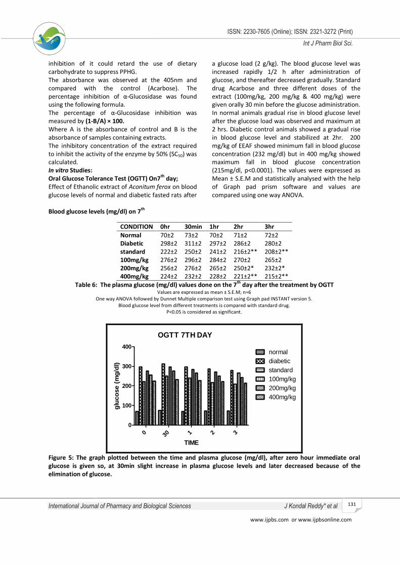
inhibition of it could retard the use of dietary carbohydrate to suppress PPHG. The absorbance was observed at the 405nm and compared with the control (Acarbose). The percentage inhibition of α-Glucosidase was found using the following formula. The percentage of α-Glucosidase inhibition was measured by (1-B/A) × 100. Where A is the absorbance of control and B is the absorbance of samples containing extracts. The inhibitory concentration of the extract required to inhibit the activity of the enzyme by 50% (SC50) was calculated. In vitro Studies: Oral Glucose Tolerance Test (OGTT) On7th day; Effect of Ethanolic extract of Aconitum ferox on blood glucose levels of normal and diabetic fasted rats after
a glucose load (2 g/kg). The blood glucose level was increased rapidly 1/2 h after administration of glucose, and thereafter decreased gradually. Standard drug Acarbose and three different doses of the extract (100mg/kg, 200 mg/kg & 400 mg/kg) were given orally 30 min before the glucose administration. In normal animals gradual rise in blood glucose level after the glucose load was observed and maximum at 2 hrs. Diabetic control animals showed a gradual rise in blood glucose level and stabilized at 2hr. 200 mg/kg of EEAF showed minimum fall in blood glucose concentration (232 mg/dl) but in 400 mg/kg showed maximum fall in blood glucose concentration (215mg/dl, p<0.0001). The values were expressed as Mean ± S.E.M and statistically analysed with the help of Graph pad prism software and values are compared using one way ANOVA.
Blood glucose levels (mg/dl) on 7
th
CONDITION 0hr 30min 1hr 2hr 3hr
Normal 70±2 73±2 70±2 71±2 72±2 Diabetic 298±2 311±2 297±2 286±2 280±2 standard 222±2 250±2 241±2 216±2** 208±2** 100mg/kg 276±2 296±2 284±2 270±2 265±2 200mg/kg 256±2 276±2 265±2 250±2* 232±2* 400mg/kg 224±2 232±2 228±2 221±2** 215±2**
Table 6: The plasma glucose (mg/dl) values done on the 7th day after the treatment by OGTT Values are expressed as mean ± S.E.M; n=6
One way ANOVA followed by Dunnet Multiple comparison test using Graph pad INSTANT version 5. Blood glucose level from different treatments is compared with standard drug.
P<0.05 is considered as significant.
OGTT 7TH DAY
0 30 1 2 3
0
100
200
300
400normal
diabetic
standard
100mg/kg
200mg/kg
400mg/kg
TIME
glu
co
se (
mg
/dl)
Figure 5: The graph plotted between the time and plasma glucose (mg/dl), after zero hour immediate oral glucose is given so, at 30min slight increase in plasma glucose levels and later decreased because of the elimination of glucose.

International Journal of Pharmacy and Biological Sciences J Kondal Reddy* et al
www.ijpbs.com or www.ijpbsonline.com
ISSN: 2230-7605 (Online); ISSN: 2321-3272 (Print)
Int J Pharm Biol Sci.
132
OGTT 7TH DAY
normal
diabet
ic
stan
dard
100m
g/kg
200m
g/kg
400m
g/kg
0
100
200
300
400normal
diabetic
standard
100mg/kg
200mg/kg
400mg/kgg
luco
se (
mg
/dl)
Figure 6: The graph showing the comparison between the normal, diabetic, standard, 100mg/kg extract, 200mg/kg extract and 400mg/kg extract doses. Oral Glucose Tolerance Test (OGTT) on 21
TH day:
Effect of EEAF on blood glucose levels of normal and diabetic fasted rats after a glucose load (2 g/kg). The blood glucose level was increased rapidly ½ h after administration of glucose, and thereafter decreased gradually. Standard drug Acarbose and three different doses of the extract (100mg/kg, 200 mg/kg & 400 mg/kg) were given orally 30 min before the glucose administration. In normal animals gradual rise in
blood glucose level after the glucose load was observed and maximum at 2 hrs. 200 mg/kg of EEAF showed minimum fall in blood glucose concentration (179 mg/dl) but in 400 mg/kg showed maximum fall in blood glucose concentration (136 mg/dl, p<0.0001). The values were expressed as Mean ± S.E.M and statistically analysed with the help of Graph pad prism software and values are compared using one way ANOVAs.
Blood glucose levels (mg/dl) on 21thday
CONDITION 0hr 30min 1hr 2hr 3hr Normal 68±1 75±1 72±1 65±1 67±1 Diabetic 284±2 296±2 287±2 290±2 277±2 Standard 164±1 178±1 160±1 144±1** 133±1 100mg/kg 236±2 239±2 228±2 231±2 219±2 200mg/kg 215±2 221±2 213±2 207±2* 198±2* 400mg/kg 162±1 179±1 169±1 155±1** 136±1**
Table 7: The plasma glucose (mg/dl) values collected on the 21st day after the treatment by OGTT
OGTT 21st day
0
30min 1hr
2hr3hr
0
100
200
300
400normal
diabetic
standard
100mg/kg
200mg/kg
400mg/kg
time
glu
cose
(m
g/d
l)
Figure 7: The graph plotted between the time and plasma glucose (mg/dl), after zero hour immediate oral glucose is given so, at 30min slight increase in plasma glucose levels and later decreased because of the elimination of glucose.

International Journal of Pharmacy and Biological Sciences J Kondal Reddy* et al
www.ijpbs.com or www.ijpbsonline.com
ISSN: 2230-7605 (Online); ISSN: 2321-3272 (Print)
Int J Pharm Biol Sci.
133
OGTT 21st day
normal
diabet
ic
stan
dard
100m
g/kg
200m
g/kg
400m
g/kg
0
100
200
300
400normal
diabetic
standard
100mg/kg
200mg/kg
400mg/kg
time
glu
co
se (
mg
/dl)
Figure 8: The graph showing the comparison between the normal, diabetic, standard, 100mg/kg extract, 200mg/kg extract and 400mg/kg extract doses. By comparing the glucose levels of the Ethanolic extract and standard drug found some inhibition from day 7 to the 28 day. The standard drug showed the 54% inhibition. 100mg/kg of EEAF showed 29.9% and 200mg/kg of EEAF showed 33.2% and 400mg/kg showed 51.5% inhibition.
CONCLUSION From the present study, It supports that therapy with alpha-glucosidase inhibitors could be an interesting adjunctive pharmacological approach to the treatment of type 2 diabetes. In this regard, the protection determined by EEAF clearly suggests the potential usefulness of this plant. We concluded that the EEAF produced a significant decrease of plasma glucose level. These results suggest that the effectiveness of the drugs depend probably, on the accumulative effect of active principles. In contrast, the significant increase in plasma glucose levels of untreated diabetic rats may be due to a progressive severity of untreated diabetes. In conclusion, EEAF showed strong inhibitory activity against α-glucosidase in vitro and in vivo. Further study need to identify the active component responsible for the inhibition of α-glucosidase is strongly recommended.
REFERENCES 1. Time line of Diabetes History http://www.enjoying-life-
with-diabetes.com/history-of-diabetes.html 2. History of diabetes http://health.savvy-cafe.com/a-
timeline-of-the-history-of-diabetes-2008-02-08/ 3. Global diabetes walk, world diabetes foundation from
http://www.worlddiabetesfoundation.org/composite-976.html
4. Diabetes statistics alarming from http://www.suite101.com/content/diabetes-a15635
5. Introduction and causes of prediabetes http://diabetesinformationhub.com/Prediabetes.php
6. Screening and diagnosis of prediabetes. http://diabetesinformationhub.com/Prediabetes_ScreeningandDiagnosis.php
7. Human anatomy pancreas from http://healthy-lifestyle.most-effective-solution.com/2010/07/29/human-anatomy-pancreas/
8. Hormones of pancreas from http://users.rcn.com/jkimball.ma.ultranet/BiologyPages/P/Pancreas.html
9. Sanger F, the Chemistry of Insulin. Science; 19591 (29):1340-1344.
10. Hopfer U Membrane transport mechanisms fo hexoses and amino acids in the small intestine. Physiology of the Gastrointestinal Tract (Johnson LR:Ed) ; 1987 : 1499-1526.
11. Haase W, Koepsell H Electron microscopic immune histochemical localization of Na-cotransporters along the rat nephron. European Journal of Cell Biology; 1989 (48):360-74.
12. Kono T, Suzuki K, Dansey, LE, Robinson RW, Blevins TL Energy-dependent and protein synthesis-independent recycling of the insulin-sensitive glucose transport mechanisms in fat cells. Journal of Biological Chemistry; 1981 (256) :6400-6407.
13. Bromer WW, Sinn LG, Staub A, Behrens OK: The amino acid sequence of glucagon. Diabetes 6:234-238, 1957.
14. Gerich JE, Lovinger R, Grodsky GM: Inhibition by somatostatin of glucagon and insulin release from the perfused rat pancreas in response to arginine, isoproterenol and theophylline: evidence for a preferential effect on glucagon secretion. Endocrinology 96:749-754, 1975.

International Journal of Pharmacy and Biological Sciences J Kondal Reddy* et al
www.ijpbs.com or www.ijpbsonline.com
ISSN: 2230-7605 (Online); ISSN: 2321-3272 (Print)
Int J Pharm Biol Sci.
134
15. Bokvist K, Olsen HL, Hoy M, Gotfredsen CF, Holmes WF, Buschard K, Rorsman P, Gromada J: Characterisation of sulphonylurea and ATP-regulated K+ channels in rat pancreatic A-cells. Pflugers Arch 438:428-436, 1999.
16. Zimmet PZ, Tuomi T, Mackay R, Rowley MJ, Knowles W, Cohen M, Lang DA: Latent autoimmune diabetes mellitus in adults (LADA): The role of antibodies to glutamic acid decarboxylase in diagnosis and prediction of insulin dependency. Diabetic Medicine ; 1994 (11):299-303.
17. Eisenbarth GS. Type I diabetes mellitus. A chronic autoimmune disease. New England Journal of Medicine 1986; 314:1360-1368.
18. Imagawa A, Hanafusa T, Miyagawa J, Matsuzawa Y. A novel subtype of type 1 diabetes mellitus characterized by a rapid onset and an absence of diabetes-related antibodies. Osaka IDDM Study Group. New England Journal of Medicine 2000; 342:301-307.
19. Rewers M, Bugawan TL, Norris JM, Blair A, Beaty B, Hoffman M et al. Newborn screening for HLA markers associated with IDDM: diabetes autoimmunity study in the young (DAISY). diabetol 1996; 39(7):807-812.
20. Ginsberg-Fellner F, Witt ME, Yagihashi S, Doberson MJ, Taub F, Fedun B et al. Congenital rubella syndrome as a model for type I (insulin- dependent) diabetes mellitus: increased prevalence of islet cell surface antibodies. diabetol 1984; 27 Supplement :87-89.
21. Graves PM, Rewers M. The role of enteroviral infections in the development of IDDM: limitations of current approaches. diabetol 1997; 46(2):161-168.
22. Bach JF. The effect of infections on susceptibility to autoimmune and allergic diseases. New England Journal of Medicine 2002; 347(12):911-920.
23. Foulis AK, McGill M, Farquharson MA, Hilton DA. A search for evidence of viral infection in panreases of newly diagnosed patients with IDDM. diabetol 1997; 40:53-61.
24. Srikanta S, Ganda OP, Rabizadeh A, Soeldner JS, Eisenbarth GS. First-degree relatives of patients with type I diabetes mellitus. Islet cell antibodies and abnormal insulin secretion. New England Journal of Medicine 1985; 313(8):461-464.
25. Lenzen S. The mechanisms of alloxan and Streptozotocin induced diabetes. Diabetologia. ; 2007 (18):216-226.
26. Weaver DC, Barry CD, McDaniel ML, Marshall GR, Lacy PE. Molecular requirements for recognition at glucoreceptor for insulin release. Molecular Pharmacology; 1975(16):361–368.
27. Winterbourn CC, Munday R. Glutathione-mediated redox cycling of alloxan. Mechanisms of superoxide dismutase inhibition and of metal-catalyzed OH_ formation. Biochem Pharmacol; 1989(38):271–277.
28. Bhuiyan M A R, Hoque M Z, Hossain S J. Free radical scavenging activity of Zizyphus mauritiana. World journal of agricultural sciences. 2009; 3: 318-322.
*Corresponding Author: J Kondal Reddy* Email: [email protected]