Comparison of mouth-to-mouth, mouth-to-mask and mouth-to-face-shield ventilation by lay persons

Infectious Disease
The Early Pathogenesis of Foot-and-MouthDisease in Cattle After Aerosol Inoculation:Identification of the Nasopharynx as thePrimary Site of Infection
J. Arzt1,2, J. M. Pacheco1, and L. L. Rodriguez1
AbstractTo characterize the early events of foot-and-mouth disease virus (FMDV) infection in cattle subsequent to simulated naturalexposure, 16 steers were aerosol inoculated with FMDV and euthanized at various times. Samples were collected from each steerantemortem (serum, nasal swabs, and oral swabs) and postmortem (up to 40 tissues per animal) and screened for FMDV by virusisolation and for FMDV RNA by real-time reverse transcription polymerase chain reaction. Tissues that tested positive for FMDVor viral RNA were examined by immunohistochemistry and multichannel immunofluorescence microscopy. In previremic steers,FMDV was most consistently localized to nasopharyngeal tissues, thereby indicating this region as the most important site ofprimary viral replication. The earliest site of microscopic localization of FMDV antigens was the lymphoid follicle-associatedepithelium of the pharyngeal mucosa-associated lymphoid tissue of the nasopharynx at 6 hours postaerosolization. At early timepoints after aerosol inoculation, viral antigens colocalized with cytokeratin-positive pharyngeal epithelial cells; intraepithelialFMDV-negative, MHCII/CD11c-double-positive dendritic cells were present in close proximity to FMDV-positive cells. Onsetof viremia coincided with marked increase of viral loads in pulmonary tissues and with substantial decrease of viral detectionin nasopharyngeal tissues. These data indicate that subsequent to aerogenous exposure to FMDV, the temporally defined criticalpathogenesis events involve (1) primary replication in epithelial cells of the pharyngeal mucosa-associated lymphoid tissue cryptsand (2) subsequent widespread replication in pneumocytes in the lungs, which coincides with (3) the establishment of sustainedviremia.
Keywordsaerosol, bovine, cattle, foot-and-mouth disease, pathogenesis, virus
Foot-and-mouth disease (FMD) is a highly contagious
picornaviral disease affecting domestic and wild cloven-
hoofed animals.2,13 Under natural transmission conditions,
within and between herds of cattle, the etiologic agent, FMD
virus (FMDV), is spread through inhalation of aerosolized
virus, and under appropriate environmental conditions, virus-
laden droplets may travel vast distances while maintaining
infectivity.1,13,22
Numerous researchers have experimentally investigated the
pathogenesis of FMD in cattle;1,4,6,7,14,15 however, a consensus
still does not exist regarding many basic aspects of the early
stages of infection. Most notably, the anatomic sites and cellu-
lar events involved in primary infection and the establishment
of viremia are not well defined. Elucidation of these critical
events and improved understanding of virus–host interactions
have high probability to favorably affect the goal of improving
efficacy of vaccines and biotherapeutic countermeasures to
protect domestic livestock against FMDV.
Early evidence that the respiratory tract was the natural route
of infection implicated the nasal cavity14 or lungs10 as the sites of
primary replication. Yet, as investigation proceeded, opinion on
this subject diverged into 2 camps favoring either the nasophar-
ynx2,7 or lungs4,6 as the primary infection site. A unique set of
experiments performed in the 1970s effectively isolated the con-
tributory roles of the upper and lower respiratory tract by
1 Plum Island Animal Disease Center, Foreign Animal Disease Research Unit,
Agricultural Research Service, US Department of Agriculture, Greenport,
New York2 Department of Microbiology, Immunology, and Pathology, Colorado State
University, Fort Collins, CO
Corresponding Author:
Jonathan Arzt, Foreign Animal Disease Research Unit, USDA/ARS Plum Island
Animal Disease Center, PO Box 848, Greenport, NY 11944
Email: [email protected]
Veterinary Pathology47(6) 1048-1063ª The American College ofVeterinary Pathologists 2010Reprints and permission:sagepub.com/journalsPermissions.navDOI: 10.1177/0300985810372509http://vet.sagepub.com
1048
placement of indwelling tracheostomy tubes in cattle.24 This
work concluded that both the lungs and the pharynx could simi-
larly serve as portals for the establishment of systemic infection.
The only studies to microscopically localize FMDV in the early
(previremic) stages of infection utilized in situ hybridization and
concluded that lungs supported infection earlier than the phar-
ynx.4,6 In consideration of the disparate findings across FMD
pathogenesis studies, it is necessary to remember that the various
studies have utilized a broad range of serotypes and subtypes of
FMDV, which may have substantial differences in virulence and
tissue tropism. Additionally, heterogeneity of inoculation sys-
tems and differences in sensitivity and/or specificity of virus
detection methods may account for some disparity among the
published studies.
Recent reports from our laboratory have described a novel
method for aerosol inoculation of cattle with FMDV and trimo-
dal systems for detection of FMDV in bovine tissues during the
early stages of infection.3,18 In the current study, we utilized
similar experimental systems to further characterize the
distribution of FMDV in cattle during the previremic and early
viremic phases of infection, with the overall conclusion that
subsequent to aerogenous inoculation of cattle, infection
initiates in the nasopharynx, it is promptly followed by pulmonary
infection, and the onset of viremia is coincident with increased
viral load in the lungs and decreased virus in the nasopharynx.
Materials and Methods
Experimental Animals, Virus, and Inoculation Systems
Sixteen 9- to 18-month-old Holstein steers weighing 400 to
500 kg were obtained from an experimental-livestock provider
accredited by the Association for Assessment and Accredita-
tion of Laboratory Animal Care (Thomas-Morris Inc, Reisters-
town, MD). For all experiments, animals were individually
housed in a biosafety level 3 animal facility from time of
inoculation until time of euthanasia. Experiments were termi-
nated by euthanasia via intravenous barbiturate overdose at
predetermined end points at 0.1 hours post aerosol inoculation
(hpa; steer No. 1), 3.0 hpa (steer Nos. 2, 3), 6.0 hpa (steer Nos.
4, 5), 12.0 hpa (steer Nos. 6, 7), 24.0 hpa (steer Nos. 8-13),
48.0 hpa (steer Nos. 14, 15), 240.0 hpa (steer No. 16).
Virus inoculum consisted of a clarified, macerate of tongue
epithelium harvested from 2 steers experimentally infected with
the FMDV strain O1-Manisa as previously described.12,18 The
inoculum was quantitated as BTID50 (bovine tongue infectious
dose 50%). All steers were aerosol inoculated with 107 BTID50
of FMDV-O1-Manisa as previously described.18 Briefly, each
steer was sedated with xylazine and fitted with a commercially
available aerosol delivery system (Aeromask-ES, Trudell Medi-
cal, London, Ontario, Canada), which was placed over the muz-
zle. The mask was attached to a jet nebulizer (Whisper Jet, Vital
Signs Inc, Totowa, NJ), which was subsequently attached to an
air compressor that generated 25 psi of pressure. Aerosolization
proceeded until the complete inoculum was expelled from the
nebulizer cup (10 to 15 minutes).
Sample Collection
Antemortem sampling consisted of collection of whole blood
in serum separation tubes, oral swabs, and nasal swabs. All
animals were sampled before inoculation to ensure FMDV-
free status and at several time points throughout the duration
of the experiment, which varied according to goals of the
individual experiments. Swabs and serum tubes were trans-
ported from the animal room to the laboratory on ice and were
immediately centrifuged for harvesting of serum, saliva, and
nasal secretion. Samples were then stored at –70�C until time
of processing.
Necropsies were performed immediately subsequent to
euthanasia at predetermined time points, and tissue specimens
were collected from oral cavity, nasal cavity, soft palate, phar-
ynx, larynx, trachea, lungs, lymph nodes, and skin (Table 1).
Tissues collected from each animal varied depending on the
goals of individual experiments. Detailed descriptions of tissue
designations and collection strategies has been published.18 For
each anatomically defined specimen, two 30-mg tissue samples
were aliquoted into separate screw-cap 1.5-ml tubes and frozen
immediately in liquid nitrogen for transfer within 2 hours to a
–70�C freezer in which they were stored until the time of
processing. An adjacent specimen from each tissue was placed
in a cryomold, embedded in Optimal Cutting Temperature
Compound (Sakura Finetek, Torrance, CA), frozen on a bath
of liquid nitrogen, and stored at –70�C for immunohistochem-
istry (IHC).
FMDV RNA Detection
Two samples of each tissue listed in Table 1 per animal were
thawed and immediately macerated in a TissueLyser bead
beater (Qiagen, Valencia, CA) as previously described.18 After
maceration, 50 ml of each sample was transferred to a 96-well
plate (Thermo Scientific, Waltham, MA) containing 150 ml of
lysis/binding solution. RNA was then extracted with Ambion’s
MagMax-96 Viral RNA Isolation Kit (Ambion, Austin, TX) on
a King Fisher-96 Magnetic Particle Processor (Thermo Scien-
tific, Waltham, MA). RNA was eluted in a final volume of
25 ml. Once extracted, 2.5 ml of RNA was analyzed by
real-time reverse transcription polymerase chain reaction
(rRT-PCR) on the ABI 7000 system (Applied Biosystems,
Austin, TX) as previously described.8 Samples with cycle
threshold values < 40 were considered positive. The remaining
macerated tissue was clarified at 1,000 rpm for 2 minutes at
4�C, and the supernatant was cleared of bacterial contamina-
tion using centrifuge tube filters (Spin-X, Costar, Corning,
NY). Clarified and cleared samples were stored at –70�C until
virus isolation (VI) was performed.
To convert cycle threshold values generated by rRT-PCR
from experimental samples to RNA genome copies per milli-
gram, serial 10-fold dilutions of in vitro synthesized FMDV
RNA of known RNA concentration were analyzed by the
rRT-PCR protocol described above. The equation of the curve
of RNA copy versus cycle threshold value, further adjusted for
Arzt et al 1049
1049
Tab
le1.
Tis
sue-
Spec
ific
Dis
trib
ution
ofFM
DV
inSt
eer
Nos.
2-1
5w
ith
Cum
ula
tive
Per
centa
gePosi
tivi
tya
Sam
ple
Iden
tific
atio
nb
3H
PA
6H
PA
12
HPA
24
HPA
48
HPA
Pre
vire
mic
Cum
ula
tive
Per
centa
gePosi
tivi
tyc
23
45
67
89
10
11
12
13
14
15
rRT
-PC
RV
IrR
T-P
CR
or
VI
Seru
mN
EG
NEG
NEG
NEG
NEG
NEG
NEG
NEG
NEG
4.6
73.9
44.0
26.6
27.2
418.2
0.0
18.2
Ora
lca
vity
/oro
phar
ynx
Low
erlip
NEG
NE
G2.6
3N
EG
NEG
3.6
625.0
25.0
50.0
Tongu
e–R
NEG
NEG
NEG
NEG
NEG
NEG
2.8
1N
EG
2.4
1N
EG
NEG
NEG
3.9
720.0
0.0
20.0
Ven
tral
soft
pal
ate–
RN
EG
NEG
NEG
NEG
NEG
2.3
42.3
93.5
5N
EG
4.5
44.4
433.3
11.1
33.3
Ven
tral
soft
pal
ate–
CN
EG
NEG
2.2
7N
EG
2.9
83.3
43.2
12.5
0N
EG
4.5
44.3
755.6
11.1
55.6
Nas
ophar
ynx/lar
ynx
Dors
also
ftpal
ate–
RN
EG
NEG
2.5
8N
EG
3.4
3N
EG
3.7
57.3
02.7
53.6
8N
EG
4.5
6N
EG
NEG
54.5
63.6
72.7
Dors
also
ftpal
ate–
C2.9
2N
EG
2.8
5N
EG
NE
G4.3
44.9
84.0
13.8
54.0
15.1
1N
EG
2.9
92.9
272.7
72.7
81.8
Dors
alnas
ophar
ynx–R
2.3
32.7
8N
EG
2.9
0N
EG
3.1
93.6
2N
EG
3.3
13.9
73.0
73.3
4N
EG
2.3
372.7
72.7
100.0
Dors
alnas
ophar
ynx–C
2.9
5N
EG
NEG
2.3
0N
EG
4.9
04.1
53.2
95.0
63.0
73.3
42.7
9N
EG
3.9
872.7
63.6
81.8
Epig
lott
is–V
2.5
02.4
7N
EG
NEG
2.9
63.5
62.3
63.9
2N
EG
3.8
8N
EG
3.7
6N
EG
NEG
63.6
27.3
72.7
Lary
nx–V
NEG
NEG
3.3
5N
EG
2.7
94.6
24.0
94.7
03.5
34.0
32.7
43.2
2N
EG
2.8
872.7
54.5
72.7
Lungs
/tra
chea
Tra
chea
–ab
ora
dN
EG
2.4
4N
EG
2.2
9N
EG
2.3
7N
EG
NEG
NEG
3.5
53.4
533.3
0.0
33.3
Tra
chea
lbifu
rcat
ion
NEG
NEG
NEG
NEG
NEG
NEG
NEG
3.7
22.4
52.4
44.9
522.2
0.0
22.2
Pro
xim
alcr
ania
llo
be
NEG
NEG
NEG
3.1
8N
EG
2.7
83.3
82.8
9N
EG
NEG
4.6
744.4
11.1
44.4
Mid
cran
iallo
be
NEG
NEG
NEG
NE
GN
EG
3.6
04.0
5N
EG
NEG
NEG
3.4
422.2
22.2
33.3
Dis
talcr
ania
llo
be
NEG
NEG
NEG
NE
GN
EG
5.1
22.2
8N
EG
NEG
4.9
3N
EG
2.5
4N
EG
5.5
427.3
36.4
36.4
Pro
xim
alm
idlo
be
NEG
NEG
NEG
2.7
5N
EG
NEG
4.6
32.6
3N
EG
5.0
43.9
733.3
11.1
33.3
Mid
mid
lobe
NEG
NEG
NEG
NEG
2.5
22.7
45.2
2N
EG
NEG
2.5
13.4
933.3
22.2
33.3
Dis
talm
idlo
be
NEG
NEG
NEG
NEG
2.8
0N
EG
4.1
13.8
3N
EG
5.1
85.4
33.8
75.0
33.7
645.5
27.3
45.5
Pro
xim
alca
udal
lobe
NEG
NEG
NEG
NEG
2.7
6N
EG
2.7
33.0
9N
EG
3.8
83.6
533.3
11.1
33.3
Mid
caudal
lobe
NEG
NEG
NEG
NEG
NEG
NEG
NEG
2.9
2N
EG
3.5
13.7
111.1
0.0
11.1
Dis
talca
udal
lobe
2.4
12.6
1N
EG
NEG
NEG
NEG
3.2
73.3
4N
EG
2.9
43.2
54.5
02.4
4N
EG
54.5
18.2
54.5
Additio
nal
tiss
ues
Lingu
alto
nsi
l2.5
8N
EG
2.7
9N
EG
NEG
NEG
NEG
NEG
NEG
3.1
02.6
53.9
03.0
14.1
336.4
9.1
36.4
Pal
atin
eto
nsi
lN
EG
NEG
NEG
NEG
NE
GN
EG
NEG
NEG
NEG
4.5
32.8
611.1
0.0
11.1
Nas
ophar
ynge
alto
nsi
lN
EG
2.8
2N
EG
NEG
NEG
NEG
2.5
7N
EG
NEG
NEG
NEG
22.2
0.0
22.2
Ret
rophar
ynge
alLN
NEG
NEG
NEG
NEG
NEG
NEG
NEG
NE
GN
EG
NEG
NEG
0.0
12.5
12.5
Hila
rLN
NEG
NEG
NEG
NEG
NEG
NEG
NE
GN
EG
NEG
NE
G0.0
12.5
12.5
Inte
rdig
ital
clef
t2.4
9N
EG
NEG
NEG
NEG
NEG
NE
GN
EG
NEG
7.7
07.9
612.5
0.0
25.0
Ala
rfo
ldN
EG
2.3
4N
EG
NEG
NEG
3.6
825.0
0.0
25.0
Turb
inat
es–C
NEG
NEG
NE
GN
EG
NEG
NEG
NEG
0.0
20.0
20.0
Thyr
oid
NEG
NEG
NEG
NEG
2.6
1N
EG
16.7
16.7
16.7
aH
PA
,hours
post
aero
soli
nocu
lation;r
RT
-PC
R,r
eal-tim
ere
vers
etr
ansc
ription
poly
mer
ase
chai
nre
action;V
I,vi
rus
isola
tion;R
,rost
ral;
C,c
audal
;V,v
entr
al;L
N,l
ymph
node;
NEG
,rR
T-P
CR
neg
ativ
e.Bla
nk
cells
indic
ate
notex
amin
ed.T
abula
ted
num
eric
aldat
aar
elo
g 10
foot-
and-m
outh
dis
ease
viru
s(F
MD
V)
RN
Age
nom
eco
pie
sper
mlo
fse
rum
or
mg
oftiss
ue
asdet
erm
ined
by
rRT
-PC
R.N
egat
ive
tiss
ues
indic
ate
that
RN
Aquan
tity
was
bel
ow
the
det
ection
thre
shold
of2.2
6lo
g 10
FMD
Vge
nom
eco
pie
sper
mg
oftiss
ue
(corr
espondin
gto
cycl
eth
resh
old¼
40.0
0).
Entr
ies
inbold
text
wer
eposi
tive
for
FMD
Vby
viru
sis
ola
tion;n
onbold
text
indic
ates
that
viru
sis
ola
tion
was
neg
ativ
e.b
Additio
nal
tiss
ues
test
edw
ere
neg
ativ
efo
ral
lpre
vire
mic
stee
rsex
amin
ed,as
follo
ws:
den
talpad
,to
ngu
e(c
audal
),to
ngu
e(t
oru
s),har
dpal
ate
(rost
ral),har
dpal
ate
(cau
dal
),nas
alpla
num
,tu
rbin
ates
(rost
ral),tr
achea
(ora
d),
trac
hea
(mid
).c
Pre
vire
mia
isdef
ined
asse
rum
viru
sis
ola
tion
neg
ativ
eat
allt
imes
sam
ple
d,i
ncl
udin
gth
etim
eofeu
than
asia
.Per
centa
geposi
tivi
ties
for
rRT
-PC
Ran
dV
Ise
par
atel
yar
edef
ined
asth
enum
ber
ofposi
tive
resu
lts
usi
ng
one
modal
ity,
div
ided
by
the
num
ber
ofst
eers
for
whic
ha
tiss
ue
type
was
exam
ined
by
that
modal
ity.
Per
centa
geposi
tivi
tyfo
rrR
T-P
CR
or
VIis
def
ined
asth
enum
ber
oftim
esth
ata
tiss
ue
was
posi
tive
by
eith
erm
odal
ity,
div
ided
by
the
num
ber
ofst
eers
for
whic
hth
attiss
ue
type
was
exam
ined
by
both
modal
itie
s.
1050

average mass of tissue samples and dilutions during processing,
was used for subsequent conversions. The cycle threshold posi-
tivity cutoff of 40 corresponded to a detection threshold value
of 2.26 log10 FMDV RNA copies per milligram (RNA/mg) of
tissue. The rRT-PCR results reported in Table 1 are the higher
RNA/mg value of the 2 samples processed per tissue per ani-
mal; the rRT-PCR results reported in Figure 1 are the mean
log10 FMDV RNA copies per milliliter (RNA/ml) for all ani-
mals sampled at each time point.
FMDV Isolation
VI was performed separately on the duplicate samples of each
tissue on LFBK cells as previously described.18,25 Upon detection
of cytopathic effect, FMDV positivity was confirmed by rRT-
PCR on cell culture supernatants. Samples in which no cytopathic
effect was observed were amplified through 3 blind passages and
the supernatants were tested by rRT-PCR before they were
deemed negative. Each VI result in Table 1 is reported positive
if either or both duplicate samples per tissue were positive.
Immunohistochemical and ImmunofluorescentLocalization of FMDV Antigens
Microscopic localization of FMDV antigens was performed in
cryosections as previously described.3,18 Briefly, tissue
sections were blocked for 2 hours at 20�C; primary antibodies
were diluted in blocking buffer and applied to tissue
sections for 18 hours at 4�C. For IHC, specific anti-FMDV
immunoreactivity was detected with a micropolymer alkaline
phosphatase kit (Biocare, Concord, CA). For multichannel
immunofluorescent (MIF), detection was performed with goat,
anti-rabbit, and isotype-specific anti-mouse secondary
antibodies labeled with AlexaFluor dyes (AF 350, 488, 594,
647). Slides were examined with a wide-field, epifluorescent
microscope, and images were captured with a cooled, mono-
chromatic digital camera. Images of individual detection
channels were adjusted for contrast and brightness and merged
in commercially available software (Adobe Photoshop CS2).
Mouse monoclonal anti-FMDV antibodies against viral struc-
tural proteins were 10GA4 and 12AF4;23 anti–nonstructural
protein antibodies were F19-6 and F19-51.27 Antibodies used
to label cell markers in MIF experiments were mouse monoclo-
nal anti-pancytokeratin plus (Biocare No. CM162), anti-bovine
cytokeratin (CK; Sigma No. C6909, Sigma-Aldrich, Inc, St.
Louis, MO), anti-CD11c (VMRD No. BAQ153A, VMRD,
Pullman, WA), anti-MHCII (VMRD No. CAT82A), and anti-
vimentin (Dako No. M0725, Dako, Carpinteria, CA), and
rabbit polyclonal anti–Von Willibrand factor (Dako No. A0082).
For each tissue screened by IHC, a duplicate negative-control
serial section treated with a mouse monoclonal anti-VSV-
Indiana antibody of similar concentration was prepared. Addi-
tional negative control tissue sections were prepared from a steer
that received a virus-free aerosol inoculum and was euthanized
24 hours post aerosol inoculation (hpa). Immunohistochemical
and MIF labeling were considered positive when there was an
intense cell-associated signal within the experimental tissue,
with the absence of such staining in the negative controls.
Results
Inoculum Detection Controls
For the purpose of determining if the FMDV screening
techniques would detect residual inoculated virus, 1 aerosol-
inoculated steer (No. 1) was euthanized immediately after
inoculation (0.1 hpa) and was subjected to standard tissue
collection and screening techniques as described for the other
animals. All 28 tissues collected from this animal were VI
negative, and only a single tissue, proximal cranial lung, was
positive by rRT-PCR (2.56 RNA/mg). This information con-
firmed that the inoculum reached as far as the lungs and
strongly affirmed that any rRT-PCR- and VI-positive findings
at subsequent time points represent FMDV replicated de novo.
Based on the positive rRT-PCR finding for proximal cranial
lung from steer No. 1, the frozen Optimal Cutting Temperature
Compound–embedded tissue specimen from this tissue was
examined by IHC to similarly test for detection of inoculated
virus. Forty serial sections were examined (alternating anti-
FMDV capsid and anti-FMDV 3D [polymerase] primary anti-
bodies) with no detection of FMDV antigens in any section,
confirming that inoculum quantity was insufficient to be
detected by IHC. Data generated from this animal are not
included in subsequent analyses.
Antemortem Profiles of FMDV-Infected Cattle
Neither vesicles nor fever (rectal temperature � 40�C) was
observed in any previremic animals. The earliest detection of
fever in any animal occurred at 60 hpa, in steer No. 16; this
animal was febrile from 60 to 96 hpa, with maximum body
temperature of 41�C detected at 60 hpa. The 3 steers euthanized
as 48 hpa or later (viremic) all developed vesicles of 1 (n ¼ 2)
or 2 (n ¼ 1) interdigital clefts at 48 hpa (Fig. 1). One steer (No.
15) had subtle blanching of the epithelium of the lingual torus
at 48 hpa, which was subsequently determined histologically to
be an early vesicle. The 1 steer that survived 240 hpa (No. 16)
developed vesicles of tongue and all 4 interdigital clefts
between 48 and 120 hpa. No other clinical signs of infection
or gross lesions were detected in the animals that survived
0.1 to 48.0 hpa.
The aerosol inoculation system provided highly consistent
patterns of viral shedding, viremia, and clinical signs across
experimental animals; however, some interanimal variation
was observed. Antemortem data are presented as cumulative
average log10 FMDV genome copy numbers per milliliter
(RNA/ml) for all animals at each time point (Fig. 1). For oral
swab samples, the quantity of FMDV RNA (inoculum) pro-
gressively decreased from 0.1 to 8.0 hpa with de novo
replicated virus first detected at 24 hpa and peaking at 72 hpa
at 8.17 RNA/ml. Similarly, nasal swab samples showed
gradual elimination of inoculum from 0.1 to 4.0 hpa with
de novo replicated viral RNA beginning to increase at 6 hpa.
Arzt et al 1051
1051
However, unlike oral swabs, the viral RNA quantity never
dropped below detection threshold before de novo replication
was detected (Fig. 1). Also unlike oral swabs, the nasal shed-
ding pattern showed 2 peaks, at 12 and 72 hpa (5.22 and 8.92
RNA/ml, respectively) with an intervening trough. Maximum
nasal and oral FMDV RNA/ml occurred coincidently at 72
hpa with the nasal detection peak being slightly higher but
decreasing more precipitously relative to salivary samples.
The earliest detection of FMDV RNA in sera occurred at
22 hpa in 1 steer. At 24 hpa, 6 of 9 steers had RNA-positive
sera, and at 48 hpa the sera of all 3 steers sampled were FMDV
rRT-PCR positive (Fig. 1). Detection of infectious FMDV in
sera was delayed relative to RNA, with a single VI-positive
steer (1 of 9) at 24 hpa and with the remaining 3 steers (3 of
3) becoming serum VI positive by 48 hpa; viremia was defined
by the detection of infectious virus in serum. Overall, FMDV
RNA was detected in sera from 22 to 120 hpa, whereas viremia
was detected from only 24 to 72 hpa.
Tissue-Specific Distribution of FMDV and Viral RNA
Steers were euthanized at predetermined time points regardless
of clinical progression of disease in individual animals. Sample
collection schemes were predetermined and standardized with
minor variation among individual animals, based on the
expected stage of disease at the time of euthanasia (Table 1).
A maximum of 40 anatomically distinct tissue specimens were
collected per animal. Both steers euthanized at 3 hpa had more
FMDV RNA–positive tissues in the nasopharyngeal/laryngeal
(NP/L) sites (6 of 12 tissues; 50.0%) than in other anatomic
regions (2 of 18 pulmonary tissues; 11.1%) (Table 1). At this
time point, FMDV RNA quantities were generally low, and
no tissues contained infectious virus. The 2 highest quantities
of FMDV RNA were detected in caudal dorsal soft palate
(2.92 RNA/mg) and caudal dorsal nasopharynx (2.95 RNA/
mg) of steer No. 2.
From 6 to 12 hpa, within the NP/L sites there was greater
quantity of tissues positive for infectious FMDV (14 of 24;
58.3%) and viral RNA (13 of 24; 54.2%) relative to all other
anatomic regions; at 12 hpa, every NP/L tissue examined was
positive by VI, rRT-PCR, or both. Within this period, rRT-
PCR and VI positivity percentages for pulmonary specimens
were also increasing but to a lesser extent than that of NP/L
tissues. At the individual tissue-specific level, the greatest
quantities of FMDV RNA between 6 and 12 hpa were detected
in distal cranial lung (5.12 RNA/mg) and caudodorsal naso-
pharynx (4.9 RNA/mg) of steer No. 7 (12 hpa).
At 24 hpa, 97.2% of NP/L samples (35 of 36 at tissue level)
were positive for either FMDV RNA or infectious virus, with
72.2% double positive (VI and rRT-PCR). By contrast,
61.1% of pulmonary specimens (22 of 36 at tissue level) were
VI or rRT-PCR positive, with 36.1% double positivity.
The highest quantity of FMDV RNA at 24 hpa (7.3 RNA/
mg) was detected within the rostral dorsal soft palate of
steer No. 9. The only positive findings (rRT-PCR or VI) from
lymph nodes of previremic steers in the study were the hilar
lymph node of steer No. 11 and the medial retropharyngeal
lymph node of steer No. 12, which were VI positive at 24 hpa.
Both steers euthanized at 48 hpa were viremic at the time of
euthanasia; as such, infectious FMDV and FMDV RNA
detected in the tissues of these animals may have been present
within and/or exterior to blood vessels in these tissues. Viral
Figure 1. Time course of foot-and-mouth disease virus RNA quantity (log10 genome copy number / ml) determined by real-time reversetranscription polymerase chain reaction in serum (red), nasal swab samples (green), and oral swab samples (blue) collected from steersaerosol-inoculated with foot-and-mouth disease virus O1-Manisa. Each data point represents the mean + SE for all steers sampled at a giventime (hours postaerosol inoculation). Lesion (vesicle) score was calculated by evaluating each foot and the head independently, with 2 pointsassigned for a small vesicle (� 1.0cm) and 4 points for a large vesicle. Viremia (*) was defined by positive findings on serum virus isolation.X-axis calibration not to scale.
1052 Veterinary Pathology 47(6)
1052
load was substantially decreased in the NP/L sites at 48 hpa
relative to 24 hpa, with 66.6% positive by VI or rRT-PCR
(8 of 12 tissues) and 33.3% double positive. By contrast, VI or
rRT-PCR positivity of the lungs from 48-hpa steers was increased
relative to 24 hpa, with 77.8% single positive (14 of 18 tissues)
and 27.8% double positive. The greatest quantity of FMDV RNA
detected in these animals was from the interdigital clefts of both
steers (7.70 and 7.96 RNA/mg) and the lingual torus of steer No.
15 (6.19 RNA/mg), all of which had grossly detected vesicles.
Among nonlesional tissues collected from the 48 hpa steers, the
greatest quantity of FMDV RNA was obtained from the distal
anterior lung of steer No. 15 (5.54 RNA/mg).
Given that the primary goal of these experiments was to
characterize the involvement of various tissues as primary
replication sites of FMDV, greater attention was focused on the
animals that had VI-negative sera at the time of euthanasia
(Table 1). For these animals (all except steer Nos. 13–16),
tissue-specific cumulative positivity percentages (PPs) were
calculated for each tissue for rRT-PCR, VI, and both. PP was
defined as follows:
total positive results at tissue X by modality Y in previremic
steers = total specimens of tissue X examined by modality
Y in previremic steers:
Thus, PP served as an indicator of the relative frequency of
involvement of each tissue in previremic FMD in these ani-
mals. The PP index demonstrated that among previremic ani-
mals, the specimens most frequently positive for infectious
FMDV or FMDV RNA were the tissues of the NP/L sites.
Specifically, the only 100% PP value occurred for rRT-PCR
or VI positivity in rostrodorsal nasopharynx. The next-
highest PP values (81.8%) were achieved in only caudodorsal
soft palate and caudodorsal nasopharynx. Overall, 15 of 18
PP values from NP/L sites were greater than all PPs from other
tissues. PP values from lung and ventral soft palate indicate les-
ser consistency of FMDV positivity relative to NP/L sites. PP
values from lymphoid tissues were uniformly low, with lingual
tonsil having the highest overall PP in this tissue category.
Microscopic Localization of FMDV Antigens andPhenotypic Characterizations of Associated Cells
Inoculation to 12 hpa. Despite numerous attempts, FMDV
antigens could not be microscopically (immunohistochemi-
cally) identified in any of the rRT-PCR-positive tissues from
the steers euthanized at 3 hpa. The earliest time point at which
viral antigens were localized in situ was at 6 hpa, within the
rostrodorsal nasopharynx of both steers (Nos. 4 and 5) eutha-
nized at this time point. Viral antigens were not localized in any
other tissues from these animals despite screening of more than
100 additional IHC slides from pulmonary and pharyngeal sites
that were rRT-PCR or VI positive. In both steers, FMDV capsid
antigen was similarly localized to microscopic epithelial crypts
of the follicle-associated epithelium (FAE) overlying pharyn-
geal mucosa–associated lymphoid tissue (PALT; Figs. 2, 3).
Although the epithelial crypts were vaguely discernable by
IHC-light microscopy (Fig. 2), the MIF examination of a serial
section clearly demonstrated that FMDV in these regions was
intraepithelial rather than within subjacent lymphoid tissue
(Fig. 3A). Furthermore, simultaneous labeling with CD11c and
MHCII indicated that the FAE and subjacent lymphoid regions
had diffuse distribution of cells positive for both these antigens
individually and as CD11c–MHCII double positives (presump-
tive dendritic cells [DCs]); Fig. 3B–3D). Despite the presence
of DCs in close proximity to FMDV antigens, virus colocalized
exclusively with CK in these tissues. Individual cells contain-
ing FMDV antigens were rarely identified in the subepithelial
dome region immediately subjacent to antigen-positive crypts
(not shown); however, phenotypic characterization of these
cells with various markers was not achieved.
At 12 hpa, the distribution of viral antigens in the nasophar-
ynx was similar to that observed at 6 hpa with the exception that
there was slightly greater intimacy of association between
FMDV antigens and the DC markers CD11c and MHCII (not
shown). However, colocalization of FMDV antigens with DC
markers was not observed. The lungs of both steers euthanized
at 12 hpa (Nos. 6, 7) had regional anti-FMDV immunoreactivity
(Figs. 4–7). Steer No. 7 had numerous FMDV capsid and
nonstructural antigen–positive cells within the distal segment
of the cranial lung lobe (Figs. 5–7). In this animal, there were
2 distinct morphological variants of immunopositive cells when
viewed by light microscopy: a squamous cell type associated
with alveolar septa (type I pneumocyte; Fig. 5, Insert A), and
a polygonal cell type variably associated with alveolar septa
or lumina (type II pneumocyte or alveolar macrophage; Fig. 5,
Insert B). MIF labeling of this tissue indicated that both morpho-
logic categories of FMDV-positive cells were additionally CK
positive (Figs. 6A, 7), von Willebrand factor (vWF) negative
(Fig. 7), vimentin negative (Fig. 6B), confirming that infected
cells were of epithelial histogenesis. However, throughout the
alveolar parenchyma, vWF- and vimentin-positive cells were
ubiquitously interspersed with CK-positive pneumocytes (Figs.
6B, 7). The other steer euthanized at 12 hpa (No. 6) had antige-
nically and morphologically distinct pulmonary viral distribu-
tion. The distal segment of the middle lung lobe of this animal
had a single, small focus of immunopositivity for FMDV 3D
protein (Fig. 4), which was negative for capsid antigens (not
shown). Unlike steer No. 7, FMDV-positive cells in this region
were exclusively squamous.
Twenty-four hpa. At 24 hpa, nasopharyngeal tissues had some
of the same qualities described at 6 and 12 hpa but with varia-
tions. Viral immunopositivity was similarly localized to FAE
of PALT regions but was less associated with crypt regions.
Rather, FAE overlying large, expansive, cryptless regions were
more commonly affected (Figs. 8–12). In such areas there
were substantially greater quantities of FMDV-positive cells
within the epithelium (Figs. 8–9) and the superficial and deep
subepithelium (Figs. 9–12) relative to earlier time points.
These cells were often positive for FMDV structural and non-
structural proteins (Fig. 12). Within subepithelial lymphoid
Arzt et al 1053
1053
Figure 2. Rostrodorsal nasopharynx, steer No. 4. A, Immunohistochemical localization of FMDV to epithelial crypt of mucosa-associatedlymphoid tissue. Anti-FMDV capsid monoclonal antibody. Micropolymer alkaline phosphatase. Gill s hematoxylin counterstain. B, highermagnification of region of interest from Figure 2A. FMDV antigens localize to crypt epithelium subjacent to crypt lumen (arrows). Anti-FMDVcapsid monoclonal antibody. Micropolymer alkaline phosphatase. Gill s hematoxylin counterstain. Figure 3. Rostrodorsal nasopharynx, steer
1054 Veterinary Pathology 47(6)
1054

regions, small quantities of FMDV structural and nonstructural
protein–positive cells had morphologic and phenotypic (CK–,
MHCIIþ, CD11cþ) characteristics of DCs (Figs. 9–12). Addi-
tionally, concurrent labeling with anti-vWF antibody indicated
that within lymphoid follicles, FMDV-positive DCs were
occasionally present within 50 mm of capillary endothelia, but
direct interaction between DCs and vascular cells was not
observed in these areas. Microvesiculation (Figs. 8, 9) and erosion
(Figs. 8–11A) of nasopharyngeal FAE were rarely observed; in
such regions, capillary endothelia (vWF-positive cells) were pres-
ent within the disrupted epithelium immediately adjacent to
FMDV-positive epithelial cells and DCs (Figs. 8, 9). Intraepithe-
lial vWF-positive cells were not observed in regions lacking
erosion, suggesting that this was a response to viral disruption
of epithelial integrity.
Compared to those at 12 hpa, the lungs at 24 hpa had more
foci of immunopositive cells with a morphologic trend toward
a greater quantity of polygonal versus squamous epithelial cell
positivity (Fig. 13). Additionally, the CK/FMDV double-
positive polygonal cells were often free within alveolar lumina
with deterioration of surrounding CK architecture.
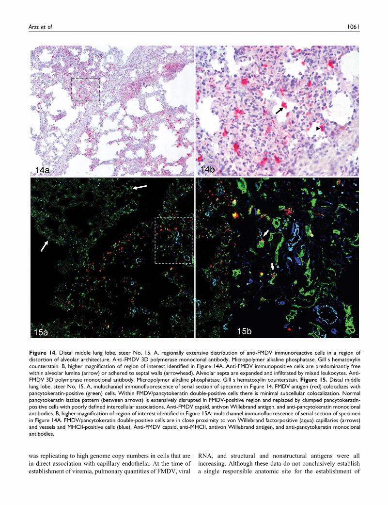
Forty-eight hpa. At 48 hpa there was a marked decrease of
detection of FMDV immunopositive cells in the nasopharynx.
Individual, and small clusters of, antigen-positive cells were
present within the strata basale and spinosum of NP/L epithelia
and lamina propria with no apparent predilection for FAE
regions. A single cluster of FMDV-positive cells within the
epiglottal epithelium of steer No. 14 was suggestive of a micro-
vesicle. This steer was the only animal in which FMDV anti-
gens were identified within the palatine tonsil (not shown).
In contrast to the lesser quantity of FMDV antigens loca-
lized to the upper respiratory tract (relative to steer euthanized
at 6–24 hpa), at 48 hpa there was substantially increased quan-
tity of structural and nonstructural FMDV antigens in pulmon-
ary samples with multifocal, coalescing distribution (Figs. 14,
15). Several tissues had regionally extensive fields spanning
up to 2.0 mm of FMDV antigen-positive cells within alveolar
parenchyma. Within these regions, alveolar septa were
expanded by infiltrates of mixed leukocytes and proteinaceous
material (fibrin). Cells containing FMDV antigens were round
to polygonal with eccentric nuclei and were more frequently
free within alveolar lumina or loosely associated with alveolar
septa when examined by IHC-light microscopy (Fig. 14). How-
ever, when examined by MIF, these cells were nearly exclu-
sively strongly CK positive (Fig. 15). In these regions there
were well-demarcated transitions from the normal CK
latticework pattern in regions lacking FMDV positivity to dis-
organized CK clumping and dissociation of cells from septa in
virus-positive regions. The pattern of MIF labelings also
revealed that a subpopulation of FMDV-positive cells had
somewhat greater association with alveolar septa than that
detectable by light microscopy. Labelings for vimentin and
vWF suggested that theses markers were present in a more nor-
mal architecture relative to the deterioration of the CK (pneu-
mocytes) pattern. FMDV/CK double-positive cells were often
directly adjacent to vWF-positive capillary endothelia (Fig.
15B). Overall, the pattern had strong similarities to the
acantholytic degeneration seen in FMD vesicles.
Discussion
In considering the early pathogenesis of FMD in any species, 2
critical issues have substantial translational relevance to the
development of vaccines and biotherapeutics. Identifying the
primary site (or sites) of infection is foremost because blocking
these sites is the only method of achieving complete sterile pro-
tection. Identifying the mechanism of the establishment of vir-
emia is secondary but still substantial. In most cases, blocking
(or impairing) this process would substantially decrease shed-
ding, transmission, and severity of clinical signs. Previous
investigations of the early pathogenesis of FMD in cattle have
independently implicated the nasopharynx7,15 and the
lungs4,6,10 as sites of primary infection. A single study indi-
cated that upper or lower respiratory tract can similarly serve
as sites of primary infection and portals for systemic generali-
zation of FMD.24 Recent reviews have suggested that subse-
quent to natural aerosol exposure, FMDV replicates in the
pharynx and establishes viremia by draining through the lym-
phatic system, but after experimental aerosol inoculation, vire-
mia is established directly through the lungs.1,2 However, the
primary experimental basis for these claims is not entirely
clear. Another pathogenesis study implicated the nasal mucosa
as the site of primary infection.14 Overall, the published litera-
ture does not provide enough information to allow a clear inter-
pretation of the critical virus–host interactions associated with
early FMDV infection of cattle.
The current study provides a more thorough description of
the early events of experimental FMD in cattle than that previ-
ously published. Additionally, the use of a controlled aerosol
inoculation system has allowed a consistently repeatable
method of exposure to virus while preserving the natural route
of infection; high-throughput techniques have facilitated
Figure (continued). No. 4. A, multichannel immunofluorescence (MIF) of serial section of region of interest identified in Figure 2A.Colocalization (orange) of FMDV capsid antigens (red) with pancytokeratin-positive (green) crypt epithelial cells. Anti-FMDV capsid and anti-pancytokeratin monoclonal antibodies. B, simultaneous MIF of specimen in Figure 3A. MHCII-positive (blue) cells in close proximity to but notcolocalized with FMDV capsid (red) antigen. Anti-FMDV capsid and anti-MHCII monoclonal antibodies. C, simultaneous MIF of specimen inFigure 3A and 3B. CD11c-positive (aqua) cells in close proximity to but not colocalized with FMDV capsid (red) antigens. Anti-FMDV capsidand anti-CD11c monoclonal antibodies. D, merge of simultaneous MIF images from Figure 3A to 3C. Intraepithelial MHCII-positive (blue) andCD11c-positive (aqua) cells (presumptive dendritic cells) are in close proximity to FMDV/pancytokeratin double-positive cells. Anti-FMDVcapsid, anti-pancytokeratin, anti-CD11c, and anti-MHCII monoclonal antibodies
Arzt et al 1055
1055
Figure 4. Distal middle lung lobe, steer No. 6. Focal region of anti-FMDV immunoreactivity with localization of antigen exclusively to alveolarsepta (insert). AntiFMDV 3D protein monoclonal antibody. Micropolymer alkaline phosphatase. Gill s hematoxylin counterstain. Figure 5. Distalcranial lung lobe, steer No. 7. Immunohistochemical localization of FMDV antigen to squamous cells of alveolar septa (Insert A) and polygonal cellsof alveolar lumina (Insert B). Anti-FMDV capsid monoclonal antibody. Micropolymer alkaline phosphatase. Gill s hematoxylin counterstain.Figure 6. A, Distal cranial lung lobe, steer No. 7. Multichannel immunofluorescence of serial section of tissue shown in Figure 5. Colocalization
1056 Veterinary Pathology 47(6)
1056
screening of numerous tissues per animal and has thus provided
a highly detailed mapping of FMDV distribution.
Screening of tissues and swab specimens by rRT-PCR and
VI indicated that there are tissue-specific temporal trends
regarding distribution of FMDV and viral RNA subsequent
to aerosol inoculation. The period of 3 to 12 hpa was generally
dominated by amplification of virus in the nasopharyngeal/lar-
yngeal (NP/L) tissues, with a trend toward progressively higher
levels of viral RNA and greater prevalence of VI positivity over
time. Similarly, within the period of 4 to 12 hours, FMDV RNA
detection from nasal swabs was increasing toward the first of 2
peaks. The temporal coincidence of these trends suggests that
the RNA in nasal swabs in this period originated from FMDV
replication in the NP/L tissues. At 24 hpa, FMDV and viral
RNA were increasingly abundant in the pulmonary tissues,
with minimal change in the NP/L sites indicating pan–respira-
tory distribution of virus associated with numerous foci of
replication in both upper and lower respiratory tract. However,
at 48 hpa, detection of FMDV by both screening tests was
decreasing in NP/L tissues, with further increase in the lungs.
This coincided with the onset of viremia and the approach to
the second peak in nasal swab RNA detection. Interpretation
of the 2 peaks of the nasal swab FMDV RNA detection curve
in the context of the tissue-specific rRT-PCR data suggests that
there are distinct pharyngeal and pulmonary phases of FMDV
release into the respiratory tract. Overall, the tissue-specific
prevalence values for all previremic animals collectively indi-
cates NP/L sites as the most important sites of primary FMDV
infection.
The rRT-PCR and VI data indicate that subsequent to aero-
sol inoculation, virus inoculum was distributed throughout the
entire respiratory tract; yet, immunolocalization of FMDV anti-
gens indicated that primary infection was limited to specific
regions of the nasopharynx and, somewhat later, the lungs.
At early time points, tissues that were positive for FMDV by
VI or rRT-PCR but IHC–MIF negative were interpreted as hav-
ing virus or viral RNA on their superficial surfaces but not
active infection and replication. Thus, VI and rRT-PCR served
as screening tests for infection at the tissue and cellular level
(high sensitivity), whereas IHC–MIF functioned as a confirma-
tory procedures (high specificity). In the present study, the
overall success of this screening and confirmation approach
provided adequate efficacy of FMDV localization.
The similar localization of FMDV antigens to the lym-
phoid FAE of PALT crypts of the rostrodorsal nasopharynx
in both steers euthanized at 6 hpa demonstrated the impor-
tance of this region in the establishment of primary infection.
The colocalization of FMDV antigens with CK and lack of
colocalization with MHCII or CD11c in these tissues demon-
strated that the first cells infected with FMDV are of epithelial
origin. This is the first microscopic documentation of locali-
zation of FMDV to the respiratory tract of any aerogenously
infected animal earlier than 24 hpa. Two previous works have
demonstrated nasopharyngeal intraepithelial FMDV RNA in
cattle by in situ hybridization at 5 days after contact expo-
sure;20,26 however, at this later phase of infection, it is impos-
sible to separate primary aerogenous from secondary
hematogenous infection. The exclusive infection of PALT
FAE cells in the context of pan–respiratory exposure to virus
suggests that there are specific, intrinsic qualities of these
cells that make them highly susceptible to infection. The cau-
dal segment of the dorsal soft palate is anatomically aligned
with the rostral dorsal nasopharynx, and these 2 tissues were
determined to be largely similar morphologically, phenotypi-
cally, and with regard to FMD pathogenesis within this study.
The laryngeal tissues (epiglottis and larynx) are morphologi-
cally similar to the nasopharyngeal sites but with less mucosa-
associated lymphoid tissue, which might explain the slightly
decreased detection of virus in the former.
Although the nature of the susceptibility of the NP/L tis-
sues remains elusive, possibilities include cell-specific
expression of virus-specific receptors (aV integrins or other)
and expression of other presently unidentified cellular factors
by the susceptible cells or by other cells in the immediate
microenvironment. Although aVb6 and aVb3 expression has
been characterized in these tissues from uninfected cattle,16,17
simultaneous localization with FMDV has not been described.
The nearly diffuse epithelial distribution of aVb6 described in
one study17 combined with the multifocal distribution of pri-
mary FMDV infection (current work) suggests that the expres-
sion of integrins does not solely dictate cellular susceptibility
to infection.
The FAE is an extremely active epithelium, and candidate
cells that might affect the microenvironment include intrae-
pithelial DCs, lymphocytes, and natural killer cells. Similarly,
M cells in these regions may directly facilitate infection by pro-
viding nonspecific portals for entry of virus into the host via
pinocytosis. Morphologic and phenotypic characterization of
bovine PALT has indicated that such cells are present in the
FAE to varying extents in FMDV-infected cattle and in the
resting (naı̈ve) state (J.A., unpublished data). Additionally,
physical characteristics of the FAE crypts may influence sus-
ceptibility to infection; it is possible that pooling of secretions
in these regions allows increased time for adsorption of virions
Figure (continued). (orange) of FMDV capsid antigens (red) with pancytokeratin-positive (green) cells of alveolar septa. Anti-FMDV capsid andanti-pancytokeratin monoclonal antibodies. B, cranial lung lobe, steer No. 7. Simultaneous multichannel immunofluorescence of specimen in Figure6A. Vimentin positive (blue) cells in close proximity to but not colocalized with FMDV capsid antigen (red). Anti-FMDV capsid and anti-vimentinmonoclonal antibodies. Figure 7. A, Distal cranial lung lobe, steer No. 7. Multichannel immunofluorescence of serial section of tissue shown inFigures 5 and 6. Triple colocalization of FMDV capsid (red) and FMDV3D protein (blue) antigens with cytokeratin (green) in pneumocytes. VonWillebrand factor positive capillary endothelial cells (aqua) are interspersed with pneumocytes and are negative for both FMDV antigens. B-E,higher-magnification single-channel views of region of interest identified in Figure 7A; individual channels: B, cytokeratin (green); C, FMDV capsid(red); D, FMDV 3D polymerase (blue); E, von Willebrand factor (aqua).
Arzt et al 1057
1057
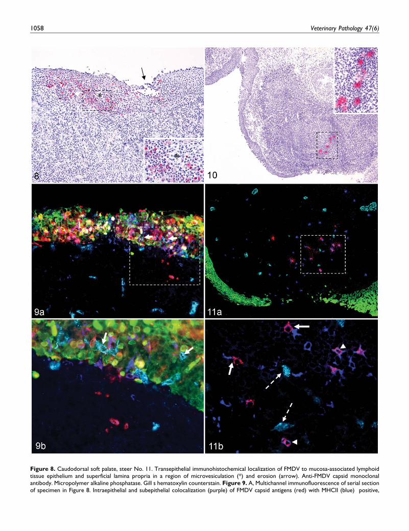
Figure 8. Caudodorsal soft palate, steer No. 11. Transepithelial immunohistochemical localization of FMDV to mucosa-associated lymphoidtissue epithelium and superficial lamina propria in a region of microvesiculation (*) and erosion (arrow). Anti-FMDV capsid monoclonalantibody. Micropolymer alkaline phosphatase. Gill s hematoxylin counterstain. Figure 9. A, Multichannel immunofluorescence of serial sectionof specimen in Figure 8. Intraepithelial and subepithelial colocalization (purple) of FMDV capsid antigens (red) with MHCII (blue) positive,
1058 Veterinary Pathology 47(6)
1058

and thus increases efficiency of infection in a highly localized
manner. Current efforts in our laboratory are directed toward
investigating these mechanisms.
Determining the role of the lungs in early infection was less
straightforward. Although viral RNA was detectable in pul-
monary tissues at 3 hpa and infectious virus at 6 hpa (similar
to NP/L sites), the prevalence of VI- and rRT-PCR-positive
samples was low at these time points compared to that in the
NP/L sites; additionally, viral antigens could not be detected
in lungs until 12 hpa (ie, 6 hours later than the nasopharynx).
At least 2 hypotheses are supported by this contrast in the data
describing processes in the NP/L sites and lung. It is possible
that the viral RNA and infectious virus detected in the lungs
before 12 hpa represent foci that were in the process of estab-
lishing primary infection independent of infection of the naso-
pharynx but with a relative lag (eclipse) period. Alternatively,
it is possible that primary infection of the NP/L sites serves as a
requisite phase in the establishment of pulmonary infection
presumably by dose amplification of inoculated virus. In this
scenario, the nasopharyngeally amplified virus would serve
as an endogenously generated secondary aerosol delivered to
the lungs via normal respiration. It is also possible that under
conditions of natural exposure (ie, direct or indirect contact),
both infection scenarios are relevant with contributory roles
determined by viral dose, aerosol droplet size, and various
aspects of virus host interactions.
Regardless of the contributory roles of lungs versus naso-
pharynx in the early pathogenesis of FMD in cattle, it is clear
that the pulmonary epithelia are permissive to FMDV replica-
tion. Our trimodal identification of FMDV in distal lung seg-
ments of both steers euthanized at 12 hpa (previremic)
indicates the consistent role of the lungs in the early progres-
sion of FMD. This is consistent with previous works that loca-
lized FMDV RNA to alveolar septa in previremic FMD in
aerosol inoculated cattle.4,6 In the present study, the morpholo-
gic localization of FMDV to alveolar septa combined with
exclusive colocalization with CK in these regions suggests that
pneumocytes are the most relevant cells supporting viral repli-
cation in the lungs. The morphologic variation of FMDV-
positive cells (squamous versus polygonal) seen in the lungs
of steer No. 7 likely represents a temporal and pathologic
continuum with the squamous variant (type I pneumocyte)
representative of early cellular infection that progresses to
infection of cells with polygonal morphology (type II pneumo-
cytes). Thus, the single, small (type I pneumocyte morphology)
focus of anti-FMDV immunopositivity in middle lung lobe of
steer No. 6 (Fig. 4) is interpreted as an earlier stage relative
to cranial lung specimen of steer No. 7 (Figs. 5–7). This con-
cept is further supported by the increasing abundance of
FMDV-immunopositive type II pnuemocytes at 24 and 48 hpa
and by ‘‘conventional wisdom’’ on the subject of responses to
insults by pulmonary epithelia.9 At 12 hpa, colocalization of
FMDV antigens and CK was rarely identified within terminal
bronchiolar epithelia, indicating that these cells are also sus-
ceptible to infection but with apparently lesser efficiency com-
pared to pneumocytes.
The question of how viremia is established in FMD has
substantial implications for ongoing and future development
of FMD prophylaxis. It has been suggested that in bovine
FMD, the mechanism of the establishment of viremia is that
virus drains from the pharynx to the regional lymph nodes
from which viremia is established.1,2 In the present study, the
lack of detection of FMDV or viral RNA in lymph nodes
draining the respiratory tract and oral cavity in the previremic
and early viremic phases of FMD suggests that this is not the
route of establishment of viremia within the current work’s
experimental conditions. A specific region’s ability to serve
as a portal for establishment of viremia depends on local
presence of virus in immediate proximity to blood vessels
and on a competent mechanism of movement of virions from
the extravascular to intravascular space. The transfer mechan-
ism is a complex subject that was not addressed by the cur-
rent study but likely involves several molecular and cellular
recognition events. However, in the current work, the require-
ment for intimate association of FMDV and blood vessels
was shown to be met in previremic animals in tissues of the
nasopharynx and lungs.
As early as 12 hpa, FMDV-positive type I pneumocytes
were present within alveolar septa that contained abundant
vWF- and vimentin-positive cells (ie, capillary endothelia).
This suggests that in the lungs, FMDV would have a high like-
lihood of gaining access to the vasculature. However, at 24 hpa,
presumptive dendritic cells. Colocalization (orange) of FMDV capsid antigens (red) with cytokeratin-positive (green) epithelial cells. VonFigure (continued). Willebrand antigenpositive (aqua) capillary endothelial cells are present within epithelium (arrows) and lamina propria inclose proximity to FMDV-positive cells. Anti-FMDV capsid, anti-MHCII, antivon Willebrand antigen, and anti-pancytokeratin monoclonalantibodies. B, Higher magnification of region of interest identified in Figure 9A, multichannel immunofluorescence of serial section of specimen inFigure 8. Von Willebrand antigenpositive (aqua) capillary endothelial cells are present within epithelium (arrows) and lamina propria in closeproximity to FMDV-positive cells. FMDV/MHCII individual and double-positive cells are present within subepithelial lymphoid region. Anti-FMDVcapsid, anti-MHCII, antivon Willebrand antigen, and anti-pancytokeratin monoclonal antibodies. Figure 10. Rostrodorsal nasopharynx, steer No.12. Immunohistochemical localization of FMDV to lymphoid follicular dark zone of mucosa-associated lymphoid tissue. Anti-FMDV capsidmonoclonal antibody. Micropolymer alkaline phosphatase. Gill s hematoxylin counterstain. Figure 11. A, Multichannel immunofluorescence ofserial section of specimen in Fig. 10. Colocalization (purple) of FMDV capsid antigens (red) with MHCII-positive (blue) dendritic cells. FMDV doesnot colocalize with cytokeratin (green) or von Willebrand antigen (aqua). Anti-FMDV capsid, anti-MHCII, antivon Willebrand antigen, and anti-bovine cytokeratin monoclonal antibodies. B, Higher magnification of region of interest identified in Figure 11A. Multichannel immunofluorescenceof serial section of specimen in Figure 10. Within a lymphoid follicle, FMDV antigens (red) are in close proximity to (arrows) and colocalize with(purple; arrowheads) MHCII-positive (blue) cells. FMDV does not colocalize with von Willebrand antigenpositive (aqua) endothelial cells (brokenarrows). Anti-FMDV capsid, anti-MHCII, antivon Willebrand antigen, and anti-pancytokeratin monoclonal antibodies.
Arzt et al 1059
1059
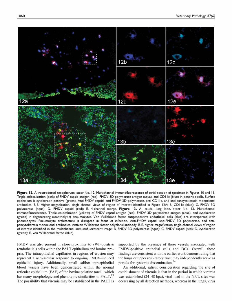
FMDV was also present in close proximity to vWF-positive
(endothelial) cells within the PALT epithelium and lamina pro-
pria. The intraepithelial capillaries in regions of erosion may
represent a neovascular response to ongoing FMDV-induced
epithelial injury. Additionally, small caliber intraepithelial
blood vessels have been demonstrated within the normal
reticular epithelium (FAE) of the bovine palatine tonsil, which
has many morphologic and phenotypic similarities to PALT.19
The possibility that viremia may be established in the PALT is
supported by the presence of these vessels associated with
FMDV-positive epithelial cells and DCs. Overall, these
findings are consistent with the earlier work demonstrating that
the lungs or upper respiratory tract may independently serve as
portals for systemic dissemination.24
An additional, salient consideration regarding the site of
establishment of viremia is that in the period in which viremia
was established (24–48 hpa), viral load in the NP/L sites was
decreasing by all detection methods, whereas in the lungs, virus
Figure 12. A, rostrodorsal nasopharynx, steer No. 12. Multichannel immunofluorescence of serial section of specimen in Figures 10 and 11.Triple colocalization (pink) of FMDV capsid antigen (red), FMDV 3D polymerase antigen (aqua), and CD11c (blue) in dendritic cells. Surfaceepithelium is cytokeratin positive (green). Anti-FMDV capsid, anti-FMDV 3D polymerase, anti-CD11c, and anti-pancytokeratin monoclonalantibodies. B-E, Higher-magnification, single-channel views of region of interest identified in Figure 12A: B, CD11c (blue); C, FMDV 3Dpolymerase (aqua); D, FMDV capsid (red); E, 4-channel merge. Figure 13. A, caudal lung lobe, steer No. 13. Multichannelimmunofluorescence. Triple colocalization (yellow) of FMDV capsid antigen (red), FMDV 3D polymerase antigen (aqua), and cytokeratin(green) in degenerating (acantholytic) pneumocytes. Von Willebrand factor antigenpositive endothelial cells (blue) are interspersed withpneumocytes. Pneumocyte architecture is disrupted in focus of infection. Anti-FMDV capsid, anti-FMDV 3D polymerase, and anti-pancytokeratin monoclonal antibodies. Antivon Willebrand factor polyclonal antibody. B-E, higher-magnification single-channel views of regionof interest identified in the multichannel immunofluorescent image: B, FMDV 3D polymerase (aqua); C, FMDV capsid (red); D, cytokeratin(green); E, von Willebrand factor (blue).
1060 Veterinary Pathology 47(6)
1060
was replicating to high genome copy numbers in cells that are
in direct association with capillary endothelia. At the time of
establishment of viremia, pulmonary quantities of FMDV, viral
RNA, and structural and nonstructural antigens were all
increasing. Although these data do not conclusively establish
a single responsible anatomic site for the establishment of
Figure 14. Distal middle lung lobe, steer No, 15. A, regionally extensive distribution of anti-FMDV immunoreactive cells in a region ofdistortion of alveolar architecture. Anti-FMDV 3D polymerase monoclonal antibody. Micropolymer alkaline phosphatase. Gill s hematoxylincounterstain. B, higher magnification of region of interest identified in Figure 14A. Anti-FMDV immunopositive cells are predominantly freewithin alveolar lumina (arrow) or adhered to septal walls (arrowhead). Alveolar septa are expanded and infiltrated by mixed leukocytes. Anti-FMDV 3D polymerase monoclonal antibody. Micropolymer alkaline phosphatase. Gill s hematoxylin counterstain. Figure 15. Distal middlelung lobe, steer No, 15. A, multichannel immunofluorescence of serial section of specimen in Figure 14. FMDV antigen (red) colocalizes withpancytokeratin-positive (green) cells. Within FMDV/pancytokeratin double-positive cells there is minimal subcellular colocalization. Normalpancytokeratin lattice pattern (between arrows) is extensively disrupted in FMDV-positive region and replaced by clumped pancytokeratin-positive cells with poorly defined intercellular associations. Anti-FMDV capsid, antivon Willebrand antigen, and anti-pancytokeratin monoclonalantibodies. B, higher magnification of region of interest identified in Figure 15A; multichannel immunofluorescence of serial section of specimenin Figure 14A. FMDV/pancytokeratin double-positive cells are in close proximity to von Willebrand factorpositive (aqua) capillaries (arrows)and vessels and MHCII-positive cells (blue). Anti-FMDV capsid, anti-MHCII, antivon Willebrand antigen, and anti-pancytokeratin monoclonalantibodies.
Arzt et al 1061
1061
FMDV viremia in cattle, interaction between processes in
upper and lower respiratory tract regions are likely, and current
efforts in our laboratory are directed toward defining the rela-
tive contributory roles of these regions.
The FMDV-associated degenerative process observed in the
lungs at 48 hpa has many microscopic similarities to the classic
acantholytic degeneration described for vesicles caused by
FMDV and other pathologic processes;11,28 however, macro-
scopic lesions were not detected in the lungs. As is seen with
keratinocytes in FMDV vesicles, the pneumocytes swell, lose
association with adjacent structures, and dissociate into an
expanding cavitary space.21,28 However, unlike a classic vesi-
cle in which the morphologic characteristics of acantholysis are
well defined and recognizable, this pulmonary acantholytic-
like degeneration could easily be mistaken for a histiocytic
alveolitis. The morphology of the acantholytic pneumocytes
is similar to that of alveolar macrophages, and it is only
the CK positivity of these cells that confirms their epithelial
histogenesis. An additional consideration on this subject is the
relationship that this pulmonary process may have to the main-
tenance of high-titer viremia during acute FMD. Although it
has been suggested that sustained FMDV viremia is maintained
by viral replication in lesional and/or nonlesional skin,1,5 this
hypothesis has never been thoroughly elucidated. Several
factors support the notion that the lungs may be important
amplifiers of FMDV and may be responsible for maintaining
high titer viremia, including (1) the relatively high FMDV
RNA/mg and large quantities of FMDV structural and non-
structural antigens detected in the lungs of viremic cattle in this
study, (2) the overall mass of the lungs, and (3) the extensive
vascularity of lungs.
In conclusion, the data presented herein support a model for
early FMDV infection of aerosol-inoculated cattle that includes
the following key events: (1) primary replication in epithelial
cells of the PALT crypts and (2) subsequent widespread repli-
cation in pneumocytes in the lung, which coincides with (3) the
establishment of sustained viremia. Current work in our labora-
tory is directed toward similar investigation of additional
serotypes and strains of FMDV and further elucidation of
FMDV–host interactions in the early pathogenesis of FMD.
Acknowledgements
This research was funded by Agricultural Research Service–Current
Research Information System project No. 1940-32000-052-00D. We
wish to acknowledge Meghan Tucker for superb histotechnological
support and Elizabeth Bishop, Ethan Hartwig, and George Smoliga for
crucial assistance in collecting and processing samples. We also thank
the animal resource unit personnel at the Plum Island Animal Disease
Center for their patience and assistance in caring for and collecting
samples from experimental animals. We thank Dr Manuel Borca and
Dr Fawzi Mohammed for thoughtful review of the article.
Declaration of Conflicting Interests
The authors declared no potential conflicts of interests with respect to
the authorship and/or publication of this article.
Funding
The authors are all salaried employees of the US federal government.
The research described and the authorship of the manuscript was all
conducted within the duties of the authors’ job posts. There was no
additional support.
References
1. Alexandersen S, Mowat N: Foot-and-mouth disease: host range
and pathogenesis. Curr Top Microbiol Immunol 288:9–42, 2005.
2. Alexandersen S, Zhang Z, Donaldson AI, Garland AJ: The patho-
genesis and diagnosis of foot-and-mouth disease. J Comp Pathol
129:1–36, 2003.
3. Arzt J, Gregg DA, Clavijo A, Rodriguez LL: Optimization of
immunohistochemical and fluorescent antibody techniques for
localization of foot-and-mouth disease virus in animal tissues.
J Vet Diagn Invest 21:779–792, 2009.
4. Brown CC, Meyer RF, Olander HJ, House C, Mebus CA: A
pathogenesis study of foot-and-mouth disease in cattle, using in
situ hybridization. Can J Vet Res 56:189–193, 1992.
5. Brown CC, Olander HJ, Meyer RF: Pathogenesis of foot-and-
mouth disease in swine, studied by in-situ hybridization. J Comp
Pathol 113:51–58, 1995.
6. Brown CC, Piccone ME, Mason PW, McKenna TS, Grubman MJ:
Pathogenesis of wild-type and leaderless foot-and-mouth disease
virus in cattle. J Virol 70:5638–5641, 1996.
7. Burrows R, Mann JA, Garland AJ, Greig A, Goodridge D: The
pathogenesis of natural and simulated natural foot-and-mouth dis-
ease infection in cattle. J Comp Pathol 91:599–609, 1981.
8. Callahan JD, Brown F, Osorio FA, Sur JH, Kramer E, Long GW,
Lubroth J, Ellis SJ, Shoulars KS, Gaffney KL, Rock DL, Nelson
WM: Use of a portable real-time reverse transcriptase-polymerase
chain reaction assay for rapid detection of foot-and-mouth disease
virus. J Am Vet Med Assoc 220:1636–1642, 2002.
9. Caswell J, Williams K. The respiratory system In: Jubb, Kennedy
& Palmer’s Pathology of Domestic Animals, ed. Maxie MG, 5th
ed., pp. 523–653. Saunders, St. Louis, MO, 2007.
10. Eskildsen M: Experimental pulmonary infection of cattle with
foot-and-mouth disease virus. Nord Vet Med 21:86–91, 1969.
11. Frenkel HS: Histologic changes in explanted bovine epithelial
tongue tissue infected with the virus of foot-and-mouth disease.
Am J Vet Res 10:142–145, 1949.
12. Golde WT, Pacheco JM, Duque H, Doel T, Penfold B, Ferman
GS, Gregg DR, Rodriguez LL: Vaccination against foot-and-
mouth disease virus confers complete clinical protection in 7 days
and partial protection in 4 days: use in emergency outbreak
response. Vaccine 23:5775–5782, 2005.
13. Grubman MJ, Baxt B: Foot-and-mouth disease. Clin Microbiol
Rev 17:465–493, 2004.
14. Korn G: Experimentelle untersuchungen zum virusnachweis im
inkubationsstadium der maul-und klauenseuche und zu ihrer
pathogenese. Arch Exp Veterinarmed 11:637–649, 1957.
15. McVicar JW, Graves JH, Sutmoller P: Growth of foot-and-mouth
disease virus in the bovine pharynx. In: Proceedings of the 74th
Annual Meeting of the United States Animal Health Association,
pp. 230–234. United States Animal Health Association, St.
Joseph, MO, 1970.
1062 Veterinary Pathology 47(6)
1062
16. Monaghan P, Gold S, Simpson J, Zhang Z, Weinreb PH, Violette
SM, Alexandersen S, Jackson T: The alpha(v)beta6 integrin
receptor for foot-and-mouth disease virus is expressed constitu-
tively on the epithelial cells targeted in cattle. J Gen Virol
86:2769–2780, 2005.
17. O’Donnell V, Pacheco JM, Gregg D, Baxt B: Analysis of
foot-and-mouth disease virus integrin receptor expression in
tissues from naive and infected cattle. J Comp Pathol
141:98–112, 2009.
18. Pacheco JM, Arzt J, Rodriguez LL: Early events in the pathogen-
esis of foot-and-mouth disease in cattle after controlled aerosol
exposure. Vet J 183:46–53, 2008.
19. Palmer MV, Thacker TC, Waters WR: Histology, immunohisto-
chemistry and ultrastructure of the bovine palatine tonsil with spe-
cial emphasis on reticular epithelium. Vet Immunol Immunopathol
127:277–285, 2009.
20. Prato Murphy ML, Forsyth MA, Belsham GJ, Salt JS: Localiza-
tion of foot-and-mouth disease virus RNA by in situ hybridization
within bovine tissues. Virus Res 62:67–76, 1999.
21. Seibold HR: A revised concept of the lingual lesions in cattle with
foot-and-mouth disease. Am J Vet Res 24:1123–1130, 1963.
22. Sellers RF, Parker J: Airborne excretion of foot-and-mouth
disease virus. J Hyg (Lond) 67:671–677, 1969.
23. Stave JW, Card JL, Morgan DO: Analysis of foot-and-mouth
disease virus type O1 Brugge neutralization epitopes using
monoclonal antibodies. J Gen Virol 67:2083–2092, 1986.
24. Sutmoller P, McVicar JW: Pathogenesis of foot-and-mouth dis-
ease: the lung as an additional portal of entry of the virus. J Hyg
(Lond) 77:235–243, 1976.
25. Swaney LM: A continuous bovine kidney cell line for routine assays
of foot-and-mouth disease virus. Vet Microbiol 18:1–14, 1988.
26. Woodbury EL, Ilott MC, Brown CC, Salt JS: Optimization of an
in situ hybridization technique for the detection of foot-and-
mouth disease virus in bovine tissues using the digoxigenin sys-
tem. J Virol Methods 51:89–93, 1995.
27. Yang M, Clavijo A, Li M, Hole K, Holland H, Wang H, Deng
MY: Identification of a major antibody binding epitope in the
non-structural protein 3D of foot-and-mouth disease virus in cat-
tle and the development of a monoclonal antibody with diagnostic
applications. J Immunol Methods 321:174–181, 2007.
28. Yilma T: Morphogenesis of vesiculation in foot-and-mouth dis-
ease. Am J Vet Res 41:1537–1542, 1980.
Arzt et al 1063
1063
Copyright © 2022 FDOKUMEN