Enhanced Tolerance to Multiple Abiotic Stresses in Transgenic Alfalfa Accumulating Trehalose
Upload
khangminh22Category
view
0download
0
i
The distinct roles of
Pseudomonas α-glucan and
trehalose in desiccation and
osmotic stress tolerances
Stuart Daniel Woodcock
September 2019
Thesis submitted to the University of East Anglia for
the degree of Doctor of Philosophy
John Innes Centre
Norwich, United Kingdom
© This copy of the thesis has been supplied on condition that anyone who consults it is
understood to recognise that its copyright rests with the author and that use of any information
derived there-from must be in accordance with current UK Copyright Law. In addition, any
quotation or extract must include full attribution.

ii
Abstract Pseudomonas aeruginosa and Pseudomonas syringae are significant pathogens of humans
and plants, respectively. An important prelude to infection is the ability of the pathogen to
survive independently of the host and to withstand environmental stresses. This is achieved
through several mechanisms including the biosynthesis of trehalose. Trehalose has
previously been implicated in the tolerance of a wide range of abiotic stresses, particularly
osmotic shock. Trehalose biosynthetic enzymes in Pseudomonas spp. were thought to be
encoded by the treS or treY/treZ operons, deletion of which reduces pathogenicity in
planta, illustrating the importance of trehalose metabolism during plant infection.
We used a combination of genetics and biochemistry to dissect trehalose metabolism. This
work has allowed us to examine the relationship between the biosynthesis of this molecule,
and its roles in stress protection. Contrary to previous understanding, we show that the
treS operon is responsible for the degradation of trehalose in Pseudomonas spp. forming
the polysaccharide α-glucan. As expected, we found that trehalose was a key molecule
during survival in osmotic conditions. An absence of intracellular trehalose yielded
osmotically-sensitive strains, whereas those with increased levels of trehalose were
osmotically-resistant. Surprisingly α-glucan conferred no discernable effect on osmotic
sensitivity but was important for survival under desiccating conditions. This phenotype was
independent of the level of trehalose, marking a clear distinction between the roles of
these two molecules in plant interactions and infection.
Other groups have observed the upregulation of genes responsible for the production of
the exopolysaccahride alginate during desiccation stress, in Pseudomonas spp. We showed
that alginate is also involved in the protection against desiccation stress. Using Arabidopsis
thaliana as an infection model, we observe attenuation when trehalose, α-glucan, and
alginate biosynthetic pathways are absent, demonstrating importance of water stress
during various stages of the Pseudomonas life cycle.

iii
Acknowledgements Firstly, I would like to thank Jake Malone, Cyril Zipfel, Stephen Bornemann and Richard Little for all
their supervision, guidance and support throughout my PhD. I would also like to thank the Malone
and Bornemann groups for all their advice, help and training along the way. A special thanks goes
to the Molecular Microbiology department for being such a welcoming, warm and fun place to
work. Additional thanks are owed to Karl Syson, James Brown, Govind Chandra, Gerhard Saalbach
and Sergey Nepogodiev for their expertise and assistance with enzymology, statistical analysis,
bioinformatics, mass-spectrometry and NMR. I am extremely grateful to Carmen Chen, Wai Hoe
Chin, Despoina Sifouna and Thomas Gate for being amazing students and putting up with my
supervision. You guys taught me a lot. A big thanks to my lovely proof-readers Catriona Thompson
and Hannah McDonald.
I could not have completed this thesis without the love and support of my friends and family. To
my Mom and Dad, I don’t know what I would have done without your endless love, encouragement
and ability to listen to my continuous ramblings. To my best friend Corinne, thank you for all the
coffee, pizza and spontaneous trips around the world. Finally, to Corinne’s appendix, you were not
helpful.
iv
Contents Abstract .................................................................................................................................. ii
Acknowledgements ............................................................................................................... iii
Contents ................................................................................................................................ iv
List of Figures ...................................................................................................................... viii
List of Tables ......................................................................................................................... xii
List of Abbreviations ........................................................................................................... xiii
General Introduction ........................................................................................ 1
1.1 Introduction ........................................................................................................................ 2
1.1.1 General Introduction to Pseudomonads ........................................................................... 2
1.1.2 Pseudomonas aeruginosa .................................................................................................. 2
1.1.3 Pseudomonas syringae ...................................................................................................... 4
1.1.4 Bacterial Stress ................................................................................................................... 6
1.1.5 Water Stress ....................................................................................................................... 7
1.1.6 Trehalose .......................................................................................................................... 13
1.1.7 The biosynthesis of bacterial α-glucan ............................................................................ 15
1.1.8 GlgE pathway ................................................................................................................... 16
1.1.9 Predicted trehalose and α-glucan biosynthesis in Pseudomonas spp. ............................ 18
1.1.10 Function of trehalose and α-glucan in Pseudomonas spp. ............................................ 21
1.2 Thesis Aims ........................................................................................................................ 23
General Materials and Methods .................................................................... 24
2.1 Media and Methods .......................................................................................................... 25
2.2 Genetic Manipulation ....................................................................................................... 25
2.2.1 Construction of gene deletion plasmids .......................................................................... 25
2.2.2 Construction of otsA/otsB expression plasmids .............................................................. 26
2.2.3 Construction of PglgA-lacZ fusion reporter plasmids ...................................................... 26
2.3 Specialised transformation of Pseudomonas.................................................................... 27
2.3.1 Transformation of Pseudomonas spp. using pTS1 and pME6032 based vectors ............ 27
2.3.2 Selection of double crossover candidates using pME3087 based vectors ...................... 27
2.3.3 Transformation of Pseudomonas spp. using pUC18 based vectors ................................. 28
2.3.4 Storage and access of biological material ........................................................................ 28
2.4 Metabolite Extraction ....................................................................................................... 28

v
2.5 Nuclear Magnetic Resonance Spectroscopy ..................................................................... 29
2.6 Iodine Staining .................................................................................................................. 29
2.7 Osmotic Stress Assays ....................................................................................................... 29
Reverse genetics and biochemical characterisation of trehalose and α-glucan
biosynthesis in Pseudomonas spp. ....................................................................................... 30
3.1 Introduction ...................................................................................................................... 31
3.2 Results ............................................................................................................................... 33
3.2.1 Reverse genetics and nuclear magnetic resonance spectroscopy can be used to explore
changes within the soluble carbohydrate metabolome of Pseudomonas spp. mutants ......... 33
3.2.2 PAO1 and Pto produce α-glucan, trehalose and maltose 1-phosphate .......................... 34
3.2.3 GlgA is a starch synthase that utilises UDP-glucose to produce linear α-glucan ............. 35
3.2.4 GlgP recycles α-glucan and produces glucose 1-phosphate from maltooligosaccharides
.................................................................................................................................................. 37
3.2.5 TreS/Pep2 utilises trehalose to produce maltose 1-phosphate ...................................... 39
3.2.6 GlgE utilises maltose 1-phosphate to produce linear α-glucan ....................................... 41
3.2.7 Absence of GlgB results in the accumulation of long linear α-glucan ............................. 43
3.2.8 GlgX functions to debranch α-glucan in Pseudomonas spp. ............................................ 45
3.2.9 TreS(2) functions as a second trehalose synthase in Pto ................................................. 46
3.2.10 TreY and TreZ produce trehalose by degrading linear α-glucan producing
maltooligosyltrehalose as an intermediate .............................................................................. 47
3.2.11 MalQ disproportinates α-glucan and is capable of synthesising maltose ..................... 49
3.2.12 Characterisation of linear α-glucan................................................................................ 51
3.3 Discussion .......................................................................................................................... 54
Trehalose and α-glucan protect against distinct abiotic stresses in
Pseudomonas ....................................................................................................................... 61
4.1 Introduction ...................................................................................................................... 62
4.2 Materials and Methods ..................................................................................................... 63
4.2.1 Desiccation Tolerance Assay ............................................................................................ 63
4.2.2 Plant Infection Assays ...................................................................................................... 64
4.3 Results - Phenotypic characterisation of trehalose and α-glucan biosynthesis in
Pseudomonas aeruginosa PAO1. .................................................................................................. 65
4.3.1 Disruption of trehalose biosynthesis results in osmotic sensitivity in Pseudomonas
aeruginosa PAO1....................................................................................................................... 65
4.3.2 Loss of α-glucan but not trehalose results in desiccation sensitivity in PAO1 ................. 68
4.3.3 In trans expression of Microbacterium koreensis 3J1 otsA/otsB recovers trehalose
biosynthesis .............................................................................................................................. 70
4.3.4 Reintroduction of trehalose biosynthesis rescues osmotic sensitivity ............................ 73

vi
4.3.5 Reintroduction of trehalose biosynthesis rescues desiccation sensitivity in PAO1 but only
when TreS/Pep2 is present ....................................................................................................... 75
4.3.6 Maltose and maltose 1-phosphate do not play a role in desiccation stress in
Pseudomonas aeruginosa PAO1. .............................................................................................. 76
4.3.7 Alginate plays a role in protecting PAO1 during desiccation stress................................. 79
4.4 Phenotypic characterisation of trehalose and α-glucan biosynthesis in Pto. ................... 82
4.4.1 Operon deletions resulted in abolition of both trehalose and α-glucan biosynthesis in Pto
but do not allow the distinction between these two molecules .............................................. 82
4.4.2 Loss of both trehalose and α-glucan in Pto results in osmotic sensitivity ....................... 84
4.4.3 Loss of both trehalose and α-glucan in Pto results in desiccation sensitivity .................. 85
4.4.4 Alginate works in combination with α-glucan to protect against desiccation stress in Pto
.................................................................................................................................................. 86
4.4.5 Trehalose, α-glucan and alginate are important during plant infection.......................... 87
4.5 Discussion .......................................................................................................................... 91
Investigating the regulation of trehalose and α-glucan biosynthesis ............ 99
5.1 Introduction .................................................................................................................... 100
5.1.1 Regulation ...................................................................................................................... 100
5.1.2 Regulation of trehalose .................................................................................................. 100
5.1.3 Regulation of classical α-glucan biosynthesis ................................................................ 101
5.1.4 Regulation of alginate biosynthesis ............................................................................... 104
5.1.5 Chapter Aims .................................................................................................................. 106
5.2 Materials and Methods ................................................................................................... 107
5.2.1 Liquid Metabolite Extraction ......................................................................................... 107
5.2.2 RNA Extraction and Retrotranscription ......................................................................... 107
5.2.3 qPCR ............................................................................................................................... 108
5.2.4 Biparental Mating .......................................................................................................... 108
5.2.5 Transposon Mutagenesis ............................................................................................... 109
5.2.6 DNA-Affinity Chromatography ....................................................................................... 109
5.3 Results ............................................................................................................................. 112
5.3.1 Regulation of trehalose and α-glucan biosynthesis occur at the level of transcription in
Pto ........................................................................................................................................... 112
5.3.2 Generation of PglgA-lacZ transcriptional fusion vectors .................................................. 114
5.3.3 Transposon mutagenesis ............................................................................................... 115
5.3.4 Identification of potential regulators of trehalose and α-glucan biosynthesis in Pto ... 116
5.3.5 Identification of potential regulators of trehalose and α-glucan biosynthesis in
Pseudomonas aeruginosa PAO1. ............................................................................................ 119
5.3.6 Deletion of cmpX resulted in increased trehalose production but sensitivity to osmotic
stress. ...................................................................................................................................... 119

vii
5.3.7 Disruption of algB indicates a role as a repressor of trehalose and α-glucan biosynthesis
in PAO1.................................................................................................................................... 122
5.3.8 Disruption of fimS implicates the AlgR/FimS two-component system as an activator of
trehalose and α-glucan biosynthesis in PAO1......................................................................... 123
5.3.9 Gac/Rsm System ............................................................................................................ 124
5.3.10 DNA-affinity chromatography ...................................................................................... 127
5.4 Discussion ........................................................................................................................ 128
General Discussion .......................................................................................136
6.1 Introduction .................................................................................................................... 137
6.2 Does the GlgE pathway exist and function in Pseudomonas spp.? ................................ 137
6.3 How do trehalose and α-glucan function in Pseudomonas spp.? ................................... 139
6.4 How are trehalose and α-glucan biosynthesis regulated in Pseudomonas spp.?........... 140
6.5 What is the molecular basis for the function of α-glucan and alginate function in
Pseudomonas? ............................................................................................................................ 142
6.6 Evolution of the GlgE Pathway ........................................................................................ 143
6.7 Widespread connections ................................................................................................ 146
6.8 Industrial application ...................................................................................................... 147
References ....................................................................................................149
Appendix ................................................................................................................................ a

viii
List of Figures Figure 1-1: Overview of Pseudomonas syringae pv. tomato DC3000 infection of plants. .... 5
Figure 1-2: A summary of water activity (AW) in bacteria during desiccation stress over
time. ................................................................................................................................ 9
Figure 1-3: Overview of alginate biosynthesis in Pseudomonas aeruginosa PAO1............. 12
Figure 1-4: Overview of classical α-glucan and trehalose biosynthesis in bacteria. ........... 14
Figure 1-5: Structural schematic of bacterial α-glucan. ....................................................... 16
Figure 1-6: Overview of the mycobacterial GlgE pathway. ................................................. 17
Figure 1-7: The genomic organisation of predicted trehalose and α-glucan biosynthesis
genes in Pseudomonas aeruginosa PAO1. ................................................................... 18
Figure 1-8: The genomic organisation of predicted trehalose and α-glucan biosynthesis
genes in Pto. .................................................................................................................. 19
Figure 1-9: Predicted pathway of trehalose and α-glucan biosynthesis in Pseudomonas
spp. ................................................................................................................................ 21
Figure 3-1: The predicted reaction catalysed by GlgA ......................................................... 35
Figure 3-2: 1H-NMR spectroscopy of the soluble metabolome following the deletion of
glgA in Pseudomonas spp. ............................................................................................ 36
Figure 3-3: The predicted reaction catalysed by GlgP ......................................................... 37
Figure 3-4: 1H-NMR spectroscopy of the soluble metabolome following the deletion of
glgP in Pseudomonas spp. ............................................................................................ 38
Figure 3-5: The predicted reactions catalysed by TreS/Pep2: the processing of trehalose
into maltose and maltose 1-phosphate (M1P). ............................................................ 39
Figure 3-6: 1H-NMR spectroscopy of the soluble metabolome following the deletion of
treS/pep2 in Pseudomonas spp. ................................................................................... 40
Figure 3-7: The predicted reaction catalysed by GlgE: the extension of linear α-glucan
using maltose 1-phosphate (M1P) as a substrate. ....................................................... 41
Figure 3-8: 1H-NMR spectroscopy of the soluble metabolome following the deletion of
glgE in Pseudomonas spp. ............................................................................................ 42
Figure 3-9: The predicted reaction catalysed by GlgB and GlgX .......................................... 43
Figure 3-10: 1H-NMR spectroscopy of the soluble metabolome following the deletion of
glgB in Pseudomonas spp. ............................................................................................ 44
Figure 3-11: Visualising the production of α-glucan in PAO1 strains using iodine staining.
...................................................................................................................................... 45
Figure 3-12: 1H-NMR spectroscopy of the soluble metabolome following the deletion of
glgX in Pseudomonas spp. ............................................................................................ 46
ix
Figure 3-13: 1H-NMR spectroscopy of the soluble metabolome following the deletion of
treS(2) in Pseudomonas syringae pv. tomato. .............................................................. 47
Figure 3-14: The predicted reactions catalysed by TreY, TreZ and TreS/Pep2 .................... 48
Figure 3-15: 1H-NMR spectroscopy of the soluble metabolome following the deletion of
treZ and treY in Pseudomonas spp. .............................................................................. 49
Figure 3-16: 1H-NMR spectroscopy of the soluble metabolome following the deletion of
malQ in Pseudomonas spp. .......................................................................................... 50
Figure 3-17: Iodine staining of linear α-glucan in Pseudomonas aeruginosa PAO1 strains
using iodine vapour. ..................................................................................................... 53
Figure 3-18: Current understanding of trehalose and α-glucan biosynthesis in
Pseudomonas spp. ........................................................................................................ 54
Figure 4-1: Growth of PAO1 and mutant strains in M9 minimal medium ± 0.85 M NaCl. .. 67
Figure 4-2: Linear mixed modelling analysis of the survival of PAO1, ΔglgA, ΔtreS/pep2
and ΔglgB strains when incubated at 100% (white bars) and 75% RH (grey bars). ..... 69
Figure 4-3: Scheme showing the genetic and biochemical basis following the production
of trehalose biosynthetic enzymes OtsA/OtsB in wildtype PAO1 (A), ΔtreS/pep2 (B)
and ΔglgA (C). ............................................................................................................... 71
Figure 4-4: 1H-NMR spectroscopy of the soluble metabolome following the over-
expression otsA/otsB in PAO1 strains........................................................................... 72
Figure 4-5: Growth of PAO1 and mutant strains with or without expression of otsA/otsB in
M9 minimal medium ± 0.85 M NaCl. ............................................................................ 74
Figure 4-6: Linear mixed modelling analysis of the survival of PAO1 and mutant strains
expressing otsA/otsB when incubated at 100% (white bars) and 75% RH (grey bars).
...................................................................................................................................... 76
Figure 4-7: Scheme representing the genetic approach used to generate a α-glucan-null
mutant capable of synthesising maltose and maltose 1-phosphate (M1P). ................ 77
Figure 4-8: 1H-NMR spectroscopy of the soluble metabolome following the deletion of
glgA, glgE and the over-expression of otsA/otsB in PAO1. .......................................... 78
Figure 4-9: Linear mixed modelling analysis of the survival of PAO1 and ΔglgA ΔglgE ::
otsA/otsB strains when incubated at 100% (white bars) and 75% RH (grey bars)....... 79
Figure 4-10: Growth of PAO1 and Δalg in M9 minimal medium ± 0.85 M NaCl. ................ 80
Figure 4-11: Linear mixed modelling analysis of the survival of PAO1 and Δalg when
incubated at 100% (white bars) and 75% RH (grey bars). ............................................ 81
Figure 4-12: The genomic organisation of the trehalose and α-glucan biosynthesis genes
in Pto. ............................................................................................................................ 82
Figure 4-13: 1H-NMR spectroscopy of the soluble metabolome following the deletion of
PSPTO_2760-2762, PSPTO_3125-3134 and PSPTO_2760-2762 PSPTO_3125-3134 (ΔΔ)
in Pto. ............................................................................................................................ 83
Figure 4-14: Growth of wild-type Pto and ΔΔ in M9 minimal medium ± 0.35 M NaCl. ...... 84

x
Figure 4-15: Linear mixed modelling analysis of the survival of Pto, ΔΔ, Δalg and Δalg ΔΔ
strains when incubated at 100% (white bars) and 75% RH (grey bars). ...................... 86
Figure 4-16: A. thaliana (Col-0) infection with Pto, ΔΔ and Δalg. ........................................ 89
Figure 4-17: A. thaliana (Col-0) infection with Pto and Δalg ΔΔ.......................................... 90
Figure 4-18: The current understanding of water stress protection mechanisms of
Pseudomonas spp. ........................................................................................................ 97
Figure 5-1: A graphical representation of the Gac/Rsm system in P. aeruginosa. ............104
Figure 5-2: 1H-NMR spectroscopy shows the effects of growth in liquid culture upon the
soluble metabolome of the Pto ΔglgB strain. ............................................................112
Figure 5-3: The addition of Lugol’s solution to metabolome extracts from Pto ΔglgB
cultured in liquid (L) or on solid medium (S). .............................................................113
Figure 5-4: Quantitative PCR data showing the absolute quantification of transcripts from
Pto cultured using solid or liquid M9 minimal medium relative to the housekeeping
gene rpoD. ...................................................................................................................114
Figure 5-5: The reporter strain Pto PglgA-lacZ cultured on L medium supplemented with 50
µg/mL X-Gal. ...............................................................................................................115
Figure 5-6: 1H-NMR spectroscopy of the soluble metabolome following transposon
insertion within Pto PglgA-lacZ. ....................................................................................118
Figure 5-7: 1H-NMR spectroscopy of the soluble metabolome following transposon
insertion within cmpX in the PAO1 PglgA-lacZ strain. ..................................................120
Figure 5-8: Growth of PAO1 and cmpX mutant strains in M9 minimal medium with and
without addition of 0.85 M NaCl. ...............................................................................121
Figure 5-9: 1H-NMR spectroscopy of the soluble metabolome following insertion within
algB of the PAO1 PglgA-lacZ strain. .............................................................................122
Figure 5-10: Growth of PAO1 PglgA-lacZ and tn::algB in M9 minimal medium with and
without addition of 0.85 M NaCl. ...............................................................................123
Figure 5-11: 1H-NMR spectroscopy of the soluble metabolome following transposon
insertion within fimS in the PAO1 PglgA-lacZ strain. ....................................................124
Figure 5-12: Growth of PAO1 PglgA-lacZ and tn::fimS in M9 minimal medium with and
without addition of 0.85 M NaCl. ...............................................................................124
Figure 5-13: 1H-NMR spectroscopy of the soluble metabolome following Gac/Rsm system
mutations in Pseudomonas aeruginosa strains. .........................................................127
Figure 5-14: The current understanding of the regulatory network governing trehalose
and α-glucan in Pseudomonas spp. ............................................................................135
Figure 6-1: Illustration of futile cycling of α-glucan biosynthesis and degradation in
Pseudomonas spp. ......................................................................................................145
Figure i: Heat map representing the statistical significance of desiccation responses
between PAO1 strains based on linear mixed modelling. .............................................. k

xi
Figure ii: Heat map representing the statistical significance of desiccation responses
between Pto strains based on linear mixed modelling. .................................................. l

xii
List of Tables Table 3-1: Concentrations of trehalose and M1P produced by PAO1 strains. .................... 33
Table 3-2: Concentrations of trehalose and M1P produced by Pto strains. ........................ 34
Table 4-1: Concentrations of trehalose and M1P produced by PAO1 mutant strains with or
without expression of otsA/otsB. ................................................................................. 73
Table 4-2: Concentrations of trehalose and M1P produced by wild-type PAO1 and PAO1
ΔglgA ΔglgE :: otsA/otsB strains. ................................................................................... 77
Table 4-3: Concentrations of trehalose and M1P produced by Pto strains. ........................ 83
Table 5-1: Transposon candidates for potential regulators of trehalose and α-glucan
biosynthesis in Pto. .....................................................................................................116
Table 5-2: Concentrations of trehalose and M1P produced by Pto PglgA-lacZ transposon
mutants. ......................................................................................................................117
Table 5-3: Transposon candidates for potential regulators of trehalose and α-glucan
biosynthesis in PAO1. .................................................................................................119
Table 5-4: Concentrations of trehalose and M1P produced by PAO1 PglgA-lacZ transposon
mutants. ......................................................................................................................120
Table 5-5: Concentrations of trehalose and M1P produced by PAO1-L, ΔgacA and ΔgacS
strains. .........................................................................................................................126
Table 5-6: Concentrations of trehalose and M1P produced by PAO1-N, ΔrsmA, ΔrsmN, and
ΔrsmA ΔrsmN strains. .................................................................................................126
Table 5-7: Putative regulators that were identified through specific binding to PAO1 PglgA.
....................................................................................................................................128
Table i: Primers used for genetic manipulations .....................................................................
Table ii: Primers used for genetic manipulations ....................................................................
Table iii: Primers used for genetic manipulations ...................................................................
Table iv: Primers used for genetic manipulations ...................................................................
Table v: Primers used for genetic manipulations ....................................................................
Table vi: Primers used for genetic manipulations ...................................................................
Table vii: Primers used for arbitrary PCR and sequencing .......................................................
Table viii: Primers used for qPCR .............................................................................................
Table ix: List of Pto strains used in this study ......................................................................... i
Table x: List of PAO1 strains used in this study ....................................................................... j

xiii
List of Abbreviations X-gal 5-bromo-4-chloro-3-indolyl β-D-galactopyranoside
Csr carbon storage regulator
cAMP cyclic adenosine monophosphate
CRP cAMP receptor protein
CFU colony forming units
CF cystic fibrosis
ddH2O double distilled H2O
DPI days post infection
DP degree of polymerisation
Δ deletion of specified gene(s) or operon
EPS exopolysaccharides
G1P glucose 1-phosphate
G6P glucose 6-phosphate
HPr histidine phosphocarrier protein
LEA late embryogenesis proteins
M1P maltose 1-phosphate
MALDI-MS matrix-assisted laser desorption/ionization mass spectrometry
Tm melting temperature
OD600 optical density at λ 600 nm
PBS phosphate buffer saline
PTS phosphoenopyruvate:carbohydrate phosphotransferase system
P-HPr phosphorylated form of the histidine phosphocarrier protein
pppGpp guanosine pentaphosphate
ppGpp guanosine tetraphosphate
1H-NMR proton nuclear magnetic resonance
Psa Pseudomonas syringae pv. actinidiae
Pto Pseudomonas syringae pv. tomato
RH relative humidity
TMSP trimethylsilyl propanoic acid

xiv
v/v volume of solute/volume of solution
w/v mass of solute/volume of solution
AW water activity

1
General Introduction

2
1.1 Introduction
The Pseudomonas genus encompasses agriculturally and clinically important pathogens
which cause significant socioeconomic cost. In order to successfully infect, they first need
to survive in the environment and cope with various stresses. They synthesise molecules
to help tolerate stresses, but this is not fully understood. One such molecule is trehalose,
which has been shown to provide protection against several abiotic stresses and ultimately
play a role in virulence. Our understanding of its metabolism has had to be substantially
revised with the recent discovery of a novel α-glucan biosynthetic pathway. This thesis
therefore addresses the role of these two molecules, trehalose and α-glucan, and their
roles in Pseudomonas species (spp.) against water stresses.
General Introduction to Pseudomonads
Pseudomonas is one of the most diverse bacterial genera. These bacteria exhibit extensive
metabolic plasticity and are found in a variety of environments, including soil [1, 2], bodies
of water [3] and precipitation [4]. Certain species are also associated with the colonisation
of animals [5, 6], plants [1], and insects [7]. Moreover, the human pathogen Pseudomonas
aeruginosa can colonise artificial environments such as hospital equipment [8, 9]. Despite
Pseudomonas spp. representing both commensal and pathogenic organisms, the two
major pathogenic species, P. aeruginosa and Pseudomonas syringae, have been most
extensively researched. As such, these two species form the primary focus of this thesis.
Pseudomonas aeruginosa
P. aeruginosa is a significant pathogen of humans and is one of the most common causes
of nosocomial infections [10]. It is an opportunistic pathogen that primarily affects
immunocompromised individuals, including patients with immunodeficiencies [11, 12],
those with disruption of the skin barrier, such as burns victims [13], and those who have
had foreign bodies introduced, such as cannulas or catheters [8]. P. aeruginosa is more
commonly known as a pneumonial agent causing acute and chronic infection of the lungs,
with the former a result of sudden trauma to the lung tissue such as smoke inhalation or
other injury [14, 15]. Acute infections are usually resolved relatively quickly by host
defences or treatment with antibiotics. However, these infections are difficult to eradicate
due to the intrinsic resistance to antibiotics exhibited by P. aeruginosa. [16, 17]. Acute
infections can become chronic if they are not resolved by the host.

3
Chronic infections are described as the persistent colonisation or infection of the lung by
P. aeruginosa where the patient’s immune system has responded by the production of anti-
pseudomonad antibodies [18]. Chronic P. aeruginosa infections occur in 80% of adult cystic
fibrosis (CF) patients where morbidity and mortality rates are significantly increased when
compared to CF patients who have not been infected with P. aeruginosa [18-20].
Following colonisation of the CF lung with P. aeruginosa, a phenotypic diversification to a
mucoid strain is extremely likely marking the onset of chronic infection [21-23]. The mucoid
phenotype is attributed to the over-expression of the exopolysaccharide (EPS) alginate
[24]. Alginate production is controlled by the sigma factor AlgU, which itself is normally
repressed by the anti-sigma factor MucA and its cognate partner MucB [25]. In mucoid
strains, MucA is typically mutated leading to the de-repression of AlgU and the over-
production of alginate [26]. Alginate over-expression and the subsequent effect on biofilm
production leads to increased antibiotic resistance [24], thereby enhancing the antibiotic
tolerance profile of an already highly resistant organism. However, the importance of the
biofilm is not limited to chronic infections, as the formation of biofilms has also been
observed with acute infection [27].
In order to establish a chronic infection, P. aeruginosa must be able to resist and tolerate
hostile conditions, including constant stimulation of the host immune system and
numerous rounds of antibiotic treatment. In order to cope with a constant barrage of
hostility and stress, P. aeruginosa has evolved various mechanisms to help it to survive [9].
P. aeruginosa can alter its immunogenicity and dampen the inflammatory response by
down-regulating or preventing the expression of immunogenic structures, such as flagella
and type IV pili [28, 29]. Furthermore, various other virulence factors that stimulate
inflammatory responses or have necrotic effects can also be down-regulated or are absent
altogether [29]. As well as evading the host immune system, P. aeruginosa is intrinsically
resistant to most antibiotics. This characteristic is attributable to the low permeability of
their outer membrane [30], alongside the expression or upregulation of chromosomally-
encoded lactamases and multi-drug efflux systems [17]. Pseudomonads can attain
additional genetic resistance through chromosomal mutations and horizontal gene
transfer. This can include the acquisition of extended spectrum β-lactamases [31] or the
mutation of regulatory elements which can result in the over-expression of efflux pumps
[32]. Antibiotic-resistant P. aeruginosa infections translates into longer hospital admissions
and up to a 500% increase in mortality rates when compared to susceptible strains [33]. As

4
a consequence of this, P. aeruginosa has been classified as a serious threat equal to
methicillin-resistant Staphylococcus aureus and extensively drug-resistant Mycobacterium
tuberculosis [34]. This emphasises the economic and health consequences of P. aeruginosa
infections.
Owing to the difficulty of treating P. aeruginosa infections due to the factors discussed
above, there has been much emphasis upon the prevention of chronic infection [35]. P.
aeruginosa is thought to be transmitted by contact with contaminated surfaces [36] and
hospital equipment [37]. Contact with health care professionals [36, 38], other patients
[39] or healthy relatives [40] can also be sources of contamination. Furthermore, there are
reports of transmission between CF patients and pet animals [41, 42]. Soil and agricultural
produce also present a significant reservoir of this pathogen [43, 44]. Investigating the
underlying mechanisms that P. aeruginosa utilises to survive and persist in these various
environments may be applicable in preventing transmission and therefore the
establishment of infection.
Pseudomonas syringae
P. syringae is one of the most significant pseudomonad plant pathogens. More than 60
varieties (pathovars) have been described based upon the host plant species and the
symptoms that are caused [45, 46]. This bacterium can infect over 180 plant species
resulting in significant economic costs and loss of yield. Hosts include cereals and other
economically relevant plants, including wheat [47], tomato [48] and kiwifruit [49].
One specific pathovar, P. syringae pv. actinidiae (Psa), is the causative agent of bacterial
canker in kiwifruit. An epidemic of Psa began to infect New Zealand kiwifruit orchards in
2010 and spread to approximately 85% of the New Zealand kiwifruit orchards by 2015 [50].
It was predicted that Psa would incur economic costs of up to NZ $410 million by 2015,
however, in 2017 it was reported that actual costs incurred were as high as NZ $930 million
[51]. The agricultural industry uses various methods to control and prevent the infection,
including streptomycin and copper sulphate sprays. However, like P. aeruginosa, P.
syringae has also developed resistance to these control mechanisms [52] exacerbating the
socio-economic costs associated with infection.
As depicted in Figure 1-1, there are two generally accepted phases of the lifecycle of P.
syringae during plant infection, these are termed epiphytic and endophytic. P. syringae is
disseminated through the water cycle or through agricultural activities such as pollination

5
[53, 54]. The epiphytic phase begins when the bacterium lands on the surface of the plant
and forms microcolonies and biofilms. The surface-exposed portion of the plant is also
known as the phyllosphere which includes the stem, leaves, flowers and fruit. Below the
soil surface, epiphytic habitats are known as the rhizosphere [55, 56]. Following the
epiphytic phase, P. syringae can migrate and enter the plant tissue through natural
openings, such as stomata, or through sites of injury [46, 57]. This marks the start of the
endophytic stage of the lifecycle and once in the apoplastic space P. syringae will multiply
and cause disease. During the course of infection, recognition of bacterial pathogen-
associated molecular patterns or effectors can stimulate the plant immune system. This
triggered immunity will activate the hypersensitive response, suppress bacterial growth
and limit the progression of infection. P. syringae responds by utilising its repertoire of
effector proteins to suppress the plant immunity and to acquire nutrient access [58-60].
Strategies regarding the entry of the bacterium into the apoplastic space can include the
secretion of phytotoxins to interfere with plant hormone signalling and prevent the closing
of the stomata [61]. An alternative strategy is to induce the nucleation of ice via the ice
nucleating protein INA, this causes cellular damage and provides additional entry points
into the plant tissue [62].
Figure 1-1: Overview of Pseudomonas syringae pv. tomato DC3000 infection of plants.Bacteria arrive on the surface of the phyllosphere where there is colony and biofilm
formation. (2) Migration across the surface of the plant to natural openings or wounds to gain
access to the apoplast. (3) Release of phytotoxins such as coronatine to prevent stomatal closure
as represented by light blue triangles. (4) The use of the ice nucleation protein, as represented by
blue stars, to wound the plant tissue and create alternative entry points. Grey colouring represents
dead or damaged plant cells. Once in the apoplastic space bacteria will secrete various effectors
and toxins to access nutrients (5) and to modulate the host (6), this is represented by the orange
and dark blue triangles respectively. This figure is reproduced with permission from Pfeilmeier et
al. (2016) [63].

6
Bacterial Stress
The bacterial response to stress is important at all stages of the life cycle. Bacteria must
survive various abiotic and biotic stresses to survive in their environment or within their
host during infection. Abiotic stresses encompass environmental factors such as
temperature, pH, ultra-violet radiation, water availability, salinity and nutrient starvation
[64-66]. By contrast, biotic stresses are factors born from other living organisms such as
microbial competition or host defences [67]. Nutrient availability can be a consequence of
either an abiotic or biotic stress, the latter of which can be caused by the competition for
resources with other microorganisms [68]. Bacteria withstand these biotic stresses using a
variety of mechanisms, such as the production of siderophores to sequester iron [69],
through the direct competition with other microorganisms via the production of antibiotics
[70] or through the deployment of type VI secretion systems [71]. Importantly, an
understanding of the mechanisms that bacteria use to respond to and resist environmental
abiotic stresses can lead to an alternative strategy to combat bacterial disease. By
exploiting these systems, bacterial persistence and environmental transmission could be
prevented.
P. syringae and P. aeruginosa spend significant parts of their life cycle in soils and in
association with host organisms. As part of this, these bacteria will be subjected to rapidly
changing levels of pH (acid stress), water stress, ultraviolet radiation, temperature and
nutrient starvation [55, 56, 72]. For example, the leaf surface is exposed to extreme
fluctuations of water availability [73]. Consequently, the ability of P. syringae to produce
water stress protection molecules has been shown to be important during epiphytic
colonisation [74]. Similarly, during colonisation of the cystic fibrosis lung P. aeruginosa is
exposed to various abiotic stresses, such as water stress, nutrient stress and oxidative
stress [75]. P. aeruginosa and P. syringae must be able to respond to these varying
conditions effectively to ensure survival in the environment, a successful life cycle and the
ability to cause disease. As an example, oxidative stress is generated by the formation of
intracellular reactive oxygen species which damages DNA, proteins and lipids [76]. This
damage can interfere with cellular processes and even result in cell death. Bacteria respond
to this stress by producing scavenging enzymes, such as superoxide dismutases, catalases
and peroxidases to convert the damaging species into harmless by-products [77]. An
alternative strategy is the expression of oxidative damage-resistant forms of enzymes [78].

7
The bacterial response to stress not only governs the protection of the cell against a given
stress but can also alter bacterial virulence and resistance to antimicrobial agents [79]. For
example, DNA damage, caused by both abiotic and biotic factors, can induce the production
of phage-encoded toxins in shiga-toxin producing Escherichia coli [80]. Furthermore, the
response to osmotic stress can induce the production of the type III secretion system
thereby promoting virulence in Salmonella enterica serovar Typhimurium [81]. The
bacterial response to various stresses can also increase resistance to antimicrobial agents
in several ways. This can include the transfer of antimicrobial resistance genes from one
cell to another [82], the prevention of cellular division [83], or the production of slow-
growing persister cells [84]. This illustrates the importance of understanding the
mechanisms that pathogenic bacteria use to survive in rapidly changing environments and
how they respond to specific stresses.
Water Stress
The availability of water is generally regarded as one of the most important abiotic factors
affecting all domains of life. It is essential for cellular functions and all living organisms.
With the current threat of climate change, water stress has become an important factor
for agriculture because 64% of the global arable land has been predicted to be affected by
water deficit or drought [85]. Microbial tolerance to water stress has become a more active
field of research as microbial-plant interactions have been shown to increase the tolerance
of plants to abiotic stresses [86] including drought [87, 88]. This is likely due to the ability
of select microorganisms to survive under dry conditions and subsequently confer stress-
tolerance to the host plant [89].
Water stress manifests itself in at least two distinct phenomena; osmotic and desiccation
stress. There are two types of osmotic stress, hypo- and hyperosmotic stress. Hypoosmotic
stress is defined by a decrease in the osmotic pressure of the external environment
translating to influx of water into the bacterial cell. This causes an increase in the cytosolic
volume and can cause cell lysis [90]. Hyperosmotic stress is defined as the loss of water and
reduction of cytosolic volume due to an increase in the osmotic pressure of the external
environment. When the concentration of external solutes is greater than the concentration
of internal cytosolic solutes, water will leave the bacterial cell by osmosis to equalise the
osmotic pressure [90]. This decreases the water availability within the bacterium,
compromising essential cellular processes. The work discussed within this thesis relates to

8
hyperosmosis therefore subsequent references to osmotic stress refers to hyperosmotic
conditions (unless otherwise stated).
Desiccation stress is defined as the dehydration of the cell and subsequent loss of water.
This differs from osmotic stress due to the fact that desiccation stress is independent of
solute concentration and the immediate environment to which water is lost [91]. Under
osmotic conditions, bacteria are surrounded by an aqueous environment of reduced water
activity. Conversely, during desiccation stress, bacteria are exposed to air and water is lost
to the atmosphere and cannot be recovered [92]. Desiccation stress is a multi-factorial
phenomenon where the removal of water from bacterial cells can cause a variety of
deleterious effects. The key stages of desiccation are summarised in Figure 1-2. Desiccation
stress consists of three main stages; drying, storage and rewetting [93]. The drying stage
can manifest as either fast or slow drying, where the rate of dehydration determines the
outcome of bacterial survival. Bacteria exposed to slow desiccating conditions exhibit a
higher rate of survival than those that are rapidly dehydrated [94]. As the intracellular
water potential decreases, bacteria are exposed to various stresses, including osmotic and
oxidative stress, toxicity due to the build-up of intracellular salts and other molecules as
well as the disruption of cellular processes due to the denaturing of enzymes and proteins.
In contrast to rapid desiccation, slow drying is likely to allow bacteria sufficient time to
mount the appropriate responses.
The storage phase begins when the water activity of the cell plateaus. During storage, the
number of viable bacteria will reduce over time. Although the exact mechanisms are
unknown, this is probably due to accumulative damage governed by oxidative and radiation
stresses [93, 95].
The final phase of desiccation is the rewetting or reviving of dehydrated bacteria. This is
governed by the reintroduction of water, most likely due to precipitation. Similar to drying,
the rate of rehydration dictates bacterial fate [93] in that fast rehydration leads to
increased cell death as compared to slow rehydration [96]. This is most likely due to cell
lysis because of rapid water influx owing to high intracellular solute concentrations from
the drying phase. In contrast to slow rehydration, rapid rehydration is likely to cause cell
lysis as the cell does not have sufficient time to respond and modulate the intracellular
solute concentration.

9
Figure 1-2: A summary of water activity (AW) in bacteria during desiccation stress over time. A depiction of the three stages of desiccation stress, drying, storage and rewetting. The additional stresses encountered during each stage are indicated. This figure is reproduced with permission from Vriezen et al. (2007) [93].
Owing to the multi-factorial components of desiccation stress, the mechanisms conferring
tolerance to dehydration are less well-understood. Mechanisms that have been found to
confer desiccation stress tolerance are also intrinsically linked to the tolerance of osmotic
stress. However, there has been some effort towards dissecting individual responses to the
two stresses [97].
One strategy to tolerate low water conditions is the modulation of metabolism. This can be
via the formation of either metabolically-inert bacteria [98] or highly stress-resistant
spores. These mechanisms ensure the long-term survival of the bacteria by shutting down
virtually all metabolic processes. Certain genera of bacteria undergo sporulation when they
encounter unfavourable abiotic stresses, such as nutrient, pH or competition stress. Spores
are extremely hardy and resistant to a variety of abiotic stresses including water stress,
ultraviolet radiation, heat and even modern disinfectants [99, 100]. Bacteria are thought
to accumulate various molecules within the spore to protect against abiotic stresses. For

10
example, Streptomyces spp. spores have been shown to accumulate the compatible solute
trehalose. Increased concentrations of which have been shown to correlate with the stress
resistance of the spore [101]. However, sporulation as a method of resisting water stress is
not widespread among bacteria because relatively few genera contain representatives that
are capable of producing spores [102].
A more widespread method of resisting water stress is the biosynthesis of protective
molecules. For example, bacterial desiccation stress resistance can be conferred by the
expression of desiccation-specific proteins. The expression of hydrophillins, such as the late
embryogenesis abundant (LEA) proteins, have been reported to protect against desiccation
stress in bacteria [103]. It has been shown that the addition of bacterial LEA proteins to
purified enzymes preserve enzymatic function during desiccating conditions [104]. In
addition to desiccation stress, LEA proteins have also been shown to improve osmotic
tolerance in human cell lines [105]. However, it is thought that the molecular mechanism
of protection conferred by LEA proteins differs to that of compatible solutes such as
trehalose [104].
Compatible Solutes
One of the most common mechanisms to protect against water stress in bacteria is the
accumulation of compatible solutes. These include betaine derivatives, sugars, amino acids
and inorganic ions [90]. Investigation into the responses of P. aeruginosa PAO1 and P.
syringae pv. tomato DC3000 (Pto) to osmotic stress revealed the accumulation of the
compatible solutes glutamate, trehalose and N-acetylglutaminylglutamine amide [74, 106,
107]. Compatible solutes protect the cell against osmotic stress by minimising water loss.
They have also been shown to play roles in stabilising proteins [108, 109] and membranes
[110] thereby preserving cellular functions during conditions of low water availability.
These stabilisation functions have also implicated compatible solutes in the protection
against other stresses, such as desiccation [111] and temperature stress [112, 113]. For
example, glycine betaine was found to protect Bacillus subtilis during low temperatures
[114] whereas trehalose has been implicated in the protection against desiccation stress in
both bacteria and yeast [113, 115].
Compatible solutes accumulate within bacteria via active uptake from the environment or
through de novo synthesis. An example of this is the biosynthesis of the compatible solute
glycine betaine in E. coli which requires the uptake of choline through the choline-specific

11
transporter BetT. Choline is then metabolised into glycine betaine through the action of
the BetA and BetB enzymes [116]. In contrast, compatible solute glutamate can be taken
up from the environment [117] or synthesised de novo [118] in response to osmotic stress.
Biofilms and alginate production
Biofilms are structured communities of bacteria which can adhere to each other or a range
of biological or inert surfaces. The biofilm itself is formed from mainly water and bacteria,
but also contains polysaccharides, nucleic acids, lipids, and proteins. These extracellular
factors are collectively known as the extracellular polymeric substance matrix [119]. The
biofilm provides a variety of functions, including protection against abiotic and biotic
stresses. For example, the microenvironment of the biofilm can affect the behaviour of the
bacteria. When in this sessile, nutrient and oxygen-limiting environment, P. aeruginosa can
adopt a different gene expression profile [120, 121]. This results in slow growth and
upregulation of stress responses under the control of the alternative sigma factor S [122].
Furthermore, the biofilm matrix can provide steric hinderance to antibacterial molecules
entering the biofilm, preventing or delaying bacterial uptake of these molecules and
subsequent killing [123].
The EPS produced by P. aeruginosa PAO1 mainly consists of three polysaccharides:
alginate, pel and psl [124]. P. syringae has been shown to produce alginate, cellulose, levans
and psl as components of their biofilm [125, 126]. Although their exact functions are
unknown, each polysaccharide plays its own role in the formation of the biofilm [124].
In addition to other abiotic stresses [127], biofilms have been implicated in protecting
against water stress. Pseudomonas spp. respond to desiccating conditions by increasing the
amount of EPS produced within the biofilm [128]. This suggests that EPS and therefore
biofilms play a role in keeping cells hydrated. Subsequently, further work has specifically
implicated the EPS component alginate in the protection of Pseudomonas spp. against
water stress [129-131]. Moreover, the biosynthesis of alginate has been shown to be a key
adaption of P. aeruginosa within the CF lungs [29] and that alginate biosynthesis is
important for P. syringae when colonising the plant host [132]. Both of these environments
are thought to be exposed to osmotic stress and desiccation [73].
The bacterial production of alginate is restricted to two genera, Pseudomonas and
Azotobacter [133], but is highly conserved within Pseudomonas spp. Alginate is formed of
β-1,4-linked L-guluronic and D-mannuronic acids. The first step in the biosynthesis of

12
alginate is the production of a linear polymer of D-mannuronic acid (Figure 1-3). The
enzymes responsible for this are encoded by algA, algC, algD and alg8. The polymannurate
is then secreted into the periplasmic space where AlgG converts D-mannuronic acid
residues to L-guluronic acid and the combined actions of AlgI, AlgJ, AlgF and AlgX O-
acetylate the mannuronic acid residues [134, 135]. Mature alginate is then secreted
through the AlgE pore. Individual deletion of the algK, algX, algG or algD genes results in
the abolition of alginate production [134, 136].
Figure 1-3: Overview of alginate biosynthesis in Pseudomonas aeruginosa PAO1.Guanosine diphosphate-mannuronic acid is formed through the enzymatic functions of AlgA, AlgC and AlgD. This is then polymerised by Alg8. Polymannurate traverses the inner and outer membranes via the complex of Alg8 Alg44, AlgX, AlgK, AlgG, AlgL and AlgE. The mannuronic acid residues are O-acetylated by AlgI, AlgJ and AlgK or epimerised into L-glucuronic acid via AlgG. The mature alginate is then secreted out of the cell via the AlgE pore. This figure is reproduced with permission from Schmid and Rehm (2015) [135].
The decoration of alginate by O-acetylation not only protects alginate against degradation
by alginate lyase (AlgL) [137], but the degree of acetylation determines the water-holding
capacity of the polysaccharide where an increase in O-acetylation results in a higher degree
of water-holding capacity [138].

13
Alginate has been shown to be important in desiccating conditions in Pseudomonas putida
as disruption of the algD biosynthesis gene leads to increased cell death at low humidity
[130]. This suggested that alginate maintains a hydrated environment to enhance survival
of P. putida during desiccating conditions. The authors claimed that disruption of alginate
biosynthesis in P. aeruginosa and P. syringae also conferred sensitivity to desiccation stress.
However, this was performed using a high molecular weight solute to stimulate stress
rather than subjecting the strains to lower relative humidity (RH). Although this could be
interpreted as osmotic stress rather than a true desiccation stress, disruption of alginate
biosynthesis did not confer sensitivity to osmotic conditions induced by NaCl.
Trehalose
Trehalose is a ubiquitous disaccharide found in virtually all organisms [139]. First studied
enzymatically in 1953 [140], this compatible solute has since been found to play a variety
of roles including protection against abiotic stresses, carbon storage, and even as a
virulence factor [141]. Trehalose has also found use in the food and pharmaceutical
industry [142, 143] creating interest in producing large amounts of trehalose using
biological systems [144, 145].
The biosynthesis of trehalose has been implicated in the tolerance of many biotic and
abiotic stresses in a range of organisms [146-148], most notably of osmotic stress [112,
149]. Trehalose has also been implicated in protecting against desiccation stress. Positive
correlations have been found between the intracellular concentration of trehalose and the
level of desiccation resistance in bacteria [150, 151]. Additionally, the introduction and
expression of trehalose biosynthesis genes from the high-trehalose producing and
markedly desiccation-resistant organism Microbacterium korensis 3J1 conferred increased
desiccation-protection upon the desiccation-sensitive P. putida KT2440 [151].
Bacterial mutants lacking trehalose biosynthetic genes have also been found to be
attenuated during infection models [152, 153] making the biosynthesis of trehalose an
attractive topic of research. Our understanding of bacterial trehalose biosynthesis has been
informed by investigating the mechanisms in various bacteria. There are three widespread
trehalose metabolic pathways in bacteria: OtsA/OtsB, TreY/TreZ and TreS (Figure 1-4) [147,
154].

14
The most prominent route of trehalose biosynthesis in bacteria is via the OtsA/OtsB
pathway [154]. The OtsA/OtsB pathway produces trehalose using the trehalose 6-
phosphate synthase OtsA and the trehalose 6-phosphatase OtsB. OtsA produces trehalose
6-phosphate from glucose 6-phosphate and UDP-glucose [155]. Trehalose 6-phosphate is
then dephosphorylated by OtsB, forming trehalose.
Figure 1-4: Overview of classical α-glucan and trehalose biosynthesis in bacteria.(G6P; Glucose 6-phosphate, G1P; Glucose 1-phosphate, UDP-Glc; UDP-glucose, M1P; maltose 1-phosphate)
The TreS pathway utilises a single enzyme, the trehalose synthase TreS, to produce
trehalose through the isomerisation of maltose [156]. However, as explained below, this is
not the physiological role of this enzyme in all organisms [157]. The TreY/TreZ pathway
utilises α-glucans, such as bacterial glycogen produced by the classical GlgC-GlgA glycogen
biosynthetic pathway, to synthesise trehalose [147, 158]. The maltooligosyl trehalose
synthase TreY facilitates the conversion of the terminal reducing-end α-1,4 bond of the α-
glucan molecule into an α-1,1 glycosidic bond forming maltooligosyl trehalose. Trehalose
is then liberated from maltooligosyl trehalose by the hydrolase action of TreZ.
Bacteria contain varying combinations of all three trehalose biosynthetic pathways or, in
some cases, multiple copies of the same pathway or components thereof [159]. For

15
example, the only route to synthesise trehalose in E. coli is through the OtsA/OtsB pathway
because it lacks gene homologues for the other trehalose biosynthetic pathways.
The biosynthesis of bacterial α-glucan
α-Glucan (also referred to as bacterial glycogen) is a large polysaccharide reported to have
a relative molecular mass of up to 1 × 10⁸. It is formed of thousands of glucose monomers
linked via α-1,4 glycosidic links with branch points formed from α-1,6 linkages [160]. Each
α-glucan molecule consists of three different types of chain (Figure 1-5) [161]. The C chain
is the chain possessing the sole free-reducing end, B chains bare α-1,6 branches whereas
the A chains do not. α-Glucan is most commonly used as a carbon store which accumulates
during the exponential phase of bacterial growth and can be recycled during unfavourable
conditions. First studied in E. coli [162], the GlgC-GlgA pathway generates a linear α-glucan
via the action of the nucleotide diphosphoglucose pyrophosphorylase GlgC and glycogen
synthase GlgA (Figure 1-4). GlgC catalyses the synthesis of ADP-glucose from glucose 1-
phosphate (G1P), generated by the phosphoglucomutase PgmA [163, 164]. The activated
glucose molecule is subsequently utilised by GlgA to extend an α-1,4 linear α-glucan chain
[165, 166]. The linear α-glucan is then branched by the branching enzyme GlgB. This
involves the transfer of a non-reducing end oligomer to the 6-position of a residue within
a linear chain to form a α-1,6 branch point [167, 168]. Degradative enzymes GlgX and GlgP
break down α-glucan by debranching [169] and removing glucose units from the non-
reducing end of the α-glucan, respectively, [170] feeding into primary metabolism (Figure
1-4).

16
Figure 1-5: Structural schematic of bacterial α-glucan. α-Glucan consists of a primary C chain possessing the sole reducing end (Ø). B chains are those that possess α-1,6-branches whereas A chains possess no branches. This figure is reproduced with permission from Syson et al. (2016) [161].
GlgE pathway
The classical GlgC-GlgA pathway was once thought to be the only widespread bacterial
method of synthesising α-glucans with the genes responsible originally found to be present
within 34% of sequenced bacterial genomes, although this was later refined to 20% [171,
172]. Furthermore, trehalose and α-glucan were only linked through the classical GlgC-GlgA
pathway and the subsequent degradation of α-glucan by TreY/TreZ producing trehalose
(Figure 1-4).
In 2010, Kalscheuer et al. identified a novel mechanism of synthesising α-glucan in
Mycobacterium spp.; this was the so-called GlgE pathway [153] (Figure 1-6). This four-step
pathway, utilising trehalose to produce α-glucan, consists of TreS, Pep2, GlgE and GlgB. This
represents an additional link between trehalose and α-glucan biosynthesis [153].

17
Figure 1-6: Overview of the mycobacterial GlgE pathway. Trehalose is isomerised into maltose by the trehalose synthase (TreS), maltose is then phosphorylated into maltose 1-phosphate by the maltokinase Pep2. M1P is then polymerised into linear α-glucan by GlgE and branched by GlgB.
The first step of this pathway is the conversion of trehalose into maltose by TreS. As
discussed above, TreS can catalyse the isomerisation of maltose into trehalose as one of
the three main routes to trehalose biosynthesis [147]. Although the production of trehalose
by TreS is energetically favourable, it was shown that in Mycobacterium spp. the overall
flux favours the isomerisation of trehalose into maltose due to the presence of the GlgE
pathway [157]. This is driven largely by the adenosine triphosphate (ATP)-dependent
activity of the maltokinase Pep2 [173]. Maltose is phosphorylated by Pep2 into maltose 1-
phosphate (M1P) [157, 174]. The maltosyltransferase GlgE enzyme then transfers the
maltosyl moiety of M1P forming α-1,4 linkages with the non-reducing ends of α-glucan
chains [175].
Mycobacterial α-glucan is found both intra- and extra-cellularly in the cytosol and capsule,
respectively. It was shown that purified capsular α-glucan mediated interaction with the
complement 3 receptor of mammalian macrophages [176] and that reduced levels of
capsular α-glucan led to reduced pathogenicity in murine infection models [177]. Taken
together, this suggests that mycobacterial α-glucan is an important disease determining
factor.
The genes encoding the GlgE pathway are predicted to exist in 14% of sequenced bacterial
genomes including those of Pseudomonas spp. [171, 178, 179]. Given that the
Pseudomonas genus encompasses bacteria from a wide variety of environments and host-
ranges, including both human and plant pathogens, it seems unlikely that the function of
the predicted GlgE pathway and α-glucan in Pseudomonas spp. would relate to disease in

18
mammals and plants. This prompts the question: Is the GlgE pathway functional and what
is its purpose in Pseudomonas spp.?
Predicted trehalose and α-glucan biosynthesis in Pseudomonas spp.
Given the intrinsic links between trehalose and α-glucan biosynthesis in bacteria and to
gain insight into the biology of α-glucan, both systems must be studied. Trehalose and α-
glucan biosynthesis has been relatively understudied in Pseudomonas spp. with trehalose
biosynthetic operons only being identified recently [74]. Comparative genomics has
predicted that Pseudomonas spp. possess the gene homologues for the classical trehalose
biosynthesis pathways, TreS and TreY/TreZ, but lack gene homologues of otsA and otsB
[74]. Previous work assumed that both the TreS and TreY/TreZ pathways were both
associated with the production of trehalose, however, they did not account for the
presence of the GlgE pathway and its impact upon subsequent interpretations.
In P. aeruginosa PAO1 and Pto, the predicted trehalose biosynthetic genes are found within
two main clusters, the treS and treY/treZ operons (Figures 1-7 and 1-8) [74, 180]. The
predicted gene homologues of the GlgE pathway and classical α-glucan biosynthesis are
clustered within the treS and treY/treZ operons, respectively [171].
Figure 1-7: The genomic organisation of predicted trehalose and α-glucan biosynthesis genes in Pseudomonas aeruginosa PAO1.

19
Figure 1-8: The genomic organisation of predicted trehalose and α-glucan biosynthesis genes in Pto.
The so-called treS operon consists of predicted gene homologues encoding GlgE,
(designated PA2151 in PAO1 and PSPTO_2760 in Pto), TreS (PA2152, PSPTO_2761) and
GlgB (PA2153, PSPTO_2762). In both the PAO1 and Pto genome the gene annotated as treS
is fused to the predicted pep2 encoding maltokinase and is henceforth referred to as
treS/pep2.
The treY/treZ operon consists of the genes encoding predicted homologues of GlgA
(PA2165, PSPTO_3125), TreZ (PA2164, PSPTO_3126), the glucanotransferase MalQ
(PA2163, PSPTO_3127) [74], TreY (PA2162, PSPTO_3128) and GlgX (PA2160, PSPTO_3130).
Pto glgX is predicted to form its own operon (PSPTO_3130 – PSPTO_3134) with four
accessory genes whose functions are unknown (Figure 1-7, Figure 1-8) [181]. PA2161 and
PSPTO_3129 encode a small predicted open reading frame designated by the grey arrow in
Figures 1-7 and 1-8 in both genomes, however these are not studied in this thesis. The gene
encoding the recycling enzyme GlgP (PA2144, PSPTO_5165) is orphaned in both species
and found elsewhere in the genome. Pto is also predicted to possess a second homologue
of TreS (PSPTO_2952). This gene is not predicted to encode the Pep2 domain.
Although homologues of the phosphomutase PgmA are present, neither the PAO1 or Pto
genomes harbour gene homologues predicted to encode GlgC. This suggests that these
organisms lack the ability to produce ADP-glucose. However, gene homologues of the UDP-
glucose pyrophosphorylase GalU are present. When galU from E. coli was expressed in
Corynebacterium glutamicum, the concentrations of α-glucan and trehalose increased

20
[182]. This suggests that there was increased flux through trehalose biosynthetic pathways.
The authors suggest that UDP-glucose could be utilised in α-glucan biosynthesis. Indeed,
subsequent work has shown that GlgA from M. tuberculosis can utilise UDP-glucose as a
donor when G1P was used as a substrate [183]. Therefore, we anticipate that GalU provides
the phosphosugar donor for α-glucan in Pseudomonas spp.
The clustering of the genes within the treY/treZ operon does indeed suggest that the
function of this operon is to produce trehalose from GlgA-synthesised α-glucan as
previously published [74, 180]. The treS operon was originally thought to be involved in the
biosynthesis of trehalose. However, the fusion of the treS gene with the maltokinase pep2
and the genomic clustering of all four homologues of the GlgE pathway (glgE, treS/pep2
and glgB) suggest that the treS operon is actually involved with the conversion of trehalose
into α-glucan rather than synthesising trehalose as previously thought.
By combining previous bioinformatic analysis of trehalose biosynthesis in Pseudomonas
spp. [74, 180] with that of the GlgE pathway in Mycobacterium spp., I propose a model of
trehalose and α-glucan biosynthesis (Figure 1-9). One limitation of my model is that it does
not account for the functions of MalQ or the presence of the second TreS homologue in
Pto. This is addressed experimentally in Chapter 3.
My model predicts that the presence of both GlgA and GlgE results in two mechanisms of
synthesising α-glucan. It is currently unclear as to why Pseudomonas spp. has evolved two
distinct biosynthetic pathways of α-glucan biosynthesis and therefore prompts the
following question: are there differences between these two pools of α-glucan?

21
Figure 1-9: Predicted pathway of trehalose and α-glucan biosynthesis in Pseudomonas spp. Red indicates the enzymatic reactions studied within this thesis (G6P; Glucose 6-phosphate, G1P; Glucose 1-phosphate, UDP-Glc; UDP-glucose, M1P; maltose 1-phosphate).
Function of trehalose and α-glucan in Pseudomonas spp.
As described above, the function of trehalose has been well-studied in other bacteria.
Indeed, trehalose has been implicated in the protection against hyperosmotic stress in
Pseudomonas spp. Metabolomic analysis shows the accumulation of trehalose and other
compatible solutes during osmotic conditions [106, 107]. Furthermore, the genes
representing the treS and treY/treZ operons are upregulated in both PAO1 and Pto under
hyperosmotic conditions [107, 184]. Furthermore, deletion of the treY/treZ and treS
operons in Pto, resulted in the abolition of trehalose production and the generation of
osmotically-sensitive strains [74]. This was not the case in P. aeruginosa PA14 where
deletion of the trehalose biosynthesis operons abolished trehalose production albeit
without an effect on osmotolerance [180].
Trehalose has also been implicated in the protection of Pseudomonas spp. against
desiccation stress. The expression of the trehalose biosynthetic genes otsA and otsB from
Microbacterium koreensis 3J1 conferred increased desiccation resistance to P. putida [151].
This suggests that trehalose can also protect Pseudomonas spp. against desiccating

22
conditions. However, given the presence of the GlgE pathway, this phenotype may have
been indirectly related to trehalose.
Trehalose has previously been shown to be an important factor for plant infection.
Transcriptomic profiling of P. syringae pv. syringae revealed that the genes representing
the treS and treY/treZ operons were upregulated during both epiphytic and apoplastic
lifestyles when compared to laboratory grown cells [132]. Furthermore, deletion of the treS
and treY/treZ operons in both Pto and PA14 results in attenuation during infection of
tomato plants or Arabidopsis thaliana, respectively [74, 180]. In the case of Pto, attenuation
was exacerbated by reduced RH [74]. These studies implicate trehalose as an important
virulence factor for survival in the phyllosphere and during apoplastic growth. Although
important for plant infection, loss of the treS and treY/treZ operons did not attenuate PA14
during infection of insect or murine models [180]. Although trehalose was implicated, the
impact of the potential abolition of α-glucan biosynthesis was not considered.
The above studies have implicated trehalose as a stress protectant and as a virulence factor
in Pseudomonas spp. However, the interpretation of these results did not consider the
intrinsic genetic and functional linkage of trehalose and α-glucan biosynthesis afforded by
the GlgE pathway. For example, during osmotic conditions, genes responsible for trehalose
and α-glucan biosynthesis are both upregulated [184], and deletion of both trehalose and
α-glucan biosynthetic operons results in osmotically-sensitive strains in Pto [74]. The
question therefore arises: does α-glucan play a role in protecting Pseudomonas spp.
against osmotic stress?
The increase in intracellular trehalose in P. putida via the expression of otsA and otsB was
correlated with increased desiccation tolerance [151]. However, based on my predicted
model (Figure 1-9) increased concentrations of trehalose could also have resulted in the
increased biosynthesis of α-glucan. Furthermore, attenuation during infection of tomato
plants with Pto lacking both trehalose and α-glucan biosynthetic genes is exacerbated by
decreased RH. This raises the question: does α-glucan play a role during desiccation
stress?
Similarly, transcriptomic data show the upregulation of both trehalose and α-glucan
biosynthesis genes during the epiphytic and apoplastic lifestyle [132]. Furthermore, the
strains used for plant infection contained whole operon deletions or large deletions of the
surrounding loci. Little consideration was given to the effects of these deletions on α-glucan

23
biosynthesis [74, 180, 181]. Conclusions drawn from the disruption of what was thought to
be solely trehalose biosynthesis should therefore be revisited and the function of α-glucan
during Pseudomonas infection should be investigated. This therefore leads to the
following question: does α-glucan play a role during the epiphytic or apoplastic lifestyle
or during infection?
1.2 Thesis Aims
Based on recent findings in Mycobacterium and Pseudomonas, it has become apparent that
the biosynthesis of trehalose in Pseudomonas spp. is poorly understood. This is evident by
the co-occurrence of trehalose biosynthetic genes with predicted homologues of the GlgE
pathway genes. Previous work characterising the functions of trehalose in Pseudomonas
spp. did not consider the presence of α-glucan biosynthesis and how this impacts on the
interpretation of subsequent data.
As described in this thesis, I probed the existence of the GlgE pathway and therefore α-
glucan biosynthesis in the human and plant pathogens P. aeruginosa PAO1 and Pto. I
functionally characterised the roles of trehalose and α-glucan in stress-tolerance
individually by decoupling their biosynthesis. I then examined the contribution of these
sugars during the bacterial infection of plants. Finally, I probed the regulation of trehalose
and α-glucan biosynthesis.

24
General Materials and Methods

25
2.1 Media and Methods
For routine experiments Pseudomonas aeruginosa PAO1 and Escherichia coli DH5α strains
were cultured in lysogeny broth (10 g/L tryptone, 5 g/L yeast extract, 10 g/L NaCl) at 37 °C.
Pseudomonas syringae pv. tomato DC3000 strains were grown in L medium (10 g/L
tryptone, 5 g/L yeast extract, 5 g/L NaCl, 1 g/L glucose anhydrous) at 28 °C [185]. The
selection of transformants was facilitated by supplementing the appropriate media with
tetracycline at concentrations of 12.5 µg/mL (100 µg/mL for P. aeruginosa) or 25 µg/mL of
gentamycin. For NMR spectroscopy and stress tolerance assays, M9 minimal medium was
used (20 mM NH4Cl, 12 mM Na2HPO4, 22 mM KH2PO4, 8.6 mM NaCl, 1 mM MgSO4, 1 mM
CaCl2, and 0.4% glucose, 0.4% casamino acids, 50 µM FeCl3) [185]. Where appropriate liquid
media were shaken or solidified with 1.5% Bacto agar (Difco, Surrey, UK).
2.2 Genetic Manipulation
Cloning was accomplished using standard molecular biology techniques, described below
[186]. Polymerase chain reaction (PCR) products and plasmid digestion derivatives were
purified using the Nucleospin® Gel and PCR Clean-up kit (Macherey-Nagel). Plasmids were
extracted from E. coli DH5α strains using the Nucleospin® Plasmid DNA Purification
commercial kit (Macherey-Nagel). DNA digestions were performed using the appropriate
restriction endonucleases (New England Biolabs). T4 DNA ligase (New England Biolabs) was
used in the construction of all vectors in this study.
Unless otherwise stated, all PCR reactions were facilitated using Q5® High-Fidelity DNA
polymerase (New England Biolabs). PCR protocols were conducted as per the
manufacturers’ guidance. Primary denaturation was performed for 30 seconds at 98 °C.
Following this, 30 cycles of denatuation (98 °C, 10 seconds), annealing (varying
temperatures, 30 seconds) and extension (72 °C, 30 seconds) was performed. Following
this a final extension was performed at 72 °C for 120 seconds. The melting temperature
(Tm) of each primer was determined using the NEB Tm Calculator
(http://tmcalculator.neb.com). The annealing temperature for a given reaction was
calculated as Tmb – 2 °C. Where Tmb represents the lowest Tm of the primer pair. All
primers and melting temperatures are listed in the appendix.
Construction of gene deletion plasmids
Primers were designed manually, and incorporate BamHI or HindIII external restriction sites
and an XbaI internal restriction site. Primers 7-12 and 61-66 were designed by Dr Jacob. G.

26
Malone and incorporate BamHI, EcoRI or KpnI external restriction sites and XbaI internal
sites. Gene deletions were achieved by amplifying the upstream and downstream regions
flanking the desired gene from the PAO1 and Pto genomes. For example the PCR reaction
to amplify the upstream region of PA2151 was performed using primers 1 and 2 (Appendix).
This annealing temperature for this reaction was 70 °C.
The deletion of the Pto treY/treZ operon was facilitated by amplifying the upstream and
downstream regions of PSPTO_3125 and PSPTO_3134 respectively using primers 117, 74
and 120, 121 (table A1). This generated approximately 1 Kb regions flanking the treY/treZ
operon. The deletion of the Pto treS operon was facilitated by amplifying the upstream and
downstream regions of PSPTO_2760 and PSPTO_2762 using primers 55, 56 and 69, 70
respectively.
The approprate purified PCR fragments were digested with the appropriate restriction
enzymes, this reaction was purified, and the PCR fragments were ligated using the internal
XbaI sites and incorporated into pTS1 utilising the unique BamHI and HindIII restriction
sites. pTS1 is a derivative of the suicide vector pME3087 containing a tetracycline resistance
gene and a counter selection sacB gene marker [187]. Knockout vectors for PA2152 and
PSPTO_2761 were constructed from pME3087 utilising BamHI and EcoRI or KpnI restriction
sites. This was performed by Jacob Malone.
Construction of otsA/otsB expression plasmids
To introduce otsA/otsB into the Tn7 site of the Pseudomonas genomes, The otsA/otsB gene
was amplified from the Microbacterium koreensis 3J1 genome [188] (Manzanera 2017,
Gift) using primers 124 and 125 [151]. The resulting PCR product was cloned into the
multiple cloning site of pME6032 [189]. Primers Ptac_F (Primer 126) and Ptac_R (Primer
127) were then used to amplify otsA/otsB in addition to the tac promoter and terminator
of pME6032. This product was then cloned into the pUC18-mini-Tn7T-Gm vector [190].
Construction of PglgA-lacZ fusion reporter plasmids
Reporter strains PAO1 and Pto PglgA-lacZ were generated by introducing β-galactosidase
gene fusion constructs under the control of the appropriate gene promoter. To ensure all
proximal promoter elements would be included, I designated the upstream 500 bp relative
to the predicted start codon of glgA in each strain as PglgA. This is because the promoter
elements of both PAO1 and Pto glgA genes had not been previously investigated. In both

27
cases, this included 19 bp of the predicted coding sequence of glgA to ensure the in-frame
translation of LacZ. The PAO1 glgA promoter was amplified using primers 19, 115, whereas
the Pto glgA promoter was amplified using primers 73 and 116. The PglgA fragments were
then individually incorporated into the multiple cloning site of pUC18-mini-Tn7T-Gm-
lacZ10 [191] utilising the BamHI and HindIII restriction sites, forming pUC18-mini-Tn7T-Gm-
PAO1-PglgA-lacZ and pUC18-mini-Tn7T-Gm-Pto-PglgA-lacZ. The respective construct was
transformed into the appropriate wild-type strain and integrated into the attTn7 genomic
site as described in Section 2.3.3.
2.3 Specialised transformation of Pseudomonas
Transformation of Pseudomonas spp. using pTS1 and pME6032 based
vectors
P. syringae and P. aeruginosa were transformed with the appropriate pTS1 based deletion
vector by adapting a previously described method [192]. This involved the preparation of
electrocompetent cells by washes with 300 mM sucrose solution and their electroporation
(2500 volts) with approximately 300 ng of the respective construct. Single crossover
integration of the plasmid into the chromosome was selected for by the presence of
tetracycline. Allele replacement was facilitated through growth to stationary phase and
selected for by supplementing appropriate agar with 10% w/v sucrose. Gene deletion in
tetracycline-sensitive, sucrose-resistant candidates was confirmed using PCR. To test for
gene deletions, we used the respective test primers (tables A1) and a GoTaq® (Promega)
based reaction. For example, primers 5 and 6 were used to test for deletion of PA2151.
Primary denaturation was performed for 300 seconds at 95 °C. Following this, 30 cycles of
denaturation (95 °C, 60 seconds), annealing (54 °C, 30 seconds) and extension (72 °C, 60
seconds) was performed. Following this a final extension was performed at 72 °C for 300
seconds.
Selection of double crossover candidates using pME3087 based vectors
As pME3087-based vectors pΔPA2152 and pΔPSPTO_2761 lack the sacB counter selection
gene, we used tetracycline enrichment to select for double crossover candidates.
Tetracycline resistant single crossover strains were grown overnight in 50 mL of the
appropriate medium, this culture was then diluted 100-fold in 3 mL of fresh medium and
incubated for 2 hours, allowing cells to enter logarithmic growth. A sub-inhibitory
concentration of tetracycline (5 µg/mL) was then added and cultures were incubated

28
further for 1 hour. Following this, the cultures were pelleted, and the supernatant was
removed. The pellet was then subsequently resuspended with 5 mL of the appropriate
media supplemented with tetracycline (5 µg/mL), phosphomycin (2 mg/mL) and
piperacillin (2 mg/mL) to kill tetracycline resistant cells. Cultures were incubated for an
additional 5 hours.
Following this final incubation, cells were pelleted and resuspended in 3 mL of fresh
medium. Samples were then serially diluted and plated onto appropriate agar and
incubated. Tetracycline-sensitive candidates, which had survived antibiotic treatment,
were then screened for the appropriate gene deletion using PCR. To test for gene deletions,
we used the respective test primers and a GoTaq® (Promega) based reaction. Tetracycline
enrichment was performed by Jacob Malone.
Transformation of Pseudomonas spp. using pUC18 based vectors
pUC18 based vectors and the helper plasmid pTNS2 were combined in a 1:1 ratio to a final
concentration of 100 ng [193]. This was then mixed with 100 µL of electrocompetent cells
with subsequent electroporation. Integration of the plasmid into the att::Tn7 chromosomal
insertion site was selected for using gentamycin.
Storage and access of biological material
Once generated, strains were cultured in 6 mL of the appropriate liquid media and
incubated overnight. Following this, 925 µL of overnight culture was added to 75 µL of
dimethylsulfoxide and strains were stored at – 80 °C until required. When necessary strains
were accessed by plating cryo-material onto appropriate solid medium and streaking for
single colonies.
2.4 Metabolite Extraction
Liquid cultures of bacteria were diluted to an optical density at λ 600 nm (OD600) of 0.5
using PBS. This was plated onto mixed cellulose ester filter discs (Merck Millipore) placed
on the surface of solidified M9 minimal medium. Once growth was sufficient
(approximately 16 hours for P. aeruginosa, 48 hours for P. syringae) cells were harvested
from the discs and resuspended in 5 mL of double-distilled H2O (ddH2O). Water was
removed from the samples using vacuum drying for 16 hours. Following this, the sample
dry weight was determined. Dry cells were resuspended in ddH2O and boiled at 95 °C for
20 minutes. Boiled cells were then centrifuged (10,000 x g, 10 minutes) and the resulting

29
supernatant was subject to additonal boiling and centrifugation. The supernatant, now
containing the total soluble metabolite content of the cells was vacuum dried for 16 hours.
The resulting dried pellet was resuspended in 1200 µL deuterium oxide (D2O).
2.5 Nuclear Magnetic Resonance Spectroscopy
Metabolite samples were subjected to NMR spectroscopy. Spectra were recorded with the
Bruker AVANCE III 400 spectrometer, at room temperature using 400 megahertz for proton
spectra with water supression. Metabolite chemical shifts were recorded as parts per
million (ppm) compared to the standard, 0.5 mM trimethylsilyl propanoic acid (TMSP; 0.00
ppm). Spectra were analysed using Topspin 3.0 (Bruker) and the concentrations of
metabolites were established through the manual integration of peaks relative to TMSP.
The area under the TMSP peak at 0.00 ppm was integrated and calibrated to a
concentration of 4.5 mM H+. The concentration of H+ of each metabolite of interest was
then calculated relative to the reference peak divided by the number of protons
responsible for the given resonance. For example, the α-1,1 peak (5.2 ppm) corresponding
to trehalose results from two identical protons. The metabolite mass was calculated as a
product of the concentration, molecular weight and volume of sample (1.2 mL). The
percentage of cell dry weight was then calculated by dividing the metabolite mass by the
cell dry weight as determined in Section 2.4. Metabolite resonances were assigned based
on previously established spectra [194].
2.6 Iodine Staining
The production of α-glucan in bacterial colonies was visualised using iodine crystals (Sigma-
aldrich). Crystals were placed in the lid of upside-down petri-dishes. Strains were stained
for up to 2 minutes. The presence of α-glucan within metabolite extracts was visualised by
the addition of aproximately 15% (v/v) Lugol’s solution (Sigma-Aldrich).
2.7 Osmotic Stress Assays
The growth kinetics of each bacterial strain were measured using a 96-well microplate
spectrometer (BioTek Instruments). P. aeruginosa cultures were diluted to OD600 of 0.01
and 5 µL of this was used to inoculate 150 µL of M9 minimal medium. P. syringae was
diluted to an OD600 of 0.1 in M9 minimal medium and 150 µL of this was used in each well.
To examine osmotic stress conditions, media were supplemented with 0.35 M or 0.85 M
NaCl for Pto and PAO1 respectively. Growth was monitored by measuring the OD600 every
hour until stationary phase had been attained.
30
Reverse genetics and biochemical
characterisation of trehalose and α-glucan biosynthesis in
Pseudomonas spp.

31
3.1 Introduction
Trehalose is a ubiquitous disaccharide found to play a variety of roles in bacteria including
protection against abiotic stresses, carbon storage, and even as a virulence factor [141]. As
described in Section 1.1.6, there are three main mechanisms of synthesising trehalose in
bacteria: the OtsA/OtsB, TreY/TreZ and TreS pathways. These trehalose biosynthetic
pathways have been characterised in a wide range of bacteria [147, 154], however,
trehalose biosynthesis in Pseudomonas spp. has been relatively understudied.
The biosynthetic operons for trehalose biosynthesis in Pto and P. aeruginosa PA14 were
identified in 2010 and 2013, respectively [74, 180]. Pseudomonas spp. are predicted to
possess gene homologues of treS, treY and treZ, clustered within the treS and treY/treZ
operons, respectively (Figure 1-7, Figure 1-8). Some species, such as Pto, are predicted to
possess an additional treS homologue. There are reports of otsB homologues found in
certain P. aeruginosa species, however, this is not representative of the genus [181, 195].
In 2010, a novel mechanism of synthesising α-glucan from trehalose was identified in
Mycobacterium spp.; the so-called GlgE pathway. This four-step pathway, utilising
trehalose to produce α-glucan, consists of TreS, Pep2, GlgE and GlgB [153]. Although
originally thought to synthesise trehalose, it was shown that TreS catalyses the degradation
of trehalose in M. smegmatis, and that flux through TreS is from trehalose into maltose
[157], driven by the ATP-dependent activity of Pep2 [173]. Maltose is then rapidly
phosphorylated by Pep2 into M1P [157, 174]. After this, GlgE transfers the maltosyl moiety
of M1P forming α-1,4 linkages with the non-reducing ends of linear α-glucan chains [175].
Pseudomonas spp. are predicted to possess homologues of the GlgE pathway genes and
they are clustered within the treS and treY/treZ operons [171, 178, 179]. The genomic
clustering of the genes within the treY/treZ operon (glgA, treZ, malQ, treY and glgX) does
indeed suggest that the function of this operon in Pseudomonas spp. is to produce
trehalose from GlgA-synthesised α-glucan as previously published [74, 180]. The genetic
organisation of the treS operon, however, may indicate something different. This operon is
predicted to contain all four homologues of the GlgE pathway (glgE, treS/pep2 and glgB).
Furthermore, the treS gene homologue is predicted to be fused with the maltokinase pep2.
Taken together, this suggests that the treS operon is actually involved with the degradation
of trehalose into α-glucan in Pseudomonas spp. rather than synthesising trehalose as

32
previously thought [74, 180]. This led to the proposition of a new biosynthetic model of
trehalose and α-glucan biosynthesis in Pseudomonas spp. (Figure 1-9).
Freeman et al. shows that following deletion of the treS or treY/treZ operons in Pto,
trehalose biosynthesis is abolished [74]. This results in an osmotically-sensitive strain which
is attenuated during tomato plant infection. Similarly, Djonović et al. reports that
transposon insertions within the glgA and treZ genes in PA14, as well as deletion of the treS
and/or treY/treZ operon, also results in the loss of trehalose production [180]. Although
this did not result in an osmotically-sensitive strain, pathogenicity during plant infection
was reduced. However, the authors did not consider the predicted presence of the GlgE
pathway and therefore potential α-glucan biosynthesis and how this impacts the
subsequent interpretations. This prompts the question: is the GlgE pathway functional
and does Pseudomonas spp. produce α-glucan?
Given the intrinsic link between trehalose and α-glucan biosynthesis, both of these systems
must be studied. This chapter aims to systematically elucidate the mechanisms that
Pseudomonas spp. use to synthesise trehalose and α-glucan. Our hypothesis-driven
approach used our biosynthetic model to predict the likely function of each gene product
within the pathway (Figure 1-9). In order to test these hypotheses and experimentally
characterise trehalose and α-glucan biosynthesis in Pseudomonas spp., I generated strains
possessing individual deletions of genes predicted to encode the key enzymes in both P.
aeruginosa PAO1 and Pto. Proton nuclear magnetic resonance (1H-NMR) spectroscopy was
then used to probe the effect of each deletion on the soluble metabolome and thereby
infer protein function. This was then used to refine our model. Where required, other
biochemical methods such as enzymology and iodine staining were used to robustly test
the hypotheses.
Our model also suggested that the presence of both GlgA and GlgE may result in two
mechanisms of synthesising linear α-glucan. It was unclear whether these pools of linear α-
glucan are structurally unique or if they were interchangeable and can be used as
substrates for other enzymes than those indicated by the model. This chapter also
attempted to elucidate differences in chain length of GlgA- and GlgE- derived linear α-
glucan using iodine staining.

33
3.2 Results
Reverse genetics and nuclear magnetic resonance spectroscopy can be
used to explore changes within the soluble carbohydrate metabolome of
Pseudomonas spp. mutants
The genes encoding each of the predicted trehalose or α-glucan biosynthesis proteins were
deleted individually and in-frame in PAO1 and Pto. Additionally, several gene deletions
were combined to create strains lacking two or more genes predicted to relate to trehalose
or α-glucan biosynthesis. To assess protein function, the effect of each gene deletion on
the soluble metabolome was measured using 1H-NMR spectroscopy. The soluble
metabolites for each mutant strain were extracted and their relative concentrations were
determined and compared to the wild-type strain. The relative concentrations of trehalose
and M1P produced by PAO1 and Pto mutant strains are summarised in Table 3-1 and Table
3-2, respectively. In most cases, glucose, maltose and α-glucan concentrations cannot be
resolved because their α-anomeric signals overlap, and other distinguishing signals are
hidden by other resonances. For example, the doublet corresponding to the α-1,4 linkage
of maltose can be obscured by the broad α-glucan peak at 5.41 ppm, therefore, the
presence or absence of glucose, maltose and α-glucan will be noted as appropriate without
quantification. The interpretation of results from individual strains and follow-on
experiments are described in the following sections.
Table 3-1: Concentrations of trehalose and M1P produced by PAO1 strains. Metabolites are presented as percentages of cellular dry weight ± standard error (n ≥ 2). – indicates no measurable metabolite, * indicates significant differences as compared to the wild-type PAO1 as determined by a student’s t-test (p ≤ 0.05). Presence or absence of α-glucan as indicated by + or – respectively, ++ indicates increased α-glucan production, a indicates linear α-glucan only.
Strain Trehalose (%) M1P (%) Presence of α-glucan
PAO1 0.13 ± 0.03 0.30 ± 0.03 +
PAO1 ΔglgE 0.12 ± 0.05 2.35 ± 0.43* ++
PAO1 ΔtreS/pep2 0.89 ± 0.25* - -
PAO1 ΔglgB 0.74 ± 0.08* 0.78 ± 0.04* ++a
PAO1 ΔglgA - - -
PAO1 ΔtreZ - 0.25 ± 0.06 ++
PAO1 ΔmalQ 0.32 ± 0.04* 0.22 ± 0.03 ++
PAO1 ΔtreY - 0.45 ± 0.13 +
PAO1 ΔglgX - 0.03 ± 0.03* -
PAO1 ΔglgP 0.24 ± 0.01* 0.29 ± 0.09 +

34
Table 3-2: Concentrations of trehalose and M1P produced by Pto strains. Metabolites are presented as percentages of cellular dry weight ± standard error (n ≥ 2). – indicates no measurable metabolite, * indicates significant differences as compared to the wild-type Pto as determined by a student’s t-test (p ≤ 0.05). Presence or absence of α-glucan as indicated by + or – respectively, a indicates linear α-glucan only.
PAO1 and Pto produce α-glucan, trehalose and maltose 1-phosphate
Wild-type PAO1 and Pto accumulated trehalose and maltose 1-phosphate (M1P) when
cultured on M9 medium. PAO1 metabolomic extracts yielded concentrations of trehalose
and M1P of 0.13 ± 0.03% and 0.30 ± 0.03% of the cellular dry weight, respectively. Pto
produced higher amounts of trehalose than PAO1, with concentrations of trehalose and
M1P corresponding to 1.1 ± 0.25% and 0.38 ± 0.12% of the cellular dry weight, respectively.
PAO1 also accumulated low levels of glucose and/or maltose.
Pseudomonas spp. are not widely reported to accumulate α-glucan [74]. Indeed α-glucan
was reportedly undetectable in both PA14 and Pto [74, 180]. However, broad peaks
corresponding to α-1,4 linkages were detected at 5.41 ppm in PAO1, indicating the
accumulation of α-glucan. Similar peaks were not convincingly present within the
metabolome of wild-type Pto. However, α-glucan was subsequently isolated from Pto, via
chloroform/ethanol extraction and this was shown to be similar to α-glucan extracted from
Mycobacterium spp. (Karl Syson, personal communication) [196]. This suggests that PAO1
and Pto both produce α-glucan and that the methods reported previously were not
sensitive enough to detect the production of this polymer in Pseudomonas spp.
Strain Trehalose (%) M1P (%) Presence of α-glucan
Wild-type Pto 1.33 ± 0.14 0.43 ± 0.15 -
Pto ΔglgE 1.24 ± 0.2 3.11 ± 0.45* +
Pto ΔtreS/pep2 0.57 ± 0.13* - -
Pto ΔglgB 1.34 ± 0.07 0.65 ± 0.31 +a
Pto ΔglgA 0.1 ± 0.01* 0.16 ± 0.04 +
Pto ΔtreZ - 0.17 ± 0.02 +
Pto ΔmalQ 1.03 ± 0.25 0.13 ± 0.09 +
Pto ΔtreY - 0.12 ± 0.05 +
Pto ΔglgX 0.01 ± 0.0* 0.09 ± 0.03* +
Pto ΔglgP 2.04 ± 0.13* 0.23 ± 0.04 +
Pto ΔtreS(2) 2.34 ± 0.15* 0.11 ± 0.02 -

35
GlgA is a starch synthase that utilises UDP-glucose to produce linear α-
glucan
Figure 3-1: The predicted reaction catalysed by GlgA: the extension of linear α-glucan using UDP-glucose as a substrate.
GlgA is annotated as a starch synthase [181] and based on our model (Figure 1-9) is the first
committed enzyme in the pathway of trehalose and α-glucan biosynthesis. It is predicted
to extend linear α-glucan using UDP-glucose and existing maltooligosaccharides as
substrates (Figure 3-1). Because of this, I hypothesise that a glgA deletion (ΔglgA) would
result in the accumulation of UDP-glucose and the absence of linear α-glucan and all
downstream metabolites. To investigate the function of GlgA and thereby test this
hypothesis, glgA was deleted in PAO1 and Pto and the soluble metabolome was extracted
and analysed using 1H-NMR spectroscopy. Although UDP-glucose was not present at
detectable levels, all downstream metabolites were absent within the metabolome of the
PAO1 ΔglgA strain (Figure 3-2). In contrast to this, trehalose, M1P and α-glucan were
present in the metabolome of Pto ΔglgA, suggesting there are alternative routes into
trehalose and α-glucan biosynthesis in this strain.

36
Figure 3-2: 1H-NMR spectroscopy of the soluble metabolome following the deletion of glgA in Pseudomonas spp. Metabolomes were extracted from wild-type PAO1 (blue) and ΔglgA (red) (A) and wild-type Pto (blue) and ΔglgA (red) (B) cultured on M9 minimal medium.Peak assignments are as indicated based on previously established spectra. (M/G; maltose and/or glucose, M1P; maltose 1-phosphate, α-1,4-; α-glucan internal linkages). Resonances corresponding to metabolites of interest were absent following deletion of glgA in PAO1. Following deletion of glgA in Pto resonances corresponding to trehalose decreased, whereas those assigned to α-glucan increased.
Because UDP-glucose was not detected upon deletion of glgA in PAO1, Karl Syson
investigated the activity of GlgA in vitro. To investigate the substrate specificity and activity
of GlgA, recombinant PAO1 GlgA protein was purified and provided with either ADP- or
UDP-glucose as donor substrates. According to 1H-NMR spectroscopy, GlgA produced linear
α-glucan from either substrate, without an acceptor maltooligosaccharide, with a
preference for UDP-glucose (Karl Syson, unpublished data). A basic local alignment search
tool revealed that Pseudomonas spp. lack ADP-glucose pyrophosphorylase homologues
required to produce ADP-glucose, therefore UDP-glucose is the likely substrate of GlgA to
produce linear α-glucan.

37
Due to time constraints, the activity of Pto GlgA was not investigated in vitro. Pto GlgA
shares 60.55% protein sequence percentage identity with PAO1 GlgA and is therefore
thought to function the same.
GlgP recycles α-glucan and produces glucose 1-phosphate from
maltooligosaccharides
Figure 3-3: The predicted reaction catalysed by GlgP: the recycling of linear α-glucan into glucose 1-phosphate (G1P).
Our predicted pathway suggests that the glycogen phosphorylase GlgP degrades the non-
reducing ends of either linear or branched α-glucan into G1P (Figure 3-3). This implies that
the deletion of glgP (ΔglgP) should result in a decrease in the intracellular concentration of
G1P and an increase in the concentration of linear α-glucan and possibly other intermediate
metabolites. However, as G1P is not accumulated to detectable levels in wild-type strains,
we would not expect to be able to determine changes in this metabolite.
The glgP genes were deleted in both PAO1 and Pto and the soluble metabolomes were
extracted and analysed. Following glgP deletion, the intracellular concentration of
trehalose significantly increased by approximately 2-fold in both species, whereas M1P
concentrations were not altered (Figure 3-4). The concentration of linear α-glucan also
appeared to increase as the corresponding peaks appeared larger when compared to the
respective wild-type strain (5.41 ppm).

38
Figure 3-4: 1H-NMR spectroscopy of the soluble metabolome following the deletion of glgP in Pseudomonas spp. Metabolomes were extracted from wild-type PAO1 (blue) and ΔglgP (red)
(A) and wild-type Pto (blue) and ΔglgP (red) (B) cultured on M9 minimal medium.Peak assignments
are as indicated based on previously established spectra. (M/G; maltose and/or glucose, M1P;
maltose 1-phosphate, α-1,4-; α-glucan internal linkages). Resonances corresponding to trehalose
and α-glucan increased following deletion of glgP in both PAO1 and Pto.
As G1P was undetectable in Pseudomonas spp. metabolome extracts under the conditions
tested, the product and substrate specificity of GlgP was investigated in vitro. Both Pto and
PAO1 GlgP protein homologues were shown by 1H-NMR spectroscopy to produce G1P
when provided rabbit liver glycogen and phosphate as substrates (Karl Syson, unpublished
data not shown).
Taken together, GlgP is a glycogen phosphorylase, absence of which leads to increased
accumulation of the GlgE pathway metabolites, trehalose and α-glucan, in vivo. GlgP also
would be expected to produce G1P from maltooligosaccharides and use a branched
substrate in vitro.

39
TreS/Pep2 utilises trehalose to produce maltose 1-phosphate
Figure 3-5: The predicted reactions catalysed by TreS/Pep2: the processing of trehalose into maltose and maltose 1-phosphate (M1P).
The fusion protein TreS/Pep2 is predicted to convert trehalose into maltose via the TreS
domain and maltose into M1P via the Pep2 domain (Figure 3-5), therefore, deletion of
treS/pep2 (ΔtreS/pep2) should result in an increase in the levels of trehalose and the
absence of M1P and other downstream metabolites. Deletion of treS/pep2 in PAO1 and
subsequent analysis of the metabolome revealed an approximate 7–fold accumulation of
trehalose when compared to wild-type PAO1. Peaks corresponding to M1P and α-glucan
were absent (Figure 3-6). This indicates that TreS/Pep2 does metabolise trehalose into M1P
in PAO1.
In contrast, the levels of trehalose were reduced by approximately 50% in Pto following the
deletion of treS/pep2. Peaks corresponding to M1P and α-glucan were also absent (Figure
3-6). Although downstream metabolites are absent, the decrease in the levels of trehalose
contradicts our predicted model. However, Pto possesses a second treS homologue, which
is investigated later (Section 3.2.9).
The function of TreS/Pep2 was further investigated in vitro. Both PAO1 and Pto TreS/Pep2
recombinant fusion proteins exhibited trehalose synthase and maltokinase activity. For
example, maltose was produced when TreS/Pep2 was incubated with trehalose.

40
Furthermore, ATP activity was detected with either trehalose or maltose as substrates (Karl
Syson, unpublished data).
Taken together, this shows that TreS/Pep2 does indeed function by metabolising trehalose
into M1P as predicted. This shows flux through the TreS domain is from trehalose into
maltose similar to the case in M. smegmatis [157].
Figure 3-6: 1H-NMR spectroscopy of the soluble metabolome following the deletion of treS/pep2 in Pseudomonas spp. Metabolomes were extracted from wild-type PAO1 (blue) and ΔtreS/pep2 (red) (A) and wild-type Pto (blue) and ΔtreS/pep2 (red) (B) cultured on M9 minimal medium.Peak assignments are as indicated based on previously established spectra. (M/G; maltose and/or glucose, M1P; maltose 1-phosphate, α-1,4-; α-glucan internal linkages). Resonances corresponding to trehalose and maltose/glucose increased following deletion of treS/pep2 in PAO1, whereas those corresponding to M1P decreased. Following deletion of treS/pep2 in Pto resonances corresponding to trehalose and M1P decreased.
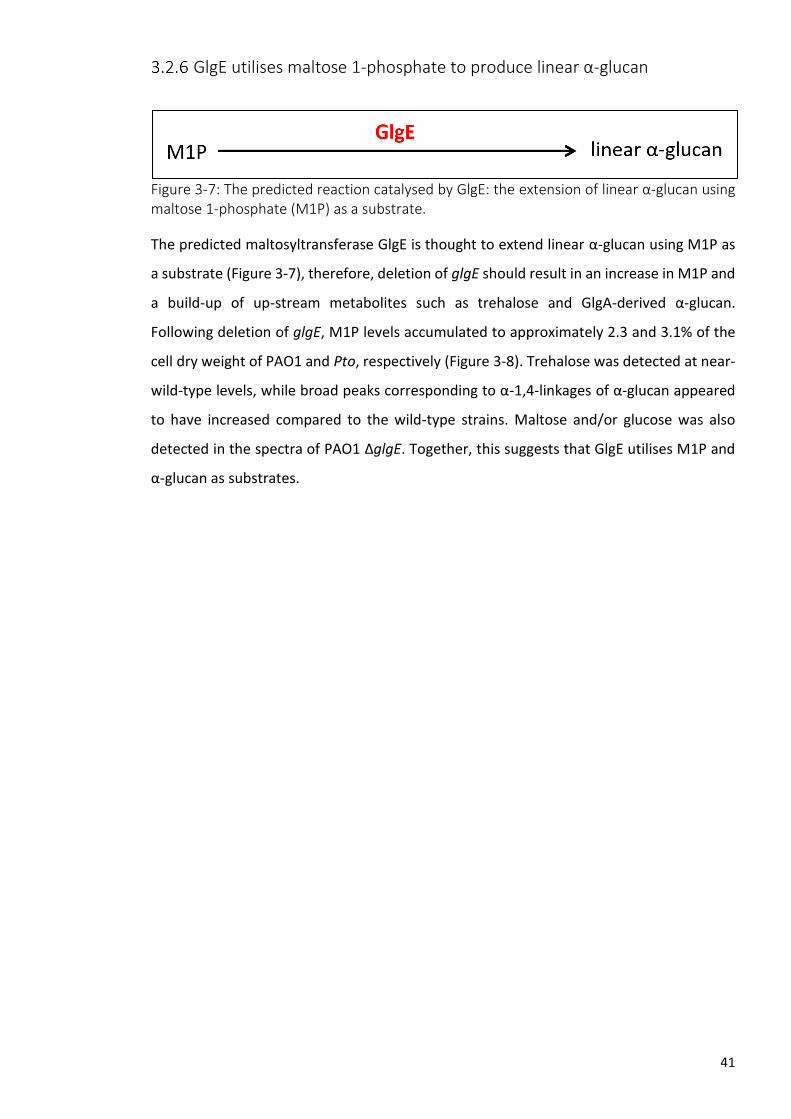
41
GlgE utilises maltose 1-phosphate to produce linear α-glucan
Figure 3-7: The predicted reaction catalysed by GlgE: the extension of linear α-glucan using maltose 1-phosphate (M1P) as a substrate.
The predicted maltosyltransferase GlgE is thought to extend linear α-glucan using M1P as
a substrate (Figure 3-7), therefore, deletion of glgE should result in an increase in M1P and
a build-up of up-stream metabolites such as trehalose and GlgA-derived α-glucan.
Following deletion of glgE, M1P levels accumulated to approximately 2.3 and 3.1% of the
cell dry weight of PAO1 and Pto, respectively (Figure 3-8). Trehalose was detected at near-
wild-type levels, while broad peaks corresponding to α-1,4-linkages of α-glucan appeared
to have increased compared to the wild-type strains. Maltose and/or glucose was also
detected in the spectra of PAO1 ΔglgE. Together, this suggests that GlgE utilises M1P and
α-glucan as substrates.

42
Figure 3-8: 1H-NMR spectroscopy of the soluble metabolome following the deletion of glgE in Pseudomonas spp. Metabolomes were extracted from wild-type PAO1 (blue) and ΔglgE (red) (A) and wild-type Pto (blue) and ΔglgE (red) (B) cultured on M9 minimal medium.Peak assignments are as indicated based on previously established spectra. (M/G; maltose and/or glucose, M1P; maltose 1-phosphate, α-1,4-; α-glucan internal linkages). Resonances corresponding to M1P and α-glucan increased following deletion of glgE in PAO1 and Pto.
To show that GlgE was capable of synthesising α-glucan, PAO1 and Pto GlgE recombinant
proteins were purified and their activities were characterised in vitro. Enzyme kinetics
confirmed that both PAO1 and Pto GlgE proteins utilised both M1P and
maltooligosaccharides as substrates and produced inorganic phosphate as a measurable
bi-product (Karl Syson, unpublished data). Taken together, the in vitro and in vivo data
suggest that GlgE is a maltosyltransferase utilising M1P to extend α-glucan chains.

43
Absence of GlgB results in the accumulation of long linear α-glucan
Figure 3-9: The predicted reaction catalysed by GlgB and GlgX: the branching of linear α-glucan into branched α-glucan via GlgB and the debranching of branched α-glucan into linear α-glucan by GlgX.
The branching enzyme GlgB is predicted to act upon linear α-glucan to produce branched
α-glucan (Figure 3-9), therefore, deletion of glgB should result in an increase in longer linear
α-glucan chains and the absence of branched α-glucan. The deletion of the gene encoding
the predicted branching enzyme in PAO1 (PAO1 ΔglgB) led to increased concentrations of
upstream metabolites: trehalose, maltose and/or glucose and M1P (Figure 3-10). The levels
of trehalose and M1P were increased by approximately 6-fold and 2.5-fold respectively
when compared to wild-type PAO1. The absence of GlgB also resulted in an increase in the
broad signal associated with α-1,4 linear glucan. The metabolome of Pto ΔglgB showed an
increase in linear α-glucan, but no significant changes were observed in the concentrations
of trehalose or M1P when compared to wild-type Pto (Figure 3-10).

44
Figure 3-10: 1H-NMR spectroscopy of the soluble metabolome following the deletion of glgB in Pseudomonas spp. Metabolomes were extracted from wild-type PAO1 (blue) and ΔglgB (red) (A) and wild-type Pto (blue) and ΔglgB (red) (B) cultured on M9 minimal medium.Peak assignments are as indicated based on previously established spectra. (M/G; maltose and/or glucose, M1P; maltose 1-phosphate, α-1,4-; α-glucan internal linkages). Resonances corresponding to trehalose, M1P and α-glucan increased following deletion of glgB in PAO1. Resonances corresponding to α-glucan increased following deletion of glgB in Pto. Trehalose and M1P concentrations did not appear to change.
Although 1H-NMR spectroscopy showed an increase in the concentration of α-1,4 linkages
in the metabolome of ΔglgB, it cannot directly indicate the average linear α-glucan chain
length. Matrix-assisted laser desorption/ionization mass spectrometry (MALDI-MS) was
initially used to investigate the length of linear α-glucan in ΔglgB strains. However, the
metabolomic extracts were too crude to identify relevant carbohydrate peaks.
As an alternative, iodine staining was used as an semi-quantitative method to investigate
the length of α-glucan [197]. When wild-type PAO1 was exposed to iodine vapour there
was no obvious staining. In contrast, when PAO1 ΔglgB was exposed to iodine vapour, the
cellular material stained a dark brown colour suggesting the increased presence of long
linear α-glucan (Figure 3-11). Similar staining was also observed when Lugol’s solution was
added to the metabolite extracts, where the absence of GlgB (ΔglgB strain) resulted in
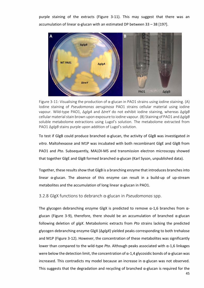
45
purple staining of the extracts (Figure 3-11). This may suggest that there was an
accumulation of linear α-glucan with an estimated DP between 33 – 38 [197].
Figure 3-11: Visualising the production of α-glucan in PAO1 strains using iodine staining. (A) Iodine staining of Pseudomonas aeruginosa PAO1 strains cellular material using iodine vapour. Wild-type PAO1, ΔglgA and ΔtreY do not exhibit iodine staining, whereas ΔglgB cellular material stain brown upon exposure to iodine vapour. (B) Staining of PAO1 and ΔglgB soluble metabolome extractions using Lugol’s solution. The metabolome extracted from PAO1 ΔglgB stains purple upon addition of Lugol’s solution.
To test if GlgB could produce branched α-glucan, the activity of GlgB was investigated in
vitro. Maltohexaose and M1P was incubated with both recombinant GlgE and GlgB from
PAO1 and Pto. Subsequently, MALDI-MS and transmission electron microscopy showed
that together GlgE and GlgB formed branched α-glucan (Karl Syson, unpublished data).
Together, these results show that GlgB is a branching enzyme that introduces branches into
linear α-glucan. The absence of this enzyme can result in a build-up of up-stream
metabolites and the accumulation of long linear α-glucan in PAO1.
GlgX functions to debranch α-glucan in Pseudomonas spp.
The glycogen debranching enzyme GlgX is predicted to remove α-1,6 branches from α-
glucan (Figure 3-9), therefore, there should be an accumulation of branched α-glucan
following deletion of glgX. Metabolomic extracts from Pto strains lacking the predicted
glycogen debranching enzyme GlgX (ΔglgX) yielded peaks corresponding to both trehalose
and M1P (Figure 3-12). However, the concentration of these metabolites was significantly
lower than compared to the wild-type Pto. Although peaks associated with α-1,6 linkages
were below the detection limit, the concentration of α-1,4 glycosidic bonds of α-glucan was
increased. This contradicts my model because an increase in α-glucan was not observed.
This suggests that the degradation and recycling of branched α-glucan is required for the

46
presence of trehalose and M1P in Pto. No relevant metabolite signals were detected in the
corresponding PAO1 strain (Figure 3-12).
Figure 3-12: 1H-NMR spectroscopy of the soluble metabolome following the deletion of glgX in Pseudomonas spp. Metabolomes were extracted from wild-type PAO1 (blue) and ΔglgX (red) (A) and wild-type Pto (blue) and ΔglgX (red) (B) cultured on M9 minimal medium.Peak assignments are as indicated based on previously established spectra. (M/G; maltose and/or glucose, M1P; maltose 1-phosphate, α-1,4-; α-glucan internal linkages). Resonances corresponding to metabolites of interest were absent following deletion of glgX in PAO1. Following deletion of glgX in Pto resonances corresponding to trehalose were absent, whereas those assigned to α-glucan increased.
To determine whether GlgX possessed debranching activity, GlgX from PAO1 and Pto was
incubated with rabbit liver glycogen. MALDI-MS showed that a carbohydrate profile was
produced following incubation of rabbit liver glycogen with GlgX consistent with
debranching activity (Karl Syson, personal communication).
TreS(2) functions as a second trehalose synthase in Pto
Unlike PAO1, bioinformatic analysis predicts that the Pto genome contains a second
homologue of TreS designated TreS(2). Assuming TreS(2) exhibits the same isomerisation
activity as the TreS domain of TreS/Pep2 (Figure 3-5), deletion of treS(2) should result in

47
altered levels of intracellular trehalose when compared to wild-type Pto. Deletion of treS(2)
in Pto resulted in an approximate 2-fold increase in the levels of intracellular trehalose,
whereas M1P levels were not significantly different when compared to the wild-type Pto
strain (Figure 3-13). This suggests that this TreS homologue is responsible for the
degradation of trehalose, presumably into maltose.
Figure 3-13: 1H-NMR spectroscopy of the soluble metabolome following the deletion of treS(2) in Pseudomonas syringae pv. tomato. Metabolomes were extracted from wild-type Pto (blue) and ΔtreS(2) (red) cultured on M9 minimal medium.Peak assignments are as indicated based on previously established spectra. (M1P; maltose 1-phosphate). Resonances corresponding to trehalose were increased following deletion of treS(2) in Pto. The concentration of M1P did not appear to change.
To show that TreS(2) can utilise both trehalose and maltose as substrates, recombinant
TreS(2) was purified and incubated with either disaccharide. 1H-NMR spectroscopy showed
that TreS(2) can utilise both trehalose to synthesise maltose and maltose to synthesise
trehalose in vitro (Karl Syson, personal communication). These results together show that
Pto possesses two functional TreS homologues, the second of which (TreS(2)) likely
functions to metabolise trehalose into maltose for entry into the GlgE pathway.
TreY and TreZ produce trehalose by degrading linear α-glucan
producing maltooligosyltrehalose as an intermediate

48
Figure 3-14: The predicted reactions catalysed by TreY, TreZ and TreS/Pep2: the degradation of linear α-glucan into trehalose and the subsequent processing of trehalose into maltose and maltose 1-phosphate (M1P) by TreS/Pep2.
The maltooligosyl trehalose synthase TreY and the maltooligosyl trehalose
trehalohydrolase TreZ are predicted to degrade the reducing end of linear α-glucan into
trehalose (Figure 3-14). First, TreY is predicted to facilitate the conversion of the terminal
α-1,4 bond of α-glucan into an α-1,α-1-glycosidic bond forming maltooligosyl trehalose.
Trehalose is then predicted to be liberated from maltooligosyltrehalose by the hydrolase
action of TreZ. Based on this prediction, I hypothesise that the deletion of either treY or
treZ would result in the abolition of trehalose production and in an increase of α-glucan
levels. Furthermore, deletion of treZ should lead to an accumulation of maltooligosyl
trehalose due to the isomerisation activity of TreY.
Deletion of treY and treZ resulted in the loss of trehalose production in both PAO1 and Pto.
This can be seen in Figure 3-15 which also shows that levels of α-glucan appeared to
increase in both ΔtreY and ΔtreZ for both species (this was more evident in ΔtreZ strains).
Deletion of treZ also resulted in the formation of a broad species at 5.2 ppm corresponding
to the terminal α-1,1 linkage of maltooligosyl trehalose. This supports the hypothesis that
in Pseudomonas spp. TreY and TreZ work in concert to produce trehalose through the
degradation of α-glucan, producing maltooligosyl trehalose as an intermediate metabolite.
Surprisingly, peaks corresponding to M1P were present in the metabolomes of both ΔtreY
and ΔtreZ for PAO1 and Pto. This was not expected as my model predicts that the only
route to M1P biosynthesis in Pseudomonas spp. is dependent on trehalose production and
therefore TreY and TreZ. This shows that an alternative route exists to maltose and M1P
biosynthesis in Pseudomonas spp.

49
Figure 3-15: 1H-NMR spectroscopy of the soluble metabolome following the deletion of treZ and treY in Pseudomonas spp. Metabolomes were extracted from wild-type PAO1 (blue), ΔtreZ (red) and ΔtreY (green) (A) and wild-type Pto (blue) and ΔtreZ (red) and ΔtreY (green) (B) cultured on M9 minimal medium.Peak assignments are as indicated based on previously established spectra. (M/G; maltose and/or glucose, M1P; maltose 1-phosphate, α-1,4-; α-glucan internal linkages α-1,1-; terminal linkage of maltooligosyl trehalose). Resonances corresponding to trehalose were absent, whilst those corresponding to α-glucan increased following deletion of treZ or treY in PAO1 and Pto. Furthermore, peaks corresponding to the terminal linkage of maltooligosyl trehalose were present within the metabolomes extracted from ΔtreZ strains. M1P was present following deletion of treZ or treY in PAO1 and Pto.
MalQ disproportinates α-glucan and is capable of synthesising maltose
To identify the source of M1P found within the ΔtreY and ΔtreZ metabolomes, it was
hypothesised that the predicted α-glucanotransferase malQ [181] may utilise α-glucan to
produce maltose that would be converted to M1P by the Pep2 kinase domain. malQ is
clustered between treZ and treY within the PAO1 and Pto genomes and the function of
MalQ had yet to be tested.
To address the function of MalQ and the potential to synthesise maltose, a strain deficient
in MalQ was generated in both Pto and PAO1. As compared to the wild-type strain, the
PAO1 ΔmalQ metabolome showed increased concentrations of trehalose (Figure 3-16).

50
Levels of α-glucan were increased in both the PAO1 and Pto ΔmalQ strains. These results
indicate that MalQ could use α-glucan as a substrate, because the deletion of malQ resulted
in an increase in the concentration of α-glucan in both species. This increase in α-glucan
may result in an increased substrate concentration for TreY and TreZ resulting in the
increase in trehalose as observed in PAO1 ΔmalQ.
Figure 3-16: 1H-NMR spectroscopy of the soluble metabolome following the deletion of malQ in Pseudomonas spp. Metabolomes were extracted from wild-type PAO1 (blue) and ΔmalQ (red) (A) and wild-type Pto (blue) and ΔmalQ (red) (B) cultured on M9 minimal medium.Peak assignments are as indicated based on previously established spectra. (M/G; maltose and/or glucose, M1P; maltose 1-phosphate, α-1,4-; α-glucan internal linkages). Resonances corresponding to trehalose increased following deletion of glgB in PAO1. Resonances corresponding to α-glucan increased following deletion of glgB in PAO1 and Pto.
As my genetic data indicated that MalQ acts upon α-glucan in vivo we then sought to
identify the product of this activity. To address this, recombinant PAO1 MalQ was purified
and provided with maltohexose as a substrate. Products of this reaction were identified
and analysed using matrix-assisted laser desorption/ionization mass spectrometry. When
maltohexose (DP6) was provided as a substrate, MalQ was shown to produce
maltooligosaccharides of varying lengths ranging from DP4 to over DP22.
Maltooligosaccharides of length DP3 or less were undetectable because these overlap with

51
the MALDI matrix signals. This disproportionation activity was also seen when MalQ was
provided with maltose (DP2) whereupon it produced maltooligosaccharides of up to DP10.
(Karl Syson, unpublished data). Although we had shown that MalQ could use maltose as a
substrate, due to the limitations of this methodology we were not able to demonstrate the
production of maltose by MalQ. However, since enzyme-catalysed reactions are reversible,
MalQ should be capable of producing maltose from maltooligosaccharides.
We then reasoned that if MalQ was capable of producing maltose, it could be acted upon
by the kinase domain of TreS/Pep2 producing M1P that would be readily detectable by 1H-
NMR spectroscopy. To test this, PAO1 recombinant MalQ and TreS/Pep2 were purified and
provided with maltohexaose and ATP. The products were analysed using 1H-NMR
spectroscopy. After 1 hour, we observed the presence of M1P, thus confirming that MalQ
can produce maltose (experimental design by Stuart Woodcock and Karl Syson, with the
experiment conducted by Karl Syson, unpublished data).
Characterisation of linear α-glucan
At this stage, one of the limitations of the predicted pathway was that there was no
distinction between the linear α-glucans produced by either GlgA or GlgE. It was unclear
whether these pools of linear α-glucan were structurally unique or if they were
interchangeable. To investigate the difference in length between the GlgA-derived linear
α-glucan and the GlgE-derived linear α-glucan, strains were generated in PAO1 to
distinguish between these two sources.
A PAO1 strain lacking GlgB accumulates only linear α-glucan as described above (Section
3.2.7). However, this may represent a pool of both GlgA- and GlgE-dependent linear α-
glucan. To generate solely GlgA-dependent linear α-glucan, a treS operon deletion strain
deficient in GlgE, TreS/Pep2 and GlgB (PA2151-2153) was generated. To test for the
accumulation of GlgA-dependent linear α-glucan, the metabolome was extracted and
analysed using 1H-NMR. This strain showed a stark increase in the levels of both GlgA-
derived linear α-glucan and trehalose (data not shown), however, peaks associated with α-
glucan were absent upon subsequent biological repetitions. This could be a result of
technical error or compensatory mutations to prevent the accumulation of linear α-glucan.
Furthermore, this strain did not exhibit staining when exposed to iodine vapour (Figure
3-17).

52
I reasoned that GlgA-dependent α-glucan could be degraded by TreY and TreZ and MalQ
based on previously described in vitro and in vivo data (Sections 3.2.10 and 3.2.11,
respectively). Deletion of malQ or treY in addition to ΔglgE ΔtreS/pep2 ΔglgB should result
in a strain producing solely GlgA-dependent linear α-glucan.
Iodine vapour staining of cellular material was used to test if deletion of malQ in addition
to ΔglgE ΔtreS/pep2 ΔglgB resulted in the accumulation of GlgA-derived α-glucan. When
exposed to iodine vapour, ΔmalQ ΔglgE ΔtreS/pep2 ΔglgB did not produce staining (Figure
3-17). This suggests that GlgA-dependent α-glucan was not present in sufficient quantities
or length.
Iodine vapour staining of cellular material was used to test if deletion of treY in addition to
ΔglgE ΔtreS/pep2 ΔglgB resulted in the accumulation of GlgA-derived α-glucan in PAO1.
When exposed to iodine vapour, ΔtreY ΔglgE ΔtreS/pep2 ΔglgB stained blue-purple (Figure
3-17). This suggested that this strain accumulated GlgA-dependent α-glucan and that this
could be estimated to be over DP 40, longer than that estimated to be accumulated by
ΔglgB [197]. This suggested that GlgA possessed the potential to synthesise long linear α-
glucan, whoever, this is likely not physiological as this is likely to be rapidly degraded by
TreY and TreZ into trehalose. However, as MalQ and GlgP are still present within this strain,
this still may not represent true GlgA-dependent α-glucan because α-glucan
disproportionation and recycling cannot be ruled out.
Taken together, this suggests that GlgA was capable of synthesising long linear α-glucan as
indicated by iodine staining. However, this is rapidly degraded by TreY and TreZ and
possibly used as a substrate by MalQ. I was unable to generate a strain producing solely
GlgE-dependent α-glucan and therefore was not able determine the relative length.
Further experiments are required to robustly determine the lengths of both GlgA- and GlgE-
dependent α-glucan.

53
Figure 3-17: Iodine staining of linear α-glucan in Pseudomonas aeruginosa PAO1 strains using iodine vapour. Wild-type PAO1 (A), ΔglgB (B), ΔPA2151-2153 (C), ΔtreY ΔPA2151-2153 (D) and ΔmalQ ΔPA2151-2153 (E). ΔglgB and ΔtreY ΔPA2151-2153 exhibit differential staining when exposed to iodine vapour.

54
3.3 Discussion
Trehalose biosynthesis in Pseudomonas spp. has up to now been relatively understudied.
The recent discovery of the GlgE pathway, a novel pathway for the biosynthesis of α-glucan
in Mycobacterium spp., has changed our understanding of trehalose biosynthesis. Although
previously thought to catalyse the biosynthesis of trehalose, flux through TreS is from
trehalose to maltose and through the rest of the GlgE pathway to produce α-glucan in
Mycobacterium spp. [157]. Pseudomonas spp. are predicted to possess gene homologues
of the GlgE pathway. The existence of the GlgE pathway implies the function of TreS is to
convert trehalose into maltose in Pseudomonas spp. This contrasts with what was
previously thought and has implications for the interpretation of past data concerning the
function of trehalose.
A combination of reverse genetics and analytical biochemistry was used to investigate the
functions of the gene products predicted to be involved in trehalose and α-glucan
biosynthesis in Pseudomonas spp. To the best of my knowledge this is the first report
characterising the genetic and biochemical basis of trehalose and α-glucan biosynthesis in
an organism possessing both the GlgA and GlgE pathway.
Figure 3-18: Current understanding of trehalose and α-glucan biosynthesis in Pseudomonas spp. Dashed lines indicate interchangeable substrate utilisation from the combined pool of α-glucan species, represented by the blue box. Disproportionation activity of MalQ is not shown for simplicity. (G6P; Glucose 6-phosphate, G1P; Glucose 1-phosphate, UDP-Glc; UDP-glucose, M1P; maltose 1-phosphate).

55
In this chapter, I demonstrated that the function of most gene products involved with
trehalose and α-glucan biosynthesis were as predicted (Figure 1-9). In consideration of the
data generated within this chapter, I have amended my model to reflect our current
understanding of trehalose and α-glucan biosynthesis in Pseudomonas (Figure 3-18).
We have demonstrated that GlgA catalyses the first committed step in this pathway, the
polymerisation of UDP-glucose into linear α-glucan. Interestingly, the preferred substrate
UDP-glucose was not accumulated by PAO1 following the deletion of glgA. UDP-glucose is
not unique to trehalose and α-glucan biosynthesis and is required for many other cellular
processes therefore it can be reasoned that UDP-glucose does not accumulate to
detectable levels because it is utilised rapidly [198]. An alternative explanation is that the
build-up of UDP-glucose is prevented because it may be toxic to the cell. The build-up of
phosphosugars trehalose 6-phosphate or M1P has direct or indirect toxic effects in M.
tuberculosis [199] therefore similar toxicity may occur with a build-up of UDP-glucose in
Pseudomonas spp. In vitro data showed that PAO1 GlgA can produce linear α-glucan from
UDP-glucose as a donor. The lack of requirement for an added acceptor substrate suggests
some UDP-glucose is hydrolysed to give glucose which then acts as the initial acceptor.
Surprisingly, M1P, trehalose and α-glucan were present in the metabolome of Pto following
deletion of glgA, albeit at reduced concentrations. Although I could not identify a second
Pto glgA homologue, this suggests that other methods of entry into trehalose and α-glucan
biosynthesis exist within this strain. Further work should be conducted to identify gene
products with α-glucan biosynthetic activity. Alternatively, the presence of these
metabolites could be a result of polar effects arising from deletion of glgA. This could be
due to inaccuracies in the prediction of the Pto glgA open reading frame. This should be
ruled out by complementation of GlgA in the Pto ΔglgA strain.
The second step of trehalose and α-glucan biosynthesis in Pseudomonas spp. is the
degradation of α-glucan into trehalose by TreY and TreZ. TreY catalyses the conversion of
the terminal α-1,4 bond of α-glucan into an α-1,α-1 glycosidic bond forming maltooligosyl
trehalose as detected by 1H-NMR spectroscopy. TreZ then hydrolyses maltooligosyl
trehalose liberating trehalose. Unexpectedly, M1P was present in strains lacking functional
TreY and TreZ (Figure 3-15) suggesting an alternative mechanism for the biosynthesis of
maltose in Pseudomonas spp.
Following this, we probed the function of proximal gene products for maltose biosynthesis
activity. We determined that MalQ possessed disproportionation activity and could

56
metabolise α-glucan producing a range of chain lengths. It was then established that MalQ
could produce maltose which would then be converted into M1P by Pep2. Along with
TreS/Pep2, this is the first report of de novo maltose biosynthesis in Pseudomonas spp. This
also suggests a mechanism of GlgE-dependent α-glucan biosynthesis which is independent
of the biosynthesis of trehalose. This therefore could circumvent the production of
trehalose in favour of α-glucan during circumstances which are inappropriate for trehalose
biosynthesis.
The first enzymatic step of the GlgE pathway is the conversion of trehalose into M1P by the
TreS/Pep2 fusion protein. As pep2 and other homologues of the GlgE pathway were
present, it was predicted that flux through TreS would be in favour of synthesising maltose
[157] and therefore TreS/Pep2 was responsible for the degradation of trehalose rather
than the biosynthesis of trehalose as previously thought [74, 180]. Metabolomic data from
PAO1 ΔtreS/pep2 was consistent with TreS/Pep2 utilising trehalose as a substrate.
In contrast to PAO1, deletion of treS/pep2 in Pto resulted in a decrease in intracellular
trehalose, inconsistent with the hypothesis that TreS/Pep2 isomerises trehalose into
maltose and subsequently M1P. Nevertheless, the activity of TreS/Pep2 was confirmed
using in vitro techniques. The decrease in trehalose concentration following deletion of
treS/pep2 could be rationalised due to the fact that high levels of trehalose has been
hypothesised to be toxic [139]. There no published reports of this in bacteria, with the
exception of trehalose 6-phosphate toxicity in M. tuberculosis [199]. Pto may respond to
increased trehalose levels by compensatory mutations or through regulatory processes. An
alternative approach to investigate the function of TreS/Pep2 in vivo is the overexpression
of treS/pep2 in Pto. My model would suggest that increased expression of treS/pep2 would
result in the reduction of trehalose concentrations, consistent with flux flowing from
trehalose to maltose. This would thereby prevent pleotropic effects induced by trehalose
accumulation.
Studies on the M. tuberculosis TreS and Pep2 proteins shows that they form a non-covalent
complex in vitro [200]. The formation of this complex strikingly increases the kinase activity
of Pep2 and subtly increases the activity of TreS. Furthermore, treS and pep2 are fused in
30% of bacterial genomes predicted to possess glgE homologues, including Pseudomonas
spp. (Govind Chandra, personal communication) [171]. The fusion of these two genes could

57
be a method of regulation within these bacteria to increase activity of the TreS/Pep2
protein.
Interestingly, deletion of the second Pto treS homologue (treS(2)) resulted in an
approximate 2-fold increase in the concentration of intracellular trehalose, consistent with
my model. This suggests redundancy within the Pto GlgE pathway. This could reflect the
environmental lifestyle of Pto where increased trehalose degradation by TreS(2) would be
required to adapt to rapidly changing environmental osmolarities. Furthermore, it was
noted by Chandra et al. that the GlgE pathway was possessed largely by plant-associated
bacteria [171].
The second step of the GlgE pathway is the polymerisation of M1P into linear α-glucan.
GlgE utilises M1P as a substrate to extend a α-glucan chain. Deletion of glgE in either PAO1
or Pto resulted in a stark increase in the levels of M1P, as expected. M1P accumulation has
been shown to be toxic in Mycobacterium spp. [153] where deletion of GlgE was lethal in
M. tuberculosis and conditionally lethal in M. smegmatis depending on the presence of
trehalose. GlgE-deficient strains were viable in Pseudomonas spp. and any detrimental
effects following the accumulation of this phosphosugar were not apparent. This suggests
that Pseudomonas spp. are resistant to the direct or indirect effects of M1P and/or that
lethality is unique to Mycobacterium spp. To further investigate this, the transcriptomic
response of ΔglgE strains would be examined to identify the existence of any M1P-induced
stress responses. Finally, GlgB introduces α-1,6 branch points to the linear chain forming
branched α-glucan, the final product of the GlgE pathway.
α-Glucan is recycled by the phosphorylase GlgP and the debranching enzyme GlgX. GlgP
degrades α-glucan chains producing G1P and GlgX removes the α-1,6 branch point,
producing linear α-glucan chains. In other bacteria GlgP degrades α-glucan chains leaving
four glucose units from the α-1,6 branch point [170], providing a suitable substrate for GlgX
suggesting that GlgP and GlgX are both required for α-glucan degradation in Pseudomonas
spp. I have also demonstrated that the recycling of α-glucan is required to produce
trehalose and M1P where loss of GlgX and therefore the ability to debranch α-glucan
results in a reduction of these metabolites.
Unexpectedly, PAO1 ΔglgX did not yield any relevant metabolites, although the absence of
trehalose and M1P supports my model, α-glucan levels were also undetectable. This is
difficult to rationalise but could be a result of regulatory or compensatory mechanisms. For

58
example, the accumulation of unrecyclable α-glucan represents a significant energy store
which becomes largely inaccessible to the bacterium leading to nutrient stress and could
result in the prevention of α-glucan biosynthesis in PAO1.
The presence of both GlgA and GlgE results in two alternative mechanisms of synthesising
linear α-glucan. It is currently unclear as to why Pseudomonas spp. have evolved two
distinct biosynthetic pathways of α-glucan biosynthesis. The most tangible explanation is
that there are differences in length between the GlgA- and GlgE-synthesised α-glucan
making them distinct molecules. Efforts to characterise the chain length of GlgA- and GlgE-
dependent α-glucan in vivo were based on the generation of PAO1 strains accumulating
either pool of linear α-glucan. The first limitation of this was due to the inability to produce
a strain accumulating solely GlgE-dependent α-glucan. This was due to the dependency of
GlgE on the rest of the pathway and the inability to prevent the production of GlgA-
dependent α-glucan. Nevertheless, based on my model, PAO1 ΔglgB was the best
representative of the accumulation of GlgE-dependent α-glucan. An absence of GlgB led to
the production of linear α-glucan, which could be estimated to be between DP 33 – 38
[197], however, this was not conclusive as this can reflect a combination of both GlgA- and
GlgE-dependent linear α-glucan.
Due to the presence of multiple enzymes such as GlgE, TreY, MalQ, and GlgP which utilise
α-glucan as a substrate, a PAO1 strain accumulating solely GlgA-dependent linear α-glucan
was not possible. Although other α-glucan modifying activities cannot be ruled out, the
GlgA-dependent linear α-glucan chain may exceed 40 glucose units. However, this is
unlikely to be physiologically-relevant as GlgA-dependent linear α-glucan seems to be
rapidly degraded by TreY.
In order to conclusively characterise the chain lengths of GlgA- and GlgE-dependent α-
glucan in vivo, the sole effects of GlgA and GlgE must be studied. To study the length of
GlgA-dependent linear α-glucan, a strain should be generated that would only possess the
glgA gene and lack every other gene product of the GlgE pathway. This strain would only
produce GlgA-dependent linear α-glucan and lack any other enzymes which utilise α-glucan
as a substrate thereby resulting in an accurate representation.
To study the length of GlgE-dependent linear α-glucan, a strain should be generated that
would only possess treS/pep2 and glgE. This strain would therefore only produce GlgE-
dependent linear α-glucan when provided with extracellular trehalose and would lack any
other enzymes which utilise α-glucan as a substrate thereby resulting in an accurate

59
representation. Although iodine staining provides an indication of linear α-glucan chain
length, this method is semi-quantitative. MALDI-MS can be used as a quantitative method
to analyse chain length, however, metabolome extracts have proven inappropriate for this
instrumentation. Therefore, in order to utilise MALDI-MS, chloroform/ethanol extractions
should be used to produce pure samples of α-glucan.
It is still unclear as to why Pseudomonas spp. possess two distinct biosynthetic pathways
of α-glucan biosynthesis and prompts the following questions: are GlgA- and GlgE-
dependent α-glucan interchangeable? For example, genomic and experimental data
suggests that TreY utilises GlgA-dependent α-glucan to produce trehalose. Can TreY utilise
GlgE-dependent α-glucan as a substrate? Can GlgB utilise both GlgA- and GlgE-dependent
α-glucan forming branched α-glucan? Can branched α-glucan be extended by both GlgA
and GlgE? These questions should be answered using in vitro techniques.
Prior to this work, understanding of trehalose biosynthesis in Pseudomonas spp. was
limited. Both TreY/TreZ and TreS pathways were thought to contribute to trehalose
biosynthesis. The only biosynthetic link between α-glucan and trehalose was thought to
occur through a requirement of α-glucan as a substrate for the TreY/TreZ pathway.
In agreement with prior studies, I have demonstrated that all genes (except malQ) within
the treY/treZ operon are involved in the production of trehalose. In contrast, I have shown
that the treS operon is not involved in the biosynthesis of trehalose as previously thought,
instead it is involved with the conversion of trehalose into maltose and the subsequent
production of α-glucan.
The identification of the GlgE pathway, a novel mechanism of α-glucan biosynthesis in
Pseudomonas spp., has wide reaching implications for the interpretation of previous data
investigating trehalose biosynthesis and its function in Pseudomonas spp. and other
bacteria possessing the GlgE pathway. For example, the deletion of both treS or treY/treZ
operons in Pto resulted in the abolition of trehalose production and the generation of an
osmotically-sensitive strain [74]. Furthermore, deletion of both treS or treY/treZ operons
resulted in compromised tomato infections, a phenomenon which was exacerbated by
lower RH. These results were interpreted as a result of the loss of trehalose, implicating
trehalose as an osmotic protectant and a virulence factor.
Similarly, deletion of the treY/treZ operon in PA14 resulted in the abolition of trehalose
production [180], while deletion of the treS operon resulted in decreased levels of

60
intracellular trehalose. Although osmotic sensitivity was not affected, deletion of either
operon resulted in attenuated virulence during infection of Arabidopsis thaliana plants,
again implicating trehalose as a virulence factor.
Furthermore, the highly trehalose-accumulating bacterium M. koreensis 3J1 is markedly
tolerant to desiccation stress [151]. This led to the positive-correlation of intracellular
trehalose concentrations and desiccation tolerance. Furthermore, the M. koreensis 3J1
trehalose biosynthetic genes otsA/otsB expressed in P. putida results in an increase in
trehalose production and subsequent desiccation tolerance. Both M. koreensis 3J1 and P.
putida both are predicted to possess homologues of the GlgE pathway and therefore
should be genetically capable of producing α-glucan. Following the proposal of my model
(figure 3-18), an increase in trehalose may result in flux through the GlgE pathway and
therefore an increase in α-glucan.
Considering our new understanding of the treS operon and the GlgE pathway in
Pseudomonas spp. previous reports investigating the role of trehalose must be re-
interpreted. For example, the biosynthesis of GlgE-dependent α-glucan is dependent on
trehalose, therefore, the modulation of intracellular trehalose concentrations also impacts
the biosynthesis of α-glucan. Subsequently, α-glucan may be at least in part responsible for
the phenotypes attributed to trehalose. The following questions then arise: does α-glucan
function as an osmotic stress protectant? Does α-glucan play a role during desiccation
stress? And does α-glucan play a role during plant infection?
The exploitation of the mutant strains and metabolomic data generated within this chapter
will allow future investigation into the individual functions of trehalose and α-glucan in
Pseudomonas spp.

61
Trehalose and α-glucan protect against distinct
abiotic stresses in Pseudomonas

62
4.1 Introduction
Trehalose functions as a protectant against a variety of stresses in bacteria. However, in
Mycobacterium and Pseudomonas spp. trehalose is used as a substrate by the novel GlgE
pathway to produce α-glucan (Chapter 3) [153, 157]. The role of classical α-glucan in
bacteria has been extensively studied and usually functions as a form of carbon and energy
storage [162]. However, the function of the GlgE pathway and therefore GlgE-dependent
α-glucan has only been investigated in M. tuberculosis. Mycobacterial GlgE-dependent α-
glucan is exported to the capsule where it interacts with the human immune system and
modulates phagocytosis during infection [176, 177].
As Pseudomonas aeruginosa and Pto are significant pathogens of humans and plants,
respectively, it is unlikely that Pseudomonas GlgE-dependent α-glucan functions to solely
modulate the human immune system. Since I have established the genetic and biosynthetic
link between trehalose and α-glucan through the GlgE pathway in Pseudomonas spp., we
can gain clues as to the function of α-glucan from previous studies investigating the roles
of trehalose.
Such studies have implicated trehalose as a stress protectant, important for both osmotic
and desiccation stress in Pseudomonas spp. Trehalose has also been shown to be important
for plant infection, implicating this sugar as a virulence factor in Pseudomonas spp. On the
contrary, the interpretation of these studies did not consider the intrinsic genetic and
functional linkage of trehalose and α-glucan biosynthesis afforded by the GlgE pathway as
shown in Chapter 3. For example, during osmotic stress, genes responsible for trehalose
and α-glucan biosynthesis are both upregulated [184], and deletion of both trehalose and
α-glucan biosynthetic operons results in osmotically-sensitive strains in Pto [74]. However,
equivalent mutations in PA14 do not result in osmotically-sensitive strains convoluting the
role of trehalose in Pseudomonas spp.
Desiccation resistance can be conferred to P. putida by increasing intracellular
concentrations of trehalose. In trans expression of M. koreensis 3J1 otsA and otsB in P.
putida increases trehalose production which subsequently correlates with increased
desiccation tolerance [151]. However, increasing the levels of trehalose could result in
higher flux through the GlgE pathway resulting in the increased biosynthesis of α-glucan
(Figure 3-18). Furthermore, infection of tomato plants with Pto lacking both trehalose and
α-glucan biosynthetic genes are exacerbated by decreased RH. Similarly, α-glucan may play

63
a role during plant colonisation and infection. For example, transcriptomic data show the
upregulation of both trehalose and α-glucan biosynthesis genes in Pto during the epiphytic
and apoplastic lifestyle [132]. Furthermore, strains used to implicate trehalose as a
virulence factor during plant infection were also deficient in α-glucan [74, 180, 181].
This chapter aims to investigate the roles of both trehalose and α-glucan in Pseudomonas
spp. With a combination of gene deletions (generated in Chapter 3), and the introduction
of non-native M. koreensis 3J1 otsA/otsB, I decoupled the biosynthesis of trehalose and α-
glucan thereby enabling the elucidation of the individual roles of both trehalose and α-
glucan. This chapter will aim to answer the following questions:
• Do trehalose and α-glucan play a role in protecting Pseudomonas spp. against
osmotic stress?
• Does α-glucan play a role during desiccation stress?
• Does α-glucan play a role during the epiphytic or apoplastic lifestyle or during
infection?
To answer these questions biosynthetic mutant strains of Pseudomonas aeruginosa PAO1
and Pto were exposed to osmotic and desiccation stresses, respectively. Osmotic stress was
conferred by the addition of NaCl to growth media and osmotic sensitivity was determined
by the subsequent effects on growth. Finally, desiccation sensitivity was determined by
measuring bacterial survival following incubation at high and low levels of RH.
In addition to this, several studies have also implicated the EPS molecule alginate in the
protection against water stress in P. putida. In this chapter I have investigated whether
alginate also functions in a similar capacity in PAO1 and Pto, and if there is interplay with
trehalose or α-glucan.
Furthermore, as shown by Chapter 3, Pseudomonas spp. are capable of producing α-glucan
by two different pathways (GalU-GlgA and GlgE). Where appropriate, I have attempted to
distinguish between these two pools of α-glucan.
4.2 Materials and Methods
Desiccation Tolerance Assay
Liquid cultures of bacteria grown in M9 minimal media were diluted to an OD600 of 0.1 using
PBS and 10 µL was spotted onto nine 15-mm grade 1 Whatman filter discs per strain (GE
Healthcare Life Sciences). After drying for a minute at room temperature these filter discs
64
were placed onto solidified M9 minimal media and incubated at either 28 °C or 37 °C for
four hours. Following incubation, time zero samples were determined by resuspending
three filter discs per strain in 3 mL of PBS. Colony forming units (CFU) were determined
following serial dilutions. The remaining filter discs were subjected to desiccation in tightly
sealed bell chambers containing either water (100% RH) or a saturated NaCl solution (75%
RH [201]) for two hours. Three technical replicates per strain were included for both
conditions. Bacteria were then recovered from the filter discs in 3 mL of PBS and following
serial dilutions, the CFU were then determined. Each experiment was repeated at least
twice.
Data transformed by logarithms to base 10 on CFU were analysed using Genstat by linear
mixed modelling using restricted maximum likelihood. The random effect was date of
experiment and Condition * Strain were fixed effects, where * is the crossing operator;
Condition includes T0, 100% RH and 75% RH. The three technical replicate samples with the
same Condition and Strain tested on the same Date provided the residual error term. The
statistical significance of Condition, Strain and their interaction was determined by F-tests
of Wald statistics. The response of a strain to lower humidity was calculated as the
difference between its predicted mean log10(CFU) at 100% RH and at 75% RH. The standard
error of the difference (SED) between the response of a mutant strain and that of the wild-
type strain was calculated from the appropriate standard errors (SE) with the formula: SED
= (SE[100%, Mutant]2 + SE[75%, Mutant]2 + SE[100%, wild-type]2 + SE[75%, wild-type]2)0.5.
The significance of the response difference was tested using the normal distribution with a
variance of SED2.
Linear mixed modelling was chosen to account for variation in between technical and
biological replications, including environmental and biological factors, allowing comparison
between all strains tested. Statistical analysis was designed by James Brown. Statistical
significance between the desiccation response of all PAO1 strains is summarised in the
appendix Figure i, whereas the significance between all Pto strains is summarised in
appendix Figure ii.
Plant Infection Assays
For Pto infection assays, Arabidopsis thaliana (Col-0) was used. These plants were grown
at 22 °C, 80% RH, under a 10-hour light period for 5-6 weeks. Pto strains were grown in M9
minimal medium overnight and diluted with 10 mM MgCl2 to OD600 0.1 for spray infection
and 0.0002 for infiltration infection. Before spray infection, cultures were amended with

65
0.04% of the surfactant Silwet-77 (De Sangosse). Plants were then sprayed until saturation.
For infiltration, cultures were injected into the underside of the leaf using a 1 mL blunt end
syringe until there was complete coverage of the leaf. For the duration of infection plants
were covered with a clear lid and watered where specified. To determine bacterial load,
two leaf discs of diameter 0.77 cm were taken from each plant and homogenised in 10 mM
MgCl2, using 3 mm glass beads (Millipore) and the Spex® Geno/Grinder. CFU counts were
calculated by serially diluting the homogenised sample and plating onto L medium
supplemented with rifampicin (50 µg/mL) and nystatin (25 µg/mL). Rifampicin was used to
select for the Pto strains and nystatin to select against fungal contaminants.
4.3 Results - Phenotypic characterisation of trehalose and α-glucan biosynthesis in Pseudomonas aeruginosa PAO1.
Disruption of trehalose biosynthesis results in osmotic sensitivity in
Pseudomonas aeruginosa PAO1
Since trehalose has been implicated in the survival of osmotic stress in Pseudomonas spp.,
loss of trehalose biosynthesis should result in an osmotically-sensitive strain. These
experiments should also determine if α-glucan plays a role during osmotic conditions. To
investigate the roles of trehalose and α-glucan during osmotic stress, PAO1 strains were
cultured in the presence and absence of NaCl. Growth kinetics were determined using
optical density at 600 nm. Osmotic stress was induced by the supplementation of 0.85 M
NaCl to M9 minimal medium. There was no difference in the growth characteristics of any
PAO1 strain when grown under non-stressed conditions (Figure 4-1). When incubated with
NaCl, PAO1 exhibited a severely delayed lag-phase, but higher maximal density as
compared to non-stressed growth (Figure 4-1). The trehalose and α-glucan null mutant
PAO1 ΔglgA (trehalose-/α-glucan-) showed an increased lag phase when compared to the
wild-type strain during osmotic stress, whereas the increased trehalose-producing, GlgE-
dependent α-glucan deficient strain PAO1 ΔtreS/pep2 (trehalose++/ GlgE-dependent α-
glucan-) yielded a wild-type-like growth profile. Additionally, the trehalose-null but α-
glucan-producing strains PAO1 ΔtreY and ΔtreZ (trehalose-/ α-glucan+) showed attenuation
during osmotic growth. Interestingly, the ΔglgB and ΔglgX strains lacking the α-glucan
branching and debranching enzymes were also attenuated during osmotic conditions, even
though basal levels of trehalose were increased in the PAO1 ΔglgB strain (3.2.7). This
suggests that efficient branching and debranching of α-glucan is needed for an optimal
response to osmotic stress. Loss of GlgE, MalQ and GlgP had little effect on PAO1 growth

66
during osmotic stress. These results support the hypothesis that trehalose protects PAO1
against osmotic stress. Although the presence of α-glucan alone does not confer osmotic
protection, efficient branching and debranching of α-glucan may be required for the
optimal response to osmotic stress.

67
Figure 4-1: Growth of PAO1 and mutant strains in M9 minimal medium ± 0.85 M NaCl. (A) PAO1 treS operon mutants: ΔglgE, ΔtreS/pep2 and ΔglgB. (B) PAO1 treY/treZ operon mutants: ΔglgA, ΔtreZ, ΔmalQ, ΔtreY and ΔglgX. (C) PAO1 and ΔglgP. Values represent the mean OD600 ± SEM of five technical replicates. Results are representative of at least three independent experiments. Error bars are not shown for growth under non-stressed conditions for reasons of legibility. Trehalose deficient strains (ΔtreZ, ΔtreY and ΔglgA) and glucan (de)branching mutants (ΔglgB and ΔglgX) are attenuated during osmotic conditions.

68
Loss of α-glucan but not trehalose results in desiccation sensitivity in
PAO1
To investigate the roles that trehalose and α-glucan play during desiccation stress, PAO1
strains were inoculated onto paper filter discs and incubated at 100% and 75% RH. CFU was
determined after incubation at each RH. The resulting data were analysed using linear
mixed modelling and represented as predicted means of log10(CFU/mL). The response of a
strain to lower humidity was calculated as the difference between its predicted mean log-
10(CFU/mL) at 100% and at 75% RH. An increased response of a mutant strain as compared
to the wild-type to lower RH translates to a more desiccation-sensitive strain.
As shown earlier (3.2.3), PAO1 ΔglgA lacks the ability to synthesise either trehalose or α-
glucan. PAO1 ΔtreS/pep2 accumulates increased levels of trehalose but lacks the genetic
capacity to convert trehalose into GlgE-dependent α-glucan. Although retaining the genetic
potential to produce GlgA-dependent α-glucan, this was not present within the
metabolome when analysed using 1H-NMR spectroscopy, nevertheless, the presence of
GlgA-dependent α-glucan cannot be ruled out. PAO1 ΔglgB accumulates both trehalose
and linear α-glucan more than wild-type PAO1.
Incubation of PAO1 and all derived strains at 100% RH resulted in the recovery of predicted
means of approximately 7.5 – 7.9 log10(CFU/mL). Incubation at 75% RH resulted in
approximately 10-fold reduction in the recovery of viable wild-type PAO1 to a predicted
mean of log10(CFU/mL) = 6.5 (Figure 4-2). This translated to in a desiccation response of
approximately 1.1 log10(CFU/mL), to which all mutant strains were compared.
Incubation of the α-glucan and trehalose null mutant PAO1 ΔglgA at 75% RH resulted in
enhanced cell death corresponding to a 1000-fold reduction in cell recovery to 4.8
log10(CFU/mL)) equating to a desiccation response of approximately 2.6 log10(CFU/mL).
Surprisingly, the trehalose-producing, GlgE-dependent α-glucan-null mutant PAO1
ΔtreS/pep2 showed similar desiccation sensitivity to the ΔglgA strain, with recovery at 75%
RH equating to a predicted mean of 4.4 log10(CFU/mL) resulting in a desiccation response
of approximately 3.1 log10(CFU/mL). In both cases the desiccation response of PAO1 ΔglgA
and PAO1 ΔtreS/pep2 was highly significant (p ≤ 0.0001) when compared to that of the
wild-type PAO1. Incubation of PAO1 ΔglgB at 75% RH resulted in very little attenuation
compared to recovery at 100% RH. This corresponded to a desiccation response of

69
approximately 0.3 log10(CFU/mL). This lack of cell death was significant (p ≤ 0.05) when
compared to wild-type PAO1.
Figure 4-2: Linear mixed modelling analysis of the survival of PAO1, ΔglgA, ΔtreS/pep2 and ΔglgB strains when incubated at 100% (white bars) and 75% RH (grey bars). Bacterial recovery is presented as the predicted mean of log10(CFU/mL). Results are representative of at least three independent experiments. PAO1 strains lacking glgA and treS/pep2 were significantly attenuated during incubation at 75% RH compared to wild-type PAO1. Conversely the desiccation resistance of ΔglgB was increased. **** indicates p ≤ 0.0001, * indicates p ≤ 0.05.
Taken together, these results show that the absence of both trehalose and α-glucan results
in desiccation sensitivity. This phenotype is not rescued by the production of trehalose even
when trehalose is overproduced as occurs in the ΔtreS/pep2 strain. Furthermore, the
absence of GlgB, resulting in the accumulation of both trehalose and linear α-glucan in the
PAO1 ΔglgB strain, increased desiccation stress protection. This implicates GlgE-dependent
α-glucan in the protection against desiccation stress, irrespective of the state of branching,
a function which was independent of trehalose.

70
In trans expression of Microbacterium koreensis 3J1 otsA/otsB recovers
trehalose biosynthesis
In contrast to previous reports, the presence of trehalose within PAO1 ΔtreS/pep2 did not
protect against desiccation stress [151]. However, the levels of intracellular trehalose
produced by ΔtreS/pep2 may not have been sufficient to confer desiccation-resistance. M.
koreensis 3J1 is a bacterium that produces high levels of intracellular trehalose and is
markedly resistant to desiccation stress [151]. The introduction and over-expression of the
non-native M. koreensis 3J1 otsA and otsB trehalose biosynthetic genes results in an
increase in trehalose production and confers desiccation resistance in P. putida [151, 188].
To investigate whether further enhanced levels of trehalose conferred desiccation stress
resistance, the M. koreensis otsA otsB (otsA/otsB) genes were introduced and
overexpressed in our PAO1 mutant strains. M. korensis 3J1 genomic DNA was kindly gifted
by Professor Maximino Manzanera, University of Granada, Spain. The otsA/otsB gene
fragment was placed under the constitutive control of a Ptac promoter. This cassette was
integrated into a neutral genomic location of wild-type PAO1, PAO1 ΔglgA and PAO1
ΔtreS/pep2, generating derivatised strains constitutively expressing the M. koreensis 3J1
otsA/otsB genes. To ensure the effective production of functional M. koreensis 3J1
OtsA/OtsB, the effect of over-expression of otsA/otsB upon the soluble metabolome was
investigated. OtsA/OtsB should catalyse the production of trehalose by using G6P and
presumably UDP-glucose as substrates in PAO1 thereby increasing the intracellular levels
of trehalose and subsequent flux through the GlgE pathway (Figure 4-3).
The production of OtsA/OtsB in ΔglgA should result in the recovery of trehalose
biosynthesis as it is predicted to bypass the glgA deletion and therefore the dependency
on TreY- and TreZ-catalysed trehalose biosynthesis. As TreS/Pep2, GlgE and GlgB are still
present within this strain, intracellular trehalose could be converted into GlgE-dependent
α-glucan (Figure 4-3).
The expression of otsA/otsB in ΔtreS/pep2 should result in enhanced production of
trehalose. As this strain lacks functional TreS/Pep2, the metabolism of trehalose into α-
glucan through the GlgE pathway is not expected. There is still the possibility that
ΔtreS/pep2 :: otsA/otsB may produce GlgA-dependent α-glucan, however, as OtsA/OtsB
and GlgA compete for UDP-glucose as a shared substrate, this pool of α-glucan is expected
to be diminished or absent (Figure 4-3).

71
Figure 4-3: Scheme showing the genetic and biochemical basis following the production of trehalose biosynthetic enzymes OtsA/OtsB in wildtype PAO1 (A), ΔtreS/pep2 (B) and ΔglgA (C). Red crosses represent gene deletions. Grey arrows, enzymes and metabolites indicates parts of the pathway not expected to be active. (G6P; Glucose 6-phosphate, G1P; Glucose 1-phosphate, UDP-Glc; UDP-glucose, M1P; maltose 1-phosphate).

72
The soluble metabolomes of PAO1 :: otsA/otsB, PAO1 ΔglgA :: otsA/otsB and PAO1
ΔtreS/pep2 :: otsA/otsB were extracted and subject to 1H-NMR spectroscopy as described
previously (Sections 2.4-2.5). Expression of otsA/otsB in PAO1 resulted in a non-significant
increase in trehalose accumulation and a significant reduction in M1P levels when
compared to the parental PAO1 strain (Table 4-1, Figure 4-4). The expression of otsA/otsB
in PAO1 ΔglgA restored the biosynthesis of trehalose, M1P and α-glucan. Expression of
otsA/otsB in PAO1 ΔtreS/pep2 resulted in a non-significant increase in the levels of
trehalose as compared to the parental ΔtreS/pep2. M1P and α-glucan were still
undetectable. Although intracellular trehalose concentrations did not significantly differ
from the parental strains following the expression of otsA/otsB in wild-type PAO1 and
ΔtreS/pep2, the recovery of trehalose and M1P biosynthesis in ΔglgA :: otsA/otsB suggests
that OtsA/OtsB is functional in PAO1.
Figure 4-4: 1H-NMR spectroscopy of the soluble metabolome following the over-expression otsA/otsB in PAO1 strains. Metabolomes were extracted from wild-type PAO1 (blue), ΔtreS/pep2 (red), ΔglgA (green), PAO1 :: otsA/otsB (purple), ΔtreS/pep2 :: otsA/otsB (yellow) and ΔglgA :: otsA/otsB (orange) cultured on M9 minimal medium.Peak assignments are as indicated based on previously established spectra (M/G; maltose and/or glucose, M1P; maltose 1-phosphate). M1P concentration decreased following over-expression of otsA/otsB in PAO1. The expression of otsA/otsB did not affect the concentration of trehalose within the soluble metabolome of PAO1 or ΔtreS/pep2. Although trehalose was not present, over-expression of otsA/otsB in ΔglgA resulted in the production of M1P.

73
Table 4-1: Concentrations of trehalose and M1P produced by PAO1 mutant strains with or without expression of otsA/otsB. Metabolites are presented as percentages of cellular dry weight ± standard error (n ≥ 2). – indicates no measurable metabolite, * indicates significant differences compared to the parental strain as determined by a student’s t-test. Parental strain data are replicated from table 3-1.
Reintroduction of trehalose biosynthesis rescues osmotic sensitivity
Although the over-expression of otsA/otsB did not result in a significant increase in
trehalose production under non-stressed conditions, OtsA/OtsB production may result in
increased tolerance to osmotic stress. Because of this, the effect of in trans expression of
otsA/otsB was investigated during growth with osmotic stress. PAO1, PAO1 ΔtreS/pep2,
PAO1 ΔglgA and the respective derivatives expressing otsA/otsB were cultured in the
presence and absence of NaCl in 96-well microplates as described earlier (Section 2.7).
Growth kinetics were determined using optical density at 600 nm.
As expected, the expression of otsA/otsB did not affect growth during non-stressed
conditions when compared to the parental strains (Figure 4-5). All strains producing
OtsA/OtsB were equally enhanced in their ability to resist osmotic stress irrespective of the
original background, with shorter lag phases when compared to their respective parental
strain. Although PAO1 :: otsA/otsB and ΔglgA :: otsA/otsB exhibited increased tolerance to
osmotic stress, these strains had the genetic capacity to produce α-glucan. As a result of
this, increased tolerance to osmotic stress could not be solely attributed to the enhanced
production of trehalose via OtsA/OtsB. Conversely, ΔtreS/pep2 :: otsA/otsB lacked the
genetic capacity to produce GlgE-dependent α-glucan from trehalose. Furthermore, there
is no evidence of the accumulation of GlgA-dependent α-glucan as this was undetectable
via 1H-NMR. Moreover, α-glucan is unlikely to be produced through the metabolism of
trehalose by TreY/TreZ in the reverse direction due to the presence of high concentrations
of water.
Taken together, this suggests that the over-expression of otsA/otsB during osmotic
conditions results in enhanced trehalose production, thereby increasing the resistance of
Strain Trehalose (%) M1P (%)
PAO1 0.13 ± 0.03 0.30 ± 0.03
ΔtreS/pep2 0.89 ± 0.25 -
ΔglgA - -
PAO1 :: otsA/otsB 0.17 ± 0.06 0.17 ± 0.02*
ΔtreS/pep2 :: otsA/otsB 0.91 ± 0.13 -
ΔglgA :: otsA/otsB 0.06 ± 0.03 0.48 ± 0.06*

74
PAO1 strains to osmotic stress and rescuing the osmotically-sensitive phenotype of PAO1
ΔglgA. This further supports the hypothesis that trehalose, not α-glucan, protects against
osmotic stress in Pseudomonas spp.
Figure 4-5: Growth of PAO1 and mutant strains with or without expression of otsA/otsB in M9 minimal medium ± 0.85 M NaCl. (A) Growth of PAO1 strains in the absence of NaCl. (B) Growth of PAO1 strains in the presence of NaCl. Values represent the mean OD600 of five replicates. Results are representative of at least three independent experiments. Error bars are not shown for growth under non-stressed conditions for reasons of legibility. Expression of otsA/otsB enhanced growth of all strains under osmotic conditions irrespective of original genetic background.

75
Reintroduction of trehalose biosynthesis rescues desiccation sensitivity
in PAO1 but only when TreS/Pep2 is present
The expression of otsA/otsB increases the desiccation stress tolerance in P. putida thereby
implicating trehalose in the protection during desiccating conditions. To investigate the
role of enhanced trehalose production during desiccation stress, the previously sensitive
parental strains, PAO1 ΔglgA and PAO1 ΔtreS/pep2, now expressing otsA/otsB were
subjected to desiccating conditions. This was performed as described in section 4.2.1.
The expression of otsA/otsB rescued the desiccation sensitivity exhibited by the PAO1
ΔglgA strain (Appendix Figure i). This resulted in a desiccation response of 0.9
log10(CFU/mL) similar to that of wild-type PAO1 (Figure 4-6). In contrast, PAO1 ΔtreS/pep2
:: otsA/otsB exhibited a desiccation response of approximately 4 log10(CFU/mL) (p ≤ 0.001)
translating to significant desiccation sensitivity when compared to the wild-type PAO1
(Figure 4-6), despite over-expression of the trehalose biosynthetic enzymes.
Taken together, the expression of otsA/otsB and therefore enhanced biosynthesis of
trehalose resulted in increased resistance to osmotic stress in PAO1 strains. Expression of
otsA/otsB also rescued desiccation sensitivity, however, this phenomenon depended on
the presence of functional TreS/Pep2. This suggests that trehalose is not directly
responsible for desiccation resistance in Pseudomonas spp. Conversely, the ability of
trehalose to rescue desiccation sensitivity in PAO1 strains is dependent on the presence of
TreS/Pep2. This implicates that the function of the GlgE pathway and thereby α-glucan is
to protect Pseudomonas spp. against desiccation stress.

76
Figure 4-6: Linear mixed modelling analysis of the survival of PAO1 and mutant strains expressing otsA/otsB when incubated at 100% (white bars) and 75% RH (grey bars). Bacterial recovery is presented as the predicted mean of log10(CFU/mL). Results are representative of at least three independent experiments. Expression of otsA/otsB resulted in the rescuing of the desiccation sensitive mutant ΔglgA. Conversely ΔtreS/pep2 :: otsA/otsB was significantly attenuated during incubation at 75% RH compared to wild-type PAO1. **** indicates p ≤ 0.0001, ns indicates non-significant.
Maltose and maltose 1-phosphate do not play a role in desiccation
stress in Pseudomonas aeruginosa PAO1.
Although α-glucan is the most likely candidate to protect against desiccation stress, it is
possible that the GlgE pathway intermediate metabolites, maltose and M1P, may
contribute to desiccation resistance. For example, maltose protects yeast against
desiccation stress [115].
To address the roles of maltose and M1P in desiccation stress, a strain able to produce
maltose and M1P but deficient in α-glucan biosynthesis was engineered. First, a strain
lacking both α-glucan biosynthesis enzymes GlgA and GlgE was generated in PAO1 (PAO1

77
ΔglgA ΔglgE). The production of trehalose, maltose and M1P was then facilitated by the
over-expression of otsA/otsB as described earlier (Section 4.3.3). This is illustrated in Figure
4-7. The soluble metabolome of PAO1 ΔglgA ΔglgE :: otsA/otsB was extracted and analysed
using 1H-NMR spectroscopy to confirm the production of the expected metabolites.
Figure 4-7: Scheme representing the genetic approach used to generate a α-glucan-null mutant capable of synthesising maltose and maltose 1-phosphate (M1P). To produce a strain unable to synthesise α-glucan, genes encoding the GlgA and GlgE enzymes were deleted. The ability to synthesise maltose and M1P was facilitated by the introduction of the trehalose biosynthesis enzymes OtsA/OtsB (red) from M. koreensis 3J1. Maltose and M1P intermediates are represented in this pathway by separating the individual domain functions of the TreS/Pep2 fusion protein. (G6P; Glucose 6-phosphate, G1P; Glucose 1-phosphate, UDP-Glc; UDP-glucose).
Table 4-2: Concentrations of trehalose and M1P produced by wild-type PAO1 and PAO1 ΔglgA ΔglgE :: otsA/otsB strains. Metabolites are presented as percentages of cellular dry weight ± standard error (n ≥ 2). – indicates no measurable metabolite, * indicates significant differences as compared to the wild-type PAO1 as determined by a student’s t-test (p ≤ 0.05). Presence or absence of α-glucan as indicated by + or – respectively. Parental PAO1 strain data is replicated from table 3-1.
Strain Trehalose (%)
M1P (%) Presence of α-glucan
PAO1 0.13 ± 0.03 0.30 ± 0.03 +
PAO1 ΔglgA ΔglgE :: otsA/otsB
0.07 ± 0.02 0.66 ± 0.11 * -
As expected, PAO1 ΔglgA ΔglgE :: otsA/otsB yieldeda significant increase in M1P to 0.66 ±
0.11%, (p ≤ 0.05) of the cell dry weight, whereas trehalose levels (0.07 ± 0.02%) were not
significantly different to that wild-type PAO1(Table 4-2). Moreover, peaks associated with
maltose and α-glucan were undetectable (Figure 4-8). Low levels of trehalose and the

78
absence of maltose likely represents the relationship between trehalose, maltose and M1P,
where the equilibrium favours the biosynthesis of M1P driven by the ATP-dependent
activity of Pep2 [157]. Moreover, trehalose is unlikely to be metabolised by the reverse
reaction of TreY and TreZ due to high concentrations of water.
Although maltose was not identified by 1H-NMR spectroscopy, the accumulation of M1P
indicated that PAO1 ΔglgA ΔglgE :: otsA/otsB was capable of producing maltose. This
showed that a strain able to produce maltose and M1P but deficient in α-glucan
biosynthesis was successfully engineered.
Figure 4-8: 1H-NMR spectroscopy of the soluble metabolome following the deletion of glgA, glgE and the over-expression of otsA/otsB in PAO1. Metabolomes were extracted from wild-type PAO1 (blue) and ΔglgA ΔglgE :: otsA/otsB (red) cultured on M9 minimal medium.Peak assignments are as indicated based on previously established spectra. (M1P; maltose 1-phosphate). Resonances corresponding to M1P were increased in the metabolome of ΔglgA ΔglgE :: otsA/otsB as compared to wild-type PAO1. Whereas, the production of α-glucan was not detected.
To investigate the roles of maltose and M1P during desiccating conditions, the survival of
PAO1 ΔglgA ΔglgE :: otsA/otsB at decreased RH was determined. When subjected to
desiccation stress, PAO1 ΔglgA ΔglgE :: otsA/otsB yielded sensitivity similar to that of
strains lacking TreS/Pep2 resulting in a desiccation response of 3.2 log10(CFU/mL) (p ≤
0.001) (Figure 4-9). This indicated that the M1P does not play a role in desiccation stress
resistance in PAO1. Although maltose was not detected, this strain possessed the genetic
and metabolic capacity to produce this metabolite under desiccation stress. Nevertheless,
this did not protect against desiccation stress.
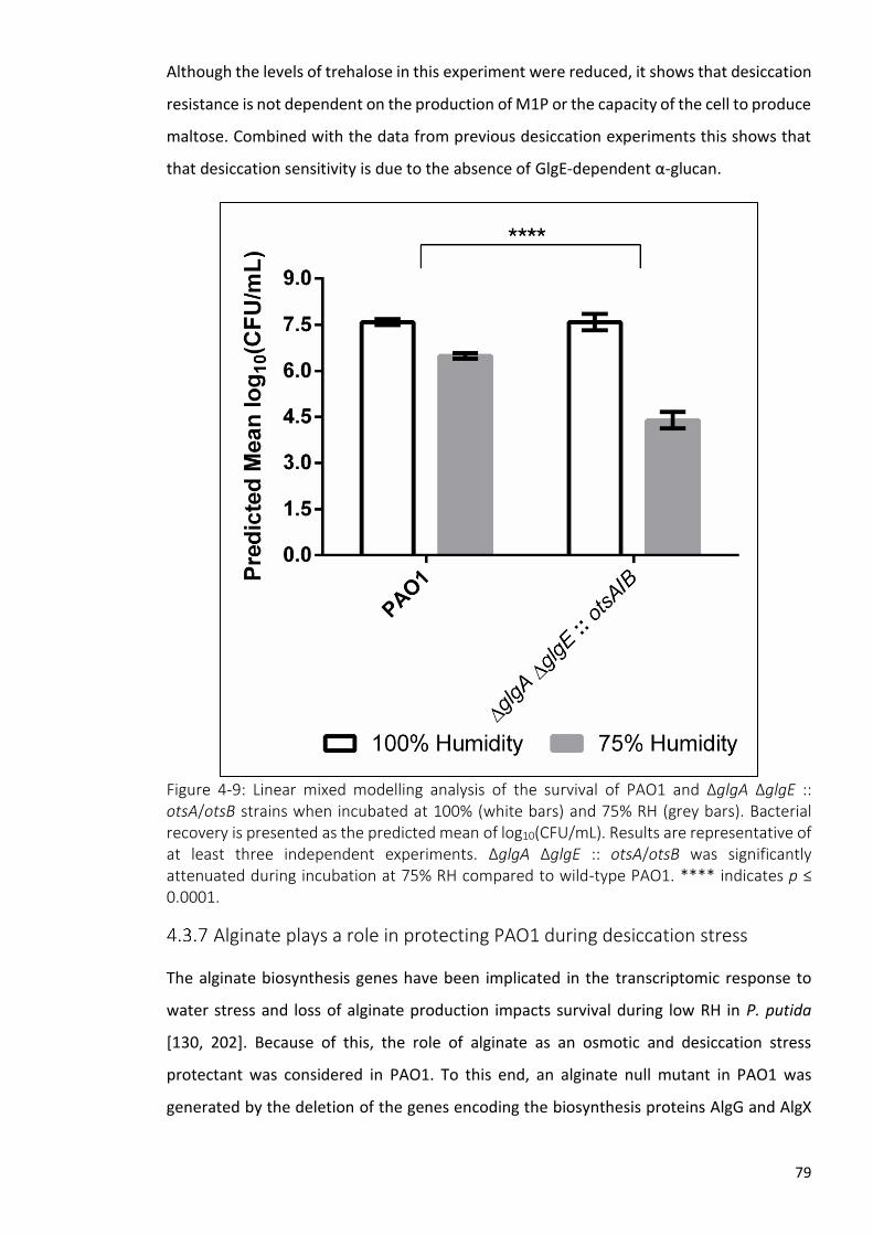
79
Although the levels of trehalose in this experiment were reduced, it shows that desiccation
resistance is not dependent on the production of M1P or the capacity of the cell to produce
maltose. Combined with the data from previous desiccation experiments this shows that
that desiccation sensitivity is due to the absence of GlgE-dependent α-glucan.
Figure 4-9: Linear mixed modelling analysis of the survival of PAO1 and ΔglgA ΔglgE :: otsA/otsB strains when incubated at 100% (white bars) and 75% RH (grey bars). Bacterial recovery is presented as the predicted mean of log10(CFU/mL). Results are representative of at least three independent experiments. ΔglgA ΔglgE :: otsA/otsB was significantly attenuated during incubation at 75% RH compared to wild-type PAO1. **** indicates p ≤ 0.0001.
Alginate plays a role in protecting PAO1 during desiccation stress
The alginate biosynthesis genes have been implicated in the transcriptomic response to
water stress and loss of alginate production impacts survival during low RH in P. putida
[130, 202]. Because of this, the role of alginate as an osmotic and desiccation stress
protectant was considered in PAO1. To this end, an alginate null mutant in PAO1 was
generated by the deletion of the genes encoding the biosynthesis proteins AlgG and AlgX

80
(PAO1 Δalg) [203, 204]. The construction of the deletion vector was performed by
Sebastian Pfeilmeier.
To assess the effect of alginate production during growth with osmotic stress, PAO1 and
PAO1 Δalg were cultured in the presence and absence of NaCl (Figure 4-10). Loss of alginate
did not affect growth in either stressed or non-stressed conditions. This suggests that the
production of alginate does not play a role in tolerating osmotic stress.
Figure 4-10: Growth of PAO1 and Δalg in M9 minimal medium ± 0.85 M NaCl. Values represent the mean OD600 ± SEM of five technical replicates. Error bars are not shown for growth under non-stressed conditions for reasons of legibility. The alginate deficient strain was not attenuated during osmotic conditions as compared to wild-type PAO1.
To investigate the role of alginate as a desiccation stress protectant, the viability of PAO1
Δalg following exposure to desiccating conditions was determined (Figure 4-11). When
subjected to desiccation stress, PAO1 Δalg yielded a statistically significant desiccation
response of 3 log10(CFU/mL) (p ≤ 0.001) compared to that of the wild-type PAO1 strain
(Figure 4-11). Taken together, this suggests that the production of alginate is important in
the resistance of desiccation stress but not osmotic stress in P. aeruginosa PAO1.

81
Figure 4-11: Linear mixed modelling analysis of the survival of PAO1 and Δalg when incubated at 100% (white bars) and 75% RH (grey bars). Bacterial recovery is presented as the predicted mean of log10(CFU/mL). Results are representative of at least three independent experiments. Δalg was significantly attenuated during incubation at 75% RH compared to wild-type PAO1. **** indicates p ≤ 0.0001.

82
4.4 Phenotypic characterisation of trehalose and α-glucan biosynthesis in Pto.
Operon deletions resulted in abolition of both trehalose and α-glucan
biosynthesis in Pto but do not allow the distinction between these two
molecules
Figure 4-12: The genomic organisation of the trehalose and α-glucan biosynthesis genes in Pto.
Since the genetic and biochemical characterisation of the predicted Pto trehalose and α-
glucan biosynthesis pathways resulted in ambiguity, I changed strategies and opted for
deletion of both the treS (PSPTO_2760 – PSPTO_2762) and treY/treZ (PSPTO_3125 –
PSPTO_3134) operons to ensure loss of trehalose and α-glucan (Figure 4-12). This resulted
in the generation of Pto ΔPSPTO_2760-2762 and Pto ΔPSPTO_3125-3134, respectively. The
operon deletions were also combined to generate Pto ΔPSPTO_2760-2762 ΔPSPTO_3125-
3134, abbreviated as Pto ΔΔ.
To show that trehalose and α-glucan biosynthesis had indeed been abolished, the soluble
metabolomes of these strains were extracted and analysed using 1H-NMR spectroscopy.
Analysis of the soluble metabolome of Pto ΔPSPTO_2760-2762, ΔPSPTO_3125-3134 and ΔΔ
revealed the absence of all relevant metabolites, except for ΔPSPTO_2760-2762 which
yielded low levels of trehalose (0.04 ± 0.006% dry cell weight) (Table 4-3, Figure 4-13).

83
Figure 4-13: 1H-NMR spectroscopy of the soluble metabolome following the deletion of PSPTO_2760-2762, PSPTO_3125-3134 and PSPTO_2760-2762 PSPTO_3125-3134 (ΔΔ) in Pto. Metabolomes were extracted from wild-type Pto (blue), ΔPSPTO_2760-2762 (red), ΔPSPTO_3125-3134 (green) and ΔΔ (purple) cultured on M9 minimal medium.Peak assignments are as indicated based on previously established spectra. (M1P; maltose 1-phosphate). Resonances corresponding to metabolites of interest were absent in the metabolomes of ΔPSPTO_3125-3134 and ΔΔ. Decreased levels of trehalose was detected in the metabolome of ΔPSPTO_2760-2762.
Table 4-3: Concentrations of trehalose and M1P produced by Pto strains. Metabolites are presented as percentages of cellular dry weight ± standard error (n ≥ 2). – indicates no measurable metabolite, * indicates significant differences as compared to the wild-type Pto as determined by a student’s t-test (p ≤ 0.05). Presence or absence of α-glucan as indicated by + or – respectively. Parental Pto strain data is replicated from table 3-1.
Strain Trehalose (%)
M1P (%) Presence of α-glucan
Pto 0.13 ± 0.03 0.30 ± 0.03 +
Pto ΔPSPTO_2760-2762 0.04 ± 0.01 - -
Pto ΔPSPTO_3125-3134 - - -
Pto ΔΔ - - -
Although trehalose was produced by the ΔPSPTO_2760-2762 strain, this was at lower
concentrations than expected based on our predicted pathway, consistent with previous
work [74, 180] (Section 3.2.5). Moreover, as the intracellular concentration of both
trehalose and α-glucan were depleted in these mutant strains as compared to the wild-
type Pto, subsequent stress sensitivity phenotypes could not be attributed to either the
loss of trehalose or α-glucan individually.
To decouple the biosynthesis of trehalose and α-glucan, and therefore distinguish their
individual roles in Pto, I attempted to increase the intracellular concentration of trehalose
within the Pto operon deletion strains by introducing M. koreensis 3J1 otsA/otsB as
described with PAO1 earlier (Section 4.3.3). However, the introduction of otsA/otsB

84
resulted in the majority of transformants not being viable. Where colonies did appear, they
exhibited morphological irregularities. This suggested that the production of these proteins
or an increase in intracellular trehalose concentration was toxic. Consequently, I did not
continue with this strategy. Although the individual roles of trehalose and α-glucan could
not be elucidated reliably in Pto, the phenotypic analysis was continued with Pto ΔΔ to
investigate the effect of the loss of both trehalose and α-glucan during water stress and to
draw comparisons to the system in PAO1.
Loss of both trehalose and α-glucan in Pto results in osmotic sensitivity
To investigate the roles of trehalose and α-glucan in Pto during osmotic stress, wild-type
Pto and ΔΔ were cultured in the presence and absence of NaCl. Wild-type Pto showed
attenuated growth with a longer lag-phase under osmotic stress when compared to non-
stressed growth (Figure 4-14) Pto ΔΔ yielded a similar growth profile to wild-type Pto under
non-stressed growth but exhibit increased osmotic sensitivity when compared to the wild-
type when NaCl was present. Due to the variable behaviour of these bacteria under osmotic
stress, these datasets were difficult to reproduce.
Figure 4-14: Growth of wild-type Pto and ΔΔ in M9 minimal medium ± 0.35 M NaCl.Values represent the mean OD600 of five replicates. Error bars are not shown for growth under non-stressed conditions for reasons of legibility. ΔΔ exhibited osmotic sensitivity compared to wild-type Pto.

85
Loss of both trehalose and α-glucan in Pto results in desiccation
sensitivity
To investigate the roles of trehalose and α-glucan in Pto during desiccation stress, wild-
type Pto and ΔΔ were examined under conditions of different RH as described above
(Section 4.2.1). Following incubation at 100% RH, recovery of wild-type Pto yielded a
predicted mean of approximately 5.8 log10(CFU/mL) (Figure 4-15). Recovery after
incubation at 75% RH resulted in a predicted mean of 5.05 log10(CFU/mL) equating to a
desiccation response of approximately 0.74 log10(CFU/mL). When subjected to either 100%
or 75% RH, the trehalose and α-glucan null mutant ΔΔ resulted in recovery of a predicted
mean of 5.9 and 4.4 log10(CFU/mL), respectively. This equated to a desiccation response of
approximately 1.5 log10(CFU/mL). Although this response was less severe than initially
expected based on equivalent data in PAO1, this was significantly different (p ≤ 0.05) when
compared to that of the wild-type Pto strain.

86
Figure 4-15: Linear mixed modelling analysis of the survival of Pto, ΔΔ, Δalg and Δalg ΔΔ strains when incubated at 100% (white bars) and 75% RH (grey bars). Bacterial recovery is presented as the predicted mean of log10(CFU/mL). Results are representative of at least three independent experiments. ΔΔ exhibited desiccation sensitivity when compared to wild-type Pto, whereas the desiccation response of Δalg was not significantly different. Δalg ΔΔ was exhibited enhanced attenuation during incubation at 75% RH compared to wild-type Pto and ΔΔ. **** indicates p ≤ 0.0001, * indicates p ≤ 0.05, ns indicates non-significant.
Alginate works in combination with α-glucan to protect against
desiccation stress in Pto
Since alginate played a role during the response to desiccation stress in PAO1 (4.3.7), I also
investigated the role of alginate production during desiccation stress in Pto. To this end, a
previously generated Pto strain harbouring a deletion of both algG and algX (Δalg) was
exposed to desiccating conditions as described earlier (Section 4.2.1). The deletion

87
construct pΔalg was constructed and the strain Pto Δalg was generated by Sebastian
Pfeilmeier.
When subjected to desiccation conditions, Pto Δalg yielded a desiccation response of 0.96
log10(CFU/mL) (Figure 4-15). This was not significantly different to the response of wild-
type Pto when the data was analysed using linear mixed modelling. Regardless of
significance, the desiccation response of Pto ΔΔ and Δalg were unexpectedly low when
compared to the desiccation response of the corresponding PAO1 strains. As a result, I
considered that α-glucan and alginate may exhibit functional redundancy in Pto strains and
that combining deletions for trehalose, α-glucan and alginate biosynthesis, may result in
desiccation susceptibility similar to that observed in the PAO1 strains.
To investigate this, the algG and algX genes were deleted to abolish alginate production in
the Pto ΔΔ strain, generating Δalg ΔΔ. This was performed using the pΔalg deletion vector
constructed by Sebastian Pfeilmeier. Exposure of Δalg ΔΔ to desiccating conditions resulted
in a desiccation response of 3.1 log10(CFU/mL), (p ≤ 0.0001).
The contribution of trehalose to desiccation stress in Pto cannot be excluded
experimentally. However, based on equivalent data in PAO1, these results suggest that Pto
utilises both alginate and α-glucan to resist desiccation stress. Furthermore, this suggests
that the polysaccharides are functionally redundant as the loss of both systems is not
simply additive.
Trehalose, α-glucan and alginate are important during plant infection
Although I have shown that alginate and α-glucan are important for desiccation survival in
vitro, the impact of this in planta has not been investigated. Since alginate and α-glucan
biosynthesis mutants have been shown to exhibit sensitivity to desiccation stress, it would
follow that the growth of these strains would be attenuated during the epiphytic lifestyle
on the surface of the leaf as they are exposed to osmotic and desiccating conditions [73].
To investigate the role of trehalose, α-glucan and alginate during plant infection, wild-type
Pto, ΔΔ, Δalg and Δalg ΔΔ strains were used to infect the model organism Arabidopsis
thaliana (Col-0).
To investigate the impact of loss of trehalose and α-glucan biosynthesis during plant
infection, Pto and ΔΔ were sprayed onto the surface of 6-week-old A. thaliana leaves. The
course of infection was monitored by determining the bacterial load by sampling the leaves

88
0, 2, and 3-days post infection (DPI). For the duration of the infection the plants were not
watered. Over the course of 3 days wild-type Pto showed marked proliferation from the
initial inoculum at day 0 indicating successful infection (Figure 4-16). Intracellular trehalose
and α-glucan did not appear to be important for pathogenesis because there was no
significant difference in the bacterial load of ΔΔ compared to the wild-type strain for the
duration of the experiment. Similarly, the production of alginate did not appear to play a
role in plant infection. There was no significant difference of the bacterial load of the Δalg
strain compared to wild-type Pto during infection. This suggests the loss of either system
individually is not important for plant infection.
When Pto lacked the ability to biosynthesise trehalose, α-glucan and alginate together,
there was an insignificant decrease in cell recovery during spray infection at 2 DPI. At 3 DPI
there was a statistically significant difference (p ≤ 0.05) between the recovery of Δalg ΔΔ
and the wild-type strain (Figure 4-17). When Pto and Δalg ΔΔ were infiltrated directly
through the stomata into the apoplast, there was no difference in bacterial recovery up to
3 DPI. This suggests that exposure on the leaf surface is required for the attenuation of
infection. Similarly, if the plants were watered regularly, thereby increasing the local RH,
the attenuation of Δalg ΔΔ during spray infection was also rescued.
Taken together, this suggests that alginate, trehalose and α-glucan are important for
survival on the surface of the leaf. Pto Δalg ΔΔ exhibits attenuation when exposed to low
RH on the surface of the leaf, a stress which is not present during higher RH or within the
apoplast. Although the contribution of trehalose during infection cannot be excluded,
equivalent data in PAO1 suggests that alginate and α-glucan protect Pto during low RH.

89
Figure 4-16: A. thaliana (Col-0) infection with Pto, ΔΔ and Δalg. (A) Spray infection with wild-type Pto or ΔΔ. (B) Spray infection with wild-type Pto or Δalg. Spray infection assays were conducted over 3 days and bacterial growth was determined by leaf disc sampling after 0-, 2- and 3-days post infection (DPI). Values represent the mean log10(CFU/cm2) of 6 replicates ± standard error of the mean. Significance was based on a two-tailed Mann-Whitney test. Results are representative of two independent experiments. No significant differences were observed between the infection profile of either mutant and wild-type Pto.

90
Figure 4-17: A. thaliana (Col-0) infection with Pto and Δalg ΔΔ. (A) Spray infection with wild-type Pto or Δalg ΔΔ. (B) Infiltration infection with wild-type Pto or Δalg ΔΔ. (C) Spray infection with wild-type Pto or Δalg ΔΔ where the plants were frequently watered. Spray infection assays are conducted over 3 days where bacterial growth is numerated by leaf disc sampling after 0-, 2- and 3-days post infection (DPI). Infiltration infection assays were conducted over 3 days where bacterial growth was determined by leaf disc sampling after 2- and 3-DPI. Values represent the mean log10(CFU/cm2) of 4-6 replicates ± standard error of the mean. Significance (* p ≤ 0.05) was based on a two-tailed Mann-Whitney U-test. Results are representative of two independent experiments. Significant attenuation of Δalg ΔΔ spray infection was observed compared to wild-type Pto at 3 DPI. This was not observed during infiltration infection or spray infection where plants were frequently watered.

91
4.5 Discussion
Prior to this work, many publications had implicated trehalose in the survival of bacteria
during water stress, namely osmotic and desiccating conditions [74, 107, 202, 205, 206].
Recent advances in our understanding of the biosynthesis of trehalose in bacteria and the
discovery of the GlgE pathway has enabled the discovery of the intrinsic genetic and
biosynthetic link between α-glucan and trehalose to be made in Pseudomonas spp.
Conclusions drawn from the disruption of what was thought to be trehalose biosynthesis
were therefore revisited and the function of trehalose and α-glucan were investigated. α-
glucan was always considered to be a carbon storage molecule providing energy to the cell
when required [207]. However, work in Mycobacterium spp. has shown that α-glucan
produced in this species is exported to the capsule where it can interact with the human
immune system and modulate virulence [208]. The fact that only a small subset of
Pseudomonas spp. are human pathogens means that the function of Pseudomonas α-
glucan is unlikely to be related solely, if at all, to the human immune system and must
therefore be investigated.
Deletion of what was thought to be the trehalose biosynthetic gene operons in
Pseudomonas aeruginosa PA14 resulted in the generation of strains attenuated during A.
thaliana infections [180], however these strains were not susceptible to osmotic stress.
Similarly, disruption of what was thought to be trehalose biosynthesis in Pto resulted in
osmotic sensitivity and strains which were attenuated during plant infection [74]. This is a
phenotype which was exacerbated by lower RH.
Furthermore, Vílchez et al. showed that trehalose concentrations were correlated with
bacterial desiccation resistance [151]. For example, M. koreensis 3J1 produces high
concentrations of trehalose and exhibits high desiccation resistance. Moreover, the in
trans expression of M. koreensis 3J1 otsA/otsB resulted in an increase of trehalose
production and desiccation resistance in P. putida.
The discovery and characterisation of the GlgE pathway in Pseudomonas spp. established
the ability of these bacteria to metabolise trehalose into α-glucan, therefore modulation of
the intracellular levels of trehalose, through gene deletion or gene introduction, may also
affect the levels of α-glucan. Phenotypes associated with Pseudomonas trehalose
biosynthesis must be reinterpreted considering recent findings. As a result, the role of α-

92
glucan and the interplay with the function of trehalose during water stress were elucidated
in Pseudomonas spp.
To investigate the role of trehalose and α-glucan in Pseudomonas spp. under osmotic and
desiccating conditions, I exploited trehalose and α-glucan biosynthesis mutant strains and
exposed individual strains to specific stress conditions. The combination of phenotypic
characterisation and the metabolomic characterisation of each of these mutant strains
enabled the assignment of individual functions to either α-glucan or trehalose.
To characterise the roles of α-glucan and trehalose during osmotic stress, PAO1 mutant
strains were cultured in media in the presence of NaCl. All strains lacking the ability to
synthesise trehalose were osmotically-sensitive suggesting that trehalose protects against
osmotic conditions. This was irrespective of the concentration of α-glucan because the α-
glucan accumulating ΔtreY and ΔtreZ strains yielded osmotic sensitivity. These strains
presumably produce a mixture of both GlgA- and GlgE-dependent α-glucan due to MalQ-
dependent maltose biosynthesis (Section 3.2.11). This suggests that α-glucan plays no
discernible role during growth in osmotic conditions.
PAO1 mutants lacking the branching and debranching enzymes GlgB and GlgX were also
osmotically-sensitive. This is surprising because the levels of trehalose in PAO1 ΔglgB
exceed that of the wild-type strain under non-stressed conditions, however, this may not
be true during hyper-osmotic conditions. This suggests that α-glucan is required as a
substrate to ensure optimal rates of trehalose production in response to osmotic stress.
Perturbation of the ability of PAO1 to branch or debranch α-glucan could result in pools of
inaccessible α-glucan. This could be due to linear α-glucan becoming too long and insoluble
in the ΔglgB strain or due to the inability of PAO1 to fully metabolise α-glucan after each
branch point to produce trehalose in the ΔglgX strain. The accumulation of long insoluble
α-glucan may interfere with the function of trehalose yielding an osmotically-sensitive
strain. Alternatively, GlgB and GlgX could be indirectly essential for the structural integrity
or function of other trehalose biosynthesis enzymes. These results suggest that α-glucan is
not directly required for osmotic tolerance, but effective recycling of α-glucan, presumably
into trehalose, is required. To further characterise this, PAO1, ΔglgB and ΔglgX should be
exposed to osmotic conditions, the soluble metabolomes extracted and the intracellular
levels of osmotically induced trehalose analysed and compared using 1H-NMR
spectroscopy. The protein-protein interactions between the enzymes of trehalose and α-

93
glucan and the subsequent impact on function should also be examined. To experimentally
validate the hypothesis that α-glucan recycling was required for osmotic tolerance,
intracellular trehalose accumulation should be determined following exposure of ΔglgB
and ΔglgX osmotic stress and compared to that of wild-type PAO1.
When subjected to 100% or 75% RH, it was clear that the loss of GlgE-dependent α-glucan
biosynthesis resulted in desiccation sensitive strains, irrespective of the ability to
synthesise trehalose. When the levels of intracellular GlgA- and GlgE-dependent linear α-
glucan are increased in the ΔglgB strain desiccation resistance was also increased. This
suggested that α-glucan protected PAO1 against desiccation stress, irrespective of the state
of branching. Although I was able to show that GlgA- and GlgE-dependent α-glucan do not
play a role during osmotic stress, I was unable to test the role of GlgA-dependent α-glucan
during desiccation stress. In order to investigate this, mutant strains accumulating solely
GlgA-dependent α-glucan should be generated and their desiccation sensitivity
determined.
Other groups have reported that the levels of intracellular trehalose in bacteria directly
correlate to desiccation resistance [150, 151]. As I speculated above, although the basal
levels of metabolite production were measured under non-stressed conditions, this may
not be representative of the metabolome under hyperosmotic and desiccation stress.
Because of this, I hypothesised that the levels of trehalose could be insufficient to confer
resistance to desiccation. Previous work had shown that the introduction of the
biosynthetic proteins OtsA/OtsB from the high trehalose-producing and markedly
desiccation-resistant organism M. koreensis 3J1 had increased desiccation resistance in P.
putida. Following this, I introduced otsA/otsB into PAO1 mutant strains to investigate the
effect of increasing the production of trehalose on the soluble metabolome and under
water stress.
Expression of otsA/otsB recovered M1P biosynthesis in the PAO1 ΔglgA mutant,
demonstrating the production of functional OtsA/OtsB. However, trehalose concentrations
did not significantly increase in any of the strains tested. Nevertheless, the production of
OtsA/OtsB by PAO1 strains conferred increased resistance to osmotic stress, consistent
with the osmoprotectant function of trehalose. This could indicate that although the OtsA
OtsB proteins are functional, trehalose does not accumulate during non-stressed
conditions either due to flux through TreS/Pep2 into M1P or via alternative trehalose
94
degradation mechanisms, such as the PAO1 periplasmic trehalase (TreA). These results also
suggest that GlgE-dependent α-glucan does not play a role under osmotic stress.
ΔtreS/pep2 :: otsA/otsB is genetically incapable of producing GlgE-dependent α-glucan
because of the loss of TreS/Pep2, but still retains osmotic resistance similar to that of PAO1
:: otsA/otsB. Although, metabolomic analysis suggested that ΔtreS/pep2 :: otsA/otsB did
not accumulate α-glucan, this strain still retains the genetic ability to produce GlgA-
dependent α-glucan. However, because PAO1 ΔtreY and ΔtreZ strains are genetically
capable of the biosynthesis of both GlgA- and GlgE-dependent α-glucan and still yield an
osmotically-sensitive phenotype, I conclude that α-glucan does not play a direct role during
osmotic stress in PAO1.
As the expression of otsA/otsB rescued the desiccation sensitivity of ΔglgA but did not
recover the phenotype of ΔtreS/pep2, I am confident that if trehalose protected against
desiccation stress, the levels of intracellular trehalose would have been sufficient to confer
protection in the ΔtreS/pep2 :: otsA/otsB strain. This suggested that TreS/Pep2 and
therefore the production of GlgE-dependent α-glucan was essential for desiccation
resistance. Even though the presence and concentration of intracellular trehalose did not
directly contribute to desiccation resistance, the levels of bacterial trehalose may still be
an indirect measure of desiccation tolerance in organisms possessing the GlgE pathway due
to the intrinsic coupling between the biosynthesis of trehalose and α-glucan.
Although GlgE-dependent α-glucan is the likely candidate for desiccation protection, I could
not rule out the products of TreS/Pep2, maltose and M1P, contributing to desiccation
resistance. It has been previously reported that the provision of maltose to Saccharomyces
cerevisiae resulted in enhanced desiccation resistance [115]. The generation of a PAO1
strain deficient in α-glucan but still capable of producing maltose and M1P required the
generation of a strain lacking both enzymes capable of synthesising α-glucan, GlgA and
GlgE. The expression of otsA/otsB then reintroduced the genetic capacity for trehalose,
maltose and M1P biosynthesis. The PAO1 ΔglgA ΔglgE :: otsA/otsB strain exhibited
desiccation sensitivity when exposed to 75% RH suggesting that the genetic capability to
produce maltose and the accumulation of M1P were not responsible for protecting the cell
from desiccation. This also further supports the hypothesis that desiccation resistance
requires GlgE-dependent α-glucan biosynthesis and that resistance is not solely dependent
on the presence of the TreS/Pep2 protein.

95
The elucidation of the roles of α-glucan and trehalose in Pto was hindered by the inability
to generate strains decoupling their individual biosynthesis. This was due to several factors
including the decrease in the levels of trehalose following a treS/pep2 or treS operon
deletion. Because levels of trehalose decreased and α-glucan was absent, I could not
confidently assign the resulting phenotypes to either of these molecules individually. An
additional factor was that attempts to increase intracellular trehalose in these strains using
the expression of M. koreensis 3J1 otsA/otsB were not successful. In lieu of this, I continued
investigating the impact of the loss of both trehalose and α-glucan biosynthesis in Pto. In
agreement with the phenotypic analysis in PAO1, the Pto ΔΔ strain was both osmotic- and
desiccation-sensitive compared to wild-type Pto. However, the desiccation response of this
strain was lower than expected when compared to the equivalent PAO1 strains. This
suggested there was some additional mechanism to resist desiccation stress in Pto.
In agreement with my findings, Gulez et al. reported that trehalose and α-glucan
biosynthesis gene homologues were upregulated during water stress in Pseudomonas
putida KT2440 [202]. Additionally, transcripts corresponding to the biosynthesis of the
polysaccharide and biofilm component alginate were also upregulated. Furthermore,
alginate has also been implicated in the desiccation survival of P. putida mt-2 [130].
Because of this the involvement of alginate during water stress was also investigated.
Alginate was also shown to be involved in the resistance of desiccation stress in PAO1. The
disruption of alginate biosynthesis in Pto did not result in a desiccation sensitive strain
when the data was analysed using linear mixed modelling. However, when alginate,
trehalose and α-glucan biosynthesis mutations were combined, the desiccation response
of this strain was greater than the sum of the responses following loss of each individual
biosynthesis pathway. This suggested that there was functional redundancy between α-
glucan and alginate in Pto and that both of these sugars function to protect against
desiccation stress.
The importance of trehalose, α-glucan and alginate were tested in planta where mutant
Pto strains were spray infected onto the surface of A. thaliana. Loss of either trehalose and
α-glucan or alginate alone was not enough to confer any defect during the course of
infection. Freeman et al. showed through spray competition assays that a strain identical
to Pto ΔΔ was attenuated compared to the wild-type strain [74]. Although the data
reported by Freeman et al. is not consistent with the results presented in this chapter, this

96
may be explained by the fact that my experiments were performed in the absence of
competition and therefore may not yield subtle differences in the ability to infect.
The loss of trehalose, α-glucan and alginate biosynthesis together resulted in a reduced
infection phenotype. This phenotype was only present when the plants were kept under
dry conditions for the duration of the infection. When the plants were frequently watered,
thereby raising the local RH, any significant attenuation was abolished. Infectious
attenuation was also reliant on epiphytic survival on the surface of the leaf because
bypassing this stage by infiltrating the Δalg ΔΔ strain through the stomata into the apoplast
resulted in a wild-type like infection. This strongly implies that alginate and α-glucan are
required by Pto to resist desiccation stress on the surface of a leaf thus enabling successful
plant infection.
Although I can assume that the RH was increased when the plants were watered, this was
not quantified. To further validate this, plant infections should be performed in dedicated
environments with controlled humidity to show that infection is proportional to RH using
methodology described by Xin et al. [209]. Nevertheless, the in planta experiments show
that the loss of both alginate and α-glucan is required for significant attenuation reflecting
the functional redundancy observed in in vitro desiccation experiments. The functions of
the hypothetical proteins PSPTO_3129, PSPTO_3131 and PSPTO_3132, the predicted
methyltransferase PSPTO_3133 and the predicted glucosyltransferase have yet to be
experimentally tested [181] and although unlikely a role during desiccation stress or growth
in planta cannot be ruled out.
Although in planta experiments were not performed with P. aeruginosa, Djonovic et al.
showed through infiltration infection assays with A. thaliana that a P. aeruginosa PA14
strain lacking both treS and treY/treZ operons was attenuated during plant growth [180].
The fact that this attenuation was observed during infiltration into the apoplast contradicts
the results in this chapter, suggesting that attenuation is a result of exposure to low RH on
the leaf surface. however, the watering regime of these plants is not stated and the
humidity in the apoplast may have been relatively low.
The results presented in this chapter have shown that although trehalose and α-glucan are
biosynthetically linked, they play distinct roles in the protection of Pseudomonas spp.
during water stress. Through this work I have been able to apply methodology to
confidently decouple the responses to the distinct water stresses, osmotic and desiccation

97
stress. I have shown that trehalose protects Pseudomonas spp. against osmotic stress, most
likely due to its role as a compatible solute and its ability to counteract the external solute
concentration and to preserve membranes and proteins from damage [139]. However,
trehalose does not directly protect against desiccation stress. This role can be attributed to
the polysaccharide α-glucan because although the presence of trehalose can correlate with
the protection of the cell against desiccation stress, this is only true when the cell retains
the ability to process trehalose into α-glucan via the GlgE pathway.
The exact mechanism of desiccation protection by α-glucan is currently unknown. Although
this work decoupled the biosynthesis of trehalose and α-glucan to study their individual
functions, the ability to interchange between both these molecules is likely to be required
for full adaptability to rapidly changing environments. This work also implicates the
external polysaccharide alginate as a desiccation stress protectant, functioning in a similar
manner to the internal α-glucan protection mechanism protecting the cell from desiccation
stress from either side of the plasma membrane. This is likely due to the capacity of alginate
to hold water [130], form a hydrogel and keep the cell hydrated by retaining localised pools
of water. I have shown that these systems are important for environmental survival as well
as during plant infection as summarised in Figure 4-18.
Figure 4-18: The current understanding of water stress protection mechanisms of Pseudomonas spp. (1) The export of alginate to the biofilm, where it is thought to form an aqueous-rich layer. (2) During osmotic stress the cell responds by biosynthesising trehalose. (3) During desiccation stress the cell responds by producing α-glucan to protect against cell death.
The importance of these systems during human infection has yet to be understood. It was
previously reported that trehalose and α-glucan biosynthetic mutants were not attenuated
in growth during metazoan, insect and mice infections [180]. However, desiccation stress
is not likely to be a factor during the course of metazoan and insect infections.

98
Furthermore, the infection of mice with the trehalose and α-glucan biosynthetic mutant
was conducted by oropharyngeal colonisation introduced through drinking water [180].
This therefore is not likely to reflect the water stress experienced within the CF lung.
This is a particularly interesting area of research as the over-production of alginate is known
to play an important role during infection of the lung in cystic fibrosis patients [29]. In
addition to this, it has been found that the trehalose and α-glucan biosynthesis genes are
highly co-expressed with the alginate biosynthesis genes in transcriptomic data of clinically-
isolated strains of P. aeruginosa (Susanne Haeussler, personal communication). This
suggests that trehalose and α-glucan may contribute to P. aeruginosa survival during CF
lung infection. This represents an exciting branch of future research. For example, the
trehalose and α-glucan content in mucoid clinical strains of P. aeruginosa could be
determined and the roles that these polysaccharides play during a cystic fibrosis lung
model.

99
Investigating the regulation of trehalose and α-
glucan biosynthesis

100
5.1 Introduction
Regulation
The ability to co-ordinate cellular processes is essential for successful bacterial lifestyles.
Pseudomonas spp. exhibit substantial genomic plasticity and can modulate and optimise
cellular processes in response to environmental signals. Therefore, it is not surprising that
Pseudomonas spp. are found in virtually every environment and can survive extreme
stresses [210]. This plasticity is largely attributed to extensive regulatory complexity [211].
Pseudomonas aeruginosa PAO1 possesses one of the largest bacterial regulatory networks,
with regulatory genes making up 12% of the genome [212].
The work described so far in this thesis involved biochemically and phenotypically
characterising trehalose and α-glucan biosynthesis in Pseudomonas spp. Additional work
has also begun to characterise the role of α-glucan in the protection against environmental
stress and its physiological relevance. To further our understanding of the physiological role
of α-glucan, we next sought to understand the factors that govern its synthesis. To achieve
this, the regulatory network controlling trehalose and α-glucan biosynthesis in
Pseudomonas spp. were studied. In this chapter I discuss our current understanding of the
regulation of trehalose and α-glucan in bacteria. In chapter 4, I show a functional link
between alginate and α-glucan, where both polysaccharides function to protect against
desiccation stress in Pseudomonas spp., therefore I also briefly discuss the relevant
mechanisms in the regulation of alginate biosynthesis.
Regulation of trehalose
The regulation of trehalose biosynthesis in bacteria is complex and surprisingly
understudied. Trehalose production is induced by a variety of environmental conditions,
including low temperature [213], salt [107], ethanol [214] and during the stationary phase
of bacterial growth [215]. Genes related to trehalose biosynthesis have been shown to be
under the control of several sigma factors. The otsA and otsB genes have been shown to
be regulated by the stress response sigma factor RpoS in E. coli [215]. Furthermore,
deletion of rpoS in P. fluorescens Pf-5 results in osmotically-sensitive strains [216]. The
alternative sigma factor RpoN has also been implicated in the regulation of trehalose
biosynthesis in bacteria. Deletion of rpoN leads to upregulation of otsA and otsB in E. coli
[217]. Conversely, rpoN mutations in P. aeruginosa results in the downregulation of genes
involved in trehalose and α-glucan biosynthesis [218]. Recently Harty et al. showed that
101
the production of trehalose in response to NaCl or ethanol was dependent on the sigma
factor AlgU (RpoE) [214]. These authors also showed that the accumulation of trehalose in
response to either NaCl or ethanol was also in-part dependent on the stringent response
and quorum sensing.
Bacteria use the stringent response to respond to nutrient-limiting conditions, producing
an alarmone called guanosine pentaphosphate (pppGpp) or tetraphosphate (ppGpp). In E.
coli and Pseudomonas spp., pppGpp is produced by RelA and SpoT [102, 219]. pppGpp is
then degraded into ppGpp by GppA in E. coli [220] and presumably by its homologue, Ppx
in Pseudomonas spp. [181]. SpoT exhibits bifunctional activity and can degrade ppGpp into
guanosine diphosphate and pyrophosphate [219, 221]. (p)ppGpp binds to RNA polymerase
and facilitates the expression of genes involved with nutrient starvation [222]. Following
exposure to ethanol or NaCl, the expression of the trehalose biosynthesis gene treZ was
significantly decreased in a P. aeruginosa relA or spoT mutant. Furthermore, these mutants
also produced less trehalose than the wild-type when exposed to either NaCl or ethanol
[214].
Regulation of classical α-glucan biosynthesis
In contrast to trehalose biosynthesis, the regulation of classical α-glucan biosynthesis has
been well studied in E. coli. I discuss some of the mechanisms of regulation below.
Control of the production of α-glucan is mainly achieved through allosteric regulation of
the biosynthetic enzymes. Products of carbon metabolism have been shown to activate or
inhibit these enzymes. For example, when carbon is not limiting, there are high levels of
glycolysis and therefore increased amounts of fructose-1,6-bisphosphate [223]. This sugar
activates the ADP-glucose pyrophosphorylase GlgC in E. coli leading to increased levels of
ADP-glucose and therefore α-glucan biosynthesis. Conversely, when carbon is limiting, the
phosphosugar adenosine monophosphate accumulates [224], and this inhibits the
enzymatic activity of GlgC, reducing the biosynthesis of ADP-glucose and therefore
inhibiting the production of α-glucan [225, 226].
The recycling enzyme glycogen phosphorylase (GlgP) is also a target of allosteric regulation
by the histidine phosphocarrier protein HPr [227], a component of the
phosphoenopyruvate:carbohydrate phosphotransferase system (PTS) [228]. GlgP binds to
both HPr and the phosphorylated form of HPr (P-HPr), with higher affinity for the latter.
Binding of HPr induces the dimerization and tetramerization of GlgP which increases its

102
phosphorylase activity, whereas, binding of the P-HPr does not show a stimulatory effect.
It is thought that under carbohydrate rich conditions, the phosphorylation state of the PTS
components and HPr is induced, which results in high affinity binding of GlgP to P-HPr,
preventing α-glucan degradation. Under low energy conditions, the PTS components will
be dephosphorylated and HPr will bind to GlgP and stimulate α-glucan degradation [229].
Another method of regulating α-glucan biosynthesis is through modulating the available
pool of the precursor substrate ADP-glucose. In E. coli ADP-glucose is degraded by the ADP-
glucose pyrophosphatase AspP, therefore the activity of AspP directly regulates the levels
of α-glucan production [230]. The activity of AspP is in turn activated by allosteric
regulation. The sugar glucose-1,6-bisphosphate and nucleoside molecules both enhance
AspP activity in vitro [231].
There is some evidence to suggest that α-glucan biosynthesis in E. coli is under the control
of the global regulators cyclic AMP (cAMP) and the cAMP receptor protein (CRP). cAMP-
or CRP-deficient mutants of E. coli were shown to be impaired in their ability to produce α-
glucan [232]. However, as transcriptomic analysis fails to show α-glucan biosynthesis genes
as part of the CRP regulon [233], it is thought that CRP/cAMP regulation of α-glucan
biosynthesis in E. coli may be indirect. This could be due to the upregulation of intermediate
regulatory elements such as the PTS components [234].
Like trehalose biosynthesis in Pseudomonas spp. the stringent response has also been
implicated in the regulation of α-glucan in E. coli. Stimulation of the stringent response
through amino acid starvation resulted in the accumulation of α-glucan [235]. Moreover,
deletion of relA results in strains deficient in their ability to produce α-glucan [236],
whereas overexpression of the genes encoding the degradative enzymes pggA and spoT
results in a reduction in α-glucan production in E. coli K-12 [237].
In E. coli α-glucan biosynthesis is controlled by the carbon storage regulator (Csr) system.
The Csr system controls the regulation of genes involved in a range of processes, notably
central carbon metabolism [238]. The Csr effector is the small RNA binding protein CsrA,
which binds to mRNA transcripts and prevents translation. This is usually a form of negative
regulation but examples of positive regulation by CsrA have also been reported [239]. CsrA
has been shown to bind to the transcript of glgC, preventing expression and therefore the
biosynthesis of α-glucan [240].

103
Homologues of CsrA are found within the Pseudomonas genera. P. aeruginosa possesses
two CsrA homologues, repressor of secondary metabolism A (RsmA) and RsmN [241],
whereas P. syringae possesses 5 RsmA homologues, one of which (PSPTO_3566) may be a
homolog of RsmE [181, 242].
In P. aeruginosa and P. syringae, the Rsm proteins are regulated by the GacA-GacS two-
component system, which I briefly summarise here (Figure 5-1). The Gac/Rsm system is
responsible for the regulation of a variety of processes, including biofilm formation,
motility, virulence and the stress response [243, 244]. This system is also implicated in the
transition from acute to chronic infection during P. aeruginosa cystic fibrosis lung infection
[244]. The histidine kinase GacS responds to as-yet unknown environmental signals by
autophosphorylation and subsequent phosphorylation of the response regulator GacA
[245]. GacA will then induce the expression of the small regulatory RNAs, RsmY and RsmZ
[246]. RsmY and RsmZ bind to and sequester the Rsm regulatory proteins, preventing Rsm
protein dependent regulation [247]. Similar to CsrA, the Rsm proteins will recognise and
bind to the transcript of target genes and usually prevent translation [248-251], but can
also have positive regulatory activity, for example, protecting RsmY and RsmZ RNA from
degradation [250]. Rsm proteins promote the expression of virulence factors such as the
type III secretion system and motility [252, 253], whereas activation of GacS and GacA
promotes biofilm production and type VI secretion [252, 254].
In P. aeruginosa the Gac/Rsm system is also regulated by the accessory sensor kinase
proteins, PA1611 [255], RetS, LadS [256, 257] and CmpX [255, 258]. RetS binds to GacS
thereby preventing the phosphorelay to activate GacA, reducing the levels of RsmY and
RsmZ and therefore promoting RsmA and RsmN regulation [245, 256]. PA1611 also
regulates the Gac/Rsm system through direct interaction with RetS [255]. PA1611 binding
to RetS results in the increased expression of RsmY and RsmZ and therefore prevents RsmA
and RsmN dependent regulation. This interaction presumably prevents RetS from binding
to GacS. LadS promotes the expression of RsmY and RsmZ by phosphorylating GacS which
subsequently activates GacA [257]. CmpX regulates the levels of PA1611, whereby deletion
of cmpX in P. aeruginosa results in elevated levels of PA1611 [255] and increased levels of
RsmY [258]. However, the mechanism of this regulation is unknown.

104
Figure 5-1: A graphical representation of the Gac/Rsm system in P. aeruginosa. GacS modulates the activity of GacA through phosphorylation. Subsequently, GacA activates the expression of the small non-coding RNAs RsmY and RsmZ. These RNA molecules sequester the regulation proteins RsmA and RsmN, modulating post-transcriptional regulation. GacS is regulated by the sensor kinases CmpX, PA1611, RetS and LadS. This figure is reproduced and modified with permission from Grenga et al. (2017) [259].
Regulation of GlgE dependent α-glucan biosynthesis
Although the regulation of classical α-glucan biosynthesis has been well studied, the
regulation of GlgE-dependent α-glucan biosynthesis is relatively unknown. To the best of
my knowledge there is only one published report concerning the regulation of the GlgE
pathway. The GlgE enzyme in Mycobacterium and Streptomyces spp. is modifiable by
phosphate groups at several positions. Phosphorylation of GlgE by the PknB kinase leads to
a reduction in GlgE enzymatic activity [260].
Regulation of alginate biosynthesis
The first elucidated mechanism of alginate regulation in Pseudomonas spp. was through
the sigma factor AlgU, driving the transcription of algD, the first gene of the alginate
biosynthesis operon [261-263]. AlgU is regulated by the anti-sigma factor MucA and its
cognate partner MucB, which sequesters AlgU to the inner membrane, preventing the
activation of transcription [25]. Upon encountering environmental signals that favour the

105
production of alginate, MucA is proteolytically degraded, liberating AlgU and allowing the
activation of alginate biosynthesis [264]. AlgU also induces the expression of other
regulatory proteins including the response regulators AlgB and AlgR [265].
The two-component system AlgB and KinB plays a role in the regulation of alginate
biosynthesis in P. aeruginosa. Mutation of the algB gene in mucoid strains results in a
marked reduction in alginate production [266]. Furthermore, it was shown that AlgB
directly regulates the expression of algD, as it binds specifically to the algD promoter in
vitro [267]. However, AlgB activity seems to be independent of its cognate sensor kinase
KinB [267] because alginate production is unaffected by kinB deletion in a mucoid strain of
P. aeruginosa [268]. Conversely, other reports show that a kinB deletion in a PAO1 mucA
mutant results in an increase of alginate production. Moreover, kinB deletion mutants yield
increased MucA degradation, subsequently inducing alginate production. This suggested
that KinB is an antagonist of alginate production [269].
The two-component system AlgR and FimS plays a role in regulating alginate production.
Deletion of algR in a mucoid strain of P. aeruginosa results in alginate deficiency with
significantly reduced levels of algD transcription [265]. AlgR also binds to the algD
promoter in vitro therefore directly regulating alginate production [270]. Interestingly,
similar to AlgB, AlgR activity is independent of phosphorylation and therefore independent
of its cognate sensor kinase FimS [268].
The response regulator CbrB and the cognate signal kinase CbrA form the CbrA/CbrB two-
component system. This system regulates carbon metabolism, biofilm formation and stress
tolerance in P. putida [271, 272]. Upon activation by CbrA, CbrB induces the transcription
of RpoN-dependent genes, such as the small regulatory RNA crcZ [273]. CbrA/CbrB
mediated regulation is achieved through modulation of the Crc protein [274].
Crc is a key regulator of carbon catabolite repression, involved in the optimisation of
bacterial metabolism based on the available nutrients by repressing specific carbon
metabolism pathways. Crc is also involved in the modulation of virulence in P. aeruginosa
[275, 276]. For example, Crc activity is highest when P. putida is cultured in rich complex
medium, conversely Crc activity is low when the bacteria are cultured using a poor carbon
source [277]. Crc functions as an RNA-binding protein, binding to the transcripts encoding
metabolic proteins and preventing translation [278]. The small regulatory RNA crcZ

106
antagonises Crc by sequestration preventing it from binding to transcripts, thereby
preventing the repressive activity of Crc.
The CbrA/CbrB system also plays a role in the production of alginate. Transposon insertion
within the cbrB response regulator results in a reduction in alginate production in P.
fluorescens [279]. Further analysis using chromatin immunoprecipitation sequencing,
revealed the genome-wide binding sites of CbrB in P. putida, which includes the algD
promoter [280].
The Gac/Rsm system is implicated in the regulation of alginate in both Azotobacter and
Pseudomonas spp [281, 282]. mucA is an RNA target of both RsmA and RsmN in P.
aeruginosa. Additional experiments showed that deletion of both Rsm proteins results in
alginate overproduction, whereas overexpression of RsmN results in reduced levels of
alginate. This shows that the Rsm proteins were negative regulators of alginate production
[282].
Chapter Aims
As very little is known about the regulation of trehalose or α-glucan biosynthesis in
Pseudomonas spp., this chapter aims to identify potential regulators in both P. aeruginosa
PAO1 and Pto.
To identify global regulators of trehalose and α-glucan biosynthesis, I first generated
reporter strains in both PAO1 and Pto that express the E. coli LacZ β-galactosidase protein
under the control of the glgA promoter, the first committed gene in the trehalose and α-
glucan biosynthesis pathway. These reporter strains indicate the level of glgA regulation by
producing a blue pigment when incubated with 5-bromo-4-chloro-3-indolyl β-D-
galactopyranoside (X-Gal). I then employed random transposon mutagenesis to identify
mutants exhibiting changes in LacZ activity. Changes in LacZ activity suggest that the
regulation of glgA has been altered. The candidate regulator was then identified in each
case using arbitrary PCR and Sanger sequencing.
In order to identify direct regulators of trehalose and α-glucan biosynthesis, I also utilised
DNA-affinity chromatography to identify DNA binding proteins specific to glgA regulation.
A biotinylated form of the PAO1 glgA promoter was used as bait to isolate regulation
candidates which were then identified using mass spectrometry.

107
5.2 Materials and Methods
Liquid Metabolite Extraction
Pto ΔglgB strains were cultured in 50 mL of M9 medium and incubated overnight at 28 °C.
The resulting cells were pelleted (4,000 x g, 8 minutes) and the residual medium was
discarded. Cell pellets were then resuspended in 5 mL of distilled H2O (dH2O). The sample
dry weight was determined after removing water through drying under vacuum. Dry
samples were then resuspended in 5 mL of dH2O and boiled at 95 °C for 1 hour. Cellular
debris was removed through centrifugation (10,000 × g, 10 minutes) and the resulting
supernatant was subject to additional boiling and centrifugation. The water was removed
from the resulting soluble metabolite extract by drying under vacuum. The dried extract
was resuspended in 1200 µL deuterium oxide (D2O).
RNA Extraction and Retrotranscription
RNA extraction was performed using the Qiagen RNeasy Mini kit, with a modified protocol.
Wild-type Pto was cultured in 50 mL of M9 minimal medium or on mixed cellulose ester
filter discs on M9 minimal agar as described earlier. RNA was protected in liquid cultures
by the addition of 30 mL RNA protect (Qiagen) or the resuspension of cells from solid media
in 5 mL of RNA protect. Samples were pelleted and resuspended with 200 µL of 10 mM Tris-
Cl pH 8.0. RLT buffer was supplemented with β-mercaptoethanol and cooled to 4 °C, before
700 µL was then added to the lysing matrix (Starlab / Thistle Scientific Ltd). The
resuspended pellet was then added to the matrix. Samples were then lysed, using two 30
second pulses with the Bio-Rad Ribolyser at a speed setting of 6.5. Samples were kept
incubated at 4 °C between pulses. Samples are then centrifuged at 11,000 × g at 4 °C for
three minutes. The sample supernatants were added to 450 µL of ethanol and this was
added to the RNeasy spin column and centrifuged at 11,000 × G for 30 seconds at room
temperature. RW1 buffer (350 µL) was then added to the column and this was centrifuged.
Contaminating DNA was degraded by adding 80 µL of DNAse I incubation mix directly to
the column and this was incubated at room temperature for 20 minutes. RW1 (350 µL) was
added to the column and this was centrifuged. RPE buffer (500 µL) was then added and
centrifuged. The column was dried by a further centrifugation for 2 minutes. RNA was
collected by the addition of 60 µL of RNAse-free water and centrifugation for one minute.
Retrotranscription was performed using the SuperScript II Reverse Transcriptase
(Invitrogen). RNA was diluted to 100 ng in a final volume of 13 µl RNAse-free water. To this,

108
1 µL of a 1/10 dilution of random primers (Invitrogen) and 1 µL of 10 mM dNTPs were
added. This was mixed and heated to 65 °C for 5 minutes. Samples were then incubated on
ice and 4 µl of 5X FirstStrand buffer and 2 µL of 0.1 M dithiothreitol was added. This was
mixed and incubated at 25 °C for 2 minutes. Samples were then incubated on ice and 1 µl
of Superscript II Reverse Transcriptase was added. The transcriptase was not added to the
controls. RNA was then retrotranscribed through the following cycle; 25 °C for 10 minutes,
42 °C for 50 minutes and 72 °C for 15 minutes.
qPCR
qPCR was performed using the SensiFast SYBR No Rox Kit (Bioline). Forward and reverse
primers were designed using http://bioinfo.ut.ee/primer3/ (and the following parameters:
fragment size 95-105, primers size min18 opt20 max22, temperature min60 opt62 max64)
[283]. The two primers were mixed to a final concentration of 10 µM each in a final volume
of 100 µL. Pto genomic DNA was serially diluted to give concentrations of 10, 1 and 0.1
ng/µL. Using a qPCR 96 well plate (4titude Ltd), each reaction contained 10 µL SensiFast
SYBR, 1 µL of the primer mix, 8 µL of dH2O and either 3.5 µL of the individual cDNA or dH2O.
Controls were conducted for each primer pair including serially diluted genomic controls,
non-retrotranscribed controls and a water control. This was then performed and analysed
using Bio Rad CFX Manager. The following protocol was used: 180 seconds at 95 °C, then,
50 cycles of 95 °C, 5 seconds, 62 °C, 10 seconds and 72 °C, 7 seconds. Melting curves were
then generated starting at 65 °C increasing by 0.5 °C until a final temperature of 95 °C.
Absolute quantification of mRNA was calculated and normalised relative to the
housekeeping gene rpoD as a reference [284].
Biparental Mating
Pto-PglgA-lacZ and E. coli S17-1 :: pALMAR3 was cultured overnight in 50 mL of media
supplemented with gentamycin and tetracycline respectively. Cultures were pelleted and
washed to remove the antibiotics. This step was repeated three times, pellets were
resuspended in a final volume of 1.5 mL. Pto-PglgA-lacZ and E. coli S17-1 :: pALMAR3 were
mixed in a 7:3 ratio to a final volume of 1 mL. This mixture was then pelleted, resuspended
in 50 µL of L medium and spotted onto a cellulose ester filter discs (Merck Millipore) placed
on the surface of L agar. Plates were then incubated at 28 °C for 3 hours. Cells were
recovered by vortexing with 2 mL of phosphate buffered saline (PBS), Pto-PglgA-lacZ ::

109
pALMAR3 conjugants were selected for by plating onto L agar containing gentamycin,
tetracycline and X-Gal.
Transposon Mutagenesis
Reporter strains PAO1 and Pto PglgA-lacZ were transformed using the mariner plasmid
pALMAR3. This was either performed by electroporation (Section 2.3.1) or by biparental
mating with E. coli S17-1 (5.2.4). Transposon insertions yielding changes in LacZ activity
were selected for on solid media supplemented with tetracycline and 50 µg/mL X-Gal.
Gentamycin was also used where appropriate to select against the S17-1 donor. Candidates
were re-streaked and the location of the transposon was determined by arbitrary PCR
[285]. The first semi-random reaction was performed using Taq polymerase (New England
Biolabs) using Arb-PCR and Arb1b (primers 130 and 131). Primary denaturation was
performed for 180 seconds at 95 °C. Following this, 30 cycles of denaturation (95 °C, 15
seconds), annealing (38 °C, 30 seconds) and extension (72 °C, 90 seconds) were performed,
and a final extension was performed at 72 °C for 180 seconds.
A second reaction to amplify the first semi-random PCR product was performed using 5 µL
of product as a template. Arb1 and Almar3-seq (primers 132 and 133) were used to amplify
the semi-random PCR product. Primary denaturation was performed for 180 seconds at 95
°C. Following this, 30 cycles of denaturation (95 °C, 15 seconds), annealing (56 °C, 30
seconds) and extension (72 °C, 90 seconds) was performed. A final extension was
performed at 72 °C for 180 seconds.
The resulting PCR product was subject to the BigDye Terminator v3.1 cycle sequencing kit
(Invitrogen). PCR product (4 µL) was added to 2.5 µL of dH2O, 1.5 µL of BigDye Terminator
buffer, 1 µL of Almar3-Seq and 1 µL of BigDye Terminator reaction mix. The sequencing
reaction was then performed using a thermocycler. Primary denaturation was performed
for 60 seconds at 96 °C. Following this, 25 cycles of denaturation (96 °C, 10 seconds),
annealing (50 °C, 5 seconds) and extension (60 °C, 240 seconds) were performed. The
sequence sample was then sent to Eurofins genomics using their ‘Ready2Load’ service.
Candidate genes were identified and annotated using the Pseudomonas Genome Project
database [181].
DNA-Affinity Chromatography
This method has been adapted from Jutras et al. (2012) [286]. The promoter region of PAO1
glgA was amplified using a Q5 polymerase-based reaction with primers 128 and 129

110
yielding a 497 bp 5ʹ-biotinylated fragment. A 500 bp 5ʹ-biotinylated region of non-promoter
DNA was also amplified as a control.
PAO1 cultures were grown overnight in 50 mL of M9 minimal medium ± 0.85 M NaCl.
Cultures were washed three times with either water or 0.85 M NaCl, respectively, and
freeze-thawed using liquid nitrogen. The pellets were resuspended in 1 mL of BS/THES
buffer. BS/THES buffer was made by combining BS buffer (10 mM HEPES, 5 mM CaCl2, 50
mM KCl, 12% Glycerol), THES buffer (50 mM TRIS-Cl pH 7.5, 10 mM EDTA, 20% Sucrose
(w/v), 140 mM NaCl, 0.7% Protease Inhibitor Cocktail company) and water in a 4.43: 2: 3.57
ratio.
The resuspended pellets were then sonicated at 40% amplitude for 10 seconds and kept at
4 °C. This was repeated five times with one-minute intervals. Samples were then
centrifuged at 11,000 × g for 20 minutes at 4 °C. Dynabeads® (Thermo Fisher Scientific)
were prepared by adding 200 µL of Dynabeads® solution to a microtube for each sample.
These were separated from solution using a 12-tube magnet (Qiagen). The beads were then
washed with 500 µL of B/W buffer (10 mM TRIS-Cl pH 7.5, 1 mM EDTA, 2 M NaCl). This step
was repeated three times with a final resuspension of 190 µL. Approximately 40 µg of
biotinylated probe (200 µL of ~200 ng/µL) was added to the Dynabeads® suspension and
incubated with end-over-end agitation at room temperature for 20 minutes. Following
incubation, the probe-bead complex was washed with TE buffer three times (10 mM TRIS-
Cl pH 8.0, 1 mM EDTA pH 8.0). The probe-bead solution was then washed twice with
BS/THES buffer and once with BS/THES buffer supplemented with 10 µg/mL of
poly(deoxyinosinic-deoxycytidylic) acid (Thermo Fisher Scientific). To the probe-bead
complex 200 µL of BS/THES and 600 µL of cell lysate were added. This was then incubated
with end-over-end agitation at room temperature for 30 minutes. Bead-probe-protein
complexes were then washed with BS/THES five times. To the bead-probe-protein
complex, 120 µL of NaCl elution buffer (25 mM TRIS-Cl, 1 M NaCl, pH 7.5) was added, the
resulting supernatant was then concentrated and desalted using a ZipTip with 0.6 µL resin
(Merck). The desalted eluate was then resolved on a 10% acrylamide resolving gel, excised
and this was subjected to mass spectrometry (Orbitrap) analysis to detect bound proteins.
Gel slices were prepared according to standard procedures adapted from Shevchenko et
al. [287]. Briefly, the slices were de-stained with ethanol, washed with 50 mM TEAB buffer
pH 8.0 (Sigma), incubated with 10 mM DTT for 30 min at 65 °C followed by incubation with
30 mM iodoacetamide (IAA) at room temperature (both in 50 mM TEAB). After washing

111
and dehydration with acetonitrile, the gels were soaked with 50 mM TEAB containing 10
ng/µl Sequencing Grade Trypsin (Promega) and incubated at 50 °C for 8 h. Peptides were
extracted, and aliquots were analysed by nanoLC-MS/MS on an Orbitrap Fusion™ Tribrid™
Mass Spectrometer coupled to an UltiMate® 3000 RSLCnano LC system (Thermo Scientific,
Hemel Hempstead, UK). The samples were loaded and trapped using a pre-column which
was then switched in-line to the analytical column for separation. Peptides were separated
on a nanoEase M/Z column (HSS C18 T3, 100 Å, 1.8 µm; Waters, Wilmslow, UK) using a
gradient of acetonitrile at a flow rate of 0.25 µl min-1 with the following steps of solvents A
(water, 0.1% formic acid) and B (80% acetonitrile, 0.1% formic acid): 0-4 min 3% B (trap
only); 4-15 min increase B to 13%; 15-77 min increase B to 38%; 77-92 min increase B to
55%; followed by a ramp to 99% B and re-equilibration to 3% B.
Data dependent analysis was performed using parallel CID and HCD fragmentation with the
following parameters: positive ion mode, orbitrap MS resolution = 120k, mass range
(quadrupole) = 300-1800 m/z, MS2 top20 in ion trap, threshold 1.9e4, isolation window 1.6
Da, charge states 2-5, AGC target 1.9e4, max inject time 35 ms, dynamic exclusion 1 count,
15 s exclusion, exclusion mass window ±5 ppm. MS scans were saved in profile mode while
MS2 scans were saved in centroid mode.
Recalibrated peaklists were generated with MaxQuant 1.6.3.4 [288] using the
Pseudomonas aeruginosa protein sequence database downloaded from Uniprot
(uniprot.org; 20161212; 5,563 sequences). The final database search was performed with
the merged HCD and CID peaklists from MaxQuant using in-house Mascot Server 2.4.1
(Matrixscience, London, UK). The search was performed on the P. aeruginosa protein
sequence database and the MaxQuant contaminants database. For the search a precursor
tolerance of 6 ppm and a fragment tolerance of 0.6 Da was used. The enzyme was set to
trypsin/P with a maximum of 2 allowed missed cleavages. Oxidation (M) and deamidation
(N/Q) were set as standard variable modifications and carbamido-methylation (CAM) of
cysteine as fixed modification. The Mascot search results were imported into Scaffold
4.4.1.1 (www.proteomsoftware.com) using identification probabilities of 99% for proteins
and 95% for peptides. DNA-affinity chromatography was performed in collaboration with
Richard Little. Peptide digestion, mass spectrometry and peptide analysis were performed
by Gerhard Saalbach and Carlo de-Oliveira-Martins.

112
5.3 Results
Regulation of trehalose and α-glucan biosynthesis occur at the level of
transcription in Pto
Since I have shown that α-glucan plays a role in the protection against desiccation in
Pseudomonas spp. (Chapter 4), I hypothesised that this molecule was most likely to be
important during epiphytic growth or surface survival. To investigate this, I examined the
effect of different growth conditions on the biosynthesis of trehalose and α-glucan in Pto.
First, the effect of growth in liquid medium on the soluble metabolome of the Pto ΔglgB
strain was examined. 1H-NMR spectroscopy showed that when Pto ΔglgB was cultured in
liquid media there were no detectable peaks for trehalose, M1P or α-glucan (Figure 5-2).
This was also reflected using iodine staining. When Lugol’s solution was added to
metabolome extracts from Pto ΔglgB grown on solid media, there was purple staining
indicating the presence of linear α-glucan (Figure 5-3). There was no staining of the
metabolome from Pto ΔglgB when cultured using liquid media. This suggested that
biosynthesis of trehalose and α-glucan was regulated, and that the system was either
activated on solid medium or repressed in liquid medium.
Figure 5-2: 1H-NMR spectroscopy shows the effects of growth in liquid culture upon the soluble metabolome of the Pto ΔglgB strain. This illustrates the presence and abundance of trehalose, maltose 1-phosphate (M1P) and α-glucan when the metabolomes are extracted from cells cultured on solid media (S) or liquid media (L). Peak assignments are as indicated. (α-1,4-; α-glucan internal linkages).

113
Figure 5-3: The addition of Lugol’s solution to metabolome extracts from Pto ΔglgB cultured in liquid (L) or on solid medium (S).
Quantitiative PCR (qPCR) was used to investigate the level of regulation controlling
trehalose and α-glucan biosynthesis. If regulation was at the level of transcription, I would
expect to see differential levels of mRNA transcripts from trehalose and α-glucan
biosynthesis genes for Pto grown on solid or liquid media.
Total RNA was isolated from Pto cultured either on solid or in liquid M9 minimal medium.
RNA was reverse transcribed into cDNA and this was subject to qPCR using primers specific
for the glgA gene. The absolute quantification of mRNA was normalised using the
housekeeping gene rpoD and compared to two other housekeeping genes, secA and gyrA,
as additional controls [284]. The levels of mRNA corresponding to rpoD, secA and gyrA did
not change significantly when isolated from wild-type Pto cultured on either media (Figure
5-4). However, the levels of glgA mRNA significantly decreased (p ≤ 0.0001) when isolated
from Pto grown in liquid compared to that of solid M9 minimal media. This suggests that
this solid/liquid regulation of trehalose and α-glucan biosynthesis occurs at the level of
transcription.

114
Figure 5-4: Quantitative PCR data showing the absolute quantification of transcripts from Pto cultured using solid or liquid M9 minimal medium relative to the housekeeping gene rpoD. Statistical significance was derived from a student’s t-test. ns indicates non-significant and **** indicates p ≤ 0.0001.
Generation of PglgA-lacZ transcriptional fusion vectors
To investigate the transcriptional regulation of trehalose and α-glucan biosynthesis I opted
to use a promoter-fusion assay. As a representative of trehalose and α-glucan biosynthesis
I elected to study the regulation of the first committed enzymatic step in this biosynthesis:
GlgA. The promoter of glgA is also predicted to control the expression of the remaining
genes in the treY/treZ operon, with glgA being the first gene in this operon [181].
PglgA was amplified from PAO1 and Pto genomic DNA and were individually incorporated
into the multiple cloning site of pUC18-mini-Tn7T-Gm-lacZ10 [191], forming pUC18-mini-
Tn7T-Gm-PAO1-PglgA-lacZ and pUC18-mini-Tn7T-Gm-Pto-PglgA-lacZ. The respective
construct was transformed into the appropriate wild-type strain and integrated into the
attTn7 genomic site as described in 2.3.3. To ensure the effective expression of lacZ by PglgA,
the resulting reporter strains PAO1 PglgA-lacZ and Pto PglgA-lacZ were cultured in the
presence of X-Gal. This resulted in the formation of blue colonies, indicative of successful
control of LacZ by the glgA promoter (Figure 5-5).

115
Figure 5-5: The reporter strain Pto PglgA-lacZ cultured on L medium supplemented with 50 µg/mL X-Gal. The blue colony pigment results from lacZ expression under the control of PglgA.
Transposon mutagenesis
Two parallel transposon mutagenesis screens were conducted to identify potential
regulators of trehalose and α-glucan biosynthesis. The transposon pALMAR3 was used to
generate random mutant libraries, which were screened to identify colonies with altered
LacZ activity based on colony appearance. The reporter strain PAO1 PglgA-lacZ was
transformed with pALMAR3 via electroporation whilst the plasmid was introduced into Pto
PglgA-lacZ via electroporation or conjugation using E. coli S17-1 as a donor. Colonies which
yielded differential levels of blue pigment were selected and the location of the transposon
was identified using arbitrary PCR and sequencing in each case. Lighter blue colonies
resulted from a decrease in LacZ activity and therefore possess transposons inserted within
genes predicted to encode activators of trehalose and α-glucan biosynthesis. Conversely,
darker blue colonies resulted from an increase in LacZ activity and therefore would possess
transposons inserted within genes predicted to encode repressors. The transposon
mutagenesis screen in PAO1 was partially conducted by Despoina Sifouna. Under my
supervision, she transformed the PAO1 PglgA-lacZ reporter strain with pALMAR3, performed

116
the arbitrary PCR, BigDye® sequencing reaction, and mapped the targets of transposon
insertions to the appropriate strain genome.
Identification of potential regulators of trehalose and α-glucan
biosynthesis in Pto
The transposon screen of Pto PglgA-lacZ consisted of over 20,000 colonies from 18 separate
transformation/conjugation events resulting in the sequencing of 46 transposon mutants.
As expected, there were several mutants with transposons found within the PglgA-lacZ
cassette resulting in dark blue or white colonies, driving or abolishing LacZ expression.
There were six candidates which harboured transposon insertions within genes predicted
to be involved in regulation or which were implicated in stress tolerance [181]. These
candidate genes are summarised in Table 5-1.
Although unlikely to regulate GlgA directly, transposon insertions in metabolic or transport
genes involved with stress tolerance resulted in reduced LacZ expression (light blue colony
phenotypes) suggesting that trehalose and α-glucan biosynthesis were down-regulated.
For example, transposon insertions within the TauC taurine ABC transporter, ArgJ, the
predicted glutamate N-acetyltransferase/amino acid acetyltransferase and SyrB
syringafactin synthetase resulted in less LacZ activity and therefore down-regulation of
glgA.
Table 5-1: Transposon candidates for potential regulators of trehalose and α-glucan biosynthesis in Pto.
Three of the six gene candidates were predicted to be involved with bacterial regulation
[181]. This included the response regulation CbrB, the histidine kinase LadS and a predicted
TetR family transcriptional regulator. Unexpectedly, most target candidates were only hit
once. To ensure these gene candidates were bona fide regulators of trehalose and α-glucan
Gene Hit Gene Product Colony Phenotype
PSPTO_0966 CbrB Light Blue
PSPTO_4796 LadS Light Blue
PSPTO_1866 TetR family transcriptional regulator
Light Blue
PSPTO_5321 TauC Taurine ABC Transporter
Light Blue
PSPTO_2830 SyrB Syringafactin Synthetase
Light Blue
PSPTO_4399 ArgJ glutamate N-acetyltransferase/amino acid acetyltransferase
White Colony

117
biosynthesis, the effects of each transposon insertion on the soluble metabolome were
investigated.
The first mutant candidate harboured a transposon insertion within the gene encoding
CbrB (PSPTO_0966). This yielded a light blue colony phenotype when incubated with X-Gal
suggesting decreased activity of LacZ. This therefore would suggest that CbrB is an activator
of GlgA expression in Pto so disruption of the cbrB gene would result in reduced levels of
trehalose and α-glucan. To further validate this, the soluble metabolome of tn::cbrB was
extracted and analysed using 1H-NMR spectroscopy as described in 2.4-2.5. The levels of
trehalose and maltose 1-phosphate (M1P) were compared to that of the Pto PglgA-lacZ
reporter strain.
The soluble metabolome of Pto PglgA-lacZ showed no significant difference, as determined
by a student’s t-test, in the levels of trehalose and M1P when compared to the wild-type
Pto strain. The metabolome of tn::cbrB yielded a decrease in the levels of both trehalose
and M1P (Table 5-2, Figure 5-6). Although this indicates that CbrB is a regulator of glgA and
hence trehalose and α-glucan biosynthesis in Pto, no statistical significance could be
derived from this as there was only one biological repeat.
Table 5-2: Concentrations of trehalose and M1P produced by Pto PglgA-lacZ transposon mutants. Metabolites are presented as percentages of cellular dry weight ± standard error (n ≥ 2). a indicates n = 1, b indicates p = 0.06, * indicates significant differences (p ≤ 0.05) as compared to the parental strain as determined by a student’s t-test.
Strain Trehalose (%) M1P (%)
Pto PglgA-lacZ 1.68 ± 0.19 0.20 ± 0.03
tn::cbrB a 0.41 0.11
tn::ladS 0.31 ± 0.07 * 0.05 ± 0.02 *
tn::PSPTO_1866 0.82 ± 0.21 b 0.26 ± 0.04

118
Figure 5-6: 1H-NMR spectroscopy of the soluble metabolome following transposon insertion within Pto PglgA-lacZ. Metabolomes were extracted from Pto PglgA-lacZ (blue), tn::cbrB (red), tn::ladS (green) and tn::PSPTO_1866 (purple) cultured on M9 minimal medium.Peak assignments are as indicated based on previously established spectra. (M1P; maltose 1-phosphate). Resonances corresponding to trehalose and M1P appeared to decrease in the metabolomes of tn::cbrB and tn::ladS as compared to the parental Pto PglgA-lacZ.
The second regulation mutant candidate harboured a transposon insertion within the gene
encoding LadS (PSPTO_4796). This gene target was identified twice during the transposon
screen. However, this arose from the same transformation event and could be a result of
clonal replication. Nevertheless, transposon insertion within this gene resulted in a light
blue colony phenotype indicating decreased LacZ activity, the down-regulation of glgA and
therefore reduced trehalose and α-glucan biosynthesis in Pto. Analysis of the soluble
metabolome from tn::ladS revealed significant decreases in both the levels of trehalose
and M1P when compared to the wild-type reporter strain (Table 5-2, Figure 5-6) indicating
that LadS is an activator of trehalose and α-glucan biosynthesis.
The third regulation mutant candidate harboured a transposon insertion within the gene
encoding a hypothetical TetR family transcriptional regulator (PSPTO_1866). Transposon
insertion within this gene resulted in a light blue colony phenotype (Table 5-2) indicating
decreased LacZ activity therefore PSPTO_1866 plays a role in the activation of trehalose
and α-glucan biosynthesis in Pto. Analysis of the soluble metabolome reveals no difference
in the levels of M1P when compared to Pto PglgA-lacZ. Levels of trehalose may have
decreased, but this was not statistically significant (p = 0.06) (Table 5-2, Figure 5-6),
therefore, this candidate was not investigated further.

119
Identification of potential regulators of trehalose and α-glucan
biosynthesis in Pseudomonas aeruginosa PAO1.
Potential regulators of trehalose and α-glucan biosynthesis in PAO1 were identified
through a second transposon mutagenesis screen. We transformed the reporter strain
PAO1 PglgA-lacZ with the pALMAR3 mariner plasmid and screened for changes in LacZ
activity, and thus changes in the regulation of GlgA. We screened approximately 10,000
mutant colonies, 52 of which yielded differences in colony colour phenotype. The location
of the transposon was determined from 11 mutant strains, resulting in the identification of
six potential regulators. This screen was performed in part by Despoina Sifouna under my
supervision. Gene candidates of interest are summarised in Table 5-3. Transposon
insertions within cmpX, algB and fimS were studied further. Due to time constraints
transposon insertions within p30/crcZ, PA1429 and PA2696 were not investigated further.
Table 5-3: Transposon candidates for potential regulators of trehalose and α-glucan biosynthesis in PAO1.
Gene Hit Gene Product Colony Phenotype
PA1775 CmpX Dark Blue
PA5483 AlgB Dark Blue
PA4726.11/2 P30/CrcZ Light Blue
PA5262 FimS Light Blue
PA1429 Probable cation-transporting P-type ATPase
Dark Blue
PA2696 Probable transcriptional Regulator
Light Blue
Deletion of cmpX resulted in increased trehalose production but
sensitivity to osmotic stress.
The first mutant candidate harboured a transposon insertion within the gene encoding
CmpX (PA1775). This gene target was only hit once during this screen. tn::cmpX yielded a
dark blue colony phenotype when incubated with X-Gal suggesting increased activity of
LacZ. This therefore would suggest that CmpX is a repressor of GlgA expression in PAO1 so
transposon insertion within the cmpX gene would result in increased levels of trehalose
and α-glucan. To further validate this, the soluble metabolome of tn::cmpX was extracted
and analysed using 1H-NMR spectroscopy. The levels of trehalose and maltose 1-phosphate
(M1P) were compared to that of the PAO1 PglgA-LacZ reporter strain. The levels of trehalose
were significantly increased whereas the concentration of M1P did not differ from the
parental reporter strain (Table 5-4, Figure 5-7). Consistent with an increase in LacZ activity,

120
these results suggest that CmpX is a repressor of GlgA and therefore trehalose and α-glucan
biosynthesis in PAO1.
Table 5-4: Concentrations of trehalose and M1P produced by PAO1 PglgA-lacZ transposon mutants. Metabolites are presented as percentages of cellular dry weight ± standard error (n ≥ 2). * indicates significant differences (p ≤ 0.05) as compared to the parental strain as determined by a student’s t-test.
Figure 5-7: 1H-NMR spectroscopy of the soluble metabolome following transposon insertion within cmpX in the PAO1 PglgA-lacZ strain. Metabolomes were extracted from PAO1 PglgA-lacZ (blue) and tn::cmpX (red) cultured on M9 minimal medium.Peak assignments are as indicated based on previously established spectra. (M1P; maltose 1-phosphate). Resonances corresponding to trehalose increased within the metabolome of tn::cmpX as compared to the parental PAO1 PglgA-lacZ.
Since the transposon insertion within cmpX resulted in increased levels of intracellular
trehalose, it would follow that tn::cmpX might exhibit increased tolerance to osmotic
stress. To test this, tn::cmpX was cultured with and without osmotic stress as described
previously (Section 2.7) and its growth characteristics were compared to that of the wild-
type reporter strain PAO1 PglgA-lacZ. Under non-stressed conditions PAO1 PglgA-lacZ and
tn::cmpX exhibited similar growth profiles. Following the addition of NaCl, PAO1 PglgA-lacZ
exhibited a delayed growth phenotype, whereas tn::cmpX did not proliferate within the
time-scale of the experiment despite increased trehalose production (Figure 5-8).
Owing to the nature of transposon mutagenesis, the decrease in viability of tn::cmpX during
osmotic stress could be due to unknown polar effects. To account for this, a non-polar
Strain Trehalose (%) M1P (%)
PAO1 PglgA-lacZ 0.21 ± 0.03 0.12 ± 0.05
tn::cmpX 0.36 ± 0.01 * 0.23 ± 0.03
tn::algB 0.57 ± 0.00 * 0.25 ± 0.08
tn::fimS 0.08 ± 0.02 * 0.07 ± 0.04

121
deletion of cmpX was generated as described previously (Section 2.2.1). When grown under
osmotic conditions, ΔcmpX behaved similarly to the phenotypes exhibited by tn::cmpX
(Figure 5-8). This suggests that the osmotic sensitivity is not due to polar effects and is a
result of cmpX disruption. The observation of osmotic sensitivity in both tn::cmpX and
ΔcmpX strains indicates that the increase in trehalose levels could be an indirect
consequence of cmpX disruption and that further investigation is required to validate CmpX
as a regulator of trehalose and α-glucan biosynthesis.
Figure 5-8: Growth of PAO1 and cmpX mutant strains in M9 minimal medium with and without addition of 0.85 M NaCl. (A) PAO1 PglgA-lacZ and tn::cmpX, (B) PAO1 and ΔcmpX. Values represent the mean OD600 of five replicates. Results are representative of two independent experiments. Error not shown for growth under non-stressed conditions. Both tn::cmpX and ΔcmpX exhibited attenuated growth under osmotic conditions when compared to the parental strain.

122
Disruption of algB indicates a role as a repressor of trehalose and α-
glucan biosynthesis in PAO1.
The second mutant candidate harboured a transposon insertion within the gene encoding
AlgB (PA5483). This gene target was hit twice arising from two independent transformation
events. tn::algB yielded a dark blue colony phenotype when incubated with X-Gal indicating
increased LacZ activity and suggesting that trehalose and α-glucan biosynthesis were
increased. Because of this, I would predict that AlgB is a repressor of GlgA expression in
PAO1 and that transposon insertion within algB would result in increased levels of
trehalose and α-glucan.
The soluble metabolome of tn::algB yielded a significant increase in the intracellular levels
of trehalose when compared to the wild-type reporter strain (p ≤ 0.01, Table 5-4, Figure
5-9). Although levels of M1P did not significantly differ, the peak corresponding to α-1,4-
glucan appears to increase in tn::algB.
Figure 5-9: 1H-NMR spectroscopy of the soluble metabolome following insertion within algB of the PAO1 PglgA-lacZ strain. Metabolomes were extracted from PAO1 PglgA-lacZ (blue) and tn:: algB (red) cultured on M9 minimal medium.Peak assignments are as indicated based on previously established spectra. (M1P; maltose 1-phosphate). Resonances corresponding to trehalose increased within the metabolome of tn::algB as compared to the parental PAO1 PglgA-lacZ.
Since transposon insertion within algB resulted in the increase of trehalose and α-glucan
production, it would follow that tn::algB would be enhanced in its ability to tolerate
osmotic stress as compared to the wild-type reporter strain. Under non-stressed conditions
PAO1 PglgA-lacZ and tn::algB exhibited similar growth profiles. When cultured in the
presence of NaCl, tn::algB possessed a shorter lag phase than that of the PAO1 PglgA-lacZ
strain, suggesting increased tolerance to osmotic stress (Figure 5-10). Taken together,

123
increased LacZ activity, intracellular trehalose concentration and osmotic tolerance
suggests that AlgB is a repressor of trehalose and α-glucan biosynthesis in PAO1.
Figure 5-10: Growth of PAO1 PglgA-lacZ and tn::algB in M9 minimal medium with and without addition of 0.85 M NaCl. Values represent the mean OD600 of five replicates. Results are representative of two independent experiments. Error not shown for growth under non-stressed conditions. tn::algB exhibited enhanced tolerance to osmotic conditions as compared to the parental PAO1 PglgA-lacZ strain.
Disruption of fimS implicates the AlgR/FimS two-component system as
an activator of trehalose and α-glucan biosynthesis in PAO1.
The third mutant candidate harboured a transposon insertion within the gene encoding
FimS (PA5483). This gene target was only hit once during this mutagenesis screen. tn::fimS
yielded a light blue colony phenotype when incubated with X-Gal indicating decreased LacZ
activity and therefore a decrease in trehalose and α-glucan biosynthesis. Therefore, I would
predict that FimS plays a role in activating the expression of glgA in PAO1 and that
disruption of fimS would result in decreased levels of trehalose and α-glucan.
Consistent with LacZ activity, 1H-NMR spectroscopy revealed that the soluble metabolome
of tn::fimS yielded significantly less trehalose than that of the wild-type reporter strain
PAO1 PglgA-lacZ (Table 5-4, Figure 5-11). Concordantly, when cultured under osmotic stress,
tn::fimS exhibited increased sensitivity (Figure 5-12). There was no difference in growth
between tn::fimS and PAO1 PglgA-lacZ when grown under non-stressed conditions.
Taken together, these results show that FimS disruption results in a decrease in glgA
expression, trehalose accumulation and osmotic tolerance in PAO1. This indicates a role of
FimS in the activation of trehalose and α-glucan biosynthesis.

124
Figure 5-11: 1H-NMR spectroscopy of the soluble metabolome following transposon insertion within fimS in the PAO1 PglgA-lacZ strain. Metabolomes were extracted from PAO1 PglgA-lacZ (blue) and tn::fimS (red) cultured on M9 minimal medium.Peak assignments are as indicated based on previously established spectra. (M1P; maltose 1-phosphate). Resonances corresponding to trehalose decreased within the metabolome of tn::fimS as compared to the parental PAO1 PglgA-lacZ.
Figure 5-12: Growth of PAO1 PglgA-lacZ and tn::fimS in M9 minimal medium with and without addition of 0.85 M NaCl. Values represent the mean OD600 of five replicates. Results are representative of two independent experiments. Error not shown for growth under non-stressed conditions. tn::fimS exhibited attenuated growth under osmotic conditions when compared to the parental PAO1 PglgA-lacZ strain.
Gac/Rsm System
The transposon mutagenesis screens of both PAO1 and Pto yielded hits within cmpX and
ladS respectively, both of which have been shown to interact with the Gac/Rsm signalling
system, discussed in more depth earlier (Section 5.1.3). To investigate whether the
Gac/Rsm system plays a role in the regulation of trehalose and α-glucan biosynthesis, I

125
collaborated with Stephen Heeb (University of Nottingham) who kindly gifted a range of
Gac/Rsm system mutants generated within P. aeruginosa sublines. Strains harbouring
disruptions in the genes encoding GacA and GacS were generated in the Lausanne subline
of PAO1 (PAO1-L). Deletion of the genes encoding RsmA, RsmN, both individually and in
combination, were generated in the Nottingham subline of PAO1 (PAO1-N) (Stephen Heeb,
personal communication). To investigate the impact of perturbations of the Gac/Rsm
system on the levels of trehalose and α-glucan, the small metabolome of these strains was
extracted and analysed using 1H-NMR spectroscopy.
PAO1-L produced trehalose and M1P to approximately 0.29% and 0.28% of the total
cellular dry weight, respectively. Following deletion of gacA, levels of trehalose were
significantly reduced to approximately 0.07% of the cell dry weight (Table 5-5, Figure 5-13).
Peaks corresponding to M1P were not detected from this strain’s metabolome. Similarly,
following deletion of gacS, trehalose levels may have been reduced, but this was not
significant (p = 0.07). M1P was not detected from the metabolome of this strain. This
suggests that the Gac/Rsm system positively regulates trehalose and α-glucan biosynthesis.
To further implicate the Gac/Rsm system in the regulation of trehalose and α-glucan
biosynthesis, it would follow that the negative regulators RsmA and RsmN would repress
the production of trehalose and α-glucan. This suggests that deletion of rsmA and rsmN
would result in an increase in the levels of these metabolites.
The soluble metabolomes of PAO1-N, ΔrsmA and ΔrsmN strains were examined using 1H-
NMR spectroscopy. PAO1-N accumulated trehalose and M1P to approximately 0.13% and
0.16% of the cellular dry weight, respectively (Table 5-6, Figure 5-13). There was no
significant difference in the levels of trehalose or M1P following deletion of rsmA or rsmN
individually. However, when RsmA and RsmN were both absent, the levels of trehalose
reduced significantly and M1P was not detected. There was also an increase in the levels
of maltose/glucose and the formation of two unknown broad sugar-like species labelled a
and b (Figure 5-13). Although the levels of trehalose and M1P did not increase as predicted,
absence of both Rsm proteins changes the metabolites produced which provides further
evidence that the Gac/Rsm system plays a role in the regulation of trehalose and α-glucan
biosynthesis.

126
Table 5-5: Concentrations of trehalose and M1P produced by PAO1-L, ΔgacA and ΔgacS strains. Metabolites are presented as percentages of cellular dry weight ± standard error (n ≥ 2). * indicates significant differences (p ≤ 0.05) as compared to the parental strain as determined by a student’s t-test. – indicates no detected metabolite.
Table 5-6: Concentrations of trehalose and M1P produced by PAO1-N, ΔrsmA, ΔrsmN, and ΔrsmA ΔrsmN strains. Metabolites are presented as percentages of cellular dry weight ±
standard error (n ≥ 2). * indicates significant differences (p ≤ 0.05) as compared to the
parental strain as determined by a student’s t-test. – indicates no detected metabolite.
Strain Trehalose (%) M1P (%)
PAO1-L 0.29 ± 0.04 0.28 ± 0.08
ΔgacA 0.07 ± 0.01 * -
ΔgacS 0.14 ± 0.01 -
Strain Trehalose (%) M1P (%)
PAO1-N 0.13 ± 0.01 0.16 ± 0.03
ΔrsmA 0.09 ± 0.01 0.07 ± 0.02
ΔrsmN 0.13 ± 0.02 0.11 ± 0.02
ΔrsmA ΔrsmN 0.02 ± 0.02 * -

127
Figure 5-13: 1H-NMR spectroscopy of the soluble metabolome following Gac/Rsm system mutations in Pseudomonas aeruginosa strains. Metabolomes were extracted from from the PAO1 Lausanne subline (PAO1-L) (blue) following disruption of gacA (red) and gacS (blue) (A) and from the PAO1 Nottingham subline (PAO1-N) (blue) following deletion of rsmA (red), rsmN (green) and rsmA and rsmN in combination (purple) (B) cultured on M9 minimal medium.Peak assignments are as indicated based on previously established spectra. (M/G; maltose and/or glucose, M1P; maltose 1-phosphate, a and b represent currently unknown species). Resonances corresponding to trehalose decreased following deletion of gacA and gacS when compared to PAO1-L. Furthermore, M1P was absent in the metabolomes of ΔgacA and ΔgacS. Deletion of either rsmA or rsmN did not affect the metabolome as compared to that of PAO1-N. When both rsmA and rsmN were absent, the peaks corresponding to unknown species were present.
DNA-affinity chromatography
To identify candidates that regulated trehalose and α-glucan biosynthesis directly, we used
DNA-affinity chromatography to select for and identify any DNA-binding proteins. A
biotinylated form of the PAO1 glgA promoter was used as bait to pull down glgA promoter-
specific binding proteins. Non-specific biotinylated DNA was used as a control to exclude
non-specific DNA binding proteins. To ensure optimal glgA regulation, cell lysates were
extracted from PAO1 cultured under hyperosmotic conditions. Candidates were selected

128
based on predicted function with at least five unique peptide reads that did not bind to the
non-specific DNA control. DNA-affinity chromatography was performed in collaboration
with Richard Little. Peptide digestion, mass spectrometry and peptide analysis were
performed by Gerhard Saalbach and Carlo de-Oliveira-Martins.
The sigma factor RpoN and five regulatory proteins FleQ, TrpI, BauR, OxyR and FleN were
predicted as glgA promoter binding proteins through DNA-affinity chromatography (Table
5-7), the implications of each of these candidates is discussed below (Section 5.4).
Table 5-7: Putative regulators that were identified through specific binding to PAO1 PglgA.
Protein Name Unique peptide count
FleQ 20
RpoN 5
FleN 5
TrpI 5
BauR 5
OxyR 7
5.4 Discussion
The regulation of bacterial trehalose biosynthesis is complex and surprisingly understudied.
Conversely the regulation of classical α-glucan biosynthesis has been extensively studied,
but regulation of the GlgE pathway has only been studied in M. tuberculosis [260].
Nevertheless, as very little is known about the regulation of trehalose or α-glucan
biosynthesis in Pseudomonas spp., I aimed to identify potential regulators in both P.
aeruginosa PAO1 and Pto., I first generated reporter strains in both PAO1 and Pto, I then
employed random transposon mutagenesis to generate and identify global regulators of
trehalose and α-glucan biosynthesis. In order to identify direct regulators of trehalose and
α-glucan biosynthesis, I also utilised DNA-affinity chromatography to identify DNA binding
proteins specific to glgA regulation.
I first identified evidence of transcriptional regulation of the trehalose and α-glucan
biosynthesis pathway in Pto during growth in liquid culture. In order to identify the
regulatory network governing trehalose and α-glucan biosynthesis in PAO1 and Pto, I
performed two complementary screens. The first screening method employed transposon
mutagenesis in an unbiased approach to identify global regulators directly or indirectly
involved in the transcription of glgA. The second approach utilised DNA-affinity
chromatography and was designed to identify regulators which directly interact with the

129
promoter of the glgA gene. The combination of these approaches has resulted in the
identification or implication of several regulatory systems that control trehalose and α-
glucan biosynthesis in Pseudomonas spp.
Although a number of known regulators were identified, transposon mutagenesis also
resulted in insertions within genes that were not predicted to have regulatory functions.
Unlikely to regulate GlgA directly, transposon insertions in metabolic or transport genes
generally resulted in reduced LacZ expression (light blue colony phenotypes) suggesting
that trehalose and α-glucan biosynthesis were down-regulated indirectly. For example,
transposon insertions within the TauC taurine ABC transporter and ArgJ, a predicted
glutamate N-acetyltransferase/amino acid acetyltransferase, most likely interfere with
taurine and glutamate metabolism. Taurine has been shown to be a compatible solute in
eukaryotic cells, but can also accumulate during osmotic conditions in E. coli [289]. ArgJ is
a component of the bacterial arginine biosynthesis pathway and utilises the
osmoprotectant glutamate as a substrate [290, 291]. Disruption of ArgJ may result in a
build-up of glutamate and therefore solute concentration. The cell may respond in turn by
decreasing the biosynthesis of other solutes such as trehalose. Similarly, SyrB, a
syringafactin synthetase, is involved in the biosynthesis of syringafactin. Syringafactin is a
hydroscopic surfactant that has been shown to have high water-holding potential and to
protect P. syringae strain B728a from water stress [292]. Increases in the levels of
syringafactin may increase the water potential of the cell, leading to a reduced requirement
for compatible solutes and therefore decreased levels of trehalose biosynthesis. However,
due to the unpredictable nature of this assay, transposon insertion in syrB may also have
resulted in the decrease in the production of syringafactin, a possibility that is harder to
rationalise. One explanation could be that disruption of syringafactin biosynthesis may
result in a differential flux in carbon metabolism, reducing inputs into trehalose and α-
glucan biosynthesis. Although these candidates are not likely to be regulators, this
illustrates a potential limitation of this methodology. Changes in osmotic potential or
carbon metabolism caused by transposon insertion has the potential to result in altered
levels of trehalose and α-glucan biosynthesis, thereby resulting in the selection of less-
relevant transposon mutant strains.
In PAO1, a transposon insertion was identified within the probable cation-transporting P-
type ATPase (PA1429). Although unlikely to be directly related to the regulation of
trehalose and α-glucan biosynthesis, disruption of this gene resulted in an increase in LacZ

130
activity. Disruption of PA1429 could lead to changes in the osmotic balance of the cell, and
therefore an increase the biosynthesis of trehalose and α-glucan. Although this protein has
not been studied, P-type ATPases are responsible for the transport of charged substrates
such as potassium ions [293]. The import of potassium ions is one strategy for resisting
osmotic stress in E. coli [294]. Therefore, interruption of potassium import, such as with
PA1429 disruption, could lead to changes in the cellular osmotic potential. Compatible
solutes, such as trehalose, would be synthesised in response accounting for the observed
increase in LacZ activity.
The first regulatory pathway identified through the transposon mutagenesis screen was
the Gac/Rsm system. The accessory sensor kinases CmpX and Lads were identified in
screens in PAO1 and Pto, respectively. Disruption of ladS resulted in reduced LacZ activity
and therefore suggested that LadS activated glgA expression. Subsequently, ladS disruption
led to decreased levels of intracellular trehalose. Conversely, cmpX disruption resulted in
increased LacZ activity and increased levels of intracellular trehalose, implicating LadS and
CmpX as an activator and repressor of glgA expression, respectively.
Although the data suggested that CmpX is a repressor of trehalose and α-glucan
biosynthesis, cmpX disruption resulted in severe growth attenuation under osmotic
conditions despite increased trehalose levels. Polar effects resulting from transposon
insertion were not responsible for osmotic sensitivity, because a strain harbouring a non-
polar deletion of cmpX also yielded a similar phenotype. This suggests that pleotropic
effects arising from the deletion of cmpX may be responsible. Bhagirath et al. reported that
a PAO1 strain lacking functional CmpX had altered levels of gene transcripts and molecules
involved in signalling or regulation. For example, cmpX deletion resulted in increased levels
of the signalling molecule cyclic di-guanosine monophosphate, and the cyclic di-guanosine
monophosphate synthase wspR. Furthermore, the transcripts corresponding to the
membrane porin oprF and the sigma factor sigX were both decreased following deletion of
cmpX. PAO1 ΔcmpX exhibited sensitivity to membrane detergents and increased
susceptibility to a range of antibiotics [258]. This demonstrates that deletion of cmpX
influences other regulatory systems and causes increased sensitivity to toxic molecules,
possibly due to membrane defects. These pleotropic effects could therefore be responsible
for the observed osmotic attenuation. For example, the OprF porin has been shown to be
required for growth in hypoosmotic conditions [295] and therefore could be required to
maintain osmotic balance in hyperosmotic conditions. Taken together, it is uncertain
131
whether trehalose accumulation is due to de-repression of the Gac/Rsm system or that
trehalose accumulation is in response to dysregulation of cellular osmotic potential due to
pleotropic effects arising from cmpX deletion.
As CmpX and LadS both play a role in modulating GacS activity as discussed earlier, I
investigated the effect of disruption of other components of the Gac/Rsm system on
trehalose and α-glucan biosynthesis in PAO1 strains. I showed that GacA and GacS function
as activators of trehalose and α-glucan biosynthesis. Therefore, RsmA and RsmN would
likely function as repressors and that deletion of the negative regulators rsmA and rsmN
would result in an increase in trehalose and α-glucan biosynthesis. Although PAO1-N ΔrsmA
ΔrsmN yielded reduced levels of trehalose and M1P, this strain produced two unknown
sugar-like species. Further analysis of the products following deletion of rsmA and rsmN is
required. Nonetheless, this supports the hypothesis of Gac/Rsm regulation of the trehalose
and α-glucan biosynthesis pathway in Pseudomonas spp. Further work is also needed to
characterise the osmotic and desiccation phenotypes of strains following Gac/Rsm system
deletions.
Literature mining further supports the hypothesis of Gac/Rsm system mediated regulation
of trehalose and α-glucan biosynthesis in Pseudomonas spp. as UTP-glucose-1-phosphate
uridylyltransferase (GalU) was shown to be a target of RsmN in PAO1 [282]. This suggests
RsmN negatively regulates the translation of GalU and prevents trehalose and α-glucan
biosynthesis. This is reminiscent of CsrA repression of GlgC in E. coli [240] where the
expression of the enzymes responsible for the metabolism of glucose 1-phosphate in both
E. coli and PAO1 are translationally repressed by CsrA homologues. Moreover, PAO1 RsmA,
but not RsmN, can functionally replace CsrA and repress α-glucan biosynthesis in E. coli
[241]. As my results suggest that both RsmA and RsmN are required to modulate levels of
trehalose and α-glucan in PAO1, it would be interesting to see if expression of E. coli CsrA
in PAO1 ΔrsmA ΔrsmN would result in a wild-type metabolome.
The CrbA/CrbB two-component system was implicated as an activator of trehalose and α-
glucan biosynthesis in Pto as CbrB disruption resulted in decreased LacZ activity and
decreased levels of intracellular trehalose. I would therefore predict that transposon
insertion within cbrB would result in an osmotically-sensitive strain. However, due to time
constraints, this hypothesis could not be tested. These results implicate either the Crc
protein in the repression of glgA or that GlgA belongs to the CbrB regulon. Although this
could be determined using in vitro DNA-binding assays to assess protein binding to the glgA

132
promoter, other authors have reported that GlgA is under the direct control of CbrB in P.
putida. Chromatin immunoprecipitation sequencing revealed the genome-wide binding
sites of CbrB in P. putida, including both the promoters of algD and glgA. Furthermore, the
mRNA transcripts of GlgA were reduced following a deletion of cbrB [280].
The crcZ RNA was also identified as a potential regulatory factor in the biosynthesis of
trehalose and α-glucan in PAO1. Disruption of crcZ would lead to de-repression of the
negative regulator Crc. The crcZ RNA is a target of the regulatory protein RsmN [282],
linking the Gac/Rsm and CbrA/B systems. Although trehalose and α-glucan biosynthesis is
under the control of both the Gac system and CbrB, there are no published data to suggest
that GlgA is under the control of Crc. However, this remains to be tested experimentally.
Finally, the alginate regulatory network was implicated in the control of trehalose and α-
glucan biosynthesis in PAO1. Disruption of FimS resulted in decreased LacZ activity,
decreased intracellular trehalose levels and yielded an osmotically-sensitive strain. This
suggests FimS plays a role in activating trehalose and α-glucan biosynthesis in PAO1,
however, there is no data reported in the literature to suggest direct regulation of glgA by
FimS.
In contrast, disruption of AlgB resulted in increased levels of LacZ activity, increased
trehalose concentration and a strain exhibiting increased osmotic resistance as compared
to the parental strain. This suggests that AlgB acts as a repressor of trehalose and α-glucan
biosynthesis.
There are contradictory reports as to the role the AlgB/KinB system plays in the regulation
of trehalose and α-glucan biosynthesis. For example, a PAO1 kinB mutant produced higher
levels of trehalose compared to the wild-type strain [214]. Conversely, other groups
reported kinB deletion resulted in decreased transcription of genes involved in trehalose
and α-glucan biosynthesis [218].
Thus far, all the regulatory networks that have been implicated in the regulation of
trehalose and α-glucan biosynthesis are also known to regulate the production of alginate
in Pseudomonas spp. Furthermore, trehalose and α-glucan biosynthesis genes are highly
co-expressed with the alginate biosynthesis genes in transcriptomic data of clinically
isolated strains of P. aeruginosa (Susanne Haeussler, personal communication). Given their
apparent functional and regulatory relationship (Chapter 4), it seems unlikely that AlgB
would activate alginate production, but repress the production of trehalose and α-glucan.

133
As discussed earlier, the nature of transposon mutagenesis can result in unpredictable
polar effects. To address this, non-polar deletions of algB and kinB should be made in Pto
and PAO1 and their effects on the synthesis of trehalose and α-glucan should be assessed.
AlgU regulates the expression of both AlgB and AlgR [265]. AlgU, AlgR, AlgB and KinB are
all upregulated in response to hyperosmotic conditions in Pseudomonas species [129, 184].
Furthermore, deletion of AlgU results in a decrease in the transcription of glgA [296], the
inability to produce trehalose in response to NaCl or ethanol [214], as well as yielding
strains sensitive to both osmotic and desiccation stress [129]. This further supports the
hypothesis that trehalose and α-glucan biosynthesis are controlled by the alginate
regulatory network, AlgU, AlgR and AlgB.
Potential regulators identified from DNA-affinity chromatography assays included RpoN,
FleQ, FleN, TrpI, BauR and OxyR. These candidates were suggested to bind directly to the
glgA promoter under osmotic conditions, indicating that they function to activate trehalose
and α-glucan biosynthesis. Additional in vitro DNA-binding assays should be performed to
validate the binding of these candidate regulators to the glgA promoter DNA.
Transcriptomic, metabolomic and phenotypic assays should also be performed to assess
the effect of individual non-polar deletions of the genes encoding these regulators.
RpoN is required for alginate overexpression in mucoid strains because deletion results in
decreased alginate production to non-mucoid levels [269]. Deletion of rpoN also resulted
in the down-regulation of genes involved with trehalose and α-glucan biosynthesis in a P.
aeruginosa ΔkinB strain [218]. However, the loss of RpoN in P. putida did not result in
osmotic sensitivity [297]. This suggests that RpoN regulates the production of trehalose
and α-glucan biosynthesis in Pseudomonas spp., but likely plays a minor role.
The transcriptional regulator FleQ and putative ATPase FleN work in concert to regulate
gene expression [298]. FleQ regulates genes involved in biofilm formation [299] and
flagellar biosynthesis [300]. FleN does not bind DNA alone [298], so its identification in the
DNA-affinity chromatography screen is likely due to interaction with FleQ. Recently the
FleQ regulon had been investigated in P. fluorescens and P. putida through chromatin
immunoprecipitation sequencing. It was found that FleQ is enriched at the promoter of
algD thereby linking FleQ to the regulation of alginate [301]. Outside of this study, no
evidence exists to suggest FleQ is involved in the regulation of trehalose and α-glucan
biosynthesis.

134
The possibility of glgA being part of the FleQ regulon is convincing for two reasons. Firstly,
20 unique peptide hits were recorded for the FleQ protein which was the highest recorded
for any of the candidates. Secondly, the cognate binding protein FleN was also identified in
the same screen suggesting a genuine interaction between FleQ, FleN and the glgA
promoter.
Other candidates included TrpI and BauR. TrpI is a tryptophan biosynthesis regulator and
activates the expression of the genes encoding the tryptophan synthases trpB and trpA
[302]. There is no published evidence to suggest involvement in the regulation of glgA.
BauR is a regulator of polyamine and β-alanine metabolism in PAO1 [303], however there
is only one report currently published regarding this regulator.
OxyR is a positive regulator of genes involved in the oxidative stress response where
mutants lacking OxyR were sensitive to hydrogen peroxide [304]. Chromatin
immunoprecipitation sequencing integrated with transcriptomic analysis in P. aeruginosa
identified rsmZ and glgA as targets of OxyR in vivo [305]. No link to alginate biosynthesis
was reported. Furthermore, E. coli mutants lacking OxyR were attenuated during
hyperosmotic conditions [306]. This strongly supports the hypothesis of OxyR acting as a
positive regulator of trehalose and α-glucan biosynthesis in Pseudomonas spp.
In order to probe the regulatory network governing trehalose and α-glucan biosynthesis in
Pseudomonas spp. I used multiple approaches to identify and experimentally characterise
key candidates. I identified three distinct systems of regulation which control the
expression of trehalose and α-glucan at the level of transcription and translation. This
regulatory network further solidifies the link between trehalose, α-glucan and alginate, as
nearly all regulatory elements control the expression of both biosynthetic systems.
This work combines experimental evidence resulting from both PAO1 and Pto. Although
regulatory elements identified in this chapter overlapped in some cases between these
organisms, further work should be conducted to identify the existence of species-specific
regulation. Further characterisation of this network would include establishing
experimental evidence of protein-DNA binding and protein-protein interactions in vitro to
determine direct interaction with the glgA promoter and thereby establish a hierarchy of
regulation.

135
The following diagram summarises the regulatory network controlling the biosynthesis of
trehalose and α-glucan in PAO1 and Pto based on experimental data presented within this
chapter combined with data reported by other researchers (Figure 5-14).
Figure 5-14: The current understanding of the regulatory network governing trehalose and α-glucan in Pseudomonas spp. The three main systems identified are the Gac/Rsm system, the CbrA/B-CrcZ-Crc system and the alginate regulatory network. Other regulators included OxyR, FleQ and RpoN. Arrows indicate activation whereas blunted lines indicate repression. Lightning indicates identification through transposon mutagenesis, with light blue colour indicating decrease in LacZ activity and dark blue indicating an increase in LacZ activity. Question marks indicate there is either no direct evidence linking the regulator to the biosynthesis pathway in the case of Crc, or whether a regulator activates or represses activation is unknown as with AlgB/KinB.

136
General Discussion

137
6.1 Introduction
Bacterial disease and infection are ubiquitous and affect almost every aspect of life with
significant social and economic costs. Pseudomonas spp. represent significant pathogens
of humans, animals and plants. Soil, water, plants, animals and even hospital surfaces
represent important reservoirs of pathogenic Pseudomonas bacteria [1-8]. During this
environmental lifestyle these bacteria will be exposed to a variety of abiotic stresses.
Bacteria employ various strategies to resist these stresses in order to survive and eventually
establish infection [66]. Understanding these bacterial stress resistance mechanisms may
offer new targets for the prevention of infection by eradicating environmental reservoirs
of pathogens.
The main aim of this thesis was to investigate the existence of a novel α-glucan biosynthesis
pathway and its role in pathogenic Pseudomonas spp. To this end, I have utilised genetic
and biochemical techniques to experimentally characterise the pathway and examined the
function of α-glucan in protecting Pseudomonas spp. against environmental stress both in
vitro and during plant infection.
6.2 Does the GlgE pathway exist and function in Pseudomonas spp.?
Prior to the work described in this thesis, the biosynthesis of trehalose and α-glucan was
relatively understudied in Pseudomonas spp. The trehalose biosynthetic loci in
Pseudomonas were discovered in 2010 but trehalose biosynthesis in these organisms has
not been extensively investigated [74, 180]. Until now, previous studies into trehalose
biosynthesis in Pseudomonas spp. consisted of the generation and analysis of trehalose-
deficient strains arising from the deletion of whole operons or large regions of the genome
[74, 180]. Although these strains did not produce trehalose, the function of individual gene
products was not assessed. Furthermore, these operon deletions also included additional
genes, including those involved with classical and GlgE-dependent α-glucan biosynthesis.
The existence of the GlgE pathway was not appreciated by previous researchers therefore
the impact of the loss of α-glucan biosynthesis was not considered.
The only work attempting to elucidate the specifics of Pseudomonas trehalose biosynthesis
involved the characterisation of some of the enzymes involved. For example, the kinetics
and activity of trehalose synthase (TreS) from P. putida was studied for its industrial
application in the production of trehalose [145, 307]. Additionally, there was one report of

138
the characterisation of a trehalose 6-phosphate phosphatase from certain P. aeruginosa
strains [195]. However, there is no evidence to suggest the existence of a trehalose 6-
phosphate synthase in P. aeruginosa or P. syringae so one can conclude that the presence
of trehalose 6-phosphate phosphatase is not representative of the species or the genus in
general [181, 195].
To address this gap in our knowledge, I described the elucidation of the biochemical
pathways linking trehalose and α-glucan for the first time in Pseudomonas spp. in Chapter
3. Pseudomonas spp. synthesise trehalose through the TreY/TreZ pathway. Contrary to
previous reports, the TreS pathway does not synthesise trehalose in Pseudomonas spp.
Instead, like Mycobacterium spp., the TreS pathway forms part of the GlgE pathway and
metabolises trehalose into α-glucan. This work demonstrates for the first time that the GlgE
pathway is functional and produces α-glucan in Pseudomonas spp. Additionally, this is the
first report of genetic and biochemical characterisation of trehalose into α-glucan
biosynthesis in an organism that contains both GlgA and GlgE mechanisms of synthesising
α-glucan. To the best of our knowledge, this is also the first report of a widely-conserved
mechanism for de novo maltose biosynthesis within Pseudomonas spp.
As a result of this work, I have shown that Pseudomonas spp. possess one conserved
mechanism of trehalose biosynthesis. However, certain species, including Pto, also possess
additional TreS homologues. Although the activity of this enzyme is described in chapter 3,
its physiological role remains unknown. The presence of TreS(2) within Pseudomonas is
restricted to dedicated pathogenic and commensal plant-associated species. The presence
of TreS(2) could therefore suggest a particular adaption reflecting the lifestyle of these
bacteria. Future work should include investigating the significance and relevance of TreS(2)
carriage. For example, is TreS(2) under distinct regulation compared to the TreY/TreZ or
TreS operons? Alternatively, transcript and protein abundance or activity could be
measured in response to different environmental conditions to determine the biological
relevance of TreS(2).
Another aspect of trehalose and α-glucan biosynthesis that requires further investigation
is the existence of additional input into the biosynthetic pathways in Pto. Deletion of glgA
in Pto did not result in the abolition of the biosynthesis of trehalose and α-glucan as
expected. The identification of enzyme(s) responsible would further our understanding of
this system in Pto.

139
6.3 How do trehalose and α-glucan function in Pseudomonas spp.?
Trehalose is thought to play an important role in a wide range of environmental stresses,
notably protecting against osmotic and desiccation stress in Pseudomonas spp. [74, 107,
151]. However, conflicting reports exist that claim trehalose does not function as a stress
protectant in Pseudomonas [180]. Furthermore, trehalose has also been implicated as a
virulence factor during Pseudomonas plant infection [74, 180].
Classical α-glucan functions as a form of carbon storage which is accumulated during
nutrient limiting conditions [234, 236]. The GlgC-GlgA α-glucan biosynthesis pathway is
possessed by approximately 20% of sequenced bacterial genomes [183]. Recently, it was
discovered that Mycobacterium spp. possesses the GlgE pathway, a novel pathway
involved with the biosynthesis of α-glucan which exists in 14% of sequenced bacterial
genomes. However, until now, the function of GlgE-dependent α-glucan had only been
studied in M. tuberculosis. Mycobacterial α-glucan interacts with the human immune
system and plays a role in the outcome of human disease [208, 308]. The GlgE pathway is
predicted to be largely possessed by plant-associated bacteria, suggesting that GlgE-
dependent α-glucan may play a role during plant infection or colonisation. The discovery
of a new function for mycobacterial α-glucan combined with the existence of a novel GlgE
pathway has renewed interest regarding the function of this polysaccharide.
In Chapter 4, I describe an investigation into the roles of trehalose and α-glucan in the
human pathogen PAO1 and the plant pathogen Pto. I showed that trehalose was required
for optimal growth during hyperosmotic conditions whereas α-glucan was required for
survival during low humidity or desiccation stress, two related but distinct environmental
stresses. This study also demonstrated functional similarities between α-glucan and the
exopolysaccharide alginate, where α-glucan and alginate exhibit functional redundancy in
Pto. This is the first report of α-glucan and alginate functioning to protect PAO1 and Pto
against desiccation stress, a role previously attributed to trehalose. Although functionally
distinct, the biosynthesis of trehalose and α-glucan are intrinsically linked through the
TreY/TreZ and GlgE pathways. This means that changes in trehalose biosynthesis would
have impacted on the biosynthesis of GlgE-dependent α-glucan, accounting for the
observed changes in desiccation tolerance [74, 151]. Deletion of the genes required for
both trehalose and α-glucan biosynthesis did not result in deleterious effects during Pto
plant infection in contrast to and contradicting published reports [74, 180].

140
However, when bacteria also lacked the ability to synthesise alginate, attenuation was
observed. Infections were only attenuated when bacteria were exposed to dry conditions
on the surface of the leaf. Infiltration into the stomata or increased RH resulted in wild-
type like infections. This illustrated the effect of water stress on survival on the surface of
leaves and its implication for subsequent infection.
The understanding of the stress tolerance mechanisms of Pseudomonas spp. could be
exploited to try and prevent Pseudomonas infections. GlgE and GlgB have been validated
as potential antibiotic targets in Mycobacterium spp. due to their essentiality [153].
Although these gene products are not essential in Pseudomonas spp., targeting these
enzymes may result in decreased environmental viability and therefore reduce the
incidence or severity of infection or the survival of bacteria in typically dry environmental
reservoirs, such as hospital surfaces and plant leaves. However, as discussed below, the
GlgE pathway is widespread and these enzymatic drug targets may not be specific solely to
pathogenic bacteria.
6.4 How are trehalose and α-glucan biosynthesis regulated in Pseudomonas spp.?
The regulation of trehalose biosynthesis in bacteria is complex and surprisingly
understudied. Although the regulation of classical α-glucan biosynthesis in bacteria has
been well studied (Section 5.1.3), there is only one report concerning the regulation of the
GlgE pathway in bacteria. Mycobacterial GlgE activity is controlled at protein activity level
by phosphorylation via the kinase PknB [260].
To address this gap in our knowledge, I described the identification of regulatory elements
controlling trehalose and α-glucan biosynthesis in Pseudomonas spp. in Chapter 5. By using
a combination of transposon mutagenesis and DNA-affinity chromatography we identified
several global regulators that play a role in regulating trehalose and α-glucan biosynthesis
in Pseudomonas spp. This includes the Gac-Rsm pathway and the CbrA/CbrA-Crc catabolite
repression system. Furthermore, trehalose and α-glucan biosynthesis were found to be
regulated by two-component systems involved with alginate biosynthesis regulation,
AlgR/FimS and AlgB/KinB, providing further evidence for a functional link between α-glucan
and alginate. Moreover, the abiotic stress regulator OxyR, the stress response sigma factor
RpoN, and the motility regulator FleQ were implicated in the regulation of PAO1 glgA.

141
As discussed earlier (Section 5.1.3) the Gac-Rsm system is involved during P. aeruginosa
colonization of the CF lung, regulating biofilm formation and the acute-chronic switch
during infection. Furthermore, the functional and regulatory links between trehalose, α-
glucan and alginate suggest that both biosynthetic systems would be activated in response
to similar environmental signals. Moreover, the trehalose and α-glucan biosynthesis genes
are highly co-expressed with the alginate biosynthesis genes in transcriptomic data of
clinically-isolated strains of P. aeruginosa (Susanne Haeussler, Personal communication).
Taken together, these results may reflect the abiotic stresses encountered in the CF lung
and suggests that trehalose and α-glucan may help promote P. aeruginosa survival and
therefore promote chronic infection. Furthermore, the Gac-Rsm system and FleQ are
involved with the switch between planktonic and biofilm lifestyles. This suggests that
trehalose and α-glucan biosynthesis are activated during the biofilm lifestyle. This may
explain why this system is not active in Pto during non-stressed growth in liquid culture.
Future work in this area could include continuing the screening for potential regulators of
trehalose and α-glucan biosynthesis in Pseudomonas spp. Further downstream analysis,
including in vitro biochemical experiments are required to further verify individual
regulators and define the regulatory hierarchy for trehalose and α-glucan biosynthesis. For
example, the promoters controlling each of the operons/genes involved in trehalose and
α-glucan biosynthesis should be characterised, including regulator-promoter interactions.
Until now, the identification of regulators in Pseudomonas spp. has been focused on the
regulation of the glgA promoter and therefore the treY/treZ operon. Although the treS
operon appears to be similarly upregulated in response to osmotic stress as compared to
the treY/treZ operon [107, 202], it is not known if there is independent regulation of the
treS operon. This therefore raises the question: do we see differential regulation between
the treS and treY/treZ operons? For example, regulation specific to the products of the treS
operon could take the form of GlgE modulation by the kinase PpkA. Pseudomonas spp.
possess genes encoding PpkA, a protein homologue of the mycobacterial kinase PknB, a
negative regulator of mycobacterial GlgB ([309]). PAO1 PpkA shares 39% protein sequence
similarity with PknB therefore PpkA interaction with Pseudomonas GlgE should be
investigated to determine if any regulatory activity exists.
The PglgA-LacZ reporter strains for this study could be utilised further to identify
environments and conditions that favour trehalose and α-glucan production. Furthermore,

142
techniques such as qRT-PCR or RNA sequencing could be used to identify the spatial and
temporal expression pattern of trehalose and α-glucan biosynthetic genes during infection
scenarios.
One important aspect in the regulation of trehalose and α-glucan biosynthesis that has not
been addressed in this thesis is the implied switch between trehalose or α-glucan
accumulation in response to water stress. It is likely that environmental signals favouring
the accumulation of trehalose also promote the recycling of α-glucan, facilitated by the
activation of the GlgX and GlgP recycling enzymes [310]. The degradation of trehalose
would also likely be prevented via the inhibition of trehalases (TreA) and modulation of
TreS/Pep2 to prevent the conversion of trehalose into M1P. This could be at the level of
transcription or translation, but because the transcription of both treS and treY/treZ
operons are upregulated in response to osmotic stress in Pseudomonas spp. [107],
regulation of GlgX and TreS/Pep2 is likely to occur at the post-transcriptional level. This
regulatory mechanism is currently unknown; identification of this would progress our
understanding of the bacterial response to both osmotic and desiccation stresses.
6.5 What is the molecular basis for the function of α-glucan and alginate function in Pseudomonas?
The work described in this thesis has implicated both α-glucan and alginate in the
protection of Pseudomonas spp. against desiccation stress. However, the molecular basis
of this protection is unknown. This prompts the following questions: how does α-glucan
function to protect the cell against desiccation stress? And how does this interplay with the
role of alginate?
Mycobacterium spp. produce both intracellular and extracellular α-glucan. α-Glucan is
exported to the capsule where it can function during infection. It is currently unknown
whether Pseudomonas spp. can export α-glucan outside of the cell or whether α-glucan is
retained within the cytosol. This could be investigated by the use of transmission electron
microscopy.
The molecular mechanism of protection conferred by α-glucan is currently unknown and
should be investigated in future. Like alginate, α-glucan might exhibit high water-binding
capacity and form a hydrogel [311]. If this is the case, the two molecules may work together
to protect the cell with α-glucan preventing loss of water from the cytosol in desiccating

143
conditions whilst alginate retains water within the biofilm matrix, increasing the external
water potential thereby reducing cellular water loss.
Alternatively, α-glucan could function similarly to trehalose. Trehalose protects against
osmotic stress by protecting intracellular macromolecules, notably proteins and
membranes. For example, there are several non-mutually exclusive hypothesises which try
to rationalise the molecular basis of this protection:
1. The water replacement hypothesis, where trehalose functions by forming hydrogen
bonds with macromolecules instead of water. This prevents the denaturing of proteins
[312]. and preserves the integrity of the membrane [313]. This hypothesis was further
refined by proposing that membrane stability is achieved through the linking of multiple
lipid headgroups forming a scaffold of hydrogen bonded trehalose molecules [314,
315].
2. The vitrification hypothesis suggests that upon dehydration trehalose forms a viscous
material known as a glass. The glass-transition state of trehalose is thought to preserve
the structural integrity of macromolecules and thereby prevent denaturation of
proteins or disruption of the membrane [316, 317].
3. The water entrapment hypothesis suggests that during dehydration trehalose can
preserve and concentrate water proximal to the macromolecule surface. This preserves
the macromolecule in a relatively hydrated state, preserving the native conformation,
and therefore function [318].
To determine the molecular function of α-glucan in Pseudomonas spp. further phenotypic
and biophysical analysis is required. For example, determining the exact location within the
cell could aid efforts in elucidating its function. Alternatively, the function of α-glucan could
be elucidated by examining the effects of co-incubating pure α-glucan with various
macromolecules during desiccating conditions. Determining the functions of bacterial α-
glucan may also result in the industrial interest and production of this molecule. For
example, if α-glucan exhibits similar properties to hydrogels, it could be utilised for medical
application [319].
6.6 Evolution of the GlgE Pathway
It is still unclear as to why Pseudomonas spp. have evolved two distinct biosynthetic
pathways of α-glucan biosynthesis. Genomic clustering suggests that GlgA-dependent α-
glucan is synthesised as a precursor to TreY/TreZ to produce trehalose whereas GlgE-

144
dependent α-glucan is produced from trehalose. This therefore begs the question: why
would Pseudomonas spp. evolve two distinct methods of synthesising α-glucan rather than
accumulating GlgA-dependent α-glucan? The most plausible explanation is that there are
differences between the lengths of GlgA- and GlgE-synthesised α-glucan making them
distinct molecules. In turn, this may determine substrate specificity. For example, are GlgA-
and GlgE-dependent α-glucan available as substrates to all α-glucan metabolising enzymes?
Is GlgB only able to branch GlgE synthesised α-glucan? Can TreY metabolise both GlgA- and
GlgE-dependent α-glucan? Can GlgA polymerise GlgE-dependent α-glucan? To try and
address these questions, we first attempted to determine differences between the length
of GlgA- and GlgE-synthesised linear α-glucan, however we were unable to show any
differences.
The length and substrate availability of α-glucan produced by either GlgA and GlgE needs
to be investigated further in Pseudomonas spp. For example, specific strains such as PAO1
ΔglgA :: otsA/otsB should produce GlgE-dependent α-glucan, exclusively, whereas, PAO1
ΔglgE ΔtreY should produce only GlgA-dependent α-glucan. These strains could therefore
be used to answer the questions alluded to above. Firstly, the length of GlgA- and GlgE-
dependent α-glucan should be determined, secondly, the ability of each enzyme to use
each purified α-glucan as a substrate should be assessed biochemically.
The action of the branching enzyme GlgB determines the structure of α-glucan in bacteria
[196]. For example, α-glucan extracted from E. coli possessed longer chains with a higher
extent of branching than α-glucan extracted from M. tuberculosis and Streptomyces
venezuelae. The β-particles of these α-glucans were also shown to be larger in diameter
than those extracted from E. coli. Furthermore, α-glucan chain length and branching
frequency was shown to modifiable by the modulation of the N-terminus of E. coli GlgB.
Truncations of the N-terminus of GlgB result in longer α-glucan chains whereas complete
deletion of glgB resulted in a strain of E. coli which produced longer chains of α-glucan
without branch points. All mutants exhibited increased nutrient starvation sensitivity as
compared to the wild-type strain. Moreover, GlgB N-terminal truncation strains exhibited
increased desiccation resistance whereas E. coli ΔglgB was more sensitive to desiccation
stress [320]. This demonstrates that the differences in the structure of α-glucan may alter
its physiological role within the cell.
To investigate this further, a bacteria-wide structural-functional analysis of α-glucan could
be conducted. Structural differences of α-glucan could be co-analysed with the associated

145
physiological traits exhibited by the bacteria thereby aiding determination of the molecular
basis for the function of α-glucan. The enzymes responsible for bacterial α-glucan
biosynthesis should also be investigated and compared. Variation in the protein sequence
or structure could result in altered function and therefore the production of novel
structures of α-glucan. Based on this information, enzymes could be introduced or modified
in target organisms, to induce the production of non-native α-glucan. This could then
confer novel or enhanced traits within the target bacterium, such as increased stress
tolerance or increased survival during nutrient starvation.
Figure 6-1: Illustration of futile cycling of α-glucan biosynthesis and degradation in Pseudomonas spp. Linear α-glucan is generated by GlgA using UDP-glucose (UDP-Glc) as a substrate, linear α-glucan is then degraded into trehalose by TreY/TreZ. Trehalose is then converted into maltose 1-phosphate (M1P) by TreS/Pep2. Finally, linear α-glucan is re-formed through the polymerisation of M1P by GlgE thus completing the cycle.
If there are no structural or functional differences between GlgA- and GlgE-dependent α-
glucan, an alternative hypothesis is that the GlgE pathway has evolved in bacteria as an
efficient mechanism to degrade intracellular trehalose. In order to cope with
environmental stress, bacteria must respond efficiently to rapidly changing environmental
conditions. The existence of the GlgE pathway could represent an efficient method of
degrading intracellular trehalose into α-glucan in response to fluctuation in the levels of
salt and RH. This may be especially true in bacteria lacking alternative trehalose degradative
enzymes such as Pto [181].

146
If structural or functional differences did not exist between α-glucan produced by either
GlgA or GlgE in Pseudomonas spp., the existence of both pathways would lead to a futile
cycling of α-glucan biosynthesis and degradation through trehalose and M1P (Figure 6-1).
Although unlikely, this could control the flux of carbon into glycolysis thereby ensuring
optimal carbon utilisation [310]. α-Glucan recycling could also provide a source of
metabolic intermediates for other processes, for example G1P and UDP-glucose can be
used for Psl biosynthesis [321]. This means that α-glucan recycling may serve as a
regulatory mechanism of several important processes, including carbon metabolism, EPS
and lipopolysaccharide biosynthesis. However, this form of regulation would be extremely
costly in terms of ATP consumption, arguing against this model.
6.7 Widespread connections
The elucidation of the function of α-glucan in Pseudomonas spp. has far reaching
implications throughout the microbial world. For example, this work has established an
additional biosynthetic link between trehalose and α-glucan in Pseudomonas spp. with
implications for other bacteria and microorganisms possessing gene homologues of the
GlgE pathway.
As discussed above, the GlgE pathway is represented in 14% of sequenced bacterial
genomes [171], largely in bacteria with soil or plant-associated lifestyles. The GlgE pathway
is absent from most pathogenic bacteria, except for human and plant pathogens
representing the Pseudomonas, Mycobacterium., Burkholderia and Xanthomonas genera.
Although, mycobacterial α-glucan plays a direct role in modulating virulence in human
infections [322], Pseudomonas α-glucan does not seem to play an active role in the
infection process. Instead, it functions by determining susceptibility to environmental
stresses (4.4.5).
The GlgE pathway is likely used as a widespread stress protection mechanism in
environmental and commensal plant bacteria. Notable exceptions withstanding, where
possessed by a pathogenic bacterium, this system may indirectly enhance infection
phenotypes due to decreased susceptibility to abiotic stresses. I would predict that the GlgE
pathway and the capacity to produce α-glucan are likely to exist in bacteria whose lifestyles
mean that they are likely to be exposed to extreme abiotic stresses like desiccation stress.
Similar to Mycobacterium spp. the existence of the GlgE pathway and therefore the
function of α-glucan in the Burkholderia genus may play a role in the outcome of infection

147
in humans. Interestingly B. cepacia is an important pathogen in the colonisation of the CF
lung [323]. Like P. aeruginosa, the production of α-glucan may decrease susceptibility to
the dry environment of the CF lung. The function of α-glucan should therefore be
determined in these bacteria. To this end, the strategies/methodologies employed in thesis
provide a genetic framework for the decoupling and dissection of trehalose and α-glucan
biosynthesis, allowing the functions of these trehalose and α-glucan to be elucidated
individually in other bacteria possessing the GlgE pathway.
Furthermore, the existence of the GlgE pathway means that previous work investigating
the function of bacterial trehalose must also be revisited because observations derived
from altering the levels of trehalose production may also interfere with levels of α-glucan,
thereby implicating α-glucan in roles typically attributed to trehalose. For example, high
trehalose-producing organisms have been shown to possess high rates of desiccation
resistance [150, 151]. Although I have shown that trehalose is not responsible for
desiccation stress protection in Pseudomonas spp., the levels of trehalose could be
indicative of α-glucan levels and therefore the desiccation tolerance of an organism.
Moreover, high trehalose-producing organisms have been shown to confer protection to
plants against salt stress and drought when part of the rhizosphere [151, 324]. Further work
must be conducted to determine whether this drought-protection ability is due to the
production of trehalose or through the associated production of α-glucan. It is predicted
that environmental droughts will increase in frequency and severity in the future, resulting
in loss of agricultural yield and the destruction of crops [325]. Desiccation-resistant bacteria
could therefore be an attractive component of a engineered rhizosphere microbiome
thereby becoming part of a strategy to promote drought tolerant plants [326]. If α-glucan
was the causal agent of this conferred drought protection, this in turn could be used as a
formulation or product to enhance plant drought tolerance directly. However, this may also
be utilised as a carbon source by commensal or pathogenic bacteria therefore the impact
on the microbiome must be carefully considered.
6.8 Industrial application
Trehalose has multiple applications within the cosmetic, pharmaceutical and food
industries [327], therefore, the optimisation of trehalose production has been a topic of
recent research. Pseudomonas spp. have been shown to have industrial significance in the
production of trehalose. Recombinant P. putida TreS was characterised and validated as a
potential candidate for efficient trehalose production on an industrial scale [145, 307]. The

148
in-depth understanding of the trehalose metabolism and regulation in Pseudomonas spp.
reported in this thesis may facilitate the bio-engineering of an enhanced trehalose-
producing strain. For example, removing genes involved in trehalose degradation and the
provision of conditions which optimise production of trehalose production. This establishes
the potential of utilising Pseudomonas bacteria as whole-cell biocatalysts.
Alternatively, the understanding of trehalose and α-glucan biosynthesis in Pseudomonas
spp. may also play indirect roles in the industrial process itself. For example, during
biotechnological applications, bacterial expression systems may be exposed to osmotic
stress limiting cell density and yield. The understanding of trehalose and α-glucan
biosynthesis in Pseudomonas spp. may therefore aid in the bioengineering of improved
stress-tolerant organisms for industrial applications [328].
Together, this work has shown the importance of α-glucan biosynthesis in Pseudomonas
spp. The existence of the novel GlgE pathway and the roles of trehalose, α-glucan and
alginate during water stress, as well as their complex global regulation, illustrates the
environmental, industrial and clinical relevance of these systems. However, several
questions remain unanswered. What is the molecular basis of α-glucan function? Are there
structural or functional differences between GlgA- and GlgE-synthesised α-glucan? What
are the regulatory elements which govern the switch between trehalose accumulation
under osmotic stress or α-glucan accumulation under desiccation stress? Finally, does α-
glucan function in the protection against any other stress in Pseudomonas spp.? Further
work will answer these questions and further our understanding of the roles of α-glucan
biosynthesis in bacteria.

149
References

150
1. Green SK, Schroth MN, Cho JJ, Kominos SD, Vitanza-Jack VB: Agricultural Plants and Soil as a Reservoir for Pseudomonas aeruginosa. Applied Microbiology 1974, 28(6):987-991.
2. Berg G, Eberl L, Hartmann A: The rhizosphere as a reservoir for opportunistic human pathogenic bacteria. Environmental Microbiology 2005, 7(11):1673-1685.
3. Khan NH, Ahsan M, Taylor WD, Kogure K: Culturability and survival of marine, freshwater and clinical Pseudomonas aeruginosa. Microbes and Environments 2010, 25(4):266-274.
4. Morris CE, Sands DC, Vinatzer BA, Glaux C, Guilbaud C, Buffiere A, Yan S, Dominguez H, Thompson BM: The life history of the plant pathogen Pseudomonas syringae is linked to the water cycle. ISME Journal 2008, 2(3):321-334.
5. Kidd TJ, Gibson JS, Moss S, Greer RM, Cobbold RN, Wright JD, Ramsay KA, Grimwood K, Bell SC: Clonal complex Pseudomonas aeruginosa in horses. Veterinary Microbiology 2011, 149(3-4):508-512.
6. Poonsuk K, Chuanchuen R: Contribution of the MexXY multidrug efflux pump and other chromosomal mechanisms on aminoglycoside resistance in Pseudomonas aeruginosa isolates from canine and feline infections. The Journal of Veterinary Medical Science 2012, 74(12):1575-1582.
7. Vodovar N, Vinals M, Liehl P, Basset A, Degrouard J, Spellman P, Boccard F, Lemaitre B: Drosophila host defense after oral infection by an entomopathogenic Pseudomonas species. Proceedings of the National Academy of Sciences of the United States of America 2005, 102(32):11414-11419.
8. Shigemura K, Arakawa S, Sakai Y, Kinoshita S, Tanaka K, Fujisawa M: Complicated urinary tract infection caused by Pseudomonas aeruginosa in a single institution (1999–2003). International Journal of Urology 2006, 13(5):538-542.
9. Gellatly SL, Hancock REW: Pseudomonas aeruginosa: new insights into pathogenesis and host defenses. Pathogens and Disease 2013, 67(3):159-173.
10. Liu W-P, Tian Y-Q, Hai Y-T, Zheng Z-N, Cao Q-L: Prevalence survey of nosocomial infections in the Inner Mongolia Autonomous Region of China [2012-2014]. Journal of Thoracic Disease 2015, 7(9):1650-1657.
11. Franzetti F, Cernuschi M, Esposito R, Moroni M: Pseudomonas infections in patients with AIDS and AIDS-related complex. Journal of Internal Medicine 1992, 231(4):437-443.
12. Kielhofner M, Atmar RL, Hamill RJ, Musher DM: Life-threatening Pseudomonas aeruginosa Infections in patients with human immunodeficiency virus infection. Clinical Infectious Diseases 1992, 14(2):403-411.
13. Chitkara YK, Feierabend TC: Endogenous and exogenous infection with Pseudomonas aeruginosa in a burns unit. International Surgery 1981, 66(3):237-240.
14. Shirani KZ, Pruitt BA, Mason AD: The influence of inhalation injury and pneumonia on burn mortality. Annals of Surgery 1987, 205(1):82-87.
15. Lange M, Hamahata A, Traber DL, Esechie A, Jonkam C, Bansal K, Nakano Y, Traber LD, Enkhbaatar P: A murine model of sepsis following smoke inhalation injury. Biochemical and Biophysical Research Communications 2010, 391(3):1555-1560.
16. Williams BJ, Dehnbostel J, Blackwell TS: Pseudomonas aeruginosa: host defence in lung diseases. Respirology 2010, 15(7):1037-1056.

151
17. Strateva T, Yordanov D: Pseudomonas aeruginosa - a phenomenon of bacterial resistance. Journal of Medical Microbiology 2009, 58(Pt 9):1133-1148.
18. Pressler T, Bohmova C, Conway S, Dumcius S, Hjelte L, Høiby N, Kollberg H, Tümmler B, Vavrova V: Chronic Pseudomonas aeruginosa infection definition: EuroCareCF Working Group report. Journal of Cystic Fibrosis 2011, 10:S75-S78.
19. Henry RL, Mellis CM, Petrovic L: Mucoid Pseudomonas aeruginosa is a marker of poor survival in cystic fibrosis. Pediatric Pulmonology 1992, 12(3):158-161.
20. Lyczak JB, Cannon CL, Pier GB: Establishment of Pseudomonas aeruginosa infection: lessons from a versatile opportunist. Microbes and Infection 2000, 2(9):1051-1060.
21. Costerton JW, Stewart PS, Greenberg EP: Bacterial biofilms: a common cause of persistent infections. Science 1999, 284(5418):1318-1322.
22. Darouiche RO: Treatment of infections associated with surgical implants. New England Journal of Medicine 2004, 350(14):1422-1429.
23. Govan JR, Deretic V: Microbial pathogenesis in cystic fibrosis: mucoid Pseudomonas aeruginosa and Burkholderia cepacia. Microbiological Reviews 1996, 60(3):539-574.
24. Martin DW, Schurr MJ, Mudd MH, Govan JR, Holloway BW, Deretic V: Mechanism of conversion to mucoidy in Pseudomonas aeruginosa infecting cystic fibrosis patients. Proceedings of the National Academy of Sciences 1993, 90(18):8377-8381.
25. Mathee K, McPherson CJ, Ohman DE: Posttranslational control of the algT (algU)-encoded sigma22 for expression of the alginate regulon in Pseudomonas aeruginosa and localization of its antagonist proteins MucA and MucB (AlgN). Journal of Bacteriology 1997, 179(11):3711-3720.
26. Schurr MJ, Yu H, Martinez-Salazar JM, Boucher JC, Deretic V: Control of AlgU, a member of the sigma E-like family of stress sigma factors, by the negative regulators MucA and MucB and Pseudomonas aeruginosa conversion to mucoidy in cystic fibrosis. Journal of Bacteriology 1996, 178(16):4997-5004.
27. Schaber JA, Triffo WJ, Suh SJ, Oliver JW, Hastert MC, Griswold JA, Auer M, Hamood AN, Rumbaugh KP: Pseudomonas aeruginosa Forms Biofilms in Acute Infection Independent of Cell-to-Cell Signaling. Infection and Immunity 2007, 75(8):3715-3721.
28. Wolfgang MC, Jyot J, Goodman AL, Ramphal R, Lory S: Pseudomonas aeruginosa regulates flagellin expression as part of a global response to airway fluid from cystic fibrosis patients. Proceedings of the National Academy of Sciences of the United States of America 2004, 101(17):6664-6668.
29. Hogardt M, Heesemann J: Adaptation of Pseudomonas aeruginosa during persistence in the cystic fibrosis lung. International Journal of Medical Microbiology 2010, 300(8):557-562.
30. Angus BL, Carey AM, Caron DA, Kropinski AM, Hancock RE: Outer membrane permeability in Pseudomonas aeruginosa: comparison of a wild-type with an antibiotic-supersusceptible mutant. Antimicrobial Agents and Chemotherapy 1982, 21(2):299-309.
31. Vahaboglu H, Oztürk R, Aygün G, Coşkunkan F, Yaman A, Kaygusuz A, Leblebicioglu H, Balik I, Aydin K, Otkun M: Widespread detection of PER-1-type extended-spectrum beta-lactamases among nosocomial Acinetobacter and Pseudomonas aeruginosa isolates in

152
Turkey: a nationwide multicenter study. Antimicrobial Agents and Chemotherapy 1997, 41(10):2265-2269.
32. Jalal S, Wretlind B: Mechanisms of quinolone resistance in clinical strains of Pseudomonas aeruginosa. Microbial Drug Resistance 1998, 4(4):257-261.
33. Nathwani D, Raman G, Sulham K, Gavaghan M, Menon V: Clinical and economic consequences of hospital-acquired resistant and multidrug-resistant Pseudomonas aeruginosa infections: a systematic review and meta-analysis. Antimicrobial Resistance and Infection Control 2014, 3:32.
34. Centres for Disease Control and Prevention: Antibiotic Resistance Threats in the United States, 2013. Atlanta: Centres for Disease Control and Prevention; 2013.
35. Conway SP, Lee TW: Prevention of chronic Pseudomonas aeruginosa infection in people with cystic fibrosis. Expert Review of Respiratory Medicine 2009, 3(4):349-361.
36. Dwivedi M, Mishra A, Singh R, Azim A, Baronia A, Prasad K: Nosocomial cross-transmission of Pseudomonas aeruginosa between patients in a tertiary intensive care unit. Indian Journal of Pathology and Microbiology 2009, 52(4):509-513.
37. DiazGranados CA, Jones MY, Kongphet-Tran T, White N, Shapiro M, Wang YF, Ray SM, Blumberg HM: Outbreak of Pseudomonas aeruginosa infection associated with contamination of a flexible bronchoscope. Infection Control and Hospital Epidemiology 2009, 30(6):550-555.
38. Kominos SD, Copeland CE, Grosiak B: Mode of transmission of Pseudomonas aeruginosa in a burn unit and an intensive care unit in a general hospital. Applied Microbiology 1972, 23(2):309-312.
39. Clifton IJ, Peckham DG: Defining routes of airborne transmission of Pseudomonas aeruginosa in people with cystic fibrosis. Expert Review of Respiratory Medicine 2010, 4(4):519-529.
40. McCallum SJ, Gallagher MJ, Corkill JE, Hart CA, Ledson MJ, Walshaw MJ: Spread of an epidemic Pseudomonas aeruginosa strain from a patient with cystic fibrosis (CF) to non-CF relatives. Thorax 2002, 57(6):559-560.
41. Mohan K, Fothergill JL, Storrar J, Ledson MJ, Winstanley C, Walshaw MJ: Transmission of Pseudomonas aeruginosa epidemic strain from a patient with cystic fibrosis to a pet cat. Thorax 2008, 63(9):839-840.
42. Miriam RF, Fábio PS, Quézia M, Marcelo PNC, Paula NR, Louise C, Nilton L: Zooanthroponotic transmission of drug-resistant Pseudomonas aeruginosa, Brazil. Emerging Infectious Disease journal 2018, 24(6):1160.
43. Green SK, Schroth MN, Cho JJ, Kominos SK, Vitanza-jack VB: Agricultural plants and soil as a reservoir for Pseudomonas aeruginosa. Applied Microbiology 1974, 28(6):987-991.
44. Noura, Salih KM, Jusuf NH, Hamid AA, Yusoff WM: High prevalence of Pseudomonas species in soil samples from Ternate island-Indonesia. Pakistan Journal of Biological Sciences 2009, 12(14):1036-1040.
45. González AJ, Landeras E, Mendoza MC: Pathovars of Pseudomonas syringae causing bacterial brown spot and halo blight in Phaseolus vulgaris L. are distinguishable by ribotyping. Applied and Environmental Microbiology 2000, 66(2):850-854.

153
46. Xin X-F, Kvitko B, He SY: Pseudomonas syringae: what it takes to be a pathogen. Nature Reviews Microbiology 2018, 16:316.
47. Valencia-Botin AJ, Cisneros-Lopez ME: A review of the studies and interactions of Pseudomonas syringae pathovars on wheat. International Journal of Agronomy 2012, 2012:5.
48. Preston GM: Pseudomonas syringae pv. tomato: the right pathogen, of the right plant, at the right time. Molecular Plant Pathology 2000, 1(5):263-275.
49. Takikawa Y, Serizawa S, Ichikawa T, Tsuyumu S, Goto M: Pseudomonas syringae pv. actinidiae pv. nov. The Causal Bacterium of Canker of Kiwifruit in Japan. Japanese Journal of Phytopathology 1989, 55(4):437-444.
50. Kiwifruit Vine Health: Psa Statistics 29 September 2015. In.: Available from: http://www.kvh.org.nz/statistics; 2015.
51. Vanneste JL: The scientific, economic, and social impacts of the New Zealand outbreak of bacterial canker of kiwifruit (Pseudomonas syringae pv. actinidiae). Annual Review of Phytopathology 2017, 55(1):377-399.
52. Cameron A, Sarojini V: Pseudomonas syringae pv. actinidiae: chemical control, resistance mechanisms and possible alternatives. Plant Pathology 2014, 63(1):1-11.
53. Morris CE, Monteil CL, Berge O: The life history of Pseudomonas syringae: linking agriculture to earth system processes. Annual Review of Phytopathology 2013, 51:85-104.
54. Donati I, Cellini A, Buriani G, Mauri S, Kay C, Tacconi G, Spinelli F: Pathways of flower infection and pollen-mediated dispersion of Pseudomonas syringae pv. actinidiae, the causal agent of kiwifruit bacterial canker. Horticulture Research 2018, 5(1):56.
55. Andrews JH, Harris RF: The ecology and biogeography of microorganisms on plant surfaces. Annual Review of Phytopathology 2000, 38(1):145-180.
56. Lindow SE, Brandl MT: Microbiology of the Phyllosphere. Applied and Environmental Microbiology 2003, 69(4):1875-1883.
57. Xin X-F, He SY: Pseudomonas syringae pv. tomato DC3000: A model pathogen for probing disease susceptibility and hormone signaling in plants. Annual Review of Phytopathology 2013, 51(1):473-498.
58. Bender CL, Alarcon-Chaidez F, Gross DC: Pseudomonas syringae phytotoxins: mode of action, regulation, and biosynthesis by peptide and polyketide synthetases. Microbiology and Molecular Biology Reviews 1999, 63(2):266-292.
59. Guo M, Block A, Bryan CD, Becker DF, Alfano JR: Pseudomonas syringae catalases are collectively required for plant pathogenesis. Journal of Bacteriology 2012, 194(18):5054-5064.
60. Guo M, Tian F, Wamboldt Y, Alfano JR: The majority of the type III effector inventory of Pseudomonas syringae pv. tomato DC3000 can suppress plant immunity. Molecular Plant-Microbe Interactions 2009, 22(9):1069-1080.
61. Zheng XY, Spivey NW, Zeng W, Liu PP, Fu ZQ, Klessig DF, He SY, Dong X: Coronatine promotes Pseudomonas syringae virulence in plants by activating a signaling cascade that inhibits salicylic acid accumulation. Cell Host & Microbe 2012, 11(6):587-596.

154
62. Hirano SS, Upper CD: Bacteria in the leaf ecosystem with emphasis on Pseudomonas syringae-a pathogen, ice nucleus, and epiphyte. Microbiology and Molecular Biology Reviews 2000, 64(3):624-653.
63. Pfeilmeier S, Caly DL, Malone JG: Bacterial pathogenesis of plants: future challenges from a microbial perspective. Molecular Plant Pathology 2016, 17(8):1298-1313.
64. Beales N: Adaptation of microorganisms to cold temperatures, weak acid preservatives, low pH, and osmotic stress: A review. Comprehensive Reviews in Food Science and Food Safety 2004, 3(1):1-20.
65. Pérez V, Hengst M, Kurte L, Dorador C, Jeffrey WH, Wattiez R, Molina V, Matallana-Surget S: Bacterial survival under extreme UV radiation: A comparative proteomics study of Rhodobacter sp., isolated from high altitude wetlands in Chile. Frontiers in Microbiology 2017, 8:1173-1173.
66. Seshasayee ASN, Bertone P, Fraser GM, Luscombe NM: Transcriptional regulatory networks in bacteria: from input signals to output responses. Current Opinion in Microbiology 2006, 9(5):511-519.
67. Majeed A, Muhammad Z, Ahmad H: Plant growth promoting bacteria: Role in soil improvement, abiotic and biotic stress management of crops. Plant Cell Reports 2018, 37(12):1599-1609.
68. Bauer MA, Kainz K, Carmona-Gutierrez D, Madeo F: Microbial wars: Competition in ecological niches and within the microbiome. Microbial Cell 2018, 5(5):215-219.
69. Leinweber A, Weigert M, Kümmerli R: The bacterium Pseudomonas aeruginosa senses and gradually responds to interspecific competition for iron. Evolution; International Journal Of Organic Evolution 2018, 72(7):1515-1528.
70. Powers MJ, Sanabria-Valentín E, Bowers AA, Shank EA: Inhibition of cell differentiation in Bacillus subtilis by Pseudomonas protegens. Journal of Bacteriology 2015, 197(13):2129-2138.
71. Murdoch SL, Trunk K, English G, Fritsch MJ, Pourkarimi E, Coulthurst SJ: The opportunistic pathogen Serratia marcescens utilizes type VI secretion to target bacterial competitors. Journal of Bacteriology 2011, 193(21):6057-6069.
72. Stockwell VO, Loper JE: The sigma factor RpoS is required for stress tolerance and environmental fitness of Pseudomonas fluorescens Pf-5. Microbiology 2005, 151(Pt 9):3001-3009.
73. Kurz M, Burch AY, Seip B, Lindow SE, Gross H: Genome-driven investigation of compatible solute biosynthesis pathways of Pseudomonas syringae pv. syringae and their contribution to water stress tolerance. Applied and Environmental Microbiology 2010, 76(16):5452-5462.
74. Freeman BC, Chen C, Beattie GA: Identification of the trehalose biosynthetic loci of Pseudomonas syringae and their contribution to fitness in the phyllosphere. Environmental Microbiology 2010, 12(6):1486-1497.
75. Bhagirath AY, Li Y, Somayajula D, Dadashi M, Badr S, Duan K: Cystic fibrosis lung environment and Pseudomonas aeruginosa infection. BMC Pulmonary Medicine 2016, 16(1):174.

155
76. Imlay JA: The molecular mechanisms and physiological consequences of oxidative stress: lessons from a model bacterium. Nature Reviews Microbiology 2013, 11:443.
77. Imlay JA: Cellular defenses against superoxide and hydrogen peroxide. Annual Review of Biochemistry 2008, 77(1):755-776.
78. Liochev SI, Fridovich I: Fumarase C, the stable fumarase of Escherichia coli, is controlled by the soxRS regulon. Proceedings of the National Academy of Sciences 1992, 89(13):5892-5896.
79. Poole K: Bacterial stress responses as determinants of antimicrobial resistance. Journal of Antimicrobial Chemotherapy 2012, 67(9):2069-2089.
80. Kimmitt PT, Harwood CR, Barer MR: Toxin gene expression by shiga toxin-producing Escherichia coli: the role of antibiotics and the bacterial SOS response. Emerging Infectious Diseases 2000, 6(5):458-465.
81. Lee AK, Detweiler CS, Falkow S: OmpR Regulates the Two-Component System SsrA-SsrB in Salmonella Pathogenicity Island 2. Journal of Bacteriology 2000, 182(3):771-781.
82. Beaber JW, Hochhut B, Waldor MK: SOS response promotes horizontal dissemination of antibiotic resistance genes. Nature 2004, 427(6969):72-74.
83. Miller C, Thomsen LE, Gaggero C, Mosseri R, Ingmer H, Cohen SN: SOS response induction by beta-lactams and bacterial defense against antibiotic lethality. Science 2004, 305(5690):1629-1631.
84. Keren I, Shah D, Spoering A, Kaldalu N, Lewis K: Specialized persister cells and the mechanism of multidrug tolerance in Escherichia coli. Journal of Bacteriology 2004, 186(24):8172-8180.
85. Cramer GR, Urano K, Delrot S, Pezzotti M, Shinozaki K: Effects of abiotic stress on plants: a systems biology perspective. BMC Plant Biology 2011, 11(1):163.
86. Glick BR: Modulation of plant ethylene levels by the bacterial enzyme ACC deaminase. FEMS Microbiology Letters 2005, 251(1):1-7.
87. Ulrich DEM, Sevanto S, Ryan M, Albright MBN, Johansen RB, Dunbar JM: Plant-microbe interactions before drought influence plant physiological responses to subsequent severe drought. Scientific Reports 2019, 9(1):249.
88. Chandra D, Srivastava R, Glick BR, Sharma AK: Drought-tolerant Pseudomonas spp. Improve the growth performance of finger millet (Eleusine coracana (L.) Gaertn.) under non-stressed and drought-stressed conditions. Pedosphere 2018, 28(2):227-240.
89. Niu X, Song L, Xiao Y, Ge W: Drought-tolerant plant growth-promoting rhizobacteria associated with foxtail millet in a semi-arid agroecosystem and their potential in alleviating drought stress. Frontiers in Microbiology 2018, 8(2580).
90. Wood JM: Bacterial responses to osmotic challenges. The Journal of General Physiology 2015, 145(5):381-388.
91. Potts M: Desiccation tolerance of prokaryotes. Microbiological Reviews 1994, 58(4):755-805.
92. Holden PA: Biofilms in unsaturated environments. In: Methods in Enzymology. Edited by Doyle RJ, vol. 337: Academic Press; 2001: 125-143.

156
93. Vriezen JAC, de Bruijn FJ, Nüsslein K: Responses of Rhizobia to Desiccation in Relation to Osmotic Stress, Oxygen, and Temperature. Applied and Environmental Microbiology 2007, 73(11):3451-3459.
94. Mary P, Ochin D, Tailliez R: Rates of drying and survival of Rhizobium meliloti strains during storage at different relative humidities. Applied and Environmental Microbiology 1985, 50(2):207-211.
95. Asada S, Takano M, Shibasaki I: Deoxyribonucleic acid strand breaks during drying of Escherichia coli on a hydorohobic filter membrane. Applied and Environmental Microbiology 1979, 37(2):266-273.
96. Kosanke JW, Osburn RM, Shuppe GI, Smith RS: Slow Rehydration Improves the Recovery of Dried Bacterial Populations. Canadian Journal of Microbiology 1992, 38(6):520-525.
97. Van De Mortel M, Halverson LJ: Cell envelope components contributing to biofilm growth and survival of Pseudomonas putida in low-water-content habitats. Molecular Microbiology 2004, 52(3):735-750.
98. Deng X, Li Z, Zhang W: Transcriptome sequencing of Salmonella enterica serovar Enteritidis under desiccation and starvation stress in peanut oil. Food Microbiology 2012, 30(1):311-315.
99. Leggett MJ, McDonnell G, Denyer SP, Setlow P, Maillard J-Y: Bacterial spore structures and their protective role in biocide resistance. Journal of Applied Microbiology 2012, 113(3):485-498.
100. Setlow P: Spores of Bacillus subtilis: their resistance to and killing by radiation, heat and chemicals. Journal of Applied Microbiology 2006, 101(3):514-525.
101. McBride MJ, Ensign JC: Effects of intracellular trehalose content on Streptomyces griseus spores. Journal of Bacteriology 1987, 169(11):4995-5001.
102. Thomas P: Isolation of an ethanol-tolerant endospore-forming Gram-negative Brevibacillus sp. as a covert contaminant in grape tissue cultures. Journal of Applied Microbiology 2006, 101(4):764-774.
103. Battista JR, Park M-J, McLemore AE: Inactivation of two homologues of proteins presumed to be involved in the desiccation tolerance of plants sensitizes Deinococcus radiodurans R1 to desiccation. Cryobiology 2001, 43(2):133-139.
104. Reyes JL, Rodrigo M-J, Colmenero-Flores JM, Gil J-V, Garay-Arroyo A, Campos F, Salamini F, Bartels D, Covarrubias AA: Hydrophilins from distant organisms can protect enzymatic activities from water limitation effects in vitro. Plant, Cell & Environment 2005, 28(6):709-718.
105. Chakrabortee S, Boschetti C, Walton LJ, Sarkar S, Rubinsztein DC, Tunnacliffe A: Hydrophilic protein associated with desiccation tolerance exhibits broad protein stabilization function. Proceedings of the National Academy of Sciences 2007, 104(46):18073-18078.
106. D'Souza-Ault MR, Smith LT, Smith GM: Roles of N-acetylglutaminylglutamine amide and glycine betaine in adaptation of Pseudomonas aeruginosa to osmotic stress. Applied and Environmental Microbiology 1993, 59(2):473-478.

157
107. Freeman BC, Chen C, Yu X, Nielsen L, Peterson K, Beattie GA: Physiological and transcriptional responses to osmotic stress of two Pseudomonas syringae strains that differ in their epiphytic fitness and osmotolerance. Journal of Bacteriology 2013.
108. Roychoudhury A, Bieker A, Haussinger D, Oesterhelt F: Membrane protein stability depends on the concentration of compatible solutes--a single molecule force spectroscopic study. Biological Chemistry 2013, 394(11):1465-1474.
109. Killian MS, Taylor AJ, Castner DG: Stabilization of dry protein coatings with compatible solutes. Biointerphases 2018, 13(6):06e401.
110. Hincha DK, Hagemann M: Stabilization of model membranes during drying by compatible solutes involved in the stress tolerance of plants and microorganisms. The Biochemical journal 2004, 383(Pt 2):277-283.
111. Zeidler S, Muller V: The role of compatible solutes in desiccation resistance of Acinetobacter baumannii. MicrobiologyOpen 2019, 8(5):e00740.
112. Reina-Bueno M, Argandoña M, Salvador M, Rodríguez-Moya J, Iglesias-Guerra F, Csonka LN, Nieto JJ, Vargas C: Role of Trehalose in Salinity and Temperature Tolerance in the Model Halophilic Bacterium Chromohalobacter salexigens. PLoS ONE 2012, 7(3):e33587.
113. Reina-Bueno M, Argandoña M, Nieto JJ, Hidalgo-García A, Iglesias-Guerra F, Delgado MJ, Vargas C: Role of trehalose in heat and desiccation tolerance in the soil bacterium Rhizobium etli. BMC Microbiology 2012, 12(1):207.
114. Hoffmann T, Bremer E: Protection of Bacillus subtilis against cold stress via compatible-solute acquisition. Journal of Bacteriology 2011, 193(7):1552-1562.
115. Tapia H, Young L, Fox D, Bertozzi CR, Koshland D: Increasing intracellular trehalose is sufficient to confer desiccation tolerance to Saccharomyces cerevisiae. Proceedings of the National Academy of Sciences of the United States of America 2015, 112(19):6122-6127.
116. Lucht JM, Bremer E: Adaptation of Escherichia coli to high osmolarity environments: osmoregulation of the high-affinity glycine betaine transport system proU. FEMS Microbiology Reviews 1994, 14(1):3-20.
117. Kang Y, Hwang I: Glutamate uptake is important for osmoregulation and survival in the rice pathogen Burkholderia glumae. PLoS ONE 2018, 13(1):e0190431.
118. Botsford JL, Lewis TA: Osmoregulation in Rhizobium meliloti: Production of glutamic acid in response to osmotic stress. Applied and Environmental Microbiology 1990, 56(2):488-494.
119. Bogino PC, Oliva MdlM, Sorroche FG, Giordano W: The role of bacterial biofilms and surface components in plant-bacterial associations. International Journal of Molecular Sciences 2013, 14(8):15838-15859.
120. Waite RD, Paccanaro A, Papakonstantinopoulou A, Hurst JM, Saqi M, Littler E, Curtis MA: Clustering of Pseudomonas aeruginosa transcriptomes from planktonic cultures, developing and mature biofilms reveals distinct expression profiles. BMC Genomics 2006, 7(1):1-14.
121. Waite RD, Papakonstantinopoulou A, Littler E, Curtis MA: Transcriptome analysis of Pseudomonas aeruginosa growth: comparison of gene expression in planktonic cultures and developing and mature biofilms. Journal of Bacteriology 2005, 187.

158
122. Mah TF, O'Toole GA: Mechanisms of biofilm resistance to antimicrobial agents. Trends in Microbiology 2001, 9(1):34-39.
123. Batoni G, Maisetta G, Esin S: Antimicrobial peptides and their interaction with biofilms of medically relevant bacteria. Biochimica et Biophysica Acta (BBA) - Biomembranes 2016, 1858(5):1044-1060.
124. Yang L, Hu Y, Liu Y, Zhang J, Ulstrup J, Molin S: Distinct roles of extracellular polymeric substances in Pseudomonas aeruginosa biofilm development. Environmental Microbiology 2011, 13(7):1705-1717.
125. Laue H, Schenk A, Li H, Lambertsen L, Neu TR, Molin S, Ullrich MS: Contribution of alginate and levan production to biofilm formation by Pseudomonas syringae. Microbiology 2006, 152(Pt 10):2909-2918.
126. Mann EE, Wozniak DJ: Pseudomonas biofilm matrix composition and niche biology. FEMS Microbiology Reviews 2012, 36(4):893-916.
127. Wai SN, Mizunoe Y, Takade A, Kawabata SI, Yoshida SI: Vibrio cholerae O1 strain TSI-4 produces the exopolysaccharide materials that determine colony morphology, stress resistance, and biofilm formation. Applied and Environmental Microbiology 1998, 64(10):3648-3655.
128. Roberson EB, Firestone MK: Relationship between desiccation and exopolysaccharide production in a soil Pseudomonas sp. Applied and Environmental Microbiology 1992, 58(4):1284-1291.
129. Schnider-Keel U, Lejbølle KB, Baehler E, Haas D, Keel C: The sigma factor AlgU (AlgT) controls exopolysaccharide production and tolerance towards desiccation and osmotic stress in the biocontrol agent Pseudomonas fluorescens CHA0. Applied and Environmental Microbiology 2001, 67(12):5683-5693.
130. Chang W-S, van de Mortel M, Nielsen L, Nino de Guzman G, Li X, Halverson LJ: Alginate production by Pseudomonas putida creates a hydrated microenvironment and contributes to biofilm architecture and stress tolerance under water-limiting conditions. Journal of Bacteriology 2007, 189(22):8290-8299.
131. Li X, Nielsen L, Nolan C, Halverson LJ: Transient alginate gene expression by Pseudomonas putida biofilm residents under water-limiting conditions reflects adaptation to the local environment. Environmental Microbiology 2010, 12(6):1578-1590.
132. Yu X, Lund SP, Scott RA, Greenwald JW, Records AH, Nettleton D, Lindow SE, Gross DC, Beattie GA: Transcriptional responses of Pseudomonas syringae to growth in epiphytic versus apoplastic leaf sites. Proceedings of the National Academy of Sciences 2013, 110(5):E425-E434.
133. Pacheco-Leyva I, Guevara Pezoa F, Diaz-Barrera A: Alginate biosynthesis in Azotobacter vinelandii: Overview of molecular mechanisms in connection with the oxygen availability. International Journal of Polymer Science 2016, 2016:12.
134. Franklin MJ, Nivens DE, Weadge JT, Howell PL: Biosynthesis of the Pseudomonas aeruginosa extracellular polysaccharides, alginate, Pel, and Psl. Frontiers in microbiology 2011, 2:167-167.
135. Schmid J, Sieber V, Rehm B: Bacterial exopolysaccharides: biosynthesis pathways and engineering strategies. Frontiers in Microbiology 2015, 6(496).

159
136. Yang J, Toyofuku M, Sakai R, Nomura N: Influence of the alginate production on cell-to-cell communication in Pseudomonas aeruginosa PAO1. Environmental Microbiology Reports 2017, 9(3):239-249.
137. SkjÅk-Bræk G, Paoletti S, Gianferrara T: Selective acetylation of mannuronic acid residues in calcium alginate gels. Carbohydrate Research 1989, 185(1):119-129.
138. Gacesa P: Bacterial alginate biosynthesis - recent progress and future prospects. Microbiology 1998, 144(5):1133-1143.
139. Elbein AD, Pan YT, Pastuszak I, Carroll D: New insights on trehalose: a multifunctional molecule. Glycobiology 2003, 13(4):17R-27R.
140. Leloir LF, Cabib E: The enzymic synthesis of trehalose phosphate. Journal of the American Chemical Society 1953, 75(21):5445-5446.
141. Piazza A, Zimaro T, Garavaglia BS, Ficarra FA, Thomas L, Marondedze C, Feil R, Lunn JE, Gehring C, Ottado J et al: The dual nature of trehalose in citrus canker disease: a virulence factor for Xanthomonas citri subsp. citri and a trigger for plant defence responses. Journal of Experimental Botany 2015, 66(9):2795-2811.
142. Patist A, Zoerb H: Preservation mechanisms of trehalose in food and biosystems. Colloids and Surfaces B: Biointerfaces 2005, 40(2):107-113.
143. Ohtake S, Wang YJ: Trehalose: current use and future applications. Journal of Pharmaceutical Sciences 2011, 100(6):2020-2053.
144. Wang L, Huang R, Gu G, Fang H: Optimization of trehalose production by a novel strain Brevibacterium sp. SY361. Journal of Basic Microbiology 2008, 48(5):410-415.
145. Wang T, Jia S, Dai K, Liu H, Wang R: Cloning and expression of a trehalose synthase from Pseudomonas putida KT2440 for the scale-up production of trehalose from maltose. Canadian Journal of Microbiology 2014, 60(9):599-604.
146. Iordachescu M, Imai R: Trehalose biosynthesis in response to abiotic stresses. Journal of Integrative Plant Biology 2008, 50(10):1223-1229.
147. Ruhal R, Kataria R, Choudhury B: Trends in bacterial trehalose metabolism and significant nodes of metabolic pathway in the direction of trehalose accumulation. Microbial Biotechnology 2013, 6(5):493-502.
148. Wiemken A: Trehalose in yeast, stress protectant rather than reserve carbohydrate. Antonie van Leeuwenhoek 1990, 58(3):209-217.
149. Larsen PI, Sydnes LK, Landfald B, Strøm AR: Osmoregulation in Escherichia coli by accumulation of organic osmolytes: betaines, glutamic acid, and trehalose. Archives of Microbiology 1987, 147(1):1-7.
150. Zhang Q, Yan T: Correlation of intracellular trehalose concentration with desiccation resistance of soil Escherichia coli populations. Applied and Environmental Microbiology 2012, 78(20):7407-7413.
151. Vílchez JI, García-Fontana C, Román-Naranjo D, González-López J, Manzanera M: Plant Drought Tolerance Enhancement by Trehalose Production of Desiccation-Tolerant Microorganisms. Frontiers in Microbiology 2016, 7:1577.

160
152. Murphy HN, Stewart GR, Mischenko VV, Apt AS, Harris R, McAlister MS, Driscoll PC, Young DB, Robertson BD: The OtsAB pathway is essential for trehalose biosynthesis in Mycobacterium tuberculosis. The Journal of Biological Chemistry 2005, 280(15):14524-14529.
153. Kalscheuer R, Syson K, Veeraraghavan U, Weinrick B, Biermann KE, Liu Z, Sacchettini JC, Besra G, Bornemann S, Jacobs WR: Self-poisoning of Mycobacterium tuberculosis by targeting GlgE in an alpha-glucan pathway. Nature Chemical Biology 2010, 6(5):376-384.
154. De Smet KAL, Weston A, Brown IN, Young DB, Robertson BD: Three pathways for trehalose biosynthesis in mycobacteria. Microbiology 2000, 146(1):199-208.
155. Giaever HM, Styrvold OB, Kaasen I, Strom AR: Biochemical and genetic characterization of osmoregulatory trehalose synthesis in Escherichia coli. Journal of Bacteriology 1988, 170(6):2841-2849.
156. Nishimoto T, Nakano M, Ikegami S, Chaen H, Fukuda S, Sugimoto T, Kurimoto M, Tsujisaka Y: Existence of a novel enzyme converting maltose into trehalose. Bioscience, Biotechnology, and Biochemistry 1995, 59(11):2189-2190.
157. Miah F, Koliwer-Brandl H, Rejzek M, Field Robert A, Kalscheuer R, Bornemann S: Flux through trehalose synthase flows from trehalose to the alpha anomer of maltose in Mycobacteria. Chemistry & Biology 2013, 20(4):487-493.
158. Maruta K, Nakada T, Kubota M, Chaen H, Sugimoto T, Kurimoto M, Tsujisaka Y: Formation of trehalose from maltooligosaccharides by a novel enzymatic system. Bioscience, Biotechnology, and Biochemistry 1995, 59(10):1829-1834.
159. Avonce N, Mendoza-Vargas A, Morett E, Iturriaga G: Insights on the evolution of trehalose biosynthesis. BMC Evolutionary Biology 2006, 6:109-109.
160. Edstrom RD: Structure of a low molecular weight form of glycogen isolated from the liver in a case of glycogen storage disease. Journal of Biological Chemistry 1972, 247(5):1360-1367.
161. Syson K, Stevenson CEM, Miah F, Barclay JE, Tang M, Gorelik A, Rashid AM, Lawson DM, Bornemann S: Ligand-bound structures and site-directed mutagenesis identify the acceptor and secondary binding sites of Streptomyces coelicolor maltosyltransferase GlgE. Journal of Biological Chemistry 2016, 291(41):21531-21540.
162. Preiss J: Bacterial glycogen synthesis and its regulation. Annual Review of Microbiology 1984, 38:419-458.
163. Espada J: Enzymic synthesis of adenosine diphosphate glucose from glucose 1-phosphate and adenosine triphosphate. Journal of Biological Chemistry 1962, 237(12):3577-3581.
164. Baecker PA, Furlong CE, Preiss J: Biosynthesis of bacterial glycogen. Primary structure of Escherichia coli ADP-glucose synthetase as deduced from the nucleotide sequence of the glgC gene. Journal of Biological Chemistry 1983, 258(8):5084-5088.
165. Fox J, Kawaguchi K, Greenberg E, Preiss J: Biosynthesis of bacterial glycogen. Purification and properties of the Escherichia coli B ADPglucose:1,4-alpha-D-glucan 4-alpha-glucosyltransferase. Biochemistry 1976, 15(4):849-857.
166. Kumar A, Larsen CE, Preiss J: Biosynthesis of bacterial glycogen. Primary structure of Escherichia coli ADP-glucose:alpha-1,4-glucan, 4-glucosyltransferase as deduced from

161
the nucleotide sequence of the glgA gene. The Journal of Biological Chemistry 1986, 261(34):16256-16259.
167. Larner J: The action of branching enzymes on outer chains of glycogen. Journal of Biological Chemistry 1953, 202(2):491-503.
168. Abad MC, Binderup K, Rios-Steiner J, Arni RK, Preiss J, Geiger JH: The X-ray crystallographic structure of Escherichia coli branching enzyme. The Journal of Biological Chemistry 2002, 277(44):42164-42170.
169. Dauvillee D, Kinderf IS, Li Z, Kosar-Hashemi B, Samuel MS, Rampling L, Ball S, Morell MK: Role of the Escherichia coli glgX gene in glycogen metabolism. Journal of Bacteriology 2005, 187(4):1465-1473.
170. Alonso-Casajús N, Dauvillée D, Viale AM, Muñoz FJ, Baroja-Fernández E, Morán-Zorzano MT, Eydallin G, Ball S, Pozueta-Romero J: Glycogen Phosphorylase, the Product of the glgP Gene, Catalyzes Glycogen Breakdown by Removing Glucose Units from the Nonreducing Ends in Escherichia coli. Journal of Bacteriology 2006, 188(14):5266-5272.
171. Chandra G, Chater KF, Bornemann S: Unexpected and widespread connections between bacterial glycogen and trehalose metabolism. Microbiology 2011, 157(Pt 6):1565-1572.
172. Bornemann S: α-Glucan biosynthesis and the GlgE pathway in Mycobacterium tuberculosis. Biochemical Society Transactions 2016, 44(1):68-73.
173. Syson K, Stevenson CEM, Rejzek M, Fairhurst SA, Nair A, Bruton CJ, Field RA, Chater KF, Lawson DM, Bornemann S: Structure of Streptomyces maltosyltransferase GlgE, a homologue of a genetically validated anti-tuberculosis target. Journal of Biological Chemistry 2011, 286(44):38298-38310.
174. Roy R, Usha V, Kermani A, Scott DJ, Hyde EI, Besra GS, Alderwick LJ, Futterer K: Synthesis of alpha-glucan in mycobacteria involves a hetero-octameric complex of trehalose synthase TreS and Maltokinase Pep2. ACS Chemical Biology 2013, 8(10):2245-2255.
175. Elbein AD, Pastuszak I, Tackett AJ, Wilson T, Pan YT: Last step in the conversion of trehalose to glycogen: a mycobacterial enzyme that transfers maltose from maltose 1-phosphate to glycogen. Journal of Biological Chemistry 2010, 285(13):9803-9812.
176. Cywes C, Hoppe HC, Daffé M, Ehlers MR: Nonopsonic binding of Mycobacterium tuberculosis to complement receptor type 3 is mediated by capsular polysaccharides and is strain dependent. Infection and Immunity 1997, 65(10):4258-4266.
177. Sambou T, Dinadayala P, Stadthagen G, Barilone N, Bordat Y, Constant P, Levillain F, Neyrolles O, Gicquel B, Lemassu A et al: Capsular glucan and intracellular glycogen of Mycobacterium tuberculosis: biosynthesis and impact on the persistence in mice. Molecular Microbiology 2008, 70(3):762-774.
178. Stover CK, Pham XQ, Erwin AL, Mizoguchi SD, Warrener P, Hickey MJ, Brinkman FS, Hufnagle WO, Kowalik DJ, Lagrou M et al: Complete genome sequence of Pseudomonas aeruginosa PA01, an opportunistic pathogen. Nature 2000, 406.
179. Buell CR, Joardar V, Lindeberg M, Selengut J, Paulsen IT, Gwinn ML, Dodson RJ, Deboy RT, Durkin AS, Kolonay JF et al: The complete genome sequence of the Arabidopsis and tomato pathogen Pseudomonas syringae pv. tomato DC3000. Proceedings of the National Academy of Sciences of the United States of America 2003, 100(18):10181-10186.

162
180. Djonović S, Urbach JM, Drenkard E, Bush J, Feinbaum R, Ausubel JL, Traficante D, Risech M, Kocks C, Fischbach MA et al: Trehalose biosynthesis promotes Pseudomonas aeruginosa pathogenicity in plants. PLoS Pathogens 2013, 9(3):e1003217.
181. Winsor GL, Griffiths EJ, Lo R, Dhillon BK, Shay JA, Brinkman FS: Enhanced annotations and features for comparing thousands of Pseudomonas genomes in the Pseudomonas genome database. Nucleic Acids Research 2016, 44(D1):D646-653.
182. Padilla L, Morbach S, Krämer R, Agosin E: Impact of heterologous expression of Escherichia coli UDP-glucose pyrophosphorylase on trehalose and glycogen synthesis in Corynebacterium glutamicum. Applied and Environmental Microbiology 2004, 70(7):3845-3854.
183. Koliwer-Brandl H, Syson K, van de Weerd R, Chandra G, Appelmelk B, Alber M, Ioerger TR, Jacobs WR, Jr., Geurtsen J, Bornemann S et al: Metabolic network for the biosynthesis of intra- and extracellular α-glucans required for virulence of Mycobacterium tuberculosis. PLOS Pathogens 2016, 12(8):e1005768.
184. Aspedon A, Palmer K, Whiteley M: Microarray analysis of the osmotic stress response in Pseudomonas aeruginosa. Journal of Bacteriology 2006, 188.
185. Miller JH: Experiments in molecular genetics: Cold Spring Harbor Laboratory; 1972.
186. Sambrook J, Fritsch EF, Maniatis T: Molecular Cloning: A Laboratory Manual: Cold Spring Harbor Laboratory; 1989.
187. Voisard C, Bull CT, Keel C, Laville J, Maurhofer M, Schnider U, Défago G, Haas D: Biocontrol of Root Diseases by Pseudomonas fluorescens CHA0: Current Concepts and Experimental Approaches. In: Molecular Ecology of Rhizosphere Microorganisms. Wiley-VCH Verlag GmbH; 1994: 67-89.
188. Manzanera M, García-Fontana C, Vílchez JI, Narváez-Reinaldo JJ, González-López J: Genome sequence of Microbacterium sp. Strain 3J1, a highly desiccation-tolerant bacterium that promotes plant growth. Genome Announcements 2015, 3(4):e00713-00715.
189. Heeb S, Blumer C, Haas D: Regulatory RNA as mediator in GacA/RsmA-dependent global control of exoproduct formation in Pseudomonas fluorescens CHA0. Journal of Bacteriology 2002, 184(4):1046-1056.
190. Choi K-H, Gaynor JB, White KG, Lopez C, Bosio CM, Karkhoff-Schweizer RR, Schweizer HP: A Tn7-based broad-range bacterial cloning and expression system. Nature Methods 2005, 2:443.
191. Choi KH, Schweizer HP: mini-Tn7 insertion in bacteria with single attTn7 sites: example Pseudomonas aeruginosa. Nature Protocols 2006, 1(1):153-161.
192. Choi KH, Kumar A, Schweizer HP: A 10-min method for preparation of highly electrocompetent Pseudomonas aeruginosa cells: application for DNA fragment transfer between chromosomes and plasmid transformation. Journal of Microbiological Methods 2006, 64(3):391-397.
193. Choi K-H, Mima T, Casart Y, Rholl D, Kumar A, Beacham IR, Schweizer HP: Genetic tools for select-agent-compliant manipulation of Burkholderia pseudomallei. Applied and Environmental Microbiology 2008, 74(4):1064-1075.

163
194. Usui T, Yokoyama M, Yamaoka N, Matsuda K, Tuzimura K, Sugiyama H, Seto S: Proton magnetic resonance spectra of D-gluco-oligosaccharides and D-glucans. Carbohydrate Research 1974, 33(1):105-116.
195. Cross M, Biberacher S, Park S-Y, Rajan S, Korhonen P, Gasser RB, Kim J-S, Coster MJ, Hofmann A: Trehalose 6-phosphate phosphatases of Pseudomonas aeruginosa. The FASEB Journal 2018, 32(10):5470-5482.
196. Rashid AM, Batey SFD, Syson K, Koliwer-Brandl H, Miah F, Barclay JE, Findlay KC, Nartowski KP, Khimyak YZ, Kalscheuer R et al: Assembly of α-Glucan by GlgE and GlgB in Mycobacteria and Streptomycetes. Biochemistry 2016, 55(23):3270-3284.
197. Bailey JM, Whelan WJ: Physical properties of starch. I. Relationship between iodine stain and chain length. Journal of Biological Chemistry 1961, 236:969-973.
198. Berbis MA, Sanchez-Puelles JM, Canada FJ, Jimenez-Barbero J: Structure and function of prokaryotic UDP-glucose pyrophosphorylase, a drug target candidate. Current Medicinal Chemistry 2015, 22(14):1687-1697.
199. Korte J, Alber M, Trujillo CM, Syson K, Koliwer-Brandl H, Deenen R, Köhrer K, DeJesus MA, Hartman T, Jacobs WR, Jr. et al: Trehalose-6-phosphate-mediated toxicity determines essentiality of OtsB2 in Mycobacterium tuberculosis in vitro and in mice. PLOS Pathogens 2016, 12(12):e1006043.
200. Roy R, Usha V, Kermani A, Scott DJ, Hyde EI, Besra GS, Alderwick LJ, Fütterer K: Synthesis of α-glucan in mycobacteria involves a hetero-octameric complex of trehalose synthase TreS and Maltokinase Pep2. ACS Chemical Biology 2013, 8(10):2245-2255.
201. De Goffau MC, Yang X, Van Dijl JM, Harmsen HJM: Bacterial pleomorphism and competition in a relative humidity gradient. Environmental Microbiology 2009, 11(4):809-822.
202. Gulez G, Dechesne A, Workman CT, Smets BF: Transcriptome dynamics of Pseudomonas putida KT2440 under water stress. Applied and Environmental Microbiology 2012, 78(3):676-683.
203. Robles-Price A, Wong TY, Sletta H, Valla S, Schiller NL: AlgX is a periplasmic protein required for alginate biosynthesis in Pseudomonas aeruginosa. Journal of Bacteriology 2004, 186(21):7369-7377.
204. Jain S, Franklin MJ, Ertesvag H, Valla S, Ohman DE: The dual roles of AlgG in C-5-epimerization and secretion of alginate polymers in Pseudomonas aeruginosa. Molecular Microbiology 2003, 47(4):1123-1133.
205. Klähn S, Hagemann M: Compatible solute biosynthesis in cyanobacteria. Environmental Microbiology 2011, 13(3):551-562.
206. Yoshida T, Sakamoto T: Water-stress induced trehalose accumulation and control of trehalase in the cyanobacterium Nostoc punctiforme IAM M-15. The Journal of General and Applied Microbiology 2009, 55(2):135-145.
207. Preiss J: Biochemistry and Molecular Biology of Glycogen Synthesis in Bacteria and Mammals and Starch Synthesis in Plants. In: Comprehensive Natural Products II. Edited by Liu H-W, Mander L. Oxford: Elsevier; 2010: 429-491.
208. Geurtsen J, Chedammi S, Mesters J, Cot M, Driessen NN, Sambou T, Kakutani R, Ummels R, Maaskant J, Takata H et al: Identification of Mycobacterial α-Glucan as a novel ligand

164
for DC-SIGN: involvement of mycobacterial capsular polysaccharides in host immune modulation. The Journal of Immunology 2009, 183(8):5221-5231.
209. Xin X-F, Nomura K, Aung K, Velásquez AC, Yao J, Boutrot F, Chang JH, Zipfel C, He SY: Bacteria establish an aqueous living space in plants crucial for virulence. Nature 2016, 539(7630):524-529.
210. Moradali MF, Ghods S, Rehm BHA: Pseudomonas aeruginosa Lifestyle: A Paradigm for Adaptation, Survival, and Persistence. Frontiers in Cellular and Infection Microbiology 2017, 7(39).
211. Rumbaugh KP: Genomic complexity and plasticity ensure Pseudomonas success. FEMS Microbiolgy Letters 2014, 356(2):141-143.
212. Galán-Vásquez E, Luna B, Martínez-Antonio A: The Regulatory Network of Pseudomonas aeruginosa. Microbial Informatics and Experimentation 2011, 1(1):3-3.
213. Kandror O, DeLeon A, Goldberg AL: Trehalose synthesis is induced upon exposure of Escherichia coli to cold and is essential for viability at low temperatures. Proceedings of the National Academy of Sciences 2002, 99(15):9727-9732.
214. Harty CE, Martins D, Doing G, Mould DL, Clay ME, Occhipinti P, Nguyen D, Hogan DA: Ethanol stimulates trehalose production through a SpoT-DksA-AlgU-dependent pathway in Pseudomonas aeruginosa. Journal of Bacteriology 2019, 201(12).
215. Hengge-Aronis R, Klein W, Lange R, Rimmele M, Boos W: Trehalose synthesis genes are controlled by the putative sigma factor encoded by rpoS and are involved in stationary-phase thermotolerance in Escherichia coli. Journal of Bacteriology 1991, 173(24):7918-7924.
216. Sarniguet A, Kraus J, Henkels MD, Muehlchen AM, Loper JE: The sigma factor sigma S affects antibiotic production and biological control activity of Pseudomonas fluorescens Pf-5. Proceedings of the National Academy of Sciences of the United States of America 1995, 92(26):12255-12259.
217. Riordan JT, Tietjen JA, Walsh CW, Gustafson JE, Whittam TS: Inactivation of alternative sigma factor 54 (RpoN) leads to increased acid resistance, and alters locus of enterocyte effacement (LEE) expression in Escherichia coli O157 : H7. Microbiology 2010, 156(Pt 3):719-730.
218. Damron FH, Owings JP, Okkotsu Y, Varga JJ, Schurr JR, Goldberg JB, Schurr MJ, Yu HD: Analysis of the Pseudomonas aeruginosa regulon controlled by the sensor kinase KinB and sigma factor RpoN. Journal of Bacteriology 2012, 194(6):1317-1330.
219. Xiao H, Kalman M, Ikehara K, Zemel S, Glaser G, Cashel M: Residual guanosine 3',5'-bispyrophosphate synthetic activity of relA null mutants can be eliminated by spoT null mutations. The Journal of Biological Chemistry 1991, 266(9):5980-5990.
220. Boes N, Schreiber K, Schobert M: SpoT-triggered stringent response controls usp dene expression in Pseudomonas aeruginosa. Journal of Bacteriology 2008, 190(21):7189-7199.
221. An G, Justesen J, Watson RJ, Friesen JD: Cloning the spoT gene of Escherichia coli: identification of the spoT gene product. Journal of Bacteriology 1979, 137(3):1100-1110.
222. Magnusson LU, Farewell A, Nyström T: ppGpp: a global regulator in Escherichia coli. Trends in Microbiology 2005, 13(5):236-242.

165
223. Neijssel OM, Snoep JL, Teixeira de Mattos MJ: Regulation of energy source metabolism in streptococci. Journal of Applied Microbiology 1997, 83(S1):12s-19s.
224. De la Fuente IM, Cortés JM, Valero E, Desroches M, Rodrigues S, Malaina I, Martínez L: On the dynamics of the adenylate energy system: homeorhesis vs homeostasis. PloS One 2014, 9(10):e108676-e108676.
225. Ballicora MA, Iglesias AA, Preiss J: ADP-glucose pyrophosphorylase, a regulatory enzyme for bacterial glycogen synthesis. Microbiology and Molecular Biology Reviews 2003, 67(2):213-225.
226. Preiss J, Shen L, Greenberg E, Gentner N: Biosynthesis of bacterial glycogen. IV. Activation and inhibition of the adenosine diphosphate glucose pyrophosphorylase of Escherichia coli B*. Biochemistry 1966, 5(6):1833-1845.
227. Seok YJ, Sondej M, Badawi P, Lewis MS, Briggs MC, Jaffe H, Peterkofsky A: High affinity binding and allosteric regulation of Escherichia coli glycogen phosphorylase by the histidine phosphocarrier protein, HPr. The Journal of Biological chemistry 1997, 272(42):26511-26521.
228. Kotrba P, Inui M, Yukawa H: Bacterial phosphotransferase system (PTS) in carbohydrate uptake and control of carbon metabolism. Journal of Bioscience and Bioengineering 2001, 92(6):502-517.
229. Deutscher J, Francke C, Postma PW: How phosphotransferase system-related protein phosphorylation regulates carbohydrate metabolism in bacteria. Microbiology and Molecular Biology Reviews 2006, 70(4):939-1031.
230. Moreno-Bruna B, Baroja-Fernandez E, Munoz FJ, Bastarrica-Berasategui A, Zandueta-Criado A, Rodriguez-Lopez M, Lasa I, Akazawa T, Pozueta-Romero J: Adenosine diphosphate sugar pyrophosphatase prevents glycogen biosynthesis in Escherichia coli. Proceedings of the National Academy of Sciences of the United States of America 2001, 98(14):8128-8132.
231. Morán-Zorzano MT, Viale AM, Muñoz FJ, Alonso-Casajús N, Eydallín GG, Zugasti B, Baroja-Fernández E, Pozueta-Romero J: Escherichia coli AspP activity is enhanced by macromolecular crowding and by both glucose-1,6-bisphosphate and nucleotide-sugars. FEBS Letters 2007, 581(5):1035-1040.
232. Leckie MP, Ng RH, Porter SE, Compton DR, Dietzler DN: Regulation of bacterial glycogen synthesis. Stimulation of glycogen synthesis by endogenous and exogenous cyclic adenosine 3':5'-monophosphate in Escherichia coli and the requirement for a functional CRP gene. The Journal of Biological Chemistry 1983, 258(6):3813-3824.
233. Zheng D, Constantinidou C, Hobman JL, Minchin SD: Identification of the CRP regulon using in vitro and in vivo transcriptional profiling. Nucleic Acids Research 2004, 32(19):5874-5893.
234. Wilson WA, Roach PJ, Montero M, Baroja-Fernandez E, Munoz FJ, Eydallin G, Viale AM, Pozueta-Romero J: Regulation of glycogen metabolism in yeast and bacteria. FEMS Microbiology Reviews 2010, 34(6):952-985.
235. Taguchi M, Izui K, Katsuki H: Augmentation of glycogen synthesis under stringent control in Escherichia coli. Journal of Biochemistry 1980, 88(2):379-387.
236. Montero M, Eydallin G, Viale Alejandro M, Almagro G, Muñoz Francisco J, Rahimpour M, Sesma María T, Baroja-Fernández E, Pozueta-Romero J: Escherichia coli glycogen

166
metabolism is controlled by the PhoP-PhoQ regulatory system at submillimolar environmental Mg2+ concentrations, and is highly interconnected with a wide variety of cellular processes. Biochemical Journal 2009, 424(1):129-141.
237. Eydallin G, Montero M, Almagro G, Sesma MT, Viale AM, Muñoz FJ, Rahimpour M, Baroja-Fernández E, Pozueta-Romero J: Genome-wide screening of genes whose enhanced expression affects glycogen accumulation in Escherichia coli. DNA Research 2010, 17(2):61-71.
238. Timmermans J, Van Melderen L: Post-transcriptional global regulation by CsrA in bacteria. Cellular and Molecular Life Sciences 2010, 67(17):2897-2908.
239. Sabnis NA, Yang H, Romeo T: Pleiotropic regulation of central carbohydrate metabolism in Escherichia coli via the gene csrA. Journal of Biological Chemistry 1995, 270(49):29096-29104.
240. Baker CS, Morozov I, Suzuki K, Romeo T, Babitzke P: CsrA regulates glycogen biosynthesis by preventing translation of glgC in Escherichia coli. Molecular Microbiology 2002, 44(6):1599-1610.
241. Morris ER, Hall G, Li C, Heeb S, Kulkarni RV, Lovelock L, Silistre H, Messina M, Cámara M, Emsley J et al: Structural rearrangement in an RsmA/CsrA ortholog of Pseudomonas aeruginosa creates a dimeric RNA-binding protein, RsmN. Structure 2013, 21(9):1659-1671.
242. Ge Y, Lee JH, Liu J, Yang HW, Tian Y, Hu B, Zhao Y: Homologues of the RNA binding protein RsmA in Pseudomonas syringae pv. tomato DC3000 exhibit distinct binding affinities with non-coding small RNAs and have distinct roles in virulence. Molecular Plant Pathology 2019.
243. Goodman AL, Kulasekara B, Rietsch A, Boyd D, Smith RS, Lory S: A signaling network reciprocally regulates genes associated with acute infection and chronic persistence in Pseudomonas aeruginosa. Developmental Cell 2004, 7(5):745-754.
244. Chambers JR, Sauer K: Small RNAs and their role in biofilm formation. Trends in Microbiology 2013, 21(1):39-49.
245. Goodman AL, Merighi M, Hyodo M, Ventre I, Filloux A, Lory S: Direct interaction between sensor kinase proteins mediates acute and chronic disease phenotypes in a bacterial pathogen. Genes & Development 2009, 23(2):249-259.
246. Brencic A, McFarland KA, McManus HR, Castang S, Mogno I, Dove SL, Lory S: The GacS/GacA signal transduction system of Pseudomonas aeruginosa acts exclusively through its control over the transcription of the RsmY and RsmZ regulatory small RNAs. Molecular Microbiology 2009, 73(3):434-445.
247. Valverde C, Heeb S, Keel C, Haas D: RsmY, a small regulatory RNA, is required in concert with RsmZ for GacA-dependent expression of biocontrol traits in Pseudomonas fluorescens CHA0. Molecular Microbiology 2003, 50(4):1361-1379.
248. Blumer C, Heeb S, Pessi G, Haas D: Global GacA-steered control of cyanide and exoprotease production in Pseudomonas fluorescens involves specific ribosome binding sites. Proceedings of the National Academy of Sciences 1999, 96(24):14073-14078.
249. Pessi G, Williams F, Hindle Z, Heurlier K, Holden MTG, Cámara M, Haas D, Williams P: The global posttranscriptional regulator RsmA modulates production of virulence

167
determinants and N-acylhomoserine lactones in Pseudomonas aeruginosa. Journal of Bacteriology 2001, 183(22):6676-6683.
250. Reimmann C, Valverde C, Kay E, Haas D: Posttranscriptional repression of GacS/GacA-controlled genes by the RNA-binding protein RsmE acting together with RsmA in the biocontrol strain Pseudomonas fluorescens CHA0. Journal of Bacteriology 2005, 187(1):276-285.
251. Irie Y, Starkey M, Edwards AN, Wozniak DJ, Romeo T, Parsek MR: Pseudomonas aeruginosa biofilm matrix polysaccharide Psl is regulated transcriptionally by RpoS and post-transcriptionally by RsmA. Molecular Microbiology 2010, 78(1):158-172.
252. Brencic A, Lory S: Determination of the regulon and identification of novel mRNA targets of Pseudomonas aeruginosa RsmA. Molecular Microbiology 2009, 72(3):612-632.
253. Heurlier K, Williams F, Heeb S, Dormond C, Pessi G, Singer D, Cámara M, Williams P, Haas D: Positive control of swarming, rhamnolipid synthesis, and lipase production by the posttranscriptional RsmA/RsmZ system in Pseudomonas aeruginosa PAO1. Journal of Bacteriology 2004, 186(10):2936-2945.
254. Parkins MD, Ceri H, Storey DG: Pseudomonas aeruginosa GacA, a factor in multihost virulence, is also essential for biofilm formation. Molecular Microbiology 2001, 40(5):1215-1226.
255. Kong W, Chen L, Zhao J, Shen T, Surette MG, Shen L, Duan K: Hybrid sensor kinase PA1611 in Pseudomonas aeruginosa regulates transitions between acute and chronic infection through direct interaction with RetS. Molecular Microbiology 2013, 88(4):784-797.
256. Ventre I, Goodman AL, Vallet-Gely I, Vasseur P, Soscia C, Molin S, Bleves S, Lazdunski A, Lory S, Filloux A: Multiple sensors control reciprocal expression of Pseudomonas aeruginosa regulatory RNA and virulence genes. Proceedings of the National Academy of Sciences of the United States of America 2006, 103(1):171-176.
257. Chambonnier G, Roux L, Redelberger D, Fadel F, Filloux A, Sivaneson M, de Bentzmann S, Bordi C: The hybrid histidine kinase LadS forms a multicomponent signal transduction system with the GacS/GacA two-component system in Pseudomonas aeruginosa. PLOS Genetics 2016, 12(5):e1006032.
258. Bhagirath AY, Somayajula D, Li Y, Duan K: CmpX affects virulence in Pseudomonas aeruginosa through the Gac/Rsm signaling pathway and by modulating c-di-GMP levels. Journal of Membrane Biology 2018, 251(1):35-49.
259. Grenga L, Little RH, Malone JG: Quick change: post-transcriptional regulation in Pseudomonas. FEMS Microbiology Letters 2017, 364(14).
260. Leiba J, Syson K, Baronian G, Zanella-Cleon I, Kalscheuer R, Kremer L, Bornemann S, Molle V: Mycobacterium tuberculosis maltosyltransferase GlgE, a genetically validated antituberculosis target, is negatively regulated by Ser/Thr phosphorylation. The Journal of Biological Chemistry 2013, 288(23):16546-16556.
261. Hershberger CD, Ye RW, Parsek MR, Xie ZD, Chakrabarty AM: The algT (algU) gene of Pseudomonas aeruginosa, a key regulator involved in alginate biosynthesis, encodes an alternative sigma factor (sigma E). Proceedings of the National Academy of Sciences 1995, 92(17):7941-7945.

168
262. DeVries CA, Ohman DE: Mucoid-to-nonmucoid conversion in alginate-producing Pseudomonas aeruginosa often results from spontaneous mutations in algT, encoding a putative alternate sigma factor, and shows evidence for autoregulation. Journal of Bacteriology 1994, 176(21):6677-6687.
263. Martin DW, Holloway BW, Deretic V: Characterization of a locus determining the mucoid status of Pseudomonas aeruginosa: AlgU shows sequence similarities with a Bacillus sigma factor. Journal of Bacteriology 1993, 175(4):1153-1164.
264. Damron FH, Goldberg JB: Proteolytic regulation of alginate overproduction in Pseudomonas aeruginosa. Molecular Microbiology 2012, 84(4):595-607.
265. Wozniak DJ, Ohman DE: Transcriptional analysis of the Pseudomonas aeruginosa genes algR, algB, and algD reveals a hierarchy of alginate gene expression which is modulated by algT. Journal of Bacteriology 1994, 176(19):6007-6014.
266. Goldberg JB, Ohman DE: Construction and characterization of Pseudomonas aeruginosa algB mutants: role of algB in high-level production of alginate. Journal of Bacteriology 1987, 169(4):1593-1602.
267. Leech AJ, Sprinkle A, Wood L, Wozniak DJ, Ohman DE: The NtrC family regulator AlgB, which controls alginate biosynthesis in mucoid Pseudomonas aeruginosa, binds directly to the algD promoter. Journal of Bacteriology 2008, 190(2):581-589.
268. Ma S, Selvaraj U, Ohman DE, Quarless R, Hassett DJ, Wozniak DJ: Phosphorylation-independent activity of the response regulators AlgB and AlgR in promoting alginate biosynthesis in mucoid Pseudomonas aeruginosa. Journal of Bacteriology 1998, 180(4):956-968.
269. Damron FH, Qiu D, Yu HD: The Pseudomonas aeruginosa sensor kinase KinB negatively controls alginate production through AlgW-dependent muca proteolysis. Journal of Bacteriology 2009, 191(7):2285-2295.
270. Mohr CD, Hibler NS, Deretic V: AlgR, a response regulator controlling mucoidy in Pseudomonas aeruginosa, binds to the FUS sites of the algD promoter located unusually far upstream from the mRNA start site. Journal of Bacteriology 1991, 173(16):5136-5143.
271. Amador CI, Canosa I, Govantes F, Santero E: Lack of CbrB in Pseudomonas putida affects not only amino acids metabolism but also different stress responses and biofilm development. Environmental Microbiology 2010, 12(6):1748-1761.
272. Nishijyo T, Haas D, Itoh Y: The CbrA–CbrB two-component regulatory system controls the utilization of multiple carbon and nitrogen sources in Pseudomonas aeruginosa. Molecular Microbiology 2001, 40(4):917-931.
273. García-Mauriño SM, Pérez-Martínez I, Amador CI, Canosa I, Santero E: Transcriptional activation of the CrcZ and CrcY regulatory RNAs by the CbrB response regulator in Pseudomonas putida. Molecular Microbiology 2013, 89(1):189-205.
274. Sonnleitner E, Abdou L, Haas D: Small RNA as global regulator of carbon catabolite repression in Pseudomonas aeruginosa. Proceedings of the National Academy of Sciences 2009, 106(51):21866-21871.
275. Hester KL, Lehman J, Najar F, Song L, Roe BA, MacGregor CH, Hager PW, Phibbs PV, Sokatch JR: Crc is involved in catabolite repression control of the bkd operons of Pseudomonas putida and Pseudomonas aeruginosa. Journal of Bacteriology 2000, 182.

169
276. Linares JF, Moreno R, Fajardo A, Martínez-Solano L, Escalante R, Rojo F, Martínez JL: The global regulator Crc modulates metabolism, susceptibility to antibiotics and virulence in Pseudomonas aeruginosa. Environmental Microbiology 2010, 12(12):3196-3212.
277. Ruiz-Manzano A, Yuste L, Rojo F: Levels and Activity of the Pseudomonas putida Global Regulatory Protein Crc Vary According to Growth Conditions. Journal of Bacteriology 2005, 187(11):3678-3686.
278. Moreno R, Marzi S, Romby P, Rojo F: The Crc global regulator binds to an unpaired A-rich motif at the Pseudomonas putida alkS mRNA coding sequence and inhibits translation initiation. Nucleic Acids Research 2009, 37(22):7678-7690.
279. Ertesvåg H, Sletta H, Senneset M, Sun Y-Q, Klinkenberg G, Konradsen TA, Ellingsen TE, Valla S: Identification of genes affecting alginate biosynthesis in Pseudomonas fluorescens by screening a transposon insertion library. BMC Genomics 2017, 18(1):11.
280. Barroso R, García-Mauriño SM, Tomás-Gallardo L, Andújar E, Pérez-Alegre M, Santero E, Canosa I: The CbrB regulon: promoter dissection reveals novel insights into the CbrAB expression network in Pseudomonas putida. PLoS ONE 2018, 13(12):e0209191.
281. Manzo J, Cocotl-Yañez M, Tzontecomani T, Martínez VM, Bustillos R, Velásquez C, Goiz Y, Solís Y, López L, Fuentes LE et al: Post-transcriptional regulation of the alginate biosynthetic gene algD by the Gac/Rsm system in Azotobacter vinelandii. Journal of Molecular Microbiology and Biotechnology 2011, 21(3-4):147-159.
282. Romero M, Silistre H, Lovelock L, Wright VJ, Chan K-G, Hong K-W, Williams P, Cámara M, Heeb S: Genome-wide mapping of the RNA targets of the Pseudomonas aeruginosa riboregulatory protein RsmN. Nucleic Acids Research 2018, 46(13):6823-6840.
283. Koressaar T, Lepamets M, Kaplinski L, Raime K, Andreson R, Remm M: Primer3_masker: integrating masking of template sequence with primer design software. Bioinformatics 2018, 34(11):1937-1938.
284. Rocha DJ, Santos CS, Pacheco LG: Bacterial reference genes for gene expression studies by RT-qPCR: survey and analysis. Antonie Van Leeuwenhoek 2015, 108(3):685-693.
285. O'Toole GA, Kolter R: Initiation of biofilm formation in Pseudomonas fluorescens WCS365 proceeds via multiple, convergent signalling pathways: a genetic analysis. Molecular Microbiology 1998, 28(3):449-461.
286. Jutras BL, Verma A, Stevenson B: Identification of novel DNA-binding proteins using DNA-affinity chromatography/pull down. Current Protocols in Microbiology 2012, Chapter 1:1F.1.1-1F.1.13.
287. Shevchenko A, Tomas H, Havlis J, Olsen JV, Mann M: In-gel digestion for mass spectrometric characterization of proteins and proteomes. Nature Protocols 2006, 1(6):2856-2860.
288. Tyanova S, Temu T, Cox J: The MaxQuant computational platform for mass spectrometry-based shotgun proteomics. Nature Protocols 2016, 11(12):2301-2319.
289. McLaggan D, Epstein W: Escherichia coli accumulates the eukaryotic osmolyte taurine at high osmolarity. FEMS Microbiology Letters 1991, 65(2):209-213.
290. Xu Y, Labedan B, Glansdorff N: Surprising arginine biosynthesis: a reappraisal of the enzymology and evolution of the pathway in microorganisms. Microbiology and Molecular Biology Reviews 2007, 71(1):36-47.

170
291. Roberts MF: Organic compatible solutes of halotolerant and halophilic microorganisms. Saline Systems 2005, 1:5.
292. Hernandez MN, Lindow SE: Pseudomonas syringae increases water availability in leaf microenvironments via production of hygroscopic syringafactin. bioRxiv 2019:626630.
293. Axelsen KB, Palmgren MG: Evolution of substrate specificities in the P-type ATPase superfamily. Journal of Molecular Evolution 1998, 46(1):84-101.
294. Rojas E, Theriot JA, Huang KC: Response of Escherichia coli growth rate to osmotic shock. Proceedings of the National Academy of Sciences 2014, 111(21):7807-7812.
295. Rawling EG, Brinkman FS, Hancock RE: Roles of the carboxy-terminal half of Pseudomonas aeruginosa major outer membrane protein OprF in cell shape, growth in low-osmolarity medium, and peptidoglycan association. Journal of Bacteriology 1998, 180(14):3556-3562.
296. Tart AH, Wolfgang MC, Wozniak DJ: The alternative sigma factor AlgT represses Pseudomonas aeruginosa flagellum biosynthesis by inhibiting expression of fleQ. Journal of Bacteriology 2005, 187(23):7955-7962.
297. Cases I, de Lorenzo V: The limits to genomic predictions: role of σN in environmental stress survival of Pseudomonas putida. FEMS Microbiology Ecology 2001, 35(2):217-221.
298. Hickman JW, Harwood CS: Identification of FleQ from Pseudomonas aeruginosa as a c-di-GMP-responsive transcription factor. Molecular Microbiology 2008, 69(2):376-389.
299. Baraquet C, Murakami K, Parsek MR, Harwood CS: The FleQ protein from Pseudomonas aeruginosa functions as both a repressor and an activator to control gene expression from the pel operon promoter in response to c-di-GMP. Nucleic Acids Research 2012, 40(15):7207-7218.
300. Arora SK, Ritchings BW, Almira EC, Lory S, Ramphal R: A transcriptional activator, FleQ, regulates mucin adhesion and flagellar gene expression in Pseudomonas aeruginosa in a cascade manner. Journal of Bacteriology 1997, 179(17):5574-5581.
301. Blanco-Romero E, Redondo-Nieto M, Martínez-Granero F, Garrido-Sanz D, Ramos-González MI, Martín M, Rivilla R: Genome-wide analysis of the FleQ direct regulon in Pseudomonas fluorescens F113 and Pseudomonas putida KT2440. Scientific Reports 2018, 8(1):13145.
302. Gao JG, Gussin GN: Activation of the trpBA promoter of Pseudomonas aeruginosa by TrpI protein in vitro. Journal of Bacteriology 1991, 173(12):3763-3769.
303. Yao X, He W, Lu C-D: Functional characterization of seven γ-glutamylpolyamine synthetase genes and the bauRABCD locus for polyamine and β-alanine utilization in Pseudomonas aeruginosa. Journal of Bacteriology 2011, 193(15):3923-3930.
304. Ochsner UA, Vasil ML, Alsabbagh E, Parvatiyar K, Hassett DJ: Role of the Pseudomonas aeruginosa oxyR-recG operon in oxidative stress defense and DNA repair: OxyR-dependent regulation of katB-ankB, ahpB, and ahpC-ahpF. Journal of Bacteriology 2000, 182(16):4533-4544.
305. Wei Q, Minh PNL, Dötsch A, Hildebrand F, Panmanee W, Elfarash A, Schulz S, Plaisance S, Charlier D, Hassett D et al: Global regulation of gene expression by OxyR in an important human opportunistic pathogen. Nucleic Acids Research 2012, 40(10):4320-4333.

171
306. Rothe M, Alpert C, Engst W, Musiol S, Loh G, Blaut M: Impact of nutritional factors on the proteome of intestinal Escherichia coli: Induction of OxyR-dependent proteins AhpF and Dps by a lactose-rich diet. Applied and Environmental Microbiology 2012, 78(10):3580-3591.
307. Yang S, Lv X, Wang X, Wang J, Wang R, Wang T: Cell-surface displayed expression of trehalose synthase from Pseudomonas putida ATCC 47054 in Pichia pastoris using Pir1p as an anchor protein. Frontiers in Microbiology 2017, 8(2583).
308. Gagliardi MC, Lemassu A, Teloni R, Mariotti S, Sargentini V, Pardini M, Daffe M, Nisini R: Cell wall-associated alpha-glucan is instrumental for Mycobacterium tuberculosis to block CD1 molecule expression and disable the function of dendritic cell derived from infected monocyte. Cell Microbiol 2007, 9(8):2081-2092.
309. Lombana TN, Echols N, Good MC, Thomsen ND, Ng HL, Greenstein AE, Falick AM, King DS, Alber T: Allosteric activation mechanism of the Mycobacterium tuberculosis receptor Ser/Thr protein kinase, PknB. Structure 2010, 18(12):1667-1677.
310. Belanger AE, Hatfull GF: Exponential-phase glycogen recycling is essential for growth of Mycobacterium smegmatis. Journal of Bacteriology 1999, 181(21):6670-6678.
311. Lee KY, Mooney DJ: Alginate: properties and biomedical applications. Progress in Polymer Science 2012, 37(1):106-126.
312. Allison SD, Chang B, Randolph TW, Carpenter JF: Hydrogen bonding between sugar and protein is responsible for inhibition of dehydration-induced protein unfolding. Archives of Biochemistry and Biophysics 1999, 365(2):289-298.
313. Golovina EA, Golovin AV, Hoekstra FA, Faller R: Water replacement hypothesis in atomic detail--factors determining the structure of dehydrated bilayer stacks. Biophysical Journal 2009, 97(2):490-499.
314. Pereira CS, Hünenberger PH: Effect of trehalose on a phospholipid membrane under mechanical stress. Biophysical Journal 2008, 95(8):3525-3534.
315. Pereira CS, Hünenberger PH: Interaction of the sugars trehalose, maltose and glucose with a phospholipid bilayer: a comparative molecular dynamics study. The Journal of Physical Chemistry B 2006, 110(31):15572-15581.
316. Sun WQ, Leopold AC, Crowe LM, Crowe JH: Stability of dry liposomes in sugar glasses. Biophysical Journal 1996, 70(4):1769-1776.
317. Green JL, Angell CA: Phase relations and vitrification in saccharide-water solutions and the trehalose anomaly. The Journal of Physical Chemistry 1989, 93(8):2880-2882.
318. Massari AM, Finkelstein IJ, McClain BL, Goj A, Wen X, Bren KL, Loring RF, Fayer MD: The influence of aqueous versus glassy solvents on protein dynamics: Vibrational echo experiments and molecular dynamics simulations. Journal of the American Chemical Society 2005, 127(41):14279-14289.
319. Camponeschi F, Atrei A, Rocchigiani G, Mencuccini L, Uva M, Barbucci R: New formulations of polysaccharide-based hydrogels for drug release and tissue engineering. Gels 2015, 1(1):3-23.
320. Wang L, Regina A, Butardo VM, Jr., Kosar-Hashemi B, Larroque O, Kahler CM, Wise MJ: Influence of in situ progressive N-terminal is still controversial truncation of glycogen

172
branching enzyme in Escherichia coli DH5α on glycogen structure, accumulation, and bacterial viability. BMC Microbiology 2015, 15:96.
321. Byrd MS, Sadovskaya I, Vinogradov E, Lu H, Sprinkle AB, Richardson SH, Ma L, Ralston B, Parsek MR, Anderson EM et al: Genetic and biochemical analyses of the Pseudomonas aeruginosa Psl exopolysaccharide reveal overlapping roles for polysaccharide synthesis enzymes in Psl and LPS production. Molecular Microbiology 2009, 73(4):622-638.
322. Geurtsen J, Chedammi S, Mesters J, Cot M, Driessen NN, Sambou T, Kakutani R, Ummels R, Maaskant J, Takata H et al: Identification of mycobacterial alpha-glucan as a novel ligand for DC-SIGN: involvement of mycobacterial capsular polysaccharides in host immune modulation. Journal of Immunology 2009, 183(8):5221-5231.
323. Scoffone VC, Chiarelli LR, Trespidi G, Mentasti M, Riccardi G, Buroni S: Burkholderia cenocepacia infections in cystic fibrosis patients: Drug resistance and therapeutic approaches. Frontiers in Microbiology 2017, 8:1592-1592.
324. Orozco-Mosqueda MD, Duan J, DiBernardo M, Zetter E, Campos-Garcia J, Glick BR, Santoyo G: The production of ACC deaminase and trehalose by the plant growth promoting bacterium Pseudomonas sp. UW4 synergistically protect tomato plants against salt stress. Frontiers in Microbiology 2019, 10:10.
325. Lesk C, Rowhani P, Ramankutty N: Influence of extreme weather disasters on global crop production. Nature 2016, 529:84.
326. Quiza L, St-Arnaud M, Yergeau E: Harnessing phytomicrobiome signaling for rhizosphere microbiome engineering. Frontiers in Plant Science 2015, 6(507).
327. Cai X, Seitl I, Mu W, Zhang T, Stressler T, Fischer L, Jiang B: Biotechnical production of trehalose through the trehalose synthase pathway: current status and future prospects. Applied Microbiology and Biotechnology 2018, 102(7):2965-2976.
328. Jia H, Fan Y, Feng X, Li C: Enhancing stress-resistance for efficient microbial biotransformations by synthetic biology. Frontiers in Bioengineering and Biotechnology 2014, 2:44-44.
329. Cuppels DA: Generation and characterization of Tn5 insertion mutations in Pseudomonas syringae pv. tomato. Applied and Environmental Microbiology 1986, 51(2):323-327.
330. Holloway BW: Genetic recombination in Pseudomonas aeruginosa. Journal of General Microbiology 1955, 13(3):572-581.
331. Reimmann C, Beyeler M, Latifi A, Winteler H, Foglino M, Lazdunski A, Haas D: The global activator GacA of Pseudomonas aeruginosa PAO positively controls the production of the autoinducer N-butyryl-homoserine lactone and the formation of the virulence factors pyocyanin, cyanide, and lipase. Molecular Microbiology 1997, 24(2):309-319.
332. Pérez-Martínez I, Haas D: Azithromycin Inhibits Expression of the GacA-Dependent Small RNAs RsmY and RsmZ in Pseudomonas aeruginosa. Antimicrobial Agents and Chemotherapy 2011, 55(7):3399-3405.

a
Appendix

b
Table i: Primers used for genetic manipulations
Target No Primer Name Primer Sequence (5′ – 3′) Restriction Site
Primer melting temperature (Tm, °C)
PAO1 glgE
(PA2151)
1 PA2151_UPFP CGCGGGATCCTGGACGAAAGCGTCAC BamHI 78
2 PA2151_UPRP CGCGTCTAGAGACAATCGAACTCATCGAG XbaI 72
3 PA2151_DNFP CGCGTCTAGAGTCGACGCATAAGGAGAAAC XbaI 73
4 PA2151_DNRP CGCGAAGCTTAATAGAAGCGGTGCCAGTAG HindIII 74
5 PA2151_TestF TGGTGCTGATCACCCTG None 65
6 PA2151_TestR GTTCGATCAGGTAGGGAATC None 62
PAO1 treS/pep2 (PA2152)
7 PA2152_UPFP CGGGATCCGACTGCTACCGGCCGAAC BamHI 80
8 PA2152_UPRP CGTCTAGACGGTTTCTCGCGTCTGG XbaI 73
9 PA2152_DNFP CGTCTAGACTCCACCGAACACCTCCTG XbaI 74
10 PA2152_DNRP CGGAATTCCTCGTACTTGTACAGTTC EcoRI 67
11 PA2152_TestF CCGAACTCAACCAGCCAC None 66
12 PA2152_TestR CGGCAGGAGGCCGTCGTG None 75
PAO1 glgB
(PA2153)
13 PA2153_UPFP CGCGGGATCCACGCTTCATCGACTTCGAG BamHI 78
14 PA2153_UPRP CGCGTCTAGAGAGGAAGGCGAATGGATC XbaI 74
15 PA2153_DNFP CGCGTCTAGAGACGTGCTGCTCAATAGC XbaI 75
16 PA2153_DNRP CGCGAAGCTTAGCAACTGGTCGCTGATG HindIII 76
17 PA2153_TestF CTGCTGATCAGGGTTCAC None 63
18 PA2153_TestR GCCCTGCAAGGCCTGCTC None 73
PAO1 glgA
(PA2165)
19 PA2165_UPFP CGCGGGATCCATTACGCAAGGTCACCAG BamHI 77
20 PA2165_UPRP CGCGTCTAGAAACAGAATGTCCCATGTC XbaI 71
21 PA2165_DNFP CGCGTCTAGACGCCTGTTGAGGAATAC XbaI 73
22 PA2165_DNRP CGCGAAGCTTCAGATGCTTGAGTTGCTC HindIII 74
23 PA2165_TestF AGCACCTGGTTCAGCAACAG None 68
24 PA2165_TestR GAAGTGGTTGTAGATCACG None 60

c
Table ii: Primers used for genetic manipulations
Target No Primer Name Primer Sequence (5′ – 3′) Restriction Site
Primer melting temperature (Tm, °C)
PAO1 treZ
(PA2164)
25 PA2164_UPFP CGCGGGATCCTGGAGATCAGCGATGC BamHI 78
26 PA2164_UPRP CGCGTCTAGATTGGAACTGGGCGCCGAAG XbaI 78
27 PA2164_DNFP CGCGTCTAGAATCGCGGTCAGCCTG XbaI 76
28 PA2164_DNRP CGCGAAGCTTCAGCAGGTCCTCGAGG HindIII 78
29 PA2164_TestF AACACGCGCTACGTGGAG None 68
30 PA2164_TestR GCAGGACATTGAAGAACAGC None 64
PAO1 malQ
(PA2163)
31 PA2163_UPFP CGCGGGATCCAACCGTTCCTGTATTTCAC BamHI 75
32 PA2163_UPRP CGCGTCTAGACAGGCGTGCGTCACTCATG XbaI 78
33 PA2163_DNFP CGCGTCTAGATGCATCGGCTATGTCGG XbaI 75
34 PA2163_DNRP CGCGAAGCTTTCCTGCCACCAGGGATTG HindIII 78
35 PA2163_TestF GATAGCGCTGCAACTGCTC None 67
36 PA2163_TestR GGATTCCCACTGAATG None 56
PAO1 treY
(PA2162)
37 PA2162_UPFP CGCGGGATCCTGCTGCTCTTCGAG BamHI 77
38 PA2162_UPRP CGCGTCTAGAGAAATAGTCCAGCCAG XbaI 71
39 PA2162_DNFP CGCGTCTAGAGACTTTCCCGTCAACCTG XbaI 74
40 PA2162_DNRP CGCGAAGCTTAACAGGCACAGCTCGACCTTG HindIII 78
41 PA2162_TestF AACGTCTCTGGCTGATCC None 64
42 PA2162_TestR GTGTACTCGGGCAATTC None 60
PAO1 glgX
(PA2160)
43 PA2160_UPFP CGCGGGATCCAGTCTCTCCGAATGCAC BamHI 77
44 PA2160_UPRP CGCGTCTAGATTCGCTGATCCGCGAAG XbaI 75
45 PA2160_DNFP CGCGTCTAGAAGTTTCCTGCTGTTCG XbaI 72
46 PA2160_DNRP CGCGAAGCTTAAGAAAGGCTCAGTAGTG HindIII 71
47 PA2160_TestF CAGTCGTCACGGTTGCCTG None 69
48 PA2160_TestR ACGGCTTTCATGGCGTTC None 66

d
Table iii: Primers used for genetic manipulations
Target No Primer Name Primer Sequence (5′ – 3′) Restriction Site
Primer melting temperature (Tm, °C)
PAO1 glgP (PA2144)
49 PA2144_UPFP CGCGGGATCCTTGGCAACGGCATTC BamHI 77
50 PA2144_UPRP CGCGTCTAGAGAGGATGCTGGCTTTCAG XbaI 74
51 PA2144_DNFP CGCGTCTAGAACGATCAGCGAATACG XbaI 71
52 PA2144_DNRP CGCGAAGCTTGAATACCGAAGAATGC HindIII 70
53 PA2144_TestF ATACGGGAAAGGTCACTG None 61
54 PA2144_TestR CCATGAATGGAATTTCGAACC None 61
Pto glgE (PSPTO_2760)
55 PSPTO_2760_UPFP CGCGAAGCTTGCACATTCATTGACCTGG HindIII 74
56 PSPTO_2760_UPRP CGCGTCTAGACTGTTCATTCATCGAGC XbaI 71
57 PSPTO_2760_DNFP CGCGTCTAGAGGAATCTGGCGTATC XbaI 71
58 PSPTO_2760_DNRP CGCGGGATCCGTTGGATGTCTCGGTGTC BamHI 79
59 PSPTO_2760_TestF TCAGACAACGCATCGTATGC None 65
60 PSPTO_2760_TestR AACCGGTGCCAGAAATACTG None 65
Pto treS/pep2 (PSPTO_2761)
61 PSPTO_2761_UPFP CGGGATCCGACATCAATCCGCGCTTC BamHI 76
62 PSPTO_2761_UPRP CGTCTAGACGCGGCATCGGGCTTC XbaI 77
63 PSPTO_2761_DNFP CGTCTAGAGGGTTGCTGCAACCGTC XbaI 74
64 PSPTO_2761_DNRP CGGGTACCAGACGCGGAACAAAGATC KpnI 74
65 PSPTO_2761_TestF GAATTTCTTCGTCAACAC None 55
66 PSPTO_2761_TestR CTCGTATTTGTAGACCTC None 56
Pto glgB (PSPTO_2762)
67 PSPTO_2762_UPFP CGCGAAGCTTAGTTGGTCAGCGACCTG HindIII 76
68 PSPTO_2762_UPRP CGCGTCTAGACGCATTCATATCGGTTC XbaI 71
69 PSPTO_2762_DNFP CGCGTCTAGAGCTTCTCTGACACTGAATC XbaI 72
70 PSPTO_2762_DNRP CGCGGGATCCAAGGCCATGCGCTAAG BamHI 79
71 PSPTO_2762_TestF TCTGGACAAGGATGATCAG None 61
72 PSPTO_2762_TestR TCGATGCGCTGGTGATGAG None 68

e
Table iv: Primers used for genetic manipulations
Target No Primer Name Primer Sequence (5′ – 3′) Restriction Site
Primer melting temperature (Tm, °C)
Pto glgA (PSPTO_3125)
73 PSPTO_3125_UPFP CGCGGGATCCGATGAGGCTCGTCAC BamHI 78
74 PSPTO_3125_UPRP CGCGTCTAGAGTTGACCTGAGACATG XbaI 71
75 PSPTO_3125_DNFP CGCGTCTAGAGTGAGCATGACCAGCAA XbaI 74
76 PSPTO_3125_DNRP CGCGAAGCTTTGTTTGAGCTGTTCG HindIII 71
77 PSPTO_3125_TestF CGGAGATTGAACATCAGG None 60
78 PSPTO_3125_TestR AGAATCACCATCAGGCCAACG None 68
Pto treZ
(PSPTO_3126)
79 PSPTO_3126_UPFP CGCGGGATCCACATCGTCAACAACGG BamHI 76
80 PSPTO_3126_UPRP CGCGTCTAGAATCGTCTGTACGCAGG XbaI 73
81 PSPTO_3126_DNFP CGCGTCTAGAGCAAGTGATGTTCTGGAG XbaI 73
82 PSPTO_3126_DNRP CGCGAAGCTTGCAGGCTGTAAAGCTG HindIII 75
83 PSPTO_3126_TestF CACCTGATCTGTCACTTCG None 62
84 PSPTO_3126_TestR AGCATTTCCAGTGCCTCC None 65
Pto malQ (PSPTO_3127)
85 PSPTO_3127_UPFP CGCGGGATCCTTCATGGGCGATGAATG BamHI 76
86 PSPTO_3127_UPRP CGCGTCTAGAAAGCCTCTCCAATTGC XbaI 72
87 PSPTO_3127_DNFP CGCGTCTAGACGACTGGAATTGCTGTC XbaI 73
88 PSPTO_3127_DNRP CGCGAAGCTTCTGGTAGTGCTCGATG HindIII 74
89 PSPTO_3127_TestF ACTGCGCTGTTGTTGCTCTC None 68
90 PSPTO_3127_TestR GTTCCACTGTCTGCAATAAC None 61
Pto treY (PSPTO_3128)
91 PSPTO_3128_UPFP CGCGGGATCCATTGGTCCGATCAGGCAC BamHI 79
92 PSPTO_3128_UPRP CGCGTCTAGAGTCATCAAGGGTGAAG XbaI 71
93 PSPTO_3128_DNFP CGCGTCTAGAATACCGCTCAGTGCAGC XbaI 75
94 PSPTO_3128_DNRP CGCGAAGCTTCGTCTCTGGATCAGAG HindIII 73
95 PSPTO_3128_TestF CTGTTGTTCGAACAGAAC None 58
96 PSPTO_3128_TestR ATTCATGGTGCCTTCCTGACG None 68

f
Table v: Primers used for genetic manipulations
Target No Primer Name Primer Sequence (5′ – 3′) Restriction Site
Primer melting temperature (Tm,
°C)
Pto glgX (PSPTO_3130)
97 PSPTO_3130_UPFP CGCGGGATCCGTCATGATGAGCGTCAC BamHI 77
98 PSPTO_3130_UPRP CGCGTCTAGAAGAACTGTCGTGCGAAG XbaI 73
99 PSPTO_3130_DNFP CGCGTCTAGAGAAGTCAAGCGTGAAG XbaI 71
100 PSPTO_3130_DNRP CGCGAAGCTTTCGGCATTCTCGAACAC HindIII 74
101 PSPTO_3130_TestF CCTATCAGATCTGGGAATC None 58
102 PSPTO_3130_TestR TCTTCACGTTTGCCGATGTG None 66
Pto glgP (PSPTO_5165)
103 PSPTO_5165_UPFP CGCGGGATCCTGGCATGAGGTGTGGAG BamHI 79
104 PSPTO_5165_UPRP CGCGTCTAGAATCAGCGTCACGAACAAG XbaI 73
105 PSPTO_5165_DNFP CGCGTCTAGAACCATTCGCGAATACG XbaI 72
106 PSPTO_5165_DNRP CGCGAAGCTTAGAATGACAGCATCCAC HindIII 72
107 PSPTO_5165_TestF CCAGCTTCCAGGTGGTATTG None 66
108 PSPTO_5165_TestR GAACTGATCTGGGACCTGC None 65
Pto Putative treS
(PSPTO_2952)
109 PSPTO_2952_UPFP CGCGGGATCCGATCATTACGTGCTTGC BamHI 76
110 PSPTO_2952_UPRP CGCGTCTAGAGTCCACGTACTGCTTGTC XbaI 74
111 PSPTO_2952_DNFP CGCGTCTAGAACCTTGCCGATCTGATC XbaI 73
112 PSPTO_2952_DNRP CGCGAAGCTTCAATGCCTTTCTCCAGC HindIII 74
113 PSPTO_2952_TestF TATCTGGAGCACCATCACTGG None 66
114 PSPTO_2952_TestR TATCGCCTGAACGTCTACC None 64
Promoter of PA2165
115 PA2165_UPR2 CGCGAAGCTTCTGCAACAGAATGTCCCATG HindIII 75
Promoter of PSPTO_3125
116 PSPTO_3125_UPRP2 CGCGAAGCTTGCCTGTTGACCTGAGACATG HindIII 77

g
Table vi: Primers used for genetic manipulations
Target No Primer Name Primer Sequence (5′ – 3′) Restriction Site
Primer melting temperature (Tm, °C)
Pto glgA (PSPTO_3125)
117 PSPTO_3125_UPFP2 CGCGGGATCCGTATAACTCGCTTGCAGTAC BamHI 76
Pto PSPTO_3131-3134
118 PSPTO_3131-3134_UPFP CGCGGGATCCCGTCGGTGAACTTCATC BamHI 77
119 PSPTO_3131-3134_UPRP CGCGTCTAGAAGTCATTGCTGATCTTC XbaI 69
120 PSPTO_3131-3134_DNFP CGCGTCTAGATCGCTGAACGCCTGAGC XbaI 77
121 PSPTO_3131-3134_DNRP CGCGAAGCTTCACGATCTGGCAACGTC HindIII 76
122 PSPTO_3121-3134_TestF GCAACATGTTCAACCAGC None 62
123 PSPTO_3121-3134_TestR TCGAAGCCAGTGAAGATGC None 65
M. korensis 3J1 otsA/otsB
124 OtsA/B FP CGCGGAATTCCACCCATGCCAGCCGCA EcoRI 82
125 OtsA/B RP CGCGAGATCTGGACATGACGAGAGTCTATTCCCG BglII 77
pME6032 tac cassette
126 ptacFP CGCGAAGCTTGACATCATAACGGTTCTG HindIII 72
127 ptacRP CGCGACTAGTGTCCGAGGAGCTTTATGC SpeI 75
Promoter of PA2165
128 [BIO]-PA2165p_UPFP [BIO]-ATTACGCAAGGTCACCAG None 62
129 PA2165p_UPRP CTGCAACAGAATGTCCCATG None 64

h
Table vii: Primers used for arbitrary PCR and sequencing
No Primer Name Primer Sequence (5′ – 3′)
130 Arb-PCR CGCAAACCAACCCTTGGCAG
131 Arb1b GGCCAGCGAGCTAACGAGACNNNNGATAT
132 Arb1 GGCCAGCGAGCTAACGAGAC
133 Almar3-seq ACATATCCATCGCGTCCGCC
Table viii: Primers used for qPCR
No Primer Name Primer Sequence (5′ – 3′)
134 RpoD_FP GAAGGCATCCGTGAAGTGAT
135 RpoD_RP GAGAACGTCGGAGAGACGAC
136 Pto_GlgA_FP AAATCAACGCCGATTACGTC
137 Pto_GlgA_RP CAGGCCTTTCTGGTAAACGA
138 SecA_FP CGACTTTGCCGTGAAAATGC
139 SecA_RP GCAACTGCGCCATGATTTTC
140 GyrA_FP AGGACTACATCGCTCACCTG
141 GyrA_RP CTTCCGGGATCTCGTAGGTC

i
Table ix: List of Pto strains used in this study
Strain Phenotype Source
Pseudomonas syringae pv. tomato DC3000
Wild-type Pseudomonas syringae pv. tomato DC3000
[329]
Pto ΔglgE Knockout of PSPTO_2760 This Study
Pto ΔtreS/pep2 Knockout of PSPTO_2761 This Study
Pto ΔglgB Knockout of PSPTO_2762 This Study
Pto ΔglgA Knockout of PSPTO_3125 This Study
Pto ΔtreZ Knockout of PSPTO_3126 This Study
Pto ΔmalQ Knockout of PSPTO_3127 This Study
Pto ΔtreY Knockout of PSPTO_3128 This Study
Pto ΔglgX Knockout of PSPTO_3130 This Study
Pto ΔglgP Knockout of PSPTO_5165 This Study
Pto ΔtreS(2) Knockout of PSPTO_2952 This Study
Pto ΔPSPTO_2760-2762 Knockout of PSPTO_2760, PSPTO_2761 and PSPTO_2762
This Study
Pto ΔPSPTO_3125-3134 Knockout of PSPTO_3125 - PSPTO_3134 This Study
Pto ΔΔ Knockout of PSPTO_2760-PSPTO_2762 and PSPTO_3125 - PSPTO_3134
This Study
Pto Δalg Knockout of PSPTO_1238 (algG) and PSPTO_1237 (algX)
Sebastian Pfeilmeier (unpublished work)
Pto Δalg ΔΔ Knockout of PSPTO_1238 (algG), PSPTO_1237 (algX), PSPTO_2760-PSPTO_2762 and PSPTO_3125 - PSPTO_3134
This Study
Pto-PglgA-lacZ Pto expressing lacZ under the control of the PSPTO_3125 promoter
This Study
Pto tn::cbrB Transposon insertion within the cbrB gene
(PSPTO_0966) in the Pto-PglgA-lacZ reporter strain
This Study
Pto tn::ladS Transposon insertion within the ladS gene
(PSPTO_4796) in the Pto-PglgA-lacZ reporter strain
This Study
Pto tn::PSPTO1866 Transposon insertion within the PSPTO_1866
gene in the Pto-PglgA-lacZ reporter strain
This Study
Pto tn::tauC Transposon insertion within the tauC gene
(PSPTO_5321) in the Pto-PglgA-lacZ reporter strain
This Study
Pto tn::syrB Transposon insertion within the syrB gene
(PSPTO_2830) in the Pto-PglgA-lacZ reporter strain
This Study
Pto tn::argJ Transposon insertion within the argJ gene
(PSPTO_4399) in the Pto-PglgA-lacZ reporter strain
This Study
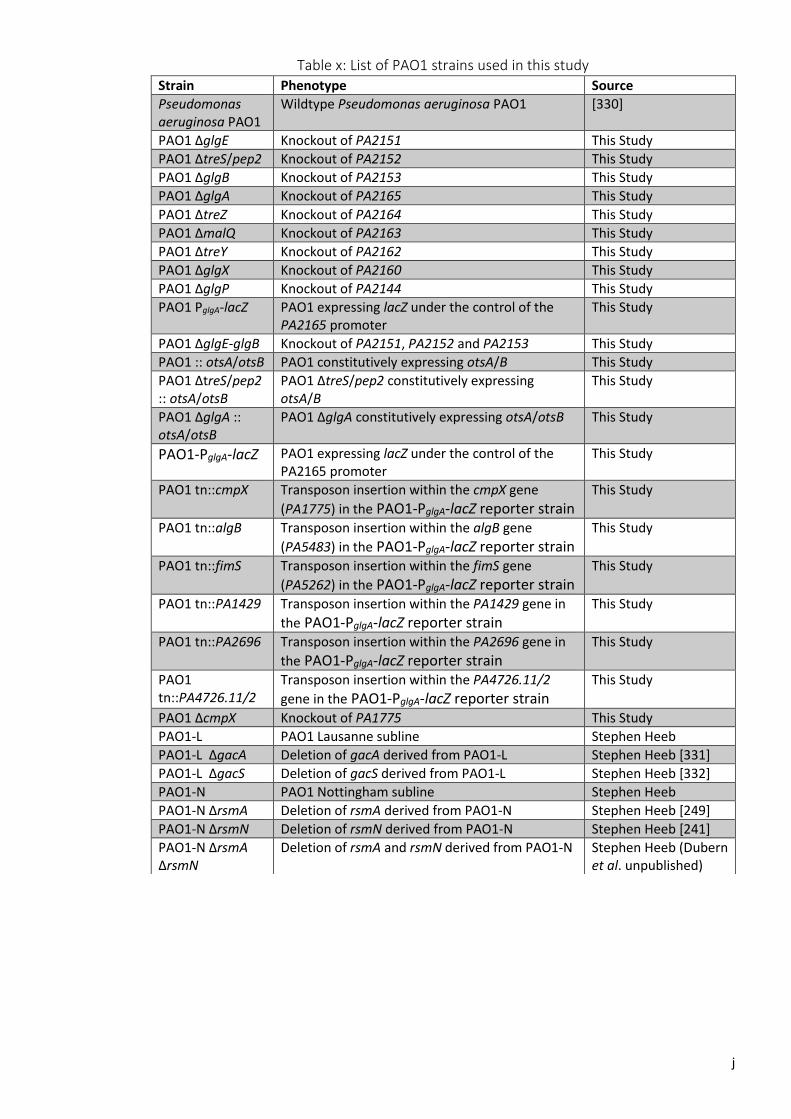
j
Table x: List of PAO1 strains used in this study
Strain Phenotype Source
Pseudomonas aeruginosa PAO1
Wildtype Pseudomonas aeruginosa PAO1 [330]
PAO1 ΔglgE Knockout of PA2151 This Study
PAO1 ΔtreS/pep2 Knockout of PA2152 This Study
PAO1 ΔglgB Knockout of PA2153 This Study
PAO1 ΔglgA Knockout of PA2165 This Study
PAO1 ΔtreZ Knockout of PA2164 This Study
PAO1 ΔmalQ Knockout of PA2163 This Study
PAO1 ΔtreY Knockout of PA2162 This Study
PAO1 ΔglgX Knockout of PA2160 This Study
PAO1 ΔglgP Knockout of PA2144 This Study
PAO1 PglgA-lacZ PAO1 expressing lacZ under the control of the PA2165 promoter
This Study
PAO1 ΔglgE-glgB Knockout of PA2151, PA2152 and PA2153 This Study
PAO1 :: otsA/otsB PAO1 constitutively expressing otsA/B This Study
PAO1 ΔtreS/pep2 :: otsA/otsB
PAO1 ΔtreS/pep2 constitutively expressing otsA/B
This Study
PAO1 ΔglgA :: otsA/otsB
PAO1 ΔglgA constitutively expressing otsA/otsB This Study
PAO1-PglgA-lacZ PAO1 expressing lacZ under the control of the PA2165 promoter
This Study
PAO1 tn::cmpX Transposon insertion within the cmpX gene
(PA1775) in the PAO1-PglgA-lacZ reporter strain
This Study
PAO1 tn::algB Transposon insertion within the algB gene
(PA5483) in the PAO1-PglgA-lacZ reporter strain
This Study
PAO1 tn::fimS Transposon insertion within the fimS gene
(PA5262) in the PAO1-PglgA-lacZ reporter strain
This Study
PAO1 tn::PA1429 Transposon insertion within the PA1429 gene in
the PAO1-PglgA-lacZ reporter strain
This Study
PAO1 tn::PA2696 Transposon insertion within the PA2696 gene in
the PAO1-PglgA-lacZ reporter strain
This Study
PAO1 tn::PA4726.11/2
Transposon insertion within the PA4726.11/2
gene in the PAO1-PglgA-lacZ reporter strain
This Study
PAO1 ΔcmpX Knockout of PA1775 This Study
PAO1-L PAO1 Lausanne subline Stephen Heeb
PAO1-L ΔgacA Deletion of gacA derived from PAO1-L Stephen Heeb [331]
PAO1-L ΔgacS Deletion of gacS derived from PAO1-L Stephen Heeb [332]
PAO1-N PAO1 Nottingham subline Stephen Heeb
PAO1-N ΔrsmA Deletion of rsmA derived from PAO1-N Stephen Heeb [249]
PAO1-N ΔrsmN Deletion of rsmN derived from PAO1-N Stephen Heeb [241]
PAO1-N ΔrsmA ΔrsmN
Deletion of rsmA and rsmN derived from PAO1-N Stephen Heeb (Dubern et al. unpublished)

k
Figure i: Heat map representing the statistical significance of desiccation responses between PAO1 strains based on linear mixed modelling. Green colouring represents statistical significance of desiccation responses between individual strains.

l
Figure ii: Heat map representing the statistical significance of desiccation responses between Pto strains based on linear mixed modelling. Green colouring represents statistical significance of desiccation responses between individual strains.
Copyright © 2022 FDOKUMEN