Elusive Recurrent Bacterial Contamination in a Diatom Culture
THE DIATOM GENUS PSEUDO-NITZSCHIA (BACILLARIOPHYCEAE) IN NEW SOUTHWALES, AUSTRALIA: MORPHOTAXONOMY, MOLECULAR PHYLOGENY,
TOXICITY, AND DISTRIBUTION1
Penelope Ajani2
Department of Biological Sciences, Macquarie University, North Ryde, New South Wales 2109, Australia
Sydney Institute of Marine Science, Chowder Bay Rd, Mosman, New South Wales 2088, Australia
Shauna Murray
Sydney Institute of Marine Science, Chowder Bay Rd, Mosman, New South Wales 2088, Australia
Plant Functional Biology and Climate Change Cluster (C3), University of Technology, PO Box 123, Broadway, Sydney,
New South Wales 2007, Australia
Gustaaf Hallegraeff
Institute for Marine and Antarctic Studies, University of Tasmania Private Bag 129, Hobart, Tasmania 7001, Australia
Nina Lundholm
Natural History Museum of Denmark, University of Copenhagen, Sølvgade 83S, Copenhagen 1307, Denmark
Michael Gillings
Department of Biological Sciences, Macquarie University, North Ryde, New South Wales 2109, Australia
Sydney Institute of Marine Science, Chowder Bay Rd, Mosman, New South Wales 2088, Australia
Steve Brett
Microalgal Services, 308 Tucker Road, Ormond, Victoria 3204, Australia
and Leanne Armand
Department of Biological Sciences, Macquarie University, North Ryde, New South Wales 2109, Australia
Sydney Institute of Marine Science, Chowder Bay Rd, Mosman, New South Wales 2088, Australia
Species belonging to the potentially harmfuldiatom genus Pseudo-nitzschia, isolated from 16localities (31 sampling events) in the coastal watersof south-eastern Australia, were examined. Clonalisolates were characterized by (i) light andtransmission electron microscopy; (ii) phylogenies,based on sequencing of nuclear-encoded ribosomaldeoxyribonucleic acid (rDNA) regions and, (iii)domoic acid (DA) production as measured by liquidchromatography–mass spectrometry (LC-MS/MS).Ten taxa were unequivocally confirmed as Pseudo-nitzschia americana, P. arenysensis, P. calliantha,P. cuspidata, P. fraudulenta, P. hasleana, P. micropora,P. multiseries, P. multistriata, and P. pungens. Anupdated taxonomic key for south-eastern AustralianPseudo-nitzschia is presented. The occurrence of twotoxigenic species, P. multistriata (maximumconcentration 11 pg DA per cell) and P. cuspidata(25.4 pg DA per cell), was documented for the firsttime in Australia. The Australian strains of
P. multiseries, a consistent producer of DA in strainsthroughout the world, were nontoxic. Data from5,888 water samples, collected from 31 oyster-growing estuaries (2,000 km coastline) from 2005 to2009, revealed 310 regulatory exceedances for“Total Pseudo-nitzschia,” resulting in six toxicepisodes. Further examination of high-risk estuariesrevealed that the “P. seriata group” had highest celldensities in the austral summer, autumn, or spring(species dependent), and lowest cell densities in theaustral winter, while the “P. delicatissima group” hadhighest in winter and spring.
Key index words: Amnesic Shellfish Poisoning;diatom; domoic acid; harmful algae; toxin
Abbreviations: ASP, amnesic shellfish poisoning;CTAB, cetyl trimethylammonium bromide; DA,domoic acid; DNA, deoxyribonucleic acid; ITS1/5.8S/ITS2, internal transcribed spacer; LC-MS/MS,liquid chromatography–mass spectrometry; LSU,large ribosomal subunit; ML, maximum likelihood;NCBI, National Centre for Biotechnology Informa-tion; NSWFA, New South Wales Food Authority;
1Received 7 March 2013. Accepted 19 May 2013.2Author for correspondence: e-mail: [email protected] Responsibility: R. Wetherbee (Associate Editor)
J. Phycol. 49, 765–785 (2013)© 2013 Phycological Society of AmericaDOI: 10.1111/jpy.12087
765

NSW, New South Wales; NSWSP, New South WalesShellfish Program; PCR, polymerase chain reaction;PSRF, potential scale reduction factor; rDNA,ribosomal deoxyribonucleic acid; TEM, transmissionelectron microscopy
Some of the most commonly occurring harmfulalgal bloom species in south-east Australian watersbelong to the diatom genus Pseudo-nitzschia (Bacil-lariophyceae; Ajani et al. 2012). The genus is foundworldwide in polar, temperate, subtropical, andtropical waters (Hasle 2002, Trainer et al. 2012) andcan be the causative organism for ASP, the toxicsyndrome that was implicated in human fatalitiescaused by the consumption of contaminated cul-tured mussels in eastern Canada in 1987 (Bateset al. 1989). DA, the potent neurotoxin causingASP, has since been implicated in contamination ofshellfish, fish, seabirds, and marine mammalsthroughout the world (reviewed in Lelong et al.2012).
The discrimination of species of Pseudo-nitzschia iscomplex as it is based on fine scale morphologicalcharacters visible only using detailed electronmicroscopy. Of the 37 species identified worldwide,14 have been confirmed toxigenic (Lelong et al.2012). In addition to complex morphometric data,the addition of molecular data and mating studieshas led to a recent increase in newly described oremended species (Trainer et al. 2012). A number ofspecies complexes are now reported with a highlevel of “cryptic” and “pseudo-cryptic” diversity(Lundholm et al. 2003, 2006, 2012, Orsini et al.2004, Amato et al. 2007, Amato and Montresor2008, Casteleyn et al. 2008, Churro et al. 2009,Quijano-Scheggia et al. 2009). Furthermore, mor-photypes that do not concur with any currentspecies descriptions continue to be detected, indi-cating the perplexing diversity within this genus(McDonald et al. 2007, Lundholm et al. 2012).
Pseudo-nitzschia is a dominant component of theAustralian phytoplankton community, reaching max-imum cell densities during the austral spring andsummer months in south-east coastal waters (Halle-graeff 1981, Ajani et al. 2001). Twelve species ofPseudo-nitzschia have been identified thus far in Aus-tralian coastal waters (Hallegraeff 1994, Lapworthet al. 2001, Jameson and Hallegraeff 2010); yet,despite its worldwide prevalence and significance insouth-east Australia, there are few studies on theecology, taxonomy, or toxicity of this genus in Aus-tralia. While no human illnesses have been reportedin Australian due to DA poisoning, no AustralianPseudo-nitzschia species or strains have been geneti-cally characterized (inter- or intra-specific) andthere has been no investigation into the linkbetween local Pseudo-nitzschia strains and DAproduction, except for a single confirmed toxigenicspecies, P. australis (Lapworth et al. 2001). Further-
more, regulatory action limits, which aim to ensurethe protection of shellfish consumers from thehazards of marine biotoxin poisoning by regularmonitoring of phytoplankton, are currently based on“Pseudo-nitzschia delicatissima group” (500,000cells � L�1), “P. multiseries and P. australis group”(50,000 cells � L�1) or “Total Pseudo-nitzschia”(500,000 cells � L�1). Each of these groups containstoxic and nontoxic representatives and without reso-lution of the genus to species level, it is most likelythat many closures of shellfish harvest areas areoccurring unnecessarily.The aim of the present study was to characterize
the diversity of the genus Pseudo-nitzschia in south-east waters of Australia using both morphologicaland molecular methods. Additionally, cultures ofPseudo-nitzschia were analyzed for DA production.
MATERIALS AND METHODS
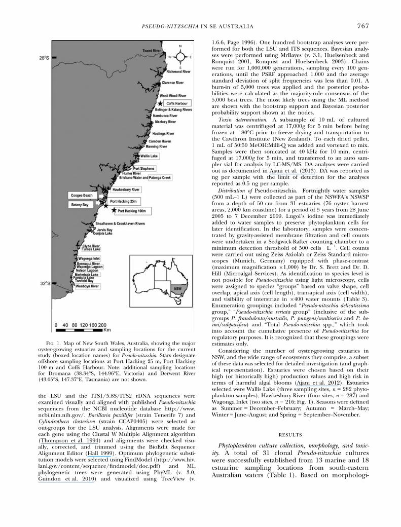
Phytoplankton collection, enumeration, and culture mainte-nance. Water samples were collected at 16 localities (31 sam-pling events) on the south-east coastline of Australia using aplankton net (20 lm mesh) hauled from a depth of 5–10 m.Sample collection points included both estuarine (E) and off-shore marine (M) waters, and major oyster-growing estuaries ofNew South Wales (Fig. 1; Table 1). Nonaxenic clonal cultureswere established by single cell isolation using micropipettesand maintained in f/2 media (Guillard 1975) at 16°C–18°Cunder a photon flux of 60–100 lmol photons � m�2 � s�1 on12:12 h dark:light cycle (white fluorescent bulbs). Cultureswere sub-sampled on day 12 (late stationary phase) for cellcounts using the Lund Cell technique (Hotzel and Croome1999), light and TEM examination, DNA sequencing based onthe large subunit (LSU) and ITS1/5.8S/ITS2 regions of theribosomal DNA and toxicity determination by LC-MS/MS forthe presence of DA.
Morphological examination. Clonal cultures were examinedto a maximum magnification of 9400 using a Leica DM5500B compound microscope (phase-contrast) equipped witha Leica DFC 450C camera (Wetzlar, Germany). Cultures werepreserved in Lugol’s iodide prior to cleaning using themethod of Hasle and Fryxell (1970) for TEM. Once cleaned,samples were then placed on formvar-coated copper gridsand loaded into a Philips CM10 TEM (Surrey, UK) equippedwith an Olympus SIS Megaview G2 digital camera (Tokyo,Japan). Micrographs were analyzed using Image J software(http://rsbweb.nih.gov/ij/), the morphometric variablesused by Lundholm et al. (2012) and the terminology anddiagnoses for the structure of diatoms by Anonymous (1975),von Stosch (1975), Ross et al. (1979) and Mann (1981).
Molecular characterization and phylogenetic analyses. A sub-sample of 10 mL of cultured material was concentrated bycentrifugation and frozen at �80°C prior to DNA extraction.DNA extraction followed the CTAB method (Doyle and Doyle1987) with modifications (Lundholm et al. 2002) and docu-mented in Ajani et al. (2013). The nuclear-encoded partialLSU and ITS1/5.8S/ITS2 rDNA regions were amplified usingthe readymade PCR mix Immomix™ (Bioline, London, UK).The primers used to amplify and sequence products are givenin Table 2. Amplicons were separated on 1% w/v agarose gelstained with Gel Red™ (Biotium, Hayward, CA, USA) andvisualized under UV light. PCR products were purified usinga SureClean purification kit (Bioline) according to the manu-facturer’s protocols and sequenced using the externalsequencing core service (Macrogen Inc., Seoul, Korea). Both
766 PENELOPE AJANI ET AL.
the LSU and the ITS1/5.8S/ITS2 rDNA sequences wereexamined visually and aligned with published Pseudo-nitzschiasequences from the NCBI nucleotide database http://www.ncbi.nlm.nih.gov/. Bacillaria paxillifer (strain Tenerife 7) andCylindrotheca closterium (strain CCAP0405) were selected asout-groups for the LSU analysis. Alignments were made foreach gene using the Clustal W Multiple Alignment algorithm(Thompson et al. 1994) and alignments were checked visu-ally, corrected, and trimmed using the BioEdit SequenceAlignment Editor (Hall 1999). Optimum phylogenetic substi-tution models were selected using FindModel (http://www.hiv.lanl.gov/content/sequence/findmodel/doc.pdf) and MLphylogenetic trees were generated using PhyML (v. 3.0,Guindon et al. 2010) and visualized using TreeView (v.
1.6.6, Page 1996). One hundred bootstrap analyses were per-formed for both the LSU and ITS sequences. Bayesian analy-ses were performed using MrBayes (v. 3.1, Huelsenbeck andRonquist 2001, Ronquist and Huelsenbeck 2003). Chainswere run for 1,000,000 generations, sampling every 100 gen-erations, until the PSRF approached 1.000 and the averagestandard deviation of split frequencies was less than 0.01. Aburn-in of 5,000 trees was applied and the posterior proba-bilities were calculated as the majority-rule consensus of the5,000 best trees. The most likely trees using the ML methodare shown with the bootstrap support and Bayesian posteriorprobability support shown at the nodes.
Toxin determination. A subsample of 10 mL of culturedmaterial was centrifuged at 17,000g for 5 min before beingfrozen at �80°C prior to freeze drying and transportation tothe Cawthron Institute (New Zealand). To each dried pellet,1 mL of 50:50 MeOH:Milli-Q was added and vortexed to mix.Samples were then sonicated at 40 kHz for 10 min, centri-fuged at 17,000g for 5 min, and transferred to an auto sam-pler vial for analysis by LC-MS/MS. DA analyses were carriedout as documented in Ajani et al. (2013). DA was reported asng per sample with the limit of detection for the analysesreported as 0.5 ng per sample.
Distribution of Pseudo-nitzschia. Fortnightly water samples(500 mL–1 L) were collected as part of the NSWFA’s NSWSPfrom a depth of 50 cm from 31 estuaries (76 oyster harvestareas, 2,000 km coastline) for a period of 5 years from 28 June2005 to 7 December 2009. Lugol’s iodine was immediatelyadded to water samples to preserve phytoplankton cells forlater identification. In the laboratory, samples were concen-trated by gravity-assisted membrane filtration and cell countswere undertaken in a Sedgwick-Rafter counting chamber to aminimum detection threshold of 500 cells � L�1. Cell countswere carried out using Zeiss Axiolab or Zeiss Standard micro-scopes (Munich, Germany) equipped with phase-contrast(maximum magnification 91,000) by Dr. S. Brett and Dr. D.Hill (Microalgal Services). As identification to species level isnot possible for Pseudo-nitzschia using light microscopy, cellswere assigned to species “groups” based on valve shape, celloverlap, apical axis (cell length), transapical axis (cell width),and visibility of interstriae in 9400 water mounts (Table 3).Enumeration groupings included “Pseudo-nitzschia delicatissimagroup,” “Pseudo-nitzschia seriata group” (inclusive of the sub-groups P. fraudulenta/australis, P. pungens/multiseries and P. he-imi/subpacifica) and “Total Pseudo-nitzschia spp.,” which tookinto account the cumulative presence of Pseudo-nitzschia forregulatory purposes. It is recognized that these groupings wereestimates only.
Considering the number of oyster-growing estuaries inNSW, and the wide range of ecosystems they comprise, a subsetof these data was selected for detailed investigation (and graph-ical representation). Estuaries were chosen based on theirhigh (or historically high) production values and high risk interms of harmful algal blooms (Ajani et al. 2012). Estuariesselected were Wallis Lake (three sampling sites, n = 282 phyto-plankton samples), Hawkesbury River (four sites, n = 287) andWagonga Inlet (two sites, n = 216; Fig. 1). Seasons were definedas Summer = December–February; Autumn = March–May;Winter = June–August; and Spring = September–November.
RESULTS
Phytoplankton culture collection, morphology, and toxic-ity. A total of 31 clonal Pseudo-nitzschia cultureswere successfully established from 13 marine and 18estuarine sampling locations from south-easternAustralian waters (Table 1). Based on morphologi-
FIG. 1. Map of New South Wales, Australia, showing the majoroyster-growing estuaries and sampling locations for the currentstudy (boxed location names) for Pseudo-nitzschia. Stars designateoffshore sampling locations at Port Hacking 25 m, Port Hacking100 m and Coffs Harbour. Note: additional sampling locationsfor Dromana (38.34°S, 144.96°E, Victoria) and Derwent River(43.05°S, 147.37°E, Tasmania) are not shown.
PSEUDO-NITZSCHIA IN SE AUSTRALIA 767

cal, molecular, and toxicological characterizations,a total of ten species were identified such asPseudo-nitzschia americana, P. arenysensis P. calliantha,P. cuspidata, P. fraudulenta, P. hasleana, P. micropora,P. multiseries, P. multistriata, and P. pungens. Molecu-lar sequencing of the nuclear-encoded rDNA wasobtained where possible and sequences have beendeposited in Genbank with accession numbers givenin Table 1. Detailed morphological and moleculardescriptions of P. micropora and P. hasleana, whichare reported for the first time from the SouthernHemisphere, are given in Ajani et al. (2013). Fur-thermore, molecular data revealed the first report
of P. arenysensis in these waters. Morphometrics ofall taxa are given in Table 4. References to previousAustralian records are included in species descrip-tions below, although earlier records by Dakin andColefax (1933, 1940) and Crosby and Wood (1959)are not included (see Discussion).Pseudo-nitzschia americana (Hasle) Fryxell (Fig. 2,
A–G; Table 4) Genbank accession nos KC017461and KC017472. Previous Australian records: Halle-graeff (1994) and Jameson and Hallegraeff (2010).Morphology: Cells occurred as single cells only (no
stepped chains observed), with a rectangular andsymmetrical valve shape (Fig. 2, A and B) and nocentral interspace (Fig. 2C). Cells had an apical axisranging between 27.9 and 41.3 lm and a transapicalaxis range of 2.3–3.5 lm (Fig. 2B) and contained18–30 fibulae per 10 lm and 26–35 interstriae per10 lm (Fig. 2C). Two (occasionally three) rows ofsmall hymenated poroid occlusions were observed,each with a hexagonally orientated array of perfora-tions (Fig. 2D). In the central valve area, poroidsnumbered between 7 and 9 per lm. The tip of thevalve appeared rounded in both girdle and valve
TABLE 1. List of Pseudo-nitzschia species, clone designation, collection location (estuarine (E) or marine (M)), date sam-pled, and accession numbers for LSU rDNA and ITS rDNA for clonal cultures established in the present study; ‘-’sequencenot obtained.
Species Clone designation Location sampled Date sampledGenbankstrain ID
LSU accessionNo.
Genbankstrain ID
ITS accessionNo.
P. americana PH25-191011 Port hacking 25 m,Sydney (M)
19/10/2011 – – – –
PH25-160412 Port hacking 25 m,Sydney (M)
16/04/2012 PH25 KC017461 PH25 KC017472
P. arenysensis CHA-280212 Coffs harbour, NSW (M) 28/02/2012 – – – –CHB-280212 Coffs harbour, NSW (M) 28/02/2012 CHB KC017456b CHB KC017466b
CHD-280212 Coffs harbour, NSW (M) 28/02/2012 – – – –P. calliantha WAL-230711 Wallis lake, NSW (E) 23/07/2011 – – – –
WONB-210911 Wonboyn lake, NSW (E) 21/09/2011 – – – –WAG-301011 Wagonga inlet, NSW (E) 30/10/2011 WAG KC017451 WAG KC017463TUR-071111 Tuross lake, NSW (E) 07/11/2011 – – – –TURA-071111 Tuross lake, NSW (E) 07/11/2011 – – – –TURB-071111 Tuross lake, NSW (E) 07/11/2011 TURB KC017452 TURB KC017464DER-220312 Derwent river, TASa (E) 22/03/2012 – – – –BB1-260612 Botany bay (M) 26/06/2012 – – – –
P. cuspidata WAG-250510 Wagonga inlet, NSW (E) 25/05/2010 – – – –PA7-190311 Patonga creek, NSW (E) 1903/2011 PA7 KC017453 – –MER-110911 Merimbula lake, NSW (E) 11/09/2011 MER KC017454 MER KC017465MER- 090112 Merimbula lake, NSW (E) 09/11/2012 – – – –PH25-120312 Port hacking 25 m, NSW (M) 12/03/2012 – – – –
P. hasleana HAWK3-090312 Hawkesbury river, NSW (E) 09/03/2012 HAWK3/1 KC017446 HAWK3/1 KC017450HAWK4-090312 Hawkesbury river, NSW (E) 09/03/2012 – – HAWK4 KC017468HAWK6-090312 Hawkesbury river, NSW (E) 09/03/2012 – – – –
P. fraudulenta MER1-110911 Merimbula lake, NSW (E) 11/09/2012 – – – –PH25-130212 Port hacking 25 m, NSW (M) 13/02/2012 PH25F KC017457 PH25F KC017467
P. micropora PS90-280311 Port Stephens, NSW (E) 28/03/2011 PS90 KC017445 PS90/1 KC017448P. multiseries COOG-161011 Coogee beach, NSW (M) 16/10/2011 COOG KC017458 COOG KC017469P. multistriata PH25B-191011 Port hacking 25 m, NSW (M) 19/10/2011 – – – –
PH25C-191011 Port hacking 25 m, NSW (M) 19/10/2011 PH25C KC017460 PH25C KC017470PH25D-191011 Port hacking 25 m, NSW (M) 19/10/2011 PH25D KC017459 – –WAPB-311011 Wapengo lake, NSW (E) 31/10/2011 – – – –
P. pungens DR-300311 Dromana, VICa (M) 30/03/2011 DR KC017462 DR KC017471WONB050811 Wonboyn lake, NSW (E) 05/08/2011 – – – –
aLocations not shown in Fig. 1.bLodged as P. delicatissima on Genbank.
TABLE 2. The primers used in this study to amplify LSUand ITS1/5.8S/ITS2 rDNA from the clonal cultures ofPseudo-nitzschia species.
Primer Sequence 5′-3′ Reference
DIR-F ACCCGCTGAATTTAAGCATA Scholin et al. 1994D3B-R TCGGAGGGAACAGCTACTA Nunn et al. 1996ITS-F ATATGCTTAAATTCAGCGGGT Murray et al. 2012ITS-R TTTCCGTAGCTGAACCTGCGG Murray et al. 2012
768 PENELOPE AJANI ET AL.
view (Fig. 2E, valve view only). The cingulum onboth the epitheca and hypotheca comprised twogirdle bands, with the valvocopula having 34–50striae per 10 lm. The valvocopula displayed a por-oid arrangement of two rows wide and one to tworows high per stria and the second copula had a2 9 1 poroid arrangement (Fig. 2F).
Culture Collection: Live material of PH25-160412was deposited at the Australian National AlgalCulture Collection (ANACC) as CS-1021.
Toxicity: No detectable toxin was determined forP. americana (two cultures tested; Table 5).
Pseudo-nitzschia arenysensis Quijano-Scheggia,Garc�es, Lundholm (Fig. 3, A–E; Table 4) Genbankaccession no’s KC017456 and KC017466. No previ-ous Australian records. May have been reportedas P. delicatissima in Lapworth et al. (2001) andJameson and Hallegraeff (2010).
Cell colonies were observed in stepped chains of nomore than nine cells in length. Cell overlap in chainswas ~1/8th–1/9th of the total cell length (Fig. 3A).Cells were symmetrical (sometimes slightly asymmet-rical) and lanceolate in valve shape, with a centralinterspace (Fig. 3, B and C). Cells had an apical axisof 33.6–45.8 lm and a transapical axis of 1.8–2.7 lm. Cells contained 20–26 fibulae per 10 lmand 38–45 interstriae per 10 lm (Fig. 3C). Tworows of round hymenated poroid occlusions wereobserved and were characterized in the central valvearea by 8–11 per lm (Fig. 3D). The tip of the cellfrustule appeared tapering and narrow in valve andgirdle view (Fig. 3E). Only one girdle band wasobserved, which comprised 40–46 striae per 10 lmand a poroid arrangement of two poroids wide andtwo or three poroids high (no image available).
Culture Collection: Live material of CHB-280212and CHD-280212 was deposited at the ANACC asCS-1025 and CS-1026, respectively. Live material ofthese strains was also deposited at CICCM asCAWB120.
Toxicity: No detectable toxin was determined forP. arenysensis (three cultures; Table 5).
Pseudo-nitzschia calliantha Lundholm, Moestrup etHasle (Fig. 4, A–G; Table 4) Genbank accessionno’s KC017451, KC017452, KC017463 and KC017464.Previous Australian records: Hallegraeff 1994; asP. pseudodelicatissima) and Jameson and Hallegraeff(2010).
Morphology: Cells formed stepped chains with upto 10 or more cells. Cell overlap was ~1/5th–1/9thof an individual cell’s length (Fig. 4A). The valveand girdle views showed symmetrically linear cells(Fig. 4B, valve view only). Apical and transapicalaxes measured 55.7–76.0 lm and 1.6–3.0 lm,respectively. A central interspace was present(Fig. 4C). There were 16–25 fibulae per 10 lm and34–45 interstriae per 10 lm. A single row of hyme-nated pore occlusions was observed, each stellate inarrangement and having four to nine sectors(Fig. 4D). A central poroid occlusion was some-times present. The number of poroids in the cen-tral valve area was 4–6 per lm. The tip of the cellfrustule appeared tapering and pointed both in valveand girdle view (Fig. 4E). Three girdle bands wereobserved with the valvocopula having 40–50 striaeper 10 lm and a poroid arrangement of two to threerows wide 9 four to five rows high per stria(Fig. 4F). The second copula had a poroid arrange-ment of two to three rows wide 9 three rows high,while the third band showed irregular smallporoids (Fig. 4F). All girdle band perforations werehexagonal.Culture Collection: Live material of DER-220312
and TURB-071111 were deposited at the ANACC asCS-1022 and CS-1023, respectively. Live material ofDER-220312 was deposited at CICCM as CAWB118.Toxicity: No detectable toxin was determined for
P. calliantha (six cultures tested; Table 5).Pseudo-nitzschia cuspidata (Hasle) Hasle (Fig. 5A–
G; Table 4) Genbank accession no’s KC017453,KC017454 and KC017465. Previous Australianrecords: Hallegraeff 1994; as P. pseudodelicatissima)and Jameson and Hallegraeff (2010).Cell colonies were observed in stepped chains
greater than eight cells in length with cell overlap~1/6th–1/8th of the total cell length (Fig. 5A). Thevalve and girdle views showed symmetrical and lan-ceolate cell shape with a central interspace present(Fig. 5, B and C). Cells had an apical axis of45–64.4 lm and transapical axis of 1.7–2.1 lm. Cellscontained 21–30 fibulae per 10 lm and 38–40 int-erstriae per 10 lm (Fig. 5C). One row of square tooval hymenated poroid occlusions, each generallybipartite but often with two to four sectors, wasobserved (Fig. 5D). In the central valve area, por-oids numbered 5–6 per lm. The tip of the cell
TABLE 3. Enumeration groupings for Pseudo-nitzschia and morphological characters used to distinguish each grouping using9400 light microscopy or water mounts as enumerated for New South Wales Food Authority (NSWFA)’s Shellfish Program(NSWSP). ‘-’ = morphological character not used to distinguish this grouping.
Pseudo-nitzschia grouping Valve shape Cell overlapApical
axis (lm)Transapicalaxis (lm)
Interstriae visible inwater mounts
P. delicatissima gp. Thin, linear 1/5–1/9 30–100 Up to 2.5–3 –P. pungens/multiseries gp. Wide, linear/lanceolate 1/4–1/3 50–140 2.5–6 YesP. fraudulenta/australis gp. Wide, spindle-shaped – 50–140 4–8 –P. heimii/subpacifica gp. Wide, symmetric one convex,
one straight margin– 30–120 4–7 –
PSEUDO-NITZSCHIA IN SE AUSTRALIA 769

TABLE4.
Speciesan
dmorphologicalinform
ation
pertaining
toclonal
culturesofPseudo-nitzschia
inthe
watersofsouth-eastAustralia
(note:n=number
of
specim
ensobserved
;dataaregivenas
minim
um
andmaxim
um
range
(above)an
dmean�
SD(below).
Species(n
)Valve
shap
eCen
tral
interspace
Cell
overlap
Apical
axis(lm)
Transapical
axis(lm)
Interstriae
per
10lm
Fibulae
per
10lm
Rowsof
Poroids
Poroids
per
1lm
Poroid
shap
e/sectors
Ban
dstriae
per
10lm
Valvoco
pulae
ban
dstriae
structure
b
P.am
erican
a(45)
Rectangu
lar,
symmetrical
No
nd
27.9–4
1.3
(32.9�
4.1)
2.3–
3.5
(3.0
�0.3)
26–3
5(29.7�
2.7)
18–3
0(20.3�
3.1)
2or3
7–9
(7.8
�0.6)
Round
34–5
0(43.5�
7.9)
29
1–2
P.arenysensis
(43)
Lan
ceolate,
symmetrical
Yes
1/8–
1/9
33.6–4
5.8
(39.8�
5.3)
1.8–
2.7
(2.1
�0.2)
38–4
5(40.4�
1.5)
20–2
6(22.8�
2.2)
28–
11(9.4
�0.9)
Round
40–4
6(42�
2.8)
29
2(3)
P.calliantha
(104
)Linear
Yes
1/5–
1/9
55.7–7
6.0
(67.4�
4.6)
1.5–1
.9(1.75�
0.1)
34–4
5(38.3�
2.1)
16–2
5(20.5�
1.9)
14–
6(4.7
�0.5)
Round,
stellate
40–5
0(46.3�
4.3)
2–39
4–5
P.cupsidata
(37)
Lan
ceolate,
symmetrical
Yes
1/6–
1/8
45–6
4.4
(54.6�
5.9)
1.7–
2.1
(1.9
�0.1)
38–4
0(39.5�
0.9)
21–3
0(24.9�
3.8)
15–
6(5.5
�0.5)
Round/
square,
bipartite
60(60�
0)19
1
P.ha
sleana
(41)
Lan
ceolate
Yes
1/5–
1/6
64.7–8
3.9
(70.3�
3.9)
2.2–
3.4
(2.6
�0.3)
34–4
0(36.2�
2.0)
15–2
5(18�
2.5)
15–
6(5.5
�0.5)
Irregu
lar
40–6
0(46.7�
8.3)
29
2–5
P.frau
dulenta
(49)
Lan
ceolate,
symmetrical
Yes
1/5–
1/7
54.7–9
4.1
(60.8�
8.7)
4.1–
6.1
(4.7
�0.5)
22–3
0(25.9�
2.3)
20–3
0(23.7�
2.5)
2–3
6–7
(6.1
�0.3)
Round,
stellate
34–5
0(40.7�
5.4)
2–39
3–11
P.micropora
(18)
Lan
ceolate,
symmetrical
No
1/9
33.1–3
6(34.9�
0.9)
1.8–
2.3
(2.0
�0.1)
42–5
0(45.9�
2.9)
23–3
0(27.1�
2.7)
29–
13(11�
1)Sm
all,
round
54–6
0(59.3�
2.1)
29
2
P.multiseries
(20)
Lan
ceolate,
symmetrical
No
1/3a
73–8
2.3
(78.5�
2.7)
3.5–
4.8
(3.8
�0.3)
13–1
5(13.7�
0.8)
13–1
5(13.7�
0.8)
3–4
4–7
(5.9
�1.1)
Round
25(25�
0)2–
39
2–3
P.multistriata
(40)
Lan
ceolate
symmetrical
No
1/6
26.8–6
3.7
(45.2�
11)
2.2–
3.9
(2.9
�0.3)
38–4
5(40.6�
1.9)
20–3
0(26.2�
2.9)
2or3
11–1
2(11.3�
0.5)
Round
55–6
0(56.7�
2.9)
29
2
P.pu
ngensvar.
pungens(33)
Linear-
lanceolate
No
1/3
68–1
11.5
(94.7�
16.1)1)
1.9–
3.7
(3.0
�0.6)
9–14
(11.6�
1.5)
9–14
(11.6�
1.5)
22–
4(2.7
�0.7)
Round
9–18
(14�
3.6)
19
1
aDatareported
inJamesonan
dHallegraeff(201
0).
bNo.poroidswide9
no.poroidshigh.
nd,nodata.
770 PENELOPE AJANI ET AL.

frustule appeared tapering and narrow in valve andgirdle view (Fig. 5E). Only one girdle band wasobserved, having 60 striae per 10 lm and a poroidarrangement of one large irregular, pentagonal-shaped poroid with hexagonal perforations (Fig. 5F).
Culture Collection: Live material of MER-090112was deposited at ANACC as CS-1024 and at CICCMas CAWB119.
Toxicity: Two of the five P. cuspidata culturestested positive to the presence of DA. Toxin concen-tration measured 24.5 pg DA per cell in cellsisolated from Lake Merimbula (strain MER- 090112)in January 2012, and 4.3 pg DA per cell in cells iso-lated offshore from Port Hacking (strain PH25-120312) in March 2012 (Table 5).
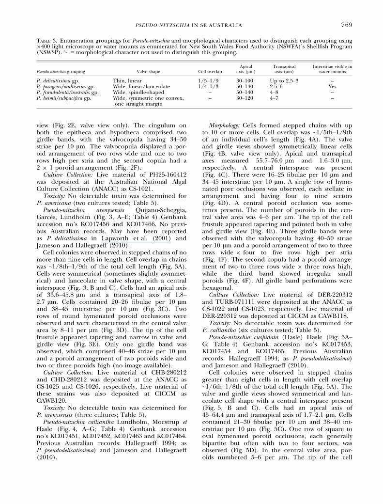
Pseudo-nitzschia fraudulenta (Cleve) Hasle (Fig. 6,A–G; Table 4) GenBank accession no’s KC017457and KC017467. Previous Australian records: Halle-graeff (1994), Lapworth et al. (2001) and Jamesonand Hallegraeff (2010).
Cells formed stepped chains up to 20 cells inlength, although most commonly seen in chains offour cells. Cell overlap was ~1/5th–1/7th of an indi-vidual cell’s length (Fig. 6A). The valve and girdleviews showed symmetrical, lanceolate cells (Fig. 6B)with apical and transapical axes measuring 54.7–94.1 lm and 4.1–6.1 lm, respectively. A centralinterspace was present (Fig. 6B). There were 22–30fibulae per 10 lm and 20–30 interstriae per 10 lm(Fig. 6C). Two to three rows of hymenated poroid
occlusions were observed, each divided into irregu-lar sectors (Fig. 6D). The number of poroids in thecentral valve area was 6–7 per lm. The tip of thecell frustule appeared tapering and pointed in bothvalve and girdle views (Fig. 6E). Only one girdleband was observed with 34–50 striae per 10 lm anda poroid arrangement of two to three rowswide 9 3–11 rows high per striae (Fig. 6F).Culture Collection: Live material of PH25-130212
was deposited at ANACC as CS-1027.Toxicity: No detectable toxin was determined for
P. fraudulenta (two cultures; Table 5).Pseudo-nitzschia hasleana Lundholm (Table 4) Gen-
bank accession no’s KC017446, KC017450 andKC017468. Previous Australian records: Ajani et al.(2013).A detailed morphological description of P. hasle-
ana isolated from the Hawkesbury River is given inAjani et al. (2013) (fig. 3A–H).Toxicity: No detectable toxin was determined for
P. hasleana (three cultures; Table 5).Pseudo-nitzschia micropora Priisholm, Moestrup et
Lundholm (Table 4) Genbank accession no.KC017445 and KC017448. Previous Australianrecords: Ajani et al. (2013).A detailed morphological description of P. micro-
pora isolated from Port Stephens is given in Ajaniet al. (2013; fig. 2A–H).Toxicity: No detectable toxin was determined for
P. micropora (one culture; Table 5).
FIG. 2. Pseudo-nitzschia americana. Culture material from strains PH25-191011 (A–E) and PH100-160412 (F). (A) Light microscopy of sin-gle cells (scale bar 20 lm); (B–F) transmission electron microscopy; (B) whole valve (5 lm); (C) mid-valve, no central nodule (2 lm);(D) mid-valve showing interstriae and two rows of poroids (0.5 lm); (E) valve end (1 lm); F) girdle bands: V= valvocopula and II=secondcopulae (0.5 lm); valvocopula showing poroid structure of two poroids wide and one or two poroids high.
PSEUDO-NITZSCHIA IN SE AUSTRALIA 771

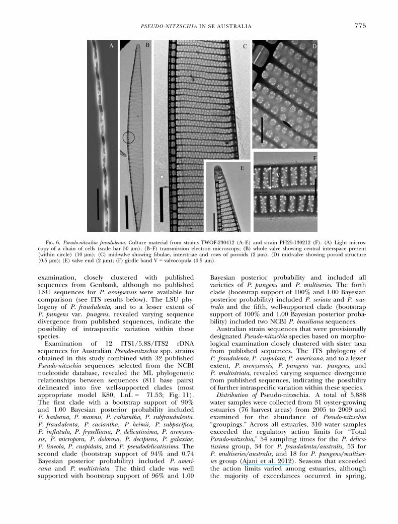
Pseudo-nitzschia multiseries (Hasle) Hasle (Fig. 7, A–H;Table 4) Genbank accession no. KC017458 andKC017469. Previous Australian records: Hallegraeff(1994), Lapworth et al. (2001) and Jameson andHallegraeff (2010).
Cells formed stepped chains up to 20 cells inlength. Unfortunately, cell overlap was notmeasured, but has been observed in cells fromAustralian waters as ~1/3 cell’s length (Jameson andHallegraeff 2010). The valve and girdle viewsshowed symmetrical lanceolate cells with no centralinterspace (Fig. 7, A and B). The apical and trans-apical axes measured 73–82.3 lm and 3.4–4.8 lm,respectively. There were both 13–15 fibulae and int-erstriae per 10 lm (Fig. 7C). Three to four rows ofsmall hymenated poroid occlusions were observedin the central valve area (Fig. 7C) with the numberof poroids measured as 4–7 per lm. The tip of thecell frustule appeared tapering and pointed in bothvalve and girdle views (Fig. 7D). Two girdle bandswere observed with the valvocopula having ~25striae per 10 lm and a poroid arrangement of twoto three rows wide 9 two to three rows high perstria (Fig. 7, E and F). The second copula had aporoid arrangement of two rows wide 9 one rowshigh (Fig. 7E). All girdle band perforations werehexagonal.
Toxicity: No detectable toxin was determined forP. multiseries (two cultures tested; Table 5).Pseudo-nitzschia multistriata (Takano) Takano
(Fig. 8, A–G; Table 4) Genbank accession no’sKC017460, KC017459 and KC017470. PreviousAustralian records: Jameson and Hallegraeff (2010).Sigmoidal shaped cells formed stepped chains,
with up to four in length and an overlap of ~1/6thof the total cell length (Fig. 8A). Cells were symmet-rical and broad lanceolate in valve view with theabsence of a central interspace (Fig. 8, B and C).Cells had an apical axis of 26.8–63.7 lm and trans-apical axis of 2.2–3.9 lm. Cells contained 20–30 fib-ulae per 10 lm and 38–45 interstriae per 10 lmand two, sometimes three, rows of small hymenatedpore occlusions, each with a hexagonally orientatedarray of perforations (Fig. 8, C and D). In the cen-tral valve area, poroids numbered 11–12 per lm.The tip of the cell frustule appeared tapering andnarrow in both valve and girdle views (Fig. 8E). Thecingulum on both the epitheca and hypotheca com-prised three girdle bands with the valvocopula hav-ing 55–60 striae per 10 lm and a poroidarrangement of two rows wide and two rows highper stria (Fig. 8, F and G). The second copula hada poroid arrangement of one to two rows wide andone to two rows high of small, singular pores
TABLE 5. List of species, clone designation, and DA determination for examined Pseudo-nitzschia strains; detection limit0.5 ng per sample.
Species Clone designation Cells examined for DA concentration DA ng per sample Pg DA per cell
P. americana PH25A-191011 2.69 9 106 ND –PH25-160412 4.86 9 106 ND –
P. arenysensis CHA-280212 1.07 9 107 ND –CHB-280212 2.77 9 106 ND –CHD-280212 – nt –
P. calliantha WAL-230711 2.95 9 106 ND –WONB-210911 1.66 9 106 ND –WAG-301011 3.96 9 106 ND –TUR-071111 – nt –TURA-071111 2.79 9 106 ND –TURB-071111 4.07 9 106 ND –DER-220312 5.07 9 105 ND –
P. cuspidata WAG-250510 – nt –PA7-190311 1.95 9 106 ND –MER-110911 – nt –MER- 090112 3.19 9 106 1.3 24.5PH25-120312 3.89 9 105 0.9 4.3
P. hasleana HAWK3-090312 2.12 9 106 ND –HAWK4-090312 1.47 9 106 ND –HAWK6-090312 2.42 9 106 ND –
P. fraudulenta MER1-110911 – nt –PH25-130212 1.18 9 106 ND –
P. micropora PS90-280311 1.8 9 106 ND –P. multiseries COOG-161011 2.42 9 105 ND –
COOG-161011 2.42 9 105 ND –P. multistriata PH25B-191011 2.01 9 106 1.8 11
PH25C-191011 9.46 9 105 5.7 1.7PH25D-191011 1.17 9 106 16.6 <1WAPB-311011 1.24 9 106 1.9 6.5
P. pungens DR-300311 3.37 9 105 ND –WONB050811 1.18 9 105 ND –
nt, not tested; ND, not detected; DA, domoic acid.
772 PENELOPE AJANI ET AL.

(Fig. 8F). Pores were not visible on the third girdleband (Fig. 8F).
Culture Collection: Live material of PH25C-191011was deposited at ANACC as CS-1028.
Toxicity: All strains of P. multistriata tested for DAwere positive, with toxin concentration ranging from<1 to 11 DA per cell. Pseudo-nitzschia multistriatastrains (PH25B-191011, PH25C-191011, PH25D-191011) were isolated from Port Hacking in October2011 and Wapengo Lake (strain WAPB-311011) alsoin October 2011 (Table 5).
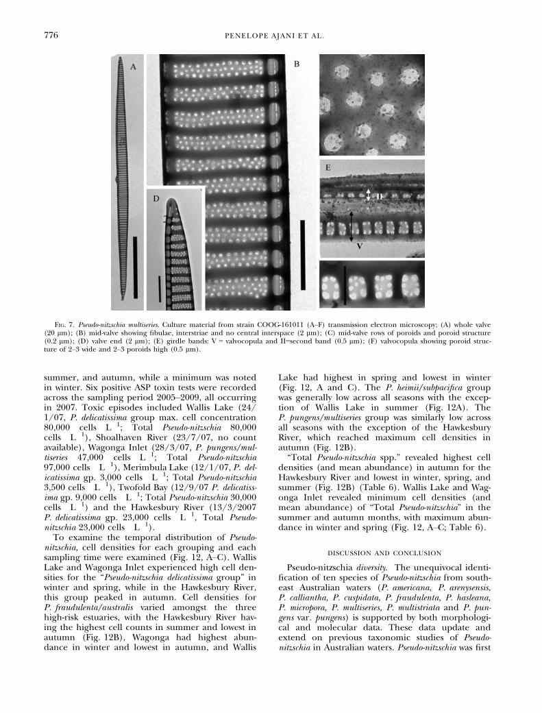
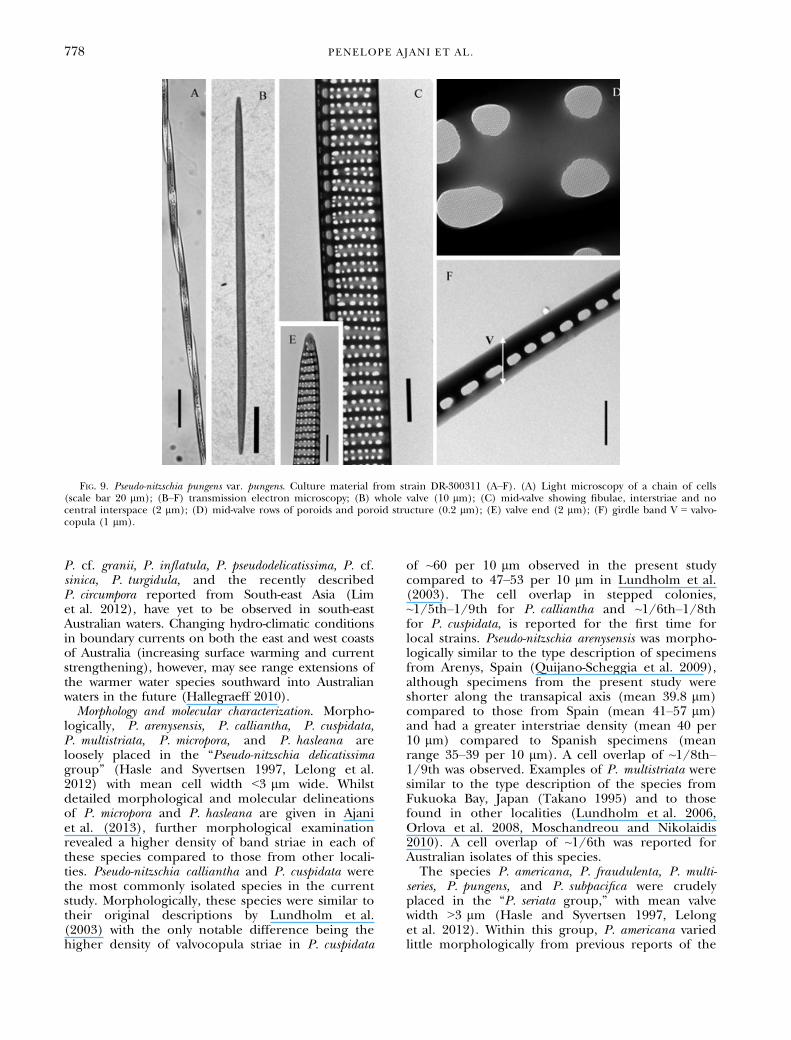
Pseudo-nitzschia pungens var. pungens (Grunowex Cleve) Hasle (Fig. 9, A–H; Table 4) Genbankaccession no. KC017462 and KC 017471. PreviousAustralian records: Hallegraeff (1994), Lapworthet al. (2001) and Jameson and Hallegraeff (2010).
Cells formed stepped chains up to fifty cells inlength with a cell overlap of ~1/3rd of the total celllength (Fig. 9A). Cells were strongly silicified with alinear to lanceolate valve shape and the absence ofa central interspace (Fig. 9, B and C). Cells had anapical axis ranging between 68 and 111.5 lm andtransapical axis between 1.9 and 3.7 lm. Cells con-tained 9–14 fibulae per 10 lm, 9–14 interstriae per10 lm, and two (sometimes three) rows of small
hymenated poroid occlusions. The latter was charac-terized by hexagonally orientated perforations(Fig. 9D). In the central valve area, poroids num-bered 2–4 per lm. The tip of the cell frustuleappeared tapering and narrow in both valve andgirdle views (Fig. 9E). Only one girdle band wasobserved with 9–18 striae per 10 lm and onelongitudinal row of poroids (Fig. 9F).Toxicity: No detectable toxin was determined for
P. pungens (two cultures tested; Table 5).Molecular characterization and phylogenetic analyses.
Phylogenetic analyses of 40 sequences (763 basepairs) of the LSU rDNA included 13 new Australiansequences of Pseudo-nitzschia obtained in this studycombined with 25 published Pseudo-nitzschiasequences and two out-groups. Based on the closestmodel available (GTR, LnL = �2243.8), the MLphylogenetic tree delineated into two highly sup-ported clades (bootstrap support 100% and 1.00Bayesian posterior probability; Fig. 10). The firstclade comprised P. multiseries and all varieties ofP. pungens for which published sequences were avail-able. Clade II comprised all other species. Australianstrain sequences that were provisionally designatedPseudo-nitzschia species based on morphological
FIG. 3. Pseudo-nitzschia arenysensis (A) light microscopy of a chain of cells (scale bar 10 lm); (B–E) transmission electron microscopy;(B) whole valve (2 lm); (C) mid-valve, showing central interspace, fibulae, interstriae, and rows of poroids (2 lm); (D) poroid structure(0.2 lm); (E) valve end (1 lm).
PSEUDO-NITZSCHIA IN SE AUSTRALIA 773

FIG. 4. Pseudo-nitzschia calliantha. Culture material from strains TURA-071111 (A–E) and WAG-301011 (F). (A) Light microscopy of achain of cells (scale bar 20 lm); (B–F) transmission electron microscopy; (B) whole valve (5 lm); (C) mid-valve showing fibulae, interst-riae and central interspace (1 lm); (D) mid-valve showing rows of poroids and poroid structure (0.2 lm); (E) valve end (2 lm); (F) girdlebands: V = valvocopula showing poroid structure of 2–3 rows wide 9 3 rows high poroids high, II = second and III = third bands (0.5 lm).
FIG. 5. Pseudo-nitzschia cuspidata. Culture material from strain WAG-250510 (A–F). (A) Light microscopy of a chain of cells (scale bar20 lm); (B–F) transmission electron microscopy; (B) whole valve (10 lm); (C) mid-valve showing fibulae, interstriae, and central inter-space (2 lm); (D) mid-valve showing rows of poroids and poroid structure (0.1 lm); (E) valve end (1 lm); (F) V = valvocopula girdleband showing poroid structure of 1 row wide 9 1 row high (0.2 lm).
774 PENELOPE AJANI ET AL.
examination, closely clustered with publishedsequences from Genbank, although no publishedLSU sequences for P. arenysensis were available forcomparison (see ITS results below). The LSU phy-logeny of P. fraudulenta, and to a lesser extent ofP. pungens var. pungens, revealed varying sequencedivergence from published sequences, indicate thepossibility of intraspecific variation within thesespecies.
Examination of 12 ITS1/5.8S/ITS2 rDNAsequences for Australian Pseudo-nitzschia spp. strainsobtained in this study combined with 32 publishedPseudo-nitzschia sequences selected from the NCBInucleotide database, revealed the ML phylogeneticrelationships between sequences (811 base pairs)delineated into five well-supported clades (mostappropriate model K80, LnL = �71.53; Fig. 11).The first clade with a bootstrap support of 90%and 1.00 Bayesian posterior probability includedP. hasleana, P. mannii, P. calliantha, P. subfraudulenta.P. fraudulenta, P. caciantha, P. heimii, P. subpacifica,P. inflatula, P. fryxelliana, P. delicatissima, P. arenysen-sis, P. micropora, P. dolorosa, P. decipiens, P. galaxiae,P. lineola, P. cuspidata, and P. pseudodelicatissima. Thesecond clade (bootstrap support of 94% and 0.74Bayesian posterior probability) included P. ameri-cana and P. multistriata. The third clade was wellsupported with bootstrap support of 96% and 1.00
Bayesian posterior probability and included allvarieties of P. pungens and P. multiseries. The forthclade (bootstrap support of 100% and 1.00 Bayesianposterior probability) included P. seriata and P. aus-tralis and the fifth, well-supported clade (bootstrapsupport of 100% and 1.00 Bayesian posterior proba-bility) included two NCBI P. brasiliana sequences.Australian strain sequences that were provisionally
designated Pseudo-nitzschia species based on morpho-logical examination closely clustered with sister taxafrom published sequences. The ITS phylogeny ofP. fraudulenta, P. cuspidata, P. americana, and to a lesserextent, P. arenysensis, P. pungens var. pungens, andP. multistriata, revealed varying sequence divergencefrom published sequences, indicating the possibilityof further intraspecific variation within these species.Distribution of Pseudo-nitzschia. A total of 5,888
water samples were collected from 31 oyster-growingestuaries (76 harvest areas) from 2005 to 2009 andexamined for the abundance of Pseudo-nitzschia“groupings.” Across all estuaries, 310 water samplesexceeded the regulatory action limits for “TotalPseudo-nitzschia,” 54 sampling times for the P. delica-tissima group, 34 for P. fraudulenta/australis, 53 forP. multiseries/australis, and 18 for P. pungens/multiser-ies group (Ajani et al. 2012). Seasons that exceededthe action limits varied among estuaries, althoughthe majority of exceedances occurred in spring,
FIG. 6. Pseudo-nitzschia fraudulenta. Culture material from strains TWOF-230412 (A–E) and strain PH25-130212 (F). (A) Light micros-copy of a chain of cells (scale bar 50 lm); (B–F) transmission electron microscopy; (B) whole valve showing central interspace present(within circle) (10 lm); (C) mid-valve showing fibulae, interstriae and rows of poroids (2 lm); (D) mid-valve showing poroid structure(0.5 lm); (E) valve end (2 lm); (F) girdle band V = valvocopula (0.5 lm).
PSEUDO-NITZSCHIA IN SE AUSTRALIA 775
summer, and autumn, while a minimum was notedin winter. Six positive ASP toxin tests were recordedacross the sampling period 2005–2009, all occurringin 2007. Toxic episodes included Wallis Lake (24/1/07, P. delicatissima group max. cell concentration80,000 cells � L�1; Total Pseudo-nitzschia 80,000cells � L�1), Shoalhaven River (23/7/07, no countavailable), Wagonga Inlet (28/3/07, P. pungens/mul-tiseries 47,000 cells � L�1; Total Pseudo-nitzschia97,000 cells � L�1), Merimbula Lake (12/1/07, P. del-icatissima gp. 3,000 cells � L�1; Total Pseudo-nitzschia3,500 cells � L�1), Twofold Bay (12/9/07 P. delicatiss-ima gp. 9,000 cells � L�1; Total Pseudo-nitzschia 30,000cells � L�1) and the Hawkesbury River (13/3/2007P. delicatissima gp. 23,000 cells � L�1, Total Pseudo-nitzschia 23,000 cells � L�1).
To examine the temporal distribution of Pseudo-nitzschia, cell densities for each grouping and eachsampling time were examined (Fig. 12, A–C). WallisLake and Wagonga Inlet experienced high cell den-sities for the “Pseudo-nitzschia delicatissima group” inwinter and spring, while in the Hawkesbury River,this group peaked in autumn. Cell densities forP. fraudulenta/australis varied amongst the threehigh-risk estuaries, with the Hawkesbury River hav-ing the highest cell counts in summer and lowest inautumn (Fig. 12B), Wagonga had highest abun-dance in winter and lowest in autumn, and Wallis
Lake had highest in spring and lowest in winter(Fig. 12, A and C). The P. heimii/subpacifica groupwas generally low across all seasons with the excep-tion of Wallis Lake in summer (Fig. 12A). TheP. pungens/multiseries group was similarly low acrossall seasons with the exception of the HawkesburyRiver, which reached maximum cell densities inautumn (Fig. 12B).“Total Pseudo-nitzschia spp.” revealed highest cell
densities (and mean abundance) in autumn for theHawkesbury River and lowest in winter, spring, andsummer (Fig. 12B) (Table 6). Wallis Lake and Wag-onga Inlet revealed minimum cell densities (andmean abundance) of “Total Pseudo-nitzschia” in thesummer and autumn months, with maximum abun-dance in winter and spring (Fig. 12, A–C; Table 6).
DISCUSSION AND CONCLUSION
Pseudo-nitzschia diversity. The unequivocal identi-fication of ten species of Pseudo-nitzschia from south-east Australian waters (P. americana, P. arenysensis,P. calliantha, P. cuspidata, P. fraudulenta, P. hasleana,P. micropora, P. multiseries, P. multistriata and P. pun-gens var. pungens) is supported by both morphologi-cal and molecular data. These data update andextend on previous taxonomic studies of Pseudo-nitzschia in Australian waters. Pseudo-nitzschia was first
FIG. 7. Pseudo-nitzschia multiseries. Culture material from strain COOG-161011 (A–F) transmission electron microscopy; (A) whole valve(20 lm); (B) mid-valve showing fibulae, interstriae and no central interspace (2 lm); (C) mid-valve rows of poroids and poroid structure(0.2 lm); (D) valve end (2 lm); (E) girdle bands: V = valvocopula and II=second band (0.5 lm); (F) valvocopula showing poroid struc-ture of 2–3 wide and 2–3 poroids high (0.5 lm).
776 PENELOPE AJANI ET AL.

observed by Dakin and Colefax (1933, 1940) andCrosby and Wood (1959; fig. 18) as a singlecosmopolitan taxon, Nitzschia seriata Cleve. The firstextensive taxonomic survey of the genus Pseudo-nitzschia in Australian coastal waters in 1984 conse-quently identified eight different species fromequatorial, tropical, subtropical, and temperatewaters (Hallegraeff 1994). Species identified wereP. americana, P. fraudulenta, P. lineola, P. multiseries,P. pseudodelicatissima, P. pungens, P. subpacifica, andP. turgidula. Since this time, using both morpho-metric and phylogenetic analyses, P. pseudodelicatiss-ima has been separated into eight species:P. pseudodelicatissima, P. cuspidata, P. calliantha, andP. caciantha (Lundholm et al. 2003), P. calliantha2(Amato et al. 2007), P. mannii (Amato and Mont-resor 2008) and both P. hasleana and P. fryxelliana(Lundholm et al. 2012). As such, the taxonomicdescriptions and figures in Hallegraeff (1994, fig.6, a–l) almost certainly included more than onespecies. Furthermore, variation in the cell widthand length and the density of fibulae and striae inP. turgidula as described by Hallegraeff (1994) com-pared with the type description from by Hasle(1965) now indicates that these strains were mostlikely P. dolorosa (Lundholm et al. 2006).
Extending from Hallegraeff’s survey in 1994, Lap-worth et al. (2001) examined limited water samples
collected from Tasmania, South Australia, WesternAustralia, and Queensland for the presence ofPseudo-nitzschia. A total of 10 species were identifiedwith P. australis and heimii being new recordings forAustralian waters. Cultures established of P. pseudo-delicatissima in their study (Lapworth et al. 2001, fig.2c) have now been re-identified as P. calliantha andthe recording of P. subfraudulenta is uncertain. Fur-thermore, Lundholm and Moestrup (2002) identi-fied a new species of Pseudo-nitzschia isolated fromSydney coastal waters as P. galaxiae in 2002, whereasJameson and Hallegraeff (2010) also recorded thepresence of P. delicatissima (possibly P. arenysensis)and P. multistriata, making a total of 14 speciesrecorded from Australian coastal waters thus far.The present study reports two further species,P. hasleana and P. micropora, recorded for the firsttime in the Southern Hemisphere (Ajani et al.2013). Furthermore, while P. subpacifica wasobserved in the 1981 survey and observed inenvironmental samples in the current study (no cul-ture was established), this taxon remains poorlydefined and warrants further investigation in east-ern Australia.Other species that have been identified in geo-
graphically nearby localities, including P. cf. heimiiand P. turgidula from New Zealand coastal waters(L. Rhodes, personal communication) and P. caciantha,
FIG. 8. Pseudo-nitzschia multistriata Culture material from strain PH25B-191011 (A–G). (A) Light microscopy of an individual cell (scalebar 25 lm); (B–G) transmission electron microscopy; (B) whole valve (10 lm); (C) mid-valve, no central interspace (2 lm); (D) mid-valveshowing fibulae, interstriae, and rows of poroids (1 lm); (E) valve end (2 lm); (F) girdle bands: V = valvocopula, II = second andIII = third bands (1 lm); (G) valvocopula showing a poroid structure of two poroids high and two poroids wide (0.02 lm).
PSEUDO-NITZSCHIA IN SE AUSTRALIA 777
P. cf. granii, P. inflatula, P. pseudodelicatissima, P. cf.sinica, P. turgidula, and the recently describedP. circumpora reported from South-east Asia (Limet al. 2012), have yet to be observed in south-eastAustralian waters. Changing hydro-climatic conditionsin boundary currents on both the east and west coastsof Australia (increasing surface warming and currentstrengthening), however, may see range extensions ofthe warmer water species southward into Australianwaters in the future (Hallegraeff 2010).Morphology and molecular characterization. Morpho-
logically, P. arenysensis, P. calliantha, P. cuspidata,P. multistriata, P. micropora, and P. hasleana areloosely placed in the “Pseudo-nitzschia delicatissimagroup” (Hasle and Syvertsen 1997, Lelong et al.2012) with mean cell width <3 lm wide. Whilstdetailed morphological and molecular delineationsof P. micropora and P. hasleana are given in Ajaniet al. (2013), further morphological examinationrevealed a higher density of band striae in each ofthese species compared to those from other locali-ties. Pseudo-nitzschia calliantha and P. cuspidata werethe most commonly isolated species in the currentstudy. Morphologically, these species were similar totheir original descriptions by Lundholm et al.(2003) with the only notable difference being thehigher density of valvocopula striae in P. cuspidata
of ~60 per 10 lm observed in the present studycompared to 47–53 per 10 lm in Lundholm et al.(2003). The cell overlap in stepped colonies,~1/5th–1/9th for P. calliantha and ~1/6th–1/8thfor P. cuspidata, is reported for the first time forlocal strains. Pseudo-nitzschia arenysensis was morpho-logically similar to the type description of specimensfrom Arenys, Spain (Quijano-Scheggia et al. 2009),although specimens from the present study wereshorter along the transapical axis (mean 39.8 lm)compared to those from Spain (mean 41–57 lm)and had a greater interstriae density (mean 40 per10 lm) compared to Spanish specimens (meanrange 35–39 per 10 lm). A cell overlap of ~1/8th–1/9th was observed. Examples of P. multistriata weresimilar to the type description of the species fromFukuoka Bay, Japan (Takano 1995) and to thosefound in other localities (Lundholm et al. 2006,Orlova et al. 2008, Moschandreou and Nikolaidis2010). A cell overlap of ~1/6th was reported forAustralian isolates of this species.The species P. americana, P. fraudulenta, P. multi-
series, P. pungens, and P. subpacifica were crudelyplaced in the “P. seriata group,” with mean valvewidth >3 lm (Hasle and Syvertsen 1997, Lelonget al. 2012). Within this group, P. americana variedlittle morphologically from previous reports of the
FIG. 9. Pseudo-nitzschia pungens var. pungens. Culture material from strain DR-300311 (A–F). (A) Light microscopy of a chain of cells(scale bar 20 lm); (B–F) transmission electron microscopy; (B) whole valve (10 lm); (C) mid-valve showing fibulae, interstriae and nocentral interspace (2 lm); (D) mid-valve rows of poroids and poroid structure (0.2 lm); (E) valve end (2 lm); (F) girdle band V = valvo-copula (1 lm).
778 PENELOPE AJANI ET AL.

FIG. 10. Pseudo-nitzschia phylogenetic tree based on the LSU region of the nuclear-encoded rRDNA (763 bp) and rooted with out-groups Bacillaria paxillifer and Cylindrotheca closterium. The tree is constructed with maximum-likelihood (ML) bootstrap values (9100 repli-cates). Numbers on the internal nodes represent ML bootstrap values and Bayesian Posterior probability values (>50%) in that order.Species, strain, and Genbank accession numbers are given for each taxon. Sequences highlighted in bold are those obtained in thepresent study.
PSEUDO-NITZSCHIA IN SE AUSTRALIA 779

FIG. 11. Pseudo-nitzschia unrooted phylogenetic tree based on the ITS region of the nuclear-encoded rRDNA (811 bp). The tree is con-structed with maximum-likelihood (ML) bootstrap values (9100 replicates). Numbers on the internal nodes represent ML bootstrap valuesand Bayesian Posterior probability values (>50%) in that order. Species, strain, and Genbank accession numbers are given for each taxon.Sequences highlighted in bold are those obtained in the present study.
780 PENELOPE AJANI ET AL.

species from Australian waters (Hallegraeff 1994,Jameson and Hallegraeff 2010), other localities(Lundholm et al. 2002), or from the type descrip-tion first documented from Atlantida, Uruguay(Hasle 1965). Lundholm et al. (2002), however,reported a slightly dissimilar girdle band structurecompared to that observed in this current study.Lundholm et al. (2002) reported that the valvocop-ula (first girdle band closest to the mantle) had a2 9 3 poroid structure, the second band had a1–2 9 1–2 poroid structure, and the third band hadno striae present. Pseudo-nitzschia americana valvesexamined in the present study showed two girdlebands, the first with a 2 9 1–2 poroid structure andthe second with a 2 9 1 poroid arrangement(Fig. 2F). Such variation could be due to a lessdeveloped valvocopula in our study, or we haveassessed the band structure toward the end of thevalvocopula rather than the middle of the cell. Con-sidering the band structure was not “intact” whenexamined in our study, the position of the poroidarrangement in relation to the middle or end of thegirdle bands could not be assessed.
Pseudo-nitzschia fraudulenta was diagnosticallycomparable to specimens described from south-east Australia (Hallegraeff 1994, Jameson andHallegraeff 2010), to those from other localities(Moschandreou and Nikolaidis 2010, Moschandreouet al. 2012), and closely resembled the type descrip-tion from Plymouth Harbour, North Sea by Cleve
(1897), and the more specific description byHasle (1965). Pseudo-nitzschia multiseries, similarly,resembled those strains previously described fromAustralian waters (Hallegraeff 1994, Jameson andHallegraeff 2010), other localities (Orlova et al.2008, Moschandreou and Nikolaidis 2010), and thetype validation from Oslofjord, Norway by Hasle in1974 (previously Nitzschia pungens Cleve 1897 andN. pungens f. multiseries Hasle 1965), although noholotypes were designated for these earlier descrip-tions). Pseudo-nitzschia pungens observed in the cur-rent study most closely resembled P. pungens var.pungens in its valve width, number of striae, fibulae,poroids, and most distinctively, the band striae(9–18 in 10 lm) and single, unsplit poroid arrange-ment (Casteleyn et al. 2008, Churro et al. 2009).Pseudo-nitzschia pungens var. cingulata, in contrast, isobserved to have a valvocopula poroid arrangementof 2 9 2–3, whilst P. pungens var. aveirensis has onerow of split poroids (molecular discussion below).Thus far, there have been no Australian molecular
sequence data available for Pseudo-nitzschia strains.Molecular data of both LSU and ITS1/5.8S/ITS2from the current study support the genetic affinity ofAustralian strains to the same taxa from other locali-ties (Lundholm et al. 2002, 2003, 2006, 2012, Amatoet al. 2007, Churro et al. 2009, Lim et al. 2012, Mosc-handreou et al. 2012). Furthermore, due to the largeextent of both cryptic and pseudo-cryptic diversity inthis genus, the molecular sequence data obtained
FIG. 12. Monthly log abundance (cells L-1; y-axis) of Pseudo-nitzschia groupings enumerated each sampling time (x-axis) as part of theNew South Wales Food Authority (NSWFA)’s Shellfish Program (NSWSP). A subset of high-production, high-risk estuaries is presented:(A) Wallis Lake, (B)Hawkesbury River; and (C) Wagonga Inlet. Note: Summer = months 12,1,2; Autumn = 3,4,5; Winter = 6,7,8 (shaded);and Spring = 9,10,11.
PSEUDO-NITZSCHIA IN SE AUSTRALIA 781

provided further validation on the biogeography ofthese species. Whilst species confirmation was theprimary goal of this study, both inter- and intraspe-cific diversities was observed in our data, warrantingfurther investigation into this genus in Australianwaters. Specifically, the ITS phylogeny of P. fraudul-enta, P. cuspidata, and P. americana, with varyingsequence divergence, indicated that geographic vari-ation may exist. Furthermore, the P. pungens strainexamined in this study was most similar to P. pungensvar. pungens in its morphology, yet revealed geneticvariability in its LSU and ITS sequence data, suggest-ing that it may be a further variety. Additional exami-nation into the molecular, mating, and morphologyof this species complex is warranted as further cryp-tic diversity within this group is likely.Given the large advances made morphologically
and molecularly to delineate the species of Pseudo-nitzschia along the south-east coast of Australia, wehere update the key of Lim et al. (2012) to clarifyand identify the species found in the region. Speciesnames highlighted in bold are those that aredescribed in the current study; others species arethose that have previously been reported fromsouth-east Australian coastal waters (Hallegraeff1994, Lapworth et al. 2001, Jameson and Hallegraeff2010, Ajani et al. (2013). Morphological features arevisible using light microscopy unless specified“TEM.”
1A. Mean transapical axis (<3 lm).....…………………………21B. Mean transapical axis (>3 lm)......…………………………31C. Mean transapical axis (>6 lm).....………………P. australis2A. Central Interspace (CIS) present, valve lanceolate/
symmetrical..…………………………………………………42B. CIS present, valve asymmetrical..………………P. dolorosa2C. CIS present, valve linear..…………………………………52D. CIS absent..…………………………………………………73A. CIS present, valve lanceolate/symmetrical...P. fraudulenta3B. CIS present, valve asymmetrical...…………………………83B. CIS absent .…………………………………………………..94A. Striae with one row of poroids (TEM).…………..………64B. Striae with two rows of poroids
(TEM)……………………..……P. delicatissima/arenysensis*4C. Striae with no discrete poroids (TEM)… .……P. galaxiae5A. Striae with one row of poroids perforated with 7-10
sectors (TEM)….....……………………………P. calliantha5B. Striae with one or two rows of circular poroids
(TEM).….………………………………………….P. lineola6A. Poroids oval/square, usually bipartite (TEM).P. cuspidata6B. Poroids irregular, many sectors (TEM)….....…P. hasleana7A. Broad valve in the middle…….....…………P. multistriata7B. Valve tapered rapidly from the middle.....…P. micropora8A. Broad round apices….....…………………………P. heimii8B. Rapidly tapering apices.....…………………P. subpacifica9A. Valve rectangular/symmetrical.....……………P. americana9B. Valve linear/lanceolate.....………………………………10
10A. Two rows of poroids......………………………P. pungens10B. Three to four rows of poroids (TEM)......….P. multiseries
*P. arenysensis delineation from P. delicatissimabased on molecular characterization of the ITS1,5.8S, and ITS2 of nuclear rDNA.Toxicity. The current study has identified the
toxigenic nature of two species, P. multistriata andTABLE6.
High-riskestuariesofNew
South
Wales:hydrology
characteristics,estuarymodificationlevel,nutrientload
,water
quality,
andseasonal
abundan
ceof‘Total
Pseudo-nitzschia’(200
5–2
009).
Estuary
Mean
annual
rainfall
(mm)a
Dilution
factora
Flushing
(days)
a
Estuary
modification
levelb
Total
SSindex
aTotalP
index
aTotalN
index
a
Turbidity
(NTU)b
min-m
ax
Tem
p(°C)b
min-m
axSal(p
pt)
b
min-m
ax
Summer
total
Pseudo-nitzschia
mean�
SE
Autumntotal
Pseudo-nitzschia
mean�
SE
Wintertotal
Pseudo-nitzschia
mean�
SE
Springtotal
Pseudo-nitzschia
mean�
SE
Wallislake
1139
7.0
76.0
Modified
MH
M0.3–
1600
7–32
0.3–
3825
,637
�6,18
410
,846
�4,01
422
8,57
2�
75,049
297,14
8�
79,430
Haw
kesbury
river
764
6.37
49.2
Exten
sively
modified
HM
M0.03
–808
4.4–
370.11
–34.15
972�
275
6,36
2�
2,54
476
9�
361
788�
435
Wagonga
inlet
817
16.48
35.3
Modified
LL
Lna
na
na
38,523
�14
,951
10,384
�2,29
492
,053
�22
,925
159,58
1�
37,241
aRoper
etal.(201
1).
bOzC
oasts,20
10(accessed25
/2/
2013
).SS
,suspen
ded
solids;
P,Phosphorus;
N,Nitroge
n;S,
Summer
(Decem
ber–F
ebruary);A,Autumn
(March
–May);
W,Winter(June–
Augu
st);
SP,Sp
ring
(Sep
tember–
November).
782 PENELOPE AJANI ET AL.

P. cuspidata. Whilst both these species have beenreported as toxic elsewhere (Rhodes et al. 2000, Billet al. 2005), DA production in Australian strains isreported for the first time. Pseudo-nitzschia cuspida-ta was responsible for a significant toxic episodeand closure of Sydney rock oyster (Saccostrea glom-erata) harvest areas in Wagonga Inlet in 2010(Fig. 1). A maximum concentration of 6,050,000cells � L�1 in water samples and 34 mg DA � kg�1
in oyster tissue were reported during this time.Harvest closures extended for 16 weeks, which wasthe longest closure ever experienced in south-eastAustralia, and resulted in significant financial lossto the AUS$35 million per year (A. Zammit, per-sonal communication). The implementation of theNSW Food Authority’s Marine Biotoxin Manage-ment Plan (2011), established in 2003 to ensurethe protection of shellfish consumers from thehazards of marine biotoxins such as DA, was suc-cessfully used to close oyster harvests in responseto this event. Curiously, while P. cuspidata was con-firmed toxic for two cultured isolates in our study(MER- 090112 and PH25-120312), another strain(PA7190311) was revealed to be nontoxic. Whilstthe age of the culture, stage of growth, light andnutrient sources are important variables in theproduction of DA (see review Lelong et al. 2012,Terseleer et al. 2013, Auro and Cochlan 2013), astudy by Thessen et al. (2009) found that most ofthe variation in toxin content within species wasattributed to strain differences. Further examina-tion into the intra- and interspecies differences ingrowth and toxicity of Pseudo-nitzschia in south-eastern Australian waters will help identify andquantify the variability of DA production amongstlocal strains.
Significant levels of DA using the enzyme-linkedimmunosorbent assay have previously confirmed thetoxigenic nature of P. australis in Australian waters,whilst isolates of P. pungens and P. fraudulentaproved nontoxic with this method (Lapworth et al.2001). Interestingly, five cultures of P. pseudodelica-tissima (now confirmed as P. calliantha) were alsotested by these authors, with only one producingdetectable levels of DA (1 ng � mL�1).
Given the culturing conditions used in this study,the nontoxic characterization of P. multiseries in thecurrent study is also a novel outcome. This specieshas been found to be a consistent producer of DA inall strains tested throughout the world (Lelong et al.2012) and, to date, our isolates from south-easternAustralia are the first nontoxic strains reported inthe world. Other species which did not produce DAin the current study, but which are toxic elsewhere,included P. calliantha, P. fraudulenta, and P. pungens(Lelong et al. 2012). Strains of P. hasleana, onlytested once before the present study (Lundholmet al. 2012), were also confirmed to be nontoxic.Lundholm and Moestrup (2002) also reported that
P. galaxiae from Sydney coastal waters did not pro-duce any detectable concentrations of DA.Distribution of Pseudo-nitzschia. Bloom dynamics
of Pseudo-nitzschia are complex and triggers caninclude seasonal drivers (irradiance, temperature,wind), regional drivers (rainfall, nutrients fromupwelling, mixing, or riverine inputs), or a combi-nation of both (Trainer et al. 2012). While the sea-sonality of Pseudo-nitzschia in Northern Hemispherecoastal waters has been demonstrated (“P. seriatagroup” most closely related to temperature and“P. delicatissima group” to salinity and ammoniumconcentration; Fehling et al. 2006), Pseudo-nitzschiablooms in other regions are stimulated by multifac-eted mechanisms that are region specific (Traineret al. 2012). Although environmental data were notmeasured concurrently with the phytoplankton col-lected in this study, water quality ranges are avail-able for New South Wales estuaries, allowing us tospeculate on the links between driving variables andbloom dynamics (Table 6). Examination of harmfulphytoplankton taxa in relation to recognized estuarydisturbance measures has revealed species abun-dance correlates to estuary modification levels andflushing time, with modified, slow flushing estuarieshaving higher abundance (Ajani et al. 2012). Boththe Hawkesbury River and Wallis Lake have reducedwater quality, low flushing and high nutrient reten-tion times, which may favor the proliferation of tol-erant harmful algal species such as Pseudo-nitzschia(Roy et al. 2001, Figueiras et al. 2006, Table 6).Conversely, Wagonga Inlet is a relatively unmodifiedestuary, although high sedimentation rates, sewagedischarge and entrance channel shoaling, mayencourage the excess growth of microalgae in thisembayment (Ajani et al. 2012, Table 6).Whilst the abundance of the “P. seriata group”
appears to be temperature-dependent (lowest abun-dance in winter), the “P. delicatissima group” may betriggered by more complex interactions betweenlight availability and water temperature (both increas-ing in the austral spring). Similarly, Alexandriumcatenella (toxic dinoflagellate) is observed to bloomin the Hawkesbury River most commonly from Sep-tember to November, when salinity ranges between21.9 and 35.4 ppt, surface water temperature is>16°C, and daily global solar exposure rises in excess15 MJ � m�2 (equiv. 400 lmol photons � m�2 � s�1
PAR; Farrell et al. 2013). However, the interannualpattern of “Total Pseudo-nitzschia” as discussed byAjani et al. (2012; fig. 5C) is highly variable acrossyears, indicating that blooms of Pseudo-nitzschia areinfluenced by multiple factors and mechanisms.Regulatory exceedances also occurred across allyears, yet only in 2007 did high cell densities result intoxic episodes. The etiology of DA production duringthis particular year is unknown; nonetheless, it high-lights that the disparity between cell abundance andbiotoxic events requires further investigation.
PSEUDO-NITZSCHIA IN SE AUSTRALIA 783

In conclusion, we have characterized the diversityof the potentially harmful diatom genus Pseudo-nitzschia from south-eastern Australia coastal waters.Ten species were unequivocally confirmed, with twotoxigenic species identified. Pseudo-nitzschia is asignificant, yet highly variable, component of thephytoplankton community in south-east Australianwaters with both food production and humanhealth importance. Future studies should includemating, growth, and ecological sampling of the mosthighly abundant, toxic representatives, for example,Pseudo-nitzschia cuspidata, to begin to understand thefactors that regulate DA production and bloomdynamics of this genus. Equipped with the detailedknowledge of local Australian strains (their underly-ing taxonomy, molecular signature and potential forproducing DA), a rapid identification method, suchas species-specific molecular probes, may be devel-oped for toxic forms of Pseudo-nitzschia in south-eastAustralian waters.
The authors thank Tim Ingleton (OEH), Anthony Zammit,Grant Webster, and Melanie Field (NSW Food Authority),Dale and Lyn Witchard, Gary, Jo-Anne and Sam Rodely, Johnand Chris Ritchie, Dave Maidment, Matt Burgoyne, StaceyLoftus, Caroline Henry, Bill Foxwell, Anthony Sciacca,Richard Ellery, Stirling Cullenward, Greg Woodford, GraemeCampbell, Bob Simpson, Tony Troup, and Tony Bell (NSWOyster farmers), and John Cipolla (beach-goer) for samplecollection. We also thank Hazel Farrell (SIMS) and LindaArmbrecht (MQU) for cell isolations (Hawkesbury River andCoffs Harbour strains), and Rouna Yauwenas (UNSW) andDr Ulysee Bove (SIMS) for laboratory assistance. Thanks toNicole Vella and Debra Birch (MQU) for electron microscopytraining, and Dr Lesley Rhodes and staff at Cawthron Insti-tute (NZ). Dr Kimon Moschandreou (University of Thessalo-niki) and Hong Chang Lim (University Malaysia Sarawak) arealso acknowledged for invaluable discussions regardingPseudo-nitzschia. This work was supported by an AustralianPostgraduate Award, the Australian Government’s AustralianBiological Resource Study (CT210-19) and the Linnean Soci-ety of New South Wales. This is contribution number 94 fromthe Sydney Institute of Marine Science.
Ajani, P., Brett, S., Krogh, M., Scanes, P., Webster, G. & Armand,L. 2012. The risk of harmful algal blooms (HABs) in theoyster-growing estuaries of New South Wales, Australia. Env.Mon. Assess. 185:5295–5316.
Ajani, P., Lee, R., Pritchard, T. & Krogh, M. 2001. Phytoplanktondynamics at a long-term coastal station off Sydney, Australia.J. Coastal Res. 34:60–73.
Ajani, P., Murray, S., Hallegraeff, G., Brett, S. & Armand, L. 2013.First reports of Pseudo-nitzschia micropora and P. hasleana (Bacil-lariaceae) from the Southern Hemisphere: morphological,molecular and toxicological characterisation. Phycol. Res.doi:10.1111/pre.12020.
Amato, A., Kooistra, W., Ghiron, J. H. L., Mann, D. G., Proschold,T. & Montresor, M. 2007. Reproductive isolation among sym-patric cryptic species in marine diatoms. Protist 158:193–207.
Amato, A. & Montresor, M. 2008. Morphology, phylogeny, andsexual cycle of Pseudo-nitzschia mannii sp. nov. (Bacillariophy-ceae): a pseudo-cryptic species within the P-pseudodelicatissimacomplex. Phycologia 47:487–497.
Anonymous. 1975. Proposals for a standardisation of diatomterminology and diagnoses. Nova Hedwigia 53:323–354.
Auro, M. E. & Cochlan, W. P. 2013. Nitrogen utilization andtoxin production by two diatoms of the Pseudo-nitzschiapseudodelicatissima complex: P. cuspidata and P. fryxelliana.J. Phycol. 49:156–169.
Bates, S. S., Bird, C. J., de Freitas, A. S. W., Foxall, R., Gilgan, M.,Hanic, L. A., Johnson, G. R. et al. 1989. Pennate diatomNitzschia pungens as the primary source of domoic acid, atoxin in shellfish from eastern Prince Edward Island,Canada. Can. J. Fish. Aq. Sci. 46:1203–1215.
Bill, B., Lundholm, N., Connell, L., Baugh, K. A. & Trainer, V. L.2005. Domoic acid in Pseudo-nitzschia cuspidata from Washing-ton State coastal waters. Proceedings of the 3rd Symposiumon Harmful Algae in the U.S., Alismor, CA. 77 pp. (Abstract)
Casteleyn, G., Chepurnov, V. A., Leliaert, F., Mann, D. G., Bates,S. S., Lundholm, N., Rhodes, L., Sabbe, K. & Vyverman, W.2008. Pseudo-nitzschia pungens (Bacillariophyceae): a cosmo-politan diatom species? Harmful Algae 7:241–257.
Churro, C. I., Carreira, C. C., Rodrigues, F. J., Craveiro, S. C.,Calado, A. J., Casteleyn, G. & Lundholm, N. 2009. Diversityand abundance of potentially toxic Pseudo-nitzschia Peragalloin Aveiro Coastal Lagoon, Portugal and description of a newvariety, P. pungens var. aveirensis var. nov. Diat. Res. 24:35–62.
Cleve, P. T. 1897. A treatise on the phytoplankton of the Atlantic and itstributaries and on the periodical changes of the plankton of Skagerak.Nya Tidnings Akriebolags Tryckeri, Upsala, Sweden, 27 pp.
Crosby, L. H. & Wood, E. J. F. 1959. Studies on Australian andNew Zealand Diatoms II.—Normally Epontic and BenthicGenera. T. Roy. Soc. New Zeal. J. Mar. Freshw. Res. 86:1–58.
Dakin, W. J. & Colefax, A. 1933. The marine plankton of thecoastal waters of New South Wales. 1. The chief planktonicforms and their seasonal distribution. Proc. Lin. Soc. NSW58:186–222.
Dakin, W. J. & Colefax, A. 1940. The plankton of the Australiancoastal waters of New South Wales. Part 1. Univ. Sydney Dept.Zool. Monogr. 1:303–314.
Doyle, J. J. & Doyle, J. L. 1987. A rapid DNA isolation procedurefor small quantities of fresh leaf tissue. Phytochem. Bull.19:11–15.
Farrell, H., Brett, S., Ajani, P. & Murray, S. (2013). Distributionof the genus Alexandrium (Halim) along the coastline of NewSouth Wales, Australia. Mar. Poll. Bull. doi:10.1016/j.marpol-bul.2013.04.009.
Fehling, J., Davidson, K., Bolch, C. & Tett, P. 2006. Seasonality ofPseudo-nitzschia spp. (Bacillariophyceae) in western Scottishwaters. Mar. Ecol. Prog. Ser. 323:91–105.
Figueiras, F. G., Pitcher, G. C. & Estrada, M. 2006. Harmful algalbloom dynamics in relation to physical processes. In Gran�eli,E. & Turner, J. T. [Eds.] Ecology of Harmful Algae. Springer,New York, USA, pp. 127–138.
Guillard, R. R. L. 1975. Culture of phytoplankton for feedingmarine invertebrates. In Smith, W. L. & Chanley, M. H.[Eds.] Culture of Marine Invertebrate Animals. Plenum Press,New York, USA, pp. 26–60.
Guindon, S., Dufayard, J. F., Lefort, V., Anisimova, M., Hordijk,W. & Gascuel, O. 2010. New algorithms and methods toestimate maximum-likelihood phylogenies: assessing theperformance of PhyML 3.0. Sys. Biol. 59:307–321.
Hall, T. A. 1999. BioEdit: a user-friendly biological sequencealignment editor and analysis program for Windows 95/98/NT. Nucl. Acids Symp. Ser. 41:95–98.
Hallegraeff, G. M. 1981. Seasonal study of phytoplankton pig-ments and species at a coastal station off Sydney: importanceof diatoms and the nanoplankton. Mar. Biol. 61:107–118.
Hallegraeff, G. M. 1994. Species of the diatom genus Pseudo-nitzschia in Australian waters. Bot. Mar. 37:397–412.
Hallegraeff, G. M. 2010. Ocean climate change, phytoplanktoncommunity responses, and harmful algal blooms: a formida-ble predictive challenge. J. Phycol. 46:220–235.
Hasle, G. R. 1965. Nitzschia and Fragilariopsis species studied inthe light and electron microscopes. II. The group Pseudo-nitzschia. Skrifter utgitt av Det Norske Vidensk-Akademi I. Mat. -Nat Klasse, Ny Series 18:1–45.
784 PENELOPE AJANI ET AL.

Hasle, G. R. 1974. Validation of the names of some marine plank-tonic species of Nitzschia (Bacillariophyceae). Taxon 23:425–428.
Hasle, G. R. 2002. Are most of the domoic acid-producing speciesof the diatom genus Pseudo-nitzschia cosmopolites? HarmfulAlgae 1:137–146.
Hasle, G. R. & Fryxell, G. A. 1970. Diatoms: cleaning and mount-ing for light and electron microscopy. T. Am. Microsc. Soc.89:469–474.
Hasle, G. R. & Syvertsen, E. E. 1997. Marine diatoms. In Thomas,C. R. [Ed.] Identifying Marine Phytoplankton. Academic Press,San Diego, USA, pp. 5–385.
Hotzel, G. & Croome, R. 1999. A Phytoplankton Methods Manual forAustralian Rivers. Land and Water Resources Research andDevelopment Corporation, Canberra, Australia, pp 1–66.
Huelsenbeck, J. P. & Ronquist, F. 2001. MRBAYES: Bayesian infer-ence of phylogenetic trees. Bioinformatics 17:754–755.
Jameson, I. & Hallegraeff, G. M. 2010. Planktonic diatoms. InHallegraeff, G. M., Bolch, C. J. S., Hill, D. R. A., Jameson, I.,LeRoi, J. M., McMinn, A., Murray, S., de Salas, M. F. & Saun-ders, K. [Eds.] Algae of Australia: Phytoplankton of TemperateCoastal Waters. ABRS, Canberra & CSIRO Publishing,Melbourne, Australia, pp. 16–82.
Lapworth, C., Hallegraeff, G. & Ajani, P. 2001. Identification ofdomoic-acid-producing Pseudo-nitzschia species in Australianwaters. In Intergovernmental Oceanographic Commission ofUNESCO Proceedings of the Ninth International Conference onHarmful Algal Blooms. Paris, pp. 38–41.
Lelong, A., Hegaret, H., Soudant, P. & Bates, S. S. 2012. Pseudo-nitzschia (Bacillariophyceae) species, domoic acid and amne-sic shellfish poisoning: revisiting previous paradigms. Phycolo-gia 51:168–216.
Lim, H. C., Leaw, C. P., Su, S. N. P., Teng, S. T., Usup, G.,Mohammad-Noor, N., Lundholm, N., Kotaki, Y. & Lim, P. T.2012. Morphology and molecular characterization ofPseudo-nitzschia (Bacillariophyceae) from Malaysian Borneo,including the new species Pseudo-nitzschia circumpora sp. nov.J. Phycol. 48:1232–1247.
Lundholm, N., Bates, S. S., Baugh, K. A., Bill, B. D., Connell, L. B.,Leger, C. & Trainer, V. L. 2012. Cryptic and pseudo-crypticdiversity in diatoms - with descriptions of Pseudo-nitzschia ha-sleana sp. nov. and P. fryxelliana sp. nov. J. Phycol. 48:436–454.
Lundholm, N., Hasle, G. R., Fryxell, G. A. & Hargraves, P. E.2002. Morphology, phylogeny and taxonomy of specieswithin the Pseudo-nitzschia americana complex (Bacillariophy-ceae) with descriptions of two new species, Pseudo-nitzschiabrasiliana and Pseudo-nitzschia linea. Phycologia 41:480–497.
Lundholm, N. & Moestrup, O. 2002. The marine diatom Pseudo-nitzschia galaxiae sp. nov. (Bacillariophyceae): morphologyand phylogenetic relationships. Phycologia 41:594–605.
Lundholm, N., Moestrup, O., Hasle, G. R. & Hoef-Emden, K.2003. A study of the Pseudo-nitzschia pseudodelicatissima/cuspi-data complex (Bacillariophyceae): what is P-pseudodelicatiss-ima? J. Phycol. 39:797–813.
Lundholm, N., Moestrup, O., Kotaki, Y., Hoef-Emden, K.,Scholin, C. & Miller, P. 2006. Inter- and intraspecific varia-tion of the Pseudo-nitzschia delicatissima complex (Bacillario-phyceae) illustrated by rRNA probes, morphological dataand phylogenetic analyses. J. Phycol. 42:464–481.
Mann, D. G. 1981. Sieves and flaps: siliceous minutiae in the poresof raphid diatoms. In Ross, R. [Ed.] Proceedings of the 6th Sym-posium on Recent and Fossil Diatom Symposium. Koeltz ScientificBooks, Konigstein, Budapest, Hungary, pp. 279–300.
McDonald, S. M., Sarno, D. & Zingone, A. 2007. IdentifyingPseudo-nitzschia species in natural samples using genus-specificPCR primers and clone libraries. Harmful Algae 6:849–860.
Moschandreou, K. K., Baxevanis, A. D., Katikou, P., Papaefthimi-ou, D., Nikolaidis, G. & Abatzopoulos, T. J. 2012. Inter- andintra-specific diversity of Pseudo-nitzschia (Bacillariophyceae)in the northeastern Mediterranean. Eur. J. Phycol. 47:321–339.
Moschandreou, K. K. & Nikolaidis, G. 2010. The genus Pseudo-nitzschia (Bacillariophyceae) in Greek coastal waters. Bot.Mar. 53:159–172.
Murray, S. A., Garby, T., Hoppenrath, M. & Neilan, B. A. 2012.Genetic diversity, morphological uniformity and polyketideproduction in dinoflagellates (Amphidinium, Dinoflagellata).PLoS ONE 7:e38253.
NSW Marine Biotoxin Management Plan 2011. NSW ShellfishProgram NSW/FA/FI115/1105. NSW Food Authority, Sydney,pp 1–36.
Nunn, G., Theisen, B., Christensen, B. & Arctander, P. 1996. Sim-plicity-correlated size growth of the nuclear 28S ribosomalRNA D3 expansion segment in the crustacean order isopoda.J. Mol. Evol. 42:211–223.
Orlova, T. Y., Stonik, I. V., Aizdaicher, N. A., Bates, S. S., Leger,C. & Fehling, J. 2008. Toxicity, morphology and distributionof Pseudo-nitzschia calliantha, P. multistriata and P. multiseries(Bacillariophyta) from the northwestern Sea of Japan. Bot.Mar. 51:297–306.
Orsini, L., Procaccini, G., Sarno, D. & Montresor, M. 2004. Multi-ple rDNA ITS-types within the diatom Pseudo-nitzschia delica-tissima (Bacillariophyceae) and their relative abundancesacross a spring bloom in the Gulf of Naples. Mar. Ecol. Prog.Ser. 271:87–98.
Page, R. D. M. 1996. TREEVIEW: an application to display phylo-genetic trees on personal computers. Comput. Appl. Biosci.12:357–358.
Quijano-Scheggia, S. I., Garces, E., Lundholm, N., Moestrup, O.,Andree, K. & Campi, J. 2009. Morphology, physiology, molec-ular phylogeny and sexual compatibility of the cryptic Pseudo-nitzschia delicatissima complex (Bacillariophyta), including thedescription of P. arenysensis sp. nov. Phycologia 48:492–509.
Rhodes, L. L., Adamson, J. & Scholin, C. 2000. Pseudo-nitzschiamultistriata (Bacillariophyceae) in New Zealand. New Zeal.J. Mar. Fresh. 34:463–467.
Ronquist, F. & Huelsenbeck, J. P. 2003. MrBayes 3: Bayesian phy-logenetic inference under mixed models. Bioinformatics19:1572–1574.
Roper, T., Creese, B., Scanes, P., Stephens, K., Williams, R., Dela-Cruz, J., Coade, G. & Coates, B. 2011. Assessing the conditionof estuaries and coastal lake ecosystems in NSW, Monitoring,evaluation and reporting program, Technical report series.Office of Environment and Heritage, Sydney, pp 1–239.
Ross, R., Cox, E. J., Karayeva, N. L., Mann, D. G., Paddock, T. B.G., Simonsen, R. & Sims, P. A. 1979. An amended terminol-ogy for the siliceous components of the diatom cell. BiehefteZur. Nova Hedwigia 64:513–533.
Roy, P. S., Williams, R. J., Jones, A. R., Yassini, I., Gibbs, P. J.,Coates, B., West, R. J., Scanes, P. R., Hudson, J. P. & Nichol,S. 2001. Structure and function of south-east Australian estu-aries. Estuar. Coast. Shelf S. 53:351–384.
Scholin, C. A., Herzog, M., Sogin, M. & Anderson, D. M. 1994.Identification of group-specific and strain-specific geneticmarkers for globally distributed Alexandrium (Dinophyceae).2. Sequence analysis of a fragment of the LSU ribosomalRNA gene. J. Phycol. 30:999–1011.
von Stosch, H. A. 1975. An amended terminology of the diatomgirdle. Biehefte Zur. Nova Hedwigia 53:1–35.
Takano, H. 1995. Pseudo-nitzschia multistriata (Takano) Takano, anew combination for the pennate diatom Nitzschia multistriat-a Takano. Diatom. Jap. J. Diatom. 10:73–74.
Terseleer, N., Gypens, N. & Lancelot, C. 2013. Factors controllingthe production of domoic acid by Pseudo-nitzschia (Bacillario-phyceae): a model study. Harmful Algae 12:45–53.
Thessen, A. E., Bowers, H. A. & Stoecker, D. K. 2009. Intra- and inter-species differences in growth and toxicity of Pseudo-nitzschiawhile using different nitrogen sources.Harmful Algae 8:792–810.
Thompson, J. D., Higgins, D. G. & Gibson, T. J. 1994. CLUSTALW: improving the sensitivity of progressive multiple sequencealignment through sequence weighting, position-specific gappenalties and weight matrix choice. Nucleic Acids Res.22:4673–4680.
Trainer, V. L., Bates, S. S., Lundholm, N., Thessen, A. E., Coch-lan, W. P., Adams, N. G. & Trick, C. G. 2012. Pseudo-nitzschiaphysiological ecology, phylogeny, toxicity, monitoring andimpacts on ecosystem health. Harmful Algae 14:271–300.
PSEUDO-NITZSCHIA IN SE AUSTRALIA 785
Copyright © 2022 FDOKUMEN