The Detection of Airborne Transmission of Tuberculosis from HIV‐Infected Patients, Using an In...
10
!∀#∃%∃ ∃ &∃ ∋∀∃ (∃ )∃ ∗∃ ∃ !∃ ∗+∃ &∃ &∃ ∃ ,∃ −∃ ∋++∃%∃ )∃ &.∃ ∗∃ /%.∃ 0∃ ., !∃ &% 12334 # ∀ # ∀ (567 ∃ 8 ∀8 ∀ & 5 /∃ 99 1:34 :;97:;< 5,,) :3<79; =8:3:3<:<; >
-
Upload
independent -
Category
Documents
-
view
1 -
download
0
Transcript of The Detection of Airborne Transmission of Tuberculosis from HIV‐Infected Patients, Using an In...
������������������ ��� �������������������������� � ��� �������������� �������� �������������������������������������� ��������������
����������������������������������������������������������������������� �
��������
!��∀#�∃�%���∃������∃�&�∃�∋��∀��∃���(�∃�)�������∃�∗�∃������∃�!�∃�∗�����+∃�&�∃�&���� ��∃���∃�,����∃�−�∃�∋�+���+∃�%�∃�)����∃�&�.�∃�∗��∃�/�%�.�∃�0��� ��� ∃�.�,���� �!����∃�&�%��1233�4����� �����������#��������∀���������#�����������∀�(567������ �������∃�����8�����������������∀����8�∀ ����&��������5��������/�������∃�99�1:34������:;9 7:;<���5,,)�:3<�79�;��
����� =� ���8�:3�:3���<:<; �
� ����������������������� ���� ������� �
������>���������������������������������������
HIV/AIDS • CID 2007:44 (15 May) • 1349
H I V / A I D SM A J O R A R T I C L E
The Detection of Airborne Transmissionof Tuberculosis from HIV-Infected Patients,Using an In Vivo Air Sampling Model
A. Roderick Escombe,1,2,4 Clarissa Oeser,4 Robert H. Gilman,4,7,8 Marcos Navincopa,5,6 Eduardo Ticona,5
Carlos Martınez,5 Luz Caviedes,7 Patricia Sheen,7 Armando Gonzalez,6 Catherine Noakes,3 David A. J. Moore,1,2,4,7,8
Jon S. Friedland,1,2 and Carlton A. Evans1,2,4,7,8
1Department of Infectious Diseases and Immunity and 2Wellcome Trust Centre for Clinical Tropical Medicine, Imperial College London, and3School of Civil Engineering, University of Leeds, United Kingdom; 4Asociacion Benefica PRISMA, 5Hospital Nacional Dos de Mayo, 6Universidad
Nacional Mayor San Marcos, and 7Universidad Peruana Cayetano Heredia, Lima, Peru; and 8Johns Hopkins Bloomberg School of Public Health,
Baltimore, Maryland
(See the editorial commentary by Fennelly on pages 1358–60)
Background. Nosocomial transmission of tuberculosis remains an important public health problem. We createdan in vivo air sampling model to study airborne transmission of tuberculosis from patients coinfected with humanimmunodeficiency virus (HIV) and to evaluate environmental control measures.
Methods. An animal facility was built above a mechanically ventilated HIV-tuberculosis ward in Lima, Peru.A mean of 92 guinea pigs were continuously exposed to all ward exhaust air for 16 months. Animals had tuberculinskin tests performed at monthly intervals, and those with positive reactions were removed for autopsy and culturefor tuberculosis.
Results. Over 505 consecutive days, there were 118 ward admissions by 97 patients with pulmonary tuberculosis,with a median duration of hospitalization of 11 days. All patients were infected with HIV and constituted aheterogeneous group with both new and existing diagnoses of tuberculosis. There was a wide variation in monthlyrates of guinea pigs developing positive tuberculin test results (0%–53%). Of 292 animals exposed to ward air,159 developed positive tuberculin skin test results, of which 129 had laboratory confirmation of tuberculosis. TheHIV-positive patients with pulmonary tuberculosis produced a mean of 8.2 infectious quanta per hour, comparedwith 1.25 for HIV-negative patients with tuberculosis in similar studies from the 1950s. The mean monthly patientinfectiousness varied greatly, from production of 0–44 infectious quanta per hour, as did the theoretical risk fora health care worker to acquire tuberculosis by breathing ward air.
Conclusions. HIV-positive patients with tuberculosis varied greatly in their infectiousness, and some werehighly infectious. Use of environmental control strategies for nosocomial tuberculosis is therefore a priority,especially in areas with a high prevalence of both tuberculosis and HIV infection.
Institutional transmission of tuberculosis is an impor-
tant public health problem. Numerous outbreaks of
tuberculosis have been reported in hospitals [1, 2],
homeless shelters [3, 4], and correctional facilities [5,
6]. Nosocomial tuberculosis is particularly important
Received 6 September 2006; accepted 12 December 2006; electronically
published 9 April 2007.
Reprints or correspondence: Dr. Rod Escombe, Dept. of Infectious Diseases and
Immunity, Imperial College London, Hammersmith Hospital Campus,
Commonwealth Bldg., Du Cane Rd., London, W12 ONN, United Kingdom
Clinical Infectious Diseases 2007; 44:1349–57
� 2007 by the Infectious Diseases Society of America. All rights reserved.
1058-4838/2007/4410-0015$15.00
DOI: 10.1086/515397
in resource-limited settings where control measures are
hardest to implement and tuberculosis burden is high-
est. Nosocomial tuberculosis is exacerbated by HIV in-
fection, which increases susceptibility to tuberculosis
infection [7] and progression to disease [8] and causes
clinic attendance and hospitalization. HIV infection has
an ill-defined effect on tuberculosis infectiousness, with
conflicting results from household contact studies for
HIV-positive and HIV-negative patients with tubercu-
losis [9]. Although delayed diagnosis because of atypical
presentation may result in increased transmission of
tuberculosis, reduced lung cavitation and decreased
sputum bacillary load may reduce infectiousness in per-
sons with HIV infection [10, 11].

1350 • CID 2007:44 (15 May) • HIV/AIDS
Figure 1. Schematic representation of the animal facility on the roof
of a tuberculosis ward. The HIV-tuberculosis ward is located on the ground
floor, and exhaust air flowed through ductwork in the false ceiling up to
the animal facility on the roof. Air passed over the guinea pigs and then
passed through 2 extractor fans before being exhausted into the atmo-
sphere through the chimney.
Measuring transmission of tuberculosis is difficult. Tuber-
culosis rates among hospital staff are confounded by exposures
outside of the workplace. Conventional mechanical air sam-
pling for viable Mycobacterium tuberculosis has been unsuc-
cessful [12], and PCR-based techniques [13, 14] also detect
nonviable organisms. Much of our understanding of airborne
transmission of tuberculosis is based on classic 1950s studies
in which guinea pigs were exposed to exhaust air from a tu-
berculosis ward [15, 16]. By matching patient and guinea pig
tubercular infection, using drug susceptibility and temporal
exposure patterns, the investigators demonstrated airborne
transmission of tuberculosis by droplet nuclei, as well as great
variability in patient infectiousness and reduced infectiousness
after the initiation of treatment.
Since the numerous hospital outbreaks associated with the
tuberculosis resurgence during the 1980s and 1990s, the US
Centers for Disease Control and Prevention developed guide-
lines for tuberculosis control in health care settings, which were
recently updated [17]. Although use of these guidelines in
North America has been paralleled by a decrease in reported
nosocomial transmission of tuberculosis, the efficacy of indi-
vidual interventions is unclear, because they are often imple-
mented simultaneously and because of the difficulties in mea-
suring transmission. It is important to determine which
interventions are effective for reducing hospital-acquired in-
fections, particularly in resource-limited settings. There has
been renewed interest recently in upper-room UV light as an
environmental control measure for tuberculosis [18, 19], but
there have been no efficacy studies in a clinical setting. The
aim of this study was to recreate the original air sampling model
(from the 1950s) in the modern era of HIV infection and
multidrug-resistant tuberculosis, to investigate the infectious-
ness of such patients, and to allow subsequent evaluation of
environmental control measures for tuberculosis.
METHODS
Patient recruitment. The study was performed at Hospital
Nacional Dos de Mayo (Lima, Peru). Persons with HIV infec-
tion with existing or suspected tuberculosis were treated in two
4-bed, negative-pressure rooms. All ward air was exhausted via
ducts to the tuberculosis infectiousness facility on the roof. This
ward operated normally as part of the tuberculosis-HIV service,
and the study had no influence on patient admission, treatment,
or duration of hospital stay.
Tuberculosis infectiousness facility. An airtight animal fa-
cility was constructed on the hospital roof (figure 1). Ward air
passed through the animal facility before extractor fans ex-
hausted it outside. Animals were housed in groups of 6–8.
Animal cages had wire mesh drop-through floors to minimize
the risk of horizontal tuberculosis spread via the fecal-oral route
[20, 21]. Airflow from the ward and into the animal house was
measured using an airflow capture hood (Alnor) on injection
and extraction vents. Airflow patterns were assessed visually
using smoke emitters (Regin HVAC Products).
Animals. Outbred male and female Peruvian guinea pigs,
weighing 600–1000 g, were maintained in quarantine for �1
month. All guinea pigs were skin tested at monthly intervals
by intradermal injection with 100 U of purified protein deriv-
ative (PPD; Evans Vaccines). The diameter of induration was
measured at 48 h. At least 2 negative monthly test results were
required before transfer from quarantine to the hospital roof
to ensure freedom from tuberculosis.
Experimental protocol. One hundred forty-four animals
were added to the exposure facility initially, with an additional
148 added after 6 months. Total duration of ward air exposure
was 505 days. Monthly tuberculin skin tests were continued,
and those with positive reactions were removed for humane
sacrifice and autopsy. A mean of 40 negative control guinea
pigs were maintained in a separate rooftop facility ventilated
with fresh air. An additional 13 animals were injected intra-
muscularly with 0.5 mL of Mycobacterium bovis sensitizing
agent [22] (Center for Veterinary Biologics) to act as positive
control animals for different PPD batches. Care of control an-
imals was identical to that of animals exposed to ward air.
Diagnosis of tuberculosis in animals. Evidence of tuber-
cular infection was sought in the lungs; bronchohilar, paratra-
cheal, and mesenteric lymph nodes; spleen; and liver. Lesions
were divided into halves, with one part stored in 10% for-
maldehyde and the other homogenized in 2 mL of sterile 0.9%
saline for culture for M. tuberculosis. If no lesion suspicious for
tuberculosis was detected, one-half of one diaphragmatic lung
HIV/AIDS • CID 2007:44 (15 May) • 1351
lobe and all bronchohilar lymph nodes were removed for ho-
mogenization and culture. Tissue homogenates were decon-
taminated for 15 min using NaOH-NALC [23], and the re-
sulting mixture was centrifuged for 15 min at 17�C. The pellet
was resuspended in 2 mL of 0.9% saline plus 0.2% bovine serum
albumin (Sigma), and 125 mL of the specimen was cultured for
M. tuberculosis using the microscopic-observation drug-sus-
ceptibility assay [24].
Patient and ward air infectiousness. Mean patient infec-
tiousness per month was calculated using the Wells-Riley model
of airborne infection [25], assuming guinea pig pulmonary
ventilation to be 0.23 m3/day [15] and adjusting for non–ward
air infiltration into ducts [26]. Ward periods relating to each
monthly skin test were calculated considering a lag of 21 days
prior to the test, reflecting the incubation period for tuber-
culosis in guinea pigs [15, 27].
Ethical approval. The study was approved by the institu-
tional review boards at Hospital Dos de Mayo, Asociacion Be-
nefica PRISMA (Lima), and Imperial College London Ham-
mersmith Hospital Campus. Animal ethics approval was
obtained from the Veterinary Medicine faculty at Universidad
Nacional Mayor San Marcos (Lima) who supervised all animal
work.
Statistical analyses. SPSS software, version 10 (SPSS), was
used. Parametric data were compared using Student’s t test,
and nonparametric data were compared using the Mann-Whit-
ney U test or Wilcoxon signed-ranks test. A x2 test was used
for data with binomial outcomes.
RESULTS
Patients. Over 505 days, there were 185 admissions to the
negative-pressure rooms by 161 patients, resulting in a total of
2667 patient-days (mean bed occupancy, 66%). The median
duration of hospitalization was 11 days (interquartile range, 6–
21days). There were 118 admissions by 97 patients with pul-
monary tuberculosis (1798 patient-days; 67%), 33 admissions
by 30 patients with extrapulmonary tuberculosis (609 patient-
days; 23%), and 34 admissions by 34 persons suspected of
having tuberculosis but with no subsequent evidence of tu-
berculosis (260 patient-days; 10%). Forty-one admissions due
to pulmonary tuberculosis (35%) were by patients with positive
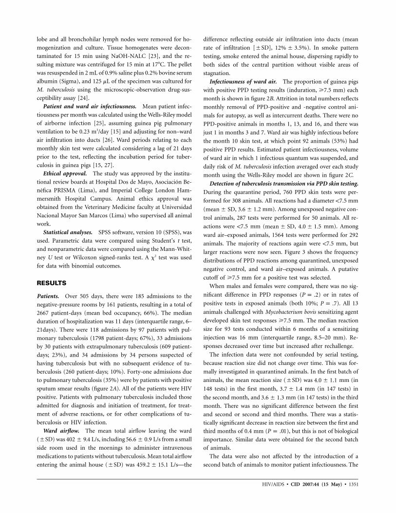
sputum smear results (figure 2A). All of the patients were HIV
positive. Patients with pulmonary tuberculosis included those
admitted for diagnosis and initiation of treatment, for treat-
ment of adverse reactions, or for other complications of tu-
berculosis or HIV infection.
Ward airflow. The mean total airflow leaving the ward
(�SD) was L/s, including L/s from a small402 � 9.4 56.6 � 0.9
side room used in the mornings to administer intravenous
medications to patients without tuberculosis. Mean total airflow
entering the animal house (�SD) was L/s—the459.2 � 15.1
difference reflecting outside air infiltration into ducts (mean
rate of infiltration [�SD], ). In smoke pattern12% � 3.5%
testing, smoke entered the animal house, dispersing rapidly to
both sides of the central partition without visible areas of
stagnation.
Infectiousness of ward air. The proportion of guinea pigs
with positive PPD testing results (induration, �7.5 mm) each
month is shown in figure 2B. Attrition in total numbers reflects
monthly removal of PPD-positive and -negative control ani-
mals for autopsy, as well as intercurrent deaths. There were no
PPD-positive animals in months 1, 13, and 16, and there was
just 1 in months 3 and 7. Ward air was highly infectious before
the month 10 skin test, at which point 92 animals (53%) had
positive PPD results. Estimated patient infectiousness, volume
of ward air in which 1 infectious quantum was suspended, and
daily risk of M. tuberculosis infection averaged over each study
month using the Wells-Riley model are shown in figure 2C.
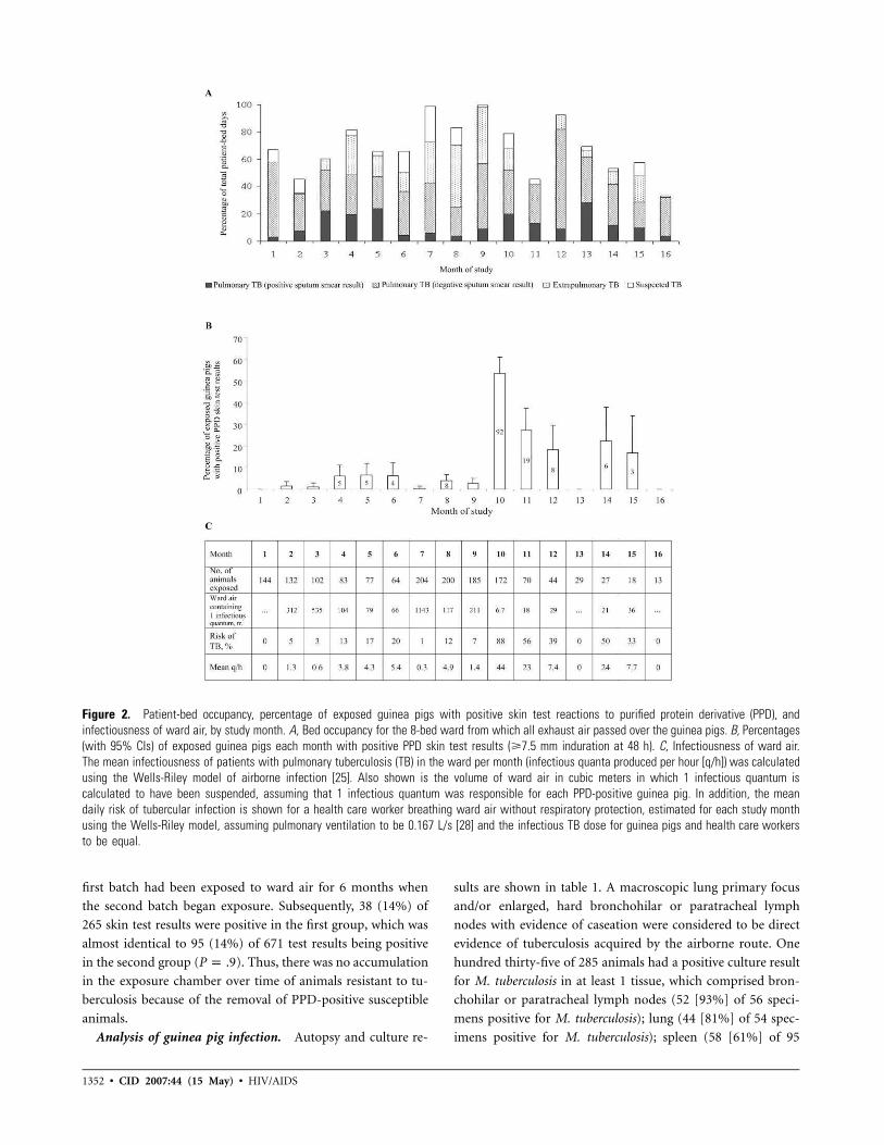
Detection of tuberculosis transmission via PPD skin testing.
During the quarantine period, 760 PPD skin tests were per-
formed for 308 animals. All reactions had a diameter !7.5 mm
( , mm). Among unexposed negative con-mean � SD 3.6 � 1.2
trol animals, 287 tests were performed for 50 animals. All re-
actions were !7.5 mm ( , mm). Amongmean � SD 4.0 � 1.5
ward air–exposed animals, 1564 tests were performed for 292
animals. The majority of reactions again were !7.5 mm, but
larger reactions were now seen. Figure 3 shows the frequency
distributions of PPD reactions among quarantined, unexposed
negative control, and ward air–exposed animals. A putative
cutoff of �7.5 mm for a positive test was selected.
When males and females were compared, there was no sig-
nificant difference in PPD responses ( ) or in rates ofP p .2
positive tests in exposed animals (both 10%; ). All 13P p .7
animals challenged with Mycobacterium bovis sensitizing agent
developed skin test responses �7.5 mm. The median reaction
size for 93 tests conducted within 6 months of a sensitizing
injection was 16 mm (interquartile range, 8.5–20 mm). Re-
sponses decreased over time but increased after rechallenge.
The infection data were not confounded by serial testing,
because reaction size did not change over time. This was for-
mally investigated in quarantined animals. In the first batch of
animals, the mean reaction size (�SD) was mm (in4.0 � 1.1
148 tests) in the first month, mm (in 147 tests) in3.7 � 1.4
the second month, and mm (in 147 tests) in the third3.6 � 1.3
month. There was no significant difference between the first
and second or second and third months. There was a statis-
tically significant decrease in reaction size between the first and
third months of 0.4 mm ( ), but this is not of biologicalP p .01
importance. Similar data were obtained for the second batch
of animals.
The data were also not affected by the introduction of a
second batch of animals to monitor patient infectiousness. The
1352 • CID 2007:44 (15 May) • HIV/AIDS
Figure 2. Patient-bed occupancy, percentage of exposed guinea pigs with positive skin test reactions to purified protein derivative (PPD), and
infectiousness of ward air, by study month. A, Bed occupancy for the 8-bed ward from which all exhaust air passed over the guinea pigs. B, Percentages
(with 95% CIs) of exposed guinea pigs each month with positive PPD skin test results (�7.5 mm induration at 48 h). C, Infectiousness of ward air.
The mean infectiousness of patients with pulmonary tuberculosis (TB) in the ward per month (infectious quanta produced per hour [q/h]) was calculated
using the Wells-Riley model of airborne infection [25]. Also shown is the volume of ward air in cubic meters in which 1 infectious quantum is
calculated to have been suspended, assuming that 1 infectious quantum was responsible for each PPD-positive guinea pig. In addition, the mean
daily risk of tubercular infection is shown for a health care worker breathing ward air without respiratory protection, estimated for each study month
using the Wells-Riley model, assuming pulmonary ventilation to be 0.167 L/s [28] and the infectious TB dose for guinea pigs and health care workers
to be equal.
first batch had been exposed to ward air for 6 months when
the second batch began exposure. Subsequently, 38 (14%) of
265 skin test results were positive in the first group, which was
almost identical to 95 (14%) of 671 test results being positive
in the second group ( ). Thus, there was no accumulationP p .9
in the exposure chamber over time of animals resistant to tu-
berculosis because of the removal of PPD-positive susceptible
animals.
Analysis of guinea pig infection. Autopsy and culture re-
sults are shown in table 1. A macroscopic lung primary focus
and/or enlarged, hard bronchohilar or paratracheal lymph
nodes with evidence of caseation were considered to be direct
evidence of tuberculosis acquired by the airborne route. One
hundred thirty-five of 285 animals had a positive culture result
for M. tuberculosis in at least 1 tissue, which comprised bron-
chohilar or paratracheal lymph nodes (52 [93%] of 56 speci-
mens positive for M. tuberculosis); lung (44 [81%] of 54 spec-
imens positive for M. tuberculosis); spleen (58 [61%] of 95
HIV/AIDS • CID 2007:44 (15 May) • 1353
Figure 3. Tuberculin purified protein derivative (PPD) skin test re-
sponses in guinea pigs exposed and not exposed to ward air. The fre-
quency distribution of induration diameter at 48 h for PPD skin tests is
shown for quarantined animals (760 tests in 308 animals over 3 months)
(A), unexposed negative control animals on the roof (287 tests in 50
animals over 13 months) (B), animals exposed to ward air (1564 tests in
292 animals over 16 months) (C), and animals exposed to ward air as in
C, with the y axis truncated at 30 mm to emphasize the distribution of
large responses �7.5 mm (D).
specimens positive for M. tuberculosis); or combined lung/
lymph node (70 [99%] of 71 specimens positive for M. tuber-
culosis). Autopsy and culture results for unexposed and exposed
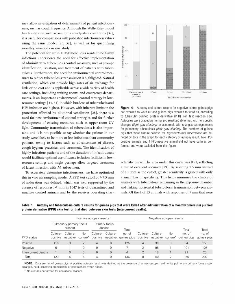
animals revealed a good relation to PPD results (figure 4). A
receiver operating characteristic [29] curve characterizing the
relative sensitivity and specificity of different cutoffs for skin
test positivity is shown in figure 5. The point closest to the
top-left corner is usually selected as the cutoff (in this case, 8.5
mm). A cutoff of �7.5 mm was adopted, because responses of
�7.5 mm were seen only in exposed animals, and this cutoff
gave increased sensitivity with only a small decrease in speci-
ficity, compared with the cutoff of 8.5 mm.
DISCUSSION
This study used a robust method of studying tuberculosis trans-
mission to demonstrate that the infectiousness of air from a
tuberculosis-HIV ward was highly variable. During the majority
of the study, ward air was relatively safe, causing few infections
in the guinea pigs, but at other times, the air was highly in-
fectious. Patient infectiousness appeared highly variable among
this heterogeneous group of patients with tuberculosis and HIV
infection, as seen in patients with tuberculosis but without HIV
infection in similar studies conducted during the 1950s.
Marked variability in the infectiousness of air from this tu-
berculosis-HIV ward was demonstrated. During the first 9
months, ward air was of relatively low infectiousness, with !7%
of exposed guinea pigs developing positive skin test results each
month. Furthermore, no PPD-positive animals were observed
in months 13 and 16. In contrast, ward air was highly infectious
prior to month 10, when 92 animals (53% of the colony) were
PPD-positive. This variation is not accounted for by changes
in bed occupancy, in patient bed-days for patients with positive
sputum smear results, or by grade of smear result positivity
(data not shown). Environmental factors, such as relative hu-
midity (highest in winter, study months 13–14), are also un-
likely to explain this variation. This variability of infectiousness
concurs with the studies by Riley et al. [15, 16, 30], in which
tuberculosis transmission occurred from a minority of patients,
and also with more recent work studying aerosols generated
by coughing patients with tuberculosis [31].
Estimated using the airborne infection model, the mean in-
fectiousness of patients with pulmonary tuberculosis each
month varied from production of 0–44 infectious quanta per
hour (1 quantum being the “infectious dose” for tuberculosis
[25]). The mean infectiousness during the whole study was 8.2
infectious quanta per hour, 16 times that calculated for the
heterogeneous mix of patients without HIV infection studied
by Riley et al. [25] during the 1950s and 1960s. This mean
infectiousness, however, masks variability in infectiousness be-
tween patients [30]. In the studies during the 1950s and 1960s,
a patient with laryngeal tuberculosis accounted for many in-
fections in guinea pigs (estimated production, 60 infectious
quanta per h [25]), and treatment was deferred in some patients
to study the effect on infectiousness. There was no evidence of
laryngeal tuberculosis in our study, and in contrast to the stud-
ies written or cowritten by Riley [15, 16, 30], all patients in
our study were immunosuppressed due to HIV infection. DNA
fingerprinting of patient and guinea pig M. tuberculosis strains
1354 • CID 2007:44 (15 May) • HIV/AIDS
Figure 4. Autopsy and culture results for negative control guinea pigs
not exposed to ward air and guinea pigs exposed to ward air, according
to tuberculin purified protein derivative (PPD) skin test reaction size.
Autopsies were graded as normal (no shading); abnormal, with nonspecific
changes (light gray shading); or abnormal, with changes pathognomonic
for pulmonary tuberculosis (dark gray shading). The numbers of guinea
pigs that were culture-positive for Mycobacterium tuberculosis are de-
noted by dots in the graph for each category of autopsy result. Two PPD-
positive animals and 1 PPD-negative animal did not have cultures per-
formed and were excluded from this figure.
Table 1. Autopsy and tuberculosis culture results for guinea pigs that were killed after administration of a monthly tuberculin purified
protein derivative (PPD) skin test or that died between skin tests (intercurrent deaths).
PPD status
Positive autopsy results Negative autopsy results
Total
no. of
guinea pigs
Pulmonary primary focus
present
Primary focus
absentTotal
no. of
guinea pigs
Culture-
positive
Culture-
negative
No
culturea
Total
no. of
guinea pigs
Culture-
positive
Culture-
negative
No
culturea
Culture-
positive
Culture-
negative
Positive 116 3 2 4 0 125 4 30 0 34 159
Negative 6 1 0 0 0 7 2 98 1 101 108
Intercurrent deaths 1 0 3 0 0 4 2 18 1 21 25
Total 123 4 5 4 0 136 8 146 2 156 292
NOTE. Data are no. of guinea pigs. A positive autopsy result was defined as the presence of a macroscopic hard, white pulmonary primary focus and/or
enlarged, hard, caseating bronchohilar or paratracheal lymph nodes.a
No cultures performed for operational reasons.
may allow investigation of determinants of patient infectious-
ness, such as cough frequency. Although the Wells-Riley model
has limitations, such as assuming steady-state conditions [32],
it is useful for comparisons with published infectiousness values
using the same model [25, 32], as well as for quantifying
monthly variations in our study.
The potential for air in HIV-tuberculosis wards to be highly
infectious underscores the need for effective implementation
of administrative tuberculosis control measures, such as prompt
identification, isolation, and treatment of patients with tuber-
culosis. Furthermore, the need for environmental control mea-
sures to reduce tuberculosis transmission is highlighted. Natural
ventilation, which can provide high rates of air exchange for
little or no cost and is applicable across a wide variety of health
care settings, including waiting rooms and emergency depart-
ments, is an important environmental control strategy in low-
resource settings [33, 34] in which burdens of tuberculosis and
HIV infection are highest. However, with inherent limits in the
protection afforded by dilutional ventilation [28], there is a
need for new environmental control strategies and for further
development of existing measures, such as upper-room UV
light. Community transmission of tuberculosis is also impor-
tant, and it is not possible to say whether the patients in our
study were likely to be more or less infectious than community
patients, owing to factors such as advancement of disease,
cough hygiene practices, and treatment. The identification of
highly infectious patients and of the duration of infectiousness
would facilitate optimal use of scarce isolation facilities in low-
resource settings and might perhaps allow targeted treatment
of latent infection with M. tuberculosis.
To accurately determine infectiousness, we have optimized
this in vivo air sampling model. A PPD test cutoff of �7.5 mm
of induration was defined, which was well supported by the
absence of responses 17 mm in 1047 tests of quarantined and
negative control animals and by the receiver operating char-
acteristic curve. The area under this curve was 0.91, reflecting
a test of excellent accuracy [29]. By selecting 7.5 mm instead
of 8.5 mm as the cutoff, greater sensitivity is gained with only
a small loss in specificity. This helps minimize the chance of
animals with tuberculosis remaining in the exposure chamber
and risking horizontal tuberculosis transmission between ani-
mals. Of the 4 of 13 animals with responses of 7 mm that were

HIV/AIDS • CID 2007:44 (15 May) • 1355
Figure 5. Receiver operating characteristic curve for 100 U of tuber-
culin purified protein derivative (PPD) skin test in guinea pigs, on the
basis of sensitivity and specificity calculated from combined autopsy and
culture data for 267 animals exposed to ward air. The area under the
curve is 0.91, suggesting a test of excellent accuracy. As indicated by
the adjacent numbers, the points on the graph correspond to PPD cutoff
points for positive tests of �14, 13, 12, 11, 10, 9.5, 9, 8.5, 8, 7.5, 7,
6.5, 6, and 5.5 mm.
found to have tuberculosis, 3 went on to develop skin test
responses �7.5 mm while awaiting autopsy.
The significance of PPD-positive animals with negative au-
topsy and culture results is of interest. Of 159 exposed guinea
pigs with PPD responses �7.5 mm, 30 (19%) had negative
culture and autopsy results (figure 4). A study by Mills et al.
[35], in which Riley was a researcher, similarly identified such
a group of animals. One possible explanation is reduced strain
virulence, and infection of guinea pigs by the intramuscular or
airborne route with clinical M. tuberculosis strains has shown
considerable differences in virulence [36–42]. For reduced M.
tuberculosis strain virulence, mycobacterial numbers in spleen
or primary lung lesions may decrease to !100 colony-forming
units by 6 weeks [40], and a lung focus may potentially resolve
over time. Because of the monthly PPD system and the ∼21-
day interval from infection to PPD conversion [27], an animal
may have been infected for 3–7 weeks by the time the skin test
was administered. If infection occurred with a reduced-viru-
lence strain, by the time of autopsy, there may have been little
macroscopic evidence of tuberculosis, and mycobacterial levels
may have been less than detection limits. Variation in the air-
borne fitness or dose of inhaled bacilli may also contribute to
the discordance between culture and skin test results. Experi-
mental airborne infection of guinea pigs with clinical M. tu-
berculosis strains has shown variability in lesion-inducing ef-
ficiency, defined as the number of colony-forming units
required to be inhaled to produce a macroscopic primary focus
[40], which ranged from 1, for fully virulent, to 4, for poorly
virulent strains. It is possible that a guinea pig infected with
fewer droplet nuclei than the lesion-inducing efficiency number
for that strain may develop a positive skin test result without
autopsy or culture evidence of tuberculosis. Therefore, if only
guinea pigs with macroscopic evidence of tuberculosis are in-
cluded, assessments of patient infectiousness may be
underestimations.
There are 2 additional potential confounders of the data.
First, 5 animals had false-negative PPD results, of which 3 had
tuberculosis at autopsy, including a grossly underweight animal
that may have developed anergy due to extensive disease. The
remaining 2 animals with apparently false-negative PPD results
may have represented laboratory cross-contamination or, al-
ternatively, early tubercular infection prior to PPD conversion.
Second, horizontal tuberculosis transmission between animals
inside the facility may confound these studies of patient infec-
tiousness. Tuberculosis transmission may occur between guinea
pigs by the respiratory and fecal-oral routes [20, 21]. However,
drop-through cages were installed to prevent coprophagy. Im-
portantly, the spatial distribution of tubercular infection in the
animal facility was random, consistent with airborne infection
from patients in the ward and not with horizontal spread.
In summary, this study has demonstrated marked variability
in infectiousness of air from an HIV-tuberculosis ward with a
heterogeneous mix of patients. Average patient infectiousness
each month varied greatly, as did the estimated risk of noso-
comial tuberculosis transmission to a health care worker on
the ward. This highlights the importance of the continuing need
for administrative and environmental control measures for re-
ducing institutional tuberculosis transmission. This air sam-
pling model is currently in use, evaluating upper-room ultra-
violet light and negative air ionization to prevent airborne
transmission of tuberculosis.
Acknowledgments
We thank the staff and patients of the Servicio de Enfermedades Infec-
ciosas y Tropicales at Hospital Nacional Dos de Mayo (Lima, Peru) for
their invaluable and continued support in this and ongoing studies. We
thank Miguel Gil Saavedra for veterinary support and Patricia Fuentes,
Pilar Navarro, Jorge Coronel, and other staff at the Laboratorio de Inves-
tigacion y Desarrollo at Universidad Peruana Cayetano Heredia for pro-
cessing of specimens. We thank SAEG Peru SA, for engineering support,
and Antonio Quispe, for creating figure 1.
Financial support. Sir Halley Stewart Trust, the Sir Samuel Scott of
Yews Trust, the Wellcome Trust (A.R.E., D.A.J.M., C.A.E., J.S.F., and R.H.G.
are funded by the Wellcome Trust [United Kingdom], and A.R.E., D.A.J.M.,
and C.A.E. have Wellcome Trust Clinical Tropical Medicine Research Fel-
lowships), USAID (HRN-5986-A-00-6006-00, GHS-A-00-03-00019-00 to
1356 • CID 2007:44 (15 May) • HIV/AIDS
R.H.G.), and Global Research Activity Cooperative Agreement from Na-
tional Institutes of Health (T35AI-07646 to R.H.G.).
Potential conflicts of interest. All authors: no conflicts.
References
1. Edlin BR, Tokars JI, Grieco MH, et al. An outbreak of multidrug-
resistant tuberculosis among hospitalized patients with the acquired
immunodeficiency syndrome. N Engl J Med 1992; 326:1514–21.
2. Ikeda RM, Birkhead GS, DiFerdinando GT Jr, et al. Nosocomial tu-
berculosis: an outbreak of a strain resistant to seven drugs. Infect
Control Hosp Epidemiol 1995; 16:152–9.
3. Curtis AB, Ridzon R, Novick LF, et al. Analysis of Mycobacterium
tuberculosis transmission patterns in a homeless shelter outbreak. Int
J Tuberc Lung Dis 2000; 4:308–13.
4. Dwyer B, Jackson K, Raios K, Sievers A, Wilshire E, Ross B. DNA
restriction fragment analysis to define an extended cluster of tuber-
culosis in homeless men and their associates. J Infect Dis 1993; 167:
490–4.
5. Mohle-Boetani JC, Miguelino V, Dewsnup DH, et al. Tuberculosis out-
break in a housing unit for human immunodeficiency virus–infected
patients in a correctional facility: transmission risk factors and effective
outbreak control. Clin Infect Dis 2002; 34:668–76.
6. Valway SE, Greifinger RB, Papania M, et al. Multidrug-resistant tu-
berculosis in the New York State prison system, 1990–1991. J Infect
Dis 1994; 170:151–6.
7. Theuer CP, Hopewell PC, Elias D, Schecter GF, Rutherford GW, Chais-
son RE. Human immunodeficiency virus infection in tuberculosis pa-
tients. J Infect Dis 1990; 162:8–12.
8. Markowitz N, Hansen NI, Hopewell PC, et al. Incidence of tuberculosis
in the United States among HIV-infected persons. The Pulmonary
Complications of HIV Infection Study Group. Ann Intern Med
1997; 126:123–32.
9. Cruciani M, Malena M, Bosco O, Gatti G, Serpelloni G. The impact
of human immunodeficiency virus type 1 on infectiousness of tuber-
culosis: a meta-analysis. Clin Infect Dis 2001; 33:1922–30.
10. Nunn P, Mungai M, Nyamwaya J, et al. The effect of human immu-
nodeficiency virus type-1 on the infectiousness of tuberculosis. Tuber
Lung Dis 1994; 75:25–32.
11. Elliott AM, Hayes RJ, Halwiindi B, et al. The impact of HIV on in-
fectiousness of pulmonary tuberculosis: a community study in Zambia.
AIDS 1993; 7:981–7.
12. Nardell EA. Air sampling for tuberculosis—homage to the lowly guinea
pig. Chest 1999; 116:1143–5.
13. Mastorides SM, Oehler RL, Greene JN, Sinnott JT, Kranik M, Sandin
RL. The detection of airborne Mycobacterium tuberculosis using mi-
cropore membrane air sampling and polymerase chain reaction. Chest
1999; 115:19–25.
14. Schafer MP, Fernback JE, Jensen PA. Sampling and analytical method
development for qualitative assessment of airborne mycobacterial spe-
cies of the Mycobacterium tuberculosis complex. Am Ind Hyg Assoc J
1998; 59:540–6.
15. Riley RL, Mills CC, O’Grady F, Sultan LU, Wittestadt F, Shivipuri DN.
Infectiousness of air from a tuberculosis ward—ultraviolet irradiation
of infected air: comparative infectiousness of different patients. Am
Rev Respir Dis 1962; 85:511–25.
16. Riley RL, Mills CC, Nyka W, et al. Aerial dissemination of pulmonary
tuberculosis: a two-year study of contagion in a tuberculosis ward:
1959. Am J Epidemiol 1995; 142:3–14.
17. Jensen PA, Lambert LA, Iademarco MF, Ridzon R. Guidelines for pre-
venting the transmission of Mycobacterium tuberculosis in health-care
settings, 2005. MMWR Recomm Rep 2005; 54:1–141.
18. Xu P, Peccia J, Fabian P, et al. Efficacy of ultraviolet germicidal irra-
diation of upper-room air in inactivating bacterial spores and Myco-
bacteria in full-scale studies. Atmos Environ 2003; 37:405–19.
19. Xu P, Kujundzic E, Peccia J, et al. Impact of environmental factors on
efficacy of upper-room air ultraviolet germicidal irradiation for inac-
tivating airborne mycobacteria. Environ Sci Technol 2005; 39:9656–64.
20. Lurie M. The effect of eliminating exposure to enteric infection on the
incidence and course of tuberculosis acquired by normal guinea pigs
confined with tuberculous cage mates. J Exp Med 1930; 51:753–68.
21. Lurie M. The route of infection in naturally acquired tuberculosis of
the guinea pig. J Exp Med 1930; 51:769–76.
22. Supplemental assay 636 for the evaluation of batches of PPD using
guinea pigs. Ames, Iowa: National Veterinary Services Laboratory (pre-
viously Centre for Veterinary Biologics Laboratory), 1998.
23. World Health Organization. Laboratory services in TB control. Parts
I–III. Geneva: World Health Organization, 1998.
24. Caviedes L, Lee TS, Gilman RH, et al. Rapid, efficient detection and
drug susceptibility testing of Mycobacterium tuberculosis in sputum by
microscopic observation of broth cultures. The Tuberculosis Working
Group in Peru. J Clin Microbiol 2000; 38:1203–8.
25. Riley RL, Nardell EA. Clearing the air: the theory and application of
ultraviolet air disinfection. Am Rev Respir Dis 1989; 139:1286–94.
26. Escombe AR. The detection and prevention of airborne tuberculosis
transmission [PhD thesis]. London: Imperial College London, 2006.
27. Fok JS, Ho RS, Arora PK, Harding GE, Smith DW. Host-parasite re-
lationships in experimental airborne tuberculosis. V. Lack of hema-
togenous dissemination of Mycobacterium tuberculosis to the lungs in
animals vaccinated with bacille Calmette-Guerin. J Infect Dis 1976;
133:137–44.
28. Nardell EA, Keegan J, Cheney SA, Etkind SC. Airborne infection: the-
oretical limits of protection achievable by building ventilation. Am Rev
Respir Dis 1991; 144:302–6.
29. Luna-Herrera J, Martinez-Cabrera G, Parra-Maldonado R, et al. Use
of receiver operating characteristic curves to assess the performance of
a microdilution assay for determination of drug susceptibility of clinical
isolates of Mycobacterium tuberculosis. Eur J Clin Microbiol Infect Dis
2003; 22:21–7.
30. Sultan L, Nyka W, Mills C, O’Grady F, Wells W, Riley RL. Tuberculosis
disseminators: a study of the variability of aerial infectivity of tuber-
culous patients. Am Rev Respir Dis 1960; 82:358–69.
31. Fennelly KP, Martyny JW, Fulton KE, Orme IM, Cave DM, Heifets
LB. Cough-generated aerosols of Mycobacterium tuberculosis: a new
method to study infectiousness. Am J Respir Crit Care Med 2004; 169:
604–9.
32. Beggs CB, Noakes CJ, Sleigh PA, Fletcher La, Siddiqi K. The trans-
mission of tuberculosis in confined spaces: an analytical review of
alternative epidemiological models. Int J Tuberc Lung Dis 2003; 7:
1015–26.
33. World Health Organization. World Health Organization guidelines for
the prevention of tuberculosis in healthcare facilities in resource-lim-
ited settings. Geneva: World Health Organization, 1999.
34. Escombe AR, Oeser CC, Gilman RH, et al. Natural ventilation for the
prevention of airborne contagion. PLoS Med 2007; 4:e68.
35. Mills CC, O’Grady F, Riley RL. Tuberculin conversion in the “naturally
infected” guinea pig. Bull Johns Hopkins Hosp 1960; 106:36–45.
36. Prabhakar R, Venkataraman P, Vallishayee RS, et al. Virulence for
guinea pigs of tubercle bacilli isolated from the sputum of participants
in the BCG trial, Chingleput District, South India. Tubercle 1987; 68:
3–17.
37. Mitchison DA, Wallace JG, Bhatia AL, Selkon JB, Subbaiah TV, Lan-
caster MC. A comparison of the virulence in guinea-pigs of South
Indian and British tubercle bacilli. Tubercle 1960; 41:1–22.
38. Mitchison DA, Bhatia AL, Radhakrishna S, Selkon JB, Subbaiah TV,
Wallace JG. The virulence in the guinea-pig of tubercle bacilli isolated
before treatment from South Indian patients with pulmonary tuber-
culosis. I. Homogeneity of the investigation and a critique of the vir-
ulence test. Bull World Health Organ 1961; 25:285–312.
39. Bhatia AL, Csillag A, Mitchison DA, Selkon JB, Somasundaram PR,
Subbaiah TV. The virulence in the guinea-pig of tubercle bacilli isolated
before treatment from South Indian patients with pulmonary tuber-

HIV/AIDS • CID 2007:44 (15 May) • 1357
culosis. 2. Comparison with virulence of tubercle bacilli from British
patients. Bull World Health Organ 1961; 25:313–22.
40. Balasubramanian V, Wiegeshaus EH, Smith DW. Growth characteristics
of recent sputum isolates of Mycobacterium tuberculosis in guinea pigs
infected by the respiratory route. Infect Immun 1992; 60:4762–7.
41. Cohn ML, Davis CL. Infectivity and pathogenicity of drug-resistant
strains of tubercle bacilli studied by aerogenic infection of guinea pigs.
Am Rev Respir Dis 1970; 102:97–100.
42. Gangadharam PR, Cohn ML, Davis CL, Middlebrook G. Infectivity
and pathogenicity of Indian and British strains of tubercle bacilli stud-
ied by aerogenic infection of guinea pigs. Am Rev Respir Dis 1963;
87:200–5.