Impact of pulmonary tuberculosis on survival of HIV-infected adults: a prospective epidemiologic...
16
Impact of pulmonary tuberculosis on survival of HIV-infected adults: a prospective epidemiologic study in Uganda Christopher C. Whalen a,b , Peter Nsubuga c,d , Alphonse Okwera c,d , John L. Johnson b , David L. Hom b , Nelson L. Michael e , Roy D. Mugerwa c , and Jerrold J. Ellner b a Department of Epidemiology and Biostatistics, Case Western Reserve University School of Medicine, Cleveland, Ohio, USA b Department of Medicine, Division of Infectious Diseases, Case Western Reserve University School of Medicine, Cleveland, Ohio, USA c Department of Medicine, Makerere University Medical School, Uganda Ministry of Health, Kampala, Uganda d National Tuberculosis and Leprosy Control Programme, Uganda Ministry of Health, Kampala, Uganda e Division of Retrovirology, Walter Reed Army Institute of Research, Rockville, Maryland, USA Abstract Background—Retrospective cohort studies of tuberculosis suggest that active tuberculosis accelerates the progression of HIV infection. The validity of these findings has been questioned because of their retrospective design, diverse study populations, variable compliance with anti- tuberculous therapy and use of anti-retroviral medication. To assess the impact of tuberculosis on survival in HIV infection we performed a prospective study among HIV-infected Ugandan adults with and without tuberculosis. Methods—In a prospective cohort study, 230 patients with HIV-associated tuberculosis and 442 HIV-infected subjects without tuberculosis were followed for a mean duration of 19 months for survival. To assess changes in viral load over 1 year, 20 pairs of tuberculosis cases and controls were selected and matched according to baseline CD4 lymphocyte count, age, sex and tuberculin skin test status. Results—During the follow-up period, 63 out of of 230 tuberculosis cases (28%) died compared with 85 out of 442 controls (19%), with a crude risk ratio of 1.4 [95% confidence interval (CI), 1.07– 1.87]. Most deaths occurred in patients with CD4 lymphocyte counts < 200 × 10 6 cells/l at baseline (n = 99) and occurred with similar frequency in the tuberculosis cases (46%) and the controls (44%). When the CD4 lymphocyte count was > 200 × 10 6 /l, however, the relative risk of death in HIV- associated tuberculosis was 2.1 (95% CI, 1.27–3.62) compared with subjects without tuberculosis. For subjects with a CD4 lymphocyte count > 200 × 10 6 /l, the 1-year survival proportion was slightly lower in the cases than in the controls (0.91 versus 0.96), but by 2 years the survival proportion was significantly lower in the cases than in the controls (0.84 versus 0.91; P < 0.02; log-rank test). For subjects with a CD4 lymphocyte count of 200 × 10 6 cells/l or fewer, the survival proportion at 1 year for the controls was lower than cases (0.59 versus 0.64), but this difference was not statistically significant (P = 0.53; logrank test). After adjusting for age, sex, tuberculin skin test status, CD4 lymphocyte count, and history of HIV-related infections, the overall relative hazard for death associated with tuberculosis was 1.81 (95% CI, 1.24–2.65). In a nested Cox regression model, the relative hazard for death was 3.0 (95% CI, 1.62–5.63) for subjects with CD4 lymphocyte counts > © 2000 Lippincott Williams & Wilkins Requests for reprints to: C.C. Whalen, Department of Epidemiology and Biostatistics, WG-49, Case Western Reserve University School of Medicine, 10900 Euclid Avenue, Cleveland, Ohio 44106-4945, USA. NIH Public Access Author Manuscript AIDS. Author manuscript; available in PMC 2010 May 13. Published in final edited form as: AIDS. 2000 June 16; 14(9): 1219–1228. NIH-PA Author Manuscript NIH-PA Author Manuscript NIH-PA Author Manuscript
Transcript of Impact of pulmonary tuberculosis on survival of HIV-infected adults: a prospective epidemiologic...
Impact of pulmonary tuberculosis on survival of HIV-infectedadults: a prospective epidemiologic study in Uganda
Christopher C. Whalena,b, Peter Nsubugac,d, Alphonse Okwerac,d, John L. Johnsonb, DavidL. Homb, Nelson L. Michaele, Roy D. Mugerwac, and Jerrold J. Ellnerb
aDepartment of Epidemiology and Biostatistics, Case Western Reserve University School ofMedicine, Cleveland, Ohio, USA bDepartment of Medicine, Division of Infectious Diseases, CaseWestern Reserve University School of Medicine, Cleveland, Ohio, USA cDepartment of Medicine,Makerere University Medical School, Uganda Ministry of Health, Kampala, Uganda dNationalTuberculosis and Leprosy Control Programme, Uganda Ministry of Health, Kampala, UgandaeDivision of Retrovirology, Walter Reed Army Institute of Research, Rockville, Maryland, USA
AbstractBackground—Retrospective cohort studies of tuberculosis suggest that active tuberculosisaccelerates the progression of HIV infection. The validity of these findings has been questionedbecause of their retrospective design, diverse study populations, variable compliance with anti-tuberculous therapy and use of anti-retroviral medication. To assess the impact of tuberculosis onsurvival in HIV infection we performed a prospective study among HIV-infected Ugandan adultswith and without tuberculosis.
Methods—In a prospective cohort study, 230 patients with HIV-associated tuberculosis and 442HIV-infected subjects without tuberculosis were followed for a mean duration of 19 months forsurvival. To assess changes in viral load over 1 year, 20 pairs of tuberculosis cases and controls wereselected and matched according to baseline CD4 lymphocyte count, age, sex and tuberculin skin teststatus.
Results—During the follow-up period, 63 out of of 230 tuberculosis cases (28%) died comparedwith 85 out of 442 controls (19%), with a crude risk ratio of 1.4 [95% confidence interval (CI), 1.07–1.87]. Most deaths occurred in patients with CD4 lymphocyte counts < 200 × 106 cells/l at baseline(n = 99) and occurred with similar frequency in the tuberculosis cases (46%) and the controls (44%).When the CD4 lymphocyte count was > 200 × 106 /l, however, the relative risk of death in HIV-associated tuberculosis was 2.1 (95% CI, 1.27–3.62) compared with subjects without tuberculosis.For subjects with a CD4 lymphocyte count > 200 × 106/l, the 1-year survival proportion was slightlylower in the cases than in the controls (0.91 versus 0.96), but by 2 years the survival proportion wassignificantly lower in the cases than in the controls (0.84 versus 0.91; P < 0.02; log-rank test). Forsubjects with a CD4 lymphocyte count of 200 × 106 cells/l or fewer, the survival proportion at 1 yearfor the controls was lower than cases (0.59 versus 0.64), but this difference was not statisticallysignificant (P = 0.53; logrank test). After adjusting for age, sex, tuberculin skin test status, CD4lymphocyte count, and history of HIV-related infections, the overall relative hazard for deathassociated with tuberculosis was 1.81 (95% CI, 1.24–2.65). In a nested Cox regression model, therelative hazard for death was 3.0 (95% CI, 1.62–5.63) for subjects with CD4 lymphocyte counts >
© 2000 Lippincott Williams & WilkinsRequests for reprints to: C.C. Whalen, Department of Epidemiology and Biostatistics, WG-49, Case Western Reserve University Schoolof Medicine, 10900 Euclid Avenue, Cleveland, Ohio 44106-4945, USA.
NIH Public AccessAuthor ManuscriptAIDS. Author manuscript; available in PMC 2010 May 13.
Published in final edited form as:AIDS. 2000 June 16; 14(9): 1219–1228.
NIH
-PA Author Manuscript
NIH
-PA Author Manuscript
NIH
-PA Author Manuscript
200 × 106/l and 1.5 (95% CI, 0.99–2.40) for subjects with a CD4 lymphocyte count of 200 × 106/lor fewer.
Conclusion—The findings from this prospective study indicate that active tuberculosis exerts itsgreatest effect on survival in the early stages of HIV infection, when there is a reserve capacity ofthe host immune response. These observations provide a theoretical basis for the treatment of latenttuberculous infection in HIV-infected persons.
KeywordsHIV-1; tuberculosis; survival; Africa; prospective cohort study
IntroductionThe World Health Organization (WHO) estimated that by 1997, 15.3 million people worldwidewere infected with both HIV-1 and Mycobacterium tuberculosis, most of them in sub-SaharanAfrica. As HIV-1 spread across Africa, and throughout the world, there has been a resurgenceof tuberculosis attributed to HIV-1. Indeed, the hallmark of HIV-1 infection is the progressivedecline and dysfunction of CD4 lymphocytes [1], cells that are critical in containing infectionwith M. tuberculosis. HIV-1 infection confers the greatest known risk for the development ofactive tuberculosis, both for reactivation of latent infection [2] or progressive primary disease[3]. Moreover, in HIV-1 infection, tuberculosis is associated with a high mortality, even whenit is properly treated [4–7]. Worldwide, tuberculosis is the major cause of death in HIV-1infection.
However, the interaction between HIV-1 and tuberculosis is bi-directional. The effects oftuberculosis on HIV-1 infection have been observed both at the cellular and clinical levels.Although there is evidence that tuberculosis enhances viral replication through mechanismsthat involve activation of the cell-mediated immune system through cytokine networks [8–11], the epidemiological findings have been inconsistent. A number of studies have shown thattuberculosis alters the progression of HIV disease, as measured by survival [12–15], whereasother studies have not [16,17]. The inconsistency in the findings may result from underlyingdifferences in the immune function of study populations, the choice of the comparison group,or the analytic methods. As a group, however, all of these studies are limited by the retrospectivedesign, small sample size, the variability in compliance with anti-tuberculous therapy orantiretroviral drug use, or a bias towards advanced HIV-1 disease. In some of the studies, acohort effect may exist as patients were recruited over a period of one decade.
The current study addresses some of these limitations as it is a prospective cohort study thatexamines the impact of tuberculosis on survival in a cohort of Ugandan adults with a spectrumof HIV-1 infection during a 3-year period. In this cohort, compliance with anti-tuberculoustherapy was monitored and antiretroviral therapy was unavailable. We found that pulmonarytuberculosis was associated with reduced survival and that the effect on disease progressionand survival may exert its greatest impact early in the course of HIV-1 infection, beforeadvanced immunosuppression develops.
MethodsStudy design and populations
In a prospective comparative cohort design, the survival experience was compared betweenHIV-infected individuals with and without pulmonary tuberculosis. Between February 1993and May 1994, 230 cases of smear-positive pulmonary tuberculosis presenting to the NationalTuberculosis and Leprosy Programme in Kampala, Uganda were eligible for the study and
Whalen et al. Page 2
AIDS. Author manuscript; available in PMC 2010 May 13.
NIH
-PA Author Manuscript
NIH
-PA Author Manuscript
NIH
-PA Author Manuscript
enrolled after history and physical examination, sputum smear for acid-fast bacilli, and chestradiography. Pulmonary tuberculosis was defined as sputum smear-positive disease confirmedby culture in a patient with the clinical signs and symptoms consistent with active tuberculosis.All cases of tuberculosis received daily self-administered anti-tuberculous therapy includingisoniazid, rifampin, pyrazinamide, and ethambutol for 2 months followed by 6 months of dailyisoniazid and rifampin. The comparison, or control group, for this study comprised subjectswithout tuberculosis who were enrolled in the placebo arm of a concurrent clinical trial ofpreventive therapy for tuberculosis [18] and were followed at Mulago Tuberculosis Clinic.Before enrollment in the preventive therapy trial, active tuberculosis was rigorously excludedwith a thorough history and physical examination, sputum smear and culture, and chestradiography. Exclusion criteria for both groups included: Karnofsky performance status < 60,positive urine pregnancy test, residence greater than 20 km from the clinic, and WHO clinicalstage IV, not including tuberculosis.
Subjects in both groups were seen monthly for 6 months then quarterly thereafter. Both groupswere monitored at Mulago Tuberculosis Clinic by the same team of physicians, nurses andancillary personnel. The same follow-up protocol and data collection forms were used for eachgroup. When a subject missed a scheduled visit or failed to show urinary metabolites ofisoniazid, home health visitors visited the homes of these subjects and motivated them to attendthe clinic for evaluation and to take the medication as prescribed. Compliance with anti-tuberculous therapy was promoted through regular health education and monitored with urinetesting for isoniazid metabolites. Subjects in the comparison group were actively screened fortuberculosis twice a year with chest radiography and sputum microscopy and culture. Iftuberculosis was suspected, these subjects underwent further diagnostic evaluation and werestarted on anti-tuberculous therapy. Both groups received standard management for HIV-1infection available in Uganda during the study which included treatment of oral thrush, herpeszoster, herpes simplex and recurrent bacterial infections. Antiretroviral therapy was not thestandard of practice in Uganda during this study and was not given to subjects.
This study was approved by the Ugandan National AIDS Scientific Subcommittee of theUganda National Council for Science and Technology and the institutional review board atCase Western Reserve University. Oral informed consent was obtained by trained counselorsin the native language of the study subject.
Study outcomesThe main study outcome was observed survival defined as the period between enrollment inthe study and death or censoring. Subjects were censored at the last clinic visit when they wereknown to be alive or at the end of the study observation. Subjects in the comparison group whodeveloped tuberculosis during follow-up were censored at the time tuberculosis was diagnosed.Mortality was assessed through a standard interview of family members or review of hospitalrecords. Failure to keep a scheduled appointment prompted a home visit to determine the vitalstatus of the study subject. The date of death and prominent symptoms at the time of deathwere also obtained from family members.
To assess the change in viral load during observation, a sample of 20 cases was matched to 20controls according to baseline age, gender, CD4 lymphocyte count, and presence of cutaneousanergy. The levels of HIV RNA were then compared within and between groups at baselineand 12 months.
MeasurementsDemographic and clinical information was obtained through standardized interviews andphysical examination performed by trained Ugandan medical officers. Socioeconomic status
Whalen et al. Page 3
AIDS. Author manuscript; available in PMC 2010 May 13.
NIH
-PA Author Manuscript
NIH
-PA Author Manuscript
NIH
-PA Author Manuscript
was assessed by a trained Ugandan social scientist after an in depth interview to characterizepersonal possessions, occupation, income, housing and mode of transportation. At the time ofscreening, venous blood was collected for HIV-1 enzyme immunoassay (EIA) testing,complete blood and differential counts (Coulter T540 system), serum creatinine and aspartateaminotransferase. HIV infection was documented by enzyme-linked immunosorbent assay(ELISA; Recombigen HIV-1 env and gag ELISA, Cambridge BioScience, Worcester,Massachusetts, USA); one of 10 HIV-1 EIA positive and one of 25 HIV-1 negative seraunderwent confirmatory testing by HIV-1 Western immunoblotting (BioRad Novapath,Hercules, Californai, USA). HIV viral load was measured in plasma using the AMPLICORHIV-1 MONITOR test with additional primers for non-B clade virus [19,20]. At the time ofscreening, all subjects underwent Mantoux skin testing using five tuberculin units of purifiedprotein derivative (PPD; Tubersol, Connaught Laboratories, Swiftwater, Pasadena, USA) and0.1 ml Candida antigen (Candida albicans allergic extract, Berkeley Biologics, Berkeley,California, USA; 1 : 50 final concentration). Between 48 and 72 h, experienced observersrecorded the results of each skin test in millimeters. Cutaneous anergy was defined as absenceof reactivity (0 mm of induration) to both PPD and Candida antigens. Subjects hadposterioranterior chest X-rays taken at baseline and then at 6-month intervals during follow-up. Expectorated sputum specimens were collected, digested with 3% NaOH, concentrated,and stained for acid fast bacilli with Ziehl–Neelsen stain at the Uganda TuberculosisInvestigations Bacteriological Unit, Wandegeya. AFB smears were reviewed by trainedtechnicians who graded the smears by the number of acid-fast organisms seen on lightmicroscopy according to criteria established by the WHO [21]. Specimens were cultured forM. tuberculosis on Lowenstem–Jensen medium slants, incubated at 37°C in air and examinedweekly until positive or for 8 weeks.
Analytic strategyBaseline characteristics for both the tuberculosis cases and the comparison group werecompared using the χ2 test for categorical data and Student’s t test for continuous variables.Mortality rates were estimated and stratified according to CD4 lymphocyte count, history ofopportunistic infection, anemia, wasting, and tuberculin skin test. Stratum specific relativerisks were estimated with 95% confidence intervals (CI). Overall survival distributions for bothgroups were estimated using the Kaplan–Meier method and compared using the log-rank test[22]. A series of Cox proportional hazards models [23] were fit after testing for the proportionalhazards assumption for each variable. In each model, the dependent variable was the observedsurvival; independent variables tested included clinical and demographic characteristics, CD4lymphocyte count, tuberculin skin test, and history of or presence of one or more HIV-relatedconditions. Variables that were associated with survival in a univariate analysis (P < 0.15) wereevaluated in a series of multivanable models. Models were compared using likelihood ratiotests. Once significant variables of survival were identified, the variable for tuberculosis statuswas added to the model and statistical significance assessed. The hazard ratio for death amongsubjects with tuberculosis compared to subjects without tuberculosis was estimated with 95%CI. A similar analysis was performed to compare survival between tuberculosis patients andHIV-infected controls without tuberculosis but with one or more HIV-related conditions. Alltwo-way interactions between tuberculosis status and other main effects were examined. Theonly significant interaction was with the body mass index. Thus, separate models weredeveloped for subjects according to the body mass index above or below the median value (21kg/m2). Once the overall model was developed, a nested Cox regression model was used toestimate the hazard ratio within two strata defined by CD4 lymphocyte counts at baseline: 0–200 × 106cells/l and > 200 × 106cells/l.
Whalen et al. Page 4
AIDS. Author manuscript; available in PMC 2010 May 13.
NIH
-PA Author Manuscript
NIH
-PA Author Manuscript
NIH
-PA Author Manuscript
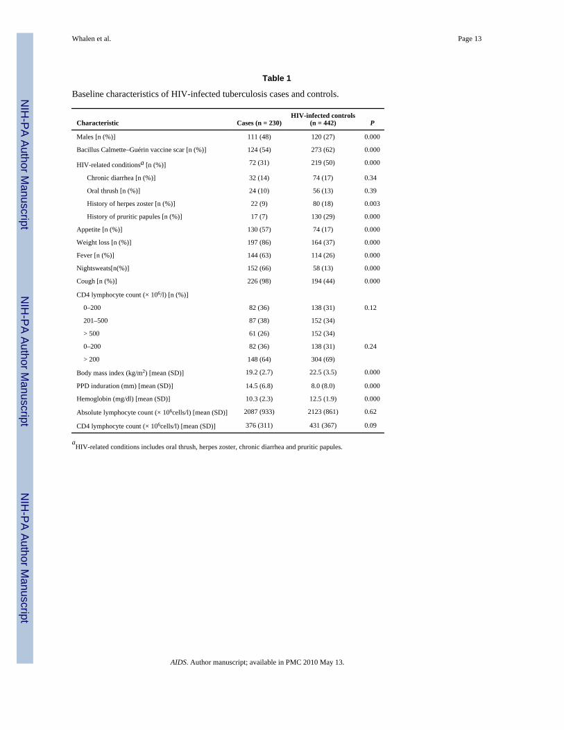
ResultsA total of 672 subjects met entry criteria for the study: 230 subjects were tuberculosis casesand 442 were HIV-infected subjects without evidence for tuberculosis. The mean age for thetuberculosis cases and the comparison group was similar (29.2 versus 30.6 years). A smallerproportion of the tuberculosis cases was female compared to the control group (51 versus 73%).The mean Karnofsky performance status was similar between the tuberculosis cases and thecontrols (83 versus 90). The distribution of subjects in high, middle, and low socioeconomicgroups was the same for the tuberculosis cases and the controls (1, 26 and 73% respectively).As expected, most patients presenting with tuberculosis had one or more classic symptoms oftuberculosis such as persistent cough, anorexia, night sweats, and fatigue (Table 1). Thefrequency of HIV-related conditions at the time of presentation was greater in the controlpopulation compared with the tuberculosis cases. Chronic diarrhea and oral thrush occurredwith comparable frequency between the two groups, but the history of herpes zoster and pruriticpapules was more common in the control subjects. Tuberculosis cases had reduced body massindex, lower hemoglobin levels and larger tuberculin skin test reactions compared with thecontrol group (Table 1). The mean absolute CD4 lymphocyte count tended to be lower in thetuberculosis cases than the controls (376 versus 431 × 10 cells/l; P = 0.09), though the overallfrequency distributions were similar between the two groups. The proportion of subjects lostto follow-up was similar in the cases and the controls (88 versus 89%); the mean duration offollow-up was also similar (1.7 versus 1.6 years).
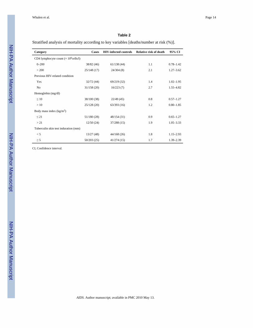
During the mean follow-up period of 19 months, 63 of 230 tuberculosis cases (28%) diedcompared with 85 of 442 controls (19%), for a crude risk ratio of 1.4 (95% CI, 1.07–1.87). Themajority of deaths (n = 99) occurred in subjects with CD4 counts < 200 × 10 cells/l at baselineand occurred with similar frequency in the tuberculosis cases and the controls (46 versus 44%;relative risk, 1.05; Table 2). When the CD4 count was above 200 cells/µL, however, the relativerisk of death in HIV-associated tuberculosis was 2.1 compared with subjects withouttuberculosis (Table 2). The relative risk of death associated with tuberculosis was also increasedafter stratifying by history of HIV-related conditions and tuberculin skin test < 5 mm. Whenstratifying by hemoglobin or body mass index, the mortality rate was slightly higher in thecontrol subjects versus cases when anemia (hemoglobin ≤ 10 mg/dl) or wasting (body massindex ≤ 21 kg/m2) were present. When subjects were not anemic or had a body mass index >21 kg/m2, the risk of death among cases was increased.
In a Kaplan–Meier analysis, the overall, unadjusted survival for cases of tuberculosis wassimilar to that of the control group (P < 0.09, log-rank test), though the cases had a lowersurvival proportion after 1 year. When this analysis was stratified according to the baselineCD4 lymphocyte count, survival in tuberculosis cases was reduced compared with the controlswhen the CD4 lymphocyte count was >200 × 10 cells/l but not when it was < 200 × 10 cells/l (Fig 1a and 1b). For subjects with CD4 lymphocyte counts > 200 × 10 cells/l the 1-yearsurvival proportion was slightly lower in the cases than in the controls (0.91 versus 0.96), butby 2 years the survival proportion was significantly lower in the cases than in the controls (0.84versus 0.91; P < 0.02, log-rank test). For subjects with CD4 counts of 200 × 10 cells/l or fewer,the survival proportion at 1 year for the controls was lower than cases (0.59 versus 0.64), butthis difference was not statistically significant (P = 0.53, log-rank test). Of note, during thefirst 3 months of observation, four deaths occurred in the cases and seven in controls, indicatingthat early mortality from tuberculosis did not account for differences in survival.
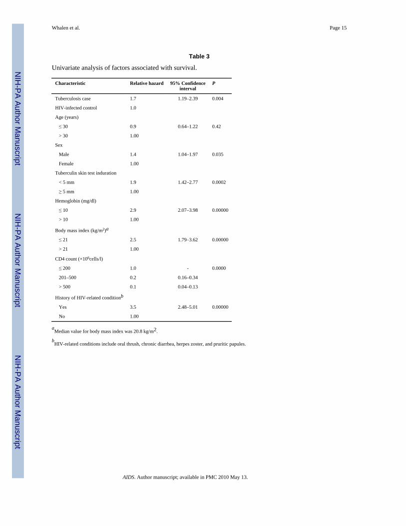
In a univariate Cox proportional hazards model, the unadjusted relative hazard for death intuberculosis cases compared with the controls was 1.7 (95% CI, 1.19–2.39; Table 3). Inaddition, gender, hemoglobin, body mass index, history of HIV-related conditions, CD4lymphocyte count, and tuberculin skin test were associated with survival (Table 3). After
Whalen et al. Page 5
AIDS. Author manuscript; available in PMC 2010 May 13.
NIH
-PA Author Manuscript
NIH
-PA Author Manuscript
NIH
-PA Author Manuscript
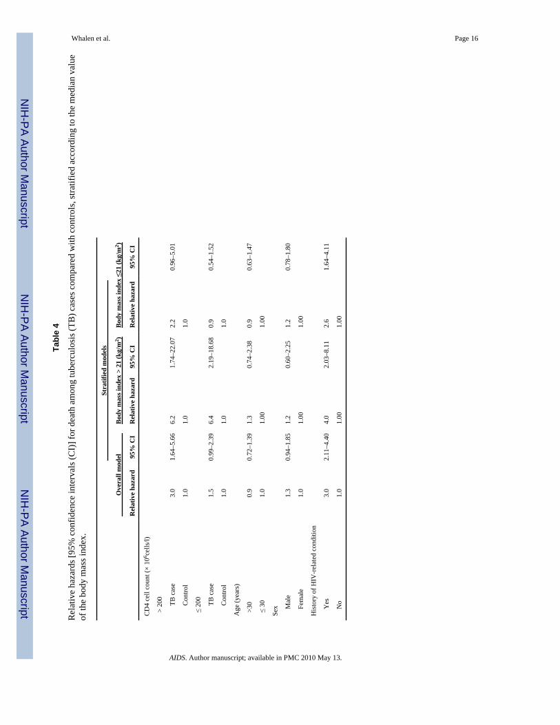
adjusting for age, sex, tuberculin skin test status, CD4 lymphocyte count, and history of HIV-related conditions, the relative hazard for death was 1.8 (95% CI, 1.24–2.65). In a nested Coxregression model adjusting for these same covariates (Table 4), the relative hazard for deathwas 3.0 (95% CI, 1.62–5.63) for subjects with CD4 lymphocyte count < 200 × 10 cells/l and1.5 (95% CI, 0.99–2.40) for subjects with a CD4 lymphocyte count of 200 × 10 cells/l or fewer.When hemoglobin and body mass index were included in this model, there was a statisticallysignificant interaction between tuberculosis and body mass index (P = 0.003) but not betweentuberculosis and hemoglobin (P = 0.19). Separate nested Cox regression models were thereforefit according to body mass index (Table 4). For subjects with body mass greater than the medianvalue of 21 kg/m2, the relative hazard for death was 6 fold greater in the tuberculosis casesthan in the control subjects, regardless of the baseline CD4 lymphocyte count (Table 4). Whenthe body mass index was < 21 kg/m2, then the risk of death was increased only among subjectswith CD4 lymphocyte counts > 200 × 10 cells/l, though this was not statistically significant(relative hazard, 2.2; P = 0.23).
When survival was compared between tuberculosis cases and HIV-infected controls with oneor more HIV-related conditions, a similar pattern of risk was found. In a Cox regression modeladjusting for age, sex, tuberculin skin test reaction, the risk of death in the tuberculosis casescompared with controls with another HIV-related condition was 1.81 (95% CI, 0.88–3.70) forsubjects with CD4 count > 200 × 10 cells/l and 0.83 (95% CI, 0.53–1.30) for subjects withCD4 count < 200 × 10 cells/l.
In a random sample of 20 tuberculosis cases drawn from the study population and 20 controlsmatched for age, sex, and CD4 count, plasma HIV RNA was measured at the time of enrollmentand again at 1 year. The mean level of the log-transformed viral load from baseline did notdiffer in the cases and controls (4.4 versus 4.5 log10 copies/ml; P = 0.34, non-parametric test).During the first year of follow-up, the viral load increased in the tuberculosis cases by anaverage of 0.49 log10 (P = 0.04, paired t test) and 0.06 log10 in the controls (P = 0.82, pairedt test), but the change in viral load between groups did not reach statistical significance (P =0.17).
DiscussionIn this prospective cohort study of HIV-infected Ugandan adults, pulmonary tuberculosis wasassociated with reduced survival, but the effect varied according to the stage of HIV disease.In subjects with less advanced immunosuppression, as indicated by a CD4 lymphocyte count> 200 × 10 cells/l, the effect of tuberculosis on survival was substantial, whereas in subjectswith advanced immunosuppression, the effect on observed survival was minimal. Thesefindings were consistent whether comparing the survival among tuberculosis cases with allcontrol subjects or only those with one or more HIV-related conditions. The effect oftuberculosis on survival also differed by body mass. In this case, wasting lessened the effectof tuberculosis on survival. The current results indicate that tuberculosis accelerates HIVdisease progression as measured by overall survival and that this effect is greatest early in thecourse of HIV-1 infection when cellular immune function is preserved.
There is a growing body of immunologic and virologic data that provides a potentialmechanism for the epidemiologic observations on the impact of tuberculosis on HIV-1. In theHIV-infected individual, the development of tuberculosis activates a T cell mediated immuneresponse, mediated through cytokine networks, that may initiate a sequence of cellular eventsthat both enhances viral production and promotes spread of infection to uninvolved cells. InHIV-infected patients with tuberculosis, there is evidence for cellular immune activation [24]and over-expression of tumor necrosis factor (TNF)-α by PPD-stimulated peripheral bloodmononuclear cells [25]. Further, the level of cellular activation of monocytes in tuberculosis
Whalen et al. Page 6
AIDS. Author manuscript; available in PMC 2010 May 13.
NIH
-PA Author Manuscript
NIH
-PA Author Manuscript
NIH
-PA Author Manuscript
is sufficient to enhance susceptibility to productive infection with HIV in vitro [8]. The immuneactivation of tuberculosis appears to augment viral replication as measured in the plasma[26] and the lung [27,28]. HIV expression may be enhanced by proinflammatory cytokines,such as TNF-α [29], and by mycobacterial products [11,30]; HIV-1 expression may be up- ordown-regulated by anti-inflammatory cytokines such as interleukm-10 [31] or transforminggrowth factor-β [32,33]. The balance of these cytokines, acting in autocrine and paracrineloops, is probably a key determinant of HIV-1 replication in tuberculosis [9].
In this study there was a modest, but significant, increase in viral load among tuberculosis casesduring the first year after diagnosis and treatment of tuberculosis. A similar increase in viralload was not observed in the control subjects who were matched by CD4 count. Although theabsolute change in viral load was not significantly different between the groups, the samplesize was small for this analysis. The rising levels of HIV RNA in tuberculosis patients seen inthis study are consistent with other studies [34–36]. This increment in viral load after theinitiation of therapy may result from the release of TNF-α [37] or from a reversal of theimmunosuppression related to tuberculosis [38]. These observations, and the proposedmechanism for co-pathogenesis, provide rationale for the use of cytokine inhibitors in thetreatment of HIV-associated tuberculosis. To date, two TNF-α inhibitors, pentoxifylline andthalidomide, have been shown to decrease plasma HIV RNA and reduce levels of immuneactivation markers, though no consistent reduction of TNF-α is seen [34,39]. The effect ofthese and other immune modulators on survival in HIV-associated tuberculosis remains to beestablished.
In this cohort, among subjects with preserved immune function, the difference in the observedsurvival distributions between tuberculosis cases and the controls is entirely consistent with aco-pathogenesis hypothesis. In subjects with advanced immunosuppression, the similarity insurvival distributions among the tuberculosis cases and controls may seem at first contradictorybut, in fact, refines the hypothesis. In advanced stages of HIV infection, we propose that viralreplication has escaped the influence of the host response to HIV-1 and that infected cells arealready stimulated to elaborate maximal amounts of intact virus. The additional effect oftuberculosis may have minimal effect in this milieu of immune activation and unchecked viralreplication. It is also possible that in more advanced HIV-1 disease, the competing risks ofdeath from other causes obscures any effect due to tuberculosis.
The findings of this study are consistent with the hypothesis that tuberculosis acts as a co-factorin disease progression of HIV, but this is not the only possible interpretation of the findings.The major competing hypothesis is that tuberculosis is a marker for advanced disease in HIVinfection and that unmeasured differences in risk of death account for the observations.Although the two groups differed in a number of baseline characteristics, they were closelymatched in terms of CD4 lymphocyte count, the marker of immune function available duringthe study. Based on the matched-pairs analysis, the viral load did not differ between cases andcontrols for a given level of CD4 count. This observation offers further support that patientshad similar risk for HIV disease progression within a given stratum of CD4 lymphocyte count.The CD4 lymphocyte count was also the most important prognostic measure in the analysis.Even when adjusting for CD4 lymphocyte count, tuberculosis remained associated withshortened survival.
One question that arises is whether the impact on survival is specific to tuberculosis. In ouranalysis, we compared the survival of tuberculosis patients to survival in HIV-infected subjectswithout tuberculosis but with one or more HIV-related conditions. Our conclusions regardingtuberculosis and survival are consistent regardless of the comparison group chosen. Previousepidemiologic studies report that other opportunistic infections besides tuberculosis areassociated with a reduced survival and have a greater relative risk of death than tuberculosis
Whalen et al. Page 7
AIDS. Author manuscript; available in PMC 2010 May 13.
NIH
-PA Author Manuscript
NIH
-PA Author Manuscript
NIH
-PA Author Manuscript
itself [16,17,40]; however, these cohorts comprised patients with advanced disease, asindicated by the presence of opportunistic infections and median CD4 cell counts < 200 × 10cells/l [41]. If immune function is an effect modifier of the interaction between tuberculosis,HIV infection and survival, as suggested by the findings of the current study, then one wouldnot expect to observe the impact of tuberculosis on survival in these cohorts. But unlikeopportunistic infections, which occur only with advanced immunosuppression, M.tuberculosis is a virulent organism which can develop at any point in the course of HIV-1disease. Early in HIV-1 disease when cell-mediated immunity is preserved, tuberculosis mayaffect the natural history of HIV-1 at a time when opportunistic infections generally do notoccur. In advanced HIV-1 disease, the impact of tuberculosis on survival may be modest whencompared to opportunistic infections which carry substantial disease-specific mortality.
The observed differences in survival may arise from deaths due to tuberculosis or from HIV-related deaths. In this study, all tuberculosis cases were given standard anti-tuberculous therapyand the majority responded with sterile sputum cultures by 2 months. We do not have detailedinformation about the cause of death from autopsy studies, but because there were nodifferences in early mortality between the case and control groups, we assume that deaths inthe cases were not caused by tuberculosis. Previous studies have indicated that when HIV-infected persons die from tuberculosis, these deaths occur during the early phase of therapy[4,6]. In this young HIV-infected Ugandan population, complications of HIV-1 besidestuberculosis are the most likely cause of death.
The analysis of this cohort points to body mass index as another effect modifier of thetuberculosis-HIV interaction. Although weight loss is a sign of tuberculosis, and hence highlycorrelated with disease, it may also be a marker for the stage of HIV-1 infection. For this reason,we examined the effect of tuberculosis after stratifying the analysis by the median value forthe body mass index. In this stratified analysis, tuberculosis was associated with reducedsurvival among subjects without wasting, whereas it had minimal effect among wastedindividuals. Thus, low body mass index acts like a marker for advanced HIV disease, similarto the low CD4 counts, so that differences in survival, if any, are minimal. An alternativeexplanation for this finding posits that control patients with wasting may have had occultdisseminated tuberculosis and hence were misclassified. In this case, the differences inobserved survival between the controls and the cases of tuberculosis would be mitigated.During the recruitment of the control group, disseminated disease was not evaluated unlesssuspected on clinical grounds alone.
Tuberculosis holds a primary place in the importance of the global HIV epidemic. Tuberculosisrepresents the leading cause of death worldwide in HIV infection. M. tuberculosis is a virulentorganism that can produce disease in HIV-infected persons at any stage of disease, even whenthe immunosuppression is minimal. In settings of high transmission of tuberculosis, HIV-infected persons are at great risk both for reactivation, progressive primary disease, andreinfection, and provide a fuel for the continuing epidemic of tuberculosis, particularly inAfrica. The findings from this study indicate that active tuberculosis will exert its greatest effecton survival when it develops during the early stages of HIV infection, when there is a reservecapacity of the host immune response. These findings provide theoretical rationale for thetreatment of latent tuberculous infection in HIV-infected persons, especially those withpreserved immune function.
AcknowledgmentsThis study was completed only through the dedicated work of many individuals. The authors acknowledge the staffof the National Tuberculosis Treatment Centre, Mulago Hospital, the Ugandan National Tuberculosis and LeprosyProgramme and the Uganda Tuberculosis Investigation Bacteriological Unit, Wandegeya for their contributions tothis study. We are most grateful to the patients and families who participated in the study.
Whalen et al. Page 8
AIDS. Author manuscript; available in PMC 2010 May 13.
NIH
-PA Author Manuscript
NIH
-PA Author Manuscript
NIH
-PA Author Manuscript
Sponsorship: Supported by a grant from the National Institutes of Health (AI32414). Training of study personnel wassupported through the AIDS International Training and Research Program of the Fogarty International Center at theNational Institutes of Health (TW-00011).
References1. Fauci AS. Host factors and the pathogenesis of HIV-induced disease. Nature 1996;384:529–534.
[PubMed: 8955267]2. O'Brien RJ, Perriens JH. Preventive therapy for tuberculosis in HIV infection: the promise and the
reality. AIDS 1995;9:665–673. [PubMed: 7546409]3. Daley CL, Small PM, Schecter GF, et al. An outbreak of tuberculosis with accelerated progression
among persons infected with the human immunodeficiency virus. N Engl J Med 1992;326:231–235.[PubMed: 1345800]
4. Small PM, Schecter GF, Goodman PC, Sande MA, Chaisson RE, Hopewell PC. Treatment oftuberculosis in patients with advanced human immunodeficiency virus infection. N Engl J Med1991;324:289–294. [PubMed: 1898769]
5. Whalen CC, Okwera A, Johnson J, et al. Predictors of survival in human immunodeficiency virus-infected patients with pulmonary tuberculosis. Am J Respir Crit Care Med 1996;153:1977–1981.[PubMed: 8665064]
6. Nunn P, Brindle R, Carpenter L, et al. Cohort study of human immunodeficiency virus infection inpatients with tuberculosis in Nairobi, Kenya. Am Rev Respir Dis 1992;146:849–854. [PubMed:1416409]
7. Ackah AN, Coubibaly D, Digbeu H, et al. Response to treatment, mortality, and CD4 lymphocytecounts in HIV-infected persons with tuberculosis in Abidjan, Cote d'lvoire. Lancet 1995;345:607–610.[PubMed: 7898177]
8. Toossi Z, Sierra-Madero JG, Blinkhorn RA, Mettler MA, Rich EA. Enhanced susceptibility of bloodmonocytes from patients with pulmonary tuberculosis to productive infection with humanimmunodeficiency virus type-1. J Exp Med 1993;177:1511–1516. [PubMed: 8478621]
9. Goletti D, Weissman D, Jackson RW, Collins F, Kinter A, Fauci AS. The in vitro induction of humanimmunodeficiency virus (HIV) replication in purified protein derivative-positive HIV-infectedpersons by recall antigen response to Mycobacterium tuberculosis is the result of a balance of theeffects of endogenous interleukin-2 and proinflammatory and antiinflammatory cytokines. J Infect Dis1998;177:1332–1338. [PubMed: 9593021]
10. Goleti D, Weissman D, Jackson RW, et al. Effect of Mycobacterium tuberculosis on HIV replication:Role of immune activation. J Immunol 1996;157:1271–1278. [PubMed: 8757635]
11. Zhang Y, Nakata K, Weiden M, Rom WN. Mycobacterium tuberculosis enhances humanimmunodeficiency virus-1 replication by transcriptional activation at the long terminal repeat. J ClinInvest 1995;95:2324–2331. [PubMed: 7738195]
12. Whalen C, Horsburgh CR, Hom D, Lahart C, Simberkoff M, Ellner J. Accelerated course of humanimmunodeficiency virus infection after tuberculosis. Am J Respir Crit Care Med 1995;151:129–135.[PubMed: 7812542]
13. Leroy V, Salmi LR, Dupon M, et al. Progression of human immunodeficiency virus in patients withtuberculosis disease. Am J Epidemiol 1997;145:293–300. [PubMed: 9054232]
14. Tacconelli, E.; Tumbarello, M.; Ardito, F.; Cauda, R. The impact of tuberculosis on survival of AIDSpatients. Presented at Fifth Conference on Retroviruses and Opportunistic Infections; Chicago.February 1998;
15. Perneger TV, Sudre P, Lundgren JD, Hirschel B. Does the onset of tuberculosis in AIDS predictshorter survival? Results of a cohort study in 17 European countries over 13 years. Br Med J1995;311:1468–1471. [PubMed: 8520334]
16. Petruckevitch A, Del Amo J, Phillips AN, et al. Disease progression and survival following specificAIDS-defining conditions: a retrospective cohort study of 2048 HIV-infected persons in London.AIDS 1998;12:1007–1013. [PubMed: 9662196]
17. Munsiff SS, Alpert PL, Gourevitch MN, Chang CJ, Klein RS. A prospective study of tuberculosisand HIV disease progression. J AIDS 1998;19:361–366.
Whalen et al. Page 9
AIDS. Author manuscript; available in PMC 2010 May 13.
NIH
-PA Author Manuscript
NIH
-PA Author Manuscript
NIH
-PA Author Manuscript
18. Whalen CC, Johnson JL, Okwera A, et al. A trial of three regimens to prevent tuberculosis in Ugandanadults infected with the human immunodeficiency virus. N Engl J Med 1997;337:801–808. [PubMed:9295239]
19. Michael, N.; Robb, M.; Birx, D., et al. Performance of the AMPLI-COR HIV-1 MONITOR test anda modified HIV-1 monitor test on HIV-1 subtypes A to F. Presented at Fourth Conference inRetroviruses and Opportunistic Infections; Washington, DC. January 1997; [abstract 116]
20. Robb, ML.; Michael, N.; Mascola, J., et al. Evaluation of a modified version of AMPLICOR HIV-1monitor test, p24 antigen, and infectious titer with an EM standardized panel of international isolates.Presented at Conference on AIDS Vaccine Development; 1997 Month; Place [abstract 205]
21. International Union Against Tuberculosis, Lung Disease. Technical guide for sputum examinationfor tuberculosis by direct microscopy. Bull IUATLD 1986;61:1–16.
22. Kaplan, El; Meier, P. Nonparametric estimation from incomplete observations. JASA 1958;53:457–481.
23. Kalbfleisch, JD.; Prentice, RL. The Statistical Analysis of Failure-Time Data. New York: Wiley;1980.
24. Vanham G, Edmonds KE, Qing L, et al. Generalized immune activation in pulmonary tuberculosis:Co-activation with HIV infection. Clin Exp Immunol 1996;103:30–34. [PubMed: 8565282]
25. Wallis RS, Vjecha M, Amir-Tahmasseb M, et al. Influence of tuberculosis on humanimmunodeficiency virus enhanced cytokine expression and elevated b2-microglobulin in HIV-1-associated tuberculosis. J Infect Dis 1993;167:43–48. [PubMed: 8418182]
26. Goletti, D.; Weissman, D.; Jackson, R., et al. Regulation of human immunodeficiency virus (HIV)replication by Mycobacterium tuberculosis: role of immune activation. Third Conference onRetroviruses and Opportunistic Infections; Washington, DC. January–February 1996; [abstract 93]
27. Toossi Z, Nicolacakis K, Xia L, Ferrari NA, Rich EA. Activation of latent HIV-1 by Mycobacteriumtuberculosis and its purified protein derivative in alveolar macrophages from HIV-infectedindividuals in vitro. J AIDS 1997;15:325–331.
28. Nakata K, Rom WN, Honda Y, et al. Mycobacterium tuberculosis enhances human immunodeficiencyvirus-1 replication in the lung. Am J Respir Crit Care Med 1997;155:996–1003. [PubMed: 9117038]
29. Poli G, Kinter A, Justement JS, et al. Tumor necrosis factor alpha functions in an autocrine mannerin the induction of human immunodeficiency virus expression. Proc Natl Acad Sci USA1990;87:782–785. [PubMed: 2300561]
30. Lederman MM, Georges DL, Kusner DJ, Mudido P, Giam CZ, Toossi Z. Mycobacteriumtuberculosis and its purified protein derivative activate expression of the human immunodeficiencyvirus. J AIDS 1994;7:727–733.
31. Naif HM, Chang J, Ho-Shon M, Li S, Cunningham AL. Inhibition of human immundeficiency virusreplication in differentiating monocytes by interleukin-10 occurs in parallel with inhibition of cellularRNA expression. AIDS Res Hum Retroviruses 1996;12:1237–1245. [PubMed: 8870845]
32. Poli G, Kinter AL, Justement JS, Bressler P, Kehrl JH, Fauci AS. Transforming growth factor betasuppresses human immunodeficiency virus expression and replication in infected cells of themonocyte/macrophage lineage. J Exp Med 1991;173:589–597. [PubMed: 1705278]
33. Shukla RR, Kumar A, Kimmel PL. Transforming growth factor beta increases the expression of HIV-1gene in transfected human mesangial cells. Kidney Int 1993;44:1022–1029. [PubMed: 8264131]
34. Wallis RS, Nsubuga P, Whalen C, et al. Pentoxifylline therapy in human immunodeficiency virus-seropositive persons with tuberculosis: A randomized, controlled trial. J Infect Dis 1996;174:727–733. [PubMed: 8843209]
35. Sassan-Morokro, M.; Nkengasong, J.; Abouya, L., et al. HIV-1 RNA plasma viral load and CD4counts before and after antituberculosis therapy in HIV-infected tuberculosis patients in Abidjan,Cote d’lvoire. International Conference on AIDS; July 1998; [abstract 12]
36. Morris, L.; Martin, DJ.; Sacks, L., et al. Persistent elevation of HIV viral load during therapy fortuberculosis. Presented at Fifth Conference on Retroviruses and Opportunistic Infections; Chicago.February 1998;
37. Bekker LG, Maartens G, Steyn L, Kaplan G. Selective increase in plasma tumor necrosis factor-αand concomitant clinical deterioration after initiating therapy in patients with severe tuberculosis. JInf Dis 1998;178:580–584. [PubMed: 9697749]
Whalen et al. Page 10
AIDS. Author manuscript; available in PMC 2010 May 13.
NIH
-PA Author Manuscript
NIH
-PA Author Manuscript
NIH
-PA Author Manuscript
38. Rooney JJJ, Crocco JA, Kramer S, Lyons HA. Further observations on tuberculin reactions in activetuberculosis. Am J Med 1976;60:517–522. [PubMed: 1274985]
39. Klausner JD, Makonkawkeyoon S, Akarasewi P, et al. The effect of thalidomide on the pathogenesisof human immunodeficiency virus type 1 M tuberculosis infection. J Acquir Immune Defic SyndrHum Retrovirol 1996;11:247–257. [PubMed: 8603261]
40. Del, Amo J.; Malin, AS.; Pozniak, AL.; De Cock, KM. Does tuberculosis accelerate the progressionof HIV disease? Evidence from basic science and epidemiology. AIDS 1999;13:1151–1158.[PubMed: 10416517]
41. Del, Amo J.; Petruckevitch, A.; Phillips, AN., et al. Spectrum of disease in Africans with AIDS inLondon. AIDS 1996;10:1563–1569. [PubMed: 8931793]
Whalen et al. Page 11
AIDS. Author manuscript; available in PMC 2010 May 13.
NIH
-PA Author Manuscript
NIH
-PA Author Manuscript
NIH
-PA Author Manuscript
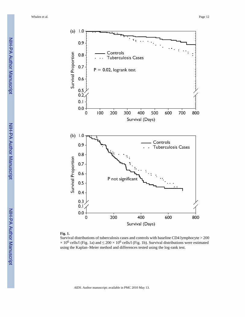
Fig. 1.Survival distributions of tuberculosis cases and controls with baseline CD4 lymphocyte > 200× 106 cells/l (Fig. 1a) and ≤ 200 × 106 cells/l (Fig. 1b). Survival distributions were estimatedusing the Kaplan–Meier method and differences tested using the log-rank test.
Whalen et al. Page 12
AIDS. Author manuscript; available in PMC 2010 May 13.
NIH
-PA Author Manuscript
NIH
-PA Author Manuscript
NIH
-PA Author Manuscript
NIH
-PA Author Manuscript
NIH
-PA Author Manuscript
NIH
-PA Author Manuscript
Whalen et al. Page 13
Table 1
Baseline characteristics of HIV-infected tuberculosis cases and controls.
Characteristic Cases (n = 230)HIV-infected controls
(n = 442) P
Males [n (%)] 111 (48) 120 (27) 0.000
Bacillus Calmette–Guérin vaccine scar [n (%)] 124 (54) 273 (62) 0.000
HIV-related conditionsa [n (%)] 72 (31) 219 (50) 0.000
Chronic diarrhea [n (%)] 32 (14) 74 (17) 0.34
Oral thrush [n (%)] 24 (10) 56 (13) 0.39
History of herpes zoster [n (%)] 22 (9) 80 (18) 0.003
History of pruritic papules [n (%)] 17 (7) 130 (29) 0.000
Appetite [n (%)] 130 (57) 74 (17) 0.000
Weight loss [n (%)] 197 (86) 164 (37) 0.000
Fever [n (%)] 144 (63) 114 (26) 0.000
Nightsweats[n(%)] 152 (66) 58 (13) 0.000
Cough [n (%)] 226 (98) 194 (44) 0.000
CD4 lymphocyte count (× 106/l) [n (%)]
0–200 82 (36) 138 (31) 0.12
201–500 87 (38) 152 (34)
> 500 61 (26) 152 (34)
0–200 82 (36) 138 (31) 0.24
> 200 148 (64) 304 (69)
Body mass index (kg/m2) [mean (SD)] 19.2 (2.7) 22.5 (3.5) 0.000
PPD induration (mm) [mean (SD)] 14.5 (6.8) 8.0 (8.0) 0.000
Hemoglobin (mg/dl) [mean (SD)] 10.3 (2.3) 12.5 (1.9) 0.000
Absolute lymphocyte count (× 106cells/l) [mean (SD)] 2087 (933) 2123 (861) 0.62
CD4 lymphocyte count (× 106cells/l) [mean (SD)] 376 (311) 431 (367) 0.09
aHIV-related conditions includes oral thrush, herpes zoster, chronic diarrhea and pruritic papules.
AIDS. Author manuscript; available in PMC 2010 May 13.
NIH
-PA Author Manuscript
NIH
-PA Author Manuscript
NIH
-PA Author Manuscript
Whalen et al. Page 14
Table 2
Stratified analysis of mortality according to key variables [deaths/number at risk (%)].
Category Cases HIV-infected controls Relative risk of death 95% CI
CD4 lymphocyte count (× 106cells/l)
0–200 38/82 (46) 61/138 (44) 1.1 0.78–1.42
> 200 25/148 (17) 24/304 (8) 2.1 1.27–3.62
Previous HIV-related condition
Yes 32/72 (44) 69/219 (32) 1.4 1.02–1.95
No 31/158 (20) 16/223 (7) 2.7 1.55–4.82
Hemoglobin (mg/dl)
≤ 10 38/100 (38) 22/49 (45) 0.8 0.57–1.27
> 10 25/128 (20) 63/393 (16) 1.2 0.80–1.85
Body mass index (kg/m2)
≤ 21 51/180 (28) 48/154 (31) 0.9 0.65–1.27
> 21 12/50 (24) 37/288 (15) 1.9 1.05–3.33
Tuberculin skin test induration (mm)
< 5 13/27 (48) 44/168 (26) 1.8 1.15–2.93
≥ 5 50/203 (25) 41/274 (15) 1.7 1.39–2.39
CI, Confidence interval.
AIDS. Author manuscript; available in PMC 2010 May 13.
NIH
-PA Author Manuscript
NIH
-PA Author Manuscript
NIH
-PA Author Manuscript
Whalen et al. Page 15
Table 3
Univariate analysis of factors associated with survival.
Characteristic Relative hazard 95% Confidenceinterval
P
Tuberculosis case 1.7 1.19–2.39 0.004
HIV-infected control 1.0
Age (years)
≤ 30 0.9 0.64–1.22 0.42
> 30 1.00
Sex
Male 1.4 1.04–1.97 0.035
Female 1.00
Tuberculin skin test induration
< 5 mm 1.9 1.42–2.77 0.0002
≥ 5 mm 1.00
Hemoglobin (mg/dl)
≤ 10 2.9 2.07–3.98 0.00000
> 10 1.00
Body mass index (kg/m2)a
≤ 21 2.5 1.79–3.62 0.00000
> 21 1.00
CD4 count (×106cells/l)
≤ 200 1.0 - 0.0000
201–500 0.2 0.16–0.34
> 500 0.1 0.04–0.13
History of HIV-related conditionb
Yes 3.5 2.48–5.01 0.00000
No 1.00
aMedian value for body mass index was 20.8 kg/m2.
bHIV-related conditions include oral thrush, chronic diarrhea, herpes zoster, and pruritic papules.
AIDS. Author manuscript; available in PMC 2010 May 13.
NIH
-PA Author Manuscript
NIH
-PA Author Manuscript
NIH
-PA Author Manuscript
Whalen et al. Page 16
Tabl
e 4
Rel
ativ
e ha
zard
s [95
% c
onfid
ence
inte
rval
s (C
I)] f
or d
eath
am
ong
tube
rcul
osis
(TB
) cas
es c
ompa
red
with
con
trols
, stra
tifie
d ac
cord
ing
to th
e m
edia
n va
lue
of th
e bo
dy m
ass i
ndex
.
Stra
tifie
d m
odel
s
Ove
rall
mod
elB
ody
mas
s ind
ex >
21
(kg/
m2 )
Bod
y m
ass i
ndex
≤21
(kg/
m2 )
Rel
ativ
e ha
zard
95%
CI
Rel
ativ
e ha
zard
95%
CI
Rel
ativ
e ha
zard
95%
CI
CD
4 ce
ll co
unt (
× 10
6 cel
ls/l)
> 2
00
TB c
ase
3.0
1.64
–5.6
66.
21.
74–2
2.07
2.2
0.96
–5.0
1
Con
trol
1.0
1.0
1.0
≤ 2
00
TB c
ase
1.5
0.99
–2.3
96.
42.
19–1
8.68
0.9
0.54
–1.5
2
Con
trol
1.0
1.0
1.0
Age
(yea
rs)
>30
0.9
0.72
–1.3
91.
30.
74–2
.38
0.9
0.63
–1.4
7
≤ 3
01.
01.
001.
00
Sex
M
ale
1.3
0.94
–1.8
51.
20.
60–2
.25
1.2
0.78
–1.8
0
F
emal
e1.
01.
001.
00
His
tory
of H
IV-r
elat
ed c
ondi
tion
Y
es3.
02.
11–4
.40
4.0
2.03
–8.1
12.
61.
64–4
.11
N
o1.
01.
001.
00
AIDS. Author manuscript; available in PMC 2010 May 13.