Tuberculosis - MSF Medical Guidelines
308
Exported on: 20/07/2022 Tuberculosis Practical guide for clinicians, nurses, laboratory technicians and medical auxiliaries © Médecins Sans Frontières
-
Upload
khangminh22 -
Category
Documents
-
view
0 -
download
0
Transcript of Tuberculosis - MSF Medical Guidelines

Exported on: 20/07/2022
TuberculosisPractical guide for clinicians, nurses, laboratory technicians and medicalauxiliaries
© Médecins Sans Frontières

ISBN
All rights reserved for all countries. No reproduction, translation and adaptation may be done without
the prior permission of the Copyright owner.
Médecins Sans Frontières and Partners in Health. Tuberculosis.
January 2022
978-2-37585-149-4
ContenidoAuthors/Contributors
Introduction
Abbreviations and acronyms
Chapter 1: Introduction and epidemiology
1.1 Characteristics of Mycobacterium tuberculosis bacillus
1.2 Transmission
1.3 Evolution of tuberculosis infection and disease in humans
1.4 Prognosis
1.5 Factors modifying tuberculosis epidemiology
1.6 Epidemiological indicators
1.7 Global burden of tuberculosis
Chapter 2: Clinical presentation
2.1 Pulmonary tuberculosis
2.2 Extrapulmonary tuberculosis
2.3 Disseminated or miliary tuberculosis
2.4 Clinical presentation in HIV-infected patients
2.5 Summary of clinical presentations of tuberculosis
Chapter 3: Diagnostic investigations
3.1 Sputum smear microscopy
3.2 Culture
3.3 Phenotypic drug susceptibility tests (DST)
3.4 Molecular techniques
3.5 Summary of bacteriological examinations
3.6 Indications for DST
3.7 Radiology
3.8 Tuberculin skin test (TST)

3.9 Interferon gamma release assays (IGRAs)
3.10 Biopsies, laboratory tests on body fluids and other biological tests
Chapter 4: Diagnostic algorithms for pulmonary tuberculosis (PTB) in adults and adolescents
4.1 Guiding principles for the use of the algorithms
4.2 Adult and adolescent algorithms
Chapter 5: Diagnosis of tuberculosis in children
5.1 Background
5.2 Characteristics of tuberculosis in children
5.3 Diagnostic approach
5.4 Key elements of the diagnosis
5.5 Collecting sputum specimens in children
5.6 Paediatric diagnostic algorithms
Chapter 6: Intensive case finding in HIV-infected individuals
6.1 Routine screening
6.2 Purposes of screening
Chapter 7: Case definitions for registration
7.1 Definition of a tuberculosis case
7.2 History of prior anti-TB treatment
7.3 Anatomical site of the disease
7.4 Bacteriological status
7.5 HIV status
7.6 Other co-morbidities
7.7 Summary of patient registration
Chapter 8: Anti-TB drugs and treatment regimens
8.1 Introduction
8.2 Anti-TB drug formulations
8.3 Quality-assured anti-TB drugs
8.4 Dosing of anti-TB drugs
8.5 Cross resistance
Chapter 9: Treatment of drug-susceptible tuberculosis
9.1 Standard first-line treatment regimens
9.2 Special situations
9.3 Adjunctive corticosteroid therapy
9.4 Follow-up for patients treated with first-line regimens
9.5 Management of adverse effects in patients on first-line regimens
9.6 Management of treatment interruption in patients on first-line regimens
Chapter 10: Treatment of multidrug-resistant TB (MDR-TB)
10.1 Design of therapeutic regimens in MDR-TB
10.2 Selection of anti-TB drugs in MDR-TB regimens
10.3 Building a treatment regimen for MDR-TB
10.4 Duration of MDR-TB regimens
10.5 Follow-up for patients treated for MDR-TB
10.6 Management of adverse effects in patients on second-line regimens
10.7 Surgery as an adjunctive treatment measure
10.8 Management of patients whose treatment failed and palliative care
10.9 Special situations
10.10 Treatment of extensively drug-resistant TB (XDR-TB)
Chapter 11: Treatment of mono- and poly-drug resistant tuberculosis (PDR-TB)
11.1 Treatment schemes
11.2 Treatment algorithms for PDR-TB
Chapter 12: Co-management and treatment of HIV in TB disease
12.1 HIV testing and counselling for patients known or suspected to have TB
12.2 Prophylaxis against opportunistic infections
12.3 Anti-TB regimens in HIV patients
12.4 Concomitant treatment TB and HIV
12.5 Drug interactions
12.6 Overlapping toxicities with anti-TB drugs and antiretrovirals
12.7 Immune reconstitution inflammatory syndrome (IRIS)
12.8 HIV-infected children with TB
12.9 HIV-infected pregnant women with TB
12.10 HIV-infected patients with DR-TB
Chapter 13: Adherence to tuberculosis treatment
13.1 Introduction
13.2 Treatment delivery model
13.3 Factors that influence adherence
13.4 Patient education and support
Chapter 14: Tuberculosis infection control

14.1 Introduction
14.2 Implementation of TB IC strategies
14.3 Administrative controls
14.4 Environmental controls
14.5 Personal protective measures
14.6 Hospital hygiene
14.7 Patients’ homes
Chapter 15: Follow-up of staff exposed to tuberculosis
15.1 Initial assessment
15.2 BCG vaccination
15.3 Follow-up
Chapter 16: Treatment of latent tuberculosis infection
16.1 Introduction
16.2 Target populations
16.3 Latent tuberculosis infection treatment regimens
16.4 Latent tuberculosis infection in HIV-infected patients
16.5 Latent tuberculosis infection in household contacts
16.6 Latent tuberculosis infection in other individuals at risk
16.7 Latent tuberculosis infection and multidrug-resistant tuberculosis
16.8 Follow-up for patients treated for latent tuberculosis infection
Chapter 17: Monitoring and evaluation
17.1 Introduction
17.2 Definitions of treatment outcomes
17.3 Recording tools
17.4 Reporting
17.5 Programme assessment
Appendices
Appendix 3. Sputum specimen: collection, storage and shipment
Appendix 3. Xpert MTB/RIF
Appendix 4. Sputum smear microscopy
Appendix 6. Adenosine desaminase assay (ADA)
Appendix 7. Lymph node fine needle aspiration
Appendix 7. Ventilated work station (VWS) and bio-safety cabinet (BSC)
Appendix 8. Protein estimation

Appendix 8. Daily dose of anti-TB drugs using FDCs
Appendix 8a. New paediatric FDCs
Appendix 8b. Former paediatric FDCs
Appendix 9. Tuberculin skin test
Appendix 10. Tuberculosis drug information sheets and patient instructions
Tuberculosis drug information sheets
Amikacin (Am)
Amoxicillin/clavulanic acid ratio 4:1 (Amx/Clv)
Clofazimine (Cfz)
Cycloserine (Cs) or terizidone (Trd)
Ethambutol (E)
Ethionamide (Eto) or prothionamide (Pto)
Imipenem/cilastatin (Ipm/Cln)
Isoniazid - Standard dose (H)
Levofloxacin (Lfx)
Linezolid (Lzd)
Meropenem (Mpm)
Moxifloxacin (Mfx)
Para-aminosalicylic acid (PAS) and sodium salt of PAS
Pyrazinamide (Z)
Rifabutin (Rfb)
Rifampicin (R)
Streptomycin (S)
Patient instructions
Patients on drug-susceptible TB treatment
Patients on drug-resistant TB treatment
Appendix 11. Compassionate use
Appendix 12. Dose adjustments in renal insufficiency
Appendix 16. Basic TB infection control risk assessment tool
Appendix 17. Management of adverse effects
Gastrointestinal disorders

Abdominal pain
Diarrhoea
Epigastric pain
Hepatotoxicity
Metallic taste
Nausea and vomiting
Neurotoxicity
Depression
Headache
Optic neuritis
Ototoxicity
Peripheral neuropathy
Psychosis
Seizures
Endocrine disorders
Gynecomastia
Hypothyroidism
Dermatological disorders
Alopecia
Fungal infection
Photosensitivity
Skin reactions
Musculoskeletal disorders
Arthralgias
Tendinitis/tendon rupture
Miscellaneous
Electrolyte disorders
Haematologic disorders
Lactic acidosis
Nephrotoxicity

QT prolongation
Appendix 17. Air change per hour (ACH) measurement recommendations
Appendix 18. Advantages and disadvantages of ventilation techniques
Appendix 19. Potential overlapping toxicities of ARVs and anti-TB drugs
Appendix 19. Upper room ultraviolet germicidal irradiation (UVGI) system
Appendix 20. Treatment supporters
Appendix 21. Informing the patient
Appendix 23. Treatment card for patients on first-line anti-TB therapy
Appendix 24. Tuberculosis register for patients on first-line anti-TB therapy
Appendix 25. Treatment card for patients on second-line anti-TB therapy
Appendix 26. Tuberculosis register for patients on second-line anti-TB therapy
Appendix 27. Respirators
Appendix 27. Request form for microscopy and Xpert MTB/RIF
Appendix 28. Surgical masks
Appendix 28. Request form for sputum culture, LPA and DST
Appendix 29. BCG vaccine
Appendix 29. Sputum smear microscopy register
Appendix 30. Xpert MTB/RIF register
Appendix 31. Drug-o-gram
Appendix 32. Quaterly report
Appendix 33. Report on detection and enrolment of TB cases with rifampicin and multidrug-
resistance
Appendix 34. Report of final outcomes of drug-resistant tuberculosis
Appendix 35. Check-list for the evaluation of a TB service
Authors/Contributors
Chief Authors
Francis Varaine, M.D.
Michael L. Rich, M.D., M.P.H.
Technical Editor
Véronique Grouzard

Contributing Authors
PIH
Amy Elizabeth Barrera-Cancedda, Salmaan Keshavjee, Carole Mitnick, Joia Mukherjee, Anne Peruski,
Kwonjune Seung
MSF
Elisa Ardizzoni, Saar Baert, Suna Balkan, Karen Day, Philipp Ducros, Gabriella Ferlazzo, Cecilia
Ferreyra, Marianne Gale, Pamela Hepple, Myriam Henkens, Cathy Hewison, Northan Hurtado, Frauke
Jochims, Jean Rigal, Joannie Roy, Peter Saranchuk, Clara Van Gulik, Carole Zen Ruffinen
Published by
Médecins Sans Frontières
Partners In Health
Introduction
Tuberculosis is an infectious disease caused by Mycobacterium tuberculosis. Tuberculosis typically
attacks the lungs, but can also affect other parts of the body. The disease has become rare in high
income countries, but is still a major public health problem in low- and middle-income countries.
It is estimated that between the years 2000 and 2010, eight to nine million new cases emerged each
year. Approximately 1.5 million people die from the disease each year. In adults, tuberculosis is the
second leading cause of death due to an infectious disease (after AIDS), with 95% of deaths occurring
in low-income countries. Tuberculosis is a major problem of children in poor countries where it kills over
100,000 children each year.
The treatment of tuberculosis remains a constraint for patients and a heavy burden for the healthcare
system. Drug-susceptible tuberculosis requires at least six months of therapy under close supervision.
A treatment for multidrug-resistant tuberculosis requires nearly two years of treatment with poorly
tolerated and less effective drugs. In most places the diagnosis still relies mainly on direct microscopy
that is unable to detect a large proportion of patients. The BCG vaccine, developed almost a century
ago, confers only partial protection.
After 40 years of minimal progress in the tools to fight tuberculosis there are some reasons for hope. A
few new drugs are reaching the final phase of development; a new molecular test that can be
decentralized to some extent and allows the rapid diagnosis of tuberculosis and of resistance to
rifampicin has been introduced. Though this is undeniable progress, much will be needed to bring the
new tools and drugs to the patients in need. Furthermore, a true “point of care” diagnostic test still
does not exist and little progress has been made in research for a more effective vaccine.

Case management of patients does not necessarily have to involve a major, vertical programme. It
should be incorporated into the framework of other medical activities in order to offer comprehensive
and integrated treatment even if the number of patients being treated is relatively small.
This guide has been developed jointly by Médecins Sans Frontières and Partners In Health. It aims at
providing useful information to the clinicians and health staff for the comprehensive management of
tuberculosis. Forms of susceptible and resistant tuberculosis, tuberculosis in children, and HIV co-
infection are all fully addressed.
As treatment protocols are constantly changing, medical staff are encouraged to check this website
for updates.
Abbreviations and acronyms

ACH Air change per hour
AFB Acid-fast bacilli
AIDS Acquired immunodeficiency syndrome
Amk Amikacin
Amx/Clv Amoxicillin/clavulanic acid
ARI Annual risk of infection
ART Antiretroviral therapy
ARV Antiretroviral
BCG Bacillus Calmette-Guérin
Bdq Bedaquiline
CDC United States Centers for Disease Control and Prevention
Cfz Clofazimine
Cm Capreomycin
CMX Cotrimoxazole
CPC Cetylpyridinum chloride
CPT Cotrimoxazole preventive therapy
Cs Cycloserine
CXR Chest X-ray
DOT Directly observed therapy
DR Drug resistance
DR-TB Drug-resistant tuberculosis

DST Drug susceptibility test(ing)
E Ethambutol
ECG Electrocardiogram
EPTB Extrapulmonary tuberculosis
Eto Ethionamide
FDC Fixed-dose combination
FNAC Fine needle aspiration cytology
FQ Fluoroquinolone
GFR Glomerular filtration rate
H Isoniazid
HCW Health care worker
HIV Human immunodeficiency virus
HPF High-power field
IC Infection control
IM Intramuscular
Imp/Cln Imipenem/cilastatin
IPT Isoniazid preventive therapy
IRIS Immune reconstitution inflammatory syndrome
IUATLD International Union against Tuberculosis and Lung Disease
Km Kanamycin
LFT Liver function test

Lfx Levofloxacin
LPA Line probe assay
Lzd Linezolid
MDR Multidrug resistance
MDR-TB Multidrug-resistant tuberculosis
Mfx Moxifloxacin
MGIT Mycobacteria growth indicator tube
MODS Microscopic observation of drug susceptibility
Mpm Meropenem
NNRTI Non-nucleoside reverse transcriptase inhibitor
NRTI Nucleoside reverse transcriptase inhibitor
NTM Non tuberculous mycobacteria
Ofx Ofloxacin
PAS Para-aminosalicylic acid
PCP Pneumocystosis
PCR Polymerase chain reaction
PI Protease inhibitor
PO Orally (per os)
Pto Prothionamide
R Rifampicin
Rfb Rifabutin

Chapter 1: Introduction and epidemiology1.1 Characteristics of Mycobacterium tuberculosis bacillus
1.2 Transmission
1.3 Evolution of tuberculosis infection and disease in humans
1.4 Prognosis
1.5 Factors modifying tuberculosis epidemiology
1.6 Epidemiological indicators
1.7 Global burden of tuberculosis
RR Rifampicin resistance
S Streptomycin
TAT Turn around time
TB Tuberculosis
Thz Thioacetazone
TLA Thin-layer agar
TSH Thyroid-stimulating hormone
TST Tuberculin skin test
UVGI Ultraviolet germicidal irradiation
WHO World Health Organization
XDR Extensive drug resistance
XDR-TB Extensively drug-resistant tuberculosis
Z Pyrazinamide
Update: January 2022
1.1 Characteristics of Mycobacteriumtuberculosis bacillus
Mycobacterium tuberculosis, along with M. bovis, M. africanum, M. microti and others, make up the
Mycobacterium tuberculosis complex, a group of bacteria that cause clinical tuberculosis (TB) in
humans.
Most TB cases are caused by M. tuberculosis. Cases due to other species are far less prevalent.
M. tuberculosis is a small, rod-shaped, strictly aerobic, acid-fast bacillus . Like other mycobacteria, it
is slow growing, resulting in more gradual development of disease when compared with other bacterial
infections.
1.2 Transmission
M. tuberculosis is transmitted from human-to-human and spread is mainly airborne. The source of
infection is usually a patient with pulmonary or laryngeal TB. During coughing, speaking, or sneezing, the
patient produces tiny infectious droplets. These particles, called droplet nuclei, are about 1 to 5
microns in diameter. Depending on the environment, they can remain suspended in the air for several
hours.
Transmission may occur when these infectious droplets are inhaled. UV light (sunshine or artificial
sources) and ventilation reduce the probability of transmission (Chapter 14).
a
Notas(a) Acid-fast bacilli are bacilli, which once stained, resist discolorat ion by acid and alcohol.

Other modes of transmission are far less common. Inoculation of cutaneous or mucous membranes
rarely occurs, although such cases have been observed in laboratory personnel. Congenital infection
(by transplacental transmission or via aspiration or swallowing of infected amniotic fluid at birth) has
been reported, but is very rare. Transmission through breast milk does not occur.
The infectiousness of a patient is associated with the quantity of bacilli contained in their sputum.
Patients with smear-positive sputum on microscopy are by far the most infectious. Those with smear-
negative/culture-positive results are less infectious, but still contribute to TB transmission due to more
frequent delays in diagnosis.
Patients infected with M. tuberculosis, but who have not developed active TB (latent tuberculosis
infection), are not infectious. Patients with extrapulmonary TB (EPTB) are only infectious in
exceptional circumstances.
Children are generally much less infectious than adults. This may be due to weaker cough mechanics,
less sputum production and lower bacillary load.
Not everyone who is exposed to an infectious TB patient becomes infected with M. tuberculosis. The
probability that TB will be transmitted depends on several factors:
Infectiousness of the source (the most important factor)
• Bacteriological status: smear-positive patients are the most infectious.
• Virulence of the bacilli: some strains are highly transmissible (and/or more likely to cause active TB).
Environment where the exposure occurred
• Outdoor environments or those with good ventilation and sunlight are less likely to lead to
transmission. Small rooms or rooms with no ventilation are conditions most likely to lead to
transmission.
• The proximity of the person to the patient is also important (e.g. the risk is higher if the person sleeps
next to the patient than if they sleep 20 metres away from the patient).
Duration of exposure
People in close and prolonged contact with TB patients are at highest risk of becoming infected with
M. tuberculosis. They may be family members, roommates, friends, co-workers or other people who
spend several hours a day with the infectious patient.
The best way to stop transmission is to start effective TB treatment as soon as possible. It is
estimated that a person with untreated smear-positive TB transmits the bacillus to 10 to 20 people a
year (with variations according to living conditions and environment).
1.3 Evolution of tuberculosis infection anddisease in humans
When a person inhales infectious droplets containing M. tuberculosis, most of the larger droplets
become lodged in the upper respiratory tract (nose and throat) where infection is unlikely to develop.
However, smaller droplet nuclei may reach the small air sacs of the lung (the alveoli) where infection can
occur.
1.3.1 Primary infection and latent tuberculosis infectionAfter transmission, M. tuberculosis multiplies slowly, in most cases in the terminal alveoli of the lungs
(primary focus) and in the lymph nodes of corresponding drainage areas: this is the primary infection.
The primary focus and related hilar lymphadenopathy form the primary complex.
In one to two months, due to the action of lymphocytes and macrophages (cellular immunity), the
primary focus is contained and encapsulated, with a central zone of parenchymal necrosis (caseous
lesions). It is not usually detectable on chest x-ray, unless it calcifies or grows substantially. Primary
infection is usually asymptomatic. In most cases (90 to 95% of non-HIV infected patients), the
pulmonary lesions gradually heal.
During the primary infection, specific immunity develops and a positive skin reaction to tuberculin is
observed . This immune response may persist without clinical signs of TB. The patient is infected by
M. tuberculosis, but does not develop the disease. This is referred to as latent tuberculosis infection
(LTBI).
In 5 to 10% of infected people, primary infection and/or LTBI progresses to active TB over their
lifetime. For HIV co-infected patients, this risk is much higher.
1.3.2 Active tuberculosisBefore immunity is established, bacilli from the primary infectious focus or from a near-by lymph node
can be transported and disseminated throughout the body via the lymph system or the bloodstream.
Secondary foci can develop this way, particularly in the lungs, lymph nodes, serous membranes,
meninges, bones and kidneys. As soon as an immune response is mounted, most of these foci resolve
spontaneously. However, some bacilli may remain dormant in the secondary foci for months and
sometimes years.
Different factors can reduce the immune response (e.g. HIV infection) and lead to reactivation of the
bacilli and their multiplication in one or more of these foci. This reactivation or progression of the
primary or secondary foci results in active TB .
An active TB lesion contains actively, slowly or sporadically multiplying bacilli as well as dormant bacilli.
While active TB may occur months or years following primary infection, half of TB cases appear in the
year following infection.
1.3.3 Risk factors for developing active tuberculosisCertain factors increase the risk of developing active TB within the first two years of being infected.
These factors include any factor that results in a weakened immune system, damaged lungs and the
intensity and duration of exposure.
[1]
[2]

Host immune response factors:
Conditions that damage the lung:
Intensity of exposure (high number of inhaled bacilli):
Referencias
HIV infection
Children under 5 years [3]
[4]
Malnutrition
Persons over 60 years
Diabetes mellitus
Other risk factors: prolonged corticosteroid therapy (g. prednisolone) and other immunosuppressive
therapies, severe kidney disease, alcoholism, substance abuse, certain types of cancer (e.g.
leukaemia, Hodgkin's lymphoma, cancer of the head and neck); pregnancy
Tobacco smoking
Silicosis
Chronic obstructive pulmonary disease (COPD)
Highly infectious source
Poorly ventilated environment
Proximity with infectious source, including residents and employees of institutions such as prisons,
boarding schools and residential care facilities
Long duration of exposure
1. Ahmad, S. Pathogenesis, immunology, and diagnosis of latent Mycobacterium tuberculosis infection. Clin Dev
Immunol. 2011: p. 814943.
ht tp://downloads.hindawi.com/journals/jir/2011/814943.pdf
2. Ai, JW, et al. Updates on the risk factors for latent tuberculosis reactivation and their managements. Emerging
Microbes & Infect ions, 2016. 5: p. e10.
ht tps://www.ncbi.nlm.nih.gov/pmc/art icles/PMC4777925/pdf /emi201610a.pdf
3. Marais, BG, et al. The natural history of childhood intra-thoracic tuberculosis: A critical review of literature from
the pre-chemotherapy era. The internat ional journal of tuberculosis and lung disease: the official journal of
the Internat ional Union against Tuberculosis and Lung Disease. 2004. 8(4) p. 392-402.
ht tps://www.ingentaconnect .com/content /iuat ld/ijt ld/2004/00000008/00000004/art00002;jsessionid=11hrh
2bb6hfpv.x-ic-live-03#
4. Erkens, CG., et al. Risk of developing tuberculosis disease among persons diagnosed with latent tuberculosis
infection in the Netherlands. Eur Respir J, 2016. 48(5): p. 1420-1428.
ht tps://erj.ersjournals.com/content /erj/48/5/1420.full.pdf

1.4 Prognosis
Without treatment, TB is a severe and potentially fatal disease. After 5 years without treatment, the
outcome of smear-positive pulmonary TB (PTB) in non-HIV-infected patients is as follows :
In non-HIV infected patients, the CFR is estimated at 3% . Untreated TB in HIV-infected patients
(not on effective antiretroviral therapy) is almost always fatal. Even on antiretroviral therapy, the CFR is
higher than in non-HIV infected patients .
Risk factors for poor outcomes of TB treatment (death and relapse) include co-morbidities (e.g. HIV
infection, diabetes, COPD), cavities on chest x-ray, high bacillary load and resistance to TB drugs.
Referencias
1.5 Factors modifying tuberculosisepidemiology
[1]
50 to 60% die (case fatality ratio (CFR) for untreated TB);
20 to 25% are cured (spontaneous cure);
20 to 25% continue to have symptoms.
[2]
[3]
[4]
1. Tiemersma EW, van der Werf MJ, Borgdorf f MW, Williams BG, Nagelkerke NJ. Natural history of tuberculosis:
duration and fatality of untreated pulmonary tuberculosis in HIV negative patients: a systematic review. PLoS
One. 2011;6(4):e17601. Published 2011 Apr 4.
ht tps://www.ncbi.nlm.nih.gov/pmc/art icles/PMC3070694/pdf /pone.0017601.pdf
2. St raetemans M, Glaziou P, Bierrenbach AL, Sismanidis C, van der Werf MJ (2011). Assessing Tuberculosis
Case Fatality Ratio: A Meta-Analysis. PLoS ONE 6(6): e20755.
ht tps://doi.org/10.1371/journal.pone.0020755
ht tps://journals.plos.org/plosone/art icle?id=10.1371/journal.pone.0020755
3. World Health Organizat ion. Global Tuberculosis Report 2021. Geneva: World Health Organizat ion; 2021.
ht tps://apps.who.int /iris/rest /bitst reams/1379788/ret rieve
4. Manosuthi, W., et al. Survival rate and risk factors of mortality among HIV/tuberculosis-coinfected patients with
and without antiretroviral therapy. J Acquir Immune Defic Syndr, 2006. 43(1): p. 42-6.
ht tps://journals.lww.com/jaids/fulltext /2006/09000/Survival_Rate_and_Risk_Factors_of_Mortality_Among.7.a
spx

Five major factors influence TB epidemiology: (1) socioeconomic conditions, (2) TB treatment, (3) HIV
infection, (4) diabetes and (5) BCG vaccination.
1.5.1 Socioeconomic conditionsThe principal factors leading to a reduction in TB cases are improved social and housing conditions.
Most cases occur in low-income countries. In industrialised countries, TB generally affects the most
disadvantaged social groups.
1.5.2 Tuberculosis treatmentDiagnosing and initiating effective treatment in a patient early during their TB disease before they can
infect multiple people is considered the most effective preventive measure against TB. Once an
effective TB treatment is started, there is a rapid reduction in transmission .
Since the introduction of TB treatment, the risk of TB infection decreased by approximately 10% per
year in industrialised countries . This trend was observed in countries with a BCG vaccination
programme as well as in those without one. Detection programmes, diagnosis and treatment of TB
contributed to this reduction in the risk of TB infection.
1.5.3 HIV infectionImmunodeficiency induced by HIV infection is a major risk factor for progression to active TB and has
a considerable impact on the epidemiology of TB. While the lifetime risk of developing active TB in the
general population is 5 to 10% after infection with M. tuberculosis, this risk is approximately 10% per
year in patients co-infected with HIV and M. tuberculosis. Approximately 8% of incident TB cases in
the world are among HIV-infected patients (highest in the WHO African Region, more than 50% in parts
of southern Africa) .
1.5.4 DiabetesThe risk of TB among people with diabetes is higher than among those without diabetes. It is
estimated that diabetes contributes to 15% of TB cases worldwide . Diabetes is also associated
with poor absorption of TB drugs and therefore higher rates of drug resistant tuberculosis (DR-TB).
1.5.5 BCG vaccinationEffectiveness of BCG at the individual level
BCG vaccination, if given at birth, is highly effective against the severe forms of TB (miliary and
meningitis) in children .
Epidemiological impact of vaccination
Despite some protection from the BCG vaccination, the impact of BCG vaccination on TB
transmission and the TB epidemic is considered negligible .
[1]
[2]
[3]
[4]
[5]
[6]
[7]

1.5.6 Other factorsOther modifying factors include infection control measures (Chapter 14) and treatment of LTBI
(Chapter 16). The degree to which in a given context the TB epidemiology is affected by these
measures is not known.
Referencias
1.6 Epidemiological indicators
When a National TB Programme (NTP) functions well, indicators can be obtained from the local
authorities and NTP.
The WHO tuberculosis country profiles also provide an estimation of TB indicators by individual
country .
Box 1.1 - Most common indicators
1. Nardell, EA. Transmission and institutional infection control of tuberculosis. Cold Spring Harb Perspect Med.
2016;6(2):1-12.
ht tps://www.ncbi.nlm.nih.gov/pmc/art icles/PMC4743075/pdf /cshperspectmed-TUB-a018192.pdf
2. Giovanni Bat t ista Migliori, Lia D'Ambrosio, Rosella Cent is, Mart in Van Den Boom, Soudeh Ehsani, Masoud
Dara. Guiding Principles to Reduce Tuberculosis Transmission in the WHO European Region. World Health
Organizat ion, 2018.
ht tps://www.euro.who.int /__data/assets/pdf_file/0008/377954/ic-principles-eng.pdf
3. E Vynnycky and PEM Fine. Interpreting the decline of tuberculosis: the role of secular trends in effective
contact. Internat ional Journal of Epidemiology. 1999; 28:327-334
4. World Health Organizat ion. Global Tuberculosis Report 2021. Geneva: World Health Organizat ion; 2021.
ht tps://apps.who.int /iris/rest /bitst reams/1379788/ret rieve
5. World Health Organizat ion & Internat ional Union against Tuberculosis and Lung
Disease. ( 2011) . Collaborative framework for care and control of tuberculosis and diabetes. World Health
Organizat ion.
ht tps://apps.who.int /iris/handle/10665/44698
6. World Health Organizat ion. (2018). BCG vaccines: WHO position paper – February 2018. Weekly
Epidemiological Record, 93( 08) ,73-96. World Health Organizat ion.
ht tps://apps.who.int /iris/bitst ream/handle/10665/260307/WER9308-73-96.pdf?sequence=1&isAllowed=y
7. Pai, M., Behr, M., Dowdy, D, et al. Tuberculosis. Nat Rev Dis Primers 2, 16076 (2016).
a

Annual incidence rate of TB cases
Numerator: number of new TB cases (all forms) that occur in a population over one year
Denominator: population at the start of the year
Annual incidence rate of smear-positive PTB cases
Numerator: number of new smear-positive PTB cases that occur in a population over one year
Denominator: population at the start of the year
Prevalence of smear-positive PTB cases over a given period of time, usually one year
Numerator: number of smear-positive PTB cases
Denominator: population at the start of the period of time
Proportion of multidrug- and rifampicin-resistant TB cases among TB cases over a given
period of time
Numerator: number of multidrug- and rifampicin-resistant TB cases
Denominators:
- Total number of TB cases
- Number of new TB cases
- Number of previously treated TB cases
Proportion of extensively drug-resistant TB cases among TB cases over a given period of
time
Numerator: number of extensively drug-resistant cases
Denominators: as for multidrug- and rifampicin-resistant TB cases
Proportion of HIV-infected patients among new TB cases over a given period of time
Numerator: number of HIV-infected patients
Denominator: number of new TB cases
(a)
(a)
(b)
(c)
(c)
(c)
(a) The rate is expressed as the number of new TB cases (or new smear-posit ive PTB cases) per 100,000
populat ion.
(b) Prevalence is expressed as the number of smear-posit ive PTB cases per 100,000 populat ion. It includes
new and pre-exist ing cases. Prevalence represents approximately double the incidence rate.
(c) Proport ion is expressed in %.
Notas(a) For more informat ion:
ht tps://worldhealthorg.shinyapps.io/tb_profiles/?_inputs_&ent ity_type=%22group%22&lan=%22FR%22

1.7 Global burden of tuberculosis1.7.1 Latent tuberculosis infectionThe global prevalence of LTBI is unknown due to difficulties in diagnosis. However, WHO estimates
that one-quarter of the world population has LTBI .
1.7.2 Active tuberculosisGlobally, active TB remains a leading cause of death from infectious disease.
WHO estimates that each year there are approximately 10 million incident cases of TB and 1.5 million
deaths due to TB, including 1.3 million among HIV-negative individuals and 214,000 among HIV-
infected individuals .
Children under 15 years account for 11% of all estimated TB cases . However, TB cases in children
are frequently undiagnosed and unreported.
While the absolute number of global TB cases is stable, there are large individual country and regional
differences in incidence and prevalence.
Most TB cases are in Southeast Asia (43%), Africa (25%) and the Western Pacific (18%), with lower
percentages in the Eastern Mediterranean, the Americas and Europe .
1.7.3 Drug-resistant tuberculosis Drug-resistant TB (DR-TB) is a growing worldwide problem, and no region is spared.
WHO estimates that annually worldwide there are :
• More than one million rifampicin-susceptible and isoniazid-resistant TB (Hr-TB) cases (11% of all
incident TB cases).
• 3.3% of new cases and 18% of previously treated cases, with multidrug-resistant TB (MDR-TB)
and rifampicin-resistant TB (RR-TB) representing 465,000 cases and 182,000 deaths.
In Eastern Europe and Central Asia, TB incidence is lower than in Southeast Asia and Africa, but up to
30% of new and 65% of retreatment cases exhibit rifampicin-resistance.
In China and India, there is a low proportion of rifampicin-resistant cases among all TB cases.
However, because of their large populations, these two countries represent 41% of global MDR/RR-
TB cases.
Resources for detecting drug resistance are limited in many parts of Africa. However, available data
suggest that the MDR-TB burden is significant, especially in the south.
The prevalence of extensively drug-resistant TB (XDR-TB) , according to the new WHO definition, is
currently unknown.
[1]
[2]
[2]
[2]
[3]
a
b
c

Referencias
Chapter 2: Clinical presentation2.1 Pulmonary tuberculosis
2.2 Extrapulmonary tuberculosis
2.3 Disseminated or miliary tuberculosis
2.4 Clinical presentation in HIV-infected patients
2.5 Summary of clinical presentations of tuberculosis
Update: January 2022
2.1 Pulmonary tuberculosis
Prolonged cough (more than 2 weeks), with or without sputum production, is a common symptom in
patients with pulmonary tuberculosis (PTB).
Notas(a) Mult idrug-resistant : resistance to at least rif ampicin and isoniazid.
(b) Rif ampicin-resistant : resistance to rif ampicin, with or without resistance to other TB drugs.
(c) Extensively drug-resistant : rif ampicin-resistance with resistance to any fluoroquinolone, and at least either
bedaquiline or linezolid.
1. Houben RM, Dodd PJ. The Global Burden of Latent Tuberculosis Infection: A Re-estimation Using
Mathematical Modelling. PLoS Med. 2016;13(10):e1002152. Published 2016 Oct 25.
ht tps://www.ncbi.nlm.nih.gov/pmc/art icles/PMC5079585/pdf /pmed.1002152.pdf
2. World Health Organizat ion. Global Tuberculosis Report 2021. Geneva: World Health Organizat ion; 2021.
ht tps://apps.who.int /iris/rest /bitst reams/1379788/ret rieve
3. World Health Organizat ion. Global Tuberculosis Report 2020. Geneva: World Health Organizat ion; 2020.
ht tps://apps.who.int /iris/rest /bitst reams/1312164/ret rieve

Other frequent, less specific, signs and symptoms include weight loss, anorexia, fatigue, haemoptysis
(blood in sputum), shortness of breath, chest pain, moderate fever and night sweats.
Signs and symptoms may vary between individuals and generally evolve in a chronic, insidious manner.
History-taking is therefore of the utmost importance.
Advanced forms and complications are common:
In endemic areas, the diagnosis of PTB should be considered in any patient consulting for respiratory
symptoms lasting more than 2 weeks.
Table 2.1 provides a differential diagnosis of PTB for non-HIV infected patients.
Table 2.1 - Differential diagnosis for PTB (non-HIV infected patients)
Respiratory insufficiency due to extensive lesions and destroyed lungs;
Massive haemoptysis due to large cavities with hyper-vascularisation and erosion of vessels;
Pneumothorax due to the rupture of a cavity in the pleural space.

For differential diagnosis in HIV-infected patients see Section 2.4.
2.2 Extrapulmonary tuberculosis
Diseases Remarks
Bacterial
pneumonia
Usually more acute and shorter in duration; high fever often present.
Response to antibiotics with no anti-TB activity suggests bacterial
pneumonia.
Lobar consolidation is typical of bacterial pneumonia; however, CXR alone
cannot differentiate PTB from bacterial pneumonia.
Pulmonary
abscess
May arise from aspiration in individuals with impaired consciousness (coma,
intoxication with alcohol/drugs, etc.).
Foul-smelling, purulent sputum.
Cavities typically have a thick wall and air fluid levels.
Bronchiectasis Frequent complication of successive, poorly-treated bronchopulmonary
infections in tropical regions.
Characterised by chronic or repeated episodes of productive cough.
Hemoptysis, usually mild, can be present.
Lung cancer History of smoking or environmental exposure (working in a mine, etc.).
Haemoptysis in 20 to 50% of patients.
Paragonimiasis
(lung flukes)
To be ruled out in presumed PTB cases in endemic areas (certain areas of
Southeast Asia, West Africa and Latin America).
Pulmonary
echinococcosis
(hydatid disease)
In Latin America, the Middle East, some Sub-Saharan African countries and
China.
Lung involvement may cause chronic cough, with or without haemoptysis.
Cysts can mimic TB cavities.
Pneumocystosis Common in patients with advanced HIV disease and patients receiving long-
term, even low dose, corticosteroid therapy.
Less common
diseases
Silicosis, sarcoidosis, melioidosis.
Cryptococcosis, aspergillosis, histoplasmosis.

Starting from a pulmonary localisation (primary infection), M. tuberculosis can spread to other organs
during a silent phase, usually soon after primary infection (Chapter 1). Active TB can develop in many
other parts of the body, particularly in lymph nodes, meninges, bones and joints, kidneys, genital organs
and the abdominal cavity.
Extrapulmonary tuberculosis (EPTB) can develop at any age. Due to relative immunodeficiency, young
children, HIV-infected and malnourished patients are more at risk of developing EPTB.
Approximately 15% of global TB cases are classified as EPTB, although this figure varies according
the local epidemiology .
A patient with EPTB may also have pulmonary involvement, which should be searched for whenever
EPTB is diagnosed or suspected.
Table 2.3 at the end of this chapter summarises the characteristics of EPTB.
2.2.1 Lymph node tuberculosisLymph node TB is common, particularly in certain areas of Africa and Asia, and especially in children
and HIV-infected patients.
The presentation of lymph node TB is a non-inflammatory adenopathy. Nodes are cold and painless,
multiple (usually bilateral) or single, evolving in a chronic mode towards softening and fistulisation.
Cervical localisation is most frequent. Axillary and mediastinal localisations are also common. Other
sites may be involved.
Diagnosis may be clinical, but whenever possible, fine needle aspiration should be performed (Chapter 3
and Appendix 7).
Adenopathy usually disappears within 3 months of treatment initiation. Paradoxical reactions may
occur at the beginning of treatment (appearance of abscesses, fistulas or other lymph nodes), but a
change in the treatment is not required.
Differential diagnoses include malignancies (lymphoma, leukaemia, ear/nose/throat tumours, Kaposi
sarcoma) and other infections (bacterial, viral, non-tuberculosis mycobacteria, toxoplasmosis, HIV
infection, syphilis, African trypanosomiasis).
2.2.2 Tuberculous meningitisTB meningitis is a serious form of TB that affects the meninges. It is most common in children under 2
years and in HIV-infected patients. It is a medical emergency. Any delay in diagnosis or treatment will
result in irreversible neurological sequelae or death .
TB meningitis typically has a subacute insidious course over days or weeks. Symptoms include
headaches, irritability, fever, vomiting and altered mental status, which worsen if treatment is delayed.
The meningeal syndrome (stiff neck, hypotonia in infants, photophobia and headache) is present in
most cases. Third cranial nerve palsy (oculomotor paralysis) may occur.
Diagnosis is assisted by examination of cerebrospinal fluid (Chapter 3).
The main differential diagnoses are other forms of meningitis.
[1]
[2]

2.2.3 Tuberculosis of bones and jointsUp to 40% of patients with TB of bones and joints have concurrent PTB .
Spinal TB (spondylodiscitis or Pott's disease)
TB can affect vertebrae and intervertebral disks, causing destruction and deformation of the spine.
The thoracic spine is the most frequently affected.
Localised back pain may precede by several months the appearance of the first radiological anomalies
(destruction of an inter-vertebral disk).
A spinal prominence (gibbus) due to destruction and deformity of the vertebral bodies may be felt.
Paravertebral cold abscesses and/or neurological complications can develop.
A missed diagnosis of thoracic or cervical spinal TB can result in paralysis.
Arthritis
TB most frequently causes a chronic mono-arthritis, starting insidiously, with little or no pain and
accompanied by joint destruction. The joints most often affected are the hips, knees, elbows and
wrists.
Osteitis
Osteitis is the least common presentation of TB of the bones. It may be a primary osteitis or an
osteitis secondary to TB arthritis. Typically, long bones are affected. Cold abscesses may
occasionally occur. Like arthritis, it is distinguished from common bacterial infections by the presence
of mild symptoms, despite bone and joint destruction.
The diagnosis is based on the patient’s history, clinical examination and radiography, as biopsy and
culture are difficult to perform in many settings. A history of prolonged and insidious osteitis or arthritis
associated with a deterioration of the general physical condition favours TB aetiology, as opposed to
bacterial osteomyelitis or brucellosis. The patient may have a history of non-response to antibiotics.
2.2.4 Urogenital tuberculosisRenal involvement is frequent and may be asymptomatic for a long period, with a slow development of
signs and symptoms: painful urination (dysuria), urinary urgency and frequency (pollakiuria), including
during the night (nocturia); back/abdominal pain; tenderness/swelling of the testes or epididymitis or
haematuria. General physical condition is generally preserved.
Diagnosis is suspected in the presence of pyuria (white blood cells in the urine) and micro- or
macroscopic haematuria, which does not respond to antibiotics. Examination of the urine helps with
diagnosis (Chapter 3).
In men, genital localisation is secondary to renal involvement. Signs are most often epididymitis with
scrotal pain.
[3]

In women, genital tract infection can also occur by a hematogenous path. Signs are non-specific:
pelvic pain, leucorrhoea and abnormal vaginal bleeding. Infertility is often the reason leading women to
seek medical attention.
Extension may be found in the peritoneum, with resulting ascites.
2.2.5 Abdominal tuberculosisAbdominal TB commonly presents as ascites resulting from the peritoneal localisation of the infection.
Abdominal mass (often in the right lower quadrant), pain and diarrhoea may be present. The frequency
of chronic ascites in tropical regions, with its many different causes, makes this relatively uncommon
form of TB difficult to diagnose .
Diagnosis is assisted by examination of the ascitic fluid via paracentesis (Chapter 3).
Constitutional symptoms (fever, night sweats, malaise and weight loss) may be present. Accumulation
of ascites may mask weight loss.
2.2.6 Tuberculous pleural effusionTuberculous pleural effusion is one of the most common forms of EPTB.
It is often asymptomatic, especially if less than 300 ml. Shortness of breath and chest pain (often
unilateral) occur when the effusion is large. Sputum production and cough are present in the case of
concurrent PTB, which is common.
Constitutional symptoms such as fever, night sweats, malaise and weight loss may also be present.
Effusion can progress to tuberculous empyema, characterised by purulent fluid containing large
numbers of bacilli. Tuberculous empyema is often associated with thickened, scarred and calcified
pleura.
Diagnosis is assisted by examination of the pleural fluid via paracentesis and chest x-ray (CXR). See
Chapter 3.
2.2.7 Tuberculous pericardial effusionClinical signs of a tuberculous pericardial effusion include chest pain, shortness of breath, oedema of
the lower limbs and sometimes ascites.
Clinical examination may show pericardial friction rub, raised jugular pressure and tachycardia.
CXR and ultrasound are key elements for diagnosis (Chapter 3).
Pericardiocentesis may be necessary in the event of acute heart failure with haemodynamic
compromise. It must be performed by experienced personnel in well-equipped hospitals, and when
possible, under direct visualisation with ultrasound.
2.2.8 Cutaneous tuberculosisThe clinical presentation of cutaneous TB is chronic, painless, non-pathognomonic lesions, ranging
from small papula and erythema to large tuberculomas.
The diagnosis is based on culture from a biopsy.
[4]

Referencias
2.3 Disseminated or miliary tuberculosis
Miliary TB is a generalised massive infection characterised by hematogenous diffusion of M.
tuberculosis throughout the body. It is a medical emergency.
The disease may manifest as a miliary pattern, or very small nodulary elements (‘millet seeds’) in the
lungs.
The classic acute form is mostly found in children, young adults and HIV-infected patients. The
presentation can be either abrupt or insidious, with progressive deterioration in the patient’s physical
condition. The clinical picture is often completed within one to two weeks and is characterised by a
profoundly altered physical condition, marked wasting, headache and constant high fever. Discrete
dyspnoea and coughing suggest a pulmonary focus; however, lungs can often be clear on auscultation.
A moderate hepatosplenomegaly is occasionally found. Certain forms of miliary TB evolve in a
subacute manner over several months.
Given this non-specific clinical picture, typhoid fever and septicaemia should be considered in the
differential diagnosis.
Diagnosis of miliary TB is confirmed by CXR (Chapter 3).
When feasible, fundoscopy may reveal choroidal tubercles.
Sputum smear examination is usually negative.
When there is no possibility of obtaining CXR, the lack of response to antibiotics is an argument in
favour of miliary TB.
The tuberculin skin test is more likely to be falsely negative than in any other form of TB.
In children, the risk of meningitis (20-40%) is high. Lumbar puncture should be routinely performed if
miliary TB is suspected.
1. World Health Organizat ion. Global Tuberculosis Report 2020. Geneva: World Health Organizat ion; 2020.
ht tps://apps.who.int /iris/rest /bitst reams/1312164/ret rieve
2. Wang, M.G., et al., Treatment outcomes of tuberculous meningitis in adults: a systematic review and meta-
analysis. BMC Pulm Med, 2019. 19(1): p. 200.
ht tps://www.ncbi.nlm.nih.gov/pmc/art icles/PMC6833188/pdf /12890_2019_Art icle_966.pdf
3. Qian, Y., et al., Characteristics and management of bone and joint tuberculosis in native and migrant population
in Shanghai during 2011 to 2015. BMC Infect Dis, 2018. 18(1): p. 543.
ht tps://bmcinfectdis.biomedcentral.com/t rack/pdf /10.1186/s12879-018-3456-3
4. Sinkala, E., et al., Clinical and ultrasonographic features of abdominal tuberculosis in HIV positive adults in
Zambia. BMC Infect Dis, 2009. 9: p. 44.
ht tps://bmcinfectdis.biomedcentral.com/art icles/10.1186/1471-2334-9-44
[1]

Referencias
2.4 Clinical presentation in HIV-infectedpatients
Among HIV-infected patients, TB is the most common opportunistic infection and the leading cause of
morbidity and mortality . According to the WHO clinical staging system for HIV/AIDS, patients with
PTB are in clinical stage 3 and patients with EPTB in clinical stage 4 .
In the early stages of HIV infection, when the immune system is functioning relatively normally, the
clinical signs of TB are similar to those in seronegative individuals.
As the immune system deteriorates in later stages of the disease, smear-negative PTB, disseminated
TB and EPTB become more common. These cases are more difficult to diagnose, and have a higher
fatality rate than smear-positive PTB cases.
Patients may have difficulty expectorating, so more advanced sputum collection techniques may be
necessary (Chapter 3 and Appendix 3).
Algorithms presented in Chapter 4 use clinical criteria combined with laboratory and other
investigations to help diagnose TB in HIV-infected individuals.
Table 2.2 provides a differential diagnosis of PTB in HIV-infected patients.
Table 2.2 - Differential diagnosis for PTB in HIV-infected patients
1. Sharma, S.K., A. Mohan, and A. Sharma. Miliary tuberculosis: A new look at an old foe. J Clin Tuberc Other
Mycobact Dis, 2016. 3: p. 13-27.
ht tps://www.ncbi.nlm.nih.gov/pmc/art icles/PMC6850233/
[1]
[2]

The most common EPTB in HIV-infected patients are miliary TB, TB meningitis and diffuse
lymphadenopathy in children, and lymph node TB, pleural effusion, pericarditis, TB meningitis and miliary
TB in adults.
Immune reconstitution inflammatory syndrome (IRIS) is a clinical presentation of TB in patients starting
antiretroviral therapy. See Chapter 12 for clinical presentation and management of IRIS.
Referencias
Diseases Comments
Other
pneumonia
(bacterial, viral,
atypical)
Bacterial pneumonia (most often S. pneumoniae, H. influenzae) is common
at all stages of HIV infection.
Atypical pneumonia (M. pneumoniae, C. pneumoniae) and viral pneumonia
are possible at any CD4 count, except in the case of cytomegalovirus, which
occurs at CD4 < 50.
Pneumocystosis
(Pneumocystis
jirovecii
pneumonia or
PCP or PJP)
Pneumocystis has many characteristics in common with PTB (insidious
onset, persistent cough, fever) but tends to occur in the advanced stages of
HIV infection (CD4 < 200).
Pneumocystosis is unlikely in patients taking co-trimoxazole prophylaxis.
It imparts a greater degree of dyspnoea, rarely produces effusions, and is
not usually accompanied by haemoptysis.
Pulmonary
Kaposi's
sarcoma
(KS)
KS can resemble PTB, with slow onset of cough, fever, haemoptysis, night
sweats and weight loss. It is a disease of advanced stage HIV, and in most
cases, is preceded or accompanied by lesions involving the skin and mucus
membranes.
Less common
diseases
Pulmonary cryptococcosis, histoplasmosis and other fungal infections.
Pulmonary nocardiosis: on direct smear, nocardia are weakly acid-fast, and
similar in appearance to mycobacteria (although they are branching
filamentous bacilli, particularly on Gram staining).
1. Ford, N., et al. TB as a cause of hospitalization and in-hospital mortality among people living with HIV
worldwide: a systematic review and meta-analysis. Journal of the Internat ional AIDS Society 2016, 19:20714.
ht tps://www.ncbi.nlm.nih.gov/pmc/art icles/PMC4712323/pdf /JIAS-19-20714.pdf
2. World Health Organizat ion. WHO Case definitions of HIV for Surveillance and Revised Clinical Staging and
Immunological Classification of HIV-related disease in adults and children. Geneva: World Health Organizat ion;
2007.
ht tps://www.who.int /hiv/pub/guidelines/HIVstaging150307.pdf

2.5 Summary of clinical presentations oftuberculosis
Table 2.3 - Clinical presentations and considerations for HIV-infected patients

Sites Clinical presentations Considerations for HIV patients
Pulmonary
TB
Prolonged cough (> 2 weeks),
with or without sputum
production.
Weight loss, anorexia, fatigue,
shortness of breath, chest pain,
moderate fever, night sweats,
haemoptysis.
Fever and weight loss are more common
and pronounced.
Cough and haemoptysis may be less
common (less inflammation and cavity
formation).
See algorithms, Chapter 4.
Disseminated
miliary TB
Non-specific symptoms:
high fever, headache,
weight loss.
Deterioration over days
or weeks.
Simultaneous involvement
of multiple organs.
High risk of meningitis in
children.
Miliary findings CXR.
May be confused with severe wasting in
advanced HIV disease.
M. tuberculosis sometimes isolated
from blood cultures.
Lymph nodes
TB
Most often in cervical region.
Non-inflammatory, painless
node > 2 cm, chronic
(> 4 weeks); fistulisation
possible.
HIV infection can cause persistent
generalised lymphadenopathy (PGL). PGL
lymph nodes are painless, and
symmetrical. Posterior cervical or
epitrochlear nodes are often involved.
Other common causes of
lymphadenopathy include lymphoma,
carcinomatous metastases,
Kaposi sarcoma.
TB meningitis Subacute, insidious.
Headaches, irritability, fever,
altered mental status.
Meningeal syndrome
usually present.
Rule out cryptococcal meningitis:
perform antigen test on serum and CSF.
Bone and
joint TB
Monoarthitis with joint
destruction and little or no pain.
Deformity of the spine (Pott’s
disease).
Multifocal disease more common.

TB is considered as non-severe if the following criteria are met:
Chapter 3: Diagnostic investigations3.1 Sputum smear microscopy
3.2 Culture
3.3 Phenotypic drug susceptibility tests (DST)
3.4 Molecular techniques
3.5 Summary of bacteriological examinations
3.6 Indications for DST
3.7 Radiology
3.8 Tuberculin skin test (TST)
3.9 Interferon gamma release assays (IGRAs)
Urogenital TB Renal: urinary symptoms, few
constitutional symptoms;
suspected when no response to
antibiotics for urinary infection.
Non-specific gynaecological
symptoms, infertility or
epididymitis with scrotal pain.
Abdominal
TB
Ascites (may mask weight loss).
Abdominal mass,
pain, diarrhoea.
PTB is more frequently associated.
Effusions Pleural: pleuritic chest
pain, dyspnoea.
Pericardial: chest pain,
dyspnoea, lower limb oedema
or ascites, pericardial friction
rub.
Serious effusions are common.
TB is the most likely aetiology in high TB-
HIV prevalence settings.
negative smear microscopy, and
uncomplicated PTB with a small infiltrate confined to one lobe and no cavities, or
uncomplicated extra-thoracic lymph node TB, or
uncomplicated intrathoracic lymph-node TB.

3.10 Biopsies, laboratory tests on body fluids and other biological tests
3.1 Sputum smear microscopy
Sputum smear microscopy allows a rapid and reliable identification of patients with pulmonary
tuberculosis (PTB) where there are more than 5000 bacilli/ml of sputum.
If the sputum has less than 5000 bacilli/ml, smear microscopy is highly unlikely to diagnose PTB, thus
has an overall low sensitivity for PTB .
Another shortcoming of smear microscopy is its non-specificity, such that M. tuberculosis appears the
same as non tuberculous mycobacteria (NTM). However, in areas of high TB prevalence, positive
smears have a very high probability of being M. tuberculosis.
The reliability of sputum microscopy depends on the quality of sputum collection. Sputum produced on
early morning often shows a higher concentration of M. tuberculosis. Importantly, the reliability of
sputum microscopy depends on the proper preparation and interpretation of slides. Thus, laboratory
technicians must be properly trained and quality control checks must be regularly carried out in a
supervising laboratory.
It is recommended that all patients suspected of PTB should submit at least two sputum specimens.
Studies have shown that, when collection and examination techniques are correctly conducted, about
80% of sputum smear-positive patients are found during the first sputum examination and over 15%
more during the second. Successive, repeated examinations yield fewer positives .
Usually, a first sample is collected at the time of the consultation when the patient is identified as a
suspected TB case. A second sample is collected in the early morning the day after the initial
consultation (and the patient brings the sample to the health facility if it is collected at home).
In order to limit the number of visits to the health facility, “frontloaded microscopy” (also referred to as
'same day' or 'spot-spot' microscopy) can be performed. Two sputum specimens are collected one
hour apart. This strategy has shown similar results to the standard strategy over two days (spot-
morning-spot) in terms of diagnostic yield .
See Appendix 3 for sputum specimen collection, storage and shipment.
The staining methods uses a technique where the mycobacteria retain a primary stain after exposure
to decolourising acid-alcohol, hence the term “acid-fast bacilli” (AFB). The two most common
methods of staining, which determine the acid-fast nature of the mycobacteria, are Ziehl-Neelsen
staining and auramine staining (Appendix 4) .
[1]
[2]
[3]
[4]
[5]
[6]

Auramine staining has the advantage of permitting a more rapid slide reading. It is recommended in
laboratories with a high workload defined as ≥ 20 slides per reader per day. It requires trained,
experienced technicians and a fluorescent microscope. LED (lightemitting-diodes) modules that can be
adapted to ordinary microscopes or new LED microscopes are simpler, cheaper and safer alternatives
to traditional mercury vapor lamp microscopes and do not require dark room.
Concentration techniques increase the sensitivity of sputum smear microscopy and fluorescence and
have also been shown to increase the detection up to 20% in some settings with high HIV
prevalence .
Referencias
3.2 Culture
Culture allows diagnostic confirmation of TB and is more sensitive than microscopy, 10-100 bacilli/ml
are required to obtain a positive result . Only specialized laboratories with regular quality assurance
procedures in place can be relied upon for culture.
[7]
1. Siddiqi K, Lambert M-L, Walley J. Clinical diagnosis of smear-negat ive pulmonary tuberculosis in low-income
countries: the current evidence. Lancet Infect Dis. 2005 Mar.21;3(5):288–96.
2. Steingart KR, Henry M, Ng V, Hopewell PC. Fluorescence versus convent ional sputum smear microscopy for
tuberculosis: a systemat ic review. Lancet Infect Dis. 2006;6:570–81.
3. Toman's Tuberculosis: Case Detect ion Treatment , and Monitoring: Quest ions and Answers. 2nd ed. Frieden
TR, editor. World Health Organizat ion, Geneva; 2004.
4. Mase SR, Ramsay A, Ng V, Henry M, Hopewell PC, Cunningham J, et al. Yield of serial sputum specimen
examinat ions in the diagnosis of pulmonary tuberculosis: a systemat ic review. Int J Tuberc Lung Dis.
2007May;11(5):485–95.
5. Ramsay A, Yassin MA, Cambanis A, Hirao S, Almotawa A, Gammo M, et al. Front-Loading Sputum Microscopy
Services: An Opportunity to Opt imise Smear-Based Case Detect ion of Tuberculosis in High Prevalence
Countries. J Trop Med. 2009;2009:1–6.
6. Laboratory services in tuberculosis cont rol Part II: microscopy. World Health Organizat ion, Geneva. 1998
(WHO/TB/98.258).
ht tp://www.ghdonline.org/drtb/discussion/laboratory-services-in-tuberculosis-controlmicr-2/
7. Bonnet M, Ramsay A, Githui W, Gagnidze L, Varaine F, Guerin PJ. Bleach Sedimentat ion: An Opportunity to
Opt imize Smear Microscopy for Tuberculosis Diagnosis in Set t ings of High Prevalence of HIV. Clin Infect
Dis. 2008 Jun.;46(11):1710–6.
[1]

After decontamination of the sputum specimen to eliminate other organisms, the sample is centrifuged.
The sediment is cultured in a special medium, in an incubator at 37°C. For specimen storage and
shipment, see Appendix 3.
M. tuberculosis is a slow-growing pathogen thus, culture results are obtained after several days. The
turn around time (TAT) for these techniques is summarized in Section 3.5, Table 3.1.
Culture should play a bigger role in diagnosis and patient follow-up due to the limited value of direct
microscopy for:
– Confirmation of failures;
– Diagnosis of EPTB;
– Confirmation of smear negative TB when the diagnosis is in doubt;
– Distinction between M. tuberculosis complex and NTM;
– Monitoring treatment and outcome evaluation for patients on second-line anti-TB drugs.
Once there is growth on either a solid or liquid media, the organism must be identified. There are a
number of ways to identify M. tuberculosis. The tests can be phenotypic (the most common being the
niacin test) or genotypic (which use DNA analysis, Section 3.4). Given the complexities associated with
phenotypic identification, genetic tests are preferred. The drawback is their cost. Nonetheless,
laboratories performing cultures, at a minimum, should be able to conduct identification tests for M.
tuberculosis that follow international guidelines.
Referencias
3.3 Phenotypic drug susceptibility tests(DST)
Phenotypic DST determines if a strain is resistant to an anti-TB drug by evaluating the growth (or
metabolic activity) in the presence of the drug . The laboratory performing phenotypic DST should be
specialised in mycobacterial cultures, reliable and subject to external quality assessment, often by a
supranational laboratory or national reference laboratory.
The turn around time (TAT) for these techniques is summarized in Section 3.5, Table 3.1.
1. Toman's Tuberculosis: Case Detect ion Treatment , and Monitoring: Quest ions and Answers. 2nd ed. Frieden
TR, editor. World Health Organizat ion, Geneva; 2004.
[1]

The reliability of DST varies from one drug to another. For Group 1 anti-TB drugs, DST is very reliable
for rifampicin and isoniazid but less so for pyrazinamide and much less for ethambutol. DST for
aminoglycosides, polypeptides and fluoroquinolones have been tested in different laboratories and
shown to have relatively good reliability and reproducibility. DST to other second-line drugs (para
aminosalicylic acid, ethionamide and cycloserine) is much less reliable and reproducible .
Referencias
3.4 Molecular techniquesMolecular (or genotypic) tests can be used to diagnose TB through the amplification of nucleic acids
(DNA or RNA). They are also used to detect drug resistance through identifying genetic mutations
(drug-resistant alleles) in the bacterium responsible (genotypic DST). Different assays and platforms
have been developed.
3.4.1 Automated real time PCR (Xpert MTB/RIF)This test can diagnose TB and resistance to rifampicin. In contrast to other techniques (in vitro culture,
DST and conventional molecular techniques) the Xpert MTB/RIF can be used in peripheral laboratories
and does not require sophisticated equipment or highly-skilled personnel .
The test is based on real-time PCR, targeting specific nucleic acid sequences in the M. tuberculosis
complex genome, while also simultaneously providing information about the most common mutations
related to rifampicin resistance.
It is a highly automated test (only 3 manual steps required), which is run in a closed system with one
cartridge per sample. Thus, it is less prone to contamination than other PCR-based tests. Each
instrument can process 4 samples at one time, with a processing time of just under 2 hours. Higher
capacity machines are available. See Appendix 3 for more information on Xpert MTB/RIF instruments.
The performances of this test are almost similar to that of the culture. Published results have shown
that for PTB detection, the assay has sensitivities of 98% for smear-positive, culture-positive
samples, and 72% for smear-negative, culture-positive samples (sensitivity can reach 90% if the test
is repeated 3 times) .
[2]
1. Kim SJ. Drug-suscept ibility test ing in tuberculosis: methods and reliability of results. Eur Respir J. 2005
Mar.1;25(3):564–9.
2. Policy guidance on drug-suscept ibility test ing (DST) of second-line ant ituberculosis drugs. World Health
Organizat ion, Geneva. (WHO/HTM/TB/2008.392).
ht tp://www.who.int /tb/publicat ions/2008/whohtmtb_2008_392/en/index.htm
[1]
[2]
[3]
[4]
[5]
[6]

The test Xpert MTB/RIF also has good sensitivity (80%) and excellent specificity (> 98%) when
performed on cerebrospinal fluid, lymph node material and gastric fluid .
Because of its excellent performance, its quick turn around time and its ease of use, this test should be
used as an initial diagnostic test in HIV-infected patients and when multidrug-resistant TB (MDR-TB) or
TB meningitis are suspected, in both adults and children.
It can also be used for diagnosis of lymph node TB. As the sensitivity of the Xpert test in pleural fluid is
low, its use is not recommended.
The sensitivity for the detection of rifampicin resistance compared with conventional DST on culture is
97.6%. The test has a high negative predictive value, therefore, non rifampicin resistant results can be
considered to be true susceptible.
In populations where the prevalence of MDR-TB is below 10%, the positive predictive value is below
85% (Appendix 3). Therefore when a Xpert with RIF positive results is found, the test should be
immediately repeated in order to rule out possible labelling or clerical errors. If the second Xpert
MTB/RIF test does not show rifampicin resistance, the patient can be considered has having a
susceptible TB. If the result of the second Xpert MTB/RIF test also shows rifampicin resistance, it
should be confirmed by a phenotypic DST or a different genotypic DST method.
Xpert MTB/RIF does not eliminate the need for conventional microscopy, culture and DST, which are
required to monitor treatment progress and to detect resistance to drugs other than rifampicin.
3.4.2 Line probe assays (LPA)To date no fully automated LPA exist. These molecular tests can only be performed by specialized
laboratories with strict quality assurance procedures in place.
There are a number of different molecular assays available:
– Conventional Nucleic Acid Amplification (NAA) amplifies M. tuberculosis-specific nucleic acid
sequences with a nucleic acid probe, enabling direct detection of the bacillus. The current NAA tests
available show a lower sensitivity than culture and therefore, are not recommended for the diagnosis of
TB. They are also too labour-intensive to be implemented for routine diagnosis in most laboratories.
– Two molecular techniques are commercially available:
• Hain assays: GenoType® MTBDRplus assay and GenoType®MTBDRsl (Hain Lifescience GmbH,
Nehren, Germany). The GenoType® MTBDRplus assay has been shown to be good at detecting
rifampicin resistance but less so for isoniazid resistance among smearpositive patients (sensitivity and
specificity values for rifampicin and isoniazid were 95.3% and 95.5% and, 89.9 and 87.1%,
respectively) . The GenoType®MTBDRsl assay can detect resistance to fluoroquinolones and
injectables drugs with a good specificity but a lower specificity (85% for fluoroquinolones and 43 to
84% for injectables) .
• The INNO-LiPA Rif. TB® line probe assay (Innogenetics, Belgium) .
[7]
[8]
[9]
[10]
[11]

The GenoType® MTBDRplus assay can identify mutations on the KatG or on the InhA genes:
– Mutation on KatG gene corresponds to resistance to high-dose isoniazid;
– Mutation on InhA gene corresponds to resistance to both isoniazid and ethionamide, but not
necessarily to high-dose isoniazid.
The GenoType®MTBDRsl assay can be used as a triage test on smear-positive patients to guide the
initial treatment in extensively drug-resistant TB (XDR-TB) suspects while awaiting confirmatory
results from conventional phenotypic testing. However, LPA assays cannot be used as replacement
tests for conventional phenotypic second-line anti-TB DST.
These molecular methods have the advantage of giving fast results, within a few hours, for smear-
positive patients (referred to as direct testing, because the sputum can be directly tested). For smear
negative patients, a primary culture is needed prior to testing (referred to as indirect testing because a
culture first has to be grown from the patient’s sputum).
In order to benefit from the short turn around time of these tests, good logistical support is required for
sample transportation to the reference laboratory with timely return of results. The main constraints
remain the high cost, high infrastructure requirements, high level of technical training and the risk of
cross-contamination.
Referencias
1. Rapid implementat ion of the Xpert MTB/RIF diagnost ic test . World Health Organizat ion, Geneva.
(WHO/HTM/TB/2011.2).
ht tp://whqlibdoc.who.int /publicat ions/2011/9789241501569_eng.pdf
2. Prerequisites to country implementat ion of Xpert MTB/RIF and key act ion points at country level. World
Health Organizat ion, Geneva. (WHO/HTM/TB/2011.12).
ht tp://whqlibdoc.who.int /hq/2011/WHO_HTM_TB_2011.12_eng.pdf
3. Automated Real-t ime Nucleic Acid Amplificat ion Technology for Rapid and Simultaneous Detect ion of
Tuberculosis and Rif ampicin Resistance: Xpert MTB/RIF System. World Health Organizat ion, Geneva.
(WHO/HTM/TB/2011.4).
ht tp://whqlibdoc.who.int /publicat ions/2011/9789241501545_eng.pdf
4. Boehme CC, Nicol MP, Nabeta P, Michael JS, Gotuzzo E, Tahirli R, et al. Feasibility, diagnost ic accuracy, and
ef fect iveness of decentralised use of the Xpert MTB/RIF test for diagnosis of tuberculosis and mult idrug
resistance: a mult icent re implementat ion study. Lancet . Elsevier Ltd; 2011 Apr.30;377(9776):1495–505.
5. Chang K, Lu W, Wang J, Zhang K, Jia S, Li F, et al. Rapid and ef f ect ive diagnosis of tuberculosis and
rif ampicin resistance with Xpert MTB/RIF assay: A meta-analysis. J Infect . Elsevier Ltd; 2012 Jun.1;64(6):580–
8.
ht tp://tbevidence.org/wp-content /uploads/2012/04/Chang-J-Infect-2012.pdf
6. Boehme CC, Nabeta P, Hillemann D, Nicol MP, Shenai S, Krapp F, et al. Rapid Molecular Detect ion of
Tuberculosis and Rif ampin Resistance. N Engl J Med. 2010 Sep.9; 363(11):1005–15.
ht tp://www.nejm.org/doi/abs/10.1056/NEJMoa0907847
7. World Health Organizat ion. The use of Xpert MTB/RIF assay for the detect ion of pulmonary and
ext rapulomnary tuberculosis and rif ampicin resistance in adults and children. Expert group meet ing.

3.5 Summary of bacteriologicalexaminations
Table 3.1 - Summary of bacteriological examinations
October 2013.
ht tp://www.stoptb.org/wg/gli/assets/documents/Xpert%20Meet ing%20Report%2024102013%20%20Pre%
20publicat ion%20FINAL.pdf
8. Mironova S, Pimkina E, Kontsevaya I, Nikolayevskyy V, Balabanova Y, Skenders G, Kummik T, Drobniewski F.
Performance of the GenoType® MTBDRPlus assay in rout ine set t ings: a mult icenter study. Eur J Clin
Microbiol Infect Dis. 2012 Jul;31(7):1381-7. Epub 2011 Oct .25.
9. Hillemann D, Rusch-Gerdes S, Richter E. Evaluat ion of the GenoType MTBDRplus Assay for Rif ampin and
Isoniazid Suscept ibility Test ing of Mycobacterium tuberculosis St rains and Clinical Specimens. J Clin
Microbiol. 2007 Aug.3;45(8):2635–40.
10. Wei-Lun Huang, Ting-Lin Chi, Mei-Hua Wu, Ruwen Jou. Performance Assessment of the GenoType MTBDRsl
Test and DNA Sequencing for Detect ion of Second-Line and Ethambutol Drug Resistance among Pat ients
Infected with Mult idrug-Resistant Mycobacterium tuberculosis. J Clin Microbiol. 2011 July; 49(7): 2502–
2508. doi: 10.1128/JCM.00197-11.
11. Rossau R, Traore H, De Beenhouwer H, Mijs W, Jannes G, De Rijk P, et al. Evaluat ion of the INNO-LiPA Rif . TB
assay, a reverse hybridizat ion assay for the simultaneous detect ion of Mycobacterium tuberculosis
complex and its resistance to rif ampin. Ant imicrob Agents Chemother. Am Soc Microbiol; 1997
Sep.19;41(10):2093–8.
[1]
[2]
[3]
[4]
[5]

Referencias
Sensitivity
AFB/ml
Median turn-
around time
Additional turn-around time
with DST
Smear microscopy
(Light, fluorescent)
> 5 000 2 hours
Culture solid
medium
LJ standard medium,
Middlebrook 7H10
and 7H11
+/- 100 16 days (smear+)
29 days (smear–)
6 weeks
Culture liquid
medium
(BACTEC®, MGIT®)
+/- 10 8 days (smear+)
16 days (smear−)
2 weeks (smear+)
2 weeks (smear−)
Culture
microcolonies
(TLA, MODS)
+/- 10 14 days 0 (H and R)
LPA
(Hain®, INNO-LiPA®)
Only on positive
smear
1 day (direct testing) 0 (H and R)
21 days (indirect testing)
Automated real-time
PCR
(Xpert MTB/RIF)
+/-10 2 hours 0 (R only)
1. Toman's Tuberculosis: Case Detect ion Treatment , and Monitoring: Quest ions and Answers. 2nd ed. Frieden
TR, editor. World Health Organizat ion, Geneva; 2004.
2. Laboratory services in tuberculosis cont rol Part II: microscopy. World Health Organizat ion, Geneva. 1998
(WHO/TB/98.258).
ht tp://www.ghdonline.org/drtb/discussion/laboratory-services-in-tuberculosis-controlmicr-2/
3. Rapid implementat ion of the Xpert MTB/RIF diagnost ic test . World Health Organizat ion, Geneva.
(WHO/HTM/TB/2011.2).
ht tp://whqlibdoc.who.int /publicat ions/2011/9789241501569_eng.pdf
4. Lee JJ, Suo J, Lin CB, Wang JD, Lin TY, Tsai YC. Comparat ive evaluat ion of the BACTEC MGIT 960 system
with solid medium for isolat ion of mycobacteria. Int J Tuberc Lung Dis 7(6):569-574.

3.6 Indications for DST
Ideally, genotypic DST is indicated for all patients at the start of TB treatment, as to ensure that the
most appropriate therapy for each individual can be determined .
At the very least, the following patients should have DST performed to isoniazid and rifampicin, or
rifampicin alone:
– Previously treated patients;
– Persons who develop active TB after exposure to a patient with documented MDR-TB;
– Patients who remain smear-positive after two months of therapy;
– New patients in countries with high prevalence of MDR-TB.
The following groups are targeted for DST for second-line drugs:
– Patients with a DST showing a resistance to at least rifampicin;
– Patients with a DST showing a resistance to at least isoniazid and another Group 1 drug;
– Patients who remain culture positive on or after Month 4 of an MDR-TB treatment or who reconvert
to a positive culture after Month 4;
– Persons who develop active TB after exposure to a patient with documented MDR-TB.
Referencias
3.7 Radiology3.7.1 X-raysChest X-ray is a non-specific investigation for TB. In many national programmes, it is not routinely
indicated in sputum smear-positive patients because of limited resources.
5. UNTAID Secretariat . Tuberculosis Diagnositc Technology Landscape 2012. UNTAID Secretariat , World
Health Organizat ion, Geneva, Switzerland. 2012.
ht tp://www.unitaid.eu/images/marketdynamics/publicat ions/UNITAID-Tuberculosis-Landscape_2012.pdf
[1]
1. Guidelines for the programmat ic management of drug-resistant tuberculosis 2011 update. World Health
Organizat ion. Geneva, Switzerland 2001 (WHO/HTM/TB/2011.6).
ht tp://whqlibdoc.who.int /publicat ions/2011/9789241501583_eng.pdf

Chest X-ray is considered as an additional diagnostic tool given its limitations of nonspecificity. Indeed,
several comparative studies have shown that the error rate of under- or over-reading the film by
specialists is around 20%. It is often difficult to detect the difference between old healed lesions of
fibrosis and active TB. They are rarely conclusive and can only complete the clinical presentation and
history to constitute a body of arguments suggestive of TB.
Chest X-ray is however recommended when the smear microscopy results are negative or when TB is
suspected in children . It is particularly useful where the proportion of bacteriologically unconfirmed
TB (i.e. smear microscopy or Xpert MTB/RIF negative) is likely to be high; for example, in populations
with a high incidence of HIV.
In HIV co-infection, infiltrates (especially in advanced immunodeficiency) tend to be more diffuse and
located in the lower lung zones; the X-ray may even appear normal in 10% of the cases. Cavitary
disease is seen less in those infected with HIV, and in one study, only 33% of HIV-infected patients
with PTB had cavities on X-ray when compared to 78% of HIV-negative patients with PTB .
Chest X-rays are valuable tools for the diagnosis of pleural and pericardial effusions, especially at the
early stages of the disease when the clinical signs are minimal. The X-ray showing an enlarged heart is a
key element for diagnosis of pericardial TB .
Chest X-ray is essential in the diagnosis of miliary TB. It shows small characteristic nodulary infiltrations
disseminated in both pulmonary fields .
Another use of radiography includes examination of the joints and bones when TB is suspected.
Radiography, including the use of computerized tomography scans (CT scans), can be useful for Pott’s
disease.
3.7.2 UltrasoundUltrasound is useful in confirming pleural effusions .
Ultrasound is extremely useful in pericardial TB as it can document that an effusion is the cause of an
enlarged heart seen on chest X-ray.
It is moderately useful in diagnosing abdominal TB, whereby documenting multiple enlarged lymph
nodes on an abdominal ultrasound is consistent with TB, however, multiple enlarged lymph nodes can
be seen in other diseases, especially in HIV. Bowel wall thickening (ileocaecal region) is also suggestive
of abdominal TB.
Referencias
[1]
[2]
[3]
[3]
[4]
1. Tuberculosis Coalit ion for Technical Assistance. Internat ional standards for tuberculosis care 2006.
ht tp://www.who.int /tb/publicat ions/2006/istc/en/index.html
2. Tshibwabwa-Tumba E, Mwinga A, Pobee J, Zumla A. Radiological f eatures of pulmonary tuberculosis in 963
HIV-infected adults at three Central Af rican Hospitals. Clin Radiol. Elsevier; 2012 Aug.17;52(11):837–41.

3.8 Tuberculin skin test (TST)
Cutaneous hypersensitivity to tuberculin reflects a delayed hypersensitivity reaction to some M.
tuberculosis antigens.
A positive reaction signifies that an infection has occurred, but it does not determine if the TB is latent
or active. It does not differentiate between infection by M. tuberculosis and hypersensitivity due to
mycobacterium other than TB.
The TST is done by injecting 5 international units of tuberculin intradermally on the ventral surface (side
of arm exposed with palm facing up) of the forearm.
The test is read by a trained health care worker, 48 to 72 hours after the injection. The reaction is the
area of induration (swelling that can be felt) around the injection. The diameter of induration is
measured with a ruler in millimetres across the forearm. The erythema (redness) around the indurated
area is not measured, because the presence of redness does not indicate a reaction.
Tuberculin used for the skin test is also known as purified protein derivative, PPD. The TST is
sometimes called PPD test or Mantoux test.
BCG vaccination induces a state of hypersensitivity which can result in a false positive TST, such that
the average diameter 1 year after BCG vaccination is 10 mm, with extremes ranging from 4 to 20 mm.
A false positive TST due to a vaccine reaction has a tendency to be less reactive with time and
disappears 5 to 10 years post-vaccination.
Positive TST
A TST is considered as positive if :
– Induration is ≥ 5 mm in HIV infected individuals, immunocompromised patients, including those
receiving prednisolone therapy of ≥ 15 mg/day for ≥ 1 month, and malnourished children;
– Induration is ≥ 10 mm in all other adults or children (BCG vaccinated or not).
A reaction that appears several minutes or several hours after injection (occasionally even after 24
hours) but which disappears on the day after its appearance is of no value.
3. Burrill J, Williams CJ, Bain G, Conder G, Hine AL, Misra RR. Tuberculosis: A Radiologic Review. Radiographics.
2007 Sep.1;27(5):1255–73.
4. Gryminski J, Krakowka P, Lypacewicz G. The diagnosis of pleural ef f usion by ult rasonic and radiologic
techniques. Chest . 1976.
[1]
In practice, TST has little value as a diagnostic tool when the annual rate of infection and BCG vaccine
coverage are high. It can only be used as an element among a body of arguments to establish the
diagnosis of active TB, and it is usually only used to help with the diagnosis in children (Chapter 5).
A highly positive (induration diameter > 20 mm) or phlyctenular reaction should be considered as an
argument in favour of active TB, but insufficient in itself for deciding on treatment.
Negative TST
Negative reactions in patients that previously presented positive reactions signify a loss of
hypersensitivity. These are considered false negative reactions and can be observed:
Temporally:
– In viral (influenza, measles) or bacterial (whooping cough) infections;
– At the start of the evolution of TB meningitis or miliary TB;
– In patients in poor general condition (e.g. malnutrition);
– During immunosuppressive treatment (e.g. corticosteroids).
Permanently:
– With natural extinction of post-vaccination reaction, observed from the fifth year that follows BCG;
– In a person with a weak immune response, such in very elderly persons;
– In persons with diseases that result in anergy: AIDS, haemopathies, sarcoidosis.
Approximately 30% of children with active TB have negative or doubtful TST when diagnosed.
TST has an essential role in identifying candidates for isoniazid prophylaxis therapy, see Chapter 16.
Referencias
3.9 Interferon gamma release assays(IGRAs)
1. Huebner RE, Schein MF, Bass JB. The tuberculin skin test . Clin Infect Dis. 1993 Dec.;17(6):968–75.

These in vitro tests of cellular immunity detect interferon. Individuals who were once exposed to M.
tuberculosis complex have lymphocytes in their blood that maintain memory for the priming TB
antigen. Addition of TB antigen to blood in vitro results in rapid stimulation of memory T lymphocytes
and release of interferon gamma, which is a specific marker of activation of the immune response .
IGRAs have the advantage that there is no cross reactivity with prior BCG vaccination and with most
environmental mycobacteria. However, overall, they offer little advantage over conventional skin
testing and may be a less sensitive test in HIV co-infected. In addition, IGRAs remain expensive and are
not routinely used in resource-constrained settings.
Referencias
3.10 Biopsies, laboratory tests on bodyfluids and other biological tests3.10.1 Biopsies and fine needle aspirate cytology (FNAC)Biopsies of lymph nodes, bone and pleural lining are often not feasible in resourceconstrained settings
given the technical skill and laboratory resources required. The cytology of the lymph nodes from
FNAC is easier to perform. Specific granulomatous tissue, the presence of giant Langhans' cells,
and/or caseous necrosis strongly correlate with TB. AFBs are not always present. For the procedure
for FNAC, see Appendix 4.
Note: molecular tests can be used on the specimens obtained from FNAC of lymph nodes.
3.10.2 Laboratory tests on body fluidsThe diagnosis of some EPTB localisations can be supported or confirmed by a combination of tests
performed in respective body fluids.
Table 3.2 - Summary of findings suggestive of TB in body fluids
[1]
[2]
1. Rangaka MX, Wilkinson KA, Glynn JR, Ling D, Menzies D, Mwansa-Kambafwile J, et al. Predict ive value of
interf eron-[gamma] release assays for incident act ive tuberculosis: a systemat ic review and meta-analysis.
Lancet Infect Dis. Elsevier Ltd; 2011 Dec.11; 12(1):45–55.
2. Use of tuberculosis interf eron-gamma release assays (IGRAs) in low–and middle income countries: policy
statement . World Health Organizat ion, Geneva. 2011.
ht tp://whqlibdoc.who.int /publicat ions/2011/9789241502672_eng.pdf

3.10.3 Other biological examinations
Fluids Tests
Ascitic fluid Typically translucent yellow-coloured liquid.
Exudate rich in lymphocytes, usually > 300 white cells/mm ; Rivalta test positive
(Appendix 8).
3
Serum-ascites albumin gradient (SAAG) < 1.1 g/dl is consistent with TB (and
many other conditions) while a SAAG > 1.1 g/dl makes peritoneal TB unlikely . [1]
Adenosine desaminase can be used as a surrogate marker for TB in peritoneal
fluid (Appendix 6).
The search for M. tuberculosis by microscopy is most often negative.
Pleural fluid Typically straw-coloured.
Proteins ≥ 30 g/l (Rivalta test, Appendix 8).
Rich in white cells (1,000-2,500/mm ), with predominant lymphocytes3
Adenosine desaminase can be used as a surrogate marker for TB in pleural fluid
(Appendix 6).
Microscopy for M. tuberculosis is most often negative.
Xpert MTB/RIF in pleural fluid has a moderate sensitivity, and therefore, is not
recommended.
Cerebrospinal
fluid
Clear, hyper-concentrated liquid.
Proteins > 0.40 g/l (Pandy test, Appendix 8).
Glucose diminished: < 60 mg/l.
CSF glucose/blood glucose < 0.5.
Between 100 and 1,000 white blood cells/ml, of which over 80% are
lymphocytes.
M. tuberculosis can be found by CSF direct microscopy in less than 10%.
Xpert MTB/RIF has a moderate sensitivity that can be increased following
centrifugation. Centrifugation is recommended if facilities for efficient and safe
centrifugation exist (high-speed centrifuge and biosafety cabinet).
In HIV+ patients, cryptococcal meningitis is a concern. Perform the antigen test
with cryptococcal antigen on serum and CSF (CrAgLFA).
Urine A culture or molecular testing, after centrifugation, are the only measures to
confirm diagnosis.
The search for M. tuberculosis in urinary microscopy is almost always negative.
Xpert MTB/RIF has a moderate sensitivity. Priority should be given to patients
with CD4 counts < 50 due to demonstrated higher sensitivity in this group . [2]
The LAM assay is useful in patients with CD4 < 200 (Section 3.10.3).

gNew TB diagnostic tests are in development for point-of-care use. These antigen-detection assays
are based on detecting liporabinomannan (LAM): a carbohydrate cell wall antigen that is excreted in the
urine of TB patients. The performances of the LAM urine assay for most populations are poor. An
exception is the sensitivity of the LAM assay in patients with CD4 counts < 200 . The test may
have some utility where advanced HIV-associated immunodeficiency is common.
Sedimentation rate is almost always higher but this examination is very non-specific. A normal
sedimentation rate makes TB less likely but still possible.
C-reactive protein is also generally increased but this test also is very non-specific.
There exist commercial rapid blood tests for “serological diagnosis of TB”, but they are so far not very
reliable in diagnosing active TB and should not be used.
Referencias
Chapter 4: Diagnostic algorithms forpulmonary tuberculosis (PTB) in adultsand adolescents4.1 Guiding principles for the use of the algorithms
[3]
[4]
[5]
1. Runyon BA, Montano AA, Akriviadis EA. The serum-ascites albumin gradient is superior to the exudate-
t ransudate concept in the dif f erent ial diagnosis of ascites. Annals of Internal Medicine; 1992. p. 215-20.
2. Lawn SD, Kerkhof f AD, Vogt M, Wood R. High diagnost ic yield of tuberculosis f rom screening urine samples
f rom HIV-infected pat ients with advanced immunodeficiency using the Xpert MTB/RIF assay. J Acquir
Immune Defic Syndr. 2012 Jul 1;60(3):289-94.
3. Lawn SD, Edwards DJ, Kranzer K, Vogt M, Bekker L-G, Wood R. Urine lipoarabinomannan assay for
tuberculosis screening before ant iret roviral therapy diagnost ic yield and associat ion with immune
reconst itut ion disease. AIDS. 2009 Sep.;23(14):1875–80.
4. Peter JG, Theron G, van Zyl-Smit R, Haripersad A, Mottay L, Kraus S, Meldau R, Hardy A, Dheda K.
Diagnost ic accuracy of a urine lipoarabinomannan st rip-test for TB detect ion in HIV-infected hospitalised
pat ients. Eur Respir J. 2012 Nov;40(5):1211-20. doi: 10.1183/09031936.00201711. Epub 2012 Feb 23.
5. Stephen D Lawn Point-of -care detect ion of lipoarabinomannan (LAM) in urine for diagnosis of HIV-
associated tuberculosis: a state of the art review. BMC Infect ious Diseases 2012, 12:103. doi:10.1186/1471-
2334-12-103.

4.2 Adult and adolescent algorithms
4.1 Guiding principles for the use of thealgorithmsThe aim of algorithms is to assist the diagnostic process and minimize incorrect diagnosis. The
following algorithms are for adults and adolescents. For diagnostic algorithms for children < 10 years,
see Chapter 5.
4.1.1 Clinical assessment– An assessment for danger signs is the first part of the clinical assessment. The adult or adolescent is
classified as seriously ill if one or more of the following danger signs are present:
• Respiratory rate > 30/minute;
• Fever > 39°C;
• Pulse rate > 120/minute;
• Unable to walk unaided.
– In cases where there is no bacteriological confirmation of TB, the clinical (and radiological)
assessment should determine if the patient needs broad-spectrum antibiotics and/or anti-TB drugs.
– HIV testing should be routinely offered to all individuals suspected of having TB. If testing is refused
or unavailable, it might be assumed that a certain patient is likely to be HIVpositive (according to
context and/or clinical presentation). In this event, follow the algorithm for HIV-infected patients.
4.1.2 Clinical responseFor patients who are treated empirically for bacterial pneumonia or pneumocystosis (PCP), a “non-
response to antibiotics” increases the likelihood of TB. The converse is not necessarily true, such that
a response to antibiotics does not automatically exclude TB in a person suspected of having TB,
particularly if respiratory symptoms persist after treatment. Pneumonia or PCP may occur in patients
with underlying TB.
Antibiotic treatment is appropriate for HIV-infected patients with cough because bacterial infections
are common both with and without TB. All seriously ill patients being started on anti-TB treatment
should also be treated empirically, with broad-spectrum antibiotics for bacterial pneumonia because
benefits outweigh the risks .
[1]
[1]

Referencias
4.2 Adult and adolescent algorithmsDiagnostic algorithm 1PTB in HIV-negative patients with low risk of MDR-TB
1. Improving the diagnosis and t reatment of smear-negat ive pulmonary and ext rapulmonary tuberculosis
among adults and adolescents. Recommendat ions for HIV prevalent and resource-const rained set t ings.
World Health Organizat ion, Geneva. (WHO/HTM/HIV/2007.01).
ht tp://www.who.int /hiv/pub/tb/pulmonary/en/index.html.

When the patient’s serological status is unknown, this algorithm should be used in settings with HIV
prevalence < 5%.
a.

Patients are considered to be at low risk of multidrug-resistant TB (MDR-TB) if they do not meet
one of the following criteria: 1) resident in areas with high MDR-TB prevalence; 2) all retreatment
categories; 3) exposure to a known MDR-TB case; 4) patient remaining smear + at 2 months; 5)
exposure to institutions with high risk of MDR-TB (e.g. prisons).
Danger signs: respiratory rate > 30/min and/or fever > 39°C and/or pulse rate > 120/min and/or unable
to walk.
Smear microscopy: two sputum examinations performed on the same day.
Broad spectrum ATB:
• If no danger signs: amoxicillin for 7 days (NO fluoroquinolones);
• If danger signs: parenteral ATB (e.g. ceftriaxone).
Clinical response to a broad spectrum antibiotic does not rule out TB. Patient should be informed to
return for reassessment if symptoms recur.
According to setting:
• Xpert MTB/RIF available: two sputum smear microscopy on the same day and one Xpert
MTB/RIF from one of the samples collected for smear microscopy;
• Xpert MTB/RIF not available: two sputum smear microscopy on the same day.
In groups of patients with high level of resistance to isoniazid (> 10%) it is recommended to perform
a conventional DST at baseline (and/or a line probe assay) in order to provide adequate treatment.
According to setting:
• In groups of patients with prevalence of MDR-TB < 10%, patients seriously ill should immediately
be initiated under empiric MDR-TB treatment. H and R will be included in the regimen until
confirmation of MDR-TB by conventional methods. If the patient is stable, the clinician may
choose to wait for confirmation before initiating a MDR treatment.
• In groups of patients with prevalence of MDR-TB ≥ 10%, patients should be initiated under
empiric MDR-TB treatment. Consider adding H in settings where mono-resistance to R is not
uncommon.
Clinical signs and chest X-ray (CXR) findings tend to be more typical in those who are HIV-negative
having active TB:
b.
c.
d.
e.
f.
g.
h.
i.
j.

When clinical signs AND CXR are strongly suggestive of active TB, treatment should be initiated
without waiting for diagnosis confirmation.
Diagnostic algorithm 2PTB in HIV-positive patients
TB Bacterial pneumonia
Clinical
signs
Weight loss, productive cough, purulent sputum,
haemoptysis, pleuritic chest pain
• Acute onset
• Fever
CXR • Infiltrates, nodules with or without cavitation in the
upper lobes and in the superior segments of the
lower lobes.
• Pleural effusions
• Adenopathy in the mediastinum or hila (rare in TB in
adults and adolescents)
• Miliary disease
• Lobar consolidation

When the patient’s serological status is unknown, this algorithm should be used in settings with HIV
prevalence > 5%.
a.

TB suspect is defined as: cough for more than 2 weeks or any cough with at least one of the
following signs: loss of weight, night sweats, fever, and suspicion based on clinical judgment.
Danger signs: respiratory rate > 30/min and/or fever > 39°C and/or pulse rate > 120/min and/or unable
to walk.
According to setting:
• Xpert MTB/RIF available: two sputum smear microscopy on the same day AND one Xpert
MTB/RIF from one of the samples collected for smear microscopy;
• Xpert MTB/RIF not available: two sputum smear microscopy on the same day.
In patients groups with high level of resistance to isoniazid (> 10%) it is recommended to perform a
conventional DST at baseline (and/or a line probe assay) in order to provide adequate treatment.
When possible a culture should be performed. A positive culture result at any point in time in the
algorithm should lead to a full TB treatment.
TB treatment should be started when clinical signs AND chest X-ray (CXR) are suggestive of TB
(Note k).
Broad spectrum ATB/PCP:
• If no danger signs: amoxicillin for 7 days (or recommended oral agent for community-acquired
pneumonia in the area). Do NOT use fluoroquinolones;
• If danger signs: parenteral ATB (e.g. ceftriaxone) AND high dose cotrimoxazole.
If no danger signs: patient should be re-assessed after 7 days.
If danger signs: patient should be assessed daily and if no response, TB treatment should be
considered after 3 to 5 days.
Clinical response to broad-spectrum ATB does not rule out TB. Patient should be informed to return
for reassessment if symptoms recur.
Differential diagnosis of a coughing HIV-infected adult/adolescent: bacterial (including atypical)
pneumonia, PCP, fungal infection, non-tuberculous mycobacteria, nocardiosis, Kaposi sarcoma and
lymphoma.
The diagnosis should be based on clinical assessment, CXR and CD4 results, whether cotrimoxazole
preventive therapy (CPT) was used, and other treatment already used in the patient. If the index of
suspicion for active TB is high, empiric TB treatment should be initiated without waiting for diagnosis
confirmation. Other treatments such as broad-spectrum ATB or therapy for PCP may be needed in
addition to TB treatment.
b.
c.
d.
e.
f.
g.
h.
i.
j.
k.

In the absence of any improvement of clinical signs (no weight gain, persistent cough, pain, etc.) AND
no improvement on CXR after 2 months of a well conducted TB treatment, diagnosis and treatment
should be reconsidered. MDR-TB should also be considered.
In addition to the differential diagnosis in Note k above, DR-TB should be considered.
Immediately start empiric MDR treatment, even if positive predictive value of Xpert MTB/RIF for R
resistance is low (this is done to avoid the rapid and high mortality due to untreated MDR-TB in HIV
patients). H and R should be included in the regimen until confirmation of MDR-TB by conventional
methods if the patient comes from a group with less than a prevalence of MDR-TB < 10%. In groups
of patients with prevalence of MDR-TB ≥ 10%, patients should be initiated under an empiric MDR
treatment without H or R, although one can consider adding H in settings where mono-resistance to R
is not uncommon.
Diagnostic algorithm 3 with Xpert MTB/RIFPTB in patients with high risk of MDR-TB
TB PCP (HIV+) Bacterial
pneumonia
Clinical
signs
• Current cough
• Weight loss
• Purulent sputum and haemoptysis less
likely if HIV-positive with low CD4 count
• Fever
• Night sweats
• Pleuritic chest pain
• Dry cough
• Dyspnoea ++
• Hypoxemia
• Not on CPT
• More likely if low CD4 count
• Acute onset
• High fever
CXR • Upper lobe infiltrates and cavitation
only likely in HIV-positive adults with
higher CD4 counts. Any lobe of the lung
may be affected
• In HIV-positive adults with lower CD4
counts, the following 4 patterns are
suggestive of TB:
1. miliary pattern
2. pleural effusion without airspace
(with straw-coloured liquid aspirate)
3. hilar and mediastinal adenopathy
4. large heart (especially if symmetrical
and rounded)
• Bilateral interstitial infiltrate
with reticulonodular markings
that are more pronounced in the
lower lobes
• Findings lag behind symptoms
and may be normal early in the
disease
• Lobar
consolidation
l.
m.
n.

The following patients are considered to be at high risk of MDR-TB: 1) resident in areas with high
MDR-TB prevalence; 2) all retreatment categories; 3) exposure to a known MDR-TB case; 4) patient
remaining smear-positive at 2 months; 5) exposure to institutions with high risk of MDR-TB (e.g.
prisons).
Groups of patients at risk of MDR-TB are also at risk of other types of DR-TB as well. DST to the
first-line should be performed in order to provide adequate treatment for possible mono- or poly-drug
resistance.
In populations with a prevalence < 10% of MDR-TB, the resistance to R diagnosed by Xpert
MTB/RIF must be confirmed by conventional methods. Drug sensitivity testing (DST) to both first-line
drugs and secondline TB drugs should be performed if possible.
• In groups of patients with prevalence of MDR-TB < 10%, the decision to start the MDR-TB
treatment will be made on clinical presentation of the patient and immunological status. Patients
seriously ill and/or HIV+ should be initiated immediately under empiric MDR-TB treatment. H and R will
be included in the regimen until confirmation by conventional methods.
• In groups of patients with prevalence of MDR-TB ≥ 10%, the patient should be initiated using an
empiric MDR-TB treatment. Consider adding H in settings where mono-resistance to R is not
uncommon.
Baseline sputum smear microscopy result on 1 specimen in order to: 1) allow patient follow-up with
microscopy; 2) take immediate decisions related to TB infection control.
a.
b.
c.
d.
e.

Chapter 5: Diagnosis of tuberculosis inchildren5.1 Background
5.2 Characteristics of tuberculosis in children
5.3 Diagnostic approach
5.4 Key elements of the diagnosis
5.5 Collecting sputum specimens in children
5.6 Paediatric diagnostic algorithms
5.1 Background
Tuberculosis (TB) is a significant cause of morbidity and mortality in children in settings of high TB
prevalence. The lack of an accurate diagnostic method has contributed to a gross underestimation of
its true burden. In high prevalence settings, children < 15 years old are expected to make up 10 to 20%
of all TB cases .
The characteristics of TB disease and approach to diagnosis in children > 10 years old are similar to
those for adults. This section focuses on children < 10 years old.
Referencias
[1]
1. Global tuberculosis cont rol 2011. (WHO/HTM/TB/2011.16).
ht tp://www.who.int /tb/publicat ions/global_report /en/

5.2 Characteristics of tuberculosis inchildren
Children have a high risk of TB infection. In endemic areas, children are likely to be exposed to TB in
their household or community. A careful contact history is extremely important in children with signs and
symptoms suggestive of TB. In children with diagnosed TB, an effort should be made to detect the
source case and any other undiagnosed cases in the household. The younger the child, the more likely
it is that a contact can be identified. Equally, all TB cases, especially children or adults who are smear-
positive, should be asked about close contact with children (Chapter 16).
Children have a high risk of progression to active TB disease and of developing severe forms: 90% of
young children, who develop TB, do so within 12 months of infection . Children < 3 years and children
with immune suppression (e.g. HIV, malnutrition, post-measles) are particularly vulnerable. TB screening
with prompt treatment or prophylaxis is especially critical in these children.
Most cases are pulmonary TB (PTB), but smear positivity is rare because children generally have low
bacillary loads. Furthermore, sputum samples can be difficult to obtain from children. As a result,
smear-positive TB represents only an estimated 10% of all TB observed in the 0 to 14 age group.
Extrapulmonary TB (EPTB) is common in children. The site of EPTB disease is age related. Miliary and
meningeal TB is more frequently seen in young children. TB lymphadenitis and osteoarticular TB are
more common in older children.
BCG administered at birth offers partial protection against severe forms in young children. It offers
little if any protection against pulmonary TB. A history of BCG vaccination does not exclude the
possibility of TB in a child with suggestive signs and symptoms.
Referencias
5.3 Diagnostic approach
[1]
1. Guidance for nat ional tuberculosis programmes on the management of tuberculosis in children, World
Health Organizat ion. Geneva 2006 (WHO/HTM/TB/2006.371).
ht tp://whqlibdoc.who.int /hq/2006/WHO_HTM_TB_2006.371_eng.pdf

For the majority of children, careful history, clinical assessment and follow up alone are sufficient to
make a diagnosis of TB, even if confirmation is not possible.
Bacteriology, chest x-ray and tuberculin skin test are useful but not essential in most cases.
A trial of treatment with anti-TB drugs is not recommended as a method to diagnose TB. The decision
to treat a child should be carefully considered. Once such a decision is made, the child should be
treated with a full course of therapy.
5.4 Key elements of the diagnosisRecommendations of how to combine the following elements to arrive at a treatment decision are
summarized in the diagnostic algorithms (Section 5.6).
5.4.1 Careful history
5.4.2 Clinical examination
Contact with a known or presumed TB case:
T iming of the exposure: greater risk if exposure occurred in the past 12 months;
Closeness of the contact: greater risk if living in same household or sleeping in the same room;
Type of TB of the source case: greater risk if smear-positive or cavities on x-ray; resistance
pattern of the source case.
Symptoms suggestive of TB:
Cough persistent for > 2 weeks and not improving;
Unexplained fever for > 1 week;
Unexplained weight loss or failure to thrive;
Unexplained fatigue, lethargy or reduced playfulness.
Vital signs: fever and increased respiratory rate may be present.
Growth: weigh the child and compare with previous records. Weight loss or flattening of the growth
curve can signal chronic disease.
Respiratory examination: abnormal auscultation or percussion may be present. Signs of severe
respiratory infection: tachypnoea, cyanosis, hypoxemia (SpO < 90%), nasal flaring, chest
indrawing, grunting and feeding difficulties in infants.
2
Physical signs of EPTB (see also Chapter 2):
Highly suggestive, e.g.:
Angular deformity of the spine;
Cervical lymph node with fistula formation.
Non specific requiring further investigation, e.g.:
Sub-acute meningitis not responding to antibiotic therapy;
Distended abdomen with ascites;
Lymphadenopathy without fistula formation;

5.4.3 Re-assessment and follow upThe diagnosis is rarely made at the first consultation, as the initial clinical presentation is usually non
specific. Follow up is critical to assess if signs and symptoms persist despite a trial of well-monitored
non-TB antibiotic treatment.
Particularly suggestive of TB disease are:
5.4.4 HIV testingHIV testing should be routinely offered to children with presumed or diagnosed TB.
5.4.5 Diagnostic investigationsThe following investigations should be performed in children suspected of TB whenever possible. The
unavailability of a test due to resource limitations should not delay the diagnosis of TB.
Tuberculin skin test (TST)
Chest x-ray
Chest x-ray can be helpful for the diagnosis of intrathoracic TB . Hilar lymphadenopathy is the most
common finding. However, obtaining quality films in children and accurately interpreting them may be
difficult. This limits their utility in many settings.
Bacteriology
Try to confirm TB, although treatment should not be delayed if clinically indicated.
For EPTB, obtain specimens from the suspected sites for microscopy and, when possible, for culture,
cytology or histopathological examination and molecular methods (e.g. Xpert MTB/RIF).
Non-painful enlarged joint.
Other: certain physical findings may point to alternative diagnoses (e.g. asthma) or relevant co-
morbidities (e.g. HIV).
Persistent pneumonia after appropriate, well-monitored antibiotic treatment;
Measured or reliably reported fever of > 38 °C for > 1 week, after common causes such as malaria
or pneumonia have been excluded;
No weight gain despite appropriate nutritional support;
Persistent or worsening fatigue.
A positive test can support a diagnosis of TB in a symptomatic child;
A negative TST does not exclude TB;
Causes of false positive: BCG, atypical mycobacteria (NTM);
Causes of false negative: HIV infection, malnutrition.
[1]

Bacterial yields are higher in older children, and in children of all ages with severe disease. Two sputum
specimens should be obtained: an on-the-spot specimen (at first evaluation), and an early morning
specimen. Alternatively, two specimens collected one hour apart are an acceptable option (see
Appendix 3).
Xpert MTB/RIF is the initial test of choice in screening for multidrug-resistant TB (MDR-TB). When
Xpert MTB/RIF is not available, conventional drug susceptibility test (DST) can be done. DST
indications are the same as for adults.
Referencias
5.5 Collecting sputum specimens inchildren
Given the importance of trying to obtain confirmation of disease in areas of high drug-resistant TB
prevalence or in contact cases of MDR-TB, optimizing the collection of appropriate specimens is
critical.
Children < 6 years old, and some as old as 10 years old, may be unable or unwilling to spontaneously
expectorate. Explanation and encouragement are important. Chest clapping is a simple, yet, often
effective measure to help expectoration.
If these measures fail, sputum specimen can be obtained by sputum induction or gastric aspiration
(Appendix 3). Given the distress caused to the child and the generally low yield on smear microscopy,
these procedures should only be done if culture or Xpert MTB/RIF is available.
5.6 Paediatric diagnostic algorithmsPaediatric diagnostic algorithm 1Contact of a TB case
1. R. Gie. Diagnostic Atlas of Intrathoracic Tuberculosis in Children: A guide for low-income countries.
Internat ional Union Against Tuberculosis and Lung Disease. 2003
ht tp://www.theunion.org/index.php/en/resources/scient ific-publicat ions/item/110-diagnost ic-at las-of -
int rathoracic-tuberculosis-in-children

Contact: child living in the same household or in close and regular contact with any known or
suspected TB case in the last 12 months.
Malnutrition or growth curve flattening.
Clinical assessment (including growth assessment), bacteriological tests, HIV testing (in high HIV
prevalence areas), and when relevant and available: X-ray (CXR), investigations for EPTB, TST.
Examples of “obvious TB” may include cases of Pott’ disease, TB meningitis, lymph node TB with
fistula formation, smear or Xpert MTB/RIF positive or highly suggestive chest X-ray (e.g. hilar
lymphadenopathy, upper lobe infiltrates, miliary picture).
Broad spectrum ATB:
Clinical response to a broad-spectrum antibiotic does not rule out TB. Carer should be informed to
consult if symptoms re-occur.
a.
b.
c.
d.
e.
If no danger signs: amoxicillin PO for 7 days;
If danger signs: parenteral ATB (e.g. ceftriaxone).
f.

Paediatric diagnostic algorithm 2Symptomatic child

Malnutrition or growth curve flattening.a.

Temperature > 38°C.
Clinical assessment (including growth assessment), bacteriological tests, HIV testing (in high HIV
prevalence areas), and when relevant and available: X-ray (CXR), investigations for EPTB, TST.
Smear microscopy positive or Xpert MTB/RIF positive, CXR showing suggestive lesions (e.g. hilar
lymphadenopathy, upper lobe infiltrates, miliary picture), gibbus.
Broad spectrum antibiotics:
In addition: PCP treatment should be given presumptively to all HIV-exposed or HIV-infected
children < 1 year of age, and any older child with severe immune suppression and not on CTX
prophylaxis. For all other HIV-exposed or HIV-infected children, it should be considered if there is
poor response to broad spectrum antibiotics after 48 hours.
Clinical response to a broad-spectrum antibiotic does not rule out TB. Carer should be informed to
consult if symptoms re-occur.
Chapter 6: Intensive case finding in HIV-infected individuals6.1 Routine screening
6.2 Purposes of screening
6.1 Routine screening
Intensive case-finding (ICF) should be in place in all projects providing care to HIV-infected patients.
Screening can be performed at multiple points in time by different levels of health care workers (e.g.
counsellors during HIV testing, health care providers during clinical consultations).
All children and adults should be regularly screened for TB using the following criteria:
Table 6.1 - Screening criteria/symptoms in children and adults
b.
c.
d.
e.
If no signs of severity:
first-line: amoxicillin PO for 7 days (NO fluoroquinolones). Advise carer to return with the child
if no improvement after 48 hours of antibiotics;
if a second course of antibiotic if needed: azithromycin PO for 5 days.
If signs of severity: parenteral antibiotics (ceftriaxone ± cloxacillin if S. aureus is suspected).
f.
[1]

Referencias
6.2 Purposes of screening6.2.1 Early detection and treatment of active TBChildren and adults found to have one or more of the above symptoms/criteria during screening may
have active TB, and they should be evaluated with an appropriate TB diagnostic algorithm in order to
rapidly diagnose those who do have TB (see Diagnostic algorithms, Chapter 4 and Chapter 5).
6.2.2 Identification of patients eligible for isoniazid preventivetherapy (IPT)The significant proportion of asymptomatic active TB described in patients eligible for ART (15 to
20%) leads to use this screening method with caution. It should only be used for ruling out TB in
patients not yet eligible for antiretroviral therapy (CD4 greater than 350 and no WHO stage 3 or 4
illnesses) or after three months of treatment in patients started under antiretroviral therapy in order to
allow for possible unmasking of TB.
For IPT, see Chapter 16.
Children Adults
Current cough (a)
Fever
Poor weight gain (b)
Contact with a contagious person
Current cough (a)
Fever
Weight loss
Night sweats
(a) Asking about “current cough”, rather than cough for 2 weeks, is more sensit ive for TB disease in HIV-
infected individuals
(b) Poor weight gain is defined as reported weight loss or underweight or confirmed weight loss > 5% since
last visit , or growth curve flat tening.
1. World Health Organizat ion. Guidelines for intensified tuberculosis case finding and isoniazid preventive
therapy for people living with HIV in resource constrained settings. World Health Organizat ion, Geneva, 2011.
ht tp://whqlibdoc.who.int /publicat ions/2011/9789241500708_eng.pdf
[1]
[2]

Referencias
Chapter 7: Case definitions forregistration7.1 Definition of a tuberculosis case
7.2 History of prior anti-TB treatment
7.3 Anatomical site of the disease
7.4 Bacteriological status
7.5 HIV status
7.6 Other co-morbidities
7.7 Summary of patient registration
7.1 Definition of a tuberculosis case
A tuberculosis (TB) case is a patient that has been diagnosed as such by a clinician, regardless if the
diagnosis has been confirmed bacteriologically or not.
The elements necessary for defining a TB case are: the TB treatment history, the bacteriological
status, the anatomical site of the disease and the patient’s HIV status.
1. Getahun H, Kit t ikraisak W, Heilig CM, Corbet t EL, Ayles H, et al. Development of a Standardized Screening
Rule for Tuberculosis in People Living with HIV in Resource- Constrained Settings: Individual Participant Data
Meta-analysis of Observational Studies. PLoS Med 8(1): e1000391. doi:10.1371/journal.pmed.1000391. 2011.
ht tp://www.plosmedicine.org/art icle/info%3Adoi%2F10.1371%2Fjournal.pmed.1000391
2. Basset t IV, Wang B, Chet ty S, Giddy J, Losina E, Mazibuko M, Bearnot B, Allen J, Walensky RP, Freedberg KA.
Intensive tuberculosis screening for HIV-infected patients starting antiretroviral therapy in Durban, South Africa.
Clin Infect Dis. 2010 Oct 1;51(7):823-9.

Note: any person receiving treatment for TB should be recorded as a TB case.
7.2 History of prior anti-TB treatment
Patients who have interrupted or failed a previous anti-TB treatment have a higher risk of developing
drug-resistance (DR). Therefore, it is important to question patients about their previous treatment
prior to treatment initiation.
Case registration distinguishes between :
Referencias
7.3 Anatomical site of the disease
[1]
New patients: patients who have never been treated for TB or have taken anti-TB drugs for less
than 1 month.
Previously treated patients: patients who have received 1 month or more of anti-TB drugs in the
past.
Previously treated patients are further sub-classified into relapse, failure and return after treatment
interruption:
Relapse: patients who were cured or completed treatment on their last TB treatment;
Failure: patients who have failed their most recent treatment (see Chapter 17 for outcome
definitions for failure);
Treatment interruption: patients who interrupted (see Chapter 17 for outcome definition of
treatment interruption) their last treatment should be classified as “Return after treatment
interruption”.
Others: patients who cannot be included in one of the above categories (e.g. patients who have
previously been treated via an erratic or unknown TB regimen).
1. World Health Organizat ion. Implementing the WHO STOP TB strategy: A handbook for national tubercuslosis
control programmes. World Health Organizat ion, Geneva. (WHO/HTM/TB/2008.401. 2008).
ht tp://www.who.int /tb/st rategy/en/

Pulmonary TB (PTB)
Refers to a case of TB presenting with involvement of the lung parenchyma.
Notes:
Extrapulmonary TB (EPTB)
Refers to a case of TB involving organs other then the lungs. Diagnosis is based on clinical signs
corresponding to extrapulmonary active TB and a decision by a clinician to treat with a full course of
anti-TB drugs .
Notes:
7.4 Bacteriological statusBacteriological status refers to the detection of M. tuberculosis by smear, culture or molecular
methods. The bacteriological status can be further sub-classified on the basis of drug sensitive and
drug resistant cases.
7.4.1 Detection of M. tuberculosisEvery TB case should be classified into one of two categories:
Confirmed TB cases are further sub-classified as:
1. smear positive/negative/not done
2. culture positive/negative/not done
3. molecular test positive/negative/not done
Miliary TB is also classified as PTB because there are lesions in the lungs.
Any patient presenting with PTB and an EPTB form at the same time is classified as a PTB case
for recording purposes.
a
Sputum smear microscopy should always be done, and if possible culture and/or molecular test.
Patients presenting with tuberculous pleural effusion, or mediastinal lymphadenopathy without
evidence of parenchymal localization are classified in this category.
Notas(a) If possible, obtain histological or bacteriological evidence (microscopy, culture or molecular test ).
Confirmed TB case: a case with a positive bacteriological result (microscopy, culture or molecular
method).
Non-confirmed TB case: a case where investigations are negative (microscopy, culture or
molecular method) and for whom a clinician prescribes anti-TB treatment.

7.4.2 Strain sensitivity/resistanceWhen possible, culture and DST should be done to determine if the strain presents resistance to some
drugs:
Patients with DR-TB should be classified in the following manner:
7.5 HIV status
Determining and recording the patient’s HIV status is critical for treatment decisions, as well as for
assessing programme performances. The TB treatment card and TB register, which should be treated
as confidential documents, should include: dates and results of HIV tests, starting date of
cotrimoxazole and antiretroviral therapy.
7.6 Other co-morbidities
Any other significant diseases, such as diabetes, hepatitis B or C, cancer and malnutrition, should be
noted at registration.
Susceptible TB: the strain is not resistant to any first-line anti-TB drugs.
Drug-resistant TB:
Monodrug-resistant TB: resistance to one first-line anti-TB drug only;
Polydrug-resistant TB (PDR-TB): resistance to more than one first-line anti-TB drug, other
than isoniazid and rifampicin;
Multidrug-resistant TB (MDR-TB): resistance to at least isoniazid and rifampicin;
Extensively drug-resistant TB (XDR-TB): MDR-TB resistant to at least one fluoroquinolone
and at least one second-line injectable drug (Km, Amk, Cm).
Confirmed isoniazid resistance and rifampicin susceptible: resistance to isoniazid but not
rifampicin. Resistance to first and second-line anti-TB drugs may be present.
Confirmed rifampicin resistant TB (RR-TB): resistance to rifampicin confirmed by phenotypic
drug susceptibility test or line probe assay or Xpert MTB/RIF (isoniazid susceptible or unknown).
Confirmed MDR-TB: resistance to isoniazid and rifampicin, with or without resistance to first and
second-line anti-TB drugs.
Confirmed XDR-TB: resistance to isoniazid and rifampicin, and to at least one fluoroquinolone,
and one second-line injectable drug (Km, Amk, Cm).
Unconfirmed DR-TB: patients treated as DR-TB but without DST results (e.g. children who are
contacts of a known case, patients with clinical failure and for whom no DST was available for
some reason).

7.7 Summary of patient registration
Table 7.1 summarizes the elements necessary for defining a TB case.
Table 7.1 - Patient registration by outcome of most recent TB treatment
Referencias
Chapter 8: Anti-TB drugs and treatmentregimens
[1]
Registration groups based on
treatment historyFurther classification
New 1. PTB or EPTB? If EPTB, indicate site.
2. Bacteriologically confirmed or non-confirmed TB case?
3. Sub-category of bacteriological status:
4. If previously treated:
5. DST pattern: susceptible to H and R, confirmed H resistance
and R susceptible, RR-TB, MDR-TB or XDR-TB
6. HIV status (negative/positive/not done)
7. Other co-morbidities?
Smear positive/negative/not done
Culture positive/negative/not done
Molecular test positive/negative/not done
Document last regimen received
History of second-line drug use
Previously
treated
Relapse
Failure
Treatment
interruption
Other
1. World Health Organizat ion. Treatment of Tuberculosis Guidelines 4 edition. World Health Organizat ion,
Geneva. 2009. (WHO/HTM/TB/2009.420).
ht tp://whqlibdoc.who.int /publicat ions/2010/9789241547833_eng.pdf
th

8.1 Introduction
8.2 Anti-TB drug formulations
8.3 Quality-assured anti-TB drugs
8.4 Dosing of anti-TB drugs
8.5 Cross resistance
8.1 IntroductionA combination of several antibacterial drugs is necessary for treating the disease and avoiding the
emergence of resistance. Treatment regimens define the specific drug combinations used and the
intended length of treatment.
Anti-TB drugs are classified into 5 groups based on efficacy, experience of use and drug class. Not all
drugs in the same group have the same efficacy, mechanisms of action, adverse effect profile or
safety. Each drug has a specific action on one or more bacillary populations but none on dormant
bacilli .
Treatment regimens are expressed in a standardised and abbreviated manner.
8.1.1 Standard code for TB treatment regimensAnti-TB drugs
Table 8.1 - Drug groups and abbreviations (adapted from the WHO )
a
[1]

Notes:
– The traditional “first-line anti-TB drugs”: H, R, Z, E and streptomycin (S) are now referred to as Group
1 drugs, with the exception of S, which is included in Group 2. Groups 2 (except S) to 5 are usually
reserved for drug-resistant (DR) TB and are referred as “second-line anti-TB drugs”.
– In these guidelines, clarithromycin (Clr) is not included in the Group 5 drugs until further data on its
efficacy is available.
Treatment regimens
Group name Anti-TB drug Abbreviation
GROUP 1
First-line oral agents
Isoniazid
Rifampicin
Pyrazinamide
Ethambutol
Rifabutin
H
R
Z
E
Rfb
GROUP 2
Injectable agents
Streptomycin
Amikacin
Kanamycin
Capreomycin
S
Amk
Km
Cm
GROUP 3
Fluoroquinolones (FQs)
Moxifloxacin
Levofloxacin
Ofloxacin
Mfx
Lfx
Ofx
GROUP 4
Oral bacteriostatic second-line anti-TB drugs
Ethionamide
Prothionamide
Cycloserine
Para-aminosalicylic acid
Eto
Pto
Cs
PAS
GROUP 5
Drugs with limited data on efficacy and/or long-
term safety in the treatment of DR-TB
Bedaquiline
Linezolid
Clofazimine
Amoxicillin/clavulanic acid
Isoniazid high dose
Thioacetazone
Imipenem/cilastatin
Meropenem
Bdq
Lzd
Cfz
Amx/Clv
High dose H
Thz
Ipm/Cln
Mpm

TB regimens are abbreviated according to the following system:
– Drugs are listed using their abbreviations.
– Treatment is divided into two phases, initial (or intensive) phase and continuation phase. These two
phases are divided by a slash.
– The number before each phase represents the duration of that phase in months.
– A number in subscript (e.g. 3) after a letter means that intermittent dosing is used (H3R3 means
isoniazid and rifampicin are given 3 times weekly).
– No number in subscript means that medications must be taken every day.
– When drugs are placed in brackets, it means that fixed-dose combinations (FDC) are used.
– When drugs are not placed in brackets, individual drugs are used.
– Second line drugs are separated by a hyphen.
Examples:
– 2 (HRZE)/4 (HR): the patient receives a FDC containing four drugs (isoniazid, rifampicin, pyrazinamide,
ethambutol) daily for two months, then a FDC containing two drugs (isoniazid, rifampicin) daily for four
months.
– 8 Km-Lfx-Eto-Cs-Z/14 Lfx-Eto-Cs-Z: the patient receives a combination of five individual drugs
daily for eight months, then a combination of four individual drugs daily for fourteen months. The
injectable drug is mentioned first, the fluoroquinolone second, Group 1 drug(s) are mentioned last.
8.1.2 Treatment approachesStandardized treatment or regimen
All patients in a defined group receive the same regimen. Different groups might receive a different
regimen.
For example:
– All patients with a strain susceptible to first-line drugs receive the same standard treatment for 6
months, or for 12 months, depending on the site involved.
– Patients who failed to respond to the first-line drugs may start an empiric standardized regimen for
multiresistant TB (MDR-TB), based on drug resistance data of first- and second-line anti-TB drugs
from representative patient populations, until the full drug susceptibility testing (DST) returns and the
patient’s regimen is individualized.
Individualized treatment or regimen
Each regimen is designed based on the patient’s previous history of TB treatment and individual DST
results.
DR-TB programmes often use a combination of the standardized and individualized approaches.
However, in situations where DST is unavailable or limited to only one or two first-line drugs,
programmes will most commonly use a purely standardized approach.

Referencias
8.2 Anti-TB drug formulations8.2.1 Fixed-dose combinations (FDCs)FDC formulations incorporate several (2, 3 or 4) individual drugs in the same tablet. FDCs are
recommended as they have many advantages in improving adherence, removing the risk of patients
taking only part of the prescribed medications, reducing the risk of failure and development of
resistance. Quality assured FDC formations only exist for Group 1 drugs, and their composition is
provided in Table 8.2
Table 8.2 - Quality-assured FDC formulations
Notas(a) An act ive TB lesion contains dist inct M. tuberculosis populat ions: act ively mult iplying bacilli in open cavit ies
(responsible for t ransmission); slowly mult iplying bacilli in acidic inflammatory t issue; sporadically mult iplying
bacilli in t issues and dormant bacilli in solid lesions.
1. World Health Organizat ion. Guidelines for the programmat ic management of drugresistant tuberculosis -
2011 update, Geneva (WHO/HTM/TB/2011.6).
ht tp://whqlibdoc.who.int /publicat ions/2011/9789241501583_eng.pdf

8.2.2 Single drug formulationsQuality assured FDCs do not exist for drugs in Groups 2 to 5. Therefore, the treatment of MDR-TB is
provided by using a combination of individual drugs.
8.2.3 Paediatric formulations
FDC tabletsFormulations available
for daily treatment (a) [1]
Treatment in adults:
4 drug FDC E275 mg/H75 mg/Z400 mg/R150 mg
3 drug FDC E275 mg/H75 mg/R150 mg
3 drug FDC H75 mg/Z400 mg/R150 mg
2 drug FDC H75 mg/R150 mg
Treatment in children:
3 drug FDC H50 mg/Z150 mg/R75 mg
3 drug FDC (a) H30 mg/Z150 mg/R60 mg
2 drug FDC H50 mg/R75 mg
2 drug FDC (a) H30 mg/R60 mg
2 drug FDC (a) H60 mg/R60 mg
(a) These formulat ions must be phased out if new paediat ric FDCs are available that correspond to the WHO
recommended doses for children.

Paediatric formulations are not available for all medicines, and the dosing thereof can be problematic.
For some drugs, the only option is to manipulate the commercial formulations available for adults by
doing the following:
– Splitting of tablets: if available use tablets with a score line. However not all tablets are meant to be
split, as this can affect the bioavailability and the efficacy of the drug (e.g. ingredients protected from
stomach acidity by an enteric coating).
– Crushing of tablets and opening of capsules: a fraction of the powder is estimated to deliver the
required dose. The remaining powder should be discarded right after the administration. These
powders can be mixed with food or liquid vehicles like juice, etc. Such manipulations should be done
immediately before administrating the medicine. There is little information on the impact of mixing drugs
with food or liquids. In some cases, interactions might occur that may change the bioavailability.
– An alternative to the manipulation of adult dosages are extemporaneous formulations. However, this
can only be considered when qualified staffs are available to ensure the preparation of these
formulations following good compounding procedures.
Referencias
8.3 Quality-assured anti-TB drugs
Using sub-standard anti-TB drugs can be disastrous both to the individual and the community. This
includes treatment failure resulting in death or resistance and transmission of resistant strains to other
individuals. Only quality-assured drugs must be used.
There are several internationally recognized mechanisms that evaluate the quality of TB drugs. These
mechanisms are: the WHO pre-qualification programme , approval by Stringent Regulatory
Authorities or evaluation and temporary approval by the Expert Review Panel of Global Fund/Global
Drug Facility .
1. Rapid advice: t reatment of tuberculosis in children. World Health Organizat ion, Geneva, 2010.
(WHO/HTM/TB/2010.13).
ht tp://whqlibdoc.who.int /publicat ions/2010/9789241500449_eng.pdf
a
b
c
Notas(a) WHO Prequalificat ion Scheme: ht tp://apps.who.int /prequal/

8.4 Dosing of anti-TB drugs
For the daily dose of anti-TB drugs to be administered:
8.5 Cross resistance
There is well-known cross-resistance between some of the antibacterial drugs used in treatment of
TB. Resistance mutations to one anti-TB drug may confer resistance to some or all of the members
of the drug family, and less commonly, to members of other families. For example, among
aminoglycosides, resistance to kanamycin is associated with near complete cross-resistance to
amikacin. In contrast, cross-resistance between kanamycin and streptomycin is generally low.
Moreover, TB isolates that are resistant to kanamycin at high doses may be resistant to capreomycin
(a polypeptide).
Table 8.3 - Summary on cross-resistance between anti-TB agents
(b) The nat ional drug regulatory authorit ies, which are members of the Internat ional Conference on
Harmonizat ion of Technical Requirements for Regist rat ion of Pharmaceut icals for Human Use (ICH) are
considered as St ringent Regulatory Authority (SRA) as per the Global Fund Quality Assurance Policy for
pharmaceut ical products f rom July 1, 2009.
(c) ht tp://www.stoptb.org/gdf /drugsupply/quality_sourcing_process.asp
See Appendix 8 for FDC tablets to be administered (number of tablets/day) based on the weight of
the patient.
See Appendix 10 for individual drugs.
[1]

Referencias
Chapter 9: Treatment of drug-susceptibletuberculosis9.1 Standard first-line treatment regimens
9.2 Special situations
9.3 Adjunctive corticosteroid therapy
9.4 Follow-up for patients treated with first-line regimens
9.5 Management of adverse effects in patients on first-line regimens
Drugs class Cross-resistance
Rifamycins R and Rfb have high levels of cross-resistance.
Isoniazid Eto/Pto can have cross-resistance to H if the inhA mutation is present.
Aminoglycosides
and polypeptides
Amk and Km have very high cross-resistance.
Km (or Amk) and Cm have low to moderate cross-resistance.
S has low cross resistance with Amk and Km.
Fluoroquinolones FQs are believed to have variable cross-resistance between each other. Some
in vitro data show that strains can be susceptible to some later-generation FQs
when resistant to earlier-generation FQs (Ofx). In these cases, it is unknown if
the later-generation FQs remain clinically effective.
Thioamides Eto and Pto have 100% cross-resistance.
1. Partners In Health. PIH Guide to the Medical Management of Mult idrug-Resistant Tuberculosis. Partners In
Health, Boston MA 2003.
ht tp://www.pih.org/publicat ions/ent ry/pih-guide-to-the-medical-management-ofmult idrug-resistant-
tuberculosis

9.6 Management of treatment interruption in patients on first-line regimens
9.1 Standard first-line treatmentregimensStandard first-line regimens are used for patients with presumed drug-susceptible tuberculosis (TB),
documented drug-susceptible TB or while waiting for drug susceptibility testing (DST) results when
drug-resistant (DR) TB is unknown but considered a low probability.
9.1.1 New patient regimensNew patients are defined as those who have no history of anti-TB treatment or who have received
less than one month of anti-TB drugs. New patients may have smear positive or smear negative
pulmonary TB (PTB) or extrapulmonary TB (EPTB).
Pulmonary TB and extrapulmonary TB
(except TB meningitis and osteoarticular/spinal TB)
In lymph node TB, adenopathies usually disappear in less than 3 months after treatment initiation.
Paradoxical reactions may be observed at the beginning of treatment (appearance of abscesses,
fistulas or other lymph nodes) and should not lead to a change in treatment. Non-steroidal anti-
inflammatory drugs can be used in patients that experience paradoxical reactions.
This regimen should NOT be used :
– In patients who develop active TB after close contact with a known DR-TB case:
Obtain DST and while waiting for results, start a regimen based on the DST of the presumed source
case (Chapter 10).
– In areas with high prevalence of resistance to isoniazid:
In these areas, all patients should get a DST at the start of treatment. The regimen 2 (HRZE)/4 (HR)E
can be used in places where DST is not available or while waiting for DST result. This
recommendation is based only on expert opinion. As a result, many patients will receive ethambutol
unnecessarily if the DST is not available, but this could prevent rifampicin resistance in theory.
[1]
[2]
The treatment lasts 6 months with an intensive phase of 2 months with 4 anti-TB drugs and a
continuation phase of 4 months with 2 anti-TB drugs.
2 (HRZE)/4 (HR)
[1]

Notes:
– An 8-month regimen 2 (HRZE)/6 (HE) or 2 S(HRZ)/6 (HE) is still used by some countries however, it
has been demonstrated that it gives more frequent relapses and failures than the 6-month regimen. It
should be replaced by the above 6-month regimen.
– Three times a week regimens are not recommended as a routine practice. However, for patients who
are: (1) under strict directly observed therapy and (2) not HIV-infected, three times a week regimen for
the continuation phase can be considered. Three times weekly administration during the intensive
phase should not be done in any situation.
TB meningitis and osteoarticular/spinal TB
TB meningitis
Treatment of TB meningitis lasts 12 months . Although 6 months are probably sufficient in most
cases, treatment lasts longer because of the uncertain cerebrospinal fluid penetration of some anti-
TB drugs. It is also recommended that all patients with TB meningitis receive a course of
corticosteroids (Section 9.3).
Osteoarticular TB and spinal TB (Pott’s disease)
Although there is limited evidence to the benefit of extending the treatment, treating for 12 months
with 2 (HRZE)/10 (HR) is recommended mainly because it is difficult to assess the response to the
treatment. Pott's disease is a severe form of TB that should be treated as a priority because of the
risk of neurological sequelae due to the chronic compression of the spinal nerve. In the absence of
significant deformity and neurological deficit, most cases of spinal TB can be successfully treated with
rest, back support bracing and anti-TB drugs. Surgery should be considered for patients with
neurological deficit, an unstable spine lesion, and/or when they are not responding to therapy.
9.1.2 Previously treated patient regimensPreviously treated patients are defined as those who have received one month or more of anti-TB
drugs in the past. It is critical in these patients to detect drug resistance, especially multidrug-resistant
TB (MDR-TB) so that an effective drug regimen can be used. First-line drug regimens are not effective
against MDR strains and their use can result in mortality and morbidity, amplification of resistance and
spread of MDR-TB.
Strategy in previously treated patients:
Drug-resistance should be determined in all previously treated patients at or before the start of
treatment. Xpert MTB/RIF is the preferred screening method for MDR-TB because of its sensitivity
and quick turnaround time. The following are strategies depending on the availability of DST:
2 (HRZE)/10 (HR)
[2]
[3]
[2]
[3]
[1]

1 - Xpert MTB/RIF is available : Xpert MTB/RIF indicating rifampicin resistance in previously treated
patients should lead to an empiric MDR regimen (see Adult and adolescent diagnostic algorithm 3,
Chapter 4). Previously treated patients with an Xpert MTB/RIF test indicating no rifampicin resistance
should have DST to first-line drugs and be started on a first-line retreatment regimen .
2 - Only conventional DST is available: Patients whose treatment has failed or other patients with
a high likelihood of MDR-TB (close contacts) should be started on an empiric MDR regimen while
waiting for DST results. Relapse patients or patients returning after interruption may receive a
retreatment regimen with first-line drugs while waiting DST . When DST result becomes available,
the regimen should be adjusted. If the clinical condition does not improve or deteriorates on the
retreatment regimen with first-line drugs while waiting DST results, change to an empiric MDR regimen.
3 - DST is not available: Strategies without DST for previously treated patients are not
recommended. Some programmes may have no choice but to care for patients under these
circumstances. In this case, it is advised for TB patients whose previous treatment has failed or
other patients with a high likelihood of MDR-TB (close contacts) to be started on an empiric MDR
regimen. Patients with low to moderate risk of having MDR-TB (relapse or returning after interruption)
may receive a retreatment regimen with first-line drugs: 2 (HRZE) /1 (HRZE)/5 (HR)E. If no response is
seen, the patient should be switched to an empiric MDR regimen. The MDR regimen should be
continued throughout the course of treatment.
Empiric MDR regimens are described in Chapter 10.
Referencias
[4]
a
b , c
a [5]
b , c
d
Notas(a) Most nat ional TB programmes use 2 S(HRZE)/1 (HRZE)/5 (HR)E while wait ing DST. These guidelines suggest
using HRZE unt il DST returns, as the benefits of st reptomycin are minimal and the daily inject ions
discomfort ing.
(b) For PTB, f ailure should be confirmed with either: a posit ive culture OR a posit ive smear and the presence
of clinical deteriorat ion. This indicates that the pat ient is a t rue f ailure and not a case of being smear-
posit ive with dead bacilli.
(c) MDR-TB rates of f ailures should be documented in all programmes to determine if their rates of MDR-TB
are high enough to warrant empiric MDR-TB t reatment while wait ing DST.
(d) Most nat ional TB programmes use 2 S(HRZE)/1 (HRZE)/5 (HR)E. These guidelines suggest using HRZE as the
benefits of st reptomycin are minimal and the daily inject ions discomfort ing.
1. World Health Organizat ion. Treatment of tuberculosis guidelines, fourth edit ion. Geneva, Switzerland 2010.
WHO/HTM/TB/2009.420.
ht tp://whqlibdoc.who.int /publicat ions/2010/9789241547833_eng.pdf
2. World Health Organizat ion. Rapid advice: t reatment of tuberculosis in children. Geneva, Switzerland 2010.

9.2 Special situationsSee references
9.2.1 WomenPregnant women
All first-line oral drugs can be administered.
Streptomycin is contra-indicated (ototoxic to the fetus).
Rifampicin can increase the metabolism of vitamin K, resulting in clotting disorders. Prophylactic
administration of vitamin K to the mother and the neonate is recommended when the mother has
received rifampicin during pregnancy:
phytomenadione (vitamin K) PO: 10 mg/day for the 15 days prior to expected date of delivery
Even with this maternal prevention, the infant still needs prophylactic IM vitamin K to prevent
haemorrhagic disease of the newborn.
phytomenadione (vitamin K) IM: 1 mg as a single dose, the day of birth
All pregnant women should also receive preventive treatment for isoniazid-related peripheral
neuropathy (pyridoxine PO: 10 mg/day along with their anti-TB drugs).
Breast-feeding women
Breast-feeding women should routinely receive preventive treatment for isoniazid-related peripheral
neuropathy (pyridoxine PO: 10 mg/day along with their anti-TB drugs). In addition, the breast-fed
infant should receive pyridoxine PO: 5 mg/day.
WHO/HTM/TB/2010.13.
ht tp://whqlibdoc.who.int /publicat ions/2010/9789241500449_eng.pdf
3. Pertuiset E, Beaudreuil J, Horusitzky A, Lioté F, Kemiche F, Richet te P, Clerc-Weyl D, Cerf -Payrast re I,
Dorfmann H, Glowinski J, Crouzet J, Z iza JM, Bardin T, Meyer O, Dryll A, Kahn MF, Kuntz D. Rev Rhum Engl
Ed. Nonsurgical t reatment of osteoart icular tuberculosis. A ret rospect ive study in 143 adults. 1999
Jan;66(1):24-8.
4. World Health Organizat ion. Rapid implementat ion of the Xpert MTB/RIF diagnost ic test : technical and
operat ional “How-to”; pract ical considerat ions. WHO/HTM/TB/2011.2.
ht tp://whqlibdoc.who.int /publicat ions/2011/9789241501569_eng.pdf
5. World Health Organizat ion. Guidelines for the Programmat ic Management of Drug-Resistant Tuberculosis.
Emergency Update 2008. (WHO/HTM/TB/2008.402).
ht tp://whqlibdoc.who.int /publicat ions/2008/9789241547581_eng.pdf
[1] [2]
For the mother:
For the newborn infant:

Women under contraception
Rifampicin and rifabutin interact with hormonal contraceptives and decreases their efficacy. Patients
may choose between: use of medroxyprogesterone IM or barrier methods (diaphragm, condom, UID),
or as a last resort, an oral contraceptive containing a high dose of estrogen (50 microgrammes/tab),
throughout the course of treatment.
9.2.2 ChildrenChildren should be treated with 2 (HRZE)/4 (HR) , except for TB meningitis and osteoarticular TB
where the treatment is 2 (HRZE)/10 (HR).
Ethambutol is considered safe regardless of child’s age, in particular regarding ocular toxicity ,
provided it is correctly dosed at 20 mg/kg/day. It is routinely used in drugsusceptible TB in children.
Streptomycin should be avoided in children because irreversible auditory nerve damage may occur and
the injections are painful. Thus, the retreatment regimen is not recommended in children.
Children with TB are often malnourished. Therapeutic feeding should be initiated in children with severe
malnutrition. Children not severely malnourished should receive nutritional supplementation with a
standard food package or ready-to-use food for at least the first two months of treatment wherever
possible.
Referencias
9.3 Adjunctive corticosteroid therapy
[3]
[4]
1. World Health Organizat ion. Treatment of tuberculosis guidelines, fourth edit ion. Geneva, Switzerland 2010.
WHO/HTM/TB/2009.420.
ht tp://whqlibdoc.who.int /publicat ions/2010/9789241547833_eng.pdf
2. World Health Organizat ion. Guidelines for the Programmat ic Management of Drug-Resistant Tuberculosis.
Emergency Update 2008. (WHO/HTM/TB/2008.402).
ht tp://whqlibdoc.who.int /publicat ions/2008/9789241547581_eng.pdf
3. World Health Organizat ion. Rapid advice: t reatment of tuberculosis in children. Geneva, Switzerland 2010.
WHO/HTM/TB/2010.13.
ht tp://whqlibdoc.who.int /publicat ions/2010/9789241500449_eng.pdf
4. World Health Organizat ion. Ethambutol ef ficacy and toxicity: literature review and recommendat ions for
daily and intermit tent dosage in children. Geneva. 2006. (WHO/HTM/TB/2006.365, WHO/FCH/CAH/2006.3).
ht tp://whqlibdoc.who.int /hq/2006/WHO_HTM_TB_2006.365_eng.pdf

Corticosteroids are indicated for:
The suggested treatment is prednisolone PO (or prednisone) for 6 to 12 weeks according to the
severity of symptoms and clinical response:
The dose should be tapered off in the last 2 weeks. For adults, decrease the dose by 5-10 mg every 2
to 3 days. Stopping the corticosteroids abruptly may result in adrenal crisis.
9.4 Follow-up for patients treated withfirst-line regimensPatients should be followed for the entire duration of treatment. Follow-up includes, in particular,
assessing the treatment results, adjusting the treatment if necessary, and detecting and managing
adverse effects and adherence problems.
9.4.1 Clinical visitsFrequency of visits will depend on the patient’s clinical condition and evolution. On average, for an
outpatient who is not having any particular problem, the recommendation is weekly visits during the first
month, a visit every other week during the second month and once a month thereafter.
The patient should be weighed at each visit and the doses should be adjusted, if necessary.
The occurrence of adverse effects should be asked at each visit.
Meningitis of all stages of severity;
Effusions: pleural effusion with severe respiratory difficulties; pericardial effusion;
Compressions: laryngitis with obstruction of upper airways; urinary tract TB (in order to prevent
ureteric stenosis); lymph node hypertrophy with bronchial or arterial compression;
Severe hypersensitivity to TB drugs (although effectiveness has not been demonstrated);
Life-threatening paradoxical reactions (immune reconstitution inflammatory syndrome) at the
beginning of antiretroviral therapy or TB treatment (Chapter 12, Section 12.7) . a
Children: 2 mg/kg once daily in the morning, up to 4 mg/kg once daily in severely ill children (max. 60
mg once daily)
Adults: 40 to 60 mg once daily in the morning
Notas(a) Though cort icosteroids are immunosuppressive, they may st ill be used safely in many HIV pat ients,
depending on the immune status and concurrent infect ions. Never start cort icosteroid t reatment before
ant i-TB therapy.

Visits should coincide with bacteriological testing, when done. In EPTB, the clinical evolution is
essential to assess the treatment response. The resolution of the symptoms and weight gain are
important elements for monitoring response to treatment.
9.4.2 Bacteriological examinationsFor EPTB, sputum smears are only performed if the patient develops pulmonary signs.
Patients with PTB should have their sputum examined as follows:
Smears at the end of intensive phaseAll PTB (smear-positive and smear-negative) should have sputum smear performed at the end of
Month 2 (new patients) or end of Month 3 (retreatment patients).
If the smear is negative, start the continuation phase.
If the smear is positive:
Smears in middle of continuation phase
Xpert
available
Evaluate for resistance to rifampicin:
Xpert RIF−: start continuation phase with first-line anti-TB drugs for one month then
repeat smear.
Xpert RIF+: switch to empiric MDR regimen (Chapter 10), perform culture and DST and
adapt treatment accordingly.
Xpert
not
available
Initially smear-positive patients:
Start continuation phase, repeat smear one month later. In most patients, sputum will
test negative a month later (patients who started out with high bacillary loads may still
have dead bacilli in their sputum at the end of the intensive phase but this is less likely a
month later).
A patient with positive smear at Month 3 (new patients) and Month 4 (retreatment
patients) should have a culture and DST performed. If clinically deteriorating, consider
switching to empiric MDR treatment while waiting for DST.
If the results show a DR-TB, adapt treatment accordingly.
Initially smear-negative patients:
Suspect a treatment failure; perform culture and DST. If clinically deteriorating, consider
switching to empiric MDR treatment while waiting for DST.
If the results show a DR-TB, adapt treatment accordingly.

If smear is negative at the end of Month 4 (new patients) or at the end of Month 5 (retreatment
patients), continue treatment until the end.
A positive smear at the end of Month 4 (end of Month 5 for retreatment patients) meets the standard
definition of “treatment failure”.
Be careful when defining failure on the basis of microscopy alone; a positive smear might be due to the
presence of dead bacilli, especially in patients who started out with a high bacillary load.
Always try to confirm the failure:
– By rapid culture;
– By clinical evaluation of the patient (if culture is not available, clinical evaluation can be sufficient).
If the culture is negative, and clinical evolution is good: a positive smear alone at Month 4 or Month 5
may not automatically indicate treatment failure. If the patient is considered highly likely not to be a
failure despite the positive smear, then continue the present treatment and monitor every two weeks
with clinical visits, smears and cultures, until it is determined with certainty the patient has been cured.
Xpert MTB/RIF (or other molecular methods) should not be used to monitor therapy. However it can be
useful to show that a positive smear during the follow-up has rifampicin resistance, making it likely that
the current therapy is not working.
End of treatment sputum examinationThe sputum smear performed at the end of Month 6 (new patients) or Month 8 (retreatment patients)
helps establish the final outcome of the treatment. Outcome definitions are discussed in Chapter 17.
9.4.3 Patient information and adherence interviewsThe clinician who makes the diagnosis and prescribes treatment should inform the patient about his
disease and its treatment. Nevertheless, this initial interview alone is not sufficient to ensure that all the
information has been given and taken in.
Interviews are recommended:
– At the start of treatment: two interviews devoted to informing the patient (one for informing him/her,
the second for making sure the information has been absorbed);
– At the end of the intensive phase: an interview to explain the treatment changes that accompany the
change in treatment phase;
– Throughout the treatment at all consultations: an interview to help assess and encourage adherence
should be performed.
See Chapter 13 for more information on adherence and patient’s support.
When there are a large number of patients, interviews devoted to treatment adherence with specially
trained personnel may be justified.
9.4.4 Follow-up schedules [1]

New patients on 6 month first-line regimen
Patients on retreatment 8 month first-line regimen
Referencias
Month 0 1 2 3 4 5 6
Clinical visits (a) * * * * * * * * * *
Bacteriological monitoring (b) * * *
(c) (d) *
Adherence * * * * * * * * *
(a) If the pat ient ’s clinical condit ion is not improving or deteriorat ing, a DST or a molecular test for resistance
should be performed.
(b) Bacteriological monitoring is not needed for EPTB, except if lung involvement is suspected.
(c) Smear-posit ivity or culture-posit ivity at Month 4 or later is defined as “t reatment f ailure” and necessitates
re-regist rat ion as “previously t reated pat ient” and a change of t reatment as described in Sect ion 9.1.2.
(d) It is not necessary to perform smear microscopy af ter Month 2 if pat ient was not bacteriologically
confirmed at the start of t reatment , smear was negat ive at Month 2, and pat ient is clinically improving.
Month 0 1 2 3 4 5 6 7 8
Clinical visits (e) * * * * * * * * * * * *
Bacteriological
monitoring (f)
* * (g) *
(h) *
Adherence * * * * * * * * * * * *
(e) If the pat ient ’s clinical condit ion is not improving or deteriorat ing at any t ime a DST or a molecular test for
resistance should be performed.
(f ) Bacteriological monitoring is not needed for EPTB, except if lung involvement is suspected.
(g) If a posit ive smear microscopy is found at Month 3, a DST or a molecular test should be performed.
(h) Smear- or culture-posit ivity at Month 5 or later is defined as t reatment f ailure and necessitates
reregist rat ion and a change of t reatment as described in Sect ion 9.1.2.
1. World Health Organizat ion. Treatment of tuberculosis guidelines, fourth edit ion. Geneva, Switzerland 2010.

9.5 Management of adverse effects inpatients on first-line regimens9.5.1 Symptom-based approach to managing adverse effectsThe drugs used to treat TB may cause adverse reactions. Managing drug reactions rapidly and
aggressively is an important means to increase tolerance. Generally with minor adverse effects, drugs
need not be stopped and encouragement to the patient and use of ancillary medicines is all that is
necessary. With major adverse effects, the drugs often have to be stopped and modified regimen
continued.
Table 9.1 - Main adverse effects and probably responsible drugs
WHO/HTM/TB/2009.420.
ht tp://whqlibdoc.who.int /publicat ions/2010/9789241547833_eng.pdf

Generally it is not necessary to monitor renal or liver function, or blood counts unless there are clinical
reasons to do so (e.g. a history of liver disease).
For more information, see individual drug sheets in Appendix 10.
9.5.2 Cutaneous or generalized hypersensitivityHypersensitivity reactions usually appear early during treatment, often in the first month, but rarely
during the first week. The drug the most likely to provoke these reactions is streptomycin however,
other drugs can be involved. Consider also other causes of skin rash (e.g. scabies).
Hypersensitivity reactions show up in the form of itching and skin rashes. General signs, such as fever,
dizziness, vomiting and headache, may occur.
Adverse effectsDrug(s) probably
responsibleManagement
Minor
Nausea, vomiting R, H, Z See Appendix 17
Arthralgia Z See Appendix 17
Peripheral neuropathy H See Section 9.5.4
Orange/red urine, tears, etc. RPatients should be told when starting
treatment that this is normal.
Major
Skin rash S, E, Z, R, H See Section 9.5.2
Auditory toxicity S See Appendix 17
Vestibular toxicity S See Appendix 17
Nephrotoxicity S See Appendix 17
Hepatitis Z, H, R See Section 9.5.3
Optic neuritis E See Appendix 17
Thrombocytopenic purpura R See Appendix 17

Severe —even lethal— exfoliative dermatitis may occur very occasionally (Stevens-Johnson's
syndrome), particularly if administration of the drug continues after signs of hypersensitivity appear.
In the event of simple itching: symptomatic treatment (e.g. antihistaminics), without interrupting or
modifying treatment.
In the event of skin rash with or without itching:
1 - Stop anti-TB drugs; give symptomatic treatment (no corticosteroids except in emergencies) and
wait for disappearance of symptoms.
2 - Identify the drug that caused the reaction in order to re-start treatment as rapidly as possible. Use
trial doses as in the table below. Test first the drugs least likely to have caused the reaction: start with
isoniazid over 3 days then add rifampicin over 3 days, etc.
For patients on re-treatment regimen including streptomycin: if isoniazid, rifampicin, pyrazinamide and
ethambutol have been all re-introduced without recurrence of rash, streptomycin should be
discontinued without testing.
Table 9.2 - Re-challenge of first-line anti-TB oral drugs and streptomycin (adapted fromthe WHO )
9.5.3 HepatotoxicityAll anti-TB drugs may cause hepatotoxicity. Pyrazinamide is the most hepatotoxic and isoniazid the
second but to a much lesser extent. Some combinations, such as rifampicinpyrazimanide potentiate
the hepatotoxic effect of each drug.
Clinical aspects resemble that of viral hepatitis: anorexia, nausea, vomiting, jaundice, etc.
[1]
Note: if the initial reaction to treatment was severe, a weaker trial dose should be used (approximately
1/10 of the dose indicated for Day 1).
Drug Likelihood Trial doses From Day 3
Day 1 Day 2
H least likely 50 mg Full dose Full dose
R 75 mg 300 mg Full dose
Z 250 mg 1000 mg Full dose
E 100 mg 500 mg Full dose
S most likely 125 mg 500 mg Full dose
th

If available, laboratory examination of liver injury is useful in diagnosing and following liver toxicity.
Serum levels of aspartate aminotransferase (AST) and alanine aminotransferase (ALT) are elevated in
liver toxicity.
AST or ALT or serum bilirubin > 3 times upper limit of normal with symptoms or > 5 times normal limit in
the absence of symptoms are considered elevated. An AST or ALT or serum bilirubin < 5 times normal
limit defines mild toxicity; 5 to 10 times normal limit defines moderate toxicity and > than 10 times
normal limit defines severe toxicity.
When such symptoms occur or if liver enzymes are moderately or severely elevated, all anti- TB drugs
should be stopped while waiting for resolution of signs. Treatment with the same drugs may, most of
the time, be resumed without incident. The objective is to resume treatment either with the initial
regimen or with another, and as rapidly as possible.
When the clinical status of the patient does not allow interruption of TB treatment, the least toxic
drugs, streptomycin and ethambutol, can be used while waiting for clinical resolution of the hepatitis.
If symptoms reappear, it might be wise to reintroduce the drugs one by one and stopping the last drug
re-introduced if symptoms recur or liver tests become abnormal. Some authors recommend starting
with rifampicin (and ethambutol) and reintroduce isoniazid 3 to 7 days later. If rifampicin, ethambutol
and isoniazid have been introduced and the biochemical abnormalities have not recurred, do not
introduce pyrazinamide as it is most likely the causative agent.
The alternative regimen depends of the drug causing the toxic hepatitis, these regimen are similar to
those recommended in case of resistance to the given drug.
– Pyrazinamide is involved: 2 S(HR)/7 (HR) or 2 (HR)E/7 (HR)
– Isoniazid is involved: 9 RZE
– Rifampicin is involved: 3 S-Lfx-HZE/12 Lfx-HZE or 3 Km-Lfx-HZE/12 Lfx-HZE
– Pyrazinamide and rifampicin are involved: 3 S-Lfx-HE/12 Lfx-HE or 3 Km-Lfx-HE/12 Lfx-HE
In the rare event of rifampicin and isoniazid are involved the treatment regimen is as an MDR regimen.
9.5.4 Isoniazid-associated neuropathyPeripheral neuropathy refers to damage to the nerves located outside of the central nervous system.
This usually occurs more commonly in pregnant and breastfeeding women and patients with HIV
infection, alcohol dependency, malnutrition, diabetes, chronic liver disease, and renal impairment. These
patients should receive preventive treatment with pyridoxine PO (5 to 10 mg/day in children; 10
mg/day in adults) along with their anti-TB drugs. Other guidelines recommend 25 mg/day but there is
some evidence that this dose may overcome the antibiotic action of isoniazid. If only 25 mg tablets
are available give 3 times weekly or cut in half and give daily.
If peripheral neuropathy develops, administer pyridoxine PO:
– Children less than 12 years: 20 to 40 mg/day in 2 divided doses
– Children over 12 years: 60 to 100 mg/day in 2 divided doses
– Adults: 100 to 200 mg daily

Referencias
9.6 Management of treatment interruptionin patients on first-line regimensThe approach depends on initial bacteriological status, the moment when the patient returns, and the
length of previous treatment. The questions of whether or not a patient still presents an active form of
the disease, and whether or not he has developed a resistance should always be determined.
Treatment interruption can be for any reason.
Every effort should be made to re-start or complete TB treatment in patients who experience
treatment interruption.
The approach is, in theory, standardised as described in Table 9.3 and Table 9.4. However, it is often
complex and should be based on rigorous study of the patient's history, meticulous clinical
examination, and bacteriological examination results. A chest X-ray might be useful, especially if
previous ones are available for comparison.
A patient who interrupted his treatment is more at risk of interrupting again. The patient should be
followed even more closely and re-motivated with the greatest attention: retreatment regimen may be
the last chance of cure, and adapted strategies should be considered to support patient’s adherence
(Chapter 13).
9.6.1 New patients on first-line regimensTable 9.3 - Management of new patients who interrupted treatment
1. World Health Organizat ion. TB/HIV: A clinical manual. Second edit ion. 2004. WHO/HTM/TB/2004.239.
ht tp://whqlibdoc.who.int /publicat ions/2004/9241546344.pdf

Length of
treatment
Length of
interruption
Sputum
result
at
return
Treatment
outcome
Classification
at return
Treatment action and
registration
< 1 month
< 2 weeks Not
needed
– – Continue treatment at
the point it was stopped.
2-7 weeks Not
needed
– – Re-start treatment.
≥ 8 weeks Smear+
Smear–
Interruption New Re-start treatment,
perform DST . (a)
1-2 months
< 2 weeks Not
needed
– – Continue treatment at
the point it was stopped.
2-7 weeks
Smear+ – – Re-start treatment,
perform DST . (a)
Smear– – – Continue treatment at
the point it was stopped.
≥ 8 weeks Smear+
Smear–
Interruption TAI
TAI
(b)
(b)
Start retreatment,
perform DST and give
a new number to the
patient.
(a)
≥ 2 months
< 2 weeks Not
needed
– – Continue treatment at
the point it was stopped.
2-7 weeks
Smear+ Cancel
previous
registration
Others Start retreatment,
perform DST , register
as “Others”.
(a)
Smear– – – Continue treatment at
the point it was stopped.
≥ 8 weeks (c) Smear+
Smear–
Interruption TAI
TAI
(b)
(b)
Start retreatment,
perform for DST and
give the patient a new
number.
(a)

9.6.2 Retreatment patients on first-line regimensTable 9.4 - Management of retreatment patients who interrupted treatment
(a) Xpert MTB/RIF and convent ional DST if available.
(b) TAI = Treatment af ter interrupt ion.
(c) For pat ients having received adequate t reatment for four months or more who return smear-negat ive and
in good clinical condit ion, the decision to start a ret reatment will be considered on a case by case basis.
Length of
treatment
Length of
interruption
Sputum
result
at
return
Treatment
outcome
Classification
at return
Treatment action and
registration
< 1 month
< 2 weeks Not
needed
– – Continue retreatment at
the point it was stopped.
2-7 weeks Not
needed
– – Re-start retreatment.
≥ 8 weeks Smear+
Smear–
Interruption
Interruption
Same as
previous
registration
Re-start retreatment and
give the patient a new
number.
> 1 month
< 2 weeks Not
needed
– – Continue retreatment at
the point it was stopped.
2-7 weeks Smear+ – – Re-start retreatment, ask
for DST . (d)
Smear– – – Continue retreatment at
the point it was stopped,
ask for DST . (d)
≥ 8 weeks Smear+
Smear–
Interruption
Interruption
TAI
TAI
(e)
(e)
Re-start retreatment,
give a new number to the
patient, ask for DST . (d)
(d) Xpert MTB/RIF and convent ional DST if available.
(e) TAI = Treatment af ter interrupt ion.

Chapter 10: Treatment of multidrug-resistant TB (MDR-TB)10.1 Design of therapeutic regimens in MDR-TB
10.2 Selection of anti-TB drugs in MDR-TB regimens
10.3 Building a treatment regimen for MDR-TB
10.4 Duration of MDR-TB regimens
10.5 Follow-up for patients treated for MDR-TB
10.6 Management of adverse effects in patients on second-line regimens
10.7 Surgery as an adjunctive treatment measure
10.8 Management of patients whose treatment failed and palliative care
10.9 Special situations
10.10 Treatment of extensively drug-resistant TB (XDR-TB)
10.1 Design of therapeutic regimens inMDR-TB
The following are the basic principles involved in MDR-TB regimen design :
[1]
The intensive phase includes at least four core Group 2 to 4 anti-TB drugs likely to be effective,
including an injectable agent − plus pyrazinamide (Z).
In the case of unclear evidence about the effectiveness of a certain drug, it can be part of the
regimen but it should not be counted as one of the four core second-line anti-TB drugs.
An anti-TB drug is considered “likely to be effective” when:

1 - The drug has not been used in a regimen that failed for the individual patient;
2 - Drug susceptibility testing (DST) performed on the patient’s strain indicates that the strain is
susceptible. Only DST for isoniazid, rifampicin, Group 2 and 3 drugs is considered reliable;
3 - No known resistance to drugs with high cross-resistance;
4 - No known close contacts with a patient infected with a strain resistant to the drug;
5 - In the absence of DST or for drugs in which individual DST is not reliable, a drug resistance
survey demonstrates that resistance to the drug is rare in patients with similar TB history.
Referencias
It is not always possible that all five criteria can be ascertained and clinical judgment is often
necessary on whether to count a drug as “likely effective”.
An important pitfall in designing MDR-TB regimens is due to the turnaround time necessary for DST,
the patient may have already received months of a treatment by the time DST results become
available from the laboratory. The possibility of further acquired resistance during this time must be
considered. If there is a high probability of acquired resistance to a drug after the specimen for
DST was collected, this drug should not be counted as one of the four second-line anti-TB drugs in
the core regimen, but can be included as an adjunctive agent.
The most effective regimens for MDR-TB include at least a fluoroquinolone (preferably a third-
generation), an injectable agent, ethionamide (or prothionamide), either cycloserine or para-
aminosalicylic acid, and pyrazinamide.
There are conditions when more than five drugs may be started, as is the case if the susceptibility
pattern is unknown or the effectiveness is questionable for a drug(s).
A drug should not be used when patient is known to have a major contraindication of usage (e.g.
known major drug-drug interactions, history of allergic reaction, pregnancy).
Each dose is given under directly observed therapy (DOT) throughout the treatment. A treatment
card is marked for each observed dose. DOT can be performed either facilitybased or home-based
(often referred to as community-based). See Chapter 13.
Treatment is given six or seven days a week. Six days a week is chosen for those patients
managed in outpatient settings where DOT cannot be done everyday.
1. World Health Organizat ion. Guidelines for the Programmat ic Management of Drug-resistant Tuberculosis.
2011 Update. Geneva, Switzerland 2011.
ht tp://whqlibdoc.who.int /publicat ions/2011/9789241501583_eng.pdf

10.2 Selection of anti-TB drugs in MDR-TBregimensSee reference
Group 1 (Oral first-line agents)Pyrazinamide is routinely added to MDR regimens if susceptibility (by DST) is documented or if DST is
unknown. If well tolerated it is used for the entire treatment, although patients doing well and with
minimal lung disease can have it stopped with the injectable agent and continue with at least three likely
effective drugs.
Ethambutol is not routinely added to MDR regimens, however it can be added if the criteria of it being a
likely effective drug are met.
For patients with strains resistant to low concentrations of isoniazid, but susceptible to higher
concentrations, the use of high-dose isoniazid may have some benefit (see Group 5).
The newer rifamycins, such as rifabutin have very high cross-resistance to rifampicin and are not used
in MDR regimens.
Group 2 (Injectable agents)All patients should receive a Group 2 injectable agent if susceptibility is documented or the drug is
considered likely to be effective.
Kanamycin or amikacin are the first choice injectable agent. Both are low cost, and have been used
extensively for the treatment of MDR-TB. They are considered to be very similar and have a high
frequency of cross-resistance.
Given the high rates of resistance to streptomycin in patients with MDR-TB, streptomycin is not used
in MDR-TB treatment regimens.
If the strain is susceptible to capreomycin or if resistance is rare in the patient population and if
aminoglycosides are contra-indicated or poorly tolerated or ineffective on the patient’s strain,
capreomycin should be used. Capreomycin should also be used while waiting for the DST results in
places where resistance to kanamycin and amikacin is common.
Group 3 (Fluoroquinolones)
[1]

The most potent available fluoroquinolones in descending order based on in vitro activity and animal
studies are: moxifloxacin > levofloxacin > ofloxacin .
This guide recommends not using ofloxacin (second-generation fluoroquinolone) as it has inferior
performance against TB compared to the other Group 3 fluoroquinolones. In addition, resistance may
develop more easily to the fluoroquinolone group when ofloxacin is used in a multidrug regimen.
Ciprofloxacin (second-generation fluoroquinolone) is not included in Group 3 and should never be used
to treat drug-susceptible or DR-TB because of its low efficacy against TB bacilli .
Third-generation fluoroquinolones (moxifloxacin and levofloxacin) may have some efficacy against
ofloxacin-resistant strains .
Mostly based on cost and availability, levofloxacin is often the fluoroquinolone used in most MDR-TB
regimens, whereas moxifloxacin is reserved for cases of high resistance (resistance to ofloxacin,
injectable agents, or other second-line anti-TB drugs).
In case of resistance to fluoroquinolones, the use of bedaquiline should be considered (see below).
Group 4 (Oral bacteriostatic second-line anti-TB drugs)
Ethionamide and prothionamide are considered the most potent Group 4 drugs . However it should
be noted that these drugs do have some cross-resistance with isoniazid. Ethionamide and
prothionamide can be included in the regimen if inhA gene is detected but should not be counted as a
likely effective drug.
Cycloserine and/or para-aminosalicylic acid should be included in MDR-TB regimens. Both share no
cross-resistance to other anti-TB drugs. Since the combination of ethionamide or prothionamide and
para-aminosalicylic acid often causes a high incidence of gastrointestinal disturbances and
hypothyroidism, these agents are usually used together only when three Group 4 agents are needed.
The drugs in Group 4 may be started at a low dose and escalated over 1 to 2 weeks to improve
tolerance.
Group 5 (Drugs with limited data on e�cacy and/or long-term safety)Group 5 drugs are recommended in cases where adequate regimens are impossible to design with the
drugs from Groups 1 to 4.
Compared to other drugs in this group bedaquiline is the only one with proven efficacy against TB.
While there is no clear evidence for the hierarchy of use of Group 5 drugs, these guidelines propose
that the three most attractive agents from this group in order of preference are: bedaquiline, linezolid,
clofazimine.
[2]
[3]
[4]
[5]
[6]

Bedaquiline : Bedaquiline is a diarylquinoline with bactericidal anti-mycobacterial activity. This
new drug was registered by the US FDA in December 2012 for MDR-TB patients with no other
therapeutic options. It is recommended in case of resistance to fluoroquinolones or when it is not
possible to have four effective anti-TB drugs from Group 2 to 4 in the regimen. The dosage in adult is
400 mg once daily for 2 weeks followed by 200 mg 3 times per week for 22 weeks.
The drug is not yet recommended for children or pregnant women. The main adverse effects are
nausea, arthralgia, headache and QT prolongation. QT prolongation can result in cardiac arrhythmia
and sudden death. Baseline and regular electrocardiogram (ECG) monitoring should be performed. QT
prolongation is more pronounced when combined with clofazimine. Combination with other QT
prolonging drugs (moxifloxacin, ondansetron, etc.) should be avoided or closely monitored. Bedaquiline
must not be combined with rifamycins and some antiretrovirals (see Chapter 12). Bedaquiline is not
registered in most high burden countries and only available through compassionate use (see also
Appendix 11).
For situations that require the use of Group 5 drugs other than bedaquiline (or when bedaquiline is not
available), use at least two other drugs from Group 5 given the limited knowledge of their efficacy.
Linezolid: Linezolid has good activity in vitro and in animal studies. There are also a number of reports
and case series in MDR-TB and XDR-TB and a recent study showing efficacy
in XDR-TB . It has numerous severe adverse effects including myelo-supression and irreversible
peripheral neuropathy. It is presently very expensive.
Clofazimine: There is a moderate amount of experience with clofazimine in MDR-TB treatment but no
clear in vivo data on efficacy against TB. It is usually added to regimens for XDR-TB.
Amoxicillin/clavulanic acid: Generally the B-lactam antibiotics are not regarded as very useful drugs
in TB. However, the addition of the B-lactamase inhibitor makes them active in vitro against TB. There
is one in vivo study that showed good early bactericidal activity. While amoxicillin/clavulanic acid is
probably a relatively weak anti-TB drug, it is often included because it is available, inexpensive and
causes only minor adverse effects.
High-dose isoniazid: High-dose isoniazid (16-20 mg/kg/day) can be used as a Group 5 drug in the
presence of resistance to low concentrations of isoniazid (> 1% of bacilli resistant to 0.2 mcg/ml
but susceptible to 1 mcg/ml of isoniazid). Isoniazid is not recommended for high-dose resistance (>
1% of bacilli resistant to 1 mcg/ml of isoniazid) or in presence of katG gene mutation (see LPA,
Chapter 3, Section 3.4.2).
Notes:
a [7] [8] [9]
b
[10]
[11]
[12]
[13]
[14]
[15]
[16]
[17]
[18]
[19]
[20]
Gatifloxacin (Group 3): Although gatifloxacin is similar to moxifloxacin in efficacy against TB, it is
associated with serious hypo/hyperglycaemia, and new onset diabetes. Thus, its use is not
recommended.
Terizidone (Group 4): It is unknown whether this drug is equally efficacious as cycloserine, therefore
these guidelines recommends the use of cycloserine over terizidone.
Imipenem/cilastatin and meropenem (Group 5): These beta-lactam/carbapenems are only given
intravenously. Given the cost and difficulty of the twice-daily intravenous administration, it is not

Referencias
commonly used in resource-constrained settings. Meropenem is preferred in children as there is
more experience with its use. Meropenem can be combined with oral doses of clavulanate. These
drugs are commonly used for a duration of two months past conversion.
Clarithromycin (Group 5): This drug is included in various TB manuals yet evidence to support
its efficacy in MDR-TB is minimal. It may have a synergistic effect on first-line anti- TB drugs with
enhanced intracellular effectiveness against the TB bacilli. However, until more information on
effectiveness in TB and MDR-TB, its use is not recommended.
[21]
Thioacetazone (Group 5): While thioacetazone is known to be active against TB bacilli, it is placed
in Group 5 because its role in DR-TB treatment is not well established. Thioacetazone has cross-
resistance with some of the other anti-TB agents (Chapter 8, Section 8.5) and overall is a weakly
bacteriostatic drug. It is contraindicated in HIVinfected individuals due to a risk of serious
adverse reactions (Stevens-Johnson syndrome and death). Persons of Asian descent also have a
higher incidence of Stevens- Johnson syndrome. For these reasons, thioacetazone is rarely added
as a Group 5 drug. Until there is more information in its role in MDR-TB therapy, its use is not
recommended.
[22]
Notas(a) Bedaquiline is relat ively well tolerated. Data and experience on its use is very limited. In a blinded
randomized placebo-control study there were an increase number of deaths in the study arm that
received bedaquiline. While none of the deaths were considered direct ly related to the drug, the possibility
that the use of the drug carries an increase risk of death cannot be ruled out . The risks and benefits of
receiving this drug should be fully explained to the pat ient .
(b) ht tp://www.accessdata.fda.gov/drugsat fdadocs/label/2012/204384s000lbl.pdf
1. Dooley KE, Mitnick CD, DeGroote MA, et al. and on behalf of the Efficacy Subgroup, RESIST-TB. Old Drugs,
New Purpose: Retooling Exist ing Drugs for Opt imized Treatment of Resistant Tuberculosis. Clinical
Infect ious Diseases 2012;55(4):572–81.
2. Alvirez-Freites EJ, Carter JL, Cynamon MH. In vitro and in vivo act ivit ies of gat ifloxacin against
Mycobacterium tuberculosis. Ant imicrob Agents Chemother 2002; 46(4): 1022-5.
3. Baohong JI, Nacer L, Maslo C, Truf fot-Pernot C, Bonnafous P, Grosset JH. In vitro and in vivo act ivit ies of
moxifloxacin and clinafloxacin against Mycobacterium tuberculosis. Ant imicrob Agents Chemother 1998;
42:2006-2069.
4. Moadebi S, Harder CK, Fitzgerald MJ, Elwood KR, Marra F. Fluoroquinolones for the t reatment of
pulmonary tuberculosis. Drugs. 2007;67(14):2077-99.
5. Yew WW, Chan CK, Leung CC, Chau CH, Tam CM, Wong PC, Lee J. Comparat ive roles of levofloxacin and
ofloxacin in the t reatment of mult idrug-resistant tuberculosis: preliminary results of a ret rospect ive study
f rom Hong Kong. Chest . 2003 Oct ;124(4):1476-81.
6. World Health Organizat ion. Guidelines for the Programmat ic Management of Drug-resistant Tuberculosis.

2011 Update. Geneva, Switzerland 2011.
ht tp://whqlibdoc.who.int /publicat ions/2011/9789241501583_eng.pdf
7. US FDA. Highlights of prescribing informat ion.
ht tp://www.who.int /tb/challenges/mdr/Package_insert_bedaquiline.pdf
8. World Health Organizat ion. Interim guidance on the use of bedaquiline to t reat MDR-TB. Geneva, 2013.
ht tp://www.who.int /tb/challenges/mdr/bedaquiline/en/index.html
9. Sundari Mase, Terence Chorba, Philip Lobue, Kenneth Cast ro. Provisional CDC Guidelines for the Use and
Safety Monitoring of Bedaquiline Fumarate (Sirturo) f or the Treatment of Mult idrug-Resistant Tuberculosis.
MMWR Recommendat ions and Reports, October 25, 2013 / 62(rr09);1-12.
ht tp://www.cdc.gov/mmwr/preview/mmwrhtml/rr6209a1.htm
10. Condos R, Hadgiangelis N, Leibert E, Jacquet te G, Harkin T, Rom WN. Case series report of a linezolid-
containing regimen for extensively drug-resistant tuberculosis. Chest 2008;134:187-92.
11. Fortún J, Mart ín-Dávila P, Navas E, et al. Linezolid for the t reatment of mult idrugresistant tuberculosis. J
Ant imicrob Chemother 2005;56:180-5.
12. Park IN, Hong SB, Oh YM, et al. Efficacy and tolerability of daily-half dose linezolid in pat ients with
int ractable mult idrug-resistant tuberculosis. J Ant imicrob Chemother 2006;58:701-4.
13. Schecter GF, Scot t C, True L, Raf tery A, Flood J, Mase S. Linezolid in the t reatment of mult idrug-resistant
tuberculosis. Clin Infect Dis 2010;50:49-55.
14. G Sotgiu, et al. Efficacy, safety and tolerability of linezolid containing regimens in t reat ing MDR-TB and
XDR-TB: systemat ic review and meta-analysis. ERJ April 10, 2012.
15. H. Cox, N. Ford: Linezolid for the t reatment of complicated drug-resistant tuberculosis: a systemat ic review
and meta-analysis. Int J Tuberc Lung Dis 2012; 16: 447-454.
16. Singla R, Caminero JA, Jaiswal A, et al. Linezolid: an ef f ect ive, safe and cheap drug for pat ients f ailing
mult idrug-resistant tuberculosis t reatment in India. Eur Respir J 2012; 39: 956-962.
17. Chang KC, Leung CC, Daley CL. Linezolid for mult idrug-resistant tuberculosis. Lancet Infect Dis 2012; 12:
502-503.
18. Lee M, Lee J, Carroll MW, et al. Linezolid for Treatment of Chronic Extensively Drug-Resistant Tuberculosis.
N Engl J Med 2012;367:1508-18.
19. Kat iyar SK, Bihari S, Prakash S, et al. A randomized controlled t rial of high-dose isoniazid adjuvant therapy
for mult idrug-reisistant tuberculosis. Int J Tuberc Lung Dis 12(2): 139-145.
20. Rom WN, Garay S, Eds. Tuberculosis. Philadelphia, Lippincot t Williams & Wilkins; 2004. p 751.
21. World Health Organizat ion. Guidelines for the Programmat ic Management of Drug-resistant Tuberculosis.
2008 Emergency Update. Geneva, Switzerland 2008.
ht tp://whqlibdoc.who.int /publicat ions/2008/9789241547581_eng.pdf
22. Nunn PP, Kibuga D, Gathua S, et al. Thiacetazone commonly causes cutaneous hypersensit ivity react ions in
HIV posit ive pat ients t reated for tuberculosis. Lancet 1991; 337: 627-630.

10.3 Building a treatment regimen forMDR-TB
Individual regimens are designed based on DST of the infecting strain, history of TB treatment, and
contact history. Figure 10.1 describes the steps to build a regimen for MDR-TB treatment.
Figure 10.1 - Building a regimen for MDR-TB
Adapted from Drug-resistant tuberculosis: a survival guide for clinicians. San Francisco, Francis J.
Curry National Tuberculosis Center and California Department of Health Services, 2004.

Box 10.1 - Examples of how to initiate and design MDR-TB regimens

Example 1 - patient doing poorly on first-line treatment
A patient receiving first-line treatment for new patients (2 HRZE/4 HR) continues to be smear
positive at Month 3 with symptoms including weight loss, fever, shortness of breath and cough. The
patient feels the shortness of breath is getting severe and he spends more than 50% of the day in
bed. No DST was performed at the start of treatment. Xpert MTB/RIF performed at Month 3
shows MTB+ and rifampicin resistance. What should be done?
Answer: A positive Xpert MTB/RIF at Month 3 in a patient doing poorly on a first-line regimen that
shows R resistance is highly likely to be a true positive. This patient should be placed on MDR-TB
therapy. A confirmatory DST with conventional methods to at least H and R and if possible to
injectable agents and fluoroquinolones should be performed.
If a rapid molecular test was not available, this patient should be placed on an MDR-TB regimen
while waiting conventional DST results.
Example 2 - Xpert RIF positive in a patient with low probability of MDR-TB
A HIV-negative smear-negative TB suspect is referred to Xpert MTB/RIF to establish the diagnosis
of TB. The result of the Xpert is MTB+ and rifampicin resistance. The patient has never been
diagnosed with TB. The MDR-TB prevalence for new patients in the area is 1%. The patient only
complains of a mild cough for 3 weeks and X-ray shows minimal lesions. What should be done?
Answer: The RIF resistance positive predictive value (PPV) for the Xpert MTB/RIF in the setting of
1% rifampicin resistance prevalence is 32% (Appendix 3). Because of the relatively low PPV of the
Xpert MTB/RIF under these circumstances and the fact that patient is HIV-negative and not
seriously ill, he can be placed on a first-line drug regimen while waiting confirmation DST. If possible,
DST confirmation should be done through a rapid phenotypic method or using LPA on culture
(indirect method). If the patient deteriorates clinically at any time while waiting confirmation DST, an
empirical MDR-TB regimen should be started. When the DST returns, the regimen should be
adjusted if the resistance to rifampicin is confirmed.
If there is low second-line drug resistance in patient’s strains with MDR-TB in the area then a
common regimen is: Km-Lfx-Eto (or Pto)-Cs-ZE.
If there is moderate to high second-line drug resistance in MDR-TB strains in the area or if the
level of resistance to second-line drugs is not known: Cm-Mfx-Eto (or Pto)-Cs-PASZE. Once
DST becomes available the regimen can be adjusted. In this case, the infecting strain was
determined to be resistant to H-R-S and susceptible to Km-Cm-Ofx-E; resistance to Z was
unknown. Given the DST results, it is recommended to continue with Km-Lfx-Eto (or Pto)-Cs-ZE
and drop the PAS if it was used in the initial regimen.
Note:
An alternative shorter 9 month standard regimen (4 Km-Gfx-Pto-Cfz-high dose H-ZE/5 Gfx- Cfz-ZE)
has shown good effectiveness in a study in Bangladesh . Adaptations are made in some countries in
Western Africa with moxifloxacin replacing gatifloxacin and extension of the regimen to 12 months.
At present, this regimen is still considered experimental .
Given the limited evidence supporting this regimen these guidelines recommend the following:
Referencias
10.4 Duration of MDR-TB regimens
a
[1]
[2]
[3]
Obtain country-level and institutional ethical approval before implementation.
Implement it under operational research conditions following good practices.
Consider this regimen on a case-by-case basis for programmes with proper follow-up and outcome
documentation in unstable settings where a 2 year-treatment is not an option.
Perform DST to the fluoroquinolones in a liquid medium and do not use in any patient with
documented fluoroquinolone resistance (the third-generation fluoroquinolones are the backbone of
the regimen and the regimen does not perform well against strains resistant to fluoroquinolones).
Use only in HIV-negative patients until more information is published on the regimen and its use in
HIV-positive patients.
Do not use in areas with a high prevalence of resistance to second-line anti-TB drugs until more
information is published.
Notas(a) The recommended dosing in the 9-month regimen is described in: Aït -Khaled N, Alarcón E, Armengol R,
Bissell K, Boillot F, Caminero J A, Chiang C-Y, Clevenbergh P, Dlodlo R, Enarson D A, Enarson P, Fujiwara P I,
Harries A D, Heldal E, Hinderaker S G, Lienhardt C, Monedero I, Rieder H L, Rusen I D, Trébucq A, Van Deun A,
Wilson N. Management of tuberculosis: a guide to the essent ials of good pract ice. Paris, France:
Internat ional Union Against Tuberculosis and Lung Disease, 2010.
1. A Van Deun, Aung Kya Jai Maug, Md Abdul Hamid Salim, Pankaj Kumar Das, Mihir Ranjan Sarker, P Daru, and
HL Rieder: Short , Highly Ef f ect ive, and Inexpensive Standardized Treatment of Mult idrug-resistant
Tuberculosis Am J Respir Crit Care Med Vol 182. pp 684–692, 2010.
ht tp://ajrccm.atsjournals.org/content /182/5/684.full.pdf
2. F Kassa, D Af folabi, G Ade, S Anagonou, M Gninafon, A Trebucq. Successful 12-month t reatment for
mult idrug-resistant tuberculosis pat ients in Benin Nat ional TB Programme, Minist ry of Health, Cotonou,
Benin. Abst ract PC-728-16 Internat ional U presented at the Internat ional Union Against Tuberculosis and
Lung Disease Conference, Kuala Lumpur, Malaysia. November 2012.
3. World Health Organizat ion. The use of short regimens for t reatment of mult idrug-resistant tuberculosis.
ht tp://www.who.int /tb/challenges/mdr/short_regimen_use/en/

10.4.1 Intensive phaseDuration of intensive phase is guided by culture. The injectable agent should be continued for at least 8
months and at least 4 months after the patient becomes culture negative – which ever is longer.
The use of an individualized approach which reviews the cultures, smears, X-rays and the patient’s
clinical status may also aid in deciding whether or not to continue an injectable agent longer than the
above recommendation, particularly in the case of patients for whom the susceptibility pattern is
unknown, effectiveness is questionable for an agent(s) or extensive or bilateral pulmonary disease is
present.
A change to intermittent therapy with the injectable agent (3 times weekly) is done when signs of
toxicity are noticed. Three times a week therapy is recommended in patients after 6 months of an
injectable agent and who have had culture conversion, as toxicity becomes a greater risk to patients
with longer periods of the injectable agent.
10.4.2 Length of treatmentThe duration of treatment is guided by culture. It is recommended continuing therapy for a minimum of
20 months and at least 18 months after the patient becomes culture negative.
Extension of therapy to 24 months may be indicated in chronic cases with extensive pulmonary
damage.
Referencias
10.5 Follow-up for patients treated forMDR-TB
Table 10.1 - Routine patient monitoring
[1]
[1]
1. World Health Organizat ion. Guidelines for the Programmat ic Management of Drug-resistant Tuberculosis.
2011 Update. Geneva, Switzerland 2011.
ht tp://whqlibdoc.who.int /publicat ions/2011/9789241501583_eng.pdf

Evaluation Frequency
Assessment
by a clinician
During intensive phase: every day during the first weeks if hospitalized and at least
every week if treated as outpatient, until the treatment is well tolerated. Once
stable, the patient is seen once or twice monthly.
During continuation phase: monthly assessment unless there is a medical necessity
to see the patient more often.
The DOT supporter sees the patient daily and signals any concerns to the clinician.
Treatment
adherence
and
tolerance
Daily at every DOT encounters by the DOT supporter.
Sputum
smear and
cultures
Monthly until the end of treatment.
Note: programmes with very limited culture capacity may consider doing smears
monthly but cultures every other month for the continuation phase.
Weight At baseline and then monthly.
DST At baseline and for any positive culture during treatment.
Chest X-rays At baseline and then every three to six months.
Serum
creatinine
Serum
potassium
(K+)
At baseline, then twice a month for the first two months, then monthly while
receiving an injectable agent. Every one to three weeks in HIV-infected patients,
diabetics throughout the course of the injectable agent.
Thyroid
stimulating
hormone
(TSH)
Every six months if receiving Eto/Pto and/or PAS (every three months in HIV
positive patients) and whenever signs/symptoms of hypothyroidism are present.
TSH is sufficient for screening for hypothyroidism and it is not necessary to
measure hormone thyroid levels.
Liver serum
enzymes
At baseline then monthly during the intensive phase. Every 3 months thereafter.
Monthly monitoring for HIV-infected.
In patients with viral hepatitis: once weekly for the first month, then every one to
four weeks.
Monthly for patients taking Bdq.
Bilirubine Monthly for patients taking Bdq.

10.6 Management of adverse effects inpatients on second-line regimens
Treating rapidly and aggressively adverse reactions is an important means to increase tolerance and is
critical to improve outcomes.
All patients should be informed that they are likely to experience adverse effects. Adverse effects
appear most commonly at the start of therapy, especially during the first few weeks of treatment
where the patient can feel quite lousy – with nausea and vomiting being the most common adverse
effect. Patients should be informed that many of the common minor adverse effects will improve with
time and medical treatment.
HIV
screening
At baseline then repeat when clinically indicated or every 6 months in high HIV
prevalence settings.
Pregnancy
tests
At baseline for women of childbearing age, and repeat if indicated.
Haemoglobin
White blood
count
If on Lzd, weekly during the first month, then monthly or as needed based on
symptoms; there is little clinical experience with prolonged use of Lzd.
For HIV-infected patients on AZT: monthly initially and then as needed based on
symptoms.
If patient not on Lzd or AZT, routine monitoring is not indicated.
Hearing tests Baseline audiogram, then monthly during intensive phase (and whenever clinically
indicated). Ask patient about changes in hearing at every clinic visit and evaluate
their ability to participate in normal conversation.
Vision tests For patients on long-term E or Lzd, use the Ishihara test (test for changes in the
vision of colour). Perform at baseline as a certain percentage of the population has
colour blindness. Then monthly in patients taking Lzd.
Psycho-
social
consultation
At baseline by trained personnel in the skills of psycho-social management, during
treatment and repeat as indicated. Refer to psychiatrist when indicated.
ECG Patients taking Bdq: at baseline then, after 2 weeks then, monthly.

Patients are monitored for general toxicities and drug-specific toxicity at every DOT encounter. They
should be educated that if serious adverse effects appear (e.g. hearing loss, dizziness, ringing in the
ears, jaundice, edema, decreased urine output, skin rash or burning in the legs), they must inform the
health care worker immediately.
It is often difficult to ascertain whether a given adverse effect is due to a single drug or is the result of
several drugs given simultaneously. If after management of adverse effects the patient remains
intolerably symptomatic, a dose reduction or elimination of one of the drugs may be necessary.
Permanent dose reduction or definitive elimination of a drug should be considered only after all other
possibilities have been exhausted i.e., in cases of significant organ dysfunction or intractable
intolerance. Ideally, any drug eliminated from a treatment regimen should be replaced with an equally
effective drug, as to not compromise the overall effectiveness of the regimen.
Dose reduction can be done in a systematic manner by starting with the most likely offending drug for
one week to see whether the symptoms diminish or disappear. If symptoms persist, the drug is returned
to its original dose and the same process repeated for the other drugs, until all potentially responsible
drugs have been tested. Systematic dose reduction of multiple drugs simultaneously would be the next
option.
Whenever reducing or holding a drug to determine the cause of an adverse effect, tell the patient that
this is a test to determine which drug is involved and that the drug dose will be increased back to
therapeutic dose in a manner that will be better tolerated. Returning back to therapeutic doses
gradually (over one to two weeks) while implementing strategies to decrease a specific adverse effect
can often allow the patient to better tolerate it.
Treatment supporters and nurses working with TB programmes should report an adverse event to the
physician at the earliest appropriate time. Only the managing physician should do dose changes or
eliminate a specific anti-TB drug.
Often, if an adverse effect cannot be completely eliminated, patients may be asked to tolerate
symptoms until they subside. Often reassurance and emotional support can result in the avoidance of
adding yet another medication to the high burden of medications the patient is already receiving.
For specific management of common adverse effects, see Appendix 17.
Ancillary medicines (anti-emetics, potassium replacement, thyroid hormone, medicines for psychiatric
conditions, etc.) should be provided free of charge to the patient.
10.7 Surgery as an adjunctive treatmentmeasure

Surgery can be considered only in optimal surgical facilities with trained thoracic surgeons.
Specialized surgical facilities should include stringent infection control measures since infectious
substances and aerosols are generated in large quantities during surgery, during mechanical ventilation
and post-operative pulmonary hygiene manoeuvres.
General indications for surgery for programmes with limited access to surgery include patients with
resistance to a large number of drugs and localized pulmonary disease. Computerized tomography,
pulmonary function testing, and quantitative lung perfusion/ventilation are recommended as part of the
preoperative work-up.
The most common operative procedure in patients with MDR-TB is resection of part or all of a lung.
Large case series analysis suggest surgical resection can be effective and safe under appropriate
surgical conditions . It is considered an adjunct to chemotherapy and appears to be beneficial for
patients when skilled thoracic surgeons and excellent postoperative care are available . It is not
indicated in patients with extensive bilateral disease.
Resection surgery should be timed so as to offer the patient the best possible chances of cure with
the least morbidity. Thus, the timing of surgery may be earlier in the course of the disease when the
patient’s risk of morbidity and mortality are lower, for example, when the disease is still localized to one
lung or one lobe. Furthermore, bacilli excretion during treatment has a “window” when the bacilli load
decreases under pressure of anti-TB drugs and it can be registered by decreasing or even disappearing
of mycobacteria in smear and/or culture. This “window” is the best time for surgery. It is critical to
operate before the mycobacterial count begins to rise. The best time for surgery is usually considered
to be between two and six months after initiation of treatment . Surgery should not be
considered a last resort.
Even with successful resection, an additional 12 to 24 months of chemotherapy should be given.
Referencias
[1]
[2]
[3]
[3]
[4]
[5]
1. Francis RS, Curwen MP. Major surgery for pulmonary tuberculosis: final report . A nat ional survey of 8232
pat ients operated on f rom April 1953 to March 1954 and followed up for five years. Tubercle 1964;
supplement 45: 5-79.
2. ED Chan, V Laurel, MJ St rand, JF Chan,Mai-Lan N. Huynh, M Goble, and MD Iseman: Treatment and outcome
analysis of 205 pat ients with mult idrug-resistant tuberculosis. Am J Respir Crit Care Med 2004; 169: 1103–
1109.
ht tp://ajrccm.atsjournals.org/content /169/10/1103.long
3. Pomerantz, B., Cleveland, J., Olson, H., Pomernatz, M. Pulmonary resect ion for mult idrugresistant
tuberculosis. Journal of Thoracic and Cardiovascular Surgery 2001; 121(3): 448-53.
4. Somocurcio J, Sotomayor A, Shin S et al. Surgical Therapy for Pat ients with Drug-Resistant Tuberculosis:
Report of 121 Cases Receiving Community- Based Treatment in Lima, Peru. Thorax published online 23 Aug
2006.
5. Kim H, Kang C, Kim Y et al. Prognost ic f actors for surgical resect ion in pat ients with mult idrug-resistant
tuberculosis. Eur Respir J 2006; 28: 576–580.

10.8 Management of patients whosetreatment failed and palliative care
When it has been determined a patient is failing therapy for DR-TB, the first priority is to design a new
regimen using the principles described in Sections 10.1 to Section 10.3. The new regimen should
contain at least two new effective drugs.
The employment of newly developed TB drugs available for compassionate use (Appendix 11) is
encouraged. For some of these drugs (delamanid), approval is expect in 2013.
When no therapeutic option or new regimen is possible, the patient can be continued on an anti-TB
regimen that is reasonably tolerated (and if the patient desires) or the regimen can be completely
stopped. The decision to stop therapy should be made after careful evaluation and consultation with
the patient, the family and the MDR-TB treatment team.
Palliative/supportive care should be continued. Supportive measures for minimizing suffering due to the
disease or the therapy should be implemented according to the patient needs.
Supportive measures may include:
Relief of respiratory symptoms: oxygen should be used to alleviate shortness of breath;
corticosteroids (prednisolone) are beneficial in severe respiratory insufficiency; codeine helps
control cough.
Identification, assessment and treatment of pain: according to the standard recommendations (non
opioids/mild opioids/strong opioids adapted to the level of pain).
All necessary ancillary medications needed should be used.
Patients with poor nutritional status should receive nutritional support.
In debilitated patients, important measures for making patients comfortable and preventing
complications must be taken. Regular scheduled movement of the bedridden patients prevents
bedsores. Bathing and oral care assistance keeps patients clean and comfortable, while preventing
skin infections.
Disorders such as anxiety or depression due to prolonged sickness, separation from family, difficult
living conditions, etc. should be addressed when present. The patient as well as the family may need
support.
Potential social problems should also be addressed. When necessary, hospice-like care should be
offered to families who want to keep the patient at home. Inpatient end-of-life care should be
available to those for whom home care is not available.

Note: the above palliative/supportive measures should be implemented to all DR-TB patients if
indicated whether or not they are failing treatment. Some measures may even need to be continued
after a patient’s TB has cured, but the patient still remains with significant respiratory damage.
10.9 Special situations10.9.1 Pregnant womenPregnant women should be carefully evaluated, such that the risks and benefits of treatment
considered according to gestational age and severity of disease.
The child should receive BCG at birth.
10.9.2 Breastfeeding womenMost anti-TB drugs will be found in the breast milk in concentrations that would equal only a small
fraction of the therapeutic dose used in an infant. Effects on infants of such exposure during the full
course of DR-TB treatment have not been established. Therefore, when resources and training are
available, it is recommended to provide infant formula as an alternative to breastfeeding. If infant
formula is used, the infant formula, clean water, fuel for boiling water and the apparatus (stove, heating
pans and bottles) must be provided to the mother, as well as training on how to prepare and use the
infant formula. If infant formula cannot be provided regularly and used safely, the child should be
breastfed and the risks/benefits explained to the mother.
Treatment administered timely and properly is the best way to prevent transmission of tubercle bacilli
to the breastfed infant.
The primary goal is culture conversion to protect the health of the mother and child, both before
and after birth.
If the patient is very stable with minimum disease, treatment may be delayed and started in the
second trimester with 3 or 4 drugs known to be safe in pregnancy and active on the infecting strain.
In most cases of moderate to severe disease the treatment should be started right away with the
risks and benefits explained to the mother.
Aminoglycosides are contraindicated. If an injectable agent is required, capreomycin is the only
option as there are case reports of safe use in pregnancy.
Ethionamide and prothiomanide should be avoided due to data suggesting teratogenicity in animals.
Fluoroquinolones are considered acceptable to use despite limited data.
Moxifloxacin, para-aminosalicylic acid, cycloserine and amoxicillin/clavulanic acid is an appropriate
initial regimen with a consideration of capreomycin in cases of advanced disease (extensive
parenchymal damage or life-threatening condition).
If some drugs were withheld because of the pregnancy, they can be added back postpartum if
needed to make a more complete regimen.

If a mother is smear-positive and there is a possibility the mother is failing treatment, the care of the
infant should be entrusted to family members until she becomes smearnegative, if feasible. Otherwise,
nursing mothers with DR-TB should not be separated from their infants.
10.9.3 Women of child-bearing ageA pregnancy test should be performed before starting anti-TB therapy (to be repeated if indicated).
Women of child-bearing age should be provided contraception in addition to MDR-TB treatment.
Patients should be advised to take their oral contraceptives at times well away from when they may
experience vomiting caused by the anti-TB drugs. Patients who vomit within the first two hours of
taking the contraceptive tablet should use a barrier method of contraception for the duration of
symptoms and for seven days after recovery.
Note: for patients with mono- and poly-drug resistant TB susceptible to rifampicin (Chapter 11),
rifampicin interacts with hormonal contraceptives and decreases their efficacy. Patients may choose
between these options, throughout the course of anti-TB treatment: medroxyprogesterone IM or
barrier methods (diaphragm, condom, UID) or, as a last resort, oral contraceptive containing a high
dose of estrogen (50 micrograms/tablet).
10.9.4 ChildrenChildren with DR-TB generally have primary resistance transmitted from an adult contact with DR-TB.
Culture and DST, if available, should be used to guide therapy. In other cases, the child should be
treated empirically, guided by the DST pattern of the index case. However, every effort should be
made to obtain a sample from the child for culture and DST.
Given the severity of DR-TB, there are no drugs that are absolutely contraindicated in children.
Children generally tolerate well second-line anti-TB drugs.
The administration of second-line drugs can be problematic due to the lack of commercially available
paediatric formulations.
10.9.5 Extrapulmonary drug-resistant TBRegimen construction and duration for extrapulmonary DR-TB is the same as for pulmonary DR-TB.
If a patient with DR-TB has symptoms suggestive of central nervous system involvement, the regimen
should include drugs with good cerebrospinal fluid (CSF) penetration : [1]
[2]
Ethionamide or prothionamide and cycloserine have good penetration into the CSF.
Kanamycin, amikacin, and capreomycin do so only in the presence of meningeal inflammation.
Para-aminosalicylic acid and ethambutol have little or no penetration.
Fluoroquinolones have variable CSF penetration, with better penetration seen in the higher
generations.

10.9.6 Renal insu�ciencyRenal insufficiency may be due an injectable anti-TB drug or other aetiologies including longstanding
TB infection.
In patients with renal insufficiency, the creatinine clearance should be calculated. If less < 30 ml/min,
anti-TB drugs should be adjusted. The formula to estimate the creatinine clearance, and the dose of
anti-TB drugs in renal insufficiency are presented in Appendix 12.
Referencias
10.10 Treatment of extensively drug-resistant TB (XDR-TB)
XDR-TB is much more difficult to treat than other MDR-TB and extremely difficult to treat in HIV-
infected patients . While reports of HIV-infected patients being promptly diagnosed with XDR-TB
and placed on adequate regimen are non-existent to date, a few reports of cohorts of HIV-negative
patients have been shown to have cure rates that exceed 50% .
There is very limited data on different clinical approaches to XDR-TB. Management of a patient with
documented, or almost certain, XDR-TB should be as follows :
1 - Consider a longer duration of use for the injectable agent (12 months or possibly the whole
treatment). If the patient’s strain is resistant to all injectable agents, use one the patient has never used
before .
2 - Use a third-generation fluoroquinolone such as moxifloxacin. The potential benefit of moxifloxacin
should be weighed against the increased risk of QT prolongation when combined with bedaquiline.
3 - Use all Group 4 agents that have not been used extensively in a previous regimen or any that are
likely to be effective.
1. Holdiness MR. Cerebrospinal fluid pharmokinet ics of ant ituberculoisis drugs. Clin Pharmacokinet
1985;10:532-534.
2. Daley CL. Mycobacterium tuberculosis Complex. In: Ant imicrobial Therapy and Vaccines. Yu VL, Merigan Jr,
TC, Barriere, SL, editors., Philadelphia: Williams & Wilkins, 1999; pp. 531-536.
[1]
[2]
[1]
[3]
[4]
[5]
a

4 - Use two or more agents from Group 5. Add bedaquiline. Consider high-dose H if lowlevel resistance
is documented or no katG mutation is detected.
5 - Use any likely effective Group 1 drugs.
6 - Consider adjuvant surgery if there is localized disease.
7 - Consider compassionate use of new agents (Appendix 11).
Extension of therapy to 24 months is the suggested minimum length of treatment for XDR-TB.
Box 10.2 - A case of XDR-TB and example regimen
Referencias
Example:
A patient is receiving Km-Ofx-Eto-Cs-Z and remains smear-positive and culture-positive after 8
months of treatment. In addition the patient is not improving clinically. The DST performed on a
sputum collected 2 months ago reveals resistance to H, R, Z, E, S, Km, Cm and Ofx. This patient
has XDR-TB.
The regimen should be designed based on the principles described in Section 10.1. Bdq should be
considered. A higher generation FQ may have some effect.
The recommended regimen to be considered in this patient would be:
Lfx-Cs-PAS-Bdq-Lzd-plus two Group 5 drugs (Cfz-Amx/Clv).
Lfx causes less QT prolongation than Mfx.
Cfz has an additive effect to the QT prolongation when used with Bdq.
ECG monitoring is required.
The risk of sudden death versus the benefits of Bdq should be fully explained to the patient.
Consider also compassionate use of new anti-TB agents under development.
Notas(a) While the reproducibility and reliability of DST to injectables is good, there is lit t le data on clinical
relevance of the test . Opt ions with XDR-TB are very limited and some st rains may be af f ected in vivo by an
injectable agent even though they are test ing resistant in vitro.
1. Migliori GB et al. Extensively drug-resistant tuberculosis, Italy and Germany [let ter]. In: Emerging Infect ious
Diseases [serial on the Internet ], May 2007.
2. Jeon CY, Hwang SH, Min JH, et al. Extensively drug-resistant tuberculosis in South Korea: risk f actors and
treatment outcomes among pat ients at a tert iary referral hospital. Clin Infect Dis. 2008 Jan 1;46(1):42-9.
3. Emergence of Mycobacterium tuberculosis with extensive resistance to second-line drugs – worldwide,
2000–2004. Morbidity and Mortality Weekly Report , 2006, 55(11):301–305.

Chapter 11: Treatment of mono- and poly-drug resistant tuberculosis (PDR-TB)11.1 Treatment schemes
11.2 Treatment algorithms for PDR-TB
11.1 Treatment schemes11.1.1 Choice of the treatment schemeMono- and poly-drug resistant tuberculosis (PDR-TB) management is based on the PDR treatment
schemes presented in Table 11.1.
Table 11.1 - Resistance pattern and recommended treatment schemes
Sus. = susceptible; Res. = resistant.
4. US FDA. Highlights of prescribing informat ion.
ht tp://www.who.int /tb/challenges/mdr/Package_insert_bedaquiline.pdf
5. World Health Organizat ion. Guidelines for the Programmat ic Management of Drug-resistant Tuberculosis.
2008 Emergency Update. Geneva, Switzerland 2008.
ht tp://whqlibdoc.who.int /publicat ions/2008/9789241547581_eng.pdf

The treatment schemes of mono/PDR-TB are based on the assumption that a full baseline drug
susceptibility testing (DST) is performed before or at the start of treatment with first line anti-TB
drugs.
There is little published evidence to determine the best treatment for mono/PDR-TB. The treatment
schemes are therefore based on the principles of TB treatment and expert opinion .
At least 3, ideally 4, likely effective drugs are included in the regimen. DST results at baseline and
previous treatment history are used to choose the appropriate scheme.
The use of Xpert MTB/RIF can greatly aid in getting patients on the proper regimens when isoniazid
resistance is present and amplification of resistance to rifampicin is a possibility.
Resistance
categoryH R E S Treatment scheme
H-R susceptible
Sus. Sus. Sus. Sus. New case regimen
Sus. Sus. Sus. Res. New case regimen
Sus. Sus. Res. Sus. New case regimen
Sus. Sus. Res. Res. New case regimen
H-resistance
Res. Sus. Sus. Sus. PDR Scheme A (a)
Res. Sus. Sus. Res. PDR Scheme A (a)
Res. Sus. Res. Sus. PDR Scheme B
Res. Sus. Res. Res. PDR Scheme B
R-resistance
Sus. Res. Sus. Sus. PDR Scheme C
Sus. Res. Sus. Res. PDR Scheme C
Sus. Res. Res. Sus. PDR Scheme C
Sus. Res. Res. Res. PDR Scheme C
(a) Except previously t reated pat ients, f or whom PDR Scheme B + ethambutol is preferred.
[1]
[2]
[3]

Perform second-line DST if patients come from a region of high second-line resistance and if there is
a history of second-line anti-TB drug use. Resistance to second-line anti-TB drugs will impact the
choice of regimen.
11.1.2 PDR Scheme A for cases with H or HS resistanceFor new patients, the treatment regimen is 9 RZE. However, the combination HRZE can be used if
more convenient since it can be given as fixed-dose combination.
At Month 2, perform smear, Xpert MTB/RIF, and culture:
Perform smear and culture every other month. If cultures or smears are positive, switch to MDR
regimen while waiting for full DST results then, adapt treatment accordingly.
For previously treated patients, it is safer to use Scheme B plus ethambutol, as DST to this drug
should not be relied upon if the patient has already received it.
11.1.3 PDR Scheme B for cases with HE or HES resistanceStart patients on 3 Cm (or Km)-Lfx-RZ/7 Lfx-RZ regardless of smear status at the time of diagnosis.
At Month 2, perform smear, Xpert MTB/RIF and culture:
Xpert
available
Xpert RIF+: switch to empiric MDR regimen while waiting for full DST results then,
adapt treatment accordingly.
Xpert RIF−: continue PDR Scheme A.
Xpert
not available
Culture+: switch to empiric MDR regimen with the inclusion of R while waiting for
full DST results.
Culture−: continue PDR Scheme A.
DST is unchanged (H or HS resistance only): stop the MDR regimen, and
resume PDR Scheme A;
DST has changed: adapt treatment accordingly.

At Month 3, perform smear, Xpert MTB/RIF, and culture. If Xpert shows RIF+ or if the culture is still
positive, this regimen is declared “failure”. Switch to MDR treatment.
Even if found susceptible, streptomycin should not be used given the high rates of resistance to this
drug in patients with DR-TB and the poor reliability of the DST.
11.1.4 PDR Scheme C for cases with R or RS or RE or RESresistanceStart patient on MDR regimen until confirmation that the strain is susceptible to fluoroquinolones and
injectable agents.
When DST results confirm resistance to R, RS, RE or RES and susceptibility to H, fluoroquinones and
an injectable agent, there are two options:
1 - Continue the full course of MDR-TB treatment plus isoniazid. This is a reasonable consideration
given that DST reliability is not 100%. This is recommended if the suspicion for MDR-TB is high (i.e. a
contact of an MDR-TB patient or failure of a first-line regimen).
2 - Start PDR Scheme C: 3 Cm (or Km)-Lfx-HZ (+/-E)/12 Lfx-HZ (+/- E). Ethambutol is added if it is
likely to be effective.
Even if found susceptible, streptomycin should not be used given the high rates of resistance to this
drug in patients with DR-TB and the poor reliability of the DST.
At Month 2, perform smear and culture:
Culture+: start empiric MDR regimen and repeat DST.
Culture−: complete PDR Scheme C.
At Month 3, perform smear and culture. If the culture is still positive, this regimen is declared “failure.”
Switch to MDR treatment.
Xpert
available
Xpert RIF+: switch to empiric MDR regimen while waiting for full DST results then,
adapt treatment accordingly.
Xpert RIF−: continue PDR Scheme B.
Xpert
not available
Culture+: switch to empiric MDR regimen with the inclusion of R while waiting for
full DST results.
Culture−: continue PDR Scheme B.
DST is unchanged (HE or HES resistance only): stop the MDR regimen, and
resume PDR Scheme B;
DST has changed: adapt treatment accordingly.
DST is unchanged: resume PDR Scheme C;
DST has changed: adapt treatment accordingly.

Note: if the baseline DST is performed by LPA (Hain® test), only DST for R and H are available. In
order to avoid possible resistance amplification, the worst scenario should be assumed:
Referencias
11.2 Treatment algorithms for PDR-TBPDR scheme A
If only resistance to H is detected, treat with Scheme B, even new patients while waiting for full
DST.
If only resistance to R is detected, treat as MDR-TB as sensitivity of Hain® test for H resistance
is low.
1. World Health Organizat ion. Guidelines for the Programmat ic Management of Drug-Resistant Tuberculosis.
Emergency Update 2008. (WHO/HTM/TB/2008.402).
ht tp://whqlibdoc.who.int /publicat ions/2011/9789241501583_eng.pdf
2. Francis J. Curry Nat ional Tuberculosis Center and Calif ornia Department of Public Health, 2008: Drug-
Resistant Tuberculosis: A Survival Guide for Clinicians, Second Edit ion.
ht tp://www.currytbcenter.ucsf .edu/drtb/
3. American Thoracic Society/Centers for Disease Control and Prevent ion/Infect ious Diseases Society of
America: Treatment of Tuberculosis, Am J Respir Crit Care Med Vol 167. pp 603–662, 2003.

PDR scheme B

PDR scheme C

Chapter 12: Co-management andtreatment of HIV in TB disease12.1 HIV testing and counselling for patients known or suspected to have TB
12.2 Prophylaxis against opportunistic infections
12.3 Anti-TB regimens in HIV patients

12.4 Concomitant treatment TB and HIV
12.5 Drug interactions
12.6 Overlapping toxicities with anti-TB drugs and antiretrovirals
12.7 Immune reconstitution inflammatory syndrome (IRIS)
12.8 HIV-infected children with TB
12.9 HIV-infected pregnant women with TB
12.10 HIV-infected patients with DR-TB
12.1 HIV testing and counselling forpatients known or suspected to have TB
HIV testing is recommended for all patients with signs and symptoms of tuberculosis (TB), whether TB
is suspected or already confirmed. HIV testing should be offered as part of an “opt out” approach,
which means the patients will have to specifically decline the HIV test after receiving the pre-test
counselling if they do not want the test performed.
HIV-infected TB patients may have household members who are also living with HIV. Testing for HIV is
recommended in immediate family members where horizontal or vertical transmission may have
occurred.
12.2 Prophylaxis against opportunisticinfections
It is recommended that cotrimoxazole preventive therapy (CPT) be initiated or continued during TB
treatment, as it is associated with a reduced risk of death. CPT prevents a number of infections such
as pneumocystosis, toxoplasmosis, some diarrhoea and other bacterial infections (respiratory, urinary
tract, etc.) and malaria.
If the patient is receiving prophylaxis against other opportunistic infections, the prophylaxis should
continue during TB therapy.

12.3 Anti-TB regimens in HIV patients
HIV patients follow the usual first-line or second-line TB regimens. Intermittent regimens should not be
used in HIV-positive TB patients.
Case definitions, treatment categories, sputum examination follow-up, and treatment outcomes are
equally applicable for HIV-infected TB patients.
12.4 Concomitant treatment TB and HIV
Antiretroviral therapy (ART) dramatically improves survival in HIV-infected patients. In addition, ART
reduces TB rates greatly both at individual and population levels.
ART must be initiated in all HIV positive patients with active TB irrespective of the CD4 cell count.
Start the anti-TB treatment first, followed by ART as soon as possible and within eight weeks of
starting TB treatment .
For the following patients, at high risk of mortality, consider starting ART within the first two weeks:
The first-line ART regimen should contain two nucleoside reverse transcriptase inhibitors (NRTIs) plus
one non nucleoside reverse transcriptase inhibitor (NNRTI). The preferred NNRTI in patients starting
ART while on TB treatment is efavirenz (EFV), since there is less interaction between EFV and
rifamycins compared to other NNRTIs. The preferred NRTI in the first-line ART regimen is tenofovir
(TDF), combined with either lamivudine (3TC) or emtricitabine (FTC). If TDF is not available, then
zidovudine (AZT) is preferred over stavudine (d4T) due to the long-term adverse effects.
In summary, for adults and adolescents:
[1]
[2]
[3]
Patients with low CD4 count (especially CD4 < 50);
Young children (especially < 1 year of age);
Patients with drug-resistant TB (DR-TB).
Tenofovir/lamivudine/efavirenz (TDF/3TC/EFV) is the preferred first-line ART regimen because of
the once daily dosing and the availability of a fixed-dose combination.
In the event of severe central nervous system intolerance to EFV:

a) Give triple NRTI ART regimen: zidovudine/lamivudine/abacavir (AZT/3TC/ABC);
or
b) Replace rifampicin (R) with rifabutin (Rfb, 300 mg daily) and start nevirapine (NVP) based ART
regimen with lead-in dosing of NVP for 2 weeks;
or
c) Give NVP based ART regimen tenofovir/lamivudine/nevirapine (TDF/3TC/NVP) but without lead-
in dose when used with rifampicin containing first-line TB treatment. In patients with CD4 > 250,
close clinical and ALT monitoring at 4, 8 and 12 weeks is recommended.
Referencias
12.5 Drug interactions12.5.1 Antituberculous and antiretroviralsRifamycins and antiretroviralsInteractions between rifamycins and 2 groups of antiretrovirals (ARVs) ⎯ NNRTIs and protease
inhibitors (PIs) ⎯ must be expected due to liver enzyme induction of the rifamycins.
For possible combinations of ARVs and rifamycins, see Table 12.1.
Patients receiving NVP when TB is diagnosed:
Patients receiving protease inhibitors (PI):
1. Abdool Karim SS et al. Integrat ion of ant iret roviral therapy with tuberculosis t reatment . (SAPIT Study). N
Engl J Med 2011 Oct 20; 365:1492.
2. Havlir DV et al. Timing of ant iret roviral therapy for HIV-1 infect ion and tuberculosis. (STRIDE Trial - ACTG
A5221). N Engl J Med 2011 Oct 20; 365:1482.
3. Blanc F-X et al. Earlier versus later start of ant iret roviral therapy in HIV-infected adults with tuberculosis.
(CAMELIA Trial). N Engl J Med 2011 Oct 20; 365:1471.
If rifabutin is available, give 2 months of HZE-Rfb followed by 4 months of H-Rfb.
If rifabutin is not available, replace NVP with EFV 600 mg. When the TB treatment is completed,
NVP may be resumed . a
If rifabutin is not available and EFV is contraindicated, see options in previous section.
When PIs and rifamycins are given to the same patient, PI serum levels can decrease to sub-
therapeutic levels, while the serum levels of rifamycins could rise to toxic levels.
Rifabutin is a less potent enzyme inducer than rifampicin; rifabutin is the preferred drug in patients
using PIs.

Table 12.1 Possible combinations of ARVs and rifamycins
If rifabutin is not available, dosages of lopinavir and ritonavir (LPV/r) must be significantly increased
in patients taking both LPV/r and rifampicin (see Table 12.1). Liver enzymes should be monitored.

Rifampicin Rifabutin
NNRTIs
Nevirapine (NVP) Do not combine unless Rfb is not available
and there are no other options.
Rfb: 300 mg/day
NVP: usual dose
Efavirenz (EFV) May be combined.
R: usual dose
EFV: 600 mg/day–
NRTIs
Abacavir (ABC)
May be combined without dose
adjustments.–
Didanosine (ddI)
Lamivudine (3TC)
Stavudine (d4T)
Zidovudine (AZT)
Tenofovir (TDF)
Pls
Indinavir (IDV) Do not combine Rfb: 300 mg/day
IDV: 1 g every 8 hours
Nelfinavir (NFV) Do not combine Rfb: 300 mg/day
NFV: usual dose
Lopinavir/ritonavir
(LPV/r)
May be combined if Rfb is not available.
LPV/r: double dose of LPV/r (800 mg/ 200
mg twice daily)
R: usual dosage
Rfb: 150 mg/day
LPV/r: usual dose
[1]
Atazanavir/ritonavir
(ATZ/r)
Do not combine Rfb: 150 mg/3 times a week
ATZ/r: usual dose
[2]

Bedaquiline and antiretroviralsData from drug-drug interaction studies with bedaquiline and ARVs to date are extremely limited.
The following ART regimens can therefore be considered in association with bedaquiline:
1) 2 NRTIs + nevirapine: e.g. AZT/3TC or FTC/NVP or TDF-3TC-NVP;
or
2) 3 NRTIs: e.g. AZT/3TC/ABC.
Fluoroquinolones and didanosineBuffered didanosine contains an aluminium/magnesium-based antacid that, if given with a
fluoroquinolone, can result in its decreased absorption. If it is not possible to avoid prescribing these
drugs together, didanosine should be given 2 hours apart fluoroquinolone administration. The enteric-
coated formulation of didnosine can be given without such precaution.
Other drug-drug interactions can occur between anti-TB drugs and ARVs, further complicating
treatment. Most of the drugs used in the treatment of DR-TB have not had drug-drug interaction
studies performed with ARVs.
12.5.2 Other interactionsRifampicin can interact with drugs commonly used in opportunistic infections.
Interaction occurs with fluconazole. Rifampicin may decrease blood levels of fluconazole by as much
as 25-50%. The two drugs can be taken 12 hours apart (i.e. rifampicin in the morning, fluconazole in the
evening) without dosage adaptation. However, the patient’s clinical condition should be carefully
monitored, as the dosage of fluconazole may need to be increased if clinical improvement is
suboptimal.
For the treatment oral candidiasis, miconazole mucoadhesive tablets (gum patches) can be used (no
interaction with rifampicin).
[3] [4]
NNRTI: EFV (enzyme inducer) is estimated to decrease bedaquiline concentrations by 50%.
Nevirapine does not significantly affect bedaquiline concentrations.
NRTI are unlikely to affect bedaquiline concentrations.
PI: ritonavir is an enzyme inhibitor. The use of ritonavir-boosted lopinavir (LPV/r) with bedaquiline
may result in a significant accumulation of bedaquiline and its metabolites. This combination is
therefore not recommended.
Notas(a) If a pat ient is changed f rom EFV back to NVP on complet ion of TB t reatment , no lead-in dosing of NVP is
necessary.

Referencias
12.6 Overlapping toxicities with anti-TBdrugs and antiretrovirals
The main potential overlapping toxicities between anti-TB drugs and ARVs are:
The use of agents with shared adverse effect profiles should be avoided if possible. Often, however,
the benefit of using drugs that have overlapping toxicities outweighs the risk. Thus, if two drugs with
overlapping toxicities are essential in a regimen, increased monitoring for potential adverse effects is
recommended rather than avoidance of a certain combination.
Important points:
1. Suhashni Naiker, C Conolly, L Weisner, D Phillips, A Harries, C Lienhardt , H McIlleron, and A Pym.
Pharmacokinet ic Evaluat ion of Dif f erent Rif abut in Dosing St rategies in Af rican TB Pat ients on
Lopinavir/ritonavir-based ART. CROI 2011 Paper # 650.
ht tp://www.ret roconference.org/2011/Abst racts/41692.htm
2. Zhang J, Zhu L. Determinat ion of rif abut in dosing regimen when administered in combinat ion with ritonavir-
boosted atazanavir” J Ant imicrob Chemother. 2011 Jun 28.
3. Sundari Mase, Terence Chorba, Philip Lobue, Kenneth Cast ro. Provisional CDC Guidelines for the Use and
Safety Monitoring of Bedaquiline Fumarate (Sirturo) f or the Treatment of Mult idrug-Resistant Tuberculosis.
MMWR Recommendat ions and Reports, October 25, 2013/62(rr09);1-12.
4. Svensson EM, Aweeka F, Park JG, Marzan F, Dooley KE, Karlsson MO. Model-based est imates of the ef f ects
of efavirenz on bedaquiline pharmacokinet ics and suggested dose adjustments for pat ients coinfected
with HIV and tuberculosis. Ant imicrob Agents Chemother. 2013 Jun;57(6).
Hepatic reactions;
Cutaneous reactions;
Neuropathy;
Nephrotoxicity.
HIV patients are more likely to develop isoniazid-related peripheral neuropathy. Thus, all patients on
isoniazid should receive pyridoxine PO (vitamin B ): 10 mg daily or 25 mg twice a week.6
The use of thioacetazone is contraindicated in HIV patients due to the high frequency of Stevens-
Johnson syndrome and corresponding risk of mortality.
Due to reports of increased renal toxicity during concurrent use of TDF and injectable agents
(kanamycin, amikacin, and capreomycin), the use of TDF is not recommended during the intensive
(i.e. injectable) phase of DR-TB treatment. If TDF is absolutely necessary, serum creatinine and
creatinine clearance, and electrolytes should be monitored frequently.

For potential overlapping toxicities of antiretrovirals and anti-TB drugs, see Appendix 19.
12.7 Immune reconstitution inflammatorysyndrome (IRIS)
Immune reconstitution inflammatory syndrome (IRIS) occurs after initiation of ART, in the presence of a
previously unrecognized, often subclinical, opportunistic infection. Patients present with paradoxical
worsening of their clinical status as the immune system recovers on ART.
Symptoms vary according to the infection, but can include fever, enlarging lymph nodes, worsening
pulmonary infiltrates, respiratory distress, neurologic signs, or exacerbation of inflammatory changes at
other sites.
IRIS can occur anytime between 10 and 180 days after ART initiation (usually 2-4 weeks) and is more
common with CD4 count < 50. Mild to moderate forms of IRIS are relatively common in TB patients
who are started on ART (seen in up to one third of patients in some studies); however, severe IRIS is
relatively rare.
IRIS occurs with respect to TB in two circumstances:
1 - Paradoxical TB IRIS: A patient is diagnosed with TB, starts TB treatment, followed by ART after a
few weeks, and then develops IRIS.
2 - Unmasking TB IRIS: A patient is screened for TB before initiation of ART and no TB is found. The
patient then starts ART, followed by onset of TB symptoms and signs.
It is important to note that IRIS with respect to TB is a diagnosis of exclusion. Patients with advanced
HIV infection may show clinical deterioration for a number of other reasons (all of which should be ruled
out before giving the diagnosis of IRIS):
The management of IRIS depends on the clinical status of the patient and the site and extent of
involvement. Nonsteroidal anti-inflammatory drugs are used in mild to moderate IRIS cases and
corticosteroids in severe IRIS cases (Chapter 9, Section 9.3). The use of corticosteroids can be
dangerous if IRIS is misdiagnosed and the clinical deterioration is in fact DR-TB or a different
opportunistic infection.
Most cases of IRIS can be treated without interruption of ART; in very severe forms of IRIS, ART may
need to be suspended.
12.8 HIV-infected children with TB
Clinical worsening due to new opportunistic infections;
Other subclinical infections unmasked following immune reconstitution with ART initiation;
TB treatment failure due to DR-TB.

Most HIV-positive children with TB respond well to the 6-month TB regimen, similar to HIV-uninfected
children. If the clinical response is slow, other causes should be considered such as poor adherence to
therapy, inadequate drug absorption, DR-TB, and other infections.
The following ARV regimens are preferred in children on TB treatment:
TDF is considered safe in children above 3 years of age.
12.9 HIV-infected pregnant women withTB
TB in HIV-positive pregnant/postpartum women is associated with significant maternal and infant
mortality. ART in pregnant women with TB is summarized below:
Referencias
12.10 HIV-infected patients with DR-TB
DR-TB does not appear to be more prevalent in HIV-infected patients compared to HIVuninfected
patients. However, high mortality rates have been reported in patients coinfected with HIV and DR-TB.
Prompt initiation of appropriate DR-TB therapy (and subsequent initiation of ART) can help to reduce
mortality.
Child < 3 years old or < 10 kg: AZT preferred or D4T/3TC + ABC;
Child > 3 years and > 10 kg: AZT preferred or ABC or D4T/3TC + EFV.
TDF is the preferred NRTI and is safe to use throughout pregnancy.
Safety of EFV is considered acceptable during pregnancy . [1]
1. World Health Organizat ion. Technical update on t reatment opt imizat ion. Use of efavirenz during
pregnancy: a public health perspect ive. June 2012.
ht tp://apps.who.int /iris/bitst ream/10665/70920/1/9789241503792_eng.pdf
Chapter 13: Adherence to tuberculosistreatment13.1 Introduction
13.2 Treatment delivery model
13.3 Factors that influence adherence
13.4 Patient education and support
Update: January 2022
13.1 Introduction
Good adherence is when the patient follows the treatment as prescribed. Patient understanding,
acceptance and motivation to start and complete TB treatment are essential to maximise chances of
cure. Good knowledge of drug dosing, length of treatment, required clinical follow-up and common
adverse effects help patients to follow the prescribed therapy.
Failure to take tuberculosis (TB) drugs consistently, or in an inappropriate manner, or stopping the
treatment too soon, can lead to treatment failure or relapse. It may also contribute to the
development of resistance, which can complicate subsequent treatment, thereby decreasing the
chances of a successful outcome.
13.2 Treatment delivery model13.2.1 Self-administered treatmentSelf-administered treatment (SAT) is taken autonomously by the patient without daily supervision. The
patient is seen at a health facility at regular intervals (e.g. monthly) to receive drugs, support and
treatment education. SMS telephone reminders may be considered to reinforce adherence.
13.2.2 Directly observed therapy

Drugs are sometimes provided daily to the patient and the treatment is taken under direct observation
(DOT) by a third party.
DOT may be provided:
DOT is labour-intensive to implement and can be inconvenient for patients. Community and home-
based DOT and VOT require fewer resources (personnel and transport) than facility-based DOT and
may be more convenient for patients.
Box 13.1 – Recommended treatment delivery models
In health facilities (facility-based DOT): in this model, DOT is implemented in a centralised setting
and treatment is administered by healthcare workers.
Outside of health facilities (community or home-based DOT): in this model, DOT is implemented in a
decentralised setting and is usually provided by supervised, trained and remunerated treatment
supporters.
For the roles and responsibilities of treatment supporters see Appendix 20.
Remotely (video-observed therapy or VOT): VOT uses secure Internet connections via a smart
phone or computer application to remotely supervise patients taking their treatment.
Drug-susceptible TB (DS-TB)
Drug-resistant TB (DR-TB)
Latent TB infection (LTBI)
DOT has not been proven to improve treatment outcomes for DS-TB when compared to SAT in
controlled trials . [1]
When there is no factor to complicate adherence, and provided the patient receives appropriate
support, treatment should be self-administered.
There are some situations in which DOT may be preferred:
Patients with mental health issues or serious socioeconomic problems (e.g. the homeless)
and all patients incapable of taking drugs on their own.
Prisoners (risk of drugs being sold or stolen).
Due to the lack of fixed-dose combinations (FDC), length of treatment, adverse effects of TB
drugs and lack of therapeutic alternatives if treatment fails, patients usually require reinforced
support.
If DOT is considered useful, home-based DOT or VOT are preferred to facility-based DOT. A
combination of approaches may be required for some patients.
[2]
LTBI treatments can be self-administered.
DOT may be preferred with the 3HP regimen, as it may cause serious hypersensitivity reactions.
However, SAT can be considered if the patient well informed and is able to seek rapid medical
attention if adverse effects develop.

Referencias
13.3 Factors that influence adherenceSeveral factors can influence adherence, including barriers related to the patient, the treatment or the
therapeutic environment. While it is not always feasible to address all these factors, at the very least it
is possible to control the treatment and therapeutic environment-related factors.
13.3.1 Patient-related factorsA discussion should be held with the patient prior to treatment initiation and then during every contact
they have with the healthcare team. The objective is to identify and anticipate barriers to treatment
adherence. Barriers may include:
Solutions depend on the context and the patient’s problem, and therefore should be identified on a
case-by-case basis.
13.3.2 Treatment-related factors
13.3.3 Factors related to the therapeutic environment
1. Karumbi, J. and P. Garner. Directly observed therapy for treating tuberculosis. Cochrane Database Syst Rev,
2015(5): p. CD003343.
ht tps://www.cochranelibrary.com/cdsr/doi/10.1002/14651858.CD003343.pub4/full
2. Williams et al. Community-based management versus traditional hospitalization in treatment of drug-resistant
tuberculosis: a systematic review and meta-analysis. Global Health Research and Policy (2016) 1:10
ht tps://www.ncbi.nlm.nih.gov/pmc/art icles/PMC5693550/pdf /41256_2016_Art icle_10.pdf
Socioeconomic factors (work and home responsibilities, treatment-related costs, decreased
income, etc.).
Psychological factors (feelings of shame, fear of stigma or marginalisation, uncertainty about the
future, conceptions about the disease and its treatment, etc.).
Physical or mental disability.
Lack of knowledge about the disease and treatment.
Perception of the disease and treatment (a patient might abandon treatment due to improvement
or absence of improvement, a negative experience with a previous treatment, etc.).
Simplicity of treatment improves adherence. The use of FDC simplifies the treatment by reducing
the number of tablets. In addition, FDC prevents omission of one or more prescribed TB drugs.
Adverse effects may lead patients to interrupt their treatment, so these should be detected and
managed promptly.

13.4 Patient education and supportPatient education and support require the involvement of the entire healthcare team (clinicians, nurses,
treatment supporters, social workers, etc.). In large-scale programmes, the healthcare team
sometimes includes trained counsellors who provide information and support.
Treatment education and support may be provided through various channels: organising educational
sessions during in-facility or home visits, video and telephone contacts.
Patient education and support are required throughout treatment, as adherence may vary over time
and patients may experience phases of treatment acceptance and rejection.
Due to the toxicity and long duration of treatment, patients on DR-TB treatment usually require
substantial support.
13.4.1 Patient educationPatient education consists of:
To ensure the widest possible access to treatment, TB diagnosis, monitoring and treatment
(including TB drugs and drugs for adverse effects and co-morbidities) should be provided free of
charge.
The relationship between patients and healthcare workers influences If patients have confidence in
healthcare workers, they are more likely to follow recommendations and engage with the treatment
process. Patients are also more likely to bring questions and concerns to the attention of
healthcare workers. The same applies to the relationship with treatment supporters.
In health facilities, the way in which patients are received is Waiting times for diagnosis or follow-up
visits should be reasonable.
Drug supply management must be rigorous. Shortages can lead to treatment interruption and
negatively impact adherence (patients waste time in unnecessary travel and lose confidence in the
health facility, ).
The proximity of drug distribution sites limits the number of patients who abandon due to
transportation problems. To anticipate potential problems, give the patients a few extra days of
treatment in case they are unable to come to get their drugs on the scheduled
For the co-management of TB and HIV infection, patients should receive TB and HIV treatment at
the same time and in the same place (“one-stop service”). This reduces the number of visits and
decreases waiting times, which results in greater patient satisfaction and improved treatment
outcomes. Co-management of other co-morbidities (e.g. diabetes, hypertension) should, when
possible, use the same approach.
Hospitalisation should be limited to patients with clinical conditions requiring hospital level care. If
hospitalisation is necessary, accommodation (comfort, food, heating, etc.) should be adequate.
The duration of stay should be as short as possible and patients should be discharged as soon as
their clinical condition allows.
Helping patients to understand the disease and treatment.

For more information see Appendix 21.
13.4.2 Emotional supportListen to patients and give them encouragement, so that they feel comfortable saying they have
forgotten or have made a mistake with their treatment. This is common, and it is important to know so
that solutions can be found.
Psychological problems, such as depression and anxiety are frequent, and may have a negative impact
on adherence. The healthcare team should be sensitised to their early detection and management.
13.4.3 Social supportImplement social support measures for patients with limited resources. Depending on the situation and
specific needs of patients:
Chapter 14: Tuberculosis infection control14.1 Introduction
14.2 Implementation of TB IC strategies
14.3 Administrative controls
14.4 Environmental controls
14.5 Personal protective measures
14.6 Hospital hygiene
14.7 Patients’ homes
14.1 Introduction
Enabling patients to acquire and maintain skills that allow them to manage their treatment and
disease in their everyday lives.
Answering patients’ questions throughout the treatment.
Social workers can help to obtain disability allowances, housing assistance, shelter for the
homeless, etc.
The programme can provide meals or food, vouchers or money for transportation or reimburse the
cost, etc.

The largest source of M. tuberculosis transmission is the contagious patients with respiratory
tuberculosis not yet diagnosed and put on treatment. Therefore, tuberculosis infection control (TB IC)
relies, above all, on:
AND
However, in health care facilities where TB patients or persons suspected of having TB congregate,
additional measures are needed to reduce the risk of transmission between patients, to health care
staff and to vulnerable (particularly immunocompromised) patients/visitors .
TB infection control (IC) consists in different strategies for preventing transmission of TB in health
care facilities.
Referencias
Early diagnosis (including in clinics and any non-tuberculosis medical wards, whereby active case
finding through cough surveillance of all admissions should avoid days or weeks of transmission
from unsuspected TB cases);
Prompt implementation of effective treatment. With effective treatment, contagiousness
decreases even after a few days and may be considered nil after 2 to 3 weeks of treatment
. It is essential the treatment is “effective,” as multidrug-resistant TB (MDR-TB) patients that are
placed on first-line anti-TB drugs are likely to remain contagious.
[1]
[2]
[3]
[4]
[5]
a
Notas(a) This chapter reviews the basic TB IC st rategies. More in depth informat ion can be found f rom the
Tuberculosis Coalit ion for Technical Assistance which has published a f ramework and developed a
website (ht tp://www.tbcta.org/Library ) that provides a comprehensive set of examples.
1. Clinical diagnosis and management of tuberculosis and measures for its prevention and control. London, UK:
Nat ional Inst itute for Health and Clinical Excellence, 2006.
2. Rouillon A, Perdrizet S, Parrot R. Transmission of tubercle bacilli: the effects of chemotherapy. Tubercle 1976;
57:275–299.
3. Long, R et al. Relative versus absolute non contagiousness of respiratory tuberculosis on treatment. Infect
Control Hosp Epidemiol 2003;24:831-838.
4. Migliori, GB et al. Review of multi-drug resistant and extensively drug-resistant TB: global perspectives with a
focus on sub-Saharan Africa. TMIH 2010; vol. 15, N° 09 PP 1052 – 1066.
5. World Health Organizat ion. WHO policy on TB infection control in health-care facilities, congregate settings
and households. World Health Organizat ion, Geneva. 2009.
ht tps://apps.who.int /iris/bitst ream/handle/10665/44148/9789241598323_eng.pdf?sequence=1

14.2 Implementation of TB IC strategiesThere is a trio of infection control levels, which include (1) administrative, (2) environmental and (3)
personal protective controls . The implementation of these measures requires a dedicated staff and
an IC plan.
14.2.1 Infection control practitionerA person should be clearly identified and designated as responsible for TB IC. This person should have
the support and authority to conduct, apply and evaluate TB IC policies. This person in some settings
is also known as IC officer.
14.2.2 Infection control committeeThe IC practitioner would evaluate the need to create an infection control committee (ICC). The ICC
might include doctors, nurses, laboratory technicians, logisticians and administration staff (including
representation from the maintenance and housekeeping services). According to the context and
degree of risk, experts in IC may be needed.
14.2.3 Infection control planAll facilities should have a detailed written IC plan that is at least annually updated and distributed to
healthcare staff.
A simplified version of the plan must be accessible to all healthcare workers including staff not directly
involved in TB patients’ management, such as cleaners, kitchen staff, etc.
The first step in developing an IC plan is assessing the health care facility’s risk for TB transmission .
This should be performed by the IC practitioner. The plan must be specific to each facility.
An example of risk assessment tool is given in Appendix 16.
The IC plan should include the different types of measures—administrative, environmental and
personal. Information on specific precautions and procedures for high-risk areas should be detailed.
It is recommended to draw a floor plan of the facility with the different areas, including the patient flow
and identifying areas of high risk.
Listed below from highest to lowest level of risk:
[1]
[2]

Referencias
14.3 Administrative controlsThe administrative controls aim at preventing the exposure to infectious droplet nuclei.
14.3.1 Patients triageUpon entry into the health facility, a member of the medical staff should identify patients with a cough
as soon as possible. Patients with a cough over two weeks should be sent to a separate waiting room
if possible.
Highest risk
Limited risk
Lowest risk (non-TB zone)
Smear-positive inpatient unit
Diagnosis department
Culture/drug susceptibility test (DST) and sputum smear preparation area (laboratory)
Sputum collection area
Radiology department
Waiting area
Children inpatient ward
Extrapulmonary TB (EPTB) and smear-negative unit
Sputum reception and smear reading area (laboratory)
Waste management area
Kitchen area
Administration
1. World Health Organizat ion. WHO policy on TB infection control in health-care facilities, congregate settings
and households.World Health Organizat ion, Geneva. 2009.
ht tps://apps.who.int /iris/bitst ream/handle/10665/44148/9789241598323_eng.pdf?sequence=1
2. Implementing the WHO policy on TB infection control. Tuberculosis Coalition for Technical Assistance.
ht tp://stoptb.org/wg/tb_hiv/assets/documents/TBICImplementat ionFramework1288971813.pdf

All patients with cough (including patients with less than two weeks of cough) should receive tissues or
face masks, and they should be requested to cover their mouth and nose when they cough.
14.3.2 Patient, visitors and attendants' flowInside the TB department, circulation of patients and attendants is controlled:
14.3.3 Segregation of hospitalized patientsPatients should preferably be treated in ambulatory care. Hospitalisation should be limited and
reserved for clinically unwell patients.
TB wards must be separated from the others wards in the health structure compound.
Ideally, within the TB department, patients should be placed in single rooms. If this is not possible,
cohort isolation must be implemented and different sections should be labelled according to the
degree of contagiousness (smear/culture status) and risk of resistance.
The following is one scheme of separation. It does involve the use of some single isolation rooms (all
TB inpatient facilities should have some isolation rooms. If none exist, a very high priority is to add
some).
Encourage patients/attendants to spend as much time as possible outdoors if weather permits or in
areas that are open on three or four sides.
Have visible signage on entry doors to TB wards that forbid visitors to enter.
Limit visitation duration, particularly for contagious patients.
Encourage visits outside the building, especially for contagious patients.
Have visiting areas well identified with signage.
Before any visit, the nurse should provide information on transmission risk, including the usage of
respirators if carers need to go in high risk areas, such as smear-positive, drug-resistant TB (DR-
TB), re-treatment smear-positive inpatient units and areas or clinics were diagnosis of TB is being
undertaken.
Avoid that known or suspect TB patients go through areas where they may infect other patients,
and vice versa, that patients without TB go through areas where they are unnecessarily exposed to
the bacillus.
Smear-positive patients with proven or suspected DR-TB, including chronic cases and retreatment
cases that are likely to have MDR-TB. MDR-TB cases should have single isolation rooms (place in
2 to 4 person rooms with other MDR-TB patients if there are no single rooms and try to match
DST patterns). It is particularly important not to mix MDRTB patients with extensively drug-
resistant TB (XDR-TB) patients.
Smear-positive patients with fully susceptible TB.
Smear-negative patients (or patients who have converted), with proven or suspected DRTB (once
patients are on effective treatment, they rapidly become non-contagious).
Less or non-contagious TB: patients with smear-negative pulmonary TB (PTB), EPTB, patients
having converted their sputum/culture and most children.
Patients who are undergoing diagnosis as suspected cases: when possible do not hospitalize
patients for diagnosis. If hospitalization is necessary, these patients need isolation rooms. Never

If women and men are to be separated, this scheme requires at least 8 different wards and enough
single rooms for suspect cases and MDR-TB patients.
14.3.4 TB IC trainingAll healthcare personnel should receive initial training on TB transmission, information on high-risk areas
in the facility and on protective measures. Continuing education should be offered annually.
The training should also include how staff can teach patients, visitors and attendants about the risk of
TB transmission and how to avoid it (cough etiquette, use of masks and respirators).
14.4 Environmental controlsThe environmental measures aim at reducing the concentration of infectious droplet nuclei in the air.
14.4.1 VentilationVentilation (replacement of inside air with outside air) is the most effective means for reducing the
concentration of M. tuberculosis in the air, and as a result, the risk of transmission.
The WHO recommends that in areas where TB transmission might occur, a minimum ventilation rate of
12 air changes per hour (ACH) should be achieved. See Appendix 17 for recommendations on ACH
measurement.
Effective ventilation can be obtained by natural (assisted or not) or mechanical means.
Natural ventilation
Natural ventilation, especially cross-ventilation (windows/doors in opposite sides of the room), has the
best cost-effective ratio. It should be done with the windows and outside doors open (as much as
weather conditions permit). Inside doors should be closed so that the flow of air is directed outside and
not toward the corridors.
Create shady spaces so that patients, attendants and visitors can stay outside during the day.
Wind-driven roof turbines (whirly birds) or chimneys can also be used to improve natural ventilation, in
that they can keep the principle of directing room air towards the exterior. In addition, fans can be used
when the natural ventilation flow rate is too low (assisted natural ventilation).
Mechanical ventilation
When natural ventilation cannot reach adequate rates, centralised mechanical ventilation should be
considered in some settings, such as within cold climates. Centralised mechanical ventilation relies on
the use of mechanical equipment to maintain an air pressure difference between two areas in order to
draw air into a room and vent it to the outside. It requires continuous and meticulous maintenance,
which renders it costly and difficult to implement and operate.
put a patient who is not receiving TB medications in a TB ward.
[1]

Advantages and disadvantages of each ventilation technique are presented in Appendix 18.
14.4.2 Architectural considerationsAirborne infection control should be always considered during the planning/construction stages of new
health facilities and those being modified. It is important to achieve the following:
Rehabilitation of existing structures in order to maximise natural ventilation could be a viable
economical option instead of building expensive systems, like centralised mechanical ventilation.
14.4.3 Ultra-violet germicidal irradiationUltra-violet germicidal irradiation (UVGI) lamps may be used when adequate ventilation cannot be
achieved in high-risk areas. When properly installed, designed, maintained and operated, an UVGI
system, in addition to 6-12 ACH ventilation, could be the equivalent of 10-25 ACH .
For technical information on upper room UVGI, see Appendix 19.
14.4.4 Areas requiring specific measuresSputum collection areas
Building layout and design with maximised natural ventilation (assisted or not) and sunlight. Waiting
areas should be open on three sides. Design of TB wards should avoid internal hallways with doors
from the rooms and wards opening into them. Instead, doors should open to outside hallways that
are open to air (this may not be feasible in cold climates).
Specific areas (open air, sputum collection booth, etc.) should be reserved for procedures with a
high risk of M. tuberculosis transmission (e.g. sputum collection, sputum induction, etc.).
Allow patient flow that reduces exposure of patients at risk to patients that are infectious (e.g.
separate waiting rooms for different cohorts, one patient per room in a hospital). If designing a new
TB ward, incorporate plenty of single rooms or at least small rooms with 2 to 4 beds for easier
separation of the different cohorts of patients.
General hospitals should also have isolation rooms available for TB suspects and contagious
patients.
a
[2]
Main requirements and constraints in UV lamps usage include:
Expertise in installation and testing;
Rigorous monitoring and maintenance;
Electricity, relative humidity less than 70%, good air mixing.
Potential hazards include: Transient eye and skin injuries from overexposure, mercury poisoning
(broken or mishandled lamp).

These areas must be settled, wherever possible, outside in open air where bacilli will naturally be
dispersed by wind rather than in a closed room where the concentration of bacilli will be high.
In cold regions, sputum collection should be performed in very well ventilated indoor rooms (at least 20
ACH) or in well ventilated rooms (at least 12 ACH) equipped with a UVGI system.
Another option for sputum collection areas in cold climate regions is to assign a specific room of small
size (1 m ) with one single glass door opening outside. Keep the door largely open for 5 minutes
between each patient. The small volume of air in this room facilitates rapid ventilation.
Laboratory
All laboratories should undergo a risk assessment, and IC measures should be adapted accordingly. In
any case, limit the access to all TB laboratories.
The use of ventilated workstation (Appendix 7) is strongly recommended for smear preparation
(microscopy and test Xpert). In laboratories where culture are carried out, biological safety cabinets
type II must be used.
Laboratories must have easy to clean working surfaces (avoid wood) to allow proper disinfection.
They should also have large windows to let in sunlight and allow natural ventilation if the laboratory has
no mechanical ventilation.
Water-filters should be used to avoid contamination by saprophyte mycobacteria that are sometimes
present in the water.
Referencias
14.5 Personal protective measuresPersonal protective measures aim at minimising the risk of bacillus transmission by providing barriers to
inhaling or exhaling infectious droplet nuclei.
2
Notas(a) UVGI inact ivate bacilli. Natural light dries the droplet but does not inact ivate bacilli.
1. Implementing the WHO policy on TB infection control. Tuberculosis Coalition for Technical Assistance.
ht tp://stoptb.org/wg/tb_hiv/assets/documents/TBICImplementat ionFramework1288971813.pdf
2. Riley RL, Knight M, Middlebrook G. Ultraviolet susceptibility of BCG and virulent tubercle bacilli. Am Rev Respir
Dis 1976;113:413–8.

14.5.1 Respirators (or high-filtration masks or anti-inhalationmasks)A respirator is personal protective equipment that prevents inhalation of infectious droplet nuclei by the
person who wears it.
Exposed staff
Staff must wear a respirator, regardless if they are the caregiver or not. Respirators should be worn:
Using respirators needs proper training, fit testing and continuous supervision. This also applies to
home-based DOT supervisors.
Visitors/attendants
Visitors and attendants must wear a respirator when entering a contagious TB patient’s room.
For more information on respirators, see Appendix 27.
14.5.2 Face or surgical masksFace masks are medical devices that prevent patients from spreading infectious droplets when talking,
coughing or sneezing. They should be worn by contagious patients (suspect or confirmed) when they
leave their rooms to go to another department or any other enclosed area. They should not be worn
when the patient is alone in his/her room and outdoors.
For more information on surgical masks, see Appendix 28.
Using a mask in public areas could be stigmatizing. Patients can use a cloth scarf to achieve the same
purpose.
14.6 Hospital hygiene14.6.1 Hygiene and disinfectionSputum containers
Patients with pulmonary TB produce sputum that may contain tubercle bacilli.
When in contact with contagious patients (suspect or confirmed TB case);
When collecting sputum samples;
When collecting and disposing of sputum containers;
In areas where droplet nuclei could be present (i.e. a room that has been occupied by a TB case,
prior to the time required for air cleaning).
In the wards, patients’ sputum containers should be large (about 200-ml), non-sterile, and sealable.
They are to be replaced daily and cannot be re-used.

Environmental cleaning
Sterilization or the use of disinfecting chemicals in a TB patient’s room is not necessary. Ordinary
cleaning of rooms and objects (linens, dishes, etc.) used by TB patients is sufficient. After the patient is
discharged, air the empty room well according to the calculated ACH.
Reusable medical items
Standard operating procedures for reprocessing items should be followed. There are no specific
measures for TB services.
Standard precautions
Standard precautions (hand hygiene, gowns, etc.) apply in TB wards, as they do in any other hospital
department.
14.6.2 Waste managementStandard operating procedures for handling and the disposal of healthcare waste (including soft, sharp,
etc.) should be followed. There are no specific measures for TB services.
Note: used sputum containers should be collected in a leak proof trash bag and incinerated without
filling the containers with chlorine solution before incineration (this can produce toxic gases).
14.7 Patients’ homes
In settings where DR-TB (and HIV) is highly prevalent, systematic TB IC evaluations on patients’ homes
are recommended.
TB IC at patients’ homes follows the same principles and measures as in healthcare facilities.
Administrative, environmental and personal measures should be followed at least until patient’s smear is
negative, ideally until culture conversion.
Administrative measures
In the laboratories, containers for sample collection are smaller (25-35 ml), with hermetic screw cap,
non-sterile and for single use.
Assess the risk of TB transmission: gather information on the number of people that live in the
house, number of rooms, etc.
Screen contacts for TB.
Children under 5 should spend as little time as possible in the same spaces as culture-positive
patient (although the risk to the child is greatly reduced once a patient starts an effective regimen).

Environmental measures
Personal protective measures
Chapter 15: Follow-up of staff exposed totuberculosis15.1 Initial assessment
15.2 BCG vaccination
15.3 Follow-up
Note: this chapter only provides general recommendations, which should be adapted to the regulations
and context of each country.
15.1 Initial assessment
New staff entering a TB facility and who will be in contact with potentially contagious patients and/or
clinical laboratory specimens must undergo baseline assessment:
The mother should use a surgical mask while taking care of the child until she becomes smear-
negative.
Offer education on TB transmission, airborne precautions (cough etiquette, masks), clinical
symptoms and waste management of sputum containers or tissues (do not empty the container;
throw it in the latrines or enclose it hermetically in plastic bags and discard in the normal waste).
Ideally, the patient should sleep in a separated room, with door closed off to the rest of the house.
Common spaces should be well ventilated (often done by keeping windows open at all times).
The patient should be encouraged to spend time outside in a shaded area if weather permits.
If smear-positive or not responding to the regimen, the patient should wear surgical masks when in
contact with persons in areas poorly ventilated. Once smear-negative, the patient can be
considered non-infectious and no longer needs to wear masks.
Any person attending to the patient in enclosed spaces should wear a respirator. A fit test should be
performed, and the person should be educated on the proper use of respirators. Once the patient is
smear-negative, respirators are no longer necessary.

In addition, the following information should be provided:
Immunocompromised health staff should not work in settings where the risk of exposure to the bacillus
is high (Chapter 14).
Pregnant women should not work in TB facilities, or at least should not be exposed to potentially
contagious patients.
15.2 BCG vaccination
Recommendations vary between countries, with some countries requiring health staff to be BCG
vaccinated if never vaccinated and TST negative. There is limited evidence regarding the benefits of
BCG vaccination in adults who have previously not had BCG vaccination.
Despite the limited evidence of efficacy, it is generally recommended to vaccinate healthcare
personnel with negative TST, particularly in situations with a significant exposure to MDR-TB
(facilities treating MDR-TB, prisons or within regions with high prevalence of MDR-TB).
BCG vaccine should only be administered if:
Inform the person vaccinated that BCG does not confer complete protection and that tuberculosis
may still occur if other protective measures are not used.
Testing for TST response soon after BCG vaccination is not recommended. More information on BCG
vaccine is provided in Appendix 29.
Determine the BCG immunization status (BCG scar check);
Perform a baseline chest x-ray;
Perform a baseline tuberculin skin test (TST);
HIV testing is strongly recommended.
M. tuberculosis occupational transmission;
TB infection control measures and good practices for preventing transmission;
Higher risk of active TB in immunocompromised (mainly HIV-infected, diabetics, pregnant women);
Suggestive symptoms of TB.
[1]
[1]
The person is HIV negative;
The person is not pregnant ; a
The person has previously never had BCG vaccination;
The person has previously never had active TB;
The person is TST negative.
Notas(a) Pregnancy is not an absolute cont ra-indicat ion but generally live vaccines should not be administered.

Referencias
15.3 Follow-up
Follow-up of the staff routinely exposed includes:
Staff presenting with a recent risk of an immunocompromised state (HIV infection, immuno-
suppressive treatment, etc.) and newly pregnant women should not remain exposed. According to the
context and the level of risk, they should be transferred to another department or to the least TB-
exposed position (Chapter 14).
Long-term (at least 36 months) isoniazid preventive therapy is recommended for all HIV-infected health
staff known to be TST-positive, including those who convert from TST-negative to TST-positive (see
Chapter 16 for more information).
Chapter 16: Treatment of latenttuberculosis infection16.1 Introduction
16.2 Target populations
16.3 Latent tuberculosis infection treatment regimens
16.4 Latent tuberculosis infection in HIV-infected patients
16.5 Latent tuberculosis infection in household contacts
16.6 Latent tuberculosis infection in other individuals at risk
16.7 Latent tuberculosis infection and multidrug-resistant tuberculosis
Women staf f ’s vaccinat ion status should be determined before they become pregnant .
1. Stevens JP, Daniel TM. Bacille Calmette Guerin immunization of heath care workers exposed to multidrug-
resistant tuberculosis: a decision analysis. Tubercl Lung Dis 1996; 77: 315-21
Clinical examination once per year;
Assessment of TB and HIV for any symptomatic staff;
Chest x-ray if clinical signs are observed (not routinely).

16.8 Follow-up for patients treated for latent tuberculosis infection
Update: January 2022
16.1 Introduction
Exposure to M. tuberculosis may result in latent tuberculosis infection (LTBI). WHO defines LTBI as a
state of persistent immune response to stimulation by M. tuberculosis antigens with no evidence of
clinically manifest active tuberculosis (TB) . This is also referred to as “tuberculosis infection”.
Identification and treatment of LTBI can reduce TB morbidity and mortality, as well as TB
transmission.
Tuberculin skin test (TST) or interferon-gamma release assay (IGRA) can be used to detect LTBI
(Chapter 3).
The goal of LTBI treatment is to reduce the risk of progression to active TB. It must be initiated only
once active TB has been ruled out by appropriate evaluation.
If a patient develops signs and symptoms of active TB while on LTBI treatment, a specimen should be
taken for diagnosis and detection of drug resistance (Xpert MTB/RIF, Xpert MTB/XDR, culture and
drug susceptibility test, DST) and according to the results, TB treatment should be initiated.
Referencias
16.2 Target populations
[1]
1. World Health Organizat ion. WHO consolidated guidelines on tuberculosis: module 1: prevention: tuberculosis
preventive treatment. Geneva: World Health Organizat ion. 2020.
ht tps://www.who.int /publicat ions/i/item/who-consolidated-guidelines-on-tuberculosis-module-1-
prevent ion-tuberculosis-prevent ive-t reatment

TST or IGRA cannot predict which patients with LTBI are likely to develop active TB. Therefore,
widespread LTBI testing and treatment are not recommended.
However, in certain populations, the risk of progression to active TB significantly exceeds that of the
general population. For these at-risk populations, the benefits of LTBI treatment of preventing active
TB and TB transmission outweigh the potential risks.
Populations who benefit most from LTBI treatment include:
16.3 Latent tuberculosis infectiontreatment regimensThere are 3 recommended LTBI treatment regimens and 2 alternative treatment regimens . The
decision to prescribe one regimen rather than the other should take into consideration:
Table 16.1 - LTBI treatment regimens
HIV-infected individuals.
Household contacts of patients with bacteriologically confirmed pulmonary TB (PTB), in
particular children under 5 years.
Other individuals or populations at risk (e.g. health staff, prisoners).
[1]
Drug-susceptibility of the strain of the presumed source patient, if known.
Co-morbidities (e.g. HIV infection, pre-existing hepatic disease or neuropathy).
Risk of drug interactions (especially with antiretrovirals), tolerability, length of treatment and
likelihood of adherence.
Individual characteristics (e.g. age, pregnancy, living conditions, individual preference).
Epidemiological and programmatic aspects (e.g. HIV prevalence, available drugs, national
recommendations).

Recommended regimens
Isoniazid daily for 6 months (6H)
or 36 months (36H)
isoniazid PO once daily:
< 30 kg: 10 mg/kg (7 to 15 mg/kg)
≥ 30 kg: 5 mg/kg (4 to 6 mg/kg)
(max. dose 300 mg daily)
OR
Isoniazid + rifapentine weekly
for 3 months (3HP)
isoniazid PO once weekly:
< 30 kg and ≥ 2 years: 20 to 30 mg/kg
≥ 30 kg: 900 mg
+
rifapentine PO once weekly :
10 to 14 kg and ≥ 2 years: 300 mg
14.1 to 25 kg and ≥ 2 years: 450 mg
25.1 to 32 kg: 600 mg
32.1 to 49.9 kg: 750 mg
≥ 50 kg: 900 mg max.
[2]
OR
Isoniazid + rifampicin daily
for 3 months (3HR)
isoniazid PO once daily:
< 30 kg: 10 mg/kg (7 to 15 mg/kg)
≥ 30 kg: 5 mg/kg (4 to 6 mg/kg)
(max. dose 300 mg daily)
+
rifampicin PO once daily:
< 30 kg: 15 mg/kg
≥ 30 kg: 10 mg/kg
(max. dose 600 mg daily)
Alternative regimens
Isoniazid + rifapentine daily
for 1 month (1HP)
isoniazid PO once daily:
≥ 13 years: 300 mg
+
rifapentine PO once daily:
≥ 13 years: 600 mg
OR
Rifampicin daily for 4 months (4R)
rifampicin PO once daily:
< 30 kg: 15 mg/kg
≥ 30 kg: 10 mg/kg
(max. dose 600 mg daily)

16.3.1 Isoniazid monotherapyIsoniazid monotherapy (or isoniazid preventive therapy, IPT) is the treatment currently most often used
for LTBI. This treatment has proven to be effective in preventing active TB in both HIV-infected and
non-HIV-infected patients .
WHO recommends this treatment in all patients regardless of their HIV status, including children of any
age and pregnant women.
The main disadvantage of isoniazid monotherapy is the length of treatment. Patients are usually
healthy and may not be motivated to complete a 6-month therapy.
Adverse effects (e.g. peripheral neuropathy, hepatotoxicity) can also lead to treatment interruption.
All patients at risk of peripheral neuropathy should receive pyridoxine (vitamin B ) for the entire duration
of treatment to prevent this risk (for doses see Appendix 17).
In HIV-infected patients, the treatment may be difficult due to additive adverse effects of
antiretrovirals and isoniazid, the extending of the duration of treatment to 36 months in some
adolescents and adults (Section 16.4.2) and the high number of tablets to be taken daily. The number
of tablets can be reduced using a fixed-dose combination (FDC) of
isoniazid/cotrimoxazole/pyridoxine.
16.3.2 Rifapentine-containing regimens Combination isoniazid-rifapentine once weekly for 3 months (3HP)
This treatment has proven to be effective in preventing active TB in both HIV-infected and non-HIV-
infected patients. WHO recommends this treatment in children 2 years and over, adolescents and
adults, regardless of their HIV status.
It is short, requires few doses, has a high completion rate and the risk of hepatoxicity is low .
The disadvantages of this regimen are the lack of FDC and the development of hypersensitivity
reaction in almost 4% of patients (Section 16.8.3).
Combination isoniazid-rifapentine once daily for 1 month (1HP)
This treatment has proven to be effective in preventing active TB in HIV-infected patients. WHO
recommends this treatment as an alternative regimen in patients 13 years and over, regardless of their
weight and HIV status.
The treatment is short, has a high completion rate and the risk of hepatoxicity is low . However,
cutaneous reactions (rash, itching) are common.
Rifapentine containing regimens are not currently recommended for pregnant women. Despite some
reassuring data , safety is not definitively established.
16.3.3 Rifampicin-containing regimensCombination isoniazid-rifampicin once daily for 3 months (3HR)
[3]
[4]
6
[5]
[6]
[4]
[7]
[8]

This treatment has proven to be effective in preventing active TB in both HIV-infected and non-HIV-
infected patients. WHO recommends this treatment in all patients regardless of their HIV status,
including children of any age and pregnant women.
It is short, safe, has a good completion rate and FDC are available for children and adults.
Hypersensitivity reaction may occur in approximately 2% of patients .
Rifampicin monotherapy once daily for 4 months (4R)
This treatment has proven to be effective in preventing active TB in non-HIV-infected patients of all
ages. WHO recommends this regimen as an alternative regimen in all patients regardless of their HIV
status, including children of any age and pregnant women.
The advantages of this regimen (better safety profile and completion rate compared to 6H) should
be weighed against the risk associated with use of rifampicin in monotherapy (development of
resistance to rifampicin in patients with undiagnosed active TB).
Notes on rifamycin-containing regimens:
Referencias
[3]
[9]
[10]
Rifapentine and rifampicin have interactions with many drugs, particularly antiretrovirals (Appendix
19) and contraceptives (Chapter 9).
For pregnant women taking rifampicin, administer phytomenadione (vitamin K) in the last few weeks
of pregnancy (Chapter 9).
Rifapentine and rifampicin are not interchangeable.
Rifabutin can replace rifampicin if rifampicin cannot be used due to drug interactions . [2]
1. World Health Organizat ion. WHO consolidated guidelines on tuberculosis: Module 1: prevention: tuberculosis
preventive treatment. Geneva: World Health Organizat ion. 2020.
ht tps://www.who.int /publicat ions/i/item/who-consolidated-guidelines-on-tuberculosis-module-1-
prevent ion-tuberculosis-prevent ive-t reatment
2. Sterling TR, Njie G, Zenner D, et al. Guidelines for the Treatment of Latent Tuberculosis Infection:
Recommendations from the National Tuberculosis Controllers Association and CDC. 2020. MMWR Recomm
Rep 2020;69(No. RR-1):1–11.
ht tp://dx.doi.org/10.15585/mmwr.rr6901a1
3. Zenner D, Beer N, Harris RJ, Lipman MC, Stagg HR, van der Werf MJ. Treatment of latent tuberculosis
infection: an updated network meta-analysis. Ann Intern Med. 2017 Aug 15; 167(4):248.
ht tps://www.acpjournals.org/doi/10.7326/M17-0609?url_ver=Z39.88-
2003&rf r_id=ori:rid:crossref .org&rf r_dat=cr_pub%20%200pubmed
4. Badje et al. Effect of isoniazid preventive therapy on risk of death in west African, HIV-infected adults with high
CD4 cell counts: long-term follow-up of the Temprano ANRS 12136 trial. Lancet Glob Health 2017; 5: e1080–
89.
ht tps://www.thelancet .com/journals/langlo/art icle/PIIS2214-109X(17)30372-8/fulltext
5. Sterling TR, Villarino ME, Borisov AS, Shang N, Gordin F, Bliven-Sizemore E, et al. Three Months of Rifapentine
and Isoniazid for Latent Tuberculosis Infection. New England Journal of Medicine. 2011;365(23):2155–66.

16.4 Latent tuberculosis infection in HIV-infected patientsTreatment of LTBI reduces the risk of active TB by 33-64% .
For patients not yet on antiretroviral treatment (ART), ART initiation should take priority over initiation
of LTBI treatment.
Among these patients, there is a high proportion of undiagnosed, asymptomatic TB cases and it is
important to use all existing diagnostic means to rule out active TB.
Note: a treatment programme for LTBI should be combined with a screening programme for active TB
in HIV-infected patients (Chapter 6).
16.4.1 ChildrenHIV-exposed children and HIV-infected children and who do not have active TB (for evaluation, see
Chapter 5) should receive LTBI treatment:
ht tps://www.nejm.org/doi/10.1056/NEJMoa1104875?url_ver=Z39.88-
2003&rf r_id=ori:rid:crossref .org&rf r_dat=cr_pub%20%200pubmed
6. Villarino ME, Scot t NA, Weis SE, Weiner M, Conde MB, Jones B, et al. Treatment for preventing tuberculosis in
children and adolescents: a randomized clinical trial of a 3-month, 12-dose regimen of a combination of
rifapentine and isoniazid. JAMA Pediat r. 2015;169(3):247–55.
ht tps://www.ncbi.nlm.nih.gov/pmc/art icles/PMC6624831/
7. BRIEF TB/A5279 Study Team. One month of Rifapentine plus Isoniazid to prevent HIV-related Tuberculosis. n
engl j med 2019; 380: 1001.
ht tps://www.nejm.org/doi/f ull/10.1056/NEJMoa1806808
8. Moro RN, Scot t NA, Vernon A, Tepper NK, Goldberg SV, Schwartzmann K, et al. Exposure to latent
tuberculosis treatment during pregnancy. The Prevent TB and the iAdhere Trials. Annals ATS. 2018 May; 15 (5):
570.
ht tps://www.ncbi.nlm.nih.gov/pmc/art icles/PMC6624829
9. World Health Organizat ion. WHO operational handbook on tuberculosis. Module 1: prevention - tuberculosis
preventive treatment. Geneva: World Health Organizat ion. 2020.
ht tps://apps.who.int /iris/rest /bitst reams/1272664/ret rieve
10. Menzies D, Adjobimey M, Ruslami R, Trajman A, Sow O, Kim H, et al. Four Months of Rifampin or Nine Months
of Isoniazid for Latent Tuberculosis in Adults. New Eng J Med. 2018 Aug 2;379(5):440.
ht tps://www.nejm.org/doi/f ull/10.1056/NEJMoa1714283
[1]
a
After contact with a TB case, including smear-positive, smear-negative and extrapulmonary TB
(EPTB), regardless of their age;
In high TB transmission areas: if aged 12 months and over, regardless of their contact history.

In addition, for children treated for active TB and living in high TB transmission areas, LTBI treatment
may also be prescribed immediately after the successful completion of TB treatment to reduce the
risk of reinfection.
16.4.2 Adolescents and adults HIV-infected adolescents and adults who do not have active TB should receive LTBI treatment,
regardless of contact history and TB prevalence in the area.
In areas with high TB transmission, HIV-infected adolescents and adults with a LTBI test positive or
unknown and who are unlikely to have active TB (no cough, no fever, no weight loss, no night sweats)
should receive the treatment for at least 36 months (long-term regimen).
This regimen is more effective in preventing TB in HIV-infected adults with a positive TST than those
with a negative TST .
If TST is not feasible, or where the national guidelines do not recommend long-term isoniazid
monotherapy, HIV-infected adolescents and adults without any TB symptoms should receive another
LTBI treatment (6H or a rifapentine- or rifampicin-containing regimen).
Table 16.2 – LTBI treatments for HIV-infected patients
Referencias
[2]
Age Recommended regimens Alternative regimens
Child < 2 years 6H or 3RH 4R
Child ≥ 2 years 6H or 3HP or 3RH 4R
Adolescent and adult 6H or 3HP or 3RH or 36H 1HP (if ≥ 13 years) or 4R
Notas(a) HIV-exposed children are children born to HIV-infected women whose HIV status has not been established
and/or are st ill at risk of infect ion (e.g. st ill breast fed).
1. Akolo C, Adet if a I, Shepperd S, Volmink J. Treatment of latent tuberculosis infection in HIV infected persons.
Cochrane Database Syst Rev. 2010;1.
ht tps://www.ncbi.nlm.nih.gov/pmc/art icles/PMC7043303/
2. Den Boon S, Matteelli A, Ford N, Getahun H. Continuous isoniazid for the treatment of latent tuberculosis
infection in people living with HIV: AIDS. 2016 Mar;30(5):797.
ht tps://pubmed.ncbi.nlm.nih.gov/26730567/

16.5 Latent tuberculosis infection inhousehold contactsA household contact is a person who has shared the same enclosed living space as the index case for
one or more nights or for frequent or extended daytime periods during 3 months before the start of the
current treatment .
16.5.1 Neonates of mothers with active pulmonary tuberculosisAll neonates born to mothers with active PTB should receive treatment for LTBI, after exclusion of
active TB, if the mother:
A test Xpert MTB/RIF and Xpert MTB/XDR should be performed to rule out resistance to rifampicin
and isoniazid before starting LTBI treatment.
The recommended regimens are 3HR or 6H. For HIV-exposed neonates receiving nevirapine, only 6H is
recommended.
BCG vaccine should be administered just after LTBI treatment completion (not during the treatment).
If a TST is feasible and the regimen chosen is 6H:
Notes:
16.5.2 Other household contacts Children under 5 years
It is not mandatory to perform TST or IGRA prior to LTBI treatment.
All children < 5 years in contact with a confirmed PTB case and who do not have active TB (for
evaluation, see Chapter 5) should receive LTBI treatment, regardless of their HIV and BCG vaccination
status.
If LTBI treatment is contra-indicated or in case of parental refusal, monitor the child closely for one
year to enable the early detection of active TB.
[1]
Has been treated for PTB less than 2 weeks at the time of birth, or
Has a positive smear microscopy result on a sputum sample collected at birth or close to the time
of birth . [2]
Administer isoniazid for 3 months, then perform a TST.
If the TST is positive, complete isoniazid monotherapy.
If the TST is negative, stop isoniazid and administer the BCG vaccine.
A neonate should not be separated from its mother unless severely ill.
Breastfeeding should continue, and breastfed neonates should receive pyridoxine (vitamin B ).6

Children 5 years and older, adolescents and adults
A TST or IGRA should be performed prior to LTBI treatment. If this is not feasible, LTBI treatment may
be considered, weighing benefits and risks.
Table 16.3 - LTBI regimens for household contacts
Referencias
16.6 Latent tuberculosis infection in otherindividuals at risk
Routine LTBI testing (TST or IGRA) and treatment after exclusion of active TB:
Children 5 years and over in contact with a confirmed PTB case and who do not have active TB
(for evaluation, see Chapter 5) may receive LTBI treatment, regardless of their HIV status.
Adolescents and adults in contact with a confirmed PTB case and who do not have active TB (no
TB symptoms and no abnormality on CXR) may receive LTBI treatment, regardless of their HIV
status.
Age Recommended regimens Alternative regimens
Child < 2 years 6H or 3RH 4R
Child ≥ 2 years and < 5 years 6H or 3HP or 3RH 4R
Child ≥ 5 years, adolescent, adult 6H or 3HP or 3RH 1HP (if ≥ 13 years) or 4R
1. World Health Organizat ion. WHO consolidated guidelines on tuberculosis: Module 1: prevention: tuberculosis
preventive treatment. Geneva: World Health Organizat ion. 2020.
ht tps://www.who.int /publicat ions/i/item/who-consolidated-guidelines-on-tuberculosis-module-1-
prevent ion-tuberculosis-prevent ive-t reatment
2. Mit tal H, Das S, Faridi MM. Management of newborn infant born to mother suffering from tuberculosis: current
recommendations & gaps in knowledge. Indian J Med Res. 2014;140(1):32-39.
ht tps://www.ncbi.nlm.nih.gov/pmc/art icles/PMC4181157/

LTBI testing should be performed periodically (e.g. once a year).
Routine LTBI testing and treatment is not recommended for diabetic, malnourished or alcoholic
patients, unless they belong to the above-mentioned risk groups.
16.7 Latent tuberculosis infection andmultidrug-resistant tuberculosisDue to limited evidence, routine LTBI treatment for all household contacts of multidrug-resistant TB
(MDR-TB) patients cannot be recommended at this time.
However, treatment of LTBI should be considered in certain high-risk household contacts based on an
individual risk-benefit assessment.
Individual assessment includes:
A TST or IGRA should be performed prior to LTBI treatment. If not feasible, LTBI treatment may be
considered, weighing benefits and risks.
16.7.1 Household contacts of multidrug-resistant tuberculosiscases eligible for treatmentEvidence is lacking on the choice of treatment to prevent disease in MDR-TB contacts. Few
observational studies, primarily using a fluoroquinolone (FQ) for 6 months, reported promising results
. Randomized clinical trials are ongoing .
For contacts of FQ-susceptible MDR-TB patients, levofloxacin PO for 6 months can be proposed at
the following doses:
Are recommended for patients with silicosis, on dialysis or taking long-term immunosuppressive
therapy.
Can be considered for health staff, populations in congregate living settings (e.g. prisoners,
refugees), migrants from countries with a high TB prevalence, homeless people and drug users.
High risk of progression to active TB: children under 5 years, individuals with HIV infection or on
immunosuppressive therapy.
Resistance pattern of the source case: the LTBI treatment regimen must be individually tailored as
contacts of MDR-TB patients are often infected with the same strain . [1]
Intensity of exposure.
Contra-indication or risk of adverse drug reactions.
[2]
[3]
[4] [5]

If active TB develops during LTBI treatment, DST including resistance to FQs is necessary due to the
potential risk associated with use of FQs in monotherapy (development of resistance to FQs in
patients with undiagnosed active TB).
Independent of LTBI treatment, monitor these patients for 2 years for the development of active TB.
16.7.2 Household contacts of multidrug-resistant tuberculosiscases not eligible for treatmentIf the contact is not eligible for LTBI treatment, closely monitor for signs and symptoms of active TB
every 3 months for the next 2 years.
If active TB develops, initiate TB treatment promptly with a regimen designed according to the DST. If
DST is not feasible, a regimen can be designed according to the resistance profile of the source case.
Referencias
Weight 5 to 9
kg
10 to 15 kg 16 to 23 kg 24 to 34 kg 35 to 45
kg
> 45 kg
Daily
dose
150 mg 200 to 300
mg
300 to 400
mg
500 to 750
mg
750 mg 1 g
1. Verver S et al. Proportion of tuberculosis transmission that takes place in households in a high-incidence area.
Lancet , 2004, 363(9404):212.
2. Trieu L, Proops DC, Ahuja SD. Moxifloxacin Prophylaxis against MDR TB. New York, New York, USA. Emerg
Infect Dis. 2015;21(3):500–3. ht tps://www.ncbi.nlm.nih.gov/pmc/art icles/PMC4344279/
3. Bamrah S, Brost rom R, Dorina F, Set ik L, Song R, Kawamura LM, et al. Treatment for LTBI in contacts of MDR-
TB patients, Federated States of Micronesia, 2009–2012. Int J Tuberc Lung Dis. 2014;18(8):912–8.
ht tps://www.ncbi.nlm.nih.gov/pmc/art icles/PMC4730114/
4. Protecting Households On Exposure to Newly Diagnosed Index Multidrug-Resistant Tuberculosis Patients
(PHOENIx MDR-TB).
ht tps://clinicalt rials.gov/ct2/show/NCT03568383
5. Tuberculosis child multidrug-resistant preventive therapy: TB CHAMP trial.
ht tps://doi.org/10.1186/ISRCTN92634082

16.8 Follow-up for patients treated forlatent tuberculosis infectionFor the modality of administration of LTBI treatments see Chapter 13.
16.8.1 Baseline assessment of liver function Before initiating LTBI treatment, look for clinical signs of hepatic disease and specific risks of
hepatotoxicity.
For patients with hepatic disease, baseline liver function tests (LFTs), i.e. aspartate aminotransferase
(AST), alanine aminotransferase (ALT) and bilirubin should be performed.
The benefit of LTBI treatment should be weighed against the potential risk of aggravation of
existing hepatic disease. LTBI treatment is contra-indicated in patients with end-stage hepatic disease
or LFTs > 5 times the upper limit of normal (ULN) and should be used with caution in patients with LFTs
> 3 times ULN .
Depending on available resources, baseline LFTs can be performed in groups at risk for hepatotoxicity
(e.g. patients with HIV infection, women during pregnancy and post-partum period, chronic alcohol
consumption, age > 35 years, concomitant use of hepatotoxic drugs, history of hepatic disease).
16.8.2 Follow-upAll patients should be evaluated monthly for signs and symptoms of active TB, adverse effects and
adherence.
TST or IGRA should not be repeated.
In patients with pre-existing hepatic disease:
Other patients should be tested if they develop symptoms of hepatotoxicity.
Any problems with adherence should be addressed with the patient.
If signs and symptoms of active TB develop, the patient should undergo full evaluation (Chapter 3,
Chapter 4 and Chapter 5).
16.8.3 Management of adverse effectsHepatotoxicity
[1]
Baseline LFTS are normal: monitor LFTs monthly.
Baseline LFTs are elevated or LFTs increase during LTBI treatment: monitor LFTs once a week . [2]

Clinical features resemble that of viral hepatitis. Early symptoms include malaise, fatigue, loss of
appetite, muscle and joint pain. Nausea, vomiting and abdominal pain are common in severe disease.
Jaundice, scleral icterus, dark (tea-coloured) urine and discoloured stool are signs of clinical worsening.
Clinical hepatitis can be fatal, so action should be taken immediately.
Note: 10-20% of patients taking isoniazid alone may have a mild, transient, asymptomatic elevation of
LFTs (AST and/or ALT). In most cases, this does not require treatment interruption.
Hypersensitivity reaction
Approximately 2% of patients on 3HR regimen and 4% of patients on 3HP regimen have
hypersensitivity reaction, typically after the first 3 to 4 doses .
Symptoms may include fever, headache, dizziness, nausea and vomiting, muscle and bone pain, rash,
itching, red eyes, angioedema, shortness of breath and, more rarely, hypotension and altered
consciousness.
In case of hypersensitivity reaction, treatment should be stopped immediately. Symptoms usually
resolve within 24 hours after TB drug withdrawal. In case of mild reaction (fever, rash, itching), consider
re-initiating the treatment. In this case, the patient should be observed at least 4 hours after each dose
is administered to detect first signs of hypersensitivity reaction.
Other adverse effects
See Appendix 17.
Referencias
Patient with symptoms of hepatitis:
Stop all TB drugs and perform LFTs:
a) AST or ALT or bilirubin ≥ 3 times ULN or severe symptoms: do not re-initiate LTBI treatment.
b) AST, ALT, and bilirubin < 3 times ULN and mild symptoms (no jaundice): after discussion with the
patient on benefits and risk, treatment may be re-initiated. Closely monitor the patient and perform
LFTs once a week. Continue treatment as long as LFTs levels remain < 3 ULN and there are no
signs of worsening hepatitis.
c) If LFTs are not available, do not re-initiate LTBI treatment.
Patient without symptoms of hepatitis, but elevated LFTs:
a) AST or ALT ≥ 5 times ULN or bilirubin ≥ 3 ULN: stop and do not re-initiate LTBI treatment.
b) AST and ALT < 5 times ULN and bilirubin < 3 ULN: stop LTBI treatment. Perform LFTs once a
week. If LFTs return to normal, after discussion with the patient on benefits and risk, treatment may
be re-initiated. Closely monitor the patient and perform LFTs once a week.
[3]
1. World Health Organizat ion. WHO operational handbook on tuberculosis. Module 1: prevention - tuberculosis
preventive treatment. Geneva: World Health Organizat ion. 2020.
ht tps://apps.who.int /iris/rest /bitst reams/1272664/ret rieveWorld Health

Chapter 17: Monitoring and evaluation17.1 Introduction
17.2 Definitions of treatment outcomes
17.3 Recording tools
17.4 Reporting
17.5 Programme assessment
17.1 Introduction
Monitoring and evaluation rely on both quantitative and qualitative information in order to provide
information on the following:
Recording and reporting are based on a set of standard case and outcome definitions.
Case definitions are presented in Chapter 7.
17.2 Definitions of treatment outcomes
2. Saukkonen JJ, Cohn DL, Jasmer RM, Schenker S, Jereb JA, Nolan CM, Peloquin CA, Gordin FM, Nunes D,
St rader DB, Bernardo J, Venkataramanan R, Sterling TR; ATS (American Thoracic Society) Hepatotoxicity of
Antituberculosis Therapy Subcommittee. An official ATS statement: hepatotoxicity of antituberculosis therapy.
Am J Respir Crit Care Med. 2006 Oct 15;174(8):935-52.
ht tps://www.atsjournals.org/doi/pdf /10.1164/rccm.200510-1666ST
3. Sterling TR, Moro RN, Borisov AS, Phillips E, Shepherd G, Adkinson NF, Weis S, Ho C, Villarino ME;
Tuberculosis Trials Consort ium. Flu-like and Other Systemic Drug Reactions Among Persons Receiving Weekly
Rifapentine Plus Isoniazid or Daily Isoniazid for Treatment of Latent Tuberculosis Infection in the PREVENT
Tuberculosis Study. Clin Infect Dis. 2015 Aug 15;61(4):527-35. doi: 10.1093/cid/civ323. Epub 2015 Apr 22.
PMID: 25904367; PMCID: PMC4560029.
ht tps://www.ncbi.nlm.nih.gov/pmc/art icles/PMC4560029/pdf /civ323.pdf
Programme performance (e.g. number of patients started on anti-TB treatment, treatment results,
number of patients tested for MDR-TB, etc.);
Planning for human resources, patient support, diagnostic tests and drug orders, etc.;
Evaluation of the functioning of the programme (quality of drugs, diagnostics, patient support, etc.).

For all forms of TB, outcome definitions have many similarities. These are:
For all forms of TB, definitions exist for:
17.2.1 Interim outcomes for drug-susceptible TB and MDR-TBGiven that TB treatment is long (6 to 18 months or more), interim outcomes provide early indicators of
programme results. Table 17.1 provides a summary on interim outcomes.
Table 17.1 - Interim outcomes
17.2.2 Final outcomes for drug-susceptible TB and DR-TBTable 17.2 provides definitions for the final outcomes.
Table 17.2 - Summary table of final outcome definitions
Outcome assignment is standardized, as to permit comparisons across clinicians, time and sites.
Outcome assignment relies heavily, but not exclusively, on bacteriologic endpoints (smear or
culture ). a
Outcomes are mutually exclusive and exhaustive.
Interim outcomes (intended to have an indication on how the programme is functioning before final
outcomes are available);
Final outcomes (cure, completion, failure, treatment interruption, death or not evaluated).
TB Interim outcomes
Drug-
susceptible TB
At 2-3 and 4-5 months:
Bacteriological status (smear negative/positive/no information)
Final outcomes in patient who had already interrupted or died
MDR-TB At 6 months:
Bacteriological status (negative/positive/no information) based on smear and
culture
Final outcomes in patient who had already interrupted or died
[1]
[2]

Outcomes TB Definitions
Cured
DS TB
Patient initially bacteriologically confirmed (microscopy, culture or
molecular test) who completed treatment
AND shows no signs of continued active disease
AND has at least 2 negative smears or cultures: one at 4-5 months
and the other at the end of treatment
AND does not meet the definition of failure.
PDR-TB
Patient initially bacteriologically confirmed (culture or molecular test),
who completed treatment
AND has been consistently culture-negative with at least 3 results on
sputum tested at least one month apart for the final 6 months of
treatment
AND does not meet the definition of failure.
MDR-TB
Patient initially bacteriologically confirmed (culture or molecular test),
who completed treatment
AND with at least 3 negative cultures in the last 8 months of
treatment
AND does not meet the definition of failure.
If there is a lone positive culture or smear reported during that time,
and no concomitant clinical evidence of deterioration, a patient may
still be considered cured, provided that this positive culture is
followed by a minimum of 3 consecutive negative cultures taken at
least 30 days apart.
Completed All
Patient who completed treatment
AND has no signs of continued active disease
AND does not meet the bacteriological criteria for cure.
Failure
DS TB
Patient with signs of continued active disease or deterioration
requiring a treatment change:
• Any patient with positive smear or culture at 4-5 months of
treatment or thereafter.
• Any patient with no significant clinical improvement, no significant
gain of weight after 4-5 months of treatment and for whom the
diagnosis of failure is established by a clinician.
DR-TB
(a)
(b) (c)
Treatment terminated or need for permanent treatment change of at
least 2 classes of anti-TB drugs because of one or more of the
following:

If treatment is continuing at the time of a cohort analysis, an outcome of “still on treatment” may be
provisionally assigned
• Lack of monitoring cultures converting to negative by 6 months for
MDR-TB (3 months for PDR-TB), and/or
• Resistance amplification to rifampicin or isoniazid (PDR-TB) or to
Group 2 or Group 3 drugs (MDR-TB), and/or
• Bacteriological reversion (at least two positive smears or cultures at
least 7 days apart after monitoring smears or cultures have become
negative), or
• A clinical decision has been made to terminate treatment early due
to poor response or adverse events. These latter failures can be
indicated separately in order to do sub-analysis.
Interrupted All Patient who interrupted treatment for 2 months or more.
Death All
Patient who died on TB treatment or while awaiting TB treatment,
irrespective of the cause of death. The cause of death should be
recorded.
Treatment
adapted (d) (e)DS TB
Patient initially treated with a standard regimen and for whom the
treatment is secondarily adapted according to the results of DST
(and not because of a treatment failure).
Not evaluated All
Patient whose treatment outcome is unknown (including patients
“transferred out” to another treatment centre, for whom the outcome
is unknown).
(a) A pat ient registered as “failure” can be re-registered as DR-TB “previously t reated 2 line” and started
again on a new regimen if possible.
nd
(b) This category does not include the changing of one drug due to an adverse ef f ect or a temporary
cessat ion of drugs in order to manage severe adverse event .
(c) If a pat ient was defined as a “f ailure”, and no appropriate t reatment was possible, but the t reatment was
cont inued and the pat ient subsequent ly interrupted the t reatment or died, the outcome is “f ailure” (the
first outcome is recorded).
(d) For programmes that report using the WHO’s mutually exclusive six outcomes, the “t reatment adapted”
outcome can be added to f ailures for report ing purposes, but should also be kept t rack of separately for
good programmat ic monitoring and evaluat ion.
(e) Not applicable for DR-TB.
Notas(a) Molecular techniques are not used to monitor t reatment response or to declare f ailure. These tests may

Referencias
17.3 Recording toolsForms used in recording and reporting can be found in the appendices. They are intended to be
examples that programmes or country can use to produce their own forms.
17.3.1 Drug-susceptible TB treatment card and drug-susceptibleTB registerDrug-susceptible TB treatment card and drug-susceptible TB register (Appendix 23 and Appendix 24)
are used for all new patients or previously treated patients treated by standard first-line regimens (with
or without confirmation of the drug susceptibility by a DST).
17.3.2 DR-TB treatment card and DR-TB registerDR-TB treatment card (Appendix 25) tracks, in particular, each dose of each drug taken during the full
course of treatment. The number of actually observed doses and the number of expected observed
doses are reported each month. In addition to the treatment card, it is recommended to keep a
medical chart with a full admission note at the time of enrolment and a progress note at each medical
encounter.
DR-TB register (Appendix 26) includes data on case definition, bacteriological exams (indicate date of
specimen collection and not the date of result), type of treatment and treatment outcome. It is a
separate register from the drug-susceptible TB register.
Each DR-TB patient detected should be registered, including patients who refuse treatment.
Transfer of patients from the drug-susceptible TB register to the DR-TB register is done usually while
on treatment when DST results are available. Patient’s outcome is reported as ‘treatment adapted’ in
the comment row of the drug-susceptible TB register (Appendix 24).
17.3.3 Laboratory request form(s) and register(s)
ident if y dead bacilli f or a long t ime and can even be posit ive af ter a pat ient is t ruly cured.
1. Laserson KF, Thorpe LE, Leimane V, Weyer K, Mitnick CD, Riekst ina V, et al. Speaking the same language:
treatment outcome definitions for multidrug-resistant tuberculosis. Int . J. Tuberc. Lung Dis. 2005:9(6):640–5.
2. World Health Organizat ion. Definitions and reporting framework for tuberculosis - 2013 revision
(WHO/HTM/TB/2013.2). WHO Geneva. 2013.
ht tp://apps.who.int /iris/bitst ream/10665/79199/1/9789241505345_eng.pdf

17.3.4 Drug-O-GramThe Drug-O-Gram is a summary of the patient’s treatment history. It includes consecutive DST and
treatment changes presented in a chronological order and gives a short summary of the patient status
(Appendix 31).
17.4 ReportingThe key evaluation tool for all forms of TB is the periodic report. It must be presented in a
standardized manner in two parts: case enrolment and treatment outcomes. The data presented in the
report comes from the TB register. It is generally completed by quarter for drug-susceptible TB and by
semester for DR-TB.
Evaluation of interim and final treatment outcomes is a fundamental stage in the evaluation. This
evaluation is done through a cohort analysis. A "cohort" is a group of individuals presenting certain
common characteristics and undergoing the same events. In respect to the evaluation of TB patients,
a cohort is represented by patients all put under treatment within a given period of time (usually a
quarter for drug-susceptible TB and a semester for DR-TB). At the end of treatment, a final outcome
is assigned to each patient (Table 17.1).
Notes:
17.4.1 Case detection and enrolment report for TBThe elements necessary for defining a TB case (treatment history, bacteriological status, anatomical
site of the disease, and HIV status) are defined in Chapter 7.
See Quarterly report for case enrolment, Appendix 32.
Main indicators
Request form for microscopy and Xpert MTB/RIF (Appendix 27);
Request form for sputum culture, LPA and DST (Appendix 28);
Sputum smear microscopy register (Appendix 29);
Xpert MTB/RIF register (Appendix 30).
The number of patients in each group should, in principle, be identical to those registered for the
same interval in the case enrolment part of the corresponding periodic report. If it is different, an
explanation should be given (e.g., patients “interrupting before treatment” can be excluded from the
outcome analysis).
The outcomes of patients "transferred in" should not be included in the outcomes of the facility to
which they were transferred. Their outcome results should be recorded in the facility that initially
enrolled the patient in TB treatment.
Proportion of confirmed pulmonary TB (PTB)
= Number of PTB cases confirmed enrolled/Total number of TB cases enrolled for the period

With the introduction of automated molecular tests and rapid cultures, it is expected that the
proportion of confirmed PTB cases will increase as compared to programmes where only smear
microscopy is available.
Proportion of smear-negative PTB
= Number of smear-negative PTB cases enrolled/Total number of TB cases enrolled for the period
This indicator essentially depends on the following: the quality of microscopy, the number of
children under treatment (children are rarely smear-positive), the prevalence of HIV infection within
the population (these patients present more smear-negative PTB), and the other diagnostics used
(culture, Xpert MTB/RIF, etc).
The proportion of smear-negative PTB is about 20% when HIV prevalence is low. It is 40 to 60%
when HIV prevalence is high. Proportions that differ significantly from these should make one
consider the possibility of under- or over-diagnosis of smear-negative forms.
Proportion of smear-positive PTB
= Number of smear-positive PTB cases enrolled/Total number of TB cases enrolled for the period
In practice, the proportion of smear-positive PTB should correspond to roughly half of all patients.
This proportion is lower, however, in areas where HIV prevalence is high. Proportion of smear-
positive PTB is around 60% where HIV prevalence is low, and it is 30 to 40% where HIV prevalence
is high. Proportions that differ significantly from these should make one consider the possibility of
under- or over-diagnosis of smear-negative pulmonary TB and extra-pulmonary B forms.
Proportion of new cases
= Number of new TB cases enrolled/Total number of cases enrolled for the period
This indicator indirectly reflects the relapse and failure rates and possible parallel treatments
outside the programme.
Proportion of children
= Number of TB patients less than 15 years enrolled/Total number of TB cases enrolled for the
period
Children should represent approximately 10 to 15% of the total number of patients. Proportions
that differ significantly from these should make one consider the possibility of under- or over-
diagnosis of TB in children.
Proportion of detected cases enrolled under treatment
= Number of cases enrolled under treatment/Total number of cases detected for the period
Patients enrolled are counted from the TB register. Patients detected are counted from the
laboratory register and include patients who “interrupted before treatment”.
Case detection rate
= Number of new smear-positive PTB cases detected/Expected number of smear-positive PTB
cases for the period
A rough estimate of the expected number of new smear-positive cases can be obtained using the
estimated TB incidence given by the WHO in the country profile, which allows an estimate of
detection efficacy.

Note: even the best programmes often do not detect more than 60 to 70% of expected new smear-
positive cases within a population. In addition, patients might come from outside the target area.
17.4.2 Case detection and enrolment report for DR-TBSee standard DR-TB case detection and enrolment reports in Appendix 33.
Early detection of resistance is intended to ensure that an appropriate treatment is initiated from the
start. DST is usually performed for patients at risk of DR-TB. Target groups vary according to local
situation, but should at a minimum always include patients who have been previously treated and
contacts of confirmed MDR-TB patients.
The indicators for detection aim at measuring the access of TB patients to DST. The frequency of
MDR-TB among individuals in different risk groups is also evaluated.
All patients in whom DR-TB is highly suspected or detected should be started on appropriate
treatment in the shortest time possible.
A comparison of enrolled patients under treatment to detected DR-TB cases gives an indication of
access to care, though some patients started on treatment may have been detected prior to the
period of assessment.
The period of assessment is six calendar months. This is usually counted from January to the end of
June and July to the end of December. Indicators are measured three months after the end of the six-
month period. All data can be extracted from the DR-TB register (Appendix 26), the laboratory register
for culture and DST and the Xpert register (Appendix 30).
Each indicator should be calculated for all patients and for each risk group of patients, including: all
cases, previously treated cases, failures, household contacts and other local risk groups according to
the strategy.
Case detection indicators
Proportion of TB patients detected with DST result for isoniazid and rifampicin (for each risk group
during the period)
= Number of TB cases detected with DST result for both isoniazid and rifampicin/Total number of
TB cases detected
Proportion of TB patients detected with Xpert MTB/RIF result (for each risk group during the period)
= Number of TB cases detected with Xpert MTB/RIF result/Total number of TB cases detected
Proportion of confirmed MDR-TB cases detected among TB patients tested for isoniazid and
rifampicin DST (for each risk group during the period)
= Number of TB cases with confirmed resistance to isoniazid and rifampicin/Total number of TB
cases tested for these 2 drugs
Proportion of Xpert RIF resistant cases detected among patients tested by Xpert MTB/RIF (for each
risk group during the period)
= Number of Xpert RIF resistant cases/Total number of TB cases with Xpert MTB/RIF result

Enrolment indicators
17.4.3 Interim treatment outcomes for drug-susceptible TB andDR-TBInterim analysis should be completed approximately 3 months after all patients who were registered
during a particular interval completed the intensive phase of treatment (three months should allow
culture results for all those patients).
Interim treatment outcomes for drug-susceptible TB
Interim results at Month 2 or 3 should be evaluated for all patients treated as new or previously treated
patients by standard first-line regimens (with or without confirmation of the drug susceptibility by a
DST). These results may be disaggregated by treatment history (new, previously treated, and by type
of previous treatment).
At the beginning of a programme, when it is not yet possible to do cohort analysis, the conversion rate
at Month 2-3 is a proxy indicator of the effectiveness of treatment, and it allows early detection of
potential problems. The smear conversion rate of new smearpositive patients is the proportion of new
smear-positive patients who are smear-negative at Month 2. The smear conversion rate of previously
treated smear-positive patients is the proportion of previously treated smear-positive patients who are
smear-negative at Month 3.
Interim treatment outcomes for DR-TB
The period of assessment is six calendar months, usually counted from January to end June, July to
end December. All patients registered and starting treatment during the period of assessment are
included in the calculation. The interim report form should be completed 9 months after the closing day
of the cohort. This allows culture information at 6 months of treatment to be included for all patients in
the cohort. For instance, interim results of TB patients who started treatment during the first semester
of a year (1 January to 30 June), should be calculated at the beginning of April of the following year.
Culture conversion (for confirmed DR-TB cases) and death by six months are used as proxies for final
outcomes. Information on treatment interruption by six months is helpful. It is also useful to know how
many patients started on second-line drugs for MDR-TB turned out not to be MDR.
All data can be extracted from the DR-TB register (Appendix 26).
Proportion of confirmed MDR-TB cases enrolled on MDR-TB treatment
= Number of confirmed MDR-TB cases registered and started on MDR-TB treatment/Total number
of confirmed MDR-TB cases detected
This can also be calculated for rifampicin resistant TB cases.
Proportion of confirmed PDR-TB cases enrolled on PDR-TB treatment
= Number of confirmed PDR-TB cases registered and started on PDR-TB treatment/Total number
of confirmed PDR-TB cases detected
This calculation does not include rifampicin resistance and unknown isoniazid resistance.

At six months:
17.4.4 Final treatment outcomes for TBSee standard TB treatment outcomes reports (Appendix 32 and Appendix 33).
The final outcome is the most important direct measurement of the effectiveness of a TB programme
in terms of patient care. All patients entered on the TB register should be assigned one of six mutually
exclusive outcomes at the end of their therapy. All patients should be assigned the first outcome they
experience for the treatment being evaluated .
Final treatment outcome cohort analysis could be carried out when all patients admitted in a given
period of time had a chance to complete their treatment. In practice:
Proportion of death
= Number of confirmed MDR-TB cases registered and started on MDR-TB treatment who died of
any cause by the end of Month 6/Total number of confirmed MDR-TB cases started on treatment
for MDR-TB during the period
Proportion of treatment interrupted
= Number of confirmed MDR-TB cases started on MDR-TB treatment who interrupted by the end
of Month 6/Total number of confirmed MDR-TB cases started on treatment for MDR-TB during the
period
Proportion with negative culture
= Number of bacteriologically confirmed pulmonary MDR-TB cases registered and started on
MDR-TB treatment with negative culture at Month 6/Total number of bacteriologically confirmed
pulmonary MDR-TB cases registered and started on treatment for MDR-TB during the period
Proportion with positive culture
= Number of bacteriologically confirmed pulmonary MDR-TB cases registered and started on
MDR-TB treatment with positive culture at Month 6/Total number of bacteriologically confirmed
pulmonary MDR-TB cases registered and started on treatment for MDR-TB during the period
Proportion found not to have MDR-TB
= Number of patients started on MDR-TB treatment during the period and later found not to be
MDR/Total number of patients started on MDR-TB treatment during the period
[1]
For drug-susceptible TB (and all patients treated by standard first-line regimens) cohort results are
analysed quarterly, one year after inclusion of the last patient of the cohort (e.g. cohort of patients
admitted during the first quarter 2014 will be evaluated at the end of the first quarter 2015).
For DR-TB, evaluation occurs 27 months after inclusion of the last patient in the cohort in order to
have the results of cultures performed at 24 months. The period of assessment is six calendar
months, usually counted from January to the end of June and July to the end of December. All
patients starting treatment during this period are included in the calculation. Indicators are measured
24 months after the end of the semester of assessment. All data can be extracted from the DR-
TB register.

Although the timing of the analysis is different for drug-susceptible TB and DR-TB, the indicators are
the same.
Indicators should be calculated for patients treated by standard first-line regimens (with or without
confirmation of drug-susceptible TB by a DST), and for patients with PDR-TB and MDR-TB.
The most important indicators are:
Proportion of cured
= Number of confirmed TB cases declared “cured”/Total number of confirmed TB cases put under
treatment during the period
This indicator is calculated for all confirmed drug-susceptible TB cases and DR-TB cases. It is the
best indicator of the success of a programme for confirmed TB patients. Though the
effectiveness of the treatment for drug-susceptible TB is theoretically above 90%, the proportion
of cure is rarely above 70%. For MDR-TB this indicator rarely exceeds 50%.
Proportion of treatment completed
= Number of patients registered as “treatment completed”/Total number of patients put under
treatment for the period
A high proportion of patients completing treatment is a positive sign for not confirmed PTB and
EPTB. For confirmed TB, it indicates insufficient bacteriological verification at the end of
treatment, thus, suggesting that a step should therefore be reinforced.
Proportion with successful outcome
= Number of patients registered as “cured” or “treatment completed”/Total number of patients put
under treatment during the period
This is the best indicator to measure the efficacy of a programme for all forms of TB (confirmed
and not confirmed, PTB and EPTB). This indicator rarely exceeds 80% for drug-susceptible TB
and 60% for MDR-TB.
Proportion of treatment interrupted
= Number of patients registered as “treatment interrupted”/Total number of patients put under
treatment during the period
Patients who interrupted treatment are at risk of not being cured or of relapsing. Treatment
interruption indicates a failure of the programme in supporting the patient to be able to successfully
complete treatment.
Proportion of death
= Number of patients registered as “death”/Total number of patients put under treatment during
the period
This ratio usually does not exceed 5% for drug-susceptible TB. Over-mortality may be related to
the poor functioning of a programme. It may also be due to a high prevalence of HIV infection
among cases or late referrals.
Proportion of failure
= Number of patients registered as “failures”/Total number of patients put under treatment during
the period
A high failure rate in new cases can be related to poor treatment adherence, high rate of primary

Referencias
17.5 Programme assessmentTo be complete, evaluation should look at how well the programme functions, particularly with respect
to three aspects: organization of care, established procedures and human resources. A set of quality
criteria is evaluated for each of these aspects. The criteria may be either qualitative (description) or
quantitative (indicators). The following tables can be used as a rough guide.
17.5.1 Organization
resistance or poor quality of anti-TB drugs. The failure rate should not be over 2% in new cases
under treatment.
Proportion of patients for whom HIV status is known
= Number of patients for whom HIV status is known by the end of treatment/Total number of
patients put under treatment during the period
This is one of the indicators that help evaluate the integration of TB and HIV services.
TB-HIV co-infection rate
= Number of HIV-infected TB patients/Total number of TB patients put under treatment during the
period and for whom HIV status is known at the end of treatment
In high HIV-prevalence regions, co-infection rate may exceed 80%. This information is important in
assessing other indicators, in particular the proportion of death.
1. Mult idrug-resistant tuberculosis (MDR-TB) indicators. A minimum set of indicators for the programmat ic
management of MDR-TB in nat ional tuberculosis cont rol program. World Health Organizat ion, Geneva.
2010.
ht tp://apps.who.int /iris/bitst ream/10665/70484/1/WHO_HTM_TB_2010.11_eng.pdf

Criteria Indicators Goals
Access to care Accessibility of treatment facilities,
decentralization, etc.
Home-based treatment available when
appropriate.
Easy access to care during
the intensive/continuation
phases
Patient comfort Patient welcome
Condition of the facility, heating (or cooling),
overall organization and cleanliness.
Food during hospitalization and/or for
outpatients (supplemental rations,
quantities, organization in charge).
Bed occupancy rate of the TB ward.
According to needs
Bed occupancy rate ≤
100%
Information and
therapeutic
education
Patient interviews conducted. Patient understanding of
treatment
Hospital hygiene Equipment (respirators, masks, gloves,
gowns, autoclaves, cleaning supplies, etc.)
Waste management (sorting, incinerator,
etc.)
All necessary equipment is
available and used.
Constant supply
of lab materials
Supplied by (government, agency or facility,
other)
Buffer stock
Number and duration of shortages
3-month buffer stock
No shortages
Constant supply
of quality-
assured
anti-TB drugs
Stock card maintenance
Order frequency, delivery time, buffer stock
Shortage(s)
Drug sources
Institution in charge of supply
Use of FDCs first-line drugs
Storage conditions
Organization of supply for peripheral
facilities
Stock cards up-to-dated
One person in charge of
the pharmacy
All adequate
No shortages
WHO-prequalified
sources (or equivalent)
Use of FDCs
Appropriate storage
conditions
Regular supply
Case detection Type of case detection (active or passive) Know the type, in order to

17.5.2 Procedures
Contacts screening
Detection rate of new smear-positive
cases
Percentage of smear-positive patients out
of the total number of patients who had a
sputum smear.
Detection rate of MDR-TB
interpret the quantitative
results of case detection
100%
Depends on the context
< 20%
Depends on the context
Diagnosis of
smear-negative
PTB and EP
forms
Automated molecular test
Culture or molecular techniques
X-rays
Others (e.g. ADA, Pandy, Rivalta, FNAC)
Algorithms used
Yes
Yes
Yes
Yes
Yes
DST DST possible (methods, quality control) Detection of DR-TB
Treatment
support
Number of patients receiving treatment
support/month
100% of those eligible for
support
Identification of
non-adherent
patients
System for identifying and looking for non-
adherent patients
Percentage of patients who resumed
treatment among those missing for less
than 2 months who had to be looked for
Yes
> 90%
Integrated
TB/HIV care
Access to voluntary counselling and testing
(VCT)
Access to ART
Access to cotrimoxazole prophylaxis
HIV treatment integrated in the TB service
(or TB treatment in the HIV service)
Yes
Yes
Yes
Yes

Criteria Indicators Goals
Registers/records Description of the documents
Consistency between TB registers and
treatment cards
Consistency between TB register and lab
registers
Records reliable
100%
100%
Standard case
definitions
Percentage of patients with exact case
definition out of a randomized sample of
patients
100%
Adequate
standard
treatment
regimens and
follow-up
Percentage of new cases correctly
treated (combinations, dosage, duration)
out of a randomized sample of patients
Percentage of patients who did not have
bacteriological follow-up according to
schedule out of a randomized sample of
patients
Percentage of MDR-TB patients who did
not have biochemistry tests according to
schedule out of a randomized sample of
patients
> 95%
< 10%
< 10%
HIV testing Percentage of new cases tested for HIV 100%
ART Percentage of HIV-positive TB cases started
on ART
ART started within:
< 2 weeks; 2 weeks-< 2 months; ≥ 2 months
100%
Criteria for cure Percentage of confirmed cases declared
cured who actually met the definition of cure
out of a randomized sample of patients
> 90%
Regular
monitoring of
drug-susceptible
TB and DR-TB
Quarterly report and cohort analysis for
drug-susceptible TB
Bi-annual report and cohort analysis for
DR-TB
Quantitative data on
inclusions and results
collected
Rapid detection of
potential problems
Adherence Percentage of patients coming in for their > 90% in both the

17.5.3 Human resources
monitoring appointment out of number of patients
expected
Percentage of doses given under DOT for
DR-TB treatment in a randomized sample
of patients
intensive and
continuation phases
100%
Prevention of M.tuberculosisairborne
transmission in TB
facilities
Isolation
Building ventilation, lights, UV lamps
(hospital wards, outpatient clinics,
laboratory); respirators for staff and
visitors in contact with contagious
patients; masks for contagious patients (if
they move about)
Written prevention plan?
Person in charge identified?
Isolation of smear
positive patients
Isolation of DR smear
positive patients
Appropriate use of
means
Yes
Yes
Standard
precautions
Description Standard precautions
followed
Laboratory quality
control
Regular evaluation of laboratory
functioning
Quarterly EQA of smear microscopy
Annual EQA of DST
Ensure the quality of
laboratory analyses for
bacteriological diagnosis
Results according to
standards
Results according to
standards

A grid for evaluating TB clinic operations can be found in Appendix 35. Each criterion is rated either
“satisfactory” or “unsatisfactory”.
AppendicesAppendix 3. Sputum specimen: collection, storage and shipment
Appendix 3. Xpert MTB/RIF
Appendix 4. Sputum smear microscopy
Appendix 6. Adenosine desaminase assay (ADA)
Appendix 7. Lymph node fine needle aspiration
Appendix 7. Ventilated work station (VWS) and bio-safety cabinet (BSC)
Appendix 8. Protein estimation
Appendix 8. Daily dose of anti-TB drugs using FDCs
Appendix 8a. New paediatric FDCs
Appendix 8b. Former paediatric FDCs
Appendix 9. Tuberculin skin test
Appendix 10. Tuberculosis drug information sheets and patient instructions
Tuberculosis drug information sheets
Amikacin (Am)
Amoxicillin/clavulanic acid ratio 4:1 (Amx/Clv)
Clofazimine (Cfz)
Criteria Indicators Goals
Staff Job descriptions (doctors, nurses, lab technicians,
cleaning staff, etc.)
Medical staff-to-patient ratio
On average:
One nurse for 10-15
patients
One doctor for 40-50
patients
Training Refer to training programme evaluation criteria Competent staff
Other
contributors
Description: other NGOs, local associations, etc.

Cycloserine (Cs) or terizidone (Trd)
Ethambutol (E)
Ethionamide (Eto) or prothionamide (Pto)
Imipenem/cilastatin (Ipm/Cln)
Isoniazid - Standard dose (H)
Levofloxacin (Lfx)
Linezolid (Lzd)
Meropenem (Mpm)
Moxifloxacin (Mfx)
Para-aminosalicylic acid (PAS) and sodium salt of PAS
Pyrazinamide (Z)
Rifabutin (Rfb)
Rifampicin (R)
Streptomycin (S)
Patient instructions
Patients on drug-susceptible TB treatment
Patients on drug-resistant TB treatment
Appendix 11. Compassionate use
Appendix 12. Dose adjustments in renal insufficiency
Appendix 16. Basic TB infection control risk assessment tool
Appendix 17. Management of adverse effects
Gastrointestinal disorders
Abdominal pain
Diarrhoea
Epigastric pain
Hepatotoxicity
Metallic taste
Nausea and vomiting
Neurotoxicity

Depression
Headache
Optic neuritis
Ototoxicity
Peripheral neuropathy
Psychosis
Seizures
Endocrine disorders
Gynecomastia
Hypothyroidism
Dermatological disorders
Alopecia
Fungal infection
Photosensitivity
Skin reactions
Musculoskeletal disorders
Arthralgias
Tendinitis/tendon rupture
Miscellaneous
Electrolyte disorders
Haematologic disorders
Lactic acidosis
Nephrotoxicity
QT prolongation
Appendix 17. Air change per hour (ACH) measurement recommendations
Appendix 18. Advantages and disadvantages of ventilation techniques
Appendix 19. Potential overlapping toxicities of ARVs and anti-TB drugs
Appendix 19. Upper room ultraviolet germicidal irradiation (UVGI) system
Appendix 20. Treatment supporters
Appendix 21. Informing the patient

Appendix 23. Treatment card for patients on first-line anti-TB therapy
Appendix 24. Tuberculosis register for patients on first-line anti-TB therapy
Appendix 25. Treatment card for patients on second-line anti-TB therapy
Appendix 26. Tuberculosis register for patients on second-line anti-TB therapy
Appendix 27. Respirators
Appendix 27. Request form for microscopy and Xpert MTB/RIF
Appendix 28. Surgical masks
Appendix 28. Request form for sputum culture, LPA and DST
Appendix 29. BCG vaccine
Appendix 29. Sputum smear microscopy register
Appendix 30. Xpert MTB/RIF register
Appendix 31. Drug-o-gram
Appendix 32. Quaterly report
Appendix 33. Report on detection and enrolment of TB cases with rifampicin and multidrug-resistance
Appendix 34. Report of final outcomes of drug-resistant tuberculosis
Appendix 35. Check-list for the evaluation of a TB service
Appendix 3. Sputum specimen: collection,storage and shipment3.1 Sputum collection techniques
3.1.1 Sputum obtained spontaneouslyTwo specimens are to be collected. When possible, specimens should be collected outside in the open
air and far away from other people.
Regardless the collection technique used, staff member present during sputum collection should
wear a respirator to prevent bacilli inhalation.

The first sample is collected on the spot, at the consultation, when the patient is identified as
suspected TB case. If the patient has recently eaten, ask him/her to rinse his/her mouth with water in
order to avoid the presence of food in the sample.
The second sample is collected the day after, in the early morning, right after the patient wakes up and
before eating. The second sample may be collected at home then the patient brings it to the health
facility.
Alternatively, two sputum specimens can be collected one hour apart (frontloaded microscopy).
Collection technique:
The quality of sample determines the reliability of the result. Always check that the sample contains
solid or purulent material and not only saliva. Take a new sample if unsatisfactory.
If the sample is collected at home, make sure that the patient has understood the technique, including
closing the container hermetically after collecting the sputum.
3.1.2 Sputum inductionSputum induction is sometimes used in children when sputa cannot be spontaneously expectorated,
and only in order to perform cultures or Xpert MTB/RIF.
Sputum induction must be performed under close medical supervision. The child should be observed for
respiratory distress during, and for 15 minutes after, the procedure. Bronchospasm may occur.
Salbutamol spray and oxygen must be ready at hand.
Equipment
Procedure
The child should fast for at least 2 hours before the procedure.
The patient must be given a labelled sputum container (or a Falcon® tube, if the sample is to be
shipped by air).
Have the patient take a deep breath, hold for a few seconds, exhale, repeat two or three times,
then cough: sputum is material brought up from the lungs after a productive cough. One or two
minutes of chest clapping are of benefit.
Collect at least 3 ml and close the container hermetically.
Gloves and respirator
Suction catheter (6, 7, 8F)
Sputum container
50 ml syringe, needle
Mask and tubing for nebulizer
Holding chamber with child’s mask (to be sterilized between each patient)
Sterile hypertonic solution of 5% sodium chloride (to be kept refrigerated)
Sterile solution of 0.9% sodium chloride (for the specimen)
Salbutamol spray
Oxygen
Prior to nebulization:

3.1.3 Gastric aspirationGastric aspiration is sometimes used in children when sputa cannot be spontaneously expectorated
nor induced using hypertonic saline, and only in order to perform cultures or Xpert MTB/RIF.
Equipment
Procedure
Explain the procedure to the child and/or the person accompanying him/her (this person must
wear a respirator).
Place the child in a sitting position in the adult’s arms.
Administer 2 puffs of salbutamol via a holding chamber, 10 minutes before nebulization.
Prepare a sputum container.
Nebulization:
Fill the nebulizer with 5 ml of 5% hypertonic saline solution (sputum inducer).
Place the nebulizer mask over the child’s mouth.
Leave the child to inhale until the reservoir is empty.
Nasopharyngeal suction:
Do 1 to 2 minutes of clapping.
Clean out the nasal cavity.
During suction, the child is laid on his /her side, back to the operator, who is behind him/her.
Fit a suction catheter to a 50 ml syringe. Lubricate the end of the catheter.
Measure the distance from the tip of the nose to the angle of the jaw. Insert the suction
catheter to that depth.
When inserting and withdrawing the tube, pull on the plunger of the syringe to create suction.
Once the syringe is filled with air and mucus, disconnect it from the suction catheter and purge
the air (tip facing upward), so that only mucus is left in the syringe.
To collect the mucus: draw 2 ml of 0.9% sodium chloride into the syringe to rinse, then empty
contents into the sample container.
Gloves and respirator
Suction catheter (6, 7, 8F)
Sputum container
50 ml syringe
Sterile water
Prior to inserting the suction catheter:
Explain the procedure to the child and/or the person accompanying him/her (this person must
wear a respirator);
Place the child in a half-sitting or sitting position in the adult’s arms.
Insert a nasogastric tube and check that it is correctly placed.
First suction to collect the gastric fluid and place it in the sputum container, then rinse the stomach
with 30 ml of sterile water and suction again. Add the suctioned fluid to the first sample.

3.2 Sputum specimen storageWhen examinations are not performed on the site of collection:
Specimen for smear microscopy
Smears should be performed within three-four days of collection and in the meanwhile stored
refrigerated (2 to 8 °C) and protected from light.
Contamination does not affect microscopy but heat make specimen liquefy, with selection of
mucopurulent part of the sample more difficult.
Specimen for culture in liquid medium
Keep the specimen refrigerated (2 to 8 °C), protected from light. Do not use cethylpyrodinium chloride
(CPC) as it is not compatible with MGIT.
The specimen should be processed as soon as possible.
Specimen for culture on Lowenstein-Jensen medium (LJ)
CPC can be used for specimens tested by Xpert MTB/RIF.
3.3 Sputum specimen shipmentTo a local laboratory
By air to a reference laboratory for culture
Samples are collected and shipped in 50 ml Falcon® conical tubes with screw caps. The tubes are
labelled UN 3373, corresponding to Category B infectious substances. If transport times are less than
12 hours, even specimens without CPC can be transported at room temperature.
Start culture within 4 hours of collecting the sample. If there will be more than four hours’ delay,
neutralize with 100 mg of sodium bicarbonate.
Specimens that can be cultured in less than 3 days after collection:
Keep refrigerated (2 to 8 °C) and protected from light until transport OR immediately transport to
the laboratory for processing.
Specimens that will be cultured more than 3 days after collection:
Use Falcon tubes and add 1% CPC to preserve the specimen for up to 2 weeks. Specimens with
CPC should not be refrigerated, as the CPC will crystallize and be ineffective.
Samples with CPC can be inoculated on LJ. For inoculation on agar, they require prior neutralization
by neutralizing buffer (Difco®).
Without CPC transport medium: between 2 and 8 °C and protected from light;
With CPC transport medium: should not be refrigerated because at low temperatures the CPC will
crystallize and ruin the sample. Specimens should be kept at room temperature, protected from
heat and light.

Samples are triple-packaged, in accordance with IATA packing instruction 650:
1. Primary container holding the sputum sample: tube tightly closed and placed into a latex glove;
2. Secondary container intended to protect the primary container: leak-proof box with enough
absorbent material to absorb the entire sample, should the primary container break;
3. Outer packaging intended to protect the secondary container, with UN 3373 labelling.
Information to be provided:
Notes:
Appendix 3. Xpert MTB/RIFXpert MTB/RIF assay is based on hemi-nested real time PCR for simultaneous detection of M.
tuberculosis (MTB) and rifampicin (RIF) resistance. The target is the rpoB gene, critical for detection
of mutations associated to rifampicin resistance.
Xpert MTB/RIF automates all aspects of real time PCR analysis, with results available in 2 hours.
3.1 Sample processingThe test can be performed using fresh sputum samples or decontaminated samples prior culture
inoculation.
Procedure with fresh sputum samples
Procedure for Xpert MTB/RIF*
Primary container: label with the patient’s name or identification number and the sample collection
date and location;
Outer package: indicate the name of the receiving laboratory, the complete address (name, street,
postal code, locality, country), and telephone number.
All samples must be accompanied by the corresponding laboratory test request form (including
clinical information).
Procedures for shipping bacterial strains obtained after culture are different, more complicated,
and rarely feasible in practice. Cultures are classified as Category A infectious substances (UN
2814).
For a detailed description of the shipment procedures, see MSF Medical catalogue, volume 4.
Ask the patient to rinse the mouth twice before collecting the sample.
Collect a minimum of 1.5 ml good quality sputum.
Follow the procedures outlined below:

* Source: National Health Laboratory Services, South Africa.
Procedure for sediment samples
3.2 Interpretation of the resultsProper test performance is ensured by 2 internal controls:
When the test is completed the display can show:
Other possible results:
3.3 Storage of samples and cartridgesSamples
Step 1 Step 2 Step 3
Add the reagent 2:1 (v/v) to the
sample and shake 10-20 times.
Incubate at room temperature for
15 min.; during incubation repeat
once shaking for 10- 20 times.
With a pipette transfer the
diluted sample into a
cartridge.
Insert the cartridge in the
machine and start the test.
Sediments can be prepared according to standard decontamination procedure (NALCNaOH
method) and re-suspend with phosphate buffer.
Ensure 0.5 ml is available for the test; add 1.5 ml of reagent for 0.5 ml of re-suspended sediment.
Follow the procedure described in the above figure.
Sample processing control (SPC) ensures adequate processing and monitors presence of inhibition.
Probe check control (PCC) verifies that the steps of the tests (rehydration, filling of the cartridge,
etc.) take place correctly.
“MTB detected” expressed by levels (the higher the level, the higher the amount of MTB detected
in the sample) or “MTB not detected”;
RIF results expressed as “detected”, “not detected” or “indeterminate” are available only if MTB is
detected.
Invalid: MTB invalid and SPC failed due to one of several reasons, such as inhibition;
Error: MTB no result, SPC no result, PCC failed; fail of system components;
No result: e.g. tests stopped during processing.

Cartridges
3.4 Logistic requirementsPower supply
The device requires stable and uninterrupted power supply.
Each GeneXpert instrument will need a uninterruptible power supply (UPS).
The minimum requirement for the functioning of the GeneXpert instrument is to have a 800VA UPS.
Operating temperature
The operating temperature for GeneXpert instrument device is 15 to 30 °C. According to climate
conditions, the installation of air conditioning can be recommended to keep the area within the
temperature ranges indicated by the manufacturer.
Calibration
The GeneXpert modules require annual calibration, which must be performed by an authorised service
provider or carried out by swapping out the modules. A detailed commercial sales contract and
customer support plan should be negotiated with the supplier, guaranteeing regular maintenance,
calibration, repair and replacement (when needed).
Cartridges and reagents shelf-life
12 months from date of production.
Storage space
Each kit contains 10 cartridges and all reagents necessary to run 10 tests.
The dimensions of the kit are 27 x 20 x 17 cm and the weight is 800 g.
Lab space
For a period ≤ 3 days: store at 35 °C maximum. A cold chain is not required for up to 3 days after
collection. During this period of time, overgrowth of normal flora does not have negative influence
on the test. However, if a cold chain is available, samples should be stored at 2 to 8 °C in order to
help their preservation.
For a period of 4 to 10 days: store refrigerated at 2 to 8 °C. If samples require other testing (i.e.
smear microscopy and/or culture), sample storing conditions adequate for microscopy and culture
have to be followed. CPC does not interfere with Xpert MTB/RIF testing.
To be stored at 2 to 28 °C.
The cartridge should be used within 30 minutes of opening the cartridge lid.
Cartridges are stable for 7 days after opening the packaging.

The GeneXpert IV instrument (4 modules allowing the processing of 4 specimens at the same time) has
the following dimensions:
29.8 cm wide, 35.6 cm high, 31.1 cm deep; weight: 12 kg.
It is designed for indoor use only.
Provide at least 5 cm of clearance on each side of the instrument to ensure adequate ventilation.
Do not place the instrument close to the vents of other instruments or air-handling units.
Safety
The personal protection requirements for microscopy should be adopted, including use of gloves and
respirators.
Waste disposal
Same procedure as for sputum containers. To be noted is the large volume of additional waste
generated by Xpert MTB/RIF compared to smear microscopy.
3.5 Predictive values for detection of rifampicin resistancewith Xpert MTB/RIFPositive predictive value (PPV) is defined as the proportion of subjects with a positive test result who
are correctly diagnosed. A high PPV means that when the test yields a positive result, it is most likely
correct. Negative predictive value (NPV) is defined as the proportion of subjects with a negative test
result who are correctly diagnosed. A high NPV means that when the test yields a negative result, it is
most likely correct. PPV and NPV are influenced by the prevalence of disease in the population being
tested.
Predictive values according to the prevalence of rifampicin resistance
Source: World Health Organization. Rapid implementation of the Xpert MTB/RIF diagnostic test .
[1]

Rifampicin
resistance
prevalence
PPV NPV True
positive (a)
False
negative (a)
False
positive (a)
True
negative (a)
1% 32.4% 99.9% 9.5 0.5 19.8 970.2
2% 49.2% 99.9% 19 1 19.6 960.4
3% 59.5% 99.8% 28.5 1.5 19.4 950.6
4% 66.4% 99.8% 38 2 19.2 940.8
5% 71.4% 99.7% 47.5 2.5 19 931
6% 75.2% 99.7% 57 3 18.8 921.2
7% 78.1% 99.6% 66.5 3.5 18.6 911.4
8% 80.5% 99.6% 76 4 18.4 901.6
9% 82.4% 99.5% 85.5 4.5 18.2 891.8
10% 84.1% 99.4% 95 5 18 882
11% 85.4% 99.4% 104.5 5.5 17.8 872.2
12% 86.6% 99.3% 114 6 17.6 862.4
13% 87.7% 99.2% 123.5 6.5 17.4 852.6
14% 88.5% 99.2% 133 7 17.2 842.8
15% 89.3% 99.1% 142.5 7.5 17 833
20% 92.2% 98.7% 190 10 16 784
25% 94.1% 98.3% 237.5 12.5 15 735
(a) Sensit ivity (95%) and specificity (98%) for Xpert MTB/RIF rif ampicin resistance, compared with reference
method (culture).

Referencias
Appendix 4. Sputum smear microscopyUpdate: January 2022
4.1 Sputum smear preparationStaff members present during sputum smear preparation should wear a respirator to prevent the
inhalation of bacilli.
Sputum smears should be prepared promptly after sputum collection.
Equipment
Technique
4.2 Ziehl-Neelsen stainingEquipment
1. World Health Organizat ion. Rapid implementation of the Xpert MTB/RIF diagnostic test [Internet ]. Geneva;
2011
ht tp://whqlibdoc.who.int /publicat ions/2011/9789241501569_eng.pdf
Gloves
Respirator (FFF2 or N95)
New, clean glass slides (never re-use sputum smear slides)
Wooden applicator sticks
Label one end of the slide with the date of sputum collection and laboratory serial number.
Select a mucopurulent or blood-stained portion of the sputum sample.
Use an applicator stick to transfer to the slide.
Smear the specimen over an area of 1.5 to 2 cm x 2 to 3 cm. Make it thin enough to be able to read
through it.
Allow the smear to air dry for 15 minutes. Do not dry the smear in direct sunlight or over a flame.
Fix the smear by passing the underside of the slide through a flame for 2 to 3 seconds. Repeat 3 or
4 times.
Allow to cool before staining.
Gloves
Distilled or filtered water
0.3% carbol fuchsin

Technique
Reading
Reporting
Table 4.1 - Grading AFB scale (WHO-IUATLD)
3% acid-alcohol
0.3% methylene blue
Binocular microscope with oil immersion objective (100x magnification)
Flood the slide with 0.3% carbol fuchsin (after filtering the carbol fuchsin).
Gently heat the underside of the slide. Begin timing as soon as steam appears. Let it steam for 5
minutes. Do not let the stain boil or dry.
Gently rinse the slide until the water runs clear, then drain off excess water.
Flood the slide with 3% acid-alcohol for 2 to 3 minutes, then drain. Repeat this operation if the slide
is not completely decolourised.
Gently rinse the slide, then drain off excess water.
Flood the slide with 0.3% methylene blue for one minute, then drain.
Gently rinse the slide until the water runs clear, then drain off excess water.
Allow to air dry. Do not wipe or blot.
The slides should be examined by an experienced technician. Technicians must be given sufficient
time to accurately read slides.
Before reading the slide, apply a drop of immersion oil to the left edge of the stained smear. Do not
touch the slide with the immersion oil applicator (risk of AFB transfer into the oil bottle and onto
another slide).
Examine at least one length (100 high power fields, HPF) before giving a negative result (this should
take at least 5 minutes).
AFB are red, straight or slightly curved rods. They may be found singly or in small groups. The
background stains blue.
[1]
Number of AFB
(1000x magnification: one length = 100 HPF)Reporting
Zero AFB/one length No AFB
1-9 AFB/one length or 100 HPF Report exact number of AFB
10-99 AFB/one length or 100 HPF 1+
1-10 AFB/one HPF in at least 50 fields 2+
> 10 AFB/one HPF in at least 20 fields 3+

Note: 1-9 AFB per 100 HPF is a positive result. Note that 1-9 AFB per 100 HPF was previously
reported as “scanty” followed by the number of AFB seen in 100 HPF (e.g. “scanty 3” meant there
were 3 AFB in 100 HPF). Do not confuse "scanty 3" (3 AFB in 100 HPF) with AFB 3+ (more than 10
AFB per HPF).
4.3 Auramine O or auramine/rhodamine stainingEquipment
Technique
Note: to control the quality of the colouration include at least one known positive smear in the batch.
Reading
Reporting
Gloves
Distilled or filtered (not chlorinated) water
0.1% auramine O or auramine/rhodamine solution
0.5% acid alcohol
0.5% potassium permanganate or 0.3% methylene blue
Fluorescence microscope (or a LED device that can be attached to a standard light microscope)
Flood the slide with auramine O or auramine/rhodamine solution for 15 minutes. Ensure that the
staining solution remains on the smear.
Gently rinse, then drain off excess water. Do not use chlorinated water to avoid disturbing the
fluorescence reading.
Flood the slide with 0.5% acid-alcohol for one minute, then drain.
Gently rinse, then drain off excess water.
Flood the slide with 0.5% potassium permanganate solution or 0.3% methylene blue for one
minute, then drain.
Gently rinse, then drain off excess water.
Allow to air dry. Do not wipe or blot.
The slides should be examined by an experienced technician (artefacts are frequent). Technicians
must be given sufficient time to read slides.
Use a 20x objective to screen the smear.
Examine one length before giving a negative result.
Always read the positive control smear first. If the positive control is not positive do not continue
with the patient smears, but re-stain the batch.
AFB are bright yellow, straight or slightly curved rods. They may be found singly or in small groups.
The background is dark. Non-specific debris stains pale yellow.

Table 4.2 - Grading AFB scale (WHO-IUATLD)
Note: the fluorescence stain remains stable when sheltered from light for only 3 days. Quality control
should be done within this time.
Referencias
Appendix 6. Adenosine desaminase assay(ADA)
Adenosine desaminase is an enzyme that is necessary for the maturation and differentiation of
lymphoid cells. It is useful surrogate marker for TB in pleural and peritoneal fluids.
ADA testing is not widely available but can be done relatively easily and cheaply if a
spectrophotometer is available. Kits can be purchased to perform the test (see MSF Medical
catalogue, volume 4).
Pleural fluid
[1]
Number of AFB
(200-250x magnification: one length = 300 HPF)Reporting
Zero AFB/one length No AFB
1-29 AFB/one length Report exact number of AFB
30-299 AFB/one length 1+
10-100 AFB/one field on average 2+
> 100 AFB/one field on average 3+
1. European Centre for Disease Prevent ion and Control. Handbook on tuberculosis laboratory diagnostic
methods in the European Union – Updated 2018. Stockholm: ECDC; 2018.
ht tps://www.ecdc.europa.eu/sites/portal/files/documents/TB-handbook-updated-2018.pdf

ADA is typically greater than 50 U/litre in TB pleural effusions. Pleural effusions with an ADA level below
40 U/litre are much less likely due to TB. The specificity is increased when ADA is greater than 50 and
the lymphocyte-neutrophil ratio is greater than 0.75.
Peritoneal fluid
A meta-analysis suggests the optimal cut off values of greater than 39 U/litre likely to be due to TB.
However, the sensitivity of ADA in peritoneal fluid is substantially lower in patients with cirrhosis.
Notes:
Appendix 7. Lymph node fine needleaspiration
FNAC is used to obtain material from lymph nodes. The material is expressed onto slides and prepared
for examination.
Two smears will be prepared with Giemsa stain to look for caseum, granuloma, giants cells, and
epithelioid cells or histocytes and 1 or 2 will be prepared with Ziehl-Neelsen (ZN) stain to look for acid-
fast bacilli (AFB).
Equipment
Technique
4
HIV-infected patients and patients already on TB medications may have lower levels of ADA.
ADA is generally not a good test in cerebrospinal fluid.
a
Needle 23G (in very few cases, it would be possible to use 19G)
10 ml syringe
2 slides for Giemsa + one or 2 slides for ZN stain
10% povidone iodine, sterile gauze, gloves
Disinfect the area.
With the needle attached to the syringe, insert the needle deep into the lymph node.
After the needle has entered the mass, pull back on the syringe plunger to create a vacuum.
Rapidly move the needle in a to-and-fro fashion to allow material entering the needle.
When blood or material appears in the needle hub the aspiration should be stopped. Try to aspirate
as much as possible of materials, the amount of materials that has been aspirated would have
effect on the specificity and sensitivity of diagnosis.
Release the negative pressure before to take out the needle from the lymph node. Do not continue
sucking while taking out the needle, this will avoid aspiration of materiel into the barrel of the syringe
and avoid mixing the sample with the possible peripheral blood in the skin.

Slide preparation
Slide should be identified prior to the aspiration and prepared immediately after the aspiration.
Prepare the smear as follow:
Reading after Giemsa staining
On each slide, one or several of the following aspects can be found:
Notes:
Detach the needle from the syringe immediately after the aspiration.
Fill the syringe with air (needle is still detached).
Giemsa
Reattach the needle to the syringe and carefully release one small drop of sample onto one end
of the slide by pushing down the plunger of the syringe (if the drop is placed in the middle of slide
it would be difficult to make smear afterwards).
Put another slide over the sample.
Slide the two slides against each other, in opposite directions, to spread the sample out
completely between them. Do not press the slides together forcefully, to avoid crushing the
cells.
Allow to air dry.
Fix the smears by methanol when they are completely dry.
Proceed to Giemsa staining.
Ziehl-Neelsen
Place a small drop of ganglion aspirate on the slide.
Make a smear that is neither to thin or too thick.
Allow to air dry.
Fix the smear by flame when it is completely dry.
Proceed to ZN staining.
Caseation necrosis (caseum): a uniform, acellular, pinkish substance.
Granuloma: cluster of epithelioid cells and lymphocytes scattered through out smear with or without
caseous necrosis.
Epithelioid cells: elongated, often semi-lunar cells with a fine granular nuclear chromatin surrounded
by pink cytoplasm.
Giant cells: huge multinuclear cells.
It would be better to look for granuloma and necrosis with the 10x and 40x power of microscope
then to look for epithelioid cells and giant cells with 100x power.
Observation of smear requires a competent reader with skills in cytology. Slides have to be sent to
a referral cytopathology laboratory for quality control or confirmation.
The quality of the specimen and the preparation are essential. The smear is to be done by skilled
technicians.

Appendix 7. Ventilated work station (VWS)and bio-safety cabinet (BSC)7.1 Ventilated workstation (VWS)The VWS provides a safe work environment while preparing sputum smears for AFB staining and
Xpert MTB/RIF. It is used when adequate natural ventilation cannot be achieved. Designed to be
placed over a bench, it is constituted by a rectangular box ducted to the outside, where the duct is
connected to an extraction fan.
VWS are used without filter and do not provide product protection. They should never be used for
preparing cultures.
7.2 Class II BSCA Class II BSC protects not only the operator and the environment, but also the material being
manipulated inside the cabinet.
The room air and the air circulating within the cabinet are drawn by a downward flowing current through
a grate then, through a HEPA filter, which protects both the operator and the product. The air exiting
the cabinet is filtered through a HEPA filter to protect the environment.
Class II BSCs are required for performing cultures.
Notes:
Appendix 8. Protein estimationUpdate: January 2022
8.1 Pandy test
Notas(a) The golden standard of diagnosis for TB on t issue samples is hematoxylin-eosin stain, but Giemsa stain
can be used as an alternat ive in remote areas with limited equipment .
Class I BSCs cannot be used for performing cultures and VWS are sufficient for preparing samples
for microscopy and tests Xpert MTB/RIF. Therefore, their use is not recommended in this manual.
Class III BSCs are generally not used for TB.

Pandy test is used to detect an increase of protein in the cerebrospinal fluid (CSF).
The normal range of protein in CSF is 0.20 to 0.45 g/litre.
The Pandy test is positive when protein is superior to 0.45 g/litre.
Equipment
Preparation of 500 ml of Pandy reagent
Pandy is a saturated phenol solution.
Pandy reagent is a highly corrosive and toxic solution:
Technique
Results
8.2 Rivalta testThe Rivalta test is used to detect an increase of protein in the body fluid (pleural fluid, ascites).
The test is positive when the proteins are superior to 30 g/litre.
Equipment
Disposable gloves
Pandy reagent
Pasteur pipettes
Conical centrifuge glass tube or test tube
1 ml pipettes
Weigh 30 g of phenol and transfer it into a 1000 ml bottle.
Add 500 ml of distilled water and shake vigorously.
Leave to stand for one 24 hours.
Check that some phenol remains undissolved:
If so, filter: the solution is ready.
If all the phenol has dissolved, add a further 10 g of phenol and wait another 24 hours before
filtering.
Label the bottle and mark it corrosive and poisonous.
Wash hands after preparation.
Place 1 ml of Pandy reagent in a centrifuge tube.
Add 3 drops of CSF, drop by drop.
After each drop, look for a white cloud in the tube.
To facilitate the reading, place a black surface behind the tube.
Presence of a white precipitate: Pandy test
Absence of a white precipitate: Pandy test
Disposable gloves

Preparation of 100 ml of Rivalta reagent
Technique
Results
Appendix 8. Daily dose of anti-TB drugsusing FDCs
Appendix 8a. New paediatric FDCsIntensive phase
Rivalta reagent
Pasteur pipettes
Conical centrifuge glass tube or test tube
5 ml pipette
Place 50 ml of distilled water in a 100 ml measuring cylinder.
With a 5 ml pipette, add 3 ml of glacial acetic acid and make up to the 100 ml mark with the
remaining 50 ml of distilled water.
Transfer the solution into a bottle.
Place 2 ml of Rivalta reagent in a centrifuge tube.
Add 3 drops of pleural fluid/ascites, drop by drop.
After each drop, look for a white cloud in the tube.
To facilitate the reading, place a dark surface behind the tube.
Presence of a white precipitate: Rivalta test positive.
Absence of a white precipitate: Rivalta test
Appendix 8a. New paediatric FDCs
Appendix 8b. Former paediatric FDCs

For example:
– A child weighing 9 kg takes 2 tablets of HZR (50 mg/150 mg/75 mg) + 2 tablets of E (100 mg) once
daily.
– A child weighing 20 kg takes 4 tablets of HZR (50 mg/150 mg/75 mg) + 1 tablet of E (400 mg) once
daily.
TABLETS ARE TO BE TAKEN ON A EMPTY STOMACH.
Continuation phase

Appendix 8b. Former paediatric FDCsIntensive phase

For example:
– A child weighing 9 kg takes 2 tablets of HZR (30 mg/150 mg/60 mg) + 2 tablets of E (100 mg) once
daily.
– A child weighing 20 kg takes 5 tablets of HZR (30 mg/150 mg/60 mg) + 1 tablet of E (400 mg) once
daily.
TABLETS ARE TO BE TAKEN ON A EMPTY STOMACH.
Continuation phase

Appendix 9. Tuberculin skin testUpdate: January 2022
9.1 Introduction

A delayed hypersensitivity reaction occurs after an intradermal injection of tuberculin (tuberculin skin
test, TST) in persons infected by M. tuberculosis or vaccinated with BCG.
The test is performed by injecting 5 international units of tuberculin (purified protein derivative, PPD)
intradermally on the ventral surface of the forearm (side of forearm exposed with palm facing up) .
The test, which should be performed by a trained healthcare worker, requires 2 visits. The reading is
done on the second visit, 48 to 72 hours after the tuberculin injection.
If the patient does not return within 72 hours, another TST should be performed.
The result is determined by the diameter of the reaction and individual characteristics of the person
being tested (Table 9.1). It should be recorded in millimetres, not as “positive” or “negative”.
The reaction is the area of induration (swelling that can be felt) around the injection site.
Using a ruler, the diameter of induration is measured transversely. The erythema (redness) around the
indurated area is not the reaction and should not be measured.
A reaction that appears several minutes, hours or even 24 hours after injection, but disappears on the
day after its appearance, is of no significance.
There is no correlation between the diameter of the induration and:
9.2 Positive TSTA positive TST signifies that a M. tuberculosis infection has occurred.
However, TST cannot differentiate between active and latent infection.
A positive test supports the diagnosis of latent TB when other diagnostic tools have been used to rule
out active TB.
In children, a positive TST may be one element among many to establish the diagnosis of active TB.
Table 9.1 - Positive TST results
a
likelihood of active TB,
risk of developing active TB,
protection against TB disease in vaccinated people.

A highly positive (induration diameter > 20 mm) or phlyctenular reaction should be considered as an
argument in favour of active TB but is not enough to decide on treatment.
Some persons may have a positive TST result even if they have not been infected with M.
tuberculosis.
Causes of false positive results include:
BCG is given at birth so previous BCG vaccination has limited impact on the interpretation of TST
results, except in small children. The average diameter of the TST reaction 1 year after BCG
vaccination is 10 mm, with extremes ranging from 4 to 20 mm. The reaction becomes weaker over
time and disappears 5 to 10 years post-vaccination.
9.3 Negative TSTUsually, a negative TST result signifies that no M. tuberculosis infection has occurred. However, a
negative TST result does not rule out TB infection.
Causes of false negative results include:
Individual characteristicsDiameter of
induration
HIV-infected persons
Severely malnourished children
Patients taking corticosteroids (e.g. prednisolone ≥ 15 mg daily ≥ 1 month) or
immunosuppressants
Recent contacts of TB patients
Persons with fibrotic changes on CXR consistent with prior TB
≥ 5 mm
Persons from countries with high TB prevalence
Mycobacteriology laboratory personnel
Persons working and/or living in congregate settings, including healthcare
facilities, prisons, homeless shelters, etc.
Children < 5 years
Children > 5 years and adolescents exposed to adults at risk of TB
Other at-risk categories (e.g. diabetes, injecting drug users, end-stage renal
disease, leukemia, low body mass index)
≥ 10 mm
All other children and adults with no other risk factors or exposure to TB ≥ 15 mm
Errors in tuberculin administration
Previous BCG vaccination
Infection with non-tuberculosis mycobacteria
Low specificity of TST
Errors in tuberculin administration

Appendix 10. Tuberculosis druginformation sheets and patientinstructions
Update: January 2022
Recent viral illness or live virus vaccination (e.g. measles)
Severe TB disease (e.g. TB meningitis or miliary TB)
Recent (< 12 weeks) or very old (many years) TB infection
Immunodepression or a weak immune response (e.g. the very elderly, children < 5 years, malnutrition,
patients taking corticosteroids or immunosuppressants)
Persons with diseases that result in anergy (e.g. AIDS, haemopathy, sarcoidosis)
Natural extinction of post-vaccination reaction from the 5 year following BCGth
Notas(a) For more informat ion on inject ion technique: WHO operat ional handbook on tuberculosis. Module 1:
prevent ion - tuberculosis prevent ive t reatment . Geneva: World Health Organizat ion; 2020.
ht tps://apps.who.int /iris/rest /bitst reams/1272664/ret rieve
Tuberculosis drug information sheets
Amikacin (Am)
Amoxicillin/clavulanic acid ratio 4:1 (Amx/Clv)
Clofazimine (Cfz)
Cycloserine (Cs) or terizidone (Trd)
Ethambutol (E)
Ethionamide (Eto) or prothionamide (Pto)
Imipenem/cilastatin (Ipm/Cln)
Isoniazid - Standard dose (H)
Levofloxacin (Lfx)
Linezolid (Lzd)
Meropenem (Mpm)
Moxifloxacin (Mfx)
Para-aminosalicylic acid (PAS) and sodium salt of PAS
Pyrazinamide (Z)
Rifabutin (Rfb)

Tuberculosis drug information sheets
Update: January 2022
Amikacin (Am)Forms, strengths and route of administration
Dosage
Rifampicin (R)
Streptomycin (S)
Patient instructions
Patients on drug-susceptible TB treatment
Patients on drug-resistant TB treatment
Amikacin (Am)
Amoxicillin/clavulanic acid ratio 4:1 (Amx/Clv)
Clofazimine (Cfz)
Cycloserine (Cs) or terizidone (Trd)
Ethambutol (E)
Ethionamide (Eto) or prothionamide (Pto)
Imipenem/cilastatin (Ipm/Cln)
Isoniazid - Standard dose (H)
Levofloxacin (Lfx)
Linezolid (Lzd)
Meropenem (Mpm)
Moxifloxacin (Mfx)
Para-aminosalicylic acid (PAS) and sodium salt of PAS
Pyrazinamide (Z)
Rifabutin (Rfb)
Rifampicin (R)
Streptomycin (S)
500 mg amikacin base, in 2 ml ampoule (250 mg/ml), for IM injection
Child and adult: 15 to 20 mg/kg once daily
Patient 60 years and over: 15 mg/kg 3 times a week
Maximum dose: 1000 mg daily
Renal insufficiency: 12 to 15 mg/kg 2 or 3 times a week

Weight
(kg)
Daily dose
(mg)Daily dose (ml) - IM injection
(500 mg in 2 ml = 250 mg/ml)
(a)
5 75-100 0.4 ml
6 90-120 0.4 ml
7 105-140 0.6 ml
8 120-160 0.6 ml
9 135-180 0.6 ml
10 150-200 0.8 ml
11 165-220 0.8 ml
12 180-240 0.8 ml
13 195-260 1 ml
14 210-280 1 ml
15 225-300 1 ml
16 240-320 1.2 ml
17 255-340 1.2 ml
18 270-360 1.2 ml
19 285-380 1.5 ml
20 300-400 1.5 ml
21 315-420 1.5 ml
22 330-440 1.5 ml
23 345-460 1.5 ml
24 360-480 1.5 ml

Contra-indications, adverse effects, precautions
Monitoring
25 375-500 2 ml
26 390-520 2 ml
27 405-540 2 ml
28 420-560 2 ml
29 435-580 2 ml
30-35 625 2.5 ml
36-45 750 3 ml
46-55 875 3.5 ml
56-70 1000 4 ml
> 70 1000 4 ml
(a) For doses less than 1 ml, use a 1 ml syringe graduated in 0.01 ml.
Do not administer to patients with allergy to aminoglycosides.
Administer with caution to patients 60 years and over or patients with pre-existing renal, vestibular,
auditory or severe hepatic impairment.
May cause:
nephrotoxicity, ototoxicity, electrolyte disturbances; rarely, hypersensitivity reactions;
local pain after injection.
For the management of adverse effects see Appendix 17.
Avoid or monitor combination with other ototoxic and/or nephrotoxic drugs (furosemide,
amphotericin B, tenofovir, etc.).
Pregnancy: CONTRA-INDICATED
Breastfeeding: no contra-indication
Symptomatic monitoring.
Audiometry, serum creatinine and electrolytes (K, Ca, Mg).

Patient instructions
Remarks
Storage – Below 25 °C
Solution may darken from colourless to a pale yellow, but this does not indicate a loss of potency.
Amoxicillin/clavulanic acid ratio 4:1(Amx/Clv)Forms and strengths
Dosage (expressed in clavulanic acid)
Maintain a good fluid intake to limit renal problems.
Use a different site for each injection (absorption may be delayed if the same site is used
repeatedly).
500 mg amoxicillin/125 mg clavulanic acid tablet
250 mg amoxicillin/62.5 mg clavulanic acid per 5 ml, powder for oral suspension
Child under 30 kg: 3 mg (0.25 ml)/kg of clavulanic acid 3 times daily, 60 minutes before each dose of
meropenem
Adolescent ≥ 15 years and ≥ 30 kg and adult: 125 mg of clavulanic acid 2 times daily, 60 minutes
before each dose of carbapenem
Maximum dose: 250 mg daily

Weight
(kg)
Daily dose
(mg)
500 mg/125 mg
tablet
250 mg/62.5 mg per 5 ml
oral suspension
5 50 − 1.3 ml x 3
6 60 − 1.5 ml x 3
7 70 − 2 ml x 3
8 80 − 2 ml x 3
9 90 − 2.5 ml x 3
10 100 − 2.5 ml x 3
11 110 − 3 ml x 3
12 120 − 3 ml x 3
13 130 − 3.5 ml x 3
14 140 − 3.5 ml x 3
15 150 − 4 ml x 3
16 160 − 4.5 ml x 3
17 170 − 4.5 ml x 3
18 180 − 5 ml x 3
19 190 − 5 ml x 3
20 200 − 5.5 ml x 3
21 210 − 5.5 ml x 3
22 220 − 6 ml x 3
23 230 − 6 ml x 3
24 240 − 6.5 ml x 3

Contra-indications, adverse effects, precautions
Monitoring
Patient instructions
25 250 − 6.5 ml x 3
26 250 − 6.5 ml x 3
27 250 − 6.5 ml x 3
28 250 − 6.5 ml x 3
29 250 − 6.5 ml x 3
30-35 250 1 tab x 2 −
36-45 250 1 tab x 2 −
46-55 250 1 tab x 2 −
56-70 250 1 tab x 2 −
> 70 250 1 tab x 2 −
Do not administer to penicillin-allergic patients and patients with history of hepatic disorders during
a previous treatment with amoxicillin/clavulanic acid.
Administer with caution to patients allergic to betalactams (cross-hypersensitivity may occur) and
to patients with hepatic impairment.
May cause: gastrointestinal disturbances (mainly diarrhoea), hypersensitivity reactions,
hepatotoxicity.
For the management of adverse effects see Appendix 17.
Pregnancy: no contra-indication
Breastfeeding: no contra-indication
Symptomatic monitoring
Take with food.

Storage – – Below 25 °C
Clofazimine (Cfz)Therapeutic action
Presentation
Dosage
The daily dose is administered in 2 divided doses or once daily, depending on tolerance and available
formulation.
Powder for oral suspension: between 15 °C and 25 °C
Once reconstituted, the oral suspension must be kept refrigerated (between 2 °C and 8 °C) and may
be used for up to 7 days.
Antibacterial with in vitro activity (no evidence in vivo)
50 mg and 100 mg soft capsules
Child under 30 kg: 2 to 3 mg/kg/day
Child over 30 kg and adult: 200 to 300 mg/day for 2 months then 100 mg/day

Weight
(kg)
Daily dose
(mg)
100 mg
capsule
50 mg
capsule
5 10-15 − −
6 12-18 − −
7 14-21 − −
8 16-24 − −
9 18-27 − −
10 20-30 − −
11 22-33 − −
12 24-36 − −
13 26-39 − −
14 28-42 − −
15 30-45 − −
16 32-48 − 1 caps
17 34-51 − 1 caps
18 36-54 − 1 caps
19 38-57 − 1 caps
20 40-60 − 1 caps
21 42-63 − 1 caps
22 44-66 − 1 caps
23 46-69 − 1 caps
24 48-72 − 1 caps

Contra-indications, adverse effects, precautions
Monitoring
Patient instructions
25 50-75 − 1 caps
26 52-78 − 1 caps
27 54-81 − 1 caps
28 56-84 − 1 caps
29 58-87 − 1 caps
30-35
First 2 months:
200 to 300 mg
then reduce to
100 mg
First 2 months:
1 caps x 2
or
2 caps (morning) + 1 caps (evening)
then
1 caps
−
36-45 −
46-55 −
56-70 −
> 70 −
Do not administer to patients with history of allergy to clofazimine.
Administer with caution to patients with severe hepatic impairment.
May cause:
gastrointestinal disturbances (nausea, vomiting, abdominal pain, diarrhoea) sometimes severe
(acute abdomen presentation, intestinal bleeding);
pink, red or brownish-black discolouration of skin, body fluids and faeces;
eye and skin dryness and irritation, hypersensitivity reactions, photosensitivity;
QT prolongation (newly reported).
For the management of adverse effects, see Appendix 17.
Pregnancy: not recommended (safety is not established)
Breast-feeding: can be used; may cause breast milk discolouration and reversible skin
discolouration in breastfed infants.
Symptomatic monitoring

Storage – Below 25 °C
Cycloserine (Cs) or terizidone (Trd)Forms and strengths
Dosage
Take with food to improve gastrointestinal tolerance.
Protect your skin from sun.
Clofazimine may cause reversible discolouration of the skin, conjunctiva, faeces, urine, sweat,
tears, saliva, sputum, breast-milk, etc. However, it may take months to years to disappear after
stopping treatment.
250 mg and 125 mg capsules
Child under 30 kg: 7.5 to 10 mg/kg 2 times daily (or 15 to 20 mg/kg once daily if tolerated)
Child 30 kg and over and adult: 5 to 7.5 mg/kg 2 times daily (or 10 to 15 mg/kg once daily if
tolerated)
Maximum dose: 1000 mg daily
Renal insufficiency: 250 mg once daily or 500 mg 3 times a week

Weight
(kg)
Daily dose
(mg)
250 mg
capsule
125 mg
capsule
5 75-100 − 1 caps
6 90-120 − 1 caps
7 105-140 − 1 caps
8 120-160 − 1 caps
9 135-180 − 1 caps
10 150-200 − 1 caps x 2
11 165-220 − 1 caps x 2
12 180-240 − 1 caps x 2
13 195-260 − 1 caps x 2
14 210-280 − 1 caps x 2
15 225-300 − 1 caps x 2
16 240-320 − 1 caps (morning) + 2 caps (evening)
17 255-340 − 1 caps (morning) + 2 caps (evening)
18 270-360 − 1 caps (morning) + 2 caps (evening)
19 285-380 − 1 caps (morning) + 2 caps (evening)
20 300-400 − 1 caps (morning) + 2 caps (evening)
21 315-420 − 1 caps (morning) + 2 caps (evening)
22 330-440 − 1 caps (morning) + 2 caps (evening)
23 345-460 − 1 caps (morning) + 2 caps (evening)
24 360-480 1 caps x 2 −

Contra-indications, adverse effects, precautions
Monitoring
25 375-500 1 caps x 2 −
26 390-520 1 caps x 2 −
27 405-540 1 caps x 2 −
28 420-560 1 caps x 2 −
29 435-580 1 caps x 2 −
30-35 500 1 caps x 2 −
36-45 500 1 caps x 2 −
46-55 750 1 caps (morning) + 2 caps (evening) −
56-70 750 1 caps (morning) + 2 caps (evening) −
> 70 750 1 caps (morning) + 2 caps (evening) −
Avoid in patients with epilepsy, depression, psychosis, severe anxiety, history of neurological or
psychiatric disorders, chronic alcohol use. However, if essential to the regimen, it can be
administered under close monitoring.
May cause:
neurotoxicity: seizure, headache, lethargy, confusion, mood change, drowsiness, anxiety,
psychosis, depression, suicidal ideation, peripheral neuropathy; rarely, vestibular toxicity;
hypersensitivity reactions.
For the management of adverse effects see Appendix 17.
Avoid or monitor combination with isoniazid and thionamides (increased risk of neurotoxicity).
Administer concomitantly pyridoxine (vitamin B ); child: 1 to 2 mg/kg (usual range: 10 to 50 mg) once
daily; adult: 100 mg once daily.
6
Pregnancy: use if the benefits outweigh the risks. Administer pyridoxine to the mother (as above).
Breastfeeding: no contra-indication. Administer pyridoxine to the mother (as above) and the
breast-fed neonate or infant (1 to 2 mg/kg once daily).
Symptomatic monitoring.

Patient instructions
Remarks
Storage – Below 25 °C
Ethambutol (E)Forms and strengths
Dosage
Take capsules with water before or after meals.
Avoid alcohol during treatment.
To increase tolerance, start with a low dose (e.g. 250 mg daily in adults), then increase over 1 to 2
weeks to achieve the requested dose.
100 mg and 400 mg tablets
100 mg dispersible tablet
Child and adult: 15 to 25 mg/kg once daily
Maximum dose: 1200 mg daily
Renal insufficiency: 15 to 25 mg/kg 3 times a week

Weight
(kg)
Daily dose
(mg)
400 mg
tablet
100 mg
tablet
5 75-125 − 1 tab
6 90-150 − 1 tab
7 105-175 − 1 tab
8 120-200 − 2 tab
9 135-225 − 2 tab
10 150-250 − 2 tab
11 165-275 − 2 tab
12 180-300 − 2 tab
13 195-325 − 2 tab
14 210-350 − 3 tab
15 225-375 − 3 tab
16 240-400 − 3 tab
17 255-425 − 3 tab
18 270-450 1 tab −
19 285-475 1 tab −
20 300-500 1 tab −
21 315-525 1 tab −
22 330-550 1 tab −
23 345-575 1 tab −
24 360-600 1 tab −

Contra-indications, adverse effects, precautions
Monitoring
Patient instructions
Remarks
25 375-625 1 tab −
26 390-650 1 tab −
27 405-675 1½ tab −
28 420-700 1½ tab −
29 435-725 1½ tab −
30-35 800 2 tab −
36-45 800 2 tab −
46-55 1200 3 tab −
56-70 1200 3 tab −
> 70 1200 3 tab −
Do not administer to patients with severe renal impairment or pre-existing optic neuritis (e.g.
diabetic retinopathy).
May cause: dose-related retrobulbar optic neuritis, exarcerbated in renal impairment.
The dosage must be carefully adjusted to the weight, especially for children under 5 years, as it is
more difficult to detect visual changes at this age.
For the management of adverse effects see Appendix 17.
Pregnancy: no contra-indication
Breastfeeding: no contra-indication
Symptomatic monitoring.
Take with or without food.

Storage – – Below 25 °C
Ethionamide (Eto) or prothionamide (Pto)Forms and strengths
Dosage
For adults on drug-susceptible TB treatment, ethambutol is given as part of a fixed-dose
combination.
Ethambutol is also used in the treatment of drug-resistant TB treatment for longer duration. For
treatment > 2 months, daily doses should be closer to 15 mg/kg and visual acuity and colour
discrimination should be monitored.
250 mg tablet (ethionamide or prothionamide)
125 mg dispersible tablet (ethionamide)
Child and adult: 15 to 20 mg/kg once daily
Maximum dose: 1000 mg daily

Weight
(kg)
Daily dose
(mg)
250 mg
tablet
125 mg dispersible
tablet
5 75-100 − 1 tab
6 90-120 − 1 tab
7 105-140 − 1 tab
8 120-160 − 1 tab
9 135-180 − 1½ tab
10 150-200 − 1½ tab
11 165-220 − 2 tab
12 180-240 − 2 tab
13 195-260 − 2 tab
14 210-280 − 2½ tab
15 225-300 − 2½ tab
16 240-320 − 2½ tab
17 255-340 − 2½ tab
18 270-360 − 2½ tab
19 285-380 − 3 tab
20 300-400 − 3 tab
21 315-420 − 3 tab
22 330-440 − 3 tab
23 345-460 − 3 tab
24 360-480 − 3 tab

Contra-indications, adverse effects, precautions
Monitoring
25 375-500 2 tab −
26 390-520 2 tab −
27 405-540 2 tab −
28 420-560 2 tab −
29 435-580 2 tab −
30-35 500 2 tab −
36-45 500 2 tab −
46-55 750 3 tab −
56-70 750 3 tab −
> 70 1000 4 tab −
Do not administer to patients with severe hepatic impairment.
Administer with caution to patients with hepatic disease, diabetes or depression.
May cause:
frequently: gastrointestinal disturbances (abdominal or epigastric pain, diarrhoea, metallic taste,
nausea and vomiting, stomatitis, etc.);
occasionally: endocrine disorders (gynecomastia, hypothyroidism), alopecia, depression, anxiety,
psychosis, hypoglycaemia, vestibular disorders, hepatotoxicity, peripheral neuropathy, optic
neuritis, hypersensitivity reactions, seizures
For the management of adverse effects see Appendix 17.
Monitor combination with: cycloserine or terizidone (increased risk of seizures) and para-
aminosalicylic acid (increased risk of gastrointestinal disturbances and hypothyroidism).
Administer concomitantly pyridoxine (vitamin B ); child: 1 to 2 mg/kg (usual range: 10 to 50 mg) once
daily; adult: 100 mg once daily.
6
Pregnancy: CONTRA-INDICATED
Breastfeeding: administer pyridoxine to the mother (as above). Observe the breast-fed neonate or
infant for adverse effects and supplement it with pyridoxine (1 to 2 mg/kg once daily).

Patient instructions
Remarks
Storage – Below 25 °C
Imipenem/cilastatin (Ipm/Cln)Forms, strengths and route of administration
Dosage (expressed in imipenem)
Symptomatic monitoring.
Liver function and thyroid function.
Take with food and/or at bedtime to limit gastrointestinal disturbances.
Avoid alcohol during treatment.
To improve tolerance, start with a low dose (e.g. 250 mg daily in adults), then increase over 1 to 2
weeks to achieve the requested dose.
For the 6HRZEto regimen for drug-susceptible TB meningitis, the dose is 20 mg/kg once daily
(max. 750 mg daily).
Powder for injection, in vial of 500 mg imipenem monohydrate/500 mg cilastatin sodium, to be
reconstituted with 20 ml of 0.9% sodium chloride (25 mg imipenem/ml).
Each dose is to be diluted in 100 ml of 0.9% sodium chloride and to be administered by IV infusion:
over 30 minutes for doses ≤ 500 mg/500 mg
over 60 minutes for doses > 500 mg/500 mg
Use a deep line, preferably an implantable venous access device (Port-a-Cath).
Adolescent 15 years and over (and ≥ 30 kg) and adult: 1000 mg (2 vials) 2 times daily with 10 hours
minimum between infusions
Maximum dose: 2000 mg daily
Renal insufficiency: 750 mg every 12 hours for CrCl 20-40 ml/minute; 500 mg every 12 hours for CrCl
< 20 ml/minute

Contra-indications, adverse effects, precautions
Monitoring
Remarks
Weight
(kg)
Daily dose
(mg)
Daily dose (ml) - IV infusion
(500 mg/500 mg per vial)
5-29 Do not used in patients < 15 years and < 30 kg
30-33 2000 2 vials (40 ml) in 100 ml of 0.9% NaCl x 2
34-40 2000 2 vials (40 ml) in 100 ml of 0.9% NaCl x 2
41-45 2000 2 vials (40 ml) in 100 ml of 0.9% NaCl x 2
46-50 2000 2 vials (40 ml) in 100 ml of 0.9% NaCl x 2
51-70 2000 2 vials (40 ml) in 100 ml of 0.9% NaCl x 2
> 70 2000 2 vials (40 ml) in 100 ml of 0.9% NaCl x 2
Do not administer to patients with allergy to carbapenems.
Administer with caution to patients allergic to other betalactams (cross-hypersensitivity may occur).
May cause:
nausea, vomiting (the infusion rate may be slowed down in case of nausea), diarrhoea;
neurotoxicity: confusional state, seizures (most frequently in patients with history of seizures or
renal impairment);
hypersensitivity reactions;
local reactions (phlebitis/thrombophlebitis).
For the management of adverse effects see Appendix 17.
Avoid or monitor combination with: valproic acid (decreased plasma concentration of valproic acid
and risk of seizures), oral or injectable ganciclovir (risk of seizures).
Pregnancy and breastfeeding: avoid unless the benefits outweigh the risks
Symptomatic monitoring.
Administer clavulanic acid 60 minutes before each dose of imipenem/cilastatin.
Do not mix with Ringer lactate (incompatibility) but may be administered via Y-site.

Storage – Below 25 °C
Isoniazid - Standard dose (H)Forms and strengths
Dosage
Do not mix with other drugs in the infusion bag.
Once reconstituted, solution:
remains stable 4 hours at room temperature or 24 hours between 2 to 8 °C;
may darken from colourless to yellow (this does not indicate a loss of potency);
should be discarded if it becomes brown.
100 mg and 300 mg tablets
100 mg dispersible tablet
Child under 30 kg: 10 mg/kg (7 to 15 mg/kg) once daily
Child 30 kg and over and adult: 5 mg/kg (4 to 6 mg/kg) once daily
Maximum dose: 300 mg daily

Weight
(kg)
Daily dose
(mg)
300 mg
tablet
100 mg
tablet
5 35-75 − ½ tab
6 42-90 − 1 tab
7 49-105 − 1 tab
8 56-120 − 1 tab
9 63-135 − 1 tab
10 70-150 − 1½ tab
11 77-165 − 1½ tab
12 84-180 − 1½ tab
13 91-195 − 2 tab
14 98-210 − 2 tab
15 105-225 − 2 tab
16 112-240 − 2 tab
17 119-255 − 2 tab
18 126-270 − 2 tab
19 133-285 − 2 tab
20 140-300 − 2 tab
21 147-300 1 tab −
22 154-300 1 tab −
23 161-300 1 tab −
24 168-300 1 tab −

Contra-indications, adverse effects, precautions
25 175-300 1 tab −
26 182-300 1 tab −
27 189-300 1 tab −
28 196-300 1 tab −
29 203-300 1 tab −
30-35 150 ½ tab −
36-45 300 1 tab −
46-55 300 1 tab −
56-70 300 1 tab −
> 70 300 1 tab −
Do not administer to patients with severe hepatic impairment.
May cause:
peripheral neuropathy;
hepatotoxicity;
hypersensitivity reactions, psychotic reactions, seizures and depression.
Monitor closely:
pregnant and breastfeeding women, patients with renal impairment or diabetes; malnourished or
HIV-infected patients (increased risk of neuropathy);
alcoholic patients (increased risk of neuropathy and hepatotoxicity);
patients with chronic hepatic disease or taking rifampicin or ≥ 35 years (increased risk of
hepatotoxicity);
patients taking anticonvulsants, benzodiazepines (risk of toxicity), warfarin (risk of bleeding).
Dose adjustment may be required.
For the management of adverse effects see Appendix 17.
Administer concomitantly pyridoxine (vitamin B ) to patients at risk of peripheral neuropathy (child: 5
to 10 mg once daily; adult: 10 mg once daily).
6
Pregnancy and breastfeeding: no contra-indication. Administer pyridoxine to the mother (as
above) and the breast-fed neonate or infant (5 mg once daily).

Monitoring
Patient instructions
Remarks
Storage – – Below 25 °C
Levofloxacin (Lfx)Therapeutic action
Presentation
Dosage
Symptomatic monitoring.
Liver function in patients with hepatic disease.
Take without food.
Avoid alcohol during treatment.
For patients on drug-susceptible TB treatment, isoniazid is given as part of a fixed-dose
combination.
For the 6HRZEto regimen for drug-susceptible TB meningitis, the dose of isoniazid is 20 mg/kg
once daily (max. 400 mg daily).
Isoniazid is also used in the treatment of latent TB infection and multidrug-resistant TB treatment
(at high dose - H ).h
Antibacterial (fluoroquinolone) with bactericidal activity
250 mg and 500 mg tablets
Child under 30 kg:
6 months-under 5 years: 15 to 20 mg/kg/day in 2 divided doses
5 years and over: 10 mg/kg once daily
Child over 30 kg and adult: 750 to 1000 mg once daily
Maximum dose: 1000 mg daily
Patient with severe renal impairment: 750 to 1000 mg/dose, 3 times per week

Weight
(kg)
Daily dose
(mg)
500 mg
tablet
250 mg
tablet
5 75-100 − −
6 90-120 − −
7 105-140 − −
8 120-160 − −
9 135-180 − −
10 150-200 − −
11 165-220 − ½ tab x 2
12 180-240 − ½ tab x 2
13 195-260 − ½ tab x 2
14 210-280 − ½ tab x 2
15 225-300 − ½ tab x 2
16 240-320 − ½ tab x 2
17 255-340 − ½ tab x 2
18 270-360 − ½ tab x 2
19 285-380 − ½ tab x 2
20 200 − 1 tab
21 210 − 1 tab
22 220 − 1 tab
23 230 − 1 tab
24 240 − 1 tab

Contra-indications, adverse effects, precautions
Monitoring
25 250 − 1 tab
26 260 − 1 tab
27 270 − 1 tab
28 280 − 1 tab
29 290 − 1 tab
30-35 750 − 3 tab
36-45 750 − 3 tab
46-55 1000 2 tab −
56-70 1000 2 tab −
> 70 1000 2 tab −
Do not administer to patients with history of allergy or tendon damage during a previous treatment
with a fluoroquinolone.
Administer with caution to children, adolescents and patients over 60 years (increased risk of
tendon damage); patients with risk factors for QT interval prolongation (heart failure, bradycardia,
hypokalaemia, etc.) or history of psychiatric disorders or seizures.
May cause: gastrointestinal disturbances, headache, central nervous system disorders (dizziness,
insomnia, depression, etc.), hypersensitivity reactions, photosensitivity, peripheral neuropathy,
myalgia, tendinitis, tendon rupture; rarely, crystalluria.
For the management of adverse effects, see Appendix 17.
Avoid or monitor combination with drugs that prolong QT interval (amiodarone, bedaquiline,
clofazimine, erythromycin, fluconazole, lopinavir, mefloquine, ondansetron, pentamidine, quinine,
etc.) and warfarin.
Do not administer simultaneously with antacids containing magnesium or aluminium, calcium, iron
and zinc salts, didanosine (except enteric-coated formulation): administer 2 hours apart.
Pregnancy and breastfeeding: safety is not established, considered as acceptable if vital for the
mother.

Patient instructions
Remarks
Storage – – Below 25 °C
Linezolid (Lzd)Therapeutic action
Presentation
Also comes in 400 mg tablet and 100 mg/5 ml powder for oral suspension.
Dosage
Symptomatic monitoring
May be taken with food but do not take milk-based product or antacids or multivitamins when
taking a tablet, wait two hours.
Maintain a good fluid intake.
Protect your skin from sun.
An oral suspension (25 mg/ml) is available, however because of its high concentration of benzyl
alcohol, it is contraindicated in children under 3 years. For long-term use in children no safety data
are available.
Antibacterial (oxazolidinone) with bactericidal activity
600 mg tablet
Child under 30 kg: 30 mg/kg/day in 3 divided doses
Child over 30 kg and adult: 600 mg once daily
Maximum dose: 600 mg daily

Weight
(kg)
Daily dose
(mg)
600 mg
tablet
100 mg per 5 ml
oral suspension
5 150 − 2.5 ml x 3
6 180 − 3 ml x 3
7 210 − 3.5 ml x 3
8 240 − 4 ml x 3
9 270 − 4.5 ml x 3
10 300 − 5 ml x 3
11 330 − 5.5 ml x 3
12 360 − 6 ml x 3
13 390 − 6.5 ml x 3
14 420 − 7 ml x 3
15 450 − 7.5 ml x 3
16 480 − 8 ml x 3
17 510 − 8.5 ml x 3
18 540 − 9 ml x 3
19 570 − 9.5 ml x 3
20 600 − 10 ml x 3
21 600 − 10 ml x 3
22 600 − 10 ml x 3
23 600 − 10 ml x 3
24 600 − 10 ml x 3

Contra-indications, adverse effects, precautions
Monitoring
25 600 − 10 ml x 3
26 600 − 10 ml x 3
27 600 − 10 ml x 3
28 600 − 10 ml x 3
29 600 − 10 ml x 3
30-35 600 1 tab −
36-45 600 1 tab −
46-55 600 1 tab −
56-70 600 1 tab −
> 70 600 1 tab −
Administer with caution to patients with blood disorders or hypertension.
May cause:
myelosuppression (decreased level of platelets after 10-14 days of treatment, decreased level
of white blood cells, and anaemia), lactic acidosis;
optic and peripheral neuropathy (can be irreversible), diarrhoea, nausea.
For the management of adverse effects, see Appendix 17.
Avoid or monitor combination with serotoninergic drugs such as tricyclic antidepressants (e.g.
amitriptyline), selective serotonin reuptake inhibitors (e.g. fluoxetine, paroxetine), lithium, etc., as it
may cause a serotonin syndrome.
Administer concomitantly pyridoxine (vitamin B ) to prevent neurotoxic effects (child: 5 to 10
mg/day; adult: 50 mg/day).
6
Pregnancy: avoid (safety is not established), except if vital. To prevent neurotoxic effects,
administer pyridoxine as above.
Breast-feeding: avoid (safety is not established), except if vital. To prevent neurotoxic effects,
administer pyridoxine as above. Supplement the breast-fed infant with pyridoxine (5 mg/day).

Patient instructions
Storage
Meropenem (Mpm)Forms, strengths and route of administration
Dosage
Symptomatic monitoring, visual acuity and colour discrimination before and during treatment;
complete blood count weekly (first month), then monthly, and as needed based on symptoms
Take with or without food.
No special temperature requirements
Powder for injection, in 500 mg vial, to be reconstituted with 10 ml of water for injection (50 mg
meropenem/ml).
Each dose is to be diluted in 5 ml/kg of 0.9% sodium chloride in children under 20 kg and in 100 ml of
0.9% sodium chloride in children 20 kg and over and adults and to be administered by IV infusion
over 15 to 30 minutes.
Use a deep line, preferably an implantable venous access device (Port-a-Cath).
Child under 30 kg: 20 to 40 mg/kg every 8 hours
Child 30 kg and over and adult: 1500 to 2000 mg 2 times daily with 10 hours minimum between
infusions
Maximum dose: 6000 mg daily
Renal insufficiency: 750 mg every 12 hours for CrCl 20-40 ml/minute; 500 mg every 12 hours for
CrCl < 20 ml/minute

Weight
(kg)
Daily dose
(mg)
Daily dose (ml) – IV infusion
(500 mg per vial)
5 300 2 ml in 25 ml of 0.9% NaCl x 3
6 300 2 ml in 30 ml of 0.9% NaCl x 3
7 600 4 ml in 35 ml of 0.9% NaCl x 3
8 600 4 ml in 40 ml of 0.9% NaCl x 3
9 600 4 ml in 45 ml of 0.9% NaCl x 3
10 900 6 ml in 50 ml of 0.9% NaCl x 3
11 900 6 ml in 55 ml of 0.9% NaCl x 3
12 900 6 ml in 60 ml of 0.9% NaCl x 3
13 900 6 ml in 65 ml of 0.9% NaCl x 3
14 900 6 ml in 70 ml of 0.9% NaCl x 3
15 900 6 ml in 75 ml of 0.9% NaCl x 3
16 1200 8 ml in 80 ml of 0.9% NaCl x 3
17 1200 8 ml in 85 ml of 0.9% NaCl x 3
18 1200 8 ml in 90 ml of 0.9% NaCl x 3
19 1200 8 ml in 95 ml of 0.9% NaCl x 3
20 1200 8 ml in 100 ml of 0.9% NaCl x 3
21 1200 8 ml in 100 ml of 0.9% NaCl x 3
22 1200 8 ml in 100 ml of 0.9% NaCl x 3
23 1200 8 ml in 100 ml of 0.9% NaCl x 3
24 1650 11 ml in 100 ml of 0.9% NaCl x 3

Contra-indications, adverse effects, precautions
Monitoring
25 1650 11 ml in 100 ml of 0.9% NaCl x 3
26 1650 11 ml in 100 ml of 0.9% NaCl x 3
27 1650 11 ml in 100 ml of 0.9% NaCl x 3
28 1650 11 ml in 100 ml of 0.9% NaCl x 3
29 1650 11 ml in 100 ml of 0.9% NaCl x 3
30-33 3000 3 vials (30 ml) in 100 ml of 0.9% NaCl x 2
34-40 3000 3 vials (30 ml) in 100 ml of 0.9% NaCl x 2
41-45 3000 3 vials (30 ml) in 100 ml of 0.9% NaCl x 2
46-50 3000 3 vials (30 ml) in 100 ml of 0.9% NaCl x 2
51-70 4000 4 vials (40 ml) in 100 ml of 0.9% NaCl x 2
> 70 4000 4 vials (40 ml) in 100 ml of 0.9% NaCl x 2
Do not administer to patients with history of allergy to carbapenems.
Administer with caution to patients allergic to cephalosporins (cross-hypersensitivity may occur).
May cause:
nausea, vomiting (the infusion rate may be slowed down in case of nausea), diarrhoea;
neurotoxicity: confusional state, seizures (rarely compared to imipenem/cilastatin, most
frequently in patients with history of seizures or renal impairment);
hypersensitivity reactions;
local reactions (phlebitis/thrombophlebitis).
For the management of adverse effects see Appendix 17.
Avoid or monitor combination with valproic acid (decreased concentration of valproic acid and risk
of seizures).
Pregnancy and breastfeeding: avoid unless the benefits outweigh the risks.
Symptomatic monitoring.
Remarks
Storage – Below 25 °C
Once reconstituted, solution should be used immediately (within 1 hour of preparation)
Moxifloxacin (Mfx)Therapeutic action
Presentation
Dosage
Administer clavulanic acid 60 minutes before each dose of meropenem.
Do not mix with other drugs in the infusion bag.
Antibacterial (fluoroquinolone) with bactericidal activity
400 mg tablet
Child under 30 kg: 7.5 to 10 mg/kg once daily
Child over 30 kg and adult: 400 mg once daily
Maximum dose: 400 mg daily

Weight
(kg)
Daily dose
(mg)
400 mg
tablet
5 37.5-50 −
6 45-60 −
7 52.5-70 −
8 60-80 −
9 67.5-90 −
10 75-100 ¼ tab
11 82.5-110 ¼ tab
12 90-120 ¼ tab
13 97.5-130 ¼ tab
14 105-140 ¼ tab
15 112.5-150 ¼ tab
16 120-160 ¼ tab
17 127.5-170 ¼ tab
18 135-180 ½ tab
19 142.5-190 ½ tab
20 150-200 ½ tab
21 157.5-210 ½ tab
22 165-220 ½ tab
23 172.5-230 ½ tab
24 180-240 ½ tab

Contra-indications, adverse effects, precautions
Monitoring
25 187.5-250 ½ tab
26 195-260 ½ tab
27 202.5-270 ½ tab
28 210-280 ½ tab
29 217.5-290 ½ tab
30-35 400 1 tab
36-45 400 1 tab
46-55 400 1 tab
56-70 400 1 tab
> 70 400 1 tab
Do not administer to patients with history of allergy or tendon damage during a previous treatment
with a fluoroquinolone.
Administer with caution to children, adolescents and patients over 60 years (increased risk of
tendon damage); patients with risk factors for QT interval prolongation (heart failure, bradycardia,
hypokalaemia, etc.) or history of psychiatric disorders or seizures.
May cause: gastrointestinal disturbances, headache, central nervous system disorders (dizziness,
insomnia, depression, etc.), hypersensitivity reactions, photosensitivity, peripheral neuropathy,
myalgia, tendinitis, tendon rupture, QT prolongation; rarely, crystalluria.
For the management of adverse effects see Appendix 17.
Avoid or monitor combination with drugs that prolong QT interval (amiodarone, bedaquiline,
clofazimine, erythromycin, fluconazole, lopinavir, mefloquine, ondansetron, pentamidine, quinine,
etc.) and warfarin.
Do not administer simultaneously with antacids containing magnesium or aluminium, calcium, iron
and zinc salts, didanosine (except enteric-coated formulation): administer 2 hours apart.
Pregnancy and breastfeeding: safety is not established, considered as acceptable when vital for
the mother.

Patient instructions
Remarks
Storage – Below 25 °C
Para-aminosalicylic acid (PAS) andsodium salt of PASTherapeutic action
Presentation
One 4 g sachet of PAS (PASER® Jacobus) = one 5.52 g sachet of PAS-sodium (PAS-Na® Olainfarm)
= one 9.2 g sachet of PAS sodium 60 % w/w (MONOPAS® Macleods).
Dosage (expressed in PAS)
For paediatric dosing: PAS (Jacobus) comes with a dosage scoop graduated in milligrams; PAS sodium
(Macleods) with a measuring spoon graduated in grams.
Symptomatic monitoring
May be taken with food but do not take milk-based product and antacids and multivitamins when
taking a tablet, wait two hours.
Maintain a good fluid intake.
Protect your skin from sun.
Moxifloxacin is more frequently associated with QT prolongation than levofloxacin and ofloxacin.
Antibacterial with bacteriostatic activity
Para-aminosalicylic acid (PAS): delayed-release granules, 4 g sachet (PASER® Jacobus)
Para-aminosalicylate sodium (sodium salt of PAS or PAS-sodium):
Powder for oral solution, 5.52 g sachet (PAS-Na® Olainfarm)
60% w/w delayed-release granules, 9.2 g sachet or 100 g jar (MONOPAS® Macleods)
Child under 30 kg: 200 to 300 mg/kg/day in 2 to 3 divided doses (max. 8 g daily)
Child over 30 kg and adult: 8 g once daily if tolerated or in 2 divided doses (max. 12 g daily)
Patient with severe renal impairment: 8 g/day in 2 divided doses

Weight
(kg)
Daily dose
(in mg of PAS)
PASER®
Jacobus (mg)
PAS-Na®
Olainfarm (sachet)
MONOPAS 9.2 g®
Mcleods (g)
5 1000-1500 500 mg x 2 − 1.5 g x 2
6 1200-1800 750 mg x 2 − 1.5 g x 2
7 1400-2100 750 mg x 2 − 2 g x 2
8 1600-2400 1000 mg x 2 − 2 g x 2
9 1800-2700 1000 mg x 2 − 3 g x 2
10 2000-3000 1000 mg x 2 − 3 g x 2
11 2200-3300 1500 mg x 2 − 3 g x 2
12 2400-3600 1500 mg x 2 − 3 g x 2
13 2600-3900 1500 mg x 2 − 3 g x 2
14 2800-4200 1500 mg x 2 − 4 g x 2
15 3000-4500 2000 mg x 2 − 4 g x 2
16 3200-4800 2000 mg x 2 − 4 g x 2
17 3400-5100 2000 mg x 2 − 4 g x 2
18 3600-5400 2000 mg x 2 − 4 g x 2
19 3800-5700 2500 mg x 2 − 6 g x 2
20 4000-6000 2500 mg x 2 − 6 g x 2
21 4200-6300 2500 mg x 2 − 6 g x 2
22 4400-6600 2500 mg x 2 − 6 g x 2
23 4600-6900 3000 mg x 2 − 6 g x 2
24 4800-7200 3000 mg x 2 − 6 g x 2

Contra-indications, adverse effects, precautions
Monitoring
Patient instructions
Remarks
25 5000-7500 3000 mg x 2 − 8 g x 2
26 5200-7800 3000 mg x 2 − 8 g x 2
27 5400-8000 3500 mg x 2 − 8 g x 2
28 5600-8000 3500 mg x 2 − 8 g x 2
29 5800-8000 3500 mg x 2 − 8 g x 2
30-70 8 g 2 sachets 2 sachets 2 sachets
> 70 8-12 g 2-3 sachets 2-3 sachets 2-3 sachets
Avoid or administer with caution to patients with hepatic or renal impairment or gastric ulcer.
May cause:
frequent gastrointestinal disturbances (nausea, vomiting, gastritis, diarrhoea);
rarely: hypothyroidism, hepatitis, blood disorders, hypersensitivity reactions.
For the management of adverse effects see Appendix 17.
Monitor combination with ethionamide/prothionamide (increased risk of hypothyroidism and
gastrointestinal disturbances).
Pregnancy: safety is not established, no known teratogenicity.
Breast-feeding: no contra-indication
Symptomatic monitoring, liver and thyroid function
Take the granules mixed with acidic juices (apple or orange).
Do not chew the granules.
Do not use if the sachet is swollen or if granules are dark brown or purple.
Shells of the granules may appear in the stool.
To increase tolerance, start with a low dose (4 g/day in 2 divided doses) then, increase the dose
over 1 to 2 weeks to achieve the target dose.

Storage –
Pyrazinamide (Z)Forms and strengths
Dosage
PAS and PAS-sodium may come in various strengths or proportions. As daily dosages are
expressed in PAS, always check the content of PAS in the product: 1 g of PAS is equivalent to 1.38
g of PAS-sodium (e.g. one 9.2 g sachet of MONOPAS® 60% w/w contains 600 mg of PAS-sodium
equivalent to approximately 435 mg of PAS per 1 g of granules).
PAS: below 15 °C; may be stored at 40°C for 8 weeks maximum;
PAS-sodium: below 25 °C
400 mg tablet
150 mg dispersible tablet
Child under 30 kg: 35 mg/kg (30 to 40 mg/kg) once daily
Child 30 kg and over and adult: 25 mg/kg (20 to 30 mg/kg) once daily
Maximum dose: 2000 mg daily
Renal insufficiency: 25 mg/kg 3 times a week

Weight
(kg)
Daily dose
(mg)
400 mg
tablet
150 mg
dispersible tablet
5 150-200 – 1 tab
6 180-240 – 1 tab
7 210-280 – 2 tab
8 240-320 – 2 tab
9 270-360 – 2 tab
10 300-400 – 3 tab
11 330-440 – 3 tab
12 360-480 – 3 tab
13 390-520 – 3 tab
14 420-560 – 3 tab
15 450-600 – 3 tab
16 480-640 – 4 tab
17 510-680 – 4 tab
18 540-720 – 4 tab
19 570-760 – 4 tab
20 600-800 – 5 tab
21 630-840 – 5 tab
22 660-880 – 5 tab
23 690-920 – 5 tab
24 720-960 2½ tab –

Contra-indications, adverse effects, precautions
Monitoring
Patient instructions
Remarks
25 750-1000 2½ tab –
26 780-1040 2½ tab –
27 810-1080 2½ tab –
28 840-1120 2½ tab –
29 870-1160 2½ tab –
30-35 800 2 tab –
36-45 1000 2½ tab –
46-55 1200 3 tab –
56-70 1600 4 tab –
> 70 2000 5 tab –
Do not administer to patients with allergy to pyrazinamide, severe hepatic impairment or severe
gout.
May cause: gout and arthralgias, hepatotoxicity, gastrointestinal disturbances (epigastric pain,
nausea and vomiting), hypersensitivity reactions; rarely, photosensitivity.
For the management of adverse effects see Appendix 17.
Pregnancy: no contra-indication
Breastfeeding: no contra-indication
Symptomatic monitoring.
Liver function in patients with hepatic impairment or under drug-resistant TB treatment.
Take with or without food.
Protect your skin from sun.

Storage – – Below 25 °C
Rifabutin (Rfb)Forms and strengths
Dosage
For patients on drug-susceptible TB treatment, pyrazinamide is given as part of a fixed-dose
combination.
For the 6HRZEto regimen for drug-susceptible TB meningitis, the dose of pyrazinamide is 40
mg/kg once daily (max. 2000 mg daily).
150 mg capsule
Child and adult: 5 to 10 mg/kg once daily
Maximum dose: 300 mg daily

Weight
(kg)
Daily dose
(mg)
150 mg
capsule
5 25-50 −
6 30-60 −
7 35-70 −
8 40-80 −
9 45-90 −
10 50-100 −
11 55-110 −
12 60-120 −
13 65-130 −
14 70-140 −
15 75-150 1 caps
16 80-160 1 caps
17 85-170 1 caps
18 90-180 1 caps
19 95-190 1 caps
20 100-200 1 caps
21 105-210 1 caps
22 110-220 1 caps
23 115-230 1 caps
24 120-240 1 caps

Contra-indications, adverse effects, precautions
25 125-250 1 caps
26 130-260 1 caps
27 135-270 1 caps
28 140-280 1 caps
29 145-290 1 caps
30-35 300 2 caps
36-45 300 2 caps
46-55 300 2 caps
56-70 300 2 caps
> 70 300 2 caps
Do not administer to patients with hypersensitivity to rifamycins or history of severe
haematological disorders (thrombocytopenia, purpura) during a previous treatment with a rifamycin.
Administer with caution to patients with severe renal impairment or hepatic or haematological
disorders.
May cause:
gastrointestinal disturbances, hepatotoxicity;
haematological disorders (leukopenia, anaemia, thrombocytopenia), hypersensitivity reactions;
reversible uveitis.
For the management of adverse effects see Appendix 17.
Reduce the dose of rifabutin:
in patients taking boosted protease inhibitors (Appendix 19);
if rifabutin toxicity is suspected in patients taking clarithromycin, fluconazole or itraconazole.
Rifabutin reduces the effect of many drugs (macrolides, some antiretrovirals, some hormones,
warfarin, etc.):
in patients taking antiretrovirals, see Appendix 19;
in women using contraception, use injectable medroxyprogesterone or an intrauterine device;
for the other drugs, adjust dosage if necessary.

Monitoring
Patient instructions
Storage
– – Below 25 °C
Rifampicin (R)Forms and strengths
Dosage
Pregnancy and breastfeeding: avoid (safety not established). If used in late pregnancy, administer
phytomenadione (vitamin K ) to the mother and the neonate.1
Symptomatic monitoring.
Liver function in patients with hepatic disease.
Full blood count.
Take with or without food.
Harmless orange-red discoloration of the urine, faeces, sweat, saliva, sputum, tears and other body
fluids.
300 mg capsule and 150 mg tablet
Child under 30 kg: 15 mg/kg (10 to 20 mg/kg) once daily
Child 30 kg and over and adult: 10 mg/kg (8 to 12 mg/kg) once daily
Maximum dose: 600 mg daily
Hepatic impairment: 8 mg/kg once daily max.

Weight
(kg)
Daily dose
(mg)
300 mg
capsule
150 mg
tablet
5 50-100 − ½ tab
6 60-120 − ½ tab
7 70-140 − ½ tab
8 80-160 − 1 tab
9 90-180 − 1 tab
10 100-200 − 1 tab
11 110-220 − 1 tab
12 120-240 − 1 tab
13 130-260 − 1½ tab
14 140-280 − 1½ tab
15 150-300 − 1½ tab
16 160-320 1 tab −
17 170-340 1 tab −
18 180-360 1 tab −
19 190-380 1 tab −
20 200-400 1 tab −
21 210-420 1 tab −
22 220-440 1 tab −
23 230-460 1 tab −
24 240-480 1 tab −

Contra-indications, adverse effects, precautions
25 250-500 1 tab −
26 260-520 1 tab −
27 270-540 1 tab −
28 280-560 1 tab −
29 290-580 1 tab −
30-35 300 1 tab −
36-45 450 1½ tab −
46-55 450 1½ tab −
56-70 600 2 tab −
> 70 600 2 tab −
Do not administer to patients with hypersensitivity to rifamycins or history of severe
haematological disorders (thrombocytopenia, purpura) during a previous treatment with a rifamycin.
Avoid or administer with caution to patients with hepatic disorders.
May cause:
gastrointestinal disturbances, headache, drowsiness, hepatotoxicity;
influenza-like symptoms, thrombocytopenia, hypersensitivity reactions.
For the management of adverse effects, see Appendix 17.
Rifampicin reduces the effect of many drugs (antimicrobials, some antiretrovirals, some hormones,
antidiabetics, corticosteroids, phenytoin, direct-acting antivirals for chronic hepatitis C, warfarin,
etc.):
in patients taking antiretrovirals, see Appendix 19;
in women using contraception, use injectable medroxyprogesterone or an intrauterine device;
in the event of concomitant fluconazole administration, administer each drug 12 hours apart
(rifampicin in the morning, fluconazole in the evening);
for the other drugs, adjust dosage if necessary.
Pregnancy and breastfeeding: no contra-indication. If used in late pregnancy, administer
phytomenadione (vitamin K ) to the mother and the neonate.1

Monitoring
Patient instructions
Remarks
Storage – – Below 25 °C
Streptomycin (S)Forms, strengths and route of administration
Dosage
The daily doses take into account the displacement volume (see note below).
Symptomatic monitoring.
Liver function in patients with hepatic disease.
Take without food (or with a small amount of food to increase gastrointestinal tolerance).
Harmless orange-red discoloration of the urine, faeces, sweat, saliva, sputum, tears and other body
fluids.
For patients on drug-susceptible TB treatment, rifampicin is given as part of a fixed-dose
combination.
For the 6HRZEto regimen for drug-susceptible TB meningitis, the dose of rifampicin is 20 mg/kg
once daily (max. 600 mg daily).
Rifampicin is also used in the treatment of latent TB infection.
Powder for injection, in vial of 1 g streptomycin base, to be dissolved in 4 ml of water for injection,
for IM injection
DO NOT ADMINISTER BY IV INJECTION.
Adolescent 30 kg and over and adult: 12 to 18 mg/kg once daily
Adult 60 years and over: 15 mg/kg 3 times a week
Maximum dose: 1000 mg daily
Renal insufficiency: 12 to 15 mg/kg 2 or 3 times a week

Note: displacement volume
Powders for injection are usually formulated such that after reconstitution the final content of the vial
corresponds to an adult dose. Errors may occur when only part of the reconstituted solution is to be
administered and no allowance is made for the displacement volume. The risk of error increases the
greater the weight of the powder and the smaller the volume of solvent used.
Contra-indications, adverse effects, precautions
Monitoring
Weight
(kg)
Daily dose
(mg)
Daily dose (ml) - IM injection
(1 g in 4 ml of water for injection;
final volume 4.83 ml; 207 mg/ml)
5-29 Not used in patients < 30 kg
30-33 500 2.4 ml
34-40 600 2.8 ml
41-45 700 3.4 ml
46-50 800 4 ml
51-70 900 4.4 ml
> 70 1000 Entire volume
Do not administer to children or adolescents under 30 kg and patients with allergy to
aminoglycosides.
Administer with caution to patients 60 years and over or patients with pre-existing renal, vestibular,
auditory or severe hepatic impairment.
May cause:
ototoxicity, nephrotoxicity, electrolyte disturbances; rarely, hypersensitivity reactions;
local pain after injection.
For the management of adverse effects see Appendix 17.
Avoid or monitor combination with other ototoxic and/or nephrotoxic drugs (furosemide,
amphotericin B, tenofovir, etc.)
Pregnancy: CONTRA-INDICATED
Breastfeeding: no contra-indication

Patient instructions
Remarks
Storage – Below 25 °C
Patient instructions
Update: January 2022
Patients on drug-susceptible TBtreatment
TB drugs are usually well tolerated. However, inform patients that they should immediately seek
medical attention in the event of:
Symptomatic monitoring.
Audiometry, serum creatinine and electrolytes (K, Ca, Mg).
Maintain a good fluid intake to limit renal problems.
Use a different site for each injection (absorption may be delayed if the same site is used
repeatedly).
Patients on drug-susceptible TB treatment
Patients on drug-resistant TB treatment
Skin rash
Yellowing of the skin or eyes or dark urine
Numbness or tingling of fingers or toes
Decreased urination
Palpitations
Blurred vision, reduced visual acuity, blind spot, green-red colour blindness, eye pain, sensitivity to
light
Pain, burning, swelling of a tendon or muscle
Pain or swelling in the joints

Patients on drug-resistant TB treatment
Inform patients that they should immediately seek medical attention in the event of:
Appendix 11. Compassionate use11.1 DefinitionsThe term “compassionate use” refers to the use of potentially life-saving experimental treatments to
patients suffering from a disease for which no satisfactory authorised therapy exists and/or who
cannot enter a clinical trial. For many patients, these treatments represent their last hope.
Experimental treatment is below referred to as investigational new drug (IND).
11.2 IndicationsBoth MDR-TB and XDR-TB can be life-threatening diseases for which approved drugs alone may be
ineffective. In some cases, experimental TB drugs, used in combination with approved drugs, could
potentially be effective or life-saving.
Compassionate use may be considered for patients presenting with a life-threatening condition (e.g.
deteriorating clinical condition due to TB and/or severe immune depression) when:
Skin rash
Yellowing of the skin or eyes or dark urine
Numbness or tingling of fingers or toes
Decreased urination
Palpitations
Dizziness or hearing loss
Blurred vision, reduced visual acuity, blind spot, green-red colour blindness, eye pain, sensitivity to
light
Muscle cramps, spasms, or weakness
Pain, burning, swelling of a tendon or muscle
Pain or swelling in the joints
Personality changes (depression, aggressive behaviour, anxiety)
Severe abdominal upset or severe nausea, vomiting, black or bloody stools
Unusual bleeding
Available treatments have failed or are very likely to fail (e.g. regimen comprises less than 3 highly
likely effective drugs and/or clinical evolution shows that the treatment is not effective).
No medical or surgical options are appropriate.
At least one highly likely effective drug is available (based in the DST result and previous use by the
patient). The IND should never be used in monotherapy. It should always be used in conjunction with

Compassionate use might be considered for a single patient or a group of patients presenting similar
characteristics.
The use of two INDs would basically follow the same indications and conditions. Possible interactions
and overlapping toxicity between the INDs have to be taken into consideration.
11.3 Minimal requirementsCompassionate use should only be considered if conditions for an adequate management of DR-TB
patients are in place: optimal treatment regimen; clinical, biological and bacteriological monitoring;
adherence support and follow-up. Results of DST by a validated laboratory are critical to decision
making.
In addition to the basic components of regular DR-TB case management a specific monitoring might
be required for the use of an IND.
It is essential that a reporting system is in place in order to diligently report any adverse events.
11.4 National regulationsIn most countries, only drugs for which a marketing authorization has been granted by the national
regulatory agency can be used in humans. Some national regulatory agencies have developed
mechanisms to facilitate the access to new drugs at different stages of development, but before
market approval. In this case, a party can apply for approval of an IND and then seek the proper
permission to import the drug to a country. The use of an IND requires permission from the proper
national regulatory authorities and/or country ethic boards.
Appendix 12. Dose adjustments in renalinsu�ciencyUpdate: January 2022
12.1 Normal values for creatinine clearance (CrCl)
other drug(s) with proven or probable efficacy in order to prevent emergence of resistance to the
IND. In that respect, will be taken into consideration on a case by case basis:
the number of remaining drug(s) and their bactericidal or bacteriostatic activity: at least one
bactericidal or 2 bacteriostatic drugs could be considered as a minimum;
the reliability of the DST to the remaining drug(s), treatment history prior to the last DST result;
the vulnerability to resistance amplification of the IND if known;
the use of the IND does not result in the discontinuation of an essential effective drug. Special
attention will be paid if the use of the IND imposes the replacement of an anti- TB drug by a
less effective one.

Women: 88 to 128 ml/minute
Men: 97 to 137 ml/minute
12.2 Estimation of CrCl (Cockcroft-Gault method)12.2.1 If serum creatinine is in µmol/litre Weight (kg) x (140 – age) x (constant)––––––––––––––––––––––––––––––––––––– Serum creatinine (µmol/litre)
The constant = 1.04 for women and 1.23 for men
12.2.2 If serum creatinine is in mg/dl Weight (kg) x (140 – age)––––––––––––––––––––––––––––– 72 x serum creatinine (mg/dl)
For women, the result must be multiplied by 0.85.
Example (calculation with serum creatinine in µmol/litre) :
A woman on cycloserine (Cs), 50 kg, 46 years, serum creatinine = 212 µmol/litre
12.2.3 Overweight and obese patientsFor overweight (BMI > 25) or obese (BMI > 30) patients, use the ideal body weight (IBW) rather than
the actual body weight to avoid overestimation of the CrCl.
The IBW is calculated using the patient’s height :
IBW women (kg) = 45.4 + 0.89 (height in cm – 152.4)
IBW men (kg) = 49.9 + 0.89 (height in cm – 152.4)
Example:
A woman, weight 70 kg, height 160 cm (BMI = 27.3, i.e. overweight)
45.4 + 0.89 (160 – 152.4) = 45.4 + 0.89 (7.6) = 45.4 + 6.76 = 52.2
For this patient, the IBW is 52 kg.
12.3 Dosing of TB drugs in renal insu�ciency
a
Step 1 - Calculate the CrCl:
50 x (140 – 46) x 1.04 = 4,888
4,888 ÷ 212 = 23.1
For this patient, the CrCl is 23.1 ml/minute
Step 2 - CrCl is < 30 ml/minute, administer 250 mg of Cs once daily or 500 mg 3 times a week.
Step 3 - Adjust each drug as required according to the table below.
b

Drugs Dose and frequency if Clcr < 30 ml/min
H No change
R No change
Z 25 mg/kg 3 times a week (not daily)
E 15-25 mg/kg 3 times a week (not daily)
Rfb No change
Mfx No change
Lfx 750-1000 mg 3 times a week (not daily)
Bdq (a) No change
Lzd No change
Cfz No change
Cs (b) 250 mg once daily or 500 mg 3 times a week
Dlm (a) No change
Ipm/Cln 750 mg every 12 hours for CrCl 20-40 ml/min
500 mg every 12 hours for CrCl < 20 ml/min
Mpm 750 mg every 12 hours for CrCl 20-40 ml/min
500 mg every 12 hours for CrCl < 20 ml/min
Am (c) 12-15 mg/kg 2 or 3 times a week (not daily)
S (c) 12-15 mg/kg 2 or 3 times a week (not daily)
Eto/Pto No change
PAS (d) 4 g 2 times daily
H h
No information

Appendix 16. Basic TB infection controlrisk assessment tool
Control risk assessment tool.pdf
Appendix 17. Management of adverseeffects
Update: January 2022
Amx/Clv (e) No change
P No change
Pa No information
(a) Use with caut ion in case of severe renal insufficiency or dialysis (limited data).
(b) Monitor carefully for signs of neurotoxicity.
(c) Use with caut ion in case of severe renal insufficiency or dialysis (increased risk of nephrotoxicity and
ototoxicity).
(d) Avoid sodium salt f ormulat ions of PAS in pat ients with renal disease (risk of excessive sodium load).
(e) On a case-by-case basis, consider once daily dosing (e.g. 500/125 mg every 24 hours) f or pat ients with CrCl
< 10 ml/minute.
Notas(a) If possible use a calculator to avoid errors, e.g.:
ht tps://www.mdcalc.com/creat inine-clearance-cockcrof t -gault -equat ion
(b) If possible use a calculator to avoid errors, e.g.:
ht tps://www.mdcalc.com/ideal-body-weight-adjusted-body-weight
Gastrointestinal disorders
Abdominal pain

Gastrointestinal disorders
Diarrhoea
Epigastric pain
Hepatotoxicity
Metallic taste
Nausea and vomiting
Neurotoxicity
Depression
Headache
Optic neuritis
Ototoxicity
Peripheral neuropathy
Psychosis
Seizures
Endocrine disorders
Gynecomastia
Hypothyroidism
Dermatological disorders
Alopecia
Fungal infection
Photosensitivity
Skin reactions
Musculoskeletal disorders
Arthralgias
Tendinitis/tendon rupture
Miscellaneous
Electrolyte disorders
Haematologic disorders
Lactic acidosis
Nephrotoxicity
QT prolongation
Abdominal pain
Diarrhoea
Epigastric pain
Hepatotoxicity
Metallic taste
Nausea and vomiting

Abdominal pain
Eto or Pto, PAS, Cfz, Lzd, FQs, H, Z
Abdominal pain is common with MDR/RR-TB treatment. It can be the early sign of severe adverse
effects such as hepatitis, pancreatitis, or lactic acidosis.
Deposition of Cfz crystals may cause severe abdominal pain (presentation of acute abdomen). In this
case, stop Cfz until symptoms resolve.
Diarrhoea
PAS, FQs, Eto or Pto, Amx/Clv, Ipm/Cln or Mpm
Diarrhoea, along with cramping, can cause significant difficulty and lead to discontinuation of
treatment.
PAS often causes diarrhoea at treatment initiation. It usually resolves or improves substantially after
some weeks.
For diarrhoea with no blood in stools and no fever, loperamide PO (adult: 4 mg followed by 2 mg after
each loose stool to a maximum of 10 mg daily) may be used intermittently, especially when the patient
needs to attend social functions or return to work, but not on a daily basis.
Encourage the patient to tolerate some degree of diarrhoea. Prevent (encourage fluid intake including
oral rehydration solution) or treat dehydration.
In the event of severe diarrhoea, particularly if associated with blood in stools, severe abdominal pain,
or fever > 38.5 °C, consider other causes such as acute bacterial enteritis, or pseudo-membranous
colitis (C. difficile) due to FQs. Do not use loperamide in bloody diarrhoea or diarrhoea associated with
fever.
Monitor serum electrolytes in patients with severe diarrhoea on QT prolonging drugs.
Epigastric pain

PAS, Eto or Pto, FQs, H, E, Z
Gastritis (epigastric burning or cramp relieved by eating) or dyspepsia (epigastric pain or discomfort
following meals, often accompanied by bloating, sensation of fullness and nausea) are frequent with
PAS, Eto or Pto.
Haematemesis (vomiting of blood) and melena (black stools) are symptoms of a bleeding gastric ulcer
and require urgent intervention.
Hepatotoxicity
Z, H, R, Eto or Pto, PAS, Bdq, Amx/Clv
All TB drugs may cause hepatotoxicity. However, certain drugs are likely more responsible than others
for this adverse effect.
The liver function tests (LFTs) used for the diagnosis and monitoring of hepatotoxicity are serum levels
of alanine aminotransferase (ALT), aspartate aminotransferase (AST) and bilirubin.
A mild, transient elevation of ALT and AST may be observed during treatment and usually remains
asymptomatic. Significant hepatotoxicity is usually symptomatic.
Clinical features resemble that of viral hepatitis. Early symptoms include malaise, fatigue, loss of
appetite, muscle and joint pain. Nausea, vomiting and abdominal pain are common in severe toxicity.
Jaundice, scleral icterus, dark (tea-coloured) urine and discoloured stool are signs of clinical worsening.
Differential diagnosis includes infections (e.g. viral hepatitis, cytomegalovirus, leptospirosis, yellow
fever, rubella), chronic alcohol use and hepatotoxicity due to other drugs (e.g. anti-epileptics,
paracetamol, sulfa drugs, erythromycin).
Clinical hepatitis can be fatal and action should be taken immediately.
1) General management
For gastritis:
omeprazole PO: 20 mg once daily in the morning for 7 to 10 days. In severe or recurrent cases,
dose may be increased to 40 mg once daily and the treatment may be prolonged for up to 8 weeks.
Histamine H2-antagonists (e.g., ranitidine) may be an alternative.
For dyspepsia:
omeprazole PO: 10 mg once daily in the morning for 4 weeks
Patient with symptoms of hepatitis:
Stop all TB drugs and perform LFTs:
a) AST or ALT or bilirubin ≥ 3 times upper limit of normal (ULN): wait for resolution of symptoms,

If LFTs continue to increase after stopping TB treatment, then ongoing progressive drug-induced
hepatitis or an unrelated cause of hepatitis should be suspected.
2) Patient on DS-TB treatment
In most cases, the same treatment can be resumed without incident. The objective is to resume the
initial regimen or an alternative regimen as rapidly as possible.
If symptoms reappear or LFTs re-increase, try to reintroduce the TB drugs one by one. Start with E
and R and reintroduce H three to 7 days later. If E, R and H have been introduced and the LFT
abnormalities have not recurred, do not introduce Z as it is most likely the causative agent.
The alternative regimen depends on the drug causing hepatotoxicity:
3) Patient on DR-TB treatment
When restarting TB treatment, start with the drugs least hepatotoxic (E, Lfx or Mfx, Cs or Trd, Dlm, Am
or S, Ipm/Cln or Mpm), then drugs moderately hepatotoxic (Bdq, Cfz, Amx/Clav), then give the most
hepatotoxic (Z, H, R, Eto or Pto, PAS). Add drugs one at a time every 5 to 7 days, and check LFTs.
The causative agent can generally be identified in this manner. It can be discontinued if not essential
and replaced with another less hepatotoxic TB drug.
Note: hepatoxicity may occur in patients receiving regimens containing pretomanid (Pa-Mfx-Z and
Bdq-Pa-Lzd). However, the responsible drug has not been determined.
Metallic taste
Eto or Pto, FQs
perform LFTs weekly and restart TB treatment when LFTs are < 3 times ULN.
b) AST, ALT and bilirubin < 3 times ULN and mild symptoms (no jaundice): restart TB treatment,
closely monitor the patient and perform LFTs weekly. Continue TB treatment as long as LFTs levels
remain < 3 ULN and there are no signs of worsening hepatitis.
Patient without symptoms of hepatitis, but elevated LFTs:
a) AST or ALT ≥ 5 times ULN or bilirubin ≥ 3 ULN: stop all TB drugs and perform LFTs weekly.
Restart TB treatment when LFTs return < 3 times ULN.
b) AST and ALT < 5 times ULN and bilirubin < 3 ULN: continue TB treatment and perform LFTs
weekly.
Z is involved: 2 (HR)E/7 (HR)
H is involved: 6RZE-Lfx
R is involved: treat as MDR/RR-TB

Encourage the patient to tolerate this adverse effect. Normal taste returns when TB treatment is
stopped.
Nausea and vomiting
Eto or Pto, PAS, Z, Amx/Clv, Cfz, Lzd, Ipm/Cln or Mpm, Bdq
Nausea and vomiting are frequent, especially with Eto or Pto and PAS during the first few weeks of
treatment. To avoid nausea and vomiting, these drugs can be initiated at low dose with gradual
increase over one to 2 weeks.
First phase - Adjust administration of the responsible drug
Second phase - Administer an antiemetic
ondansetron PO 30 minutes before TB drugs:
Child 6 months to < 2 years: 2 mg once daily
Child 2 to < 4 years: 2 mg 2 times daily
Child 4 to < 12 years: 4 mg 2 times daily
Child ≥ 12 years and adult: 4 to 8 mg 2 times daily
Ondansetron is a QT prolonging drug and should be avoided in patients on Cfz, Bdq, Mfx, Dlm, Lfx.
Always look for:
Signs of dehydration (thirst, dry mouth, sunken eyes)
Serum electrolytes disorders if vomiting
Signs of hepatitis
Haematemesis and melena
Dehydration and electrolyte disorders should be corrected as necessary.
Treat nausea and vomiting aggressively, using a stepwise approach:
Try to identify the drug(s) causing nausea and vomiting. Stop it/them for 2 or 3 days, then gradually
reintroduce.
Administer the drug(s) causing nausea at bedtime.
Patient on PAS:
Take one hour after taking other TB drugs.
If PAS is taken once daily, take in 2 divided doses.
Encourage the patient: nausea and vomiting often improve over the first weeks and may resolve
entirely with time.

In adults, when ondansetron is not available or is to be avoided:
metoclopramide PO:
Adult < 60 kg: 5 mg 3 times daily
Adult ≥ 60 kg: 10 mg 3 times daily
The interval between each dose should be at least 6 hours (even in the event of vomiting). Do not use
metoclopramide if neurological problems develop.
or
promethazine PO 30 minutes before TB drugs:
Adult: 25 mg
Third phase - Reduce the dose or temporarily stop the responsible drug
Note: if there is excessive anxiety over the nausea caused by TB drugs, consider adding diazepam PO
(adult: 5 mg 30 minutes before TB drugs). This can help to avoid “anticipation nausea”. The treatment
must be short as benzodiazepines may cause dependence and tolerance. Do not exceed 10 days of
treatment.
Neurotoxicity
Depression
Patient on Eto or Pto: consider reducing dose by one weight class (e.g. if taking 1000 mg daily,
reduce to 750 mg). Avoid giving an adult weighing more than 33 kg less than 500 mg daily of Eto or
Pto.
Patient on Cfz: reduce the dose by half.
In the event of intractable nausea and vomiting, stop all TB drugs until symptoms resolve.
Depression
Headache
Optic neuritis
Ototoxicity
Peripheral neuropathy
Psychosis
Seizures

Cs or Trd, Eto or Pto
The treatment of MDR/RR-TB may contribute to depression. Depressive symptoms may fluctuate
during TB treatment. History of depression may increase the risk of developing depression during
treatment, but is not a contra-indication to use of any of the above TB drugs.
Consider lowering the dose or discontinuing a suspected TB drug, provided this does not compromise
the effectiveness of TB treatment.
Other interventions include psychological support to patient (and family if needed) and, when necessary
antidepressant treatment.
Avoid selective serotonin reuptake inhibitors and tricyclic antidepressants with Lzd (risk of serotonin
syndrome).
Suicidal ideation is more commonly associated with Cs or Trd. Evidence of suicidal ideation should
prompt immediate action:
Headache
Cs or Trd, Bdq, Dlm, FQs
Headache is common during the first months of treatment. It can be treated with analgesics.
Headache due to Cs or Trd can be prevented by starting at low dose (250 to 500 mg daily), with
gradual increase over 1 to 2 weeks.
Optic neuritis
Lzd, E; rarely Eto or Pto
This adverse effect is typically due to Lzd and E.
Symptoms include loss of red-green colour distinction, reduced visual acuity and central scotoma.
Loss of red-green colour distinction is the first sign. In this case, stop the suspect drug immediately
and permanently.
Keep the patient in the hospital for surveillance.
Stop Cs or Trd.
Lower the dose of Eto or Pto to 500 mg daily until the patient is stable.
Refer to mental health consultation.

Symptoms are usually reversible after discontinuation of the drug, but optic neuritis due to Lzd may be
irreversible.
Ototoxicity
Aminoglycosides; rarely: Cs or Trd, FQs, Eto or Pto, H, Lzd
Hearing loss, tinnitus and/or vestibular disorders (vertigo, dizziness, imbalance) are signs of ototoxicity.
Ototoxicity is most commonly observed in patients receiving large cumulative doses of
aminoglycosides. Concomitant use of loop diuretics (furosemide), particularly in patients with renal
insufficiency, may exacerbate ototoxicity.
Baseline and follow-up audiometry is required to detect early hearing loss. Hearing loss in high
frequencies (> 4000 Hz) is often the first sign of auditory toxicity due to aminoglycosides and can be
unnoticed by the patient.
In case of hearing loss, tinnitus or vestibular disorders, discontinue the suspected drug if this does not
compromise the effectiveness of TB treatment.
If no alternative is available, reduce the dose of aminoglycoside (3 times weekly rather than daily, e.g.
on Monday, Wednesday and Friday). Continuation of aminoglycoside therapy despite hearing loss
almost always results in deafness.
T innitus and vestibular disorders can rarely be due to the following drugs: Cs or Trd, FQs, Eto or Pto, H
and Lzd. If stopping the aminoglycoside does not improve symptoms, other drugs can be discontinued
to see if the symptoms improve, then reintroduced one by one to see if symptoms return.
Drug-induced tinnitus and vestibular disorders can be irreversible.
Peripheral neuropathy
Lzd, Cs or Trd, H, Eto or Pto; rarely E, FQs
Peripheral neuropathy refers to damage to the nerves located outside of the central nervous system.
This adverse effect is associated to several TB drugs but is commonly due to Lzd, Cs or Trd and H.

Peripheral neuropathy occurs most commonly in the lower extremities. Signs and symptoms include
sensory disturbances (e.g. numbness, tingling, burning, pain, loss of temperature sensation), difficulty
walking, weakness and decreased or absent deep tendon reflexes. At times, sensory changes may
occur in upper extremities.
Linezolid-induced neuropathy is extremely painful and may be non-reversible.
1) Patient on DS-TB treatment
2) Patient on DR-TB treatment
Other contributing causes should be addressed (e.g., diabetes or malnutrition).
Administer pyridoxine PO: 100 mg daily in adults until symptoms resolve.
For pain management: ibuprofen or paracetamol.
Physiotherapy may be of benefit.
If these measures are insufficient, treat as chronic neuropathic pain.
Do not use tricyclic antidepressants in patients on Lzd (risk of serotonin syndrome).
Do not use carbamazepine (strong CYP450 inducer) in patients on Bdq or Dlm.
To prevent isoniazid-induced peripheral neuropathy:
Administer pyridoxine PO to patients at risk (pregnant and breastfeeding women, neonates and
breastfed infants, and patients with HIV infection, alcohol dependency, malnutrition, diabetes,
chronic hepatic disease, and renal impairment) along with their TB treatment:
Neonate, infant and child < 5 kg: 5 mg once daily
Child ≥ 5 kg and adult: 10 mg once daily
If peripheral neuropathy develops:
Administer pyridoxine PO
Child < 12 years: 10 to 20 mg 2 times daily
Child ≥ 12 years: 50 mg 2 times daily
Adult: 50 mg 3 times daily
For pain management: ibuprofen or paracetamol.
To prevent peripheral neuropathy:
Administer pyridoxine PO:
Patient on H: all patients at risk, as for DS-TB.
Patient on Cs or Trd, Lzd, H and Eto or Pto:
Neonate, infant, child: 1 to 2 mg/kg (usual range in child: 10 to 50 mg) once daily
Adult: 100 mg once daily
h
If peripheral neuropathy develops:
Patient on Lzd: stop Lzd immediately. For mild symptoms not requiring analgesics, Lzd can be
restarted at a lower dose once symptoms subside. For moderate or severe symptoms, stop
Lzd permanently. Consider additional TB drugs to reinforce the therapeutic regimen.
Patient on Cs or Trd or H : stop these drugs. If they are essential to the regimen, they may be
re-introduced once symptoms subside.
h

Psychosis
Cs or Trd, FQs, H, Eto or Pto
Visual or auditory hallucinations, delusions, paranoia and bizarre behaviour are hallmarks of psychosis.
Health personnel should be familiar with these symptoms to allow early detection.
The most likely TB drug involved is Cs or Trd, but psychotic symptoms may occur with FQs, H, Eto or
Pto.
History of psychosis is not a contra-indication to the use of the above-mentioned drugs, though
psychiatric symptoms are more likely to occur in such circumstances.
Some patients may need antipsychotic treatment throughout the duration of TB treatment.
Psychosis is generally reversible upon discontinuation of TB treatment.
For acute psychosis:
Once psychotic symptoms have resolved, antipsychotic treatment can be tapered most of the time.
Cs or Trd can be resumed, generally at lower dose.
Antipsychotic treatment should be continued until the end of Cs or Trd treatment and then can usually
be stopped gradually (do not stop it abruptly).
If the patient does not tolerate the reintroduction of Cs or Trd, another TB drug should be considered.
Whenever psychosis occurs in a patient on Cs or Trd, check the serum creatinine. Cs or Trd is 100%
renally excreted and a decrease in renal function can result in toxic levels of Cs or Trd. In this case, a
temporary suspension of Cs or Trd and re-introduction at an adjusted dose may be needed (Appendix
12).
Seizures
Cs or Trd, H, FQs, Eto or Pto, Ipm/Cln or Mpm
All the above-mentioned drugs may cause seizures. However, rule out or treat other possible causes
(e.g., epilepsy, meningitis, encephalitis, alcohol withdrawal, hypoglycaemia, stroke, cancer, or
toxoplasmosis in HIV-infected patients).
If patients are at risk of harming themselves or others: urgent hospitalisation.
Stop Cs or Trd.
Treat the acute psychosis.

In the event of seizures, measure blood glucose level and blood electrolytes. Measure also serum
creatinine. With impaired renal function, TB drugs can reach toxic levels, causing seizures. Dosage
adjustment may be necessary (Appendix 12).
A history of seizures is not an absolute contra-indication to the use of the above-mentioned drugs.
However, do not use Cs or Trd if there is an alternative. In patients with epilepsy, seizures should be
controlled with anti-epileptic therapy before starting TB treatment.
The use of TB drugs (especially H and R) in patients on antiepileptics may lead to decreased blood
levels of antiepileptics and seizures.
In patients without history of seizures, a first episode of seizures on TB treatment is likely due to the
TB drugs. However, none of the above drugs leave permanent damage.
If a patient has a seizure for the first time:
Antiepileptic treatment may be necessary until the end of the TB treatment.
Endocrine disorders
Gynecomastia
Eto or Pto
Eto or Pto may cause breast enlargement in men and women. Galactorrhoea has been reported.
Encourage the patient to tolerate this adverse effect. Symptoms resolve when Eto or Pto is stopped.
Hypothyroidism
Stop suspected TB drugs for a short period.
Start antiepileptic treatment, especially in the event of repeated seizures after stopping suspected
drugs. Do not use carbamazepine or phenytoin in patients receiving Bdq or Dlm (strong CYP450
inducers).
Reintroduce TB drugs that are essential to TB treatment. Usually, they can be resumed at a lower
dose, but the effective dose should be reached as soon as possible.
Gynecomastia
Hypothyroidism

Eto or Pto, PAS
Symptoms appear slowly, are nonspecific and may include fatigue, muscle weakness, daytime
sleepiness, excessive sensitivity to cold, dry skin, coarse hair, constipation, facial puffiness, and
depression.
Thyroid enlargement and delayed deep tendon reflexes may be seen on examination.
The diagnosis is confirmed by a serum level of thyroid-stimulating hormone (TSH) ≥ 10 mIU/litre.
Eto or Pto and PAS may cause hypothyroidism, even more frequently when used together. If possible
the responsible TB drugs should be replaced but may be continued if there is no alternative.
In both cases, replacement hormone therapy is required:
levothyroxine PO
Adult < 60 years: initially 75 to 100 micrograms once daily then, adjust in 25 microgram increments
every 4 to 12 weeks according to response. Usual maintenance dose is 100 to 200 micrograms daily.
Adult ≥ 60 years and/or with significant cardiovascular disease: initially 25 micrograms once daily then,
adjust in 25 microgram increments every 4 to 12 weeks according to response. Usual maintenance
dose is 100 to 125 micrograms daily.
The daily dose should be taken at the same time each day, 30 to 60 minutes before a meal or a
caffeine-containing drink (e.g. coffee, tea) or other drugs to improve absorption.
Monitor TSH until it normalizes below 5 mIU/litre.
Thyroid dysfunction resolves upon discontinuation of TB treatment. Hormone replacement may be
discontinued several months after TB treatment completion.
Dermatological disorders
Alopecia
Alopecia
Fungal infection
Photosensitivity
Skin reactions
H, Eto or Pto
Temporary and mild hair loss may (rarely) occur in the first months of treatment. Encourage the patient
to tolerate this adverse effect. Symptoms resolve when TB treatment is stopped.
Fungal infection
FQs
Vaginal, penile, skin fold and oral candidiasis may occur in patients taking FQs.
Topical antifungals or short-course oral antifungals are usually effective.
Photosensitivity
Cfz, FQs; rarely Z
Advise patient to avoid direct exposure to the sun, wear protecting clothes (e.g. long sleeves) and use
sunscreen.
Skin reactions
All TB drugs
Skin reactions such as itch and skin rash may be hypersensitivity reactions due to any TB drug. General
signs of hypersensitivity such as fever, dizziness, vomiting and headache may also occur.
Skin reactions usually appear early during treatment, often in the first month, but rarely during the first
week. Most skin reactions are mild or moderate. Severe – even lethal – exfoliative dermatitis
(Stevens Johnson's syndrome) may occasionally occur, particularly if administration of the TB drug
continues after first signs of hypersensitivity appear.
Minor skin reactions

Major skin reactions
Rechallenge of TB drugs
Each TB drug can be reinstated as a “challenge” (a test-dose). Introduce one drug at a time, starting
with the drugs least likely to have caused the reaction.
Give the drugs in a setting where a health care provider can respond to any severe allergic reaction.
If a test-dose of any drug causes a reaction, discontinue this drug, unless it is deemed essential to the
regimen (in this case, desensitisation can be considered).
Note: if the initial reaction to treatment is severe, a weaker trial dose should be used (approximately
1/10 of the dose indicated for trial dose 1).
Simple itching: symptomatic treatment (e.g. antihistamine) without interrupting or modifying the TB
treatment.
Localised, mild skin rash, with or without itching:
Rule out other possible causes unrelated to TB drugs (i.e. scabies, contact dermatitis).
If no obvious other cause, stop all TB drugs.
Give symptomatic treatment (an antihistamine, no corticosteroids except in emergencies) and
wait for disappearance of symptoms.
Once the reaction has resolved, try to determine which drug caused the reaction (see re-
challenge of TB drugs below).
Stop all TB drugs.
In the event of anaphylaxis, manage according to standard emergency protocol (epinephrine, etc.).
For severe generalised rash, a parenteral corticosteroid may be needed.
Once the reaction has resolved, try to determine which TB drug caused the reaction (see re-
challenge of TB drugs below).
Never re-introduce any drug resulting in Stevens-Johnson syndrome or anaphylaxis.
First- line TB drugs
Start with isoniazid over 3 days then add rifampicin over 3 days, etc.
Drug Likelihood Trial dose 1 Trial dose 2 Trial dose 3
H Least likely 50 mg Full dose Full dose
R Least likely 75 mg 300 mg Full dose
Z Likely 250 mg 1000 mg Full dose
E Likely 100 mg 500 mg Full dose
th
Second- line TB drugs
Start with the most important drug in a regimen unless there is suspicion that it is the cause of the

Musculoskeletal disorders
Arthralgias
Z, Rfb, H, Bdq, FQs
Arthralgias generally diminish over time. Serum uric acid levels are frequently elevated, but this is of little
clinical relevance. Anti-hyperuricaemic therapy is of no proven benefit in these patients.
Begin therapy with an anti-inflammatory agent, e.g. ibuprofen PO (adult: 400 to 800 mg 3 times daily).
Paracetamol PO (adult: 500 to 1000 mg 3 times daily) may also help bring relief when given together
with an anti-inflammatory drug.
If symptoms fail to resolve, consider lowering the dose of the suspected agent (most often Z), if this
does not compromise the effectiveness of TB treatment.
Tendinitis/tendon rupture
FQs
In the acute phase, the main symptom of tendinitis is pain when moving the affected joint or palpating
the tendon.
In later phase, continuous pain and tendon thickening or nodularity may be present.
The Achilles tendon is involved in most cases, but other joints may be affected (shoulder, hand, etc.).
New and intense physical activities are not recommended during a treatment with a FQ.
Tendinitis is more common in older patients, patients with renal insufficiency or under corticosteroids.
reaction. Restart each TB drug one after the other, starting at about 1/10 of the dose on Day 1,
half-dose on Day 2 and full dose on Day 3.
Arthralgias
Tendinitis/tendon rupture

Tendon rupture is a complication of tendinitis. Signs and symptoms include a snap or pop sound at the
time of rupture, bruising, inability to move the joint and a lack of continuity of the tendon on palpation.
Early detection of tendinitis, symptomatic treatment, and discontinuation of FQ can prevent tendon
rupture. If the TB treatment is likely to fail without the FQ, try to continue the FQ. Inform the patient
that tendon rupture may occur, but that FQ is essential to prevent TB treatment failure.
Symptomatic treatment:
Miscellaneous
Electrolyte disorders
Aminoglycosides
Electrolyte disorders can occur with the aminoglycosides and are typically reversible with
discontinuation of therapy.
Other potential causes (vomiting and diarrhoea) should be treated if present.
If clinical signs of mild to moderate hypokalaemia develop (i.e. muscle cramps, spasms or weakness) or
if serum potassium level is between 2.5-3.4 mmol/litre, potassium replacement is required:
Potassium chloride PO
Child under 45 kg: 2 mmol/kg (2 ml/kg) daily in divided doses
Child 45 kg and over and adult: 30 mmol (30 ml) 3 times daily
If clinical signs of severe hypokalaemia develop (i.e. marked muscle weakness, cardiac arrhythmias) or
if serum potassium level is < 2.5 mmol/litre, hospitalise and urgently administer potassium chloride by
slow IV infusion.
For a patient with hypokalaemia:
Rest the joint involved.
Pain management: application of ice, and ibuprofen PO:
Adult: 400 to 600 mg every 4 to 6 hours when required, maximum dose: 2400 mg daily.
Electrolyte disorders
Haematologic disorders
Lactic acidosis
Nephrotoxicity
QT prolongation

Haematologic disorders
Lzd, R, H, Rfb, E
Most TB drugs can cause hematological disorders that may involve any blood cells (red cells, white
cells, platelets). However, the TB drugs most involved are Lzd, R and H.
1) Patient on DS-TB treatment
R can cause potentially life-threatening thrombocytopenia. This is more common when R is used
intermittently.
Clinical features may include minor haemorrhage (e.g. epistaxis) or severe haemorrhage and
thrombocytopenic purpura.
Measure platelets when thrombocytopenia is suspected:
In any event R should not be reintroduced.
2) Patient on DR-TB treatment
Lzd may cause anemia, neutropenia and/or thrombocytopenia.
Monitor serum potassium levels and QT interval until they return to normal.
Consider magnesium PO if serum magnesium cannot be measured. Untreated hypomagnesaemia
may lead to "resistance" to correction of hypokalaemia. Magnesium should be taken at least 2
hours before or 4 to 6 hours after the FQs.
Severity grade
in adults (a)Anaemia Neutropenia Thrombocytopenia
Mild 10.5 - 9.5 g/dl 1500 - 1000/mm³ 100,000 - 75,000/mm³
Moderate < 9.5 - 8.0 g/dl < 1000 - 750/mm³ < 75,000 - 50,000/mm³
Severe < 8.0 - 6.5 g/dl < 750 - 500/mm³ < 50,000 - 20,000/mm³
Life-threatening < 6.5 g/dl < 500/mm³ < 20,000/mm³
(a) Adapted f rom NIAID Division of Microbiology and Infect ious Diseases, severity scale, Nov-2007.
Moderate thrombocytopenia: stop R and monitor platelets weekly until > 75,000/mm .3
Severe thrombocytopenia: stop all TB drugs. Hospitalise. Treat shock or severe haemorrhage.

Lactic acidosis
Lzd
Lactic acidosis is a rare but potentially life-threatening increase of lactic acid in the bloodstream, that
can be due to mitochondrial toxicity of certain TB drugs, usually Lzd.
Signs and symptoms include nausea and vomiting, abdominal pain, extreme fatigue, muscle cramps and
increased respiratory rate.
If lactic acidosis is suspected, measure blood lactate and pH. Blood lactate ≥ 4 mmol/litre and pH <
7.35 confirm the diagnosis. Stop Lzd and hospitalise for adequate management.
Note that lactic acidosis may also be due to ART (NRTIs).
Toxicity Management
Mild to moderate
In all cases:
Monitor carefully.
Consider reduction of dose of Lzd (e.g. 300 mg once daily or 600 mg 3
times weekly in adults).
For moderate anemia: consider adding erythropoietin (EPO).
For moderate neutropenia:
Stop Lzd.
Restart at reduced dose once toxicity has decreased to ‘mild’.
Severe
In all cases:
Stop Lzd and monitor carefully.
If Lzd is essential to the regimen, restart at reduced dose once toxicity
has decreased to ‘mild’.
For severe anemia: consider adding EPO.
Life-threatening
Stop Lzd and monitor carefully.
Hospitalise.
Perform blood transfusion
If Lzd is essential to the regimen consider restarting at reduced dose once
toxicity has decreased to ‘mild’.

Nephrotoxicity
Aminoglycosides
Nephrotoxicity is diagnosed by a rise in serum creatinine above baseline. In its early form it is usually
asymptomatic, which means it is very important to monitor serum creatinine while on aminoglycosides.
Symptomatic cases may present with decreased urine output, evidence of volume overload (edema,
anasarca or shortness of breath) or uremic symptoms such as mental status changes (confusion,
somnolence).
Comorbidities such as diabetes or chronic renal failure are not a contra-indication to treatment with
aminoglycosides, though caution must be exercised in such circumstances.
QT prolongation
Cfz, Mfx , Bdq, Mfx, Dlm, Lfx
Some TB drugs may cause QT prolongation and predispose to torsades de pointes, arrhythmias, and
sudden death.
ECG should be performed before starting TB treatment then monitored throughout the course of
treatment in patients taking these drugs.
Possible other causes include other QT prolonging drugs (Appendix 19), hypothyroidism and genetic
causes such as long QT syndrome.
Mild or moderate QT prolongation (QTcF > 470 in women and > 450 ms in men and ≤ 500 ms) is
common. Severe QT prolongation (QTcF > 500 ms or increase > 60 ms from baseline) is relatively rare.
If renal failure occurs:
Stop the aminoglycoside.
Rule out other causes of renal failure (e.g. diabetes, dehydration, other drugs, congestive heart
failure, urinary obstruction, urinary tract infection, prostate hypertrophy).
Adjust doses of other TB drugs to creatinine clearance (Appendix 12).
Monitor serum creatinine and electrolytes every 1 to 2 weeks until stable.
If renal function stabilises or improves and if the drug is essential, resume the aminoglycoside
adjusted to creatinine clearance (Appendix 12).
h
In all cases:
Measure serum electrolytes and correct electrolyte disorders if necessary.
Measure thyroid stimulating hormone (TSH) and, if necessary, treat hypothyroidism.

Appendix 17. Air change per hour (ACH)measurement recommendations
The ACH in a mechanically ventilated room should remain more or less constant, whereas natural
ventilation will vary according to:
The ACH rate is one tool among others to assess if:
To calculate the ACH in a given room:
ACH = 0.65 x air speed (m/s) x opening area (m ) x 3600––––––––––––––––––––––––––––––––––––––––––––––––––––––––– Room volume (m )
Summary of proposed specifications:
For mild and moderate QT prolongation: monitor ECG at least weekly.
For severe QT prolongation: stop QT prolonging drugs, hospitalise, perform continuous ECG
monitoring until QT returns to normal. Once the patient is stable (normal QTCF and no electrolyte
disorders), critical QT prolonging TB drugs can be reintroduced:
Patient on Bdq: consider resuming while suspending all other QT prolonging drugs.
Patient on Mfx: use Lfx instead.
Patient on Cfz or Dlm: consider stopping if alternatives are available.
Patient on QT prolonging non-TB drug: consider stopping it.
Whether the doors/windows/vents in that room are open or not;
Wind speed and direction;
Temperature and humidity differential between inside and outside.
The efficiency of the system in delivering the outdoor air and in removing the pollutants to each
location in the room;
The overall airflow direction is from clean to dirty zones.
Start by drawing a sketch of the room;
Measure the dimensions of the room and calculate the volume (in m );3
Measure the surface (in m ) of all the openings/vents in the room and air direction across the
openings/vents;
2
Measure the air speed (in meters per second) using an anemometer.
2
3

There are two main techniques to measure the ventilation. The most commonly used is the
anemometer that measures the velocity (speed) of air (see manufacturer’s recommendations for
various types of anemometers). The technique using the gas analyser is difficult and should only be
used by trained staff.
Appendix 18. Advantages anddisadvantages of ventilation techniques
Surface
(m²)
Height
(m)ACH
Opening window
surface area (m²)
Single rooms > 7.5 (2.5 x 3) > 3 > 12 > 25%
Wards 4.5 m²/patient > 3.5 > 12 > 15%
Waiting rooms
(preferably outside)3 m²/patient > 3.5 > 12 > 15%
Sputum collection areas
(preferably outside)> 1.5 > 2.5 > 20 > 50%
Toilets > 1.2 > 2.5 > 12 > 25%
Consultation rooms > 7.5 (2.5 x 3) > 3 > 12 > 25%
Central corridors
(avoid in new buildings)> 2 > 3 > 12 > 25%

Installation/
equipment
Climate Technical considerations
Cost CommentsCold Hot Assessment/
calculation
Installation/
maintenance
Natural
ventilation
Windows and
doorsno yes simple very simple low
Wind
depending.
No
guarantee
on direction
intensity and
frequency.
Whirly birds no yes very simple very simple low
Wind
depending
(no
electricity
needed).
No
guarantee
on intensity
and
frequency.
Chimney no yes very simple simple medium
Climate and
wind
depending.
No
guarantee
on intensity.
Assisted
natural
ventilation
Ceiling, wall
and desk
fans
no yes simple simple low
Improve
dilution.
Needs small
energy
power.
Extractors/
exhaust fans
no yes simple very simple low Small energy
power
needed.
Could be

Appendix 19. Potential overlappingtoxicities of ARVs and anti-TB drugs
Drugs that are more strongly associated with the listed toxicities appear in bold lettering.
solar power
(R&D).
Mechanical
Heating
ventilation
and air
conditioning
yes ideal difficult difficult high
Relative
pressure
between
rooms. High
consumption
of energy.
Could need
filter, HEPA
filter and
heat
exchange.

Toxicity ARV
agent
Anti-TB agent Comments
Abdominal pain All ARVs Eto/Pto, PAS,
Cfz, FQs, H, Lzd,
E, Z
Abdominal pain is common and often benign;
however, it may be an early symptom of
severe adverse effects such as pancreatitis,
hepatitis or lactic acidosis.
Central nervous
system (CNS)
toxicity
EFV Cs, H, Eto/Pto,
FQs
EFV has a high rate of CNS adverse effects
(dizziness, impaired concentration,
depersonalization, abnormal dreams,
insomnia and confusion) in the first 2-3
weeks of use, but they typically resolve on
their own. If they do not resolve, consider
substitution of the agent. There are limited
data on the use of EFV with Cs; concurrent
use is accepted practice as long as there is
frequent monitoring for CNS toxicity.
Depression EFV Cs, FQ, Eto/Pto Severe depression can be seen in 2.4% of
patients receiving EFV. Consider substitution
of EFV if severe depression develops.
Diarrhoea All
protease
inhibitors,
ddI
(buffered
formulation)
Eto/Pto, PAS,
FQs Amx/Clv,
Ipm/Cln
Diarrhoea is common. Also consider
opportunistic infections as a cause of
diarrhoea, or Clostridium difficile
(pseudomembranous colitis).
Dysglycaemia
(disturbed blood
sugar regulation)
Protease
inhibitors
Eto/Pto PI tend to cause insulin resistance and
hyperglycaemia. Eto/Pto may cause
hypoglycaemia and poor glucose regulation
in diabetics.
Electrolyte
disturbances
TDF (rare) Cm,
aminoglycosides
Headache AZT, EFV Cs Rule out more serious causes of headache
such as bacterial or cryptococcal meningitis,
toxo- plasmosis, etc. Use of analgesics
(ibuprofen, paracetamol) and good hydration
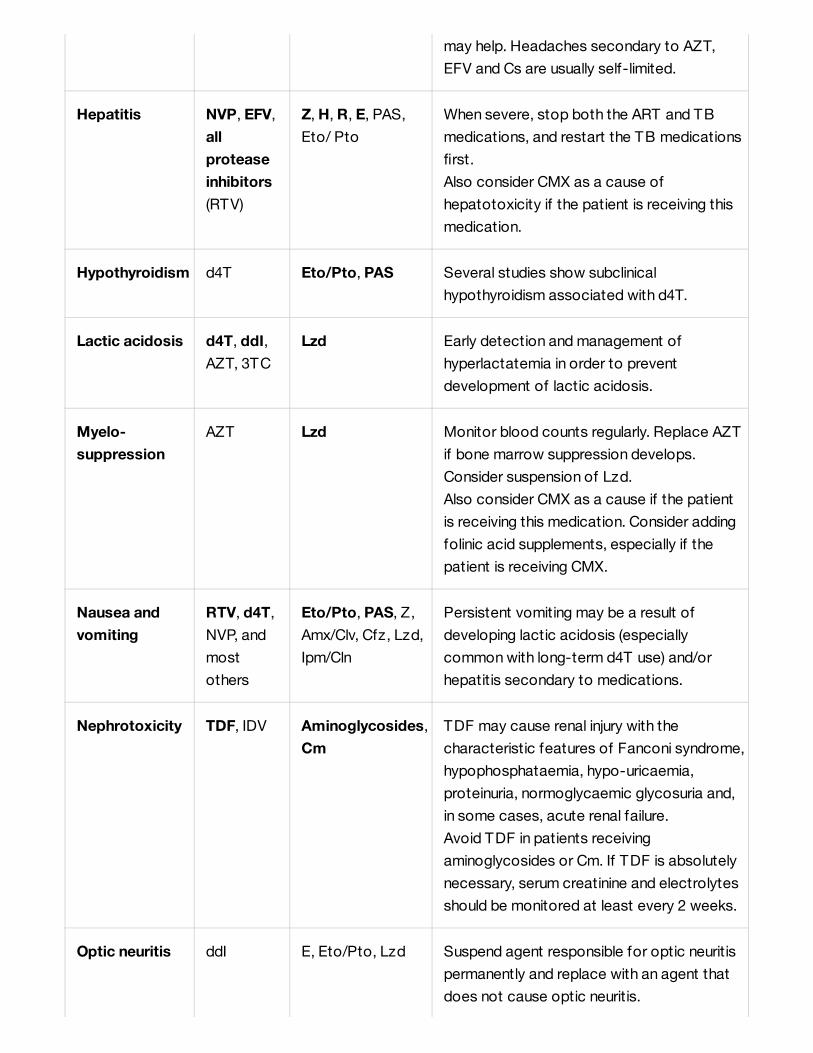
may help. Headaches secondary to AZT,
EFV and Cs are usually self-limited.
Hepatitis NVP, EFV,
all
protease
inhibitors
(RTV)
Z, H, R, E, PAS,
Eto/ Pto
When severe, stop both the ART and TB
medications, and restart the TB medications
first.
Also consider CMX as a cause of
hepatotoxicity if the patient is receiving this
medication.
Hypothyroidism d4T Eto/Pto, PAS Several studies show subclinical
hypothyroidism associated with d4T.
Lactic acidosis d4T, ddI,
AZT, 3TC
Lzd Early detection and management of
hyperlactatemia in order to prevent
development of lactic acidosis.
Myelo-
suppression
AZT Lzd Monitor blood counts regularly. Replace AZT
if bone marrow suppression develops.
Consider suspension of Lzd.
Also consider CMX as a cause if the patient
is receiving this medication. Consider adding
folinic acid supplements, especially if the
patient is receiving CMX.
Nausea and
vomiting
RTV, d4T,
NVP, and
most
others
Eto/Pto, PAS, Z,
Amx/Clv, Cfz, Lzd,
Ipm/Cln
Persistent vomiting may be a result of
developing lactic acidosis (especially
common with long-term d4T use) and/or
hepatitis secondary to medications.
Nephrotoxicity TDF, IDV Aminoglycosides,
Cm
TDF may cause renal injury with the
characteristic features of Fanconi syndrome,
hypophosphataemia, hypo-uricaemia,
proteinuria, normoglycaemic glycosuria and,
in some cases, acute renal failure.
Avoid TDF in patients receiving
aminoglycosides or Cm. If TDF is absolutely
necessary, serum creatinine and electrolytes
should be monitored at least every 2 weeks.
Optic neuritis ddI E, Eto/Pto, Lzd Suspend agent responsible for optic neuritis
permanently and replace with an agent that
does not cause optic neuritis.

Adapted from WHO Guidelines for the programmatic management of drug-resistant tuberculosis .
Referencias
Appendix 19. Upper room ultravioletgermicidal irradiation (UVGI) systemThe use of UVGI in the upper part of rooms may be effective in killing or inactivating M. tuberculosis
generated by infected persons.
19.1 Mechanism of action
Pancreatitis d4T, ddI Lzd Avoid concomitant use of these agents. If an
agent causes pancreatitis, suspend it
permanently and do not use any of the
potentially pancreatitis-producing ARVs (d4T
or ddI) in the future.
Peripheral
neuropathy
d4T, ddI Lzd, Cs, H,
Eto/Pto, S, Km,
Amk, Cm, E, FQs
Avoid use of d4T or ddI in combination with
Cs or Lzd (increased risk of peripheral
neuropathy).
QT
prolongation
RTV
boosted PI
Bdq, Cfz, Mfx,
other FQs
Skin rash ABC, NVP,
EFV, d4T
and others
All anti-TB drugs Do not re-challenge with ABC (risk of life-
threatening anaphylaxis). Do not re-challenge
with any agent that may have caused
Stevens-Johnson syndrome.
Also consider CMX as a cause of skin rash if
the patient is receiving this medication.
[1]
1. World Health Organizat ion. Guidelines for the Programmatic Management of Drug- Resistant Tuberculosis.
Emergency Update 2008. (WHO/HTM/TB/2008.402)
ht tp://whqlibdoc.who.int /publicat ions/2008/9789241547581_eng.pdf

UV lamps are installed into fixtures suspended from a ceiling or mounted on a wall. Fixtures are shielded
with louvers or bafflers in order to block radiation below the horizontal plane of the fixtures. UV lights
create in the upper portion of the room a germicidal zone where the bacilli are killed (Figure 1). Patients
in the lower portion of the room are not exposed to UVGI lights. Good air mixing is needed to transport
the air (and thereby the bacilli) to the upper portion of the room. Disinfection is achieved through the
rapid dilution of contaminated lower room air with clean irradiated upper room air.
Figure 1
* 1 feet = 0.3048 m
From the WHO, Implementing the WHO Policy on TB Infection Control in Health-Care Facilities,
Congregate Settings and Households
The lamps should irradiate the entire surface of the upper part of the room (Figure 2), in order to
disinfect the largest possible volume of air mixed at a low speed between the upper and lower part of
the room.
UVGI Upper-room Irradiation
Figure 2

From Guidelines for the Utilization of Ultraviolet Germicidal Irradiation technology in controlling
transmission of tuberculosis in health care facilities in South Africa
Several factors influence the efficiency of UVGI systems:
19.2 MaintenanceDust-covered and/or old UVGI lamps are less effective, hence the need for a careful maintenance,
including regular cleaning:
[1]
Ventilation rate: in controlled environment, at rates up to 6 air change per hour (ACH), UVGI systems
increase the effect of air cleaning to > 12 ACH . But when ventilation rates are increased above 6
ACH, UVGI system effectiveness could be reduced because the time for bacteria irradiation is
shorter .
[2]
[3]
[4]
Effective mixing within the room may be provided by natural convection currents or fans, preferably,
ceiling ones. Low velocity ceiling fans boosted UVGI system’s effectiveness up to 33% when ACH
was below 6 . [5]
[6]
[7]
Relative humidity: studies have reported rapidly decreasing air cleaning effectiveness in UVGI
systems when the relative humidity goes above 70%.
[8]
[9]
[10]
Installation: the height of the room should be minimum 2.5 m and UVGI fixtures should be installed at
the minimum height of 2.1 m. As a thumb rule, a 30W lamp should be sufficient for 18 m of
surface , but room shape and type of fixture should be taken into consideration when
calculating the needs. For instance, wall-mounted lamps would have a smaller germicidal area than
ceiling-mounted ones. Lamps should be on whenever there is a risk of TB transmission. For
example, in rooms with hospitalized patients, the lamps should be turned on 24 hours a day.
2
[11]
[12]
Maintenance: see below.
Lamps and fixture surfaces should be wiped at least monthly (more often if necessary) with a cloth
dampened with 70% alcohol. Do not use water and soap or any detergent. The cleaning should be
performed when lamps and fixtures are cool.
Measurement of UVGI level must be done at installation and at least once a year. A UV light meter
programmed to detect UV light on a wavelength of 254 nm is needed. Measurements should be
performed at eye level in the occupied zone (~ 1.6 m) and in upper irradiated portion of the room, at
a distance of 1.2 m from the fixture in all possible directions (imitating a circle with measurements

19.3 DisposalUV lamps contain mercury and quartz and are considered as hazardous waste. Disposal is extremely
difficult in many countries; this should be considered before implementing them. If adequate disposal of
the lamps by specialized enterprises is not possible in the country, neither their repatriation; UV lamps
should be disposed of by encapsulation (sealed in a metal 200 litre drum filled with concrete and then
buried away from water sources).
Safety considerations
Reflecting surfaces in the irradiation area of UV lamps must be avoided (i.e. oil painted ceilings, etc.).
At certain wavelengths (including UV-C) UV exposure may be harmful. Skin exposure can produce
sunburn (erythema). Exposure of the eyes can produce conjunctivitis (feeling of sand in the eyes,
tearing) and/or keratitis (intense pain, sensitivity to light). These symptoms typically commence 6 to 12
hours after exposure.
Despite the fact that these are reversible conditions, health care workers should immediately report
them to the IC officer. This could mean that UV irradiation is higher than previously thought at lower
room level (lamp poorly positioned? Reflecting surface?).
The USA National Institute for Occupational Safety and Health (NIOSH) states that safe exposure
limits are set below those found to initiate eye irritation, the body surface most susceptible to UV. Next
table shows the permissible exposure times for given effective irradiances at 254 nm wavelength.
done while moving in circumference spaced of 1 m). Ideally, all upper room measurements should be
around 30 μW/cm to 50 μW/cm . Persons doing these measurements should wear protective
equipment (UVprotective glasses, clothing made of tightly woven fabric, soft cotton gloves) and
cover exposed skin with opaque creams with solar-protection factors > 15.
2 2
UV lamps last between 5 000 and 10 000 hours of continue use (7 to 14 months). Check
manufacturer’s information. After this period, UV lamps rapidly lose effectiveness and need to be
changed.

Exposures exceeding this limit would require the use of personal protection equipment to protect the
skin and eyes.
In order to avoid overexposure of UVGI, education of health care workers should include basic
information on UVGI systems and their potential harmful effects of if overexposure occurs.
Permissible exposure time (a)
Effective irradiance
(μW/cm )2
(Units given) (Seconds)
8 h 28,800 0.2
4 h 14,400 0.4
2 h 7,200 0.8
1 h 3,600 1.7
30 min 1,800 3.3
15 min 900 6.7
10 min 600 10
5 min 300 20
1 min 60 100
30 s 30 200
10 s 10 600
1 s 1 6,000
0.5 s 0.5 12,000
0.1 s 0.1 60,000
(a) The occupat ional exposure limit f or UV-C at 254 nm is 6,000 μJ/cm . This can be also calculated with the
following formula: Dose (in μJ/cm ) = Time (in seconds) * Irradiance (in μW/cm).
2
2

Referencias
Appendix 20. Treatment supportersUpdate: January 2022
Treatment supporters need specific training to know and understand their role in order to provide the
patient with adequate treatment education and support.
They should be compensated for their time and services and reimbursed for expenses incurred.
1. I Coker, E Nardell, B Fourie, P Brickner, S Parsons, N Bhagwandin and P Onyebujoh. Guidelines for the
Utilisation of Ultraviolet Germicidal Irradiation technology in controlling transmission of tuberculosis in health
care facilities in South Africa. MRC.
ht tp://www.sahealthinfo.org/tb/guidelines.pdf
2. Xu P. Ultraviolet germicidal irradiation for preventing infectious disease transmission. Boulder, CO: University of
Colorado, Department of Civil, Environmental, and Architectural Engineering; 2001.
3. Collins FM. Relative susceptibility of acid-fast and non-acid-fast bacteria to ultraviolet light. Appl Microbiol
1971;21:411–3.
4. Kethley TW, Branch K. Ultraviolet lamps for room air disinfection. Effect of sampling location and particle size of
bacterial aerosol. Arc Environ Health 1972;25:205–14.
5. Riley RL, Permutt S. Room air disinfection by ultraviolet irradiation of upper air. Air mixing and germicidal
effectiveness. Arch Environ Health 1971;22:208–19.
6. Riley RL, Kaufman JE. Air disinfection in corridors by upper air irradiation with ultraviolet. Arch Environ Health
1971;22:551–3.
7. Ko G, First MW, Burge HA. The characterization of upper-room ultraviolet germicidal irradiation in inactivating
airborne microorganisms. Environmental Health Perspectives 2002;110:95–101.
8. Ko G, First MW, Burge HA. Influence of relative humidity on particle size and UV sensitivity of Serratia
marcescens and Mycobacterium bovis BCG aerosols. Tubercle Lung Dis 2000;80:217–28.
9. Peccia J, Werth HM, Miller S, Hernandez M. Effects of relative humidity on the ultraviolet induced inactivation of
airborne bacteria. Aerosol Science and Technology 2001;35:728–40.
10. Riley RL, Kaufman JE. Effect of relative humidity on the inactivation of airborne Serratia marcescens by
ultraviolet radiation. Appl Microbiol 1972;23:1113–20.
11. First MW, Nardell EA, Chaisson W, Riley R. Guidelines for the application of upper-room ultraviolet germicidal
irradiation for preventing transmission of airborne contagion—part I: basic principles. ASHRAE Trans
1999:105:869–876.
12. Riley RL, Nardell EA [1989]. Clearing the air: the theory and application of ultraviolet air disinfection. Am Rev
Respir Dis 139(5):1286–1294.

20.1 Selecting a treatment supporterThe treatment supporter :
It is usually not recommended to have family members as treatment supporters. The family relationship
may interfere with the ability to administer TB treatment, especially if the patient is a child.
20.2 Roles and responsibilitiesRole and responsibilities of a treatment supporter may include:
Appendix 21. Informing the patient
a
Is someone from the patient’s commmunity;
Is preferably a community health worker or a person with a background in health (e.g. pharmacist),
but can also be a non health worker (co-worker or neighbour);
Is chosen by, or is acceptable to, the patient and their family (e.g. supporter and patient of the
same sex) ;
Is able to observe the patient’s confidentiality;
Has a stable living situation;
Has basic literacy skills (can read and write and has basic numeracy skills);
Is motivated to care for TB patients and committed to supporting them for the full duration of
treatment;
Lives near enough to the patient to be able to make regular visits (daily or weekly) and go to their
home immediately in the event of an emergency;
Is in good physical condition and not immune depressed . b
Supervision of all drug intakes and keeping records on TB treatment card.
Detection of adverse effects and, when necessary, prompt referal of the patient to a health facility.
Accompanying the patient to medical consultations.
Collection and transport of sputum specimens for smear and culture.
Provision of health education to family members, including the risk of transmission and
implementation of infection control measures in the home.
Detection of signs and symptoms of TB in family members.
Participation in refresher trainings.
Notas(a) From USAID TB CARE II (2017) Community-based Care for Drug-resistant Tuberculosis: A Guide for
Implementers. Version 3. Updated in 2017.
ht tps://tbcare2.org/wp-content /uploads/2018/09/Community-Based-DR-TB-20180830-1.pdf
(b) The most common cause of immunosuppression is HIV infect ion, but chronic illnesses such as diabetes
also alter the immune system and are a risk f actor for TB infect ion and act ive TB.

21.1 At the start of treatmentArrange two interviews (allow about 20 minutes for each): one to supply the patients with the
information they need to follow the treatment, the second to make sure they have assimilated the
information. These interviews should coincide with the first two clinical visits. The first interview should
occur before the treatment begins. Depending on how the clinic is organized, the interviews are done
either by the prescribing clinician alone at the time of the clinical visit, or with the help of a specially-
trained staff member at an interview just for this purpose. Patients may bring someone with them, if
they wish.
Outpatients
First interview
For example: take rifampicin in the morning, and fluconazole at night.
Second interview (one week later)
Explain:
The disease and how it spreads
For example: this is a serious, but generally curable, infection that affects the lungs and can be
spread if not treated (tailor the information according to the focus of the infection, the
resistance pattern).
The treatment process:
Length, intensive/continuation phases, clinical and bacteriological monitoring, visit schedule
(tailor the information according to the regimen); how DOT will work and why it is important when
relevant.
The medications:
Management:
Where, when, and from whom to get medications;
Number of tablets per day; number of doses per day, etc.;
Keep tablets in their blister pack until taken, no removing them from their package ahead of
time.
Main adverse effects and what to do if they occur:
For example: for rifampicin, point out that it turns the urine, stools, tear, etc. reddish-orange,
that this is normal and not a cause for concern. For ethambutol, advise the patient to consult
the doctor immediately if s/he notices a decrease in his/her vision or ability to correctly
distinguish colours, etc.
Special precautions (depending on concomitant treatment):
Any incentives or enablers the patient may qualify for and how the patient can access them.
Stress the importance of adherence, anticipate problems, and think about possible solutions.
Answer any questions.
Give the date of the second interview (one week later).

Hospitalized patients
First interview
Same as above, plus explain:
Second interview (when patient is ready for discharge)
21.2 In the course of treatmentAdherence interviews should take place at least monthly (more frequently if needed) throughout the
entire course of treatment. Their purpose is to identify/resolve any problems resulting in poor
adherence. Assessment is done either by the clinician at the monthly clinical visit, or by the nurse
responsible for individual distribution of drugs.
Adherence interviews should be quick (about 5 minutes); on the other hand, devote as much time as
necessary to resolving any problems.
The interview at the end of the intensive phase is more specifically devoted to informing the patient,
because of the changes in drug regimen for the continuation phase.
Appendix 23. Treatment card for patientson first-line anti-TB therapy
Treatment card for patients on first-line anti-TB therapy.pdf
Check to make sure that information has been assimilated; ask open-ended questions, give the
patient time to answer. Give more information, if necessary.
Answer any questions.
Remind the patient of the date of the next visit.
Hospital infection control measures:
Isolation and why it is indicated; the importance of covering the mouth when coughing or sneezing,
the use of sputum containers, visits outside the building, face masks/respirators (who, when, why),
airing out the room, etc.
T imetable for injections and distribution of drugs.
Explain:
Where and when to get medications, the visit schedule;
DOT and other treatment support as relevant.
Make sure that the information the patient needs to continue treatment as an outpatient has been
assimilated (treatment process, medications, adverse effects and what to do, etc.).
Stress the importance of adherence, anticipate problems, and think about possible solutions.
Answer any questions.

Appendix 24. Tuberculosis register forpatients on first-line anti-TB therapy
Tuberculosis register for patients on first-line anti-TB therapy.pdf
Appendix 25. Treatment card for patientson second-line anti-TB therapy
Treatment card for patients on second-line anti-TB therapy.pdf
Appendix 26. Tuberculosis register forpatients on second-line anti-TB therapy
Tuberculosis register for patients on second-line anti-TB therapy.pdf
Appendix 27. RespiratorsUpdate: January 2022
27.1 Introduction

Respirators are masks designed to protect the wearer from inhaling bacilli.
They must be worn by all staff in areas where the risk of TB transmission is high:
Visitors and attendants must wear a respirator when entering a contagious TB patient’s room.
Recommended respirators include:
27.2 Instructions for useRespirators are for personal use. The same respirator cannot be shared between staff members or
between caregivers.
The respirator should be put on before entering the room and removed after exiting the room.
Respirators must be worn covering the nose, mouth and chin and provide a tight seal around the edge.
Every time that a respirator is put on, a seal check has to be performed:
Different factors may not allow proper sealing of respirators to the face: respirator size and/or model;
respirator wearer’s facial features, including beard and facial hair; headscarves, etc.
Inpatient department with smear positive or drug-resistant TB patients
Consultation room for TB diagnosis
Laboratory (sputum smear preparation and culture/drug susceptibility testing)
Sputum collection area
Radiology department
Waiting areas in high TB prevalence settings
The CE-certified filtering facepiece EN 149 FFP2, filtering efficiency 94% if challenged with 0.4μm
particles;
or
The United States Centre for Disease Control and Prevention/National Institute for Occupational
Safety and Health (NIOSH) certified N95, filtering efficiency > 95% if challenged with 0.3µm
particles.
Fully open the respirator and slightly bend the nose wire to form a curve.
Separate the two elastic straps and position the respirator under the chin.
Stretch the two straps over the head, place the first strap at neck-height and the second strap
across the top of the head.
Model the nose wire around the bridge of the nose and secure the edges until you achieve a perfect
Check for leaks by covering the respirator with both hands and forcefully inhaling and exhaling
several times. The respirator should collapse when inhaling and expand when exhaling and no air leak
between the face and the respirator should be Otherwise, straps should be readjusted and/or the
respirator repositioned until is sealed properly.

There is limited evidence on the acceptable length of time a respirator can be worn with maintained
efficiency. The filter materials remain functional for weeks or months, but with frequent wearing the
respirator will become less adjusted.
An extensively used respirator should be discarded after 7 days. However, if for example, it is only used
a few hours 2 to 3 times a week, it can be reused for several weeks . During this period, staff can
reuse their respirator provided it is not wet or damaged and its straps are not loosened. Each staff
member should keep their respirator in the pocket of their personal gown without creasing it. If the filter
material is damaged or the mask has loose straps, the respirator should be discarded immediately.
Note: TB bacillus is trapped in the filter of a mask and will not be released with shaking or other
physical movements of the mask.
27.3 StorageStore in a dry, well ventilated place. Respirators should not be crushed during storage.
27.4 DisposalRespirators are disposed of as “soft waste” and do not need to be disinfected before being discarded.
27.5 Fit testingProper fit of a respirator is critical to ensure respiratory protection. Therefore, all staff members who
could be exposed to M. tuberculosis should before being required to wear a respirator perform a “fit
testing” to determine if the respirators being used fit them properly.
At least two models of respirators should be available. If a worker cannot be fitted with one model, the
other one should be used.
Testing is performed using a fit testing kit. The kit contains all the supplies and instructions needed to
perform the test.
Fist testing kit
[1]

Referencias
Appendix 27. Request form formicroscopy and Xpert MTB/RIF
Request form for microscopy and Xpert MTB/RIF.pdf
Appendix 28. Surgical masksUpdate: January 2022
1. Roland Diel, Albert Nienhaus, Peter Wit te, Renate Ziegler. Protection of healthcare workers against
transmission of Mycobacterium tuberculosis in hospitals: a review of the evidence. ERJ Open
Research 2020 6: 00317-2019.
ht tps://openres.ersjournals.com/content /6/1/00317-2019

28.1 IntroductionThe purpose of surgical masks is to catch droplet nuclei that patients expel while talking, breathing or
coughing.
Surgical masks should be worn by contagious or potentially contagious patients (confirmed or
presumed cases) when they leave their rooms to go to another department or any other enclosed
area. Wearing a surgical mask is not necessary when patients are alone in their room or outdoors.
The terms “surgical”, “medical” or “procedure” are sometimes used interchangeably to qualify masks.
Only masks that conform to the norms EN 14683 or ASTM F2100 should be used.
28.2 Instructions for useSurgical masks are for personal use. The same mask cannot be shared.
Surgical masks must be replaced at least once a day and when they become wet or damaged.
It is not recommended to wear masks for large portions of the day or while sleeping, as they restrict air
movement and are not comfortable.
28.3 StorageStore in a dry, well ventilated place.
28.4 DisposalMasks are disposed of as “soft waste” and do not need to be disinfected before being discarded.
Appendix 28. Request form for sputumculture, LPA and DST
Request form for sputum culture, LPA and DST.pdf
Open the mask.
Bend the nasal bar (if included).
Put the chin into the mask.
Attach the two straps behind the head or over the ears.

Appendix 29. BCG vaccineUpdate: January 2022
Composition, forms and route of administration
Dosage and vaccination scheduleRefer to national recommendations. In countries with a high incidence of TB (> 40 cases per 100,000),
WHO recommends :
Technique and site of administration
Contra-indications
Adverse effects
Live attenuated bacterial vaccine
Powder for injection, to be dissolved with the entire vial of the specific solvent supplied by the
manufacturer, in multidose vial, for intradermal injection
[1]
Child under 12 months: 0.05 ml single dose as soon as possible after birth
Child 12 months and over and adult: 0.1 ml single dose a
Clean the injection site with clean water. Do not use antiseptics as risk of inactivation of vaccine).
Allow to dry.
Administer intradermally. If the injection is correctly performed, an “orange-skin” papule measuring
5-8 mm in diameter should appear at the injection site.
The vaccine is administered in the deltoid region of the arm, about one-third down the upper arm
over the insertion of the deltoid muscle.
The vaccine should be injected in the same place for each child so that the BCG scar is easier to
locate.
Do not administer to patients with congenital or acquired immunodeficiency (e.g. HIV infection or
serologic status unknown, but symptoms consistent with HIV infection, immunosuppressive therapy,
malignant haemopathy).
Postpone vaccination until recovery in the event of acute extensive dermatosis, acute complicated
malnutrition or severe acute febrile illness (minor infections are not contra-indications).
Local reaction 2-4 weeks after injection: papule that ends up as an ulcer and usually heals
spontaneously (dry dressing only) after 2 to 5 months, leaving a permanent
Complications requiring no specific treatment and which almost always evolve favourably:
persistent ulcer with serous discharge for over 4 months after injection;
non-suppurated adenitis, most often axillary, sometimes cervical;

Precautions
Storage
Referencias
abscess at the injection site due to infection (red, hot and painful abscess) or inadvertent
intradermal injection (cold and painless abscess).
Uncommon complications:
suppurative lymphadenitis, mostly observed in neonates, usually due to inadvertent intradermal
injection. The lymph node, which can have a diameter of over 3 cm, evolves toward softening
and fistulisation with chronic osteomyelitis in exceptional cases.
disseminated BCG disease , most commonly in immunocompromised children under 2 years old
(mortality rate > 70%) .
b
[2]
If administered simultaneously with other vaccines, use different syringes and injection sites. Do not
mix with other vaccines in the same syringe.
Pregnancy: CONTRA-INDICATED
Breastfeeding: no contra-indication
Reconstituted vaccine: between 2 °C and 8 °C for 6 hours max.
Powder: between 2 °C and 8 °C.
Solvent: a cold chain is not required for However, at least 24 hours before reconstitution of the
vaccine, the solvent must be refrigerated between 2 °C and 8 °C so that the solvent and lyophilised
powder are at the same temperature: a temperature difference during reconstitution may reduce
vaccine efficacy. Do not freeze.
Notas(a) BCG vaccine provides high protect ion for neonates, but only moderate for school age TST negat ive
children.
(b) If disseminated BCG disease is diagnosed, a 6-month TB t reatment should be administered.
1. World Health Organizat ion. BCG vaccines: WHO position paper – February 2018/Vaccins BCG: Note de
synthèse de l’OMS – Février 2018. Weekly epidemiological record/Relevé épidémiologique hebdomadaire, 23
FEBRUARY 2018, 93 YEAR/23 FÉVRIER 2018, 93 ANNÉE, No 8, 2018, 93, 73–96.
ht tps://apps.who.int /iris/bitst ream/handle/10665/260306/WER9308.pdf ;jsessionid=872A2E82241BA438A3C
9953650A92DF0?sequence=1
th e
2. Hesseling AC, Rabie H, Marais BJ, Manders M, Lips M, Schaaf HS, et al. Bacille Calmette-Guérin vaccine-
induced disease in HIV-infected and HIV-uninfected children. Clin Infect Dis. 2006;42:548–58.
ht tps://pubmed.ncbi.nlm.nih.gov/16421800/

Appendix 29. Sputum smear microscopyregister
Sputum smear microscopy register.pdf
Appendix 30. Xpert MTB/RIF register
Xpert MTB/RIF register.pdf
Appendix 31. Drug-o-gram
Drug-o-gram.pdf
Appendix 32. Quaterly report
Quaterly report.pdf
Appendix 33. Report on detection andenrolment of TB cases with rifampicinand multidrug-resistance

Report on detection and enrolment of TB cases with rifampicin and multidrug-resistance.pdf
Appendix 34. Report of final outcomes ofdrug-resistant tuberculosis
Report of final outcomes of drug-resistant tuberculosis.pdf
Appendix 35. Check-list for the evaluationof a TB service
Check-list for the evaluation of a TB service.pdf