Effects of nitration on the structure and aggregation of α-synuclein
pubs.acs.org/BiochemistryrXXXX American Chemical Society
Biochemistry XXXX, XXX, 000–000 A
DOI: 10.1021/bi1010927
The Conformation and the Aggregation Kinetics of R-SynucleinDepend on the Proline Residues in Its C-Terminal Region†
Jessika Meuvis,‡ Melanie Gerard,§ Linda Desender,§ Veerle Baekelandt, ) and Yves Engelborghs*,‡
‡Laboratory of Biomolecular Dynamics, Katholieke Universiteit Leuven, Celestijnenlaan 200G, B-3001 Leuven, Belgium,§Laboratory of Biochemistry, Katholieke Universiteit Leuven Campus Kortrijk, Etienne Sabbelaan 53, B-8500 Kortrijk, Belgium, and
)Laboratory for Neurobiology and Gene Therapy, Katholieke Universiteit Leuven, Kapucijnenvoer 33, B-3000 Leuven, Belgium
Received July 9, 2010; Revised Manuscript Received September 6, 2010
ABSTRACT: The neuronal protein R-synuclein (R-syn) plays a central role in Parkinson’s disease (PD).The pathological features of PD are the loss of dopaminergic neurons in the substantia nigra pars compactaand the presence of Lewy bodies. The C-terminal domain ofR-syn is characterized by the presence of 15 acidicamino acids and all five proline residues of the protein (P108, P117, P120, P128, and P138). The aggregation ofthis natively unfolded protein is accelerated in vitro by FK506 binding proteins (FKBPs) showingpeptidyl-prolyl cis-trans isomerase activity. These proteins catalyze the cis-trans conformational changeof the X-Pro peptide bond, often a rate-limiting step in protein folding. The acceleration of the folding ofR-syn by FKBPs may accelerate disease-associated aggregation. To further elucidate the role of the prolineresidues in the conformation and aggregation of R-syn, we constructed several mutants of R-syn in which oneor more proline residues are mutated to alanine via site-directed mutagenesis. For this purpose, we producedand purified His-WT R-syn, a recombinant R-syn with a polyhistidine tag (six His residues) and a linker, anda number of Pro-to-Ala mutants. The aggregation kinetics of these mutants and His-WT R-syn were studiedby turbidity, thioflavin T fluorescence, and CDmeasurements. We can conclude that mutation of the prolineresidues to alanine accelerates the aggregation kinetics ofR-synwhile all prolinemutants formed fibrils similarto His-WT R-syn, as visualized via transmission electron microscopy. We also demonstrate that theaccelerating effect of hFKBP12 is abolished via removal of the proline residues from the C-terminus. Finally,we show that themutant ofHisR-synwith all five proline residuesmutated to alanine ismore structured (moreR-helix) thanHis-WT R-syn, indicating the role of the Pro residues as potential helix breakers in the inhibitoryconformation of the C-terminus.
Aggregation of unstructured proteins is a key element inseveral neurodegenerative diseases such as Parkinson’s disease(PD)1 and Alzheimer’s disease (AD) (1). The pathologicalfeatures of PD are a loss of dopaminergic neurons in thesubstantia nigra pars compacta and the presence of Lewy bodies(LB). LB are intracellular inclusions mainly composed of amy-loid fibrils of R-synuclein (R-syn) (2). It has been implied thatsmall oligomers rather than fibrils are the toxic species ofR-syn (3, 4). R-syn was also genetically linked to PD by thediscovery of three point mutations that cause a familial form ofPD (5-7). Moreover, it was shown more recently that locusduplication or triplication of the WT R-syn gene causes familialforms of PD (8, 9).
Finally, the central role of R-syn in PD was corroboratedexperimentally in different animal models in which overexpres-sion leads to formation of neuronal inclusions and motoricsymptoms reminiscent of PD (10-12).
R-syn contains three domains. The N-terminal part (aminoacids 1-60) forms R-helices when bound to vesicles (13). Thecentral NAC domain (amino acids 61-95) is necessary andsufficient for aggregation (14, 15), and the C-terminal domain(amino acids 96-140) contains 15 acidic side chains (10 Glu and5 Asp residues) but also the only five proline residues of theprotein (P108, P117, P120, P128, and P138). Deletion of theC-terminus enhances the aggregation rate of R-syn (16, 17).Consequently, the C-terminal part is considered as a regulatorypart of the protein. Remarkably though, this negatively chargeddomain is not incorporated into the fibrils (18, 19).
R-syn is a natively unfolded protein in solution that can adoptan ensemble of conformations without rigid structure butstabilized by long-range tertiary interactions. On the basis ofNMR studies, it was suggested that these long-range interactionsbetween the NAC and C-terminal domain inhibit fibril forma-tion (20). It was also shown thatMet oxidation strengthens theselong-range interaction and leads to the formation of stablenontoxic oligomers (21).
The aggregation of R-syn is a nucleation-dependent processwith a lag phase, exponential growth phase, and development of asteady state. In the lag phase, an aggregation nucleus, suggestedto be a dimer (22), is formed,which is preceded by the formation apartially folded intermediate (23), and followed by oligomerformation (24). In the exponential phase, oligomers grow intoprotofibrils and finally into mature fibrils (25).
†This work was financially supported by the Research Foundation-Flanders (FWO Project G.0584.06N), the Belgian Federal ScienceCouncil (Contract IUAP P6/19), and research funds from the KatholiekeUniversiteit Leuven (GOA 2006/02 and IOF/KP/07/001).*To whom correspondence should be addressed: Laboratory of
Biomolecular Dynamics, Katholieke Universiteit Leuven, Celestijnenlaan200G, B-3001 Leuven, Belgium. Telephone:þ32 16 32 71 60. Fax:þ32 1632 79 74. E-mail: [email protected].
1Abbreviations: PD, Parkinson’s disease;R-syn,R-synuclein; FKBPs,FK506 binding proteins; PPIase, peptidyl-prolyl cis-trans isomerase;PA, Pro-to-Ala mutants; CD, circular dichroism; ThT, thioflavin T.
B Biochemistry, Vol. XXX, No. XX, XXXX Meuvis et al.
We have found previously that the aggregation of R-syn isaccelerated in vitro by FK506 binding proteins (FKBPs) (26).Early aggregate formation, followed with turbidity, was acceler-ated by the addition of recombinant hFKBP12 in the micro-molar range (26, 27). Fibril formation (measured by thioflavin Tfluorescence), however, was accelerated in the presence ofpicomolar to nanomolar and micromolar concentrations ofhFKBP12. Addition of the inhibitor FK506 abolished the accel-eration of fibril formation. An enzymatically inactive mutant ofhFKBP12 (D37L/F99Y) did not have any effect on fibril forma-tion, confirming the importance of the enzymatic activity for theeffect of hFKBP12 on R-syn fibril formation. However, atmicromolar concentrations of theD37L/F99YhFKBP12mutant,an acceleration of early aggregate formation was still observedwith turbidity, indicating that at these higher concentrations,another type of interaction (e.g., merely binding) was involved.
FKBP12 belongs to the family of immunophilins, enzymesthat have a PPIase activity. These proteins catalyze the cis-transconformational change of a peptidyl-prolyl bond (X-Pro),often the rate-limiting step in protein folding (28). The immuno-suppressant FK506 inhibits the peptidyl-prolyl isomerase activ-ity of FKBP12 (29-31).
FKBP12 is strongly expressed in the deep gray matter ofthe human brain. A first indication that FKBP12 plays a role inPD comes from the observation that its level of expression isincreased in a 6-OHDA rodent model of PD as well as in thebrain of PD patients (32, 33). Also, FKBP12 was found to oftencolocalize with R-syn in LB and with tau in AD brain (34). Wehave recently reported that overexpression and inhibition ofFKBP12 also affects R-syn aggregation in neuronal cells and inmouse brain, demonstrating the physiological relevance of ourfindings (35).
Parvulins and cyclophilins are non-FKBP members of thefamily of immunophilins. Pin1, a human parvulin, has beenimplicated in AD (36). Pin1 was also shown to colocalize withR-syn in PD brain and to induce R-syn aggregation in cellculture (37). These results link peptidyl-prolyl bond isomeriza-tion to the pathogenesis of neurodegenerative diseases such as PDand AD.
To further elucidate the role of proline residues in theconformation and aggregation of R-syn, we constructed differentPro-to-Ala mutants (PA), as well a mutant with all five prolineresiduesmutated to alanine via site-directedmutagenesis (His-PA5
R-syn). The aggregation kinetics of these mutant R-syn’s werestudied with turbidity, thioflavin T fluorescence, and CDmeasurements. We also analyzed the effect of hFKBP12 onthe aggregation of His-PA5 R-syn with turbidity and thioflavinT fluorescence measurements. Finally, we also comparedthe secondary structure of His-WT and His-PA5 R-syn withcircular dichroism.
EXPERIMENTAL PROCEDURES
Construction of R-syn Expression Plasmids and Purifi-cation of Recombinant Protein. The proline mutants of R-synwere constructed via site-directed mutagenesis using a two-steppolymerase chain reaction (PCR). The presence of the mutationswas confirmed by DNA sequencing. His-WT R-syn and the His-PAmutant were purified as described previously (26, 27). Briefly,BL21(DE3) PLysS Escherichia coli cells were transformed withpRSET B R-syn (His-WT or His mutant). Cells were grown at37 �C in the presence of 100 μg/mL ampicillin and 30 μg/mL
chloramphenicol (Roth, Karlsruhe, Germany). Cells were in-duced at an OD600 of 0.6 with 1 mM IPTG (Roth) and leftshaking for 3 h at 30 �C. Cells were harvested and suspended in50mL of sonication buffer [20mMHepes, 100 mMNaCl, 1 mMPMSF, and 0.05 mM EDTA (pH 7.4)]. The lysate was frozenovernight. After sonication (10� 40 s, Branson sonifier 450), thelysate was heated for 45 min at 65 �C. The cell debris was pelletedby centrifugation at 39100g for 30 min (Sorvall RC-24 refriger-ated centrifuge, SLA-1500 rotor). The clear lysate was filteredand subjected to Ni-Ted affinity chromatography (Macherey-Nagel, D€uren, Germany). The fractions that contained R-synwere pooled and applied to anion exchange chromatography(Hitrap HP Q, GE Healthcare, Buckinghamshire, U.K.). Thecolumn was washed with 20 mM Tris-HCl and 100 mM NaCl(pH 8.0). A gradient was applied from 100 mM to 1MNaCl. Allfractions that contained R-syn were pooled and desalted on aHiPrep 26/10 desalting column (GE healthcare). The sample waseluted in 20 mM NH4HCO3 (pH 7.4) and lyophilized (Heto drywinner model DW3, Heto Holten A/S, Allerod, Sweden). Thepurity was checked via sodium dodecyl sulfate-polyacrylamidegel electrophoresis (SDS-PAGE). WT R-syn was expressedand purified as described previously (38). It was previouslyshown that His-tagged R-syn forms the same fibrils as WTR-syn (26, 27). Although His-WT R-syn fibrillates much slowerthan R-syn, we also showed that hFKBP12 accelerates theformation of fibrils of His-WT andWT R-syn to the same extent,showing that the presence of the His tag does not substantiallyaffect the acceleration effect of hFKBP12 (Figure 1 of theSupporting Information).Purification of GST-FKBP12. The pGEX-2TK-FKBP12
expression plasmid was transformed in BL21(DE3) competentcells. The cells were transferred to 5 mL LB cultures andgrown overnight in the presence of chloramphenicol (30 μg/mL)and ampicillin (100 μg/mL) (Roth). Cells were transferred toa 100 mL culture and grown overnight. The next morning, cellswere transferred to a 2 L culture and grown to an OD600 of 0.8.The cells were induced with 1 mM IPTG (Roth) and left shakingfor 4 h at 28 �C.Afterward, cells were harvested by centrifugationand resolubilized in sonication buffer [20 mM Hepes, 1 mMPMSF, and 1% Triton X-100 (pH 7.4)]. The pellet was frozenovernight. After sonication, the lysate was filtered with a 0.45 μmfilter. The lysate was applied to glutathione Sepharose beads(GE healthcare). The beads were washed with 20 mM Hepes,1 mM PMSF, 100 mM NaCl, and 25 mM β-mercaptoethanol(pH 7.4). The protein was eluted with 20 mM Hepes, 1 mMPMSF, 100 mM NaCl, 25 mM β-mercaptoethanol, and 10 mMreduced glutathione (pH 7.4). The purity was checked withSDS-PAGE.Peptidyl-Prolyl Cis-Trans Isomerase Activity. The
activity of GST-FKBP12 was measured via the method ofKullertz et al. (39). Forty microliters of chymotrypsin [1 g/Lchymotrypsin in 35 mM Hepes (pH 7.8)] was added to a 4 μLsample or 4 μL of 35 mM Hepes (pH 7.8) in the blank. Thereaction was monitored at 390 nm after the addition of 400 μL of125 mg/mL succinimidyl-Phe-Pro-Phe-4-nitroanilide (Bachem).Measurements were performed at room temperature. The data asa function of time were fitted to the equation
y ¼ A0ð1- e- kobstÞþB0 ð1Þ
to obtain the first-order rate constant (k) of the cis-transisomerization reaction (A0 is the extrapolated absorbance at time
Article Biochemistry, Vol. XXX, No. XX, XXXX C
zero, and B0 is the total increase in the magnitude of the ab-sorbance signal). The specific enzymatic activity (kcat/KM) wasdetermined via the equation
kcat
KM¼ kobs - k0
½enzyme� ð2Þ
where [enzyme] is the enzyme concentration used, k0 is theobserved rate constant of the uncatalyzed reaction, kobs isthe observed rate constant in the presence of FKBP12, and KM
is the Michaelis-Menten constant.Turbidity Measurements. Lyophilized protein was dis-
solved in 20 mM Hepes, 150 mM NaCl, and 0.02% NaN3 (pH7.4). Small aggregates were removed with the increase in the pHof the sample to 11 with 0.6 NNaOH for 10 min, after which thepH was lowered again to 7.4 by the addition of 0.6 N HCl (40).Samples were centrifuged for 10 min at 14000 rpm (Eppendorftabletop centrifuge). Samples were incubated at 37 �C in a 96-wellplate (Greiner Bio-one) while being shaken (∼270 rpm) in asafire2 (Tecan, Mechelen, Belgium). The turbidity was measuredevery 1000 s at 350 nm. All experiments were performed at leastthree times, and representative graphs of these experiments areshown.Thioflavin T Measurements. Sample preparation was the
same as for the turbidity measurements with the exceptionthat 50 μM thioflavin T (Sigma Aldrich) was added prior tothe shaking (∼270 rpm) at 37 �C. Every 1000 s, the sample wasmeasured by excitation at 446 nm and fluorescence emission wasdetected at 482 nm.All experiments were performed at least threetimes, and representative graphs of these experiments are shown.Fitting of the Aggregation Curves. All the aggregation
curves were fitted to a sigmoid model with four parameters (41)(using Sigmaplot 8.0).
y ¼ y0 þ Δy
1þ e- kðt- t1=2Þ ð3Þ
where t1/2 is the half-time of aggregation, k is the rate constant ofaggregation determining the steepness of the curve,Δy is the totalincrease in the magnitude of the signal, and y0 is the signal at timezero (provided kt1/2 . 1) .Circular Dichroism. Sample preparation was the same as for
the turbidity measurements. Far-UV CD measurements wereperformed on a Jasco-800 spectrophotometer (Jasco, Ijsselstein,The Netherlands) in a 0.1 cm path length cuvette (Hellma,M€ullheim, Germany). The protein concentration was 0.2 mg/mL [20 mMTris-HCl and 100 mMNaCl (pH 7.4)]. Spectra wererecorded at room temperature, from 195 to 260 nm with a stepsize of 1 nm and a scanning speed of 20 nm/min. An average ofthree scans were recorded. Spectra were corrected for the buffer
contribution. CD spectra were deconvoluted using the CDSSTRalgorithm (42) on Dichroweb (http://dichroweb.cryst.bbk.ac.uk/html/home.shtml) (43, 44).Transmission ElectronMicroscopy. Samples for transmis-
sion electronmicroscopy (TEM)were absorbed for 2min on 300-mesh carbon-coated copper Formvar grids and stained for 1 minwith 1% uranyl acetate. Samples were examined with a JEOLJEM 2100 LaB6 electron microscope operating at 200 keV.
RESULTS
Mutation of One orMore Proline Residues to Alanine inthe C-Terminal Part of R-syn Accelerates Its Aggregationand Fibril Formation. We have shown previously with fluo-rescence correlation spectroscopy, thioflavin T fluorescence, andturbidity measurements that FK506 binding proteins acceleratethe aggregation of His-WT R-syn (26, 27). This effect wasdependent on the concentration of hFKBP12 and on its enzy-matic activity, because it was significantly reduced in the presenceof the inhibitor FK506 or an enzymatically inactive mutant ofhFKBP12 (26, 27).
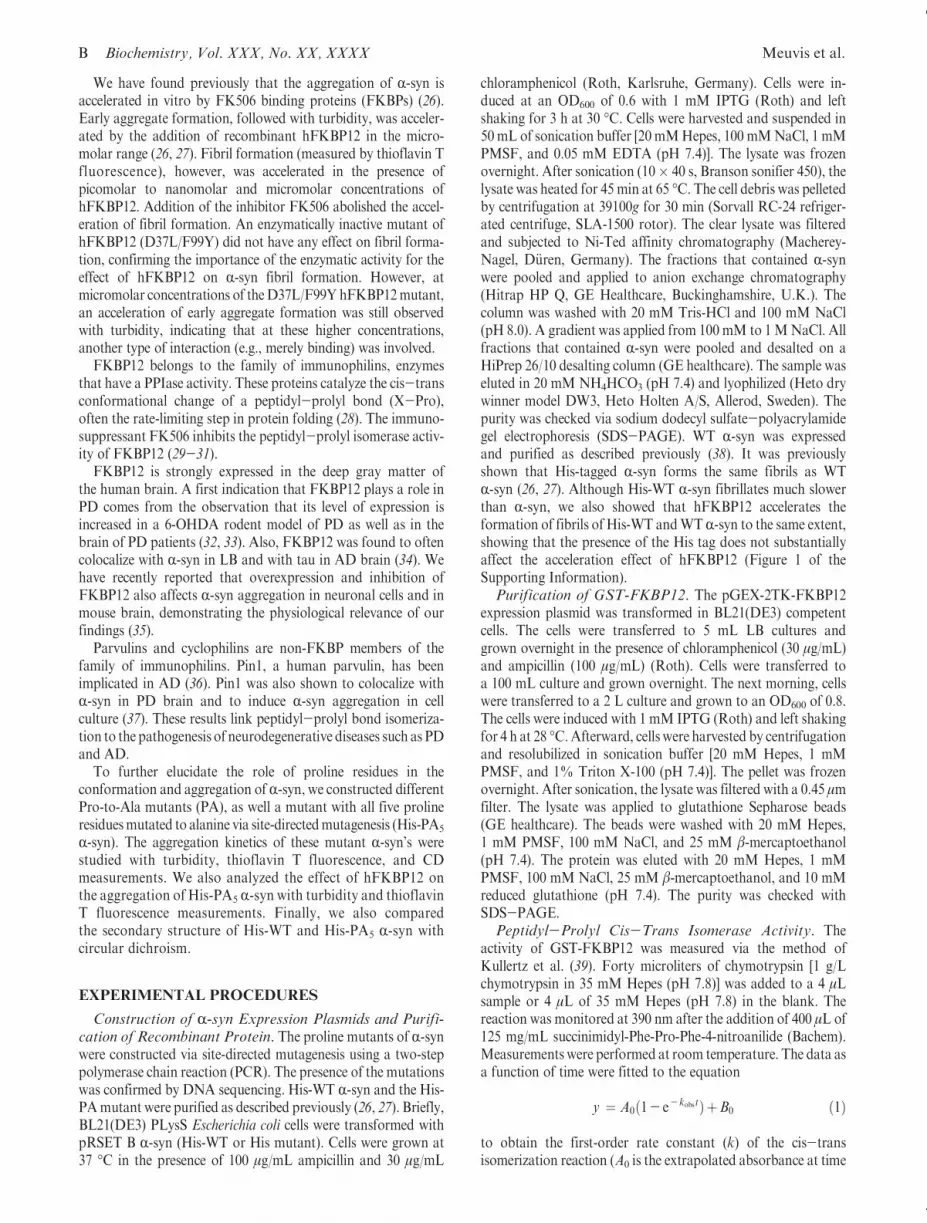
To elucidate the role of the different Pro residues in theaggregation and fibril formation of R-syn, the kinetics ofaggregation of the constructed PA mutants of His-R-syn werestudied and compared with those of His-WT R-syn usingturbidity and ThT fluorescence measurements (Figure 1).
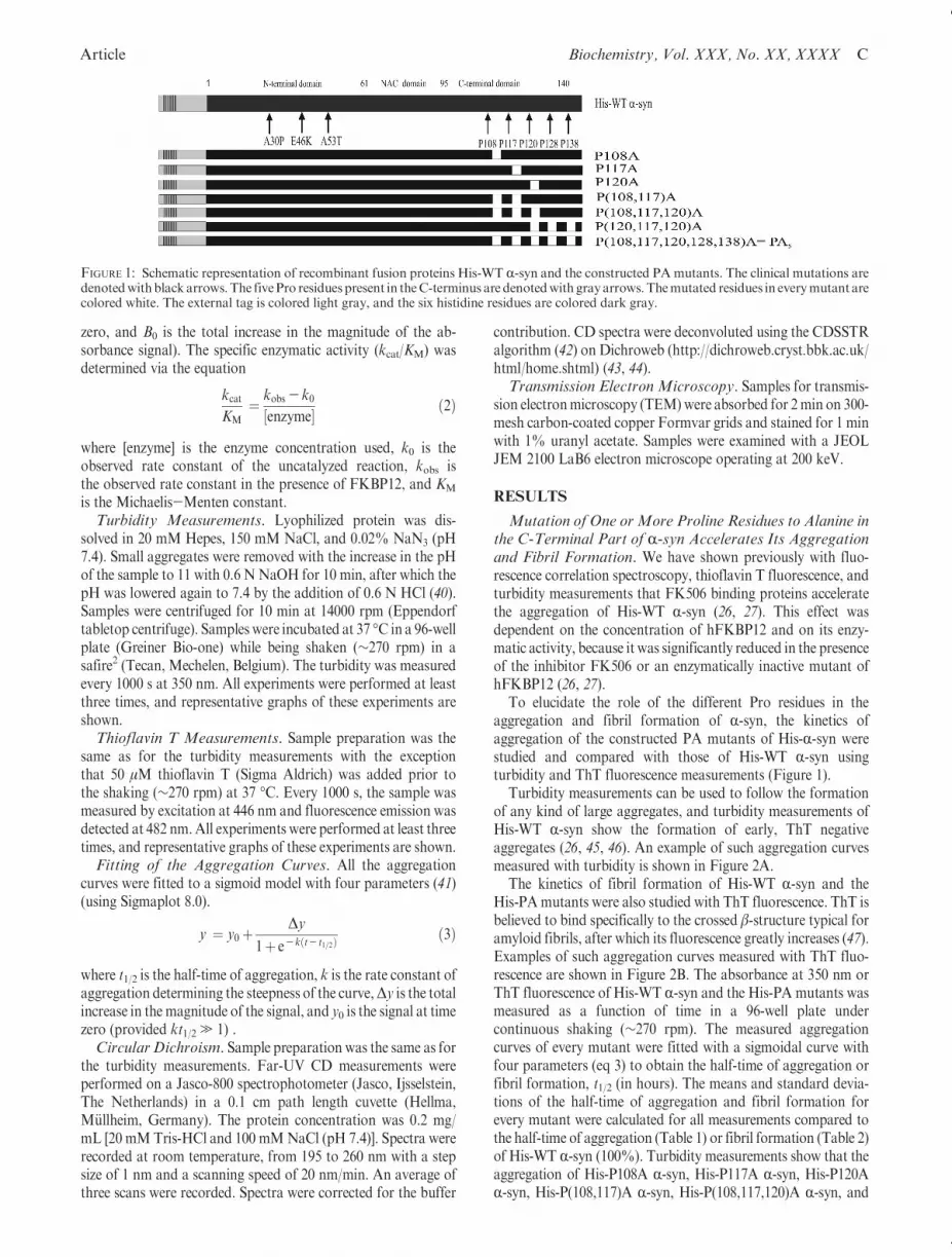
Turbidity measurements can be used to follow the formationof any kind of large aggregates, and turbidity measurements ofHis-WT R-syn show the formation of early, ThT negativeaggregates (26, 45, 46). An example of such aggregation curvesmeasured with turbidity is shown in Figure 2A.
The kinetics of fibril formation of His-WT R-syn and theHis-PAmutants were also studied with ThT fluorescence. ThT isbelieved to bind specifically to the crossed β-structure typical foramyloid fibrils, after which its fluorescence greatly increases (47).Examples of such aggregation curves measured with ThT fluo-rescence are shown in Figure 2B. The absorbance at 350 nm orThT fluorescence of His-WT R-syn and the His-PA mutants wasmeasured as a function of time in a 96-well plate undercontinuous shaking (∼270 rpm). The measured aggregationcurves of every mutant were fitted with a sigmoidal curve withfour parameters (eq 3) to obtain the half-time of aggregation orfibril formation, t1/2 (in hours). The means and standard devia-tions of the half-time of aggregation and fibril formation forevery mutant were calculated for all measurements compared tothe half-time of aggregation (Table 1) or fibril formation (Table 2)of His-WT R-syn (100%). Turbidity measurements show that theaggregation of His-P108A R-syn, His-P117A R-syn, His-P120AR-syn, His-P(108,117)A R-syn, His-P(108,117,120)A R-syn, and
FIGURE 1: Schematic representation of recombinant fusion proteins His-WT R-syn and the constructed PAmutants. The clinical mutations aredenotedwithblack arrows.The fivePro residues present in theC-terminus are denotedwith grayarrows.Themutated residues in everymutant arecolored white. The external tag is colored light gray, and the six histidine residues are colored dark gray.
D Biochemistry, Vol. XXX, No. XX, XXXX Meuvis et al.
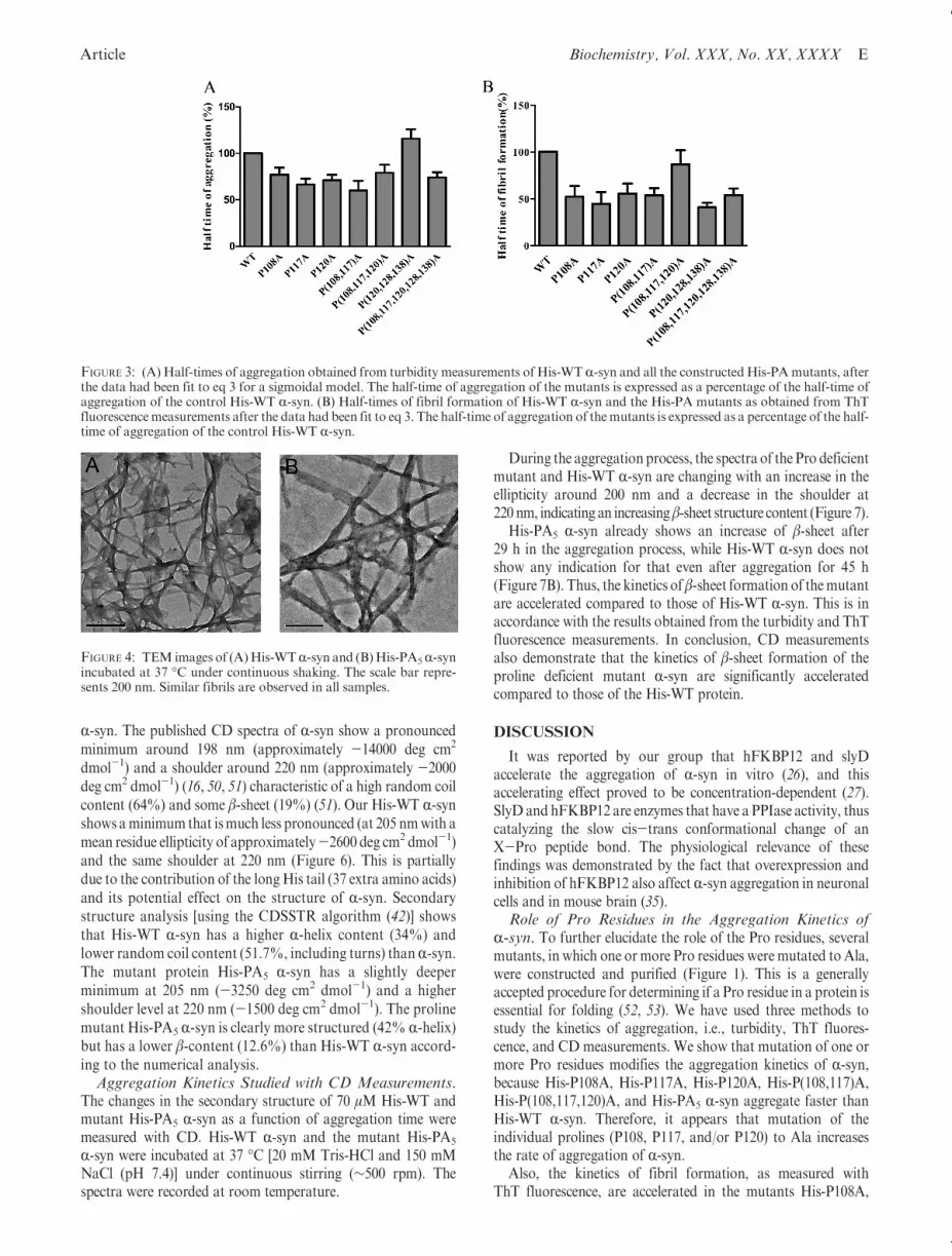
His-PA5 R-syn is faster than that of His-WT R-syn. Thekinetics of aggregation of His-P(120,128,138)A R-syn, how-ever, are slower than the kinetics of aggregation of His-WTR-syn (Figure 3).
From the ThT measurements, we can conclude that the fibrilformation of His-P108A R-syn, His-P117A R-syn, His-P120AR-syn, His-P(108,117)A R-syn, His-P(120,128,138)A R-syn, andHis-PA5 R-syn is faster than that of His-WT R-syn. Fibrilformation of His-P(108,117,120)A R-syn, however, seems to beonly marginally faster than fibril formation of His-WT R-syn.
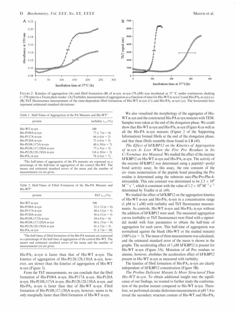
We also visualized the morphology of the aggregates of His-WTR-syn and the constructedHis-PAR-synmutants with TEM.Samples were taken at the end of the elongation phase. We couldshow that His-WT R-syn andHis-PA5 R-syn (Figure 4) as well asall the His-PA R-syn mutants (Figure 2 of the SupportingInformation) formed fibrils at the end of the elongation phase,and that these fibrils resemble those found in LB (48).The Effect of hFKBP12 on the Kinetics of Aggregation
of R-syn Is Lost When the Five Pro Residues in ItsC-Terminus AreMutated.We studied the effect of the enzymehFKBP12 on His-WT R-syn and His-PA5 R-syn. The activity ofthe enzyme hFKBP12 was determined using a peptidyl-prolylbond activity assay. In this assay, the rate constant of thecis-trans isomerization of the peptide bond preceding the Proresidue is determined using the substrate succ-Phe-Pro-Phe-4-nitroanilide. This rate constant was determined to be 2.3 � 106
M-1 s-1, which is consistent with the value of 1.2� 106 M-1 s-1
determined by Tradler et al. (49).We studied the effect of hFKBP12 on the aggregation kinetics
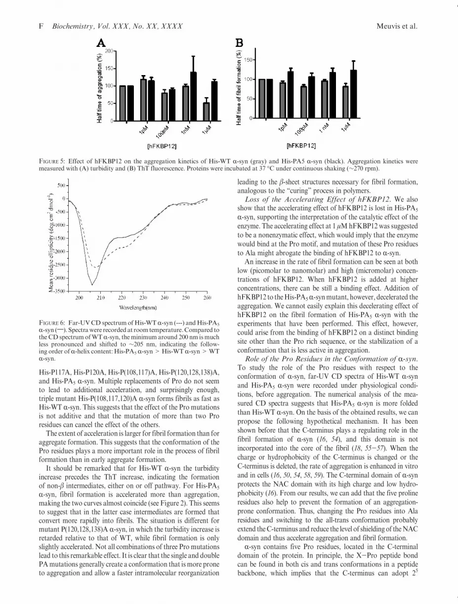
of His-WT R-syn and His-PA5 R-syn in a concentration range(1 pM to 1 μM) with turbidity and ThT fluorescence measure-ments. As controls, His-WT R-syn and His-PA5 R-syn withoutthe addition of hFKBP12 were used. The measured aggregationcurves (turbidity or ThT fluorescence) were fitted with a sigmoi-dal model with four parameters to obtain the half-time ofaggregation for each curve. This half-time of aggregation wasnormalized against the blank (His-WT or the studied mutant)(100%) (n=3). Themean of threemeasurementswas calculated,and the estimated standard error of the mean is shown in thegraphs. The accelerating effect of 1 μM hFKBP12 is present forHis-WT R-syn (Figure 5A). Mutation of all Pro residues toalanine, however, abolishes the acceleration effect of hFKBP12present in His-WT R-syn as measured with turbidity.
The kinetics of fibril formation of His-PA5 R-syn are clearlyindependent of hFKBP12 concentration (Figure 5B).The Proline Deficient Mutant Is More Structured Than
His-WT R-syn. To obtain additional insight into the signifi-cance of our findings, we wanted to further study the conforma-tion of the proline mutant compared to His-WT R-syn. There-fore, we performed circular dichroismmeasurements at pH 7.4 toreveal the secondary structure content of His-WT and His-PA5
FIGURE 2: Kinetics of aggregation (A) and fibril formation (B) of R-syn. R-syn (70 μM) was incubated at 37 �C under continuous shaking(∼270 rpm) in aTecanplate reader. (A)Turbiditymeasurement of aggregation as a functionof time forHis-WTR-syn (O) andHis-PA5R-syn (4).(B) ThT fluorescence measurement of the time-dependent fibril formation of His-WT R-syn (O) and His-PA5 R-syn (4). The horizontal barsrepresent estimated standard deviations.
Table 1: Half-Times of Aggregation of the PA Mutants and His-WTa
protein turbidity t1/2 (%)
His-WT R-syn 100
His-P108A R-syn 77( 7 (n=4)
His-P117A R-syn 66( 6 (n=3)
His-P120A R-syn 71( 6 (n=3)
His-P(108,117)A R-syn 60( 10 (n=7)
His-P(108,117,120)A R-syn 77( 9 (n=5)
His-P(120,128,138)A R-syn 116( 10 (n=3)
His-PA5 R-syn 74 ( 6 (n=7)
aThe half-times of aggregation of the PA mutants are expressed as apercentage of the half-time of aggregation of the control His-WT. Themeans and estimated standard errors of the mean and the number ofmeasurements (n) are given.
Table 2: Half-Times of Fibril Formation of the His-PA Mutants and
His-WTa
protein ThT t1/2 (%)
His-WT R-syn 100
His-P108A R-syn 52( 11 (n=8)
His-P117A R-syn 44( 12 (n=5)
His-P120A R-syn 56( 11 (n=5)
His-P(108,117)A R-syn 54( 8 (n=6)
His-P(108,117,120)A R-syn 86( 15 (n=9)
His-P(120,128,138)A R-syn 41( 5 (n=5)
His-PA5 R-syn 51 ( 7 (n=10)
aThe half-times of fibril formation of the His-PA mutants are expressedas a percentage of the half-time of aggregation of the control His-WT. Themeans and estimated standard errors of the mean and the number ofmeasurements (n) are given.
Article Biochemistry, Vol. XXX, No. XX, XXXX E
R-syn. The published CD spectra of R-syn show a pronouncedminimum around 198 nm (approximately -14000 deg cm2
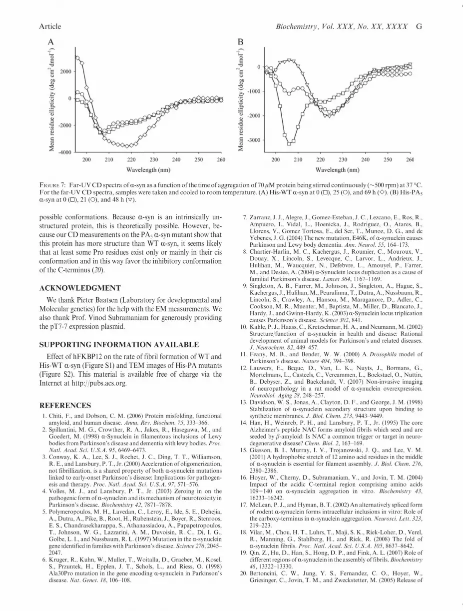
dmol-1) and a shoulder around 220 nm (approximately -2000deg cm2 dmol-1) (16, 50, 51) characteristic of a high random coilcontent (64%) and some β-sheet (19%) (51). Our His-WT R-synshows aminimum that ismuch less pronounced (at 205 nmwith amean residue ellipticity of approximately-2600 deg cm2 dmol-1)and the same shoulder at 220 nm (Figure 6). This is partiallydue to the contribution of the longHis tail (37 extra amino acids)and its potential effect on the structure of R-syn. Secondarystructure analysis [using the CDSSTR algorithm (42)] showsthat His-WT R-syn has a higher R-helix content (34%) andlower random coil content (51.7%, including turns) thanR-syn.The mutant protein His-PA5 R-syn has a slightly deeperminimum at 205 nm (-3250 deg cm2 dmol-1) and a highershoulder level at 220 nm (-1500 deg cm2 dmol-1). The prolinemutant His-PA5 R-syn is clearly more structured (42% R-helix)but has a lower β-content (12.6%) than His-WT R-syn accord-ing to the numerical analysis.Aggregation Kinetics Studied with CD Measurements.
The changes in the secondary structure of 70 μM His-WT andmutant His-PA5 R-syn as a function of aggregation time weremeasured with CD. His-WT R-syn and the mutant His-PA5
R-syn were incubated at 37 �C [20 mM Tris-HCl and 150 mMNaCl (pH 7.4)] under continuous stirring (∼500 rpm). Thespectra were recorded at room temperature.
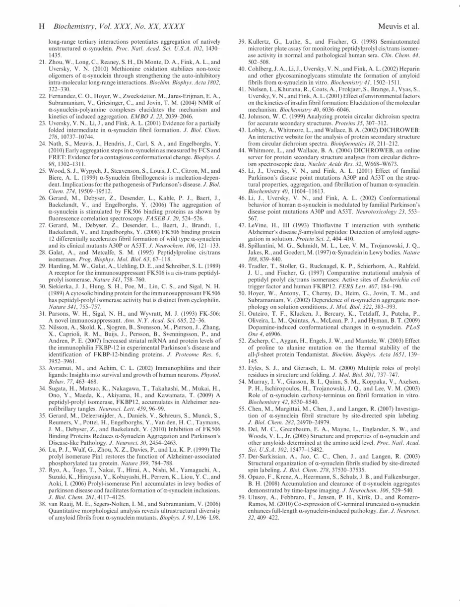
During the aggregation process, the spectra of the Pro deficientmutant and His-WT R-syn are changing with an increase in theellipticity around 200 nm and a decrease in the shoulder at220 nm, indicating an increasingβ-sheet structure content (Figure 7).
His-PA5 R-syn already shows an increase of β-sheet after29 h in the aggregation process, while His-WT R-syn does notshow any indication for that even after aggregation for 45 h(Figure 7B). Thus, the kinetics ofβ-sheet formation of themutantare accelerated compared to those of His-WT R-syn. This is inaccordance with the results obtained from the turbidity and ThTfluorescence measurements. In conclusion, CD measurementsalso demonstrate that the kinetics of β-sheet formation of theproline deficient mutant R-syn are significantly acceleratedcompared to those of the His-WT protein.
DISCUSSION
It was reported by our group that hFKBP12 and slyDaccelerate the aggregation of R-syn in vitro (26), and thisaccelerating effect proved to be concentration-dependent (27).SlyD and hFKBP12 are enzymes that have a PPIase activity, thuscatalyzing the slow cis-trans conformational change of anX-Pro peptide bond. The physiological relevance of thesefindings was demonstrated by the fact that overexpression andinhibition of hFKBP12 also affect R-syn aggregation in neuronalcells and in mouse brain (35).Role of Pro Residues in the Aggregation Kinetics of
R-syn. To further elucidate the role of the Pro residues, severalmutants, in which one ormore Pro residues were mutated to Ala,were constructed and purified (Figure 1). This is a generallyaccepted procedure for determining if a Pro residue in a protein isessential for folding (52, 53). We have used three methods tostudy the kinetics of aggregation, i.e., turbidity, ThT fluores-cence, and CDmeasurements. We show that mutation of one ormore Pro residues modifies the aggregation kinetics of R-syn,because His-P108A, His-P117A, His-P120A, His-P(108,117)A,His-P(108,117,120)A, and His-PA5 R-syn aggregate faster thanHis-WT R-syn. Therefore, it appears that mutation of theindividual prolines (P108, P117, and/or P120) to Ala increasesthe rate of aggregation of R-syn.
Also, the kinetics of fibril formation, as measured withThT fluorescence, are accelerated in the mutants His-P108A,
FIGURE 3: (A) Half-times of aggregation obtained from turbidity measurements of His-WT R-syn and all the constructed His-PAmutants, afterthe data had been fit to eq 3 for a sigmoidal model. The half-time of aggregation of the mutants is expressed as a percentage of the half-time ofaggregation of the control His-WT R-syn. (B) Half-times of fibril formation of His-WT R-syn and the His-PA mutants as obtained from ThTfluorescencemeasurements after the data had been fit to eq 3. The half-time of aggregation of themutants is expressed as a percentage of the half-time of aggregation of the control His-WT R-syn.
FIGURE 4: TEM images of (A)His-WTR-syn and (B)His-PA5R-synincubated at 37 �C under continuous shaking. The scale bar repre-sents 200 nm. Similar fibrils are observed in all samples.
F Biochemistry, Vol. XXX, No. XX, XXXX Meuvis et al.
His-P117A, His-P120A, His-P(108,117)A, His-P(120,128,138)A,and His-PA5 R-syn. Multiple replacements of Pro do not seemto lead to additional acceleration, and surprisingly enough,triple mutant His-P(108,117,120)A R-syn forms fibrils as fast asHis-WT R-syn. This suggests that the effect of the Pro mutationsis not additive and that the mutation of more than two Proresidues can cancel the effect of the others.
The extent of acceleration is larger for fibril formation than foraggregate formation. This suggests that the conformation of thePro residues plays a more important role in the process of fibrilformation than in early aggregate formation.
It should be remarked that for His-WT R-syn the turbidityincrease precedes the ThT increase, indicating the formationof non-β intermediates, either on or off pathway. For His-PA5
R-syn, fibril formation is accelerated more than aggregation,making the two curves almost coincide (see Figure 2). This seemsto suggest that in the latter case intermediates are formed thatconvert more rapidly into fibrils. The situation is different formutant P(120,128,138)A R-syn, in which the turbidity increase isretarded relative to that of WT, while fibril formation is onlyslightly accelerated. Not all combinations of three Pro mutationslead to this remarkable effect. It is clear that the single and doublePAmutations generally create a conformation that is more proneto aggregation and allow a faster intramolecular reorganization
leading to the β-sheet structures necessary for fibril formation,analogous to the “curing” process in polymers.Loss of the Accelerating Effect of hFKBP12. We also
show that the accelerating effect of hFKBP12 is lost in His-PA5
R-syn, supporting the interpretation of the catalytic effect of theenzyme. The accelerating effect at 1 μMhFKBP12was suggestedto be a nonenzymatic effect, which would imply that the enzymewould bind at the Pro motif, and mutation of these Pro residuesto Ala might abrogate the binding of hFKBP12 to R-syn.
An increase in the rate of fibril formation can be seen at bothlow (picomolar to nanomolar) and high (micromolar) concen-trations of hFKBP12. When hFKBP12 is added at higherconcentrations, there can be still a binding effect. Addition ofhFKBP12 to theHis-PA5R-synmutant, however, decelerated theaggregation. We cannot easily explain this decelerating effect ofhFKBP12 on the fibril formation of His-PA5 R-syn with theexperiments that have been performed. This effect, however,could arise from the binding of hFKBP12 on a distinct bindingsite other than the Pro rich sequence, or the stabilization of aconformation that is less active in aggregation.Role of the Pro Residues in the Conformation of R-syn.
To study the role of the Pro residues with respect to theconformation of R-syn, far-UV CD spectra of His-WT R-synand His-PA5 R-syn were recorded under physiological condi-tions, before aggregation. The numerical analysis of the mea-sured CD spectra suggests that His-PA5 R-syn is more foldedthan His-WT R-syn. On the basis of the obtained results, we canpropose the following hypothetical mechanism. It has beenshown before that the C-terminus plays a regulating role in thefibril formation of R-syn (16, 54), and this domain is notincorporated into the core of the fibril (18, 55-57). When thecharge or hydrophobicity of the C-terminus is changed or theC-terminus is deleted, the rate of aggregation is enhanced in vitroand in cells (16, 50, 54, 58, 59). The C-terminal domain of R-synprotects the NAC domain with its high charge and low hydro-phobicity (16). From our results, we can add that the five prolineresidues also help to prevent the formation of an aggregation-prone conformation. Thus, changing the Pro residues into Alaresidues and switching to the all-trans conformation probablyextend theC-terminus and reduce the level of shielding of theNACdomain and thus accelerate aggregation and fibril formation.
R-syn contains five Pro residues, located in the C-terminaldomain of the protein. In principle, the X-Pro peptide bondcan be found in both cis and trans conformations in a peptidebackbone, which implies that the C-terminus can adopt 25
FIGURE 5: Effect of hFKBP12 on the aggregation kinetics of His-WT R-syn (gray) and His-PA5 R-syn (black). Aggregation kinetics weremeasured with (A) turbidity and (B) ThT fluorescence. Proteins were incubated at 37 �C under continuous shaking (∼270 rpm).
FIGURE 6: Far-UVCD spectrumofHis-WTR-syn (---) andHis-PA5
R-syn (;). Spectrawere recordedat room temperature.Compared tothe CD spectrumofWTR-syn, theminimum around 200 nm ismuchless pronounced and shifted to ∼205 nm, indicating the follow-ing order ofR-helix content: His-PA5 R-syn>His-WTR-syn>WTR-syn.
Article Biochemistry, Vol. XXX, No. XX, XXXX G
possible conformations. Because R-syn is an intrinsically un-structured protein, this is theoretically possible. However, be-cause our CDmeasurements on the PA5 R-syn mutant show thatthis protein has more structure than WT R-syn, it seems likelythat at least some Pro residues exist only or mainly in their cisconformation and in this way favor the inhibitory conformationof the C-terminus (20).
ACKNOWLEDGMENT
We thank Pieter Baatsen (Laboratory for developmental andMolecular genetics) for the help with the EMmeasurements. Wealso thank Prof. Vinod Subramaniam for generously providingthe pT7-7 expression plasmid.
SUPPORTING INFORMATION AVAILABLE
Effect of hFKBP12 on the rate of fibril formation of WT andHis-WT R-syn (Figure S1) and TEM images of His-PA mutants(Figure S2). This material is available free of charge via theInternet at http://pubs.acs.org.
REFERENCES
1. Chiti, F., and Dobson, C. M. (2006) Protein misfolding, functionalamyloid, and human disease. Annu. Rev. Biochem. 75, 333–366.
2. Spillantini, M. G., Crowther, R. A., Jakes, R., Hasegawa, M., andGoedert, M. (1998) R-Synuclein in filamentous inclusions of Lewybodies from Parkinson’s disease and dementia with lewy bodies.Proc.Natl. Acad. Sci. U.S.A. 95, 6469–6473.
3. Conway, K. A., Lee, S. J., Rochet, J. C., Ding, T. T., Williamson,R. E., and Lansbury, P. T., Jr. (2000) Acceleration of oligomerization,not fibrillization, is a shared property of both R-synuclein mutationslinked to early-onset Parkinson’s disease: Implications for pathogen-esis and therapy. Proc. Natl. Acad. Sci. U.S.A. 97, 571–576.
4. Volles, M. J., and Lansbury, P. T., Jr. (2003) Zeroing in on thepathogenic form of R-synuclein and its mechanism of neurotoxicity inParkinson’s disease. Biochemistry 42, 7871–7878.
5. Polymeropoulos, M. H., Lavedan, C., Leroy, E., Ide, S. E., Dehejia,A., Dutra, A., Pike, B., Root, H., Rubenstein, J., Boyer, R., Stenroos,E. S., Chandrasekharappa, S., Athanassiadou, A., Papapetropoulos,T., Johnson, W. G., Lazzarini, A. M., Duvoisin, R. C., Di, I. G.,Golbe, L. I., andNussbaum, R. L. (1997)Mutation in the R-synucleingene identified in families with Parkinson’s disease.Science 276, 2045–2047.
6. Kruger, R., Kuhn, W., Muller, T., Woitalla, D., Graeber, M., Kosel,S., Przuntek, H., Epplen, J. T., Schols, L., and Riess, O. (1998)Ala30Pro mutation in the gene encoding R-synuclein in Parkinson’sdisease. Nat. Genet. 18, 106–108.
7. Zarranz, J. J., Alegre, J., Gomez-Esteban, J. C., Lezcano, E., Ros, R.,Ampuero, I., Vidal, L., Hoenicka, J., Rodriguez, O., Atares, B.,Llorens, V., Gomez Tortosa, E., del Ser, T., Munoz, D. G., and deYebenes, J. G. (2004) The newmutation, E46K, of R-synuclein causesParkinson and Lewy body dementia. Ann. Neurol. 55, 164–173.
8. Chartier-Harlin, M. C., Kachergus, J., Roumier, C., Mouroux, V.,Douay, X., Lincoln, S., Levecque, C., Larvor, L., Andrieux, J.,Hulihan, M., Waucquier, N., Defebvre, L., Amouyel, P., Farrer,M., and Destee, A. (2004) R-Synuclein locus duplication as a cause offamilial Parkinson’s disease. Lancet 364, 1167–1169.
9. Singleton, A. B., Farrer, M., Johnson, J., Singleton, A., Hague, S.,Kachergus, J., Hulihan,M., Peuralinna, T.,Dutra, A.,Nussbaum,R.,Lincoln, S., Crawley, A., Hanson, M., Maraganore, D., Adler, C.,Cookson, M. R., Muenter, M., Baptista, M., Miller, D., Blancato, J.,Hardy, J., andGwinn-Hardy,K. (2003)R-Synuclein locus triplicationcauses Parkinson’s disease. Science 302, 841.
10. Kahle, P. J., Haass, C., Kretzschmar, H. A., andNeumann,M. (2002)Structure/function of R-synuclein in health and disease: Rationaldevelopment of animal models for Parkinson’s and related diseases.J. Neurochem. 82, 449–457.
11. Feany, M. B., and Bender, W. W. (2000) A Drosophila model ofParkinson’s disease. Nature 404, 394–398.
12. Lauwers, E., Beque, D., Van, L. K., Nuyts, J., Bormans, G.,Mortelmans, L., Casteels, C., Vercammen, L., Bockstael, O., Nuttin,B., Debyser, Z., and Baekelandt, V. (2007) Non-invasive imagingof neuropathology in a rat model of R-synuclein overexpression.Neurobiol. Aging 28, 248–257.
13. Davidson, W. S., Jonas, A., Clayton, D. F., and George, J. M. (1998)Stabilization of R-synuclein secondary structure upon binding tosynthetic membranes. J. Biol. Chem. 273, 9443–9449.
14. Han, H., Weinreb, P. H., and Lansbury, P. T., Jr. (1995) The coreAlzheimer’s peptide NAC forms amyloid fibrils which seed and areseeded by β-amyloid: Is NAC a common trigger or target in neuro-degenerative disease? Chem. Biol. 2, 163–169.
15. Giasson, B. I., Murray, I. V., Trojanowski, J. Q., and Lee, V. M.(2001) A hydrophobic stretch of 12 amino acid residues in the middleof R-synuclein is essential for filament assembly. J. Biol. Chem. 276,2380–2386.
16. Hoyer, W., Cherny, D., Subramaniam, V., and Jovin, T. M. (2004)Impact of the acidic C-terminal region comprising amino acids109-140 on R-synuclein aggregation in vitro. Biochemistry 43,16233–16242.
17. McLean, P. J., andHyman, B. T. (2002) An alternatively spliced formof rodent R-synuclein forms intracellular inclusions in vitro: Role ofthe carboxy-terminus in R-synuclein aggregation.Neurosci. Lett. 323,219–223.
18. Vilar, M., Chou, H. T., Luhrs, T., Maji, S. K., Riek-Loher, D., Verel,R., Manning, G., Stahlberg, H., and Riek, R. (2008) The fold ofR-synuclein fibrils. Proc. Natl. Acad. Sci. U.S.A. 105, 8637–8642.
19. Qin, Z., Hu, D., Han, S., Hong, D. P., and Fink, A. L. (2007) Role ofdifferent regions ofR-synuclein in the assembly of fibrils.Biochemistry46, 13322–13330.
20. Bertoncini, C. W., Jung, Y. S., Fernandez, C. O., Hoyer, W.,Griesinger, C., Jovin, T. M., and Zweckstetter, M. (2005) Release of
FIGURE 7: Far-UVCDspectra ofR-syn as a function of the time of aggregation of 70 μMprotein being stirred continuously (∼500 rpm) at 37 �C.For the far-UV CD spectra, samples were taken and cooled to room temperature. (A) His-WT R-syn at 0 (0), 25 (O), and 69 h (]). (B) His-PA5
R-syn at 0 (0), 21 (O), and 48 h (3).
H Biochemistry, Vol. XXX, No. XX, XXXX Meuvis et al.
long-range tertiary interactions potentiates aggregation of nativelyunstructured R-synuclein. Proc. Natl. Acad. Sci. U.S.A. 102, 1430–1435.
21. Zhou,W., Long, C., Reaney, S. H., DiMonte, D. A., Fink, A. L., andUversky, V. N. (2010) Methionine oxidation stabilizes non-toxicoligomers of R-synuclein through strengthening the auto-inhibitoryintra-molecular long-range interactions.Biochim. Biophys. Acta 1802,322–330.
22. Fernandez, C. O., Hoyer,W., Zweckstetter, M., Jares-Erijman, E. A.,Subramaniam, V., Griesinger, C., and Jovin, T. M. (2004) NMR ofR-synuclein-polyamine complexes elucidates the mechanism andkinetics of induced aggregation. EMBO J. 23, 2039–2046.
23. Uversky, V. N., Li, J., and Fink, A. L. (2001) Evidence for a partiallyfolded intermediate in R-synuclein fibril formation. J. Biol. Chem.276, 10737–10744.
24. Nath, S., Meuvis, J., Hendrix, J., Carl, S. A., and Engelborghs, Y.(2010) Early aggregation steps inR-synuclein asmeasured byFCS andFRET: Evidence for a contagious conformational change. Biophys. J.98, 1302–1311.
25. Wood, S. J., Wypych, J., Steavenson, S., Louis, J. C., Citron, M., andBiere, A. L. (1999) R-Synuclein fibrillogenesis is nucleation-depen-dent. Implications for the pathogenesis of Parkinson’s disease. J. Biol.Chem. 274, 19509–19512.
26. Gerard, M., Debyser, Z., Desender, L., Kahle, P. J., Baert, J.,Baekelandt, V., and Engelborghs, Y. (2006) The aggregation ofR-synuclein is stimulated by FK506 binding proteins as shown byfluorescence correlation spectroscopy. FASEB J. 20, 524–526.
27. Gerard, M., Debyser, Z., Desender, L., Baert, J., Brandt, I.,Baekelandt, V., and Engelborghs, Y. (2008) FK506 binding protein12 differentially accelerates fibril formation of wild type R-synucleinand its clinical mutants A30P or A53T. J. Neurochem. 106, 121–133.
28. Galat, A., and Metcalfe, S. M. (1995) Peptidylproline cis/transisomerases. Prog. Biophys. Mol. Biol. 63, 67–118.
29. Harding,M.W., Galat, A., Uehling, D. E., and Schreiber, S. L. (1989)A receptor for the immunosuppressant FK506 is a cis-trans peptidyl-prolyl isomerase. Nature 341, 758–760.
30. Siekierka, J. J., Hung, S. H., Poe, M., Lin, C. S., and Sigal, N. H.(1989) A cytosolic binding protein for the immunosuppressant FK506has peptidyl-prolyl isomerase activity but is distinct from cyclophilin.Nature 341, 755–757.
31. Parsons, W. H., Sigal, N. H., and Wyvratt, M. J. (1993) FK-506:A novel immunosuppressant. Ann. N.Y. Acad. Sci. 685, 22–36.
32. Nilsson, A., Skold, K., Sjogren, B., Svensson, M., Pierson, J., Zhang,X., Caprioli, R. M., Buijs, J., Persson, B., Svenningsson, P., andAndren, P. E. (2007) Increased striatal mRNA and protein levels ofthe immunophilin FKBP-12 in experimental Parkinson’s disease andidentification of FKBP-12-binding proteins. J. Proteome Res. 6,3952–3961.
33. Avramut, M., and Achim, C. L. (2002) Immunophilins and theirligands: Insights into survival and growth of human neurons. Physiol.Behav. 77, 463–468.
34. Sugata, H., Matsuo, K., Nakagawa, T., Takahashi, M., Mukai, H.,Ono, Y., Maeda, K., Akiyama, H., and Kawamata, T. (2009) Apeptidyl-prolyl isomerase, FKBP12, accumulates in Alzheimer neu-rofibrillary tangles. Neurosci. Lett. 459, 96–99.
35. Gerard, M., Deleersnijder, A., Daniels, V., Schreurs, S., Munck, S.,Reumers, V., Pottel, H., Engelborghs, Y., Van den, H. C., Taymans,J. M., Debyser, Z., and Baekelandt, V. (2010) Inhibition of FK506Binding Proteins Reduces R-Synuclein Aggregation and Parkinson’sDisease-like Pathology. J. Neurosci. 30, 2454–2463.
36. Lu, P. J., Wulf, G., Zhou, X. Z., Davies, P., and Lu, K. P. (1999) Theprolyl isomerase Pin1 restores the function of Alzheimer-associatedphosphorylated tau protein. Nature 399, 784–788.
37. Ryo, A., Togo, T., Nakai, T., Hirai, A., Nishi, M., Yamaguchi, A.,Suzuki,K., Hirayasu, Y.,Kobayashi,H., Perrem,K., Liou, Y.C., andAoki, I. (2006) Prolyl-isomerase Pin1 accumulates in lewy bodies ofparkinson disease and facilitates formation of R-synuclein inclusions.J. Biol. Chem. 281, 4117–4125.
38. van Raaij, M. E., Segers-Nolten, I. M., and Subramaniam, V. (2006)Quantitative morphological analysis reveals ultrastructural diversityof amyloid fibrils from R-synuclein mutants. Biophys. J. 91, L96–L98.
39. Kullertz, G., Luthe, S., and Fischer, G. (1998) Semiautomatedmicrotiter plate assay for monitoring peptidylprolyl cis/trans isomer-ase activity in normal and pathological human sera. Clin. Chem. 44,502–508.
40. Cohlberg, J. A., Li, J., Uversky, V. N., and Fink, A. L. (2002)Heparinand other glycosaminoglycans stimulate the formation of amyloidfibrils from R-synuclein in vitro. Biochemistry 41, 1502–1511.
41. Nielsen, L., Khurana, R., Coats, A., Frokjaer, S., Brange, J., Vyas, S.,Uversky, V.N., andFink,A. L. (2001) Effect of environmental factorson the kinetics of insulin fibril formation: Elucidation of themolecularmechanism. Biochemistry 40, 6036–6046.
42. Johnson, W. C. (1999) Analyzing protein circular dichroism spectrafor accurate secondary structures. Proteins 35, 307–312.
43. Lobley, A., Whitmore, L., andWallace, B. A. (2002) DICHROWEB:An interactive website for the analysis of protein secondary structurefrom circular dichroism spectra. Bioinformatics 18, 211–212.
44. Whitmore, L., and Wallace, B. A. (2004) DICHROWEB, an onlineserver for protein secondary structure analyses from circular dichro-ism spectroscopic data. Nucleic Acids Res. 32, W668–W673.
45. Li, J., Uversky, V. N., and Fink, A. L. (2001) Effect of familialParkinson’s disease point mutations A30P and A53T on the struc-tural properties, aggregation, and fibrillation of human R-synuclein.Biochemistry 40, 11604–11613.
46. Li, J., Uversky, V. N., and Fink, A. L. (2002) Conformationalbehavior of human R-synuclein is modulated by familial Parkinson’sdisease point mutations A30P and A53T. Neurotoxicology 23, 553–567.
47. LeVine, H., III (1993) Thioflavine T interaction with syntheticAlzheimer’s disease β-amyloid peptides: Detection of amyloid aggre-gation in solution. Protein Sci. 2, 404–410.
48. Spillantini, M. G., Schmidt, M. L., Lee, V. M., Trojanowski, J. Q.,Jakes, R., andGoedert,M. (1997)R-Synuclein in Lewy bodies.Nature388, 839–840.
49. Tradler, T., Stoller, G., Rucknagel, K. P., Schierhorn, A., Rahfeld,J. U., and Fischer, G. (1997) Comparative mutational analysis ofpeptidyl prolyl cis/trans isomerases: Active sites of Escherichia colitrigger factor and human FKBP12. FEBS Lett. 407, 184–190.
50. Hoyer, W., Antony, T., Cherny, D., Heim, G., Jovin, T. M., andSubramaniam, V. (2002) Dependence of R-synuclein aggregate mor-phology on solution conditions. J. Mol. Biol. 322, 383–393.
51. Outeiro, T. F., Klucken, J., Bercury, K., Tetzlaff, J., Putcha, P.,Oliveira, L. M., Quintas, A., McLean, P. J., and Hyman, B. T. (2009)Dopamine-induced conformational changes in R-synuclein. PLoSOne 4, e6906.
52. Zscherp, C., Aygun, H., Engels, J. W., and Mantele, W. (2003) Effectof proline to alanine mutation on the thermal stability of theall-β-sheet protein Tendamistat. Biochim. Biophys. Acta 1651, 139–145.
53. Eyles, S. J., and Gierasch, L. M. (2000) Multiple roles of prolylresidues in structure and folding. J. Mol. Biol. 301, 737–747.
54. Murray, I. V., Giasson, B. I., Quinn, S. M., Koppaka, V., Axelsen,P. H., Ischiropoulos, H., Trojanowski, J. Q., and Lee, V. M. (2003)Role of R-synuclein carboxy-terminus on fibril formation in vitro.Biochemistry 42, 8530–8540.
55. Chen, M., Margittai, M., Chen, J., and Langen, R. (2007) Investiga-tion of R-synuclein fibril structure by site-directed spin labeling.J. Biol. Chem. 282, 24970–24979.
56. Del, M. C., Greenbaum, E. A., Mayne, L., Englander, S. W., andWoods, V. L., Jr. (2005) Structure and properties of R-synuclein andother amyloids determined at the amino acid level. Proc. Natl. Acad.Sci. U.S.A. 102, 15477–15482.
57. Der-Sarkissian, A., Jao, C. C., Chen, J., and Langen, R. (2003)Structural organization of R-synuclein fibrils studied by site-directedspin labeling. J. Biol. Chem. 278, 37530–37535.
58. Opazo, F., Krenz, A., Heermann, S., Schulz, J. B., and Falkenburger,B. H. (2008) Accumulation and clearance of R-synuclein aggregatesdemonstrated by time-lapse imaging. J. Neurochem. 106, 529–540.
59. Ulusoy, A., Febbraro, F., Jensen, P. H., Kirik, D., and Romero-Ramos,M. (2010) Co-expression of C-terminal truncatedR-synucleinenhances full-length R-synuclein-induced pathology.Eur. J. Neurosci.32, 409–422.
Copyright © 2022 FDOKUMEN