Influence of Esophageal Endoscopic Submucosal Dissection ...
Upload
independentCategory
view
5download
0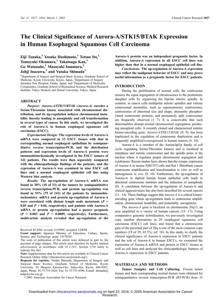
The Clinical Significance of Aurora-A/STK15/BTAK Expression
in Human Esophageal Squamous Cell Carcinoma
Eiji Tanaka,1 Yosuke Hashimoto,1 Tetsuo Ito,1
Tomoyuki Okumura,1 Takatsugu Kan,1
Go Watanabe,1 Masayuki Imamura,2
Johji Inazawa,3 and Yutaka Shimada1
1Department of Surgery and Surgical Basic Science, Graduate School ofMedicine, Kyoto University, Kyoto, Japan, 2Department of SurgerySaiseikai Noe Hospital, Osaka, Japan; and 3Department of MolecularCytogenetics, Graduate School of Biomedical Science, Medical ResearchInstitute, Tokyo Medical and Dental University, Tokyo, Japan
ABSTRACT
Purpose: Aurora-A/STK15/BTAK (Aurora-A) encodes a
Serine/Threonine kinase associated with chromosomal dis-
tribution, and its up-regulation induces chromosomal insta-
bility thereby leading to aneuploidy and cell transformation
in several types of cancer. In this study, we investigated the
role of Aurora-A in human esophageal squamous cell
carcinoma (ESCC).
Experimental Design: The expression levels of Aurora-A
mRNA were compared in 33 ESCC tissues with that in
corresponding normal esophageal epithelium by semiquan-
titative reverse transcription-PCR, and the distribution
patterns and expression levels of Aurora-A protein were
immunohistochemically investigated in the ESCC tumors of
142 patients. The results were then separately compared
with the clinicopathologic findings of the patients, and the
expression of Aurora-A was examined in nine ESCC cell
lines and a normal esophageal epithelial cell line using
Western blot analysis.
Results: The up-regulation of Aurora-A mRNA was
found in 30% (10 of 33) of the tumors by semiquantitative
reverse transcription-PCR, and protein up-regulation was
found in 53% (75 of 142) of the patients by immunohis-
tochemistry. mRNA and protein up-regulation of Aurora-A
were correlated with distant lymph node metastasis (P =
0.05 and P = 0.04, respectively), and patients with Aurora-A
mRNA or protein up-regulation had a poorer prognosis
(P = 0.003 and P = 0.0009, respectively). Furthermore,
multivariate analysis revealed that up-regulation of the
Aurora-A protein was an independent prognostic factor. In
addition, Aurora-A expression in all ESCC cell lines was
higher than that in a normal esophageal epithelial cell line.
Conclusions: The up-regulation of Aurora-A expression
may reflect the malignant behavior of ESCC and may prove
useful information as a prognostic factor for ESCC patients.
INTRODUCTION
During the proliferation of normal cells, the centrosome
ensures the equal segregation of chromosomes to the postmitotic
daughter cells by organizing the bipolar mitotic spindle. In
contrast, in cancer cells multipolar mitotic spindles and various
centrosomal anomalies, such as supernumerary centrosomes,
centrosomes of abnormal size and shape, aberrantly phosphor-
ylated centrosome proteins, and premaurely split centrosomes
are frequently observed (1–7). It is conceivable that such
abnormalities disrupt normal chromosomal segregation, produc-
ing aneuploid cells. A recently cloned and characterized mitotic
kinase-encoding gene, Aurora-A/STK15/BTAK (8, 9), has been
implicated in the regulation of centrosome duplication and is
frequently amplified/overexpressed in human tumors (9, 10).
Aurora-A is a member of the Aurora/Ipl1p family of cell
cycle–regulating Serine/Threonine kinases and is localized at
interphase and mitotic centrosomes and the spindle poles in the
nucleus where it regulates proper chromosome segregation and
cytokinesis. Recent studies have shown that the ectopic expression
of Aurora-A in mouse NIH/3T3 cells and Rat 1 fibroblasts causes
centrosome amplification and transformation in vitro as well as
tumorgenesis in vivo (9, 10). Furthermore, the up-regulation of
Aurora-A in diploid human breast epithelial cells leads to
abnormal centrosome numbers and the induction of aneuploidy
(9). A correlation between the up-regulation of Aurora-A and
clinical aggressiveness has also been described for several cancers
(11–14). These findings suggest that Aurora-A is a critical kinase-
encoding gene whose up-regulation leads to centrosome amplifi-
cation, chromosomal instability, and potentially, oncogenesis.
The Aurora-A gene is localized on chromosome 20q13, an
area amplified in a variety of human cancers (15–17). Using
comparative genomic hybridization, we previously investigated
copy number aberrations in 29 esophageal squamous cell
carcinoma (ESCC) cell lines, and found that a chromosome
gain of the proximal part of 20q is one of the most common copy
numbers (19 of 29, 65.5%; ref. 18). In this study, to clarify the
clinical significance of Aurora-A expression in ESCC patients
and the role of Aurora-A in human ESCCs, we examined the
expression of Aurora-A mRNA and protein in ESCC tissues as
well as cell lines and analyzed the clinicopathologic features of
Aurora-A expression in ESCC patients.
MATERIALS AND METHODS
Tumor Samples and Cell Culturing. Frozen tumor
tissues and their corresponding normal tissues were obtained for
semiquantitative reverse transcription-PCR (RT-PCR) from 33
Received 8/12/04; revised 11/19/04; accepted 12/8/04.Grant support: Japanese Ministry of Education, Culture, Sports,Science and Technology grant 14370385.The costs of publication of this article were defrayed in part by thepayment of page charges. This article must therefore be hereby markedadvertisement in accordance with 18 U.S.C. Section 1734 solely toindicate this fact.Note: Supplementary data for this article are available at Clinical CancerResearch Online (http://clincancerres.aacrjournals.org/).Requests for reprints: Yutaka Shimada, Department of Surgery andSurgical Basic Science, Graduate School of Medicine, KyotoUniversity, 54 Shogoin Kawara-Cho, Sakyo-Ku, Kyoto, 606-8507,Japan. Phone: 81-75-751-3626; Fax: 81-75-751-4390; E-mail: [email protected].
#2005 American Association for Cancer Research.
Vol. 11, 1827–1834, March 1, 2005 Clinical Cancer Research 1827
Research. on April 22, 2016. © 2005 American Association for Cancerclincancerres.aacrjournals.org Downloaded from

patients, and paraffin-embedded sections were acquired from
142 patients who underwent surgery at Kyoto University
Hospital from 1990 to 2001 for immunohistochemistry using
primary ESCC. All tumors were confirmed as being ESCC by
the Clinicopathologic Department of the hospital. The cases
were diagnosed as formalin-fixed, paraffin-embedded, H&E-
stained representative specimens and were classified according
to the 5th edition of the tumor-node-metastasis classification (19,
20). Thus, lymph node metastasis observed outside the regional
lymph node was classified as ‘‘distant lymph node metastasis.’’
All M1a and M1b cases exhibited regional lymph node
metastasis (N1), and there was no distant organ metastasis
observed for any case. These tumors were excluded from this
study, and there were no operative mortalities. In addition, all
preoperative chemotherapy was cisplatin-based. The tumor
characteristics for semiquantitative RT-PCR are summarized in
Table 1. Their median follow-up for survival was 31.3 months.
The tumor characteristics for immunohistochemistry are
summarized in Table 2, for which the median follow-up was
32.6 months. Information regarding patient gender, age, stage of
the disease, preoperative chemotherapy, and histopathologic
factors was extracted from past medical records.
Written informed consent was obtained from the patients
regarding the performance of surgery and the use of resected
samples for research. The approval numbers of the Institutional
Review Board of Kyoto University are 232 and G48.
All tested ESCC cell lines of the KYSE series were
established in our laboratory and maintained in RPMI 1640 (Life
Technologies, Gaithersburg, MD) and Ham’s F12 (Nissui
Pharmaceutical, Tokyo, Japan) mixed (1:1) medium containing
2% fetal bovine serum (21). The normal esophageal epithelial cell
line NEK2 was established in our laboratory and maintained in
keratinocyte serum-free medium containing 2.5 Ag of epidermal
growth factor and 25 Ag of bovine pituitary extract (22). HeLa
cells were purchased from the American Type Culture Collection
(Rockville, MD), cultured in DMEM (Life Technologies) with
10% FCS and used as a positive control (23, 24).
Purification of Total Cellular RNA and Semiquantita-
tive Reverse Transcription-PCR. Total cellular RNA was
purified from frozen stored tissues of ESCC patients by the
TRIzol reagent (Invitrogen, Carlsbad, CA) method (25, 26).
Reverse transcription of total cellular RNA (5 Ag) was done for
each sample using a First-Strand cDNA Synthesis Kit (Amer-
sham, Buckinghamshire, United Kingdom), and cDNA was
subjected to PCR for 25 cycles of amplification using an
Advantage cDNA PCR kit (Becton Dickinson Biosciences, Palo
Alto, CA). Amplification was done for 30 seconds at 94jC and
30 seconds at 54jC, and the final extension step was carried out
for 5 minutes at 72jC. The amplification products were
separated on 1.5% agarose gels and visualized by ethidium
bromide staining. The PCR primers used for Aurora-A were 5V-GGCAAGAGAAAAGCAAAGC-3Vfor the forward primer and
5V-ATCATTTCAGGGGGCAGGTA-3V for the reverse primer.
For glyceraldehyde-3-phosphate dehydrogenase, the forward
primer used was 5V-TGGTATCGTGGAAGGACTCATGAC-3Vand the reverse primer used was 5V-ATGCCAGT-
GAGCTTCCCGTTCAGC-3V. For the positive controls, cDNA
from HeLa cells was used for each analysis.
Immunohistochemical Staining. Resected esophageal
specimens were fixed in 10% formaldehyde and then
embedded in paraffin within 4 days. Sections were cut and
mounted on aminopropyltriethoxysilane-coated glass slides, and
immunohistochemical staining was done using an Envision +
kit (Dako Cytomation, Glostrup, Denmark). Briefly, the tissue
sections were deparaffinized and rehydrated in water, after
which antigen retrieval was carried out by incubation in Target
retrieval solution (Dako Cytomation) buffer at 121jC for 5
minutes in an autoclave. Endogenous peroxidase and nonspe-
cific antibody reactivity was blocked with peroxidase blocking
reagent (Dako Cytomation) at room temperature for 15
minutes. The sections were then incubated overnight at 4jCwith anti-human Aurora-A polyclonal antibody (Trans Genic
Inc., Kumamoto, Japan. diluted 1:100) in PBS containing 1%
bovine serum albumin. After next rinsing in TBS containing
Table 1 Patient and tumor characteristics for semiquantitative RT-PCR
Total 33 (%)
Mean age (range), y 63.0 (47-84)<63 18 (54.5)z63 15 (45.5)
GenderMale 29 (87.9)Female 4 (12.1)
Extent of the primary tumorT1, T2 7, 6 (21.2, 18.2)T3, T4 12, 8 (36.4, 24.2)
Lymph node metastasisN0 10 (30.3)N1 23 (69.7)
Distant lymph node metastasisM0 27 (81.8)M1a, M1b 4, 2 (12.1, 6.1)
StageI, IIa, IIb 5, 4, 3 (15.2, 12.1, 9.0)III, IVa, IVb 15, 1, 5 (45.5, 3.0, 15.2)
Preoperative chemotherapyPerformed 17 (51.5)Not performed 16 (48.5)
Table 2 Patient and tumor characteristics for immunohistochemistry
Total 142 (%)
Mean age (range), y 64.0 (48-90)<64 61 (43.0)z64 81 (57.0)
GenderMale 118 (83.1)Female 24 (16.9)
Extent of the primary tumorT1, T2 47, 23 (33.1, 16.2)T3, T4 44, 28 (31.0, 19.7)
Lymph node metastasisN0 56 (39.4)N1 86 (60.6)
Distant lymph node metastasisM0 119 (83.8)M1a, M1b 13, 10 (9.2, 7.0)
StageI, IIa, IIb 32, 20, 24III, IVa, IVb 42, 7, 17 (29.6, 4.9, 12.0)
Preoperative chemotherapyPerformed 49 (34.5)Not performed 93 (65.5)
Aurora-A Expression in ESCC1828
Research. on April 22, 2016. © 2005 American Association for Cancerclincancerres.aacrjournals.org Downloaded from

1% Tween 20, the sections were incubated with anti-rabbit
labeled polymer horseradish peroxidase for 30 minutes at room
temperature, and after rinsing again, they were incubated with
3,3V-diaminobenzidine liquid system (Dako Cytomation) for 5
minutes, counterstained with Mayer’s hematoxylin, dehydrated,
and then mounted. The immunoreactivity of each slide was
confirmed using anti-human cytokeratin antibody (Dako
Cytomation, clone MNF 116; see supplemental data).
Evaluation of Immunohistochemical Staining. A sec-
tion without primary antibody was used as a negative control for
each case. Positive staining of the nucleus was evaluated in five
areas of each section. Each sample was divided into two groups
according to the percentage of Aurora-A nuclear staining–
positive cells among the tumor cells, for which we classified
tumors as positive when >30% nucleus of the tumor was stained.
All slides were independently evaluated by two investigators
(E.T. and Y.H.) without prior knowledge of each patient’s clinical
information. When the opinions of the two evaluators were
different, agreement was reached by careful discussion. The
reproducibility of the evaluations of each investigator was tested
via second assessment for all cases.
Statistical Analyses. Cumulative survival analysis and
disease-free survival analysis were done using the Kaplan-Meier
method and analyzed by the log-rank test. Multivariate analysis
was done using the Cox’s regression model and logistic
multivariate regression model. The correlation between Auro-
ra-A expression and each clinicopathologic factor was evaluated
by Fisher’s exact test. Each statistical analysis was done using
StatView 4.5 (Abacus Concept, Inc., Berkley, CA). A P < 0.05
was considered to indicate statistical significance.
Western Blot Analysis. Cells were washed with PBS
and treated with lysis buffer [50 mmol/L Tris-HCl (pH 7.5),
150 mmol/L NaCl, 5 mmol/L EDTA, and 1% Triton X-100]
containing Complete Mini protease inhibitor (Roche Diagnos-
tics, Mannheim, Germany) on ice for 15 minutes and then
centrifuged. Next, the protein content was measured using a
bicinchoninic acid protein assay reagent (Pierce, Rockford,
MA) after which the cell lysates (50 Ag) were electrophoresed
Fig. 1 Semiquantitative RT-PCR of ESCC tissues. Aurora-A mRNAexpression in tumor samples (T) and their corresponding normalepithelium (N) were investigated. mRNA from HeLa cells was used inthe same analysis as a positive control. The signal intensity of eachsample was calculated using NIH image software, and the ratio Aurora-A/GAPDH was then scored. Next, Aurora-A expression in eachspecimen was evaluated using the ratio of tumor/corresponding normalepithelial part (T/N ratio), and using this result the patients were dividedinto two groups.
Table 3 The relationship between Aurora-A mRNA expression andpatient clinicopathologic characteristics
Status of Aurora-Aexpression
Negative (n = 23),ratio < 2.0
Positive (n = 10),ratio z 2.0 *P
Age (y) 0.07<63 10 8z63 13 2
Gender 0.07Male 22 7Female 1 3
Extent of the primary tumor 0.70T1, T2 10 (4, 6) 3 (3, 0)T3, T4 13 (10, 3) 7 (2, 5)
Lymph node metastasis 0.68N0 8 2N1 15 8
Distant lymph node metastasis 0.05M0 21 6M1a, M1b 2 (0, 2) 4 (4, 0)
Stage 0.26I, IIa, IIb 10 (3, 4, 3) 2 (2, 0, 0)III, IVa, IVb 13 (11, 0, 2) 8 (4, 1, 3)
*Fisher’s exact test.
Fig. 2 The association between Aurora-A mRNA expression andpatient prognosis. A, cumulative survival rate in relation to Aurora-AmRNA expression. The cumulative survival rate of patients with Aurora-A mRNA–positive tumors was lower than that of patients with Aurora-A-negative tumors. Positive: T/N ratio > 2.0. Negative: T/N ratio < 2.0.We classified 10 tumors as positive and 23 as negative. B, disease-freesurvival rate in relation to Aurora-A mRNA expression. The disease-freesurvival rate of patients with Aurora-A mRNA–positive tumors waslower than that of patients with Aurora-A-negative tumors. Positive: T/Nratio > 2.0. Negative: T/N ratio < 2.0.
Clinical Cancer Research 1829
Research. on April 22, 2016. © 2005 American Association for Cancerclincancerres.aacrjournals.org Downloaded from

on 2% to 15% gradient polyacrylamide gel (Daiichi Pure
Chemicals, Tokyo, Japan) and transferred to a polyvinylidene
difluoride membranes (Millipore, Bedford, MA) using a
semidry transfer blot system (Bio-Rad, Hercules, CA). After
blocking with TBS containing 1% Tween 20 and 5% skimmed
milk for 1 hour, the membranes were incubated at 4jC over
night with anti-human Aurora-A polyclonal antibody (Trans
Genic, diluted 1:100) or anti-human h-actin monoclonal
antibody (Sigma Inc., St. Louis, MO; diluted 1:2000), washed,
and incubated with horseradish peroxidase-labeled anti-rabbit
or anti-mouse IgG (Zymed, San Francisco, CA) as a
secondary antibody. Proteins were detected using Western
Blotting Luminol Reagent (Santa Cruz Biotechnology, San
Diego, CA).
RESULTS
Semiquantitative Reverse Transcription-PCR. The sig-
nal intensity of each sample was calculated using NIH image
(NIH, Bethesda, MD), and the ratio of Aurora-A/glyceralde-
hyde-3-phosphate dehydrogenase was then scored. Next,
Aurora-A expression in each specimen was then evaluated
by the ratio of tumor / corresponding normal epithelial part
(T/N ratio), and divided into four subgroups according to the
T/N ratio.
Parts of the results are shown in Fig. 1. The distribution
of Aurora-A mRNA expression among 33 patients was as
follows: 14 tumors had a T/N ratio of 0 to 1.0, 4 a ratio of
1.0 to 1.5, 5 a ratio of 1.5 to 2.0, and 10 a ratio of >2.0. The
median T/N ratio was 1.45, and we classified the tumors with
a T/N ratio over 2.0 as positive, whereas the tumors with a
ratio of <2.0 were classified as negative (Fig. 1).
Correlation between Aurora-A mRNA Expression and
Clinicopathologic Findings. Among the 33 patients, positive
Aurora-A expression was correlated with distant lymph node
metastasis (P = 0.05). However, it was not correlated with age
(P = 0.07), gender (P = 0.07), extent of the primary tumor
(P = 0.70), regional lymph node metastasis (P = 0.68), and
the tumor-node-metastasis, system for staging cancer stage
(P = 0.26; Table 3).
Association between Aurora-A mRNA Expression and
Patient Prognosis. Overall, the cumulative survival rate of
the patients with Aurora-A-positive tumors was significantly
lower than that of the patients with Aurora-A-negative tumors
(P = 0.003; Fig. 2A). The disease-free survival rate of the
patients with Aurora-A-positive tumors was also significantly
lower (P = 0.002; Fig. 2B).
Fig. 3 Immunohistochemical staining of Aurora-A. A, staining patternof normal parts. We observed diffuse cytoplasmic staining in the hornylayer, nuclear staining in the granular layer, no staining in thespindle layer, and weak cytoplasmic and moderate nuclear staining inthe basal layer. B and C, positive staining for Aurora-A expression.Positive tumors showed strong nuclear staining, and occasionalcytoplasmic staining was also observed. D and E, negative stainingpattern for Aurora-A expression. Negative tumors did not show nuclearstaining. F, this tumor did not show cytoplasmic staining, but did exhibitnuclear staining. We classified it as positive according to its nuclear-staining rate, which was 38.8%. (Original magnification: A , B , D , and Fat �200; C and E at �400).
Aurora-A Expression in ESCC1830
Research. on April 22, 2016. © 2005 American Association for Cancerclincancerres.aacrjournals.org Downloaded from

Immunohistochemical Staining Patterns. The findings
for semiquantitative RTPCR led us to speculate that up-
regulation of the Aurora-A protein in the surgical specimens
might have occurred. For the immunohistochemical study, we
first examined the staining pattern of Aurora-A protein in the
specimens. Preliminary data for immunofluorescent staining of
Aurora-A in HeLa cells showed heterogeneous nuclear staining
Table 4 The relationship between Aurora-A protein expression andpatient clinicopathologic characteristics
Status of Aurora-Aexpression
Negative (n = 67),nuclear staining <30%
Positive (n = 75),nuclear staining >30% *P
Age (y) 0.31<64 32 2964 35 46
Gender 0.51Male 54 64Female 13 11
Extent of theprimary tumor
0.24
T1, T2 37 (26, 11) 33 (21, 12)T3, T4 30 (21, 9) 42 (23, 19)
Lymph nodemetastasis
0.13
N0 31 25N1 36 50
Distant lymph nodemetastasis
0.04
M0 61 58M1a, M1b 6 (2, 4) 17 (10, 7)
Stage 0.18I, IIa, IIb 40 (17, 12, 11) 36 (15, 8, 13)III, IVa, IVb 27 (20, 1, 6) 39 (22, 6, 11)
*Fisher’s exact test
Fig. 4 The association between Aurora-A protein expression andpatient prognosis. A, cumulative survival rate in relation to Aurora-Aimmunohistochemical expression. The cumulative survival rate ofpatients with immunohistochemically Aurora-A-positive tumors waslower than that of patients with Aurora-A-negative tumors. Positive:nuclear-staining ratio >30%. Negative: nuclear-staining ratio <30%.We classified 75 tumors as positive and 67 as negative. B, disease-freesurvival rate in relation to Aurora-A immunohistochemical expression.The disease-free survival rate of patients with immunohistochemicallyAurora-A-positive tumors was lower than that of patients with Aurora-A-negative tumors. Positive: nuclear-staining ratio >30%. Negative:nuclear-staining ratio <30%. C, cumulative survival rate in relation toAurora-A immunohistochemical expression among patients whoreceived preoperative chemotherapy at stage I or II. The cumulativesurvival rate of patients with immunohistochemically Aurora-A-positive tumors did not show statistical significance. D, cumulativesurvival rate in relation to Aurora-A immunohistochemical expressionamong patients who received preoperative chemotherapy at stage III orIV. The cumulative survival rate of patients with immunohistochemi-cally Aurora-A-positive tumors was lower than that of patients withAurora-A-negative tumors. E, cumulative survival rate in relation toAurora-A immunohistochemical expression among patients who didnot receive preoperative chemotherapy at stage I or II. The cumulativesurvival rate of patients with immunohistochemically Aurora-A-positive tumors did not show statistical significance. F, cumulativesurvival rate in relation to Aurora-A immunohistochemical expressionamong the patients who did not receive preoperative chemotherapy atstage III or IV. The cumulative survival rate of patients withimmunohistochemically Aurora-A-positive tumors was lower than thatof patients with Aurora-A-negative tumors.
Clinical Cancer Research 1831
Research. on April 22, 2016. © 2005 American Association for Cancerclincancerres.aacrjournals.org Downloaded from
and a high concentration in the centrosome (data not shown),
and therefore we then examined the nuclear staining of the
surgical specimens. For normal esophageal epithelium, we
observed diffuse cytoplasmic staining in the horny layer,
nuclear staining in the granular layer, no staining in the spindle
layer, and weak cytoplasmic and moderate nuclear staining in
the basal layer (Fig. 3A). In contrast, the tumors often showed
strong nuclear staining compared with the normal epithelium.
Occasional cytoplasmic staining was also observed in the
tumors, but we classified them only according to the percentage
of nuclear staining. A representative tumor with intense
staining due to Aurora-A expression in the nucleus is shown
in Fig. 3B and C . On the other hand, some specimens did
not show nuclear staining (Fig. 3D and E). We scored the
percentage of nuclear staining–positive cells among the tumor
cells and the distribution of Aurora-A immunohistochemical
expression among 142 patients as follows: 17 tumors were
<10%; 26 tumors, 10% to 20%; 24 tumors, 20% to 30%; 27
tumors, 30% to 40%; 28 tumors, 40% to 50%; 8 tumors, 50%
to 60%; and 12 tumors were >80%. For these specimens, the
median nuclear staining percentage was 33.9. A sample case
which scored 38.8% is shown in Fig. 3F. We classified 75
tumors with staining >30% of the nucleus as positive,
whereas 67 tumors with staining <30% were classified as
negative.
Correlation between Aurora-A Immunohistochemical
Expression and Clinicopathologic Findings. Among the 142
patients, positive Aurora-A expression was correlated with
distant lymph node metastasis (P = 0.04). However, it was not
correlated with gender (P = 0.51), age (P = 0.31), extent of the
primary tumor (P = 0.24), regional lymph node metastasis (P =
0.13), and the tumor-node-metastasis stage (P = 0.18; Table 4).
In addition, we analyzed the Aurora-A-positive staining using a
logistic multivariate regression model and found that distant
lymph node metastasis (M) made the most contribution to
Aurora-A protein expression (odds ratio, 2.61; 95%confidence
interval, 0.92-8.23; P = 0.08) compared with the other factors
(Table 5).
Association between Aurora-A Immunohistochemical
Expression and Patient Prognosis. Overall, the cumulative
survival rate of the patients with Aurora-A-positive tumors was
significantly lower than that of thepatientswithAurora-A-negative
tumors (P = 0.0009; Fig. 4A). The disease-free survival rate of the
patients with Aurora-A-positive tumors was also significantly
lower (P = 0.0012; Fig. 4B). Univariate analysis showed that the
extent of the primary tumor (risk ratio, 2.33; P = 0.0013), regional
lymph nodemetastasis (risk ratio, 4.44;P <0.0001), distant lymph
node metastasis (risk ratio, 2.84; P = 0.0002), and Aurora-A status
(risk ratio, 2.33; P = 0.0013) were statistically significant
prognostic factors. Among the patients, 23 with distant lymph
node metastasis had the poorest prognosis (P = 0.019; see
supplemental data). Moreover, we divided the patients into four
subgroups according to whether or not they received preoperative
chemotherapy and their tumor-node-metastasis stage, and ana-
lyzed each subgroup. Regardless of receiving preoperative
chemotherapy or not, among the patients at stage III or IV,
the cumulative survival rate of those with immunohistochemically
Aurora-A-positive tumorswas lower thanthatof thosewithAurora-
A-negative tumors (Fig. 4D and F; P = 0.041 and P = 0.007,
respectively). On the other hand, regardless of receiving preoper-
ative chemotherapy or not, among the patients at stage I or II, the
cumulative survival rate for those with Aurora-A-positive tumors
was not statistically significant (Fig. 4C and E; P = 0.44 and P =
0.88, respectively). Finally, Cox’s multivariate analysis revealed
that the extent of the primary tumor (P = 0.05), lymph node
metastasis (P = 0.001), and Aurora-A status (P = 0.01) were
independent prognostic factors (Table 6).
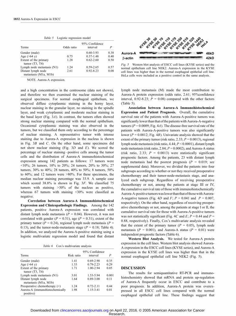
Western Blot Analysis. We tested for Aurora-A protein
expression in the cell lines. Western blot analysis showed Aurora-
A expression in the ESCC cell lines (KYSE series), and Aurora-A
expression in the KYSE cell lines was higher than that in the
normal esophageal epithelial cell line NEK2 (Fig. 5).
DISCUSSION
The results for semiquantitative RT-PCR and immuno-
histochemistry showed that mRNA and protein up-regulation
of Aurora-A frequently occur in ESCC and contribute to a
poor prognosis. In addition, Aurora-A protein was overex-
pressed in all ESCC cell lines compared with the normal
esophageal epithelial cell line. These findings suggest that
Table 6 Cox’s multivariate analysis
Terms Risk ratio95% Confidence
interval P
Gender (male) 1.41 0.69-2.90 0.35Age (>64 y) 1.33 0.79-2.23 0.29Extent of the primarytumor (T3, T4)
1.71 1.00-2.94 0.05
Lymph node metastasis (N1) 3.01 1.53-5.94 0.001Distant lymph nodemetastasis (M1a, M1b)
1.64 0.89-3.00 0.11
Preoperative chemotherapy (+) 1.24 0.72-2.11 0.44Aurora-A (immunohistochemicallypositive)
1.98 1.15-3.41 0.01
Fig. 5 Western blot analysis of ESCC cell lines (KYSE series) and thenormal epithelium cell line NEK2. Aurora-A expression in the KYSEcell lines was higher than in the normal esophageal epithelial cell line.HeLa cells were included as a positive control in the same analysis.
Table 5 Logistic regression model
Terms Odds ratio95% Confidence
interval P
Gender (male) 0.60-3.93 0.38Age (>64 y) 0.75 0.37-1.48 0.40Extent of the primarytumor (T3, T4)
1.28 0.62-2.60 0.50
Lymph node metastasis (N1) 1.24 0.59-2.65 0.57Distant lymph nodemetastasis (M1a, M1b)
2.61 0.92-8.23 0.08
NOTE. Aurora-A expression.
Aurora-A Expression in ESCC1832
Research. on April 22, 2016. © 2005 American Association for Cancerclincancerres.aacrjournals.org Downloaded from

Aurora-A up-regulation is a common abnormality in ESCC
and may play a role in its progression.
Aurora-A status and distant lymph node metastasis was
found to be significantly associated using Fisher’s exact test, and
similarly, logistic regression analysis showed a tendency toward
their association, albeit not statistically significant. Several
previous reports showed that the up-regulation of Aurora-A is
correlated with these malignant phenotypes in many cancers
(11–14). The up-regulation of Aurora-A leads to centrosome
amplification and consequently causes chromosome instability
(9), which may help tumor cells obtain invasive and metastatic
phenotypes. In addition, a more recent report showed that the
Aurora-A Phe31Ile polymorphism is associated with advanced
stage ESCC and its occurrence (27). For esophageal carcinoma,
when a patient has distant lymph node metastasis, it can be
considered a systemic disease (20). We think that some
biological changes might occur at this time. The number of
patients who had distant lymph node metastasis in our study
was small, but we speculate that the excessive expression
of Aurora-A may be related to the progression from a regional
disease state to a systemic disease state for ESCC. Furthermore,
not only the up-regulation of Aurora-A but also the frequent
change in Aurora-A polymorphism may affect the invasiveness
and metastatic properties of tumor cells in ESCC.
With regard to the correlation between immunohisto-
chemical Aurora-A staining and the prognosis of ESCC, the
cumulative survival rate and disease-free survival rate of
patients with Aurora-A-positive tumors was significantly lower
than that of patients with Aurora-A-negative tumors. Thus, an
Aurora-A-positive status is an independent prognostic factor,
implying that the elevated expression of Aurora-A may be an
indicator of the patient’s prognosis. Recent reports showed
that the up-regulation of Aurora-A results in resistance to
apoptosis induced by paclitaxel in a human cancer cell line
(28, 29). This raises the possibility that Aurora kinase
inhibition may provide for a new approach for the treatment
of multiple human malignancies (30). Currently, additional
studies including those examining the association between
Aurora-A expression status and chemosensitivity of ESCC are
under way in our laboratory.
In conclusion, our findings suggest that mRNA and the
immunohistochemical up-regulation of Aurora-A may be useful
as a prognostic factor for ESCC patients. Future studies on the
physiologic targets of Aurora-A and its potential role in the
pathogenesis of ESCC will be helpful for finding a novel
therapeutic strategy for the treatment of ESCC.
ACKNOWLEDGMENTSWe thank Sakiko Shimada for culturing and providing the ESCC
cell lines; Junichiro Kawamura, Kan Kondo, Shiro Nagatani, Toshiya
Soma, Naoki Teratani, Masato Kondo, and Yukiko Mori for their
technical advice; and Takako Murai and Akane Iwase for their technical
assistance.
REFERENCES
1. Ghadimi BM, Sackett DL, Difilippantonio MJ, et al. Centrosomeamplification and instability occurs exclusively in aneuploid, but not indiploid colorectal cancer cell lines, and correlates with numericalchromosomal aberrations. Genes Chromosomes Cancer 2000;27:183–90.
2. Kuo KK, Sato N, Mizumoto K, et al. Centrosome abnormalities inhuman carcinomas of the gallbladder and intrahepatic and extrahepaticbile ducts. Hepatology 2000;31:59–64.
3. Lingle WL, Lutz WH, Ingle JN, Maihle NJ, Salisbury JL. Centrosomehypertrophy in human breast tumors: implications for genomic stabilityand cell polarity. Proc Natl Acad Sci U S A 1998;95:2950–5.
4. Pihan GA, Purohit A, Wallace J, Malhotra R, Liotta L, Doxsey SJ.Centrosome defects can account for cellular and genetic changes thatcharacterize prostate cancer progression. Cancer Res 2001;61:2212–9.
5. Pihan GA, Purohit A, Wallace J, et al. Centrosome defects and geneticinstability in malignant tumors. Cancer Res 1998;58:3974–85.
6. Sato N, Mizumoto K, Nakamura M, et al. Correlation betweencentrosome abnormalities and chromosomal instability in humanpancreatic cancer cells. Cancer Genet Cytogenet 2001;126:13–9.
7. Saunders WS, Shuster M, Huang X, et al. Chromosomal instabilityand cytoskeletal defects in oral cancer cells. Proc Natl Acad Sci U S A2000;97:303–8.
8. Nigg EA. Mitotic kinases as regulators of cell division and itscheckpoints. Nat Rev Mol Cell Biol 2001;2:21–32.
9. Zhou H, Kuang J, Zhong L, et al. Tumour amplified kinase STK15/BTAK induces centrosome amplification, aneuploidy and transformation.Nat Genet 1998;20:189–93.
10. Bischoff JR, Anderson L, Zhu Y, et al. A homologue of Drosophilaaurora kinase is oncogenic and amplified in human colorectal cancers.EMBO J 1998;17:3052–65.
11. Li D, Zhu J, Firozi PF, et al. Overexpression of oncogenic STK15/BTAK/Aurora A kinase in human pancreatic cancer. Clin Cancer Res2003;9:991–7.
12. Sen S, Zhou H, Zhang RD, et al. Amplification/overexpression of amitotic kinase gene in human bladder cancer. J Natl Cancer Inst2002;94:1320–9.
13. Royce ME, Xia W, Sahin AA, et al. STK15/Aurora-A expression inprimary breast tumors is correlated with nuclear grade but not withprognosis. Cancer 2004;100:12–9.
14. Hamada M, Yakushijin Y, Ohtsuka M, Kakimoto M, Yasukawa M,Fujita S. Aurora2/BTAK/STK15 is involved in cell cycle checkpoint andcell survival of aggressive non-Hodgkin’s lymphoma. Br J Haematol2003;121:439–47.
15. Isola JJ, Kallioniemi OP, Chu LW, et al. Genetic aberrations detectedby comparative genomic hybridization predict outcome in node-negativebreast cancer. Am J Pathol 1995;147:905–11.
16. Sato N, Mizumoto K, Nakamura M, et al. Correlation betweencentrosome abnormalities and chromosomal instability in humanpancreatic cancer cells. Cancer Genet Cytogenet 2001;126:13–9.
17. Fukushige S, Waldman FM, Kimura M, et al. Frequent gain ofcopy number on the long arm of chromosome 20 in humanpancreatic adenocarcinoma. Genes Chromosomes Cancer 1997;19:161–9.
18. Pimkhaokham A, Shimada Y, Fukuda Y, et al. Nonrandomchromosomal imbalances in esophageal squamous cell carcinoma celllines: possible involvement of the ATF3 and CENPF genes in the 1q32amplicon. Jpn J Cancer Res 2000;91:1126–33.
19. Sobin LH, Fleming ID. TNM classification of malignant tumors, 5thedition. Cancer 1997;80:1803–4.
20. Fleming ID, Cooper JS, Henson DE, et al, editors. American JointCommittee on Cancer. AJCC cancer staging manual. 5th ed. Philadel-phia: J.B. Lippincott; 1997.
21. Shimada Y, Imamura M, Wagata T, Yamaguchi N, Tobe T.Characterization of 21 newly established esophageal cancer cell lines.Cancer 1992;69:277–84.
22. Okumura T, Shimada Y, Imamura M, Yasumoto S. Neurotrophinreceptor p75(NTR) characterizes human esophageal keratinocyte stemcells in vitro . Oncogene 2003;22:4017–26.
23. Cremet JY, Descamps S, Verite F, Martin A, Prigent C. Preparationand characterization of a human aurora-A kinase monoclonal antibody.Mol Cell Biochem 2003;243:123–31.
Clinical Cancer Research 1833
Research. on April 22, 2016. © 2005 American Association for Cancerclincancerres.aacrjournals.org Downloaded from

24. Marumoto T, Honda S, Hara T, et al. Aurora-A kinase maintains thefidelity of early and late mitotic events in HeLa cells. J Biol Chem2003;278:51786–95.
25. Chomczynski P, Sacchi N. Single-step method of RNA isolation byacid guanidinium thiocyanate-phenol-chloroform extraction. Anal Bio-chem 1987;162:156–9.
26. Chomczynski P. A reagent for the single-step simultaneous isolationof RNA, DNA and proteins from cell and tissue samples. Biotechniques1993;15:532–4,536–7.
27. Miao X, Sun T, Wang Y, Zhang X, Tan W, Lin D. Functional STK15Phe31Ile polymorphism is associated with the occurrence and advanced
disease status of esophageal squamous cell carcinoma. Cancer Res2004;64:2680–3.
28. Anand S, Penrhyn-Lowe S, Venkitaraman AR. AURORA-Aamplification overrides the mitotic spindle assembly checkpoint,inducing resistance to Taxol. Cancer Cell 2003;3:51–62.
29. Dutertre S, Prigent C. Aurora-A overexpression leads to override ofthe microtubule-kinetochore attachment checkpoint. Mol Interv2003;3:127–30.
30. Harrington EA, Bebbington D, Moore J, et al. VX-680, a potent andselective small-molecule inhibitor of the Aurora kinases, suppressestumor growth in vivo . Nat Med 2004;10:262–7.
Aurora-A Expression in ESCC1834
Research. on April 22, 2016. © 2005 American Association for Cancerclincancerres.aacrjournals.org Downloaded from

2005;11:1827-1834. Clin Cancer Res Eiji Tanaka, Yosuke Hashimoto, Tetsuo Ito, et al.
CarcinomaExpression in Human Esophageal Squamous Cell The Clinical Significance of Aurora-A/STK15/BTAK
Updated version
http://clincancerres.aacrjournals.org/content/11/5/1827
Access the most recent version of this article at:
Cited articles
http://clincancerres.aacrjournals.org/content/11/5/1827.full.html#ref-list-1
This article cites 28 articles, 9 of which you can access for free at:
Citing articles
http://clincancerres.aacrjournals.org/content/11/5/1827.full.html#related-urls
This article has been cited by 17 HighWire-hosted articles. Access the articles at:
E-mail alerts related to this article or journal.Sign up to receive free email-alerts
Subscriptions
Reprints and
To order reprints of this article or to subscribe to the journal, contact the AACR Publications
Permissions
To request permission to re-use all or part of this article, contact the AACR Publications
Research. on April 22, 2016. © 2005 American Association for Cancerclincancerres.aacrjournals.org Downloaded from
Copyright © 2022 FDOKUMEN