The class 6 semaphorin SEMA6A is induced by interferon-gamma and defines an activation status of...
13
Immunopathology and Infectious Diseases The Class 6 Semaphorin SEMA6A Is Induced by Interferon- and Defines an Activation Status of Langerhans Cells Observed in Pathological Situations Gregory Gautier,* Blandine de Saint-Vis,* Brigitte Se ´ne ´ chal, † Jean-Jacques Pin,* Elizabeth E.M. Bates,* Christophe Caux,* Fre ´de ´ ric Geissmann, † and Pierre Garrone* From the Laboratory for Immunological Research,* Schering- Plough, Dardilly; and Equipe INSERM “Avenir” and Laboratoire d’Anatomie Pathologique, † IFR94, Faculte ´ de Me ´decine Necker, Universite ´ Paris V, Paris, France Originally implicated in axon guidance , semaphorins represent a large family of molecules that are now known to be expressed in the immune system. Among different semaphorins tested by reverse tran- scriptase-polymerase chain reaction in human im- mune cells , the expression of class 6 transmembrane semaphorin SEMA6A was restricted to dendritic cells (DCs). Using in-house generated monoclonal antibod- ies , SEMA6A expression appeared further restricted to Langerhans cells (LCs). In vivo , SEMA6A mRNA was expressed in freshly isolated skin LCs but SEMA6A protein was not detectable on normal skin and ton- sillar epithelium. Of interest , SEMA6A protein was strongly expressed on skin and bone LCs and on LCs in draining lymph nodes from patients with LC histi- ocytosis or dermatopathic lymphadenitis , respec- tively , representing two inflammatory conditions in which LCs display an immature DC-LAMP low , CD83 low , and CCR7 phenotype. SEMA6A expression was low in resting LCs generated in vitro and was enhanced by interferon (IFN)- but not by interleu- kin-4 , interleukin-10 , IFN-/ , or lipopolysaccharide. Most IFN--induced SEMA6A-positive cells remained immature with low CD83 and DC-LAMP/CD208 ex- pression , but they expressed CCR7 and responded to macrophage inflammatory protein-3 (MIP-3/ CCL19). The expression of SEMA6A, for which the ligand and function remain unknown , may therefore identify an alternative IFN--dependent activation sta- tus of LCs in vivo. (Am J Pathol 2006, 168:453– 465; DOI: 10.2353/ajpath.2006.050288) Dendritic cells (DCs) are bone marrow-derived profes- sional antigen-presenting cells required to initiate spe- cific T-cell immune responses. 1 Immature DCs reside in nonlymphoid tissues, similar to Langerhans cells (LCs), which form an organized network in the epidermis. Dur- ing pathogen invasion, they are believed to capture an- tigen and, after inflammatory stimuli, leave the tissues to reach the draining lymphoid organs where they fit to- gether in a meshwork favoring interaction with T cells. During their migration, DCs usually undergo phenotypical and functional maturation including loss of antigen cap- ture capacity and up-regulation of co-stimulatory mole- cules such as CD80 and CD86. Thus, migration and organization in network constitute integral parts of DC function. Different studies have demonstrated that traf- ficking of DCs is controlled by soluble chemotactic fac- tors known as chemokines, 2–5 but other molecules are expected to play a role in the regulation of DC tissue trafficking. Guidance of axon during the development of the ner- vous system involves molecules such as Slit, Robo, Eph, and semaphorins, which function by either attractive or repulsive effects through interactions with their specific receptors and regulation of cytoskeleton organization. 6–8 In particular, semaphorins represent a large phylogeneti- cally conserved family (25 genes) of secreted, glyco- sylphosphatidylinositol-anchored and transmembrane glycoproteins that are divided in eight subclasses (sub- classes 1 to 7 and V for viral). 9,10 One characteristic of semaphorins is an N-terminal 500-amino acid extracellu- Supported by the Fondation Marcel Me ´ rieux, Lyon, France (grant to G.G.), and INSERM (“Avenir” grant to B.S. and F.G. for work in Necker). Accepted for publication October 7, 2005. Address reprint requests to Christophe Caux, INSERM U590, Centre Leon Berard, 28 rue Laennec, 69373 Lyon Cedex 08, France. E-mail: [email protected]. American Journal of Pathology, Vol. 168, No. 2, February 2006 Copyright © American Society for Investigative Pathology DOI: 10.2353/ajpath.2006.050288 453
-
Upload
univ-paris-diderot -
Category
Documents
-
view
1 -
download
0
Transcript of The class 6 semaphorin SEMA6A is induced by interferon-gamma and defines an activation status of...

Immunopathology and Infectious Diseases
The Class 6 Semaphorin SEMA6A Is Induced byInterferon-� and Defines an Activation Status ofLangerhans Cells Observed in PathologicalSituations
Gregory Gautier,* Blandine de Saint-Vis,*Brigitte Senechal,† Jean-Jacques Pin,*Elizabeth E.M. Bates,* Christophe Caux,*Frederic Geissmann,† and Pierre Garrone*From the Laboratory for Immunological Research,* Schering-
Plough, Dardilly; and Equipe INSERM “Avenir” and Laboratoire
d’Anatomie Pathologique,† IFR94, Faculte de Medecine Necker,
Universite Paris V, Paris, France
Originally implicated in axon guidance, semaphorinsrepresent a large family of molecules that are nowknown to be expressed in the immune system.Among different semaphorins tested by reverse tran-scriptase-polymerase chain reaction in human im-mune cells, the expression of class 6 transmembranesemaphorin SEMA6A was restricted to dendritic cells(DCs). Using in-house generated monoclonal antibod-ies, SEMA6A expression appeared further restrictedto Langerhans cells (LCs). In vivo , SEMA6A mRNA wasexpressed in freshly isolated skin LCs but SEMA6Aprotein was not detectable on normal skin and ton-sillar epithelium. Of interest, SEMA6A protein wasstrongly expressed on skin and bone LCs and on LCsin draining lymph nodes from patients with LC histi-ocytosis or dermatopathic lymphadenitis, respec-tively, representing two inflammatory conditions inwhich LCs display an immature DC-LAMPlow,CD83low, and CCR7� phenotype. SEMA6A expressionwas low in resting LCs generated in vitro and wasenhanced by interferon (IFN)-� but not by interleu-kin-4, interleukin-10, IFN-�/� , or lipopolysaccharide.Most IFN-�-induced SEMA6A-positive cells remainedimmature with low CD83 and DC-LAMP/CD208 ex-pression, but they expressed CCR7 and respondedto macrophage inflammatory protein-3� (MIP-3�/CCL19). The expression of SEMA6A, for which theligand and function remain unknown, may thereforeidentify an alternative IFN-�-dependent activation sta-
tus of LCs in vivo. (Am J Pathol 2006, 168:453–465; DOI:10.2353/ajpath.2006.050288)
Dendritic cells (DCs) are bone marrow-derived profes-sional antigen-presenting cells required to initiate spe-cific T-cell immune responses.1 Immature DCs reside innonlymphoid tissues, similar to Langerhans cells (LCs),which form an organized network in the epidermis. Dur-ing pathogen invasion, they are believed to capture an-tigen and, after inflammatory stimuli, leave the tissues toreach the draining lymphoid organs where they fit to-gether in a meshwork favoring interaction with T cells.During their migration, DCs usually undergo phenotypicaland functional maturation including loss of antigen cap-ture capacity and up-regulation of co-stimulatory mole-cules such as CD80 and CD86. Thus, migration andorganization in network constitute integral parts of DCfunction. Different studies have demonstrated that traf-ficking of DCs is controlled by soluble chemotactic fac-tors known as chemokines,2–5 but other molecules areexpected to play a role in the regulation of DC tissuetrafficking.
Guidance of axon during the development of the ner-vous system involves molecules such as Slit, Robo, Eph,and semaphorins, which function by either attractive orrepulsive effects through interactions with their specificreceptors and regulation of cytoskeleton organization.6–8
In particular, semaphorins represent a large phylogeneti-cally conserved family (�25 genes) of secreted, glyco-sylphosphatidylinositol-anchored and transmembraneglycoproteins that are divided in eight subclasses (sub-classes 1 to 7 and V for viral).9,10 One characteristic ofsemaphorins is an N-terminal 500-amino acid extracellu-
Supported by the Fondation Marcel Merieux, Lyon, France (grant to G.G.),and INSERM (“Avenir” grant to B.S. and F.G. for work in Necker).
Accepted for publication October 7, 2005.
Address reprint requests to Christophe Caux, INSERM U590, CentreLeon Berard, 28 rue Laennec, 69373 Lyon Cedex 08, France. E-mail:[email protected].
American Journal of Pathology, Vol. 168, No. 2, February 2006
Copyright © American Society for Investigative Pathology
DOI: 10.2353/ajpath.2006.050288
453

lar domain (called sema domain) that contains cysteine-rich repeats. Members of the semaphorin family havealso been implicated in organogenesis, vascularization,angiogenesis, neuronal apoptosis, and the progressionof cancer.11,12 The best characterized receptors forsemaphorins are the plexins and neuropilins.6,13,14 Re-cently, a role for semaphorins in the immune system hasemerged.15–17 In particular, the semaphorin SEMA4D,known as CD100, is expressed on T cells, resting Bcells,18 and DCs, and it can be up-regulated after acti-vation. Its cross-linking enhances T-cell activation andproliferation in humans.19 More recently, human andmouse sema4D have been found to enhance B cell,monocyte, and monocyte-derived DC (moDC) activationwith secretion of proinflammatory cytokines and prolifer-ation by turning off the negative signals of CD72.19–21
Soluble human SEMA4D can also inhibit monocyte andB-cell migration.22 The class 4 semaphorin sema4a, ex-pressed by mouse bone marrow-derived DCs and Bcells, enhances the in vitro activation and differentiation ofT cells and the in vivo generation of antigen-specific Tcells through binding and activation of the receptor Tim-2(T cell conserved immunoglobulin and mucin domain),which is expressed on T cells.23 Additionally, virus-en-coded semaphorins SEMAVA/A39R and SEMAVB/AHV,and human SEMA7A/CDw108 induce aggregation ofmonocytes, secretion of the proinflammatory cytokinesinterleukin (IL)-6 and IL-8, and expression of CD54through activation of their receptor Plexin-C1.13,24 Thesefindings indicate that semaphorins not only have a role inthe nervous system but are also involved in regulation ofthe immune system through interactions with plexins andneuropilins as well as with other receptors such as CD72and Tim-2.17
In recent efforts to determine the expression and pos-sible functions of cell guidance molecules on DCs, wehave shown that Eph kinase receptors regulate integrin-mediated adhesion of DCs.25 Herein, we have analyzedthe presence of mRNA for several different semaphorinsin human immune cells, in particular in DCs. We reportthat LCs generated in vitro from cord blood CD34� pro-genitors and LCs isolated from skin preferentially ex-pressed the class 6 semaphorin SEMA6A-1.26 The class6 semaphorins are transmembrane semaphorins com-posed of four members: SEMA6A, 6B, 6C, and 6D.26–29
Two isoforms of SEMA6A have been described:SEMA6A-1 and SEMA6A-2. The full-length SEMA6A-1exhibits an intracellular domain that displays a small re-gion of homology to Zyxin, with a proline-rich motif in-volved in the regulation of actin polymerization via bind-ing to Ena/VASP proteins,26 suggesting a role in LCtrafficking or activation.
Maximal SEMA6A expression on LCs required a cer-tain level of activation such as that provided by interferon(IFN)-�. These cells migrated in response to CCR7 li-gands but remained immature with respect to CD83 andDC-LAMP expression. In vivo SEMA6A protein was notdetected on LCs from normal skin but was abundantlyexpressed on skin and bone LCs from patients with Lang-erhans cell histiocytosis (LCH) and on LCs in draininglymph nodes from patients with dermatopathic lymphad-
enitis (DL), which also display a phenotype similar to theone of IFN-�-treated cells in vitro with respect to CD83,DC-LAMP,30–33 and CCR7 expression.30,31 The pres-ence of SEMA6A on immature or partially mature LCsmay delineate a particular pathway of LC activation invivo.
Materials and Methods
Hematopoietic Factors, Cytokines, Reagents,and Cell Lines
All cultures were performed in RPMI 1640 medium sup-plemented with 10% heat-inactivated fetal calf serum, 2mmol/L L-glutamine (all from Life Technologies, Gaithers-burg, MD), and 100 �g/ml gentamicin (Schering-Plough,Levallois-Perret, France). All hematopoietic factors andcytokines were recombinant human proteins and wereused at optimal concentration as indicated: granulocytemacrophage-colony stimulating factor (100 ng/ml GM-CSF: specific activity, 2 � 106 U/mg; Schering-PloughResearch Institute, Kenilworth, NJ), tumor necrosis factor(TNF)-� (2.5 ng/ml: specific activity, 2 � 107 U/mg; Gen-zyme, Boston, MA), stem cell factor (SCF) (25 ng/ml:specific activity, 4 � 105 U/mg; R&D Systems, Abington,UK), IL-4 (10 ng/ml specific activity, 107 U/mg; Schering-Plough Research Institute), IL-10 (100 ng/ml: specificactivity, 107 U/mg; Schering-Plough Research Institute),IFN-� (20 ng/ml: specific activity,: 107 U/mg; R&D Sys-tems), and transforming growth factor (TGF)-�1 (10 ng/ml: specific activity, 5 � 107 U/mg; R&D Systems). Thechemokines macrophage inflammatory protein-3� (MIP-3�/CCL20), MIP-3�/CCL19, MIG/CXCL9, and IP-10/CXCL10 were from R&D Systems. In some experiments,cells were activated with 1 ng/ml of phorbol 12-myristate13-acetate (PMA) (Sigma-Aldrich, St. Louis, MO) and 1�g/ml of ionomycin (Calbiochem, La Jolla, CA) or with 25ng/ml of Escherichia coli lipopolysaccharide (LPS; Sigma-Aldrich). Murine fibroblasts transfected with humanCD40 ligand (CD40L-L cells) were produced in thelaboratory.25
Cell Preparation
Umbilical cord blood samples, adult peripheral bloodsamples, and tonsils were obtained according to institu-tional guidelines. Peripheral blood mononuclear cells(PBMCs) were purified from human peripheral blood byFicoll-Hypaque centrifugation. Monocytes (�95% CD14�)were purified from PBMCs by centrifugation over a 52%Percoll gradient followed by immunomagnetic depletion ofcontaminating T, B, and NK cells as described else-where.25,34 Granulocytes were purified from whole blood, Tlymphocytes (�95% CD3�) were purified from PBMCs byimmunomagnetic depletion, and B cells (�98% CD19�)were isolated from tonsils as previously described.34
CD34� hematopoietic progenitors were purified from um-bilical cord blood through positive selection using anti-CD34 monoclonal antibody (mAb)-coated microbeads(Miltenyi Biotech, Bergish Gladbach, Germany) and was
454 Gautier et alAJP February 2006, Vol. 168, No. 2

achieved using Minimacs separation columns (Miltenyi Bio-tech) as previously described.35 Epidermal cell suspen-sions were obtained from normal skin of patients undergo-ing reconstructive plastic surgery of breast or abdomen, asdescribed elsewhere.36 LC enrichment was achieved bysuccessive density gradient centrifugation steps and deple-tion of basal keratinocytes. The isolated cells contained 55to 75% CD1a� LCs.
Generation of DCs from CD34� Progenitorsand from Monocytes
Cultures of CD34� cells were established in the presenceof SCF, GM-CSF, TNF-�, and 5% AB� pooled humanserum, as described.35,37 By day 6, human serum wasremoved, and cells were further cultured in the presenceof GM-CSF and TNF-�, with or without TGF-�1 until day12. At this time point, aliquots of cells were activated withPMA and ionomycin for 1 and 6 hours, pooled, and thenlysed for RNA extraction. For analysis of SEMA6A expres-sion by flow cytometry, cells were collected at the timepoint indicated. DCs were activated by co-culture withirradiated (7500 rads) CD40L-L cells (one CD40L-L cellfor five DCs) for 48 hours. In some experiments, CD1a�
and CD14� DC precursor subsets were separated at day6 by sequential immunomagnetic-positive selection usingMinimacs separation columns (CD1a and CD14 immuno-beads) (Miltenyi Biotech) and further cultured until day 10for the CD1a� DC subset in the presence of GM-CSF andTNF-� and until day 12 for the CD14� DC subset in thepresence of GM-CSF and TNF-� with or without TGF-�1.moDCs were produced by culturing purified blood mono-cytes for 6 days in the presence of GM-CSF and IL-4. Insome experiments, 5 � 105 moDCs/well (24-well cultureplate) were further activated with 25 ng/ml LPS for 72hours or by co-culture with 105 irradiated CD40L-L cells.
RNA Extraction, cDNA Reverse Transcription,and Reverse Transcriptase (RT)-PolymeraseChain Reaction (PCR) Analysis
Cells were lysed and total RNA was extracted by RNeasykit (Qiagen, Valencia, CA). First-strand cDNAs were pre-pared after DNase I treatment (in the presence of RNaseinhibitor) of 5 �g of total RNA using oligo(dT) primers(Pharmacia, Uppsala, Sweden) and Superscript kit.Genomic contamination was excluded by quantitativePCR (Light Cycler; Bio-Rad, Hercules, CA) using humanCD4 promoter probe (forward primer: 5�-TTCCACACT-GGGCCACCTAT-3�, reverse primer: 5�-TTGTGGGCT-TACCACTGCTG-3�, probe: CACTGGACACAATTGC-CCTCAGG-3�). Synthesis of cDNAs was controlled byperforming RT-PCR using �-actin sequence (21, 28, and35 cycles).25 RT-PCRs were performed using Ready MixREDTaq (Sigma Chemical Co., St. Louis, MO) with theprimers 5�-GTCCTGCTCCCCGCCGATTC-3� (forward)and 5�-CACTGCTGTCCCCACTTACC-3� (reverse) forSEMA4A, 5�-CAAGGGACGAGAAGGGGAAT-3� (for-ward) and 5�-AAAAGGGTGTCACGCCAGTC-3� (reverse)
for SEMA4B, 5�-GTCCGACTGCTATGCCGAGC-3� (for-ward) and 5�-CGCCGATGCCAGTTGTTAAT-3� (reverse)for SEMA4C, 5�-AACGAGCCTAGTTTCGTGTTTGC-3�(forward) and 5�-GGCCTGGGTCCGGTCCACCACG-3�(reverse) for SEMA4D, 5�-CGGCACTTTGGCTCATCT-CT-3� (forward) and 5�-AAGTTCCCTCTGTCGCCGTC-3�(reverse) for SEMA4F, and 5�-GCGTGATGTTGTCTG-GCAA-3� (forward) and 5�-CACGCTCTATGTCCT-GCTCA-3� (reverse) for human SEMA6A cDNA. Cycleconditions (35 cycles) were 92°C for 1 minute, 60°C for 1minute, and 72°C for 2 minutes. PCR products werecloned using the pCRIITOPO vector (TA cloning kit; In-vitrogen, San Diego, CA). Double-stranded plasmid wassequenced on an ABI 373A sequencer (Applied Biosys-tems, Foster City, CA) using dye terminator technology.Sequencher (Gene Codes, Ann Arbor, MI) software wasused to analyze sequences and verify identity with hu-man SEMA6A (accession number AF279659).
Generation of Anti-SEMA6A mAbs
Full-length SEMA6A was cloned from cDNA of total hu-man brain (Clontech, Palo Alto, CA). Full-length SEMA6Awith a C-terminal c-myc tag (intracellular domain)(SEMA6A-c-myc) was cloned into the pMET7 plasmidand was used for mouse immunization and for flow cyto-metric screening of mAbs. Full-length SEMA6A with anN-terminal HA tag (extracellular domain) (HA-SEMA6A)was cloned into the pDisplay (Invitrogen) plasmid andwas used for immunocytochemical screening of mAbs.For the SEMA6A-c-myc construct, SalI restriction enzymesite (underlined) followed by the start codon was in-cluded in the forward cloning primer (5�-ATAGTCGACAT-GAGGTCAGAAGCCTTGCTGC-3�), and a c-myc epitopefollowed by the stop codon and NotI restriction enzymesite (underlined) (5�-ATAGCGGCCGCTTAAGATCCTCT-TCAGAGATGAGTTTCTGCTCCTGTACACGCATCATTG-GGCT-3�) was introduced in the reverse cloning primer.For the HA-SEMA6A construct, the forward primer con-taining SalI restriction enzyme site (underlined) and ex-cluding SEMA6A signal peptide (5�-CCCGGTCGACAC-CAGAAGATTCTGAGCCAAT-3�) (peptide signal and HAtag are included in pDisplay) and the reverse primercontaining a stop codon and a NotI restriction enzymesite (underlined) (5�-ATAGCGGCCGCTTATGTACACG-CATCATTGGGCT-3�) were used. mAbs against SEMA6Awere produced by immunizing BALB/c mice (CharlesRivers, Les Oncins, France) with three intraperitonealinjections of 106 SEMA6A-c-myc-transfected COP-5 cellsin Freund’s adjuvant (Sigma). Spleens were removed 3days after a final intravenous injection of SEMA6A-c-myc-transfected COP-5, and splenocytes were fused with mu-rine SP2 myeloma cell line using polyethylene glycol-1000 and cultured in 96-well plates using standardprocedures. Hybridoma supernatants were screened fortheir reactivity against acetone-fixed HA-SEMA6A-trans-fected COP-5 cells by immunocytochemistry. Selectedsupernatants were confirmed by flow cytometry onSEMA6A-c-myc-transfected COP-5 cells and by immuno-cytochemistry. Surface and intracytoplasmic (cells were
SEMA6A Expression by Human DCs 455AJP February 2006, Vol. 168, No. 2

first permeabilized with saponine) staining were per-formed and analyzed on a FACSCalibur flow cytometer(BD Biosciences, Mountain View, CA). After cloning ofselected hybridomas, mAbs were produced by high-density culture INTEGRA CL 350 (IBS Integra Bio-sciences, Chur, Switzerland) and purified by HPLC on ananion exchange column. The mAbs 118E7 and 104B3used in this study are of IgG1 kappa isotype and recog-nize the extracellular domain of SEMA6A.
Flow Cytometry Analysis
Cell-surface expression of SEMA6A was determined byflow cytometry analysis with a FACSCalibur flow cytome-ter (BD Biosciences). For single staining, cells were in-cubated for 30 minutes at 4°C with 10 �g/ml purifiedanti-SEMA6A mAb 118E7 in phosphate-buffered saline,1% human serum, washed twice, and then labeled withPE-conjugated goat anti-mouse IgG (DAKO, Glostrup,Denmark). Nonspecific staining was determined usingisotype-matched control mAb. PE-conjugated anti-CD25,anti-CD83, anti-CD86, and anti-CCR7 were used for thephenotype of in vitro-generated DCs.
Immunohistochemistry
Biopsy samples from 12 patients referred to Necker En-fants Malades Hospital and diagnosed as having LCH31
or DL30 were examined by immunohistochemistry. Serialcryostat sections of biopsy specimens were stained withCD1a or SEMA6A mAb 104B3 mouse primary antibodiesat the appropriate concentration as determined by titra-tion and then labeled with a goat anti-mouse peroxidase-conjugated antibody. AEC (Sigma) was used as a sub-strate for peroxidase. Isotype-matched antibodies wereused as negative controls. Double staining was per-formed with double stain EnVision kit (DAKO) accordingto the manufacturer’s instructions.
Migration Assay
Migration assays were performed using Transwell cham-bers (6.5-mm diameter; Costar, Cambridge, MA) with 5 �105 cells/well. CD34� progenitor cell-derived DCs wereincubated for 1 hour at 37°C in 5-�m pore size inserts inthe presence or the absence of MIP-3�, MIP-3�, IP-10, orMIG tested at 1 �g/ml in the lower chamber. Migratingcells (in the lower chamber) were collected and countedby flow cytometry. Each experiment was performed induplicate, and less than 10% variation was observed.
Results
Expression of Class 4 Semaphorins andSEMA6A mRNA in Cells of the Immune System
All RNA preparations were first controlled for the ab-sence of genomic DNA contamination, and cDNA werenormalized according to the �-actin PCR amplification.
Moreover, primers used in the study allowed us todiscriminate amplification of cDNA and genomic DNAand were designed to be specific for each semaphorinwithout cross-reacting with any other semaphorinsbased on available published sequences in NCBI da-tabase. Amplified fragments were also sequenced toconfirm the specificity. In a first set of experiments,RT-PCR reactions were performed with DCs derivedfrom CD34� progenitors cultured in the presence ofGM-CSF and TNF-� for 12 days. cDNA for class 5semaphorins were not amplified (data not shown),whereas mRNA for class 4 semaphorins (SEMA4A, 4B,4C, 4D, and 4F) and class 6 SEMA6A were detected inCD34�-derived DCs (Figure 1). Class 3 and 7 sema-phorins were not tested. In agreement with previousreports,17,22,38 – 40 SEMA4A and SEMA4D were alsodetected in T cells, B cells, monocytes, and granulo-cytes. Very faint or no band of SEMA6A mRNA couldbe amplified from T lymphocytes, granulocytes andPBMCs with or without activation (anti-CD3/anti-CD28for T cells and PMA plus ionomycin for granulocytesand PBMCs). Weak SEMA6A expression was detectedin B cells only after CD40L activation. Among in vitro-generated DCs, weak amplification of SEMA6A wasdetected in activated but not resting monocyte-derivedDCs (moDCs), contrasting with the strong expressionof SEMA4A, 4D, and 4F in DCs derived from mono-cytes with GM-CSF and IL-4 (Figure 1). But in CD34�-derived DCs, strong SEMA6A expression was detectedafter 12 days of culture (no signal was observed at day
Figure 1. Expression of class 4 semaphorins and SEMA6A mRNA in hema-topoietic cells. RT-PCR analysis of human class 4 and 6 semaphorin expres-sion in hematopoietic cells (left). cDNAs were prepared from resting andactivated (1 hour and 6 hours) freshly isolated PBMCs, blood T cells, mono-cytes, and granulocytes or tonsil B cells. RT-PCR analysis of semaphorinexpression in in vitro-generated CD34�-derived DCs and moDCs (right).cDNAs were prepared from cord blood CD34� progenitors cultured in thepresence of GM-CSF and TNF-� for 6 and 12 days, then for an additional 4days with CD40L-L cells, or from monocytes cultured in the presence ofGM-CSF and IL-4 for 6 days (unactivated), followed by 24-hour activationwith CD40L-L cells, or from CD40L-L cells as control. RT-PCR was performedunder standard conditions using 50 ng of cDNA for 35 cycles. All cDNAsamples were normalized according to the results of human �-actin PCRamplification of 21, 28, and 35 cycles (only the amplification of 28 cycles isshown) except for CD40L-L cells normalized using mouse �-actin PCR am-plification. The absence of genomic contamination was controlled in all RNAsamples (before the reverse transcription step) by quantitative PCR usinghuman CD4 promotor probe. Results are representative of RT-PCR of threeindependent samples.
456 Gautier et alAJP February 2006, Vol. 168, No. 2

6) and was maintained after CD40L activation (Figure1). SEMA6A sequences from these DCs were analyzedand found to be identical to the full-length sequencedescribed by Klostermann and colleagues.26 All to-gether these results show that SEMA6A is preferentiallyexpressed in DCs, in particular in CD34�-derived DCs,in contrast to other tested semaphorins that are ex-pressed on most leukocytes.
SEMA6A mRNA Is Preferentially Expressed inLCs
In vitro CD34�-derived DCs are composed of two subsetsof DC progenitors: the CD1a� subpopulation that differ-entiates into LCs and the CD14� subpopulation that maycorrespond to interstitial DCs.35,41 To analyze SEMA6Aexpression, these two subpopulations of DCs weresorted by flow cytometry according to CD14 and CD1aexpression after 6 days of culture of CD34� progenitorsand then recultured for 5 additional days with GM-CSFand TNF-� as previously described.37 As shown in Figure2A, SEMA6A mRNA was expressed at high levels in cellsderived from the resting and activated CD1a� subpopu-lation compared to that from the CD14� subpopulation in
which only a weak signal was observed after CD40Lactivation.
In DCs isolated ex vivo from tonsils, no SEMA6A ex-pression was detected by RT-PCR in CD11c� DCs, anda very faint signal was detected in cDNA of CD11c� cellsafter 35 cycles of PCR amplification (Figure 2B). In cor-relation with data obtained with in vitro-generated CD1a�
DCs, SEMA6A messenger was detected in both nonac-tivated and TNF-�-activated (48 hours) LCs isolated fromskin (Figure 2C). In contrast, no SEMA6A expression wasdetected in basal keratinocytes. All together, these re-sults indicate that SEMA6A is preferentially expressed inin vitro-generated CD1a� LCs and in ex vivo-isolated skinLCs.
Cell Surface Expression of SEMA6A IsRestricted to the in Vitro-Generated LCs
To confirm the expression of SEMA6A at the protein leveland to further search for its function on DCs, we gener-ated monoclonal antibodies (mAbs) against humanSEMA6A. The mAbs 118E7 (IgG1) and 104B3 (IgG1)recognizing the extracellular domain of recombinantSEMA6A were selected and used throughout this studyfor flow cytometry and immunocytochemistry/immunohis-tochemistry, respectively. Their specificity was demon-strated by flow cytometric analysis on different cell linespreviously tested by RT-PCR for the presence of varioussemaphorins including SEMA6A (not shown). On totalpopulation of CD34�-derived DCs generated in the pres-ence of GM-CSF and TNF-� at day 12, only a smallfraction of cells was generally stained by the anti-SEMA6A mAb (Figure 3A, top left). In agreement withRT-PCR analysis showing preferential expression ofSEMA6A mRNA in the CD1a� subset (Figure 2A), flowcytometry analysis showed a clear staining of anti-SEMA6A mAb 118E7 on DCs derived from CD1a� pre-cursors but not from CD14� precursors in the presenceof GM-CSF and TNF-� (Figure 3A). Exogenous TGF-�1 isrequired for LCs differentiation from CD14� precursors orfrom monocytes, but it is not necessary for LC differenti-ation from CD1a� precursors.35,37,42–45 Addition ofTGF-�1 from day 6 to day 12 of culture increased thedetection of cell surface SEMA6A on DCs derived fromtotal CD34� or from CD14� DC precursors (Figure 3B).Further activation by CD40L or LPS did not significantlyup-regulate the intensity of SEMA6A expression (notshown).
In accordance with the RT-PCR results, no staining ofanti-SEMA6A mAb 118E7 was detected by flow cytom-etry on blood T cells, B cells, NK cells, granulocytes, ormonocytes, even after activation (anti-CD3/anti-CD28 forT cells and LPS plus IL-4 or IFN-� for monocytes) (notshown). SEMA6A expression was very weakly inducedon tonsil B cells cultured with CD40L and IL-4 but not withCD40L and IL-2 plus IL-10 or IFN-� (not shown). Finally,we did not detect by flow cytometry any staining of anti-SEMA6A mAb 118E7 on blood CD11c� or CD11c� plas-macytoid DCs (pDCs) (not shown) or on moDCs (notshown).
Figure 2. RT-PCR analysis of human SEMA6A expression in different DCsubsets generated in vitro and in ex vivo-isolated CD11c� and CD11c� tonsilDCs and skin LCs. A: Cord blood CD34� progenitors were cultured in thepresence of GM-CSF, SCF, TNF-�, and 2.5% AB� human serum. At day 6,CD1a� and CD14� precursors were sorted by flow cytometry, reculturedwith GM-CSF and TNF-� for 6 additional days, and then activated or not asindicated. B: CD11c� and CD11c� DCs were sorted by flow cytometry fromtonsil. C: cDNAs were prepared from basal keratinocytes and from LCsfreshly isolated from normal skin activated or not with TNF-� for 48 hours.RT-PCR for SEMA6A was performed under standard conditions using 50 ng ofcDNA for 35 cycles. All cDNA samples were normalized according to theresults of �-actin PCR amplification and the absence of genomic contamina-tion was controlled as described in Figure 1. Results are representative ofRT-PCR of three independent samples.
SEMA6A Expression by Human DCs 457AJP February 2006, Vol. 168, No. 2
All together, these observations confirm that SEMA6Ais preferentially expressed at the cell surface of in vitro-generated LCs and identify TGF-� as a determinant cy-tokine for the induction of SEMA6A expression on invitro-generated LCs. However, we failed to detect cellsurface SEMA6A expression on freshly isolated skin LCsby flow cytometry (not shown), despite the fact thatSEMA6A mRNA was amplified in these cells (Figure 2C).
SEMA6A Is Expressed in LC-AssociatedPathologies
To analyze the in vivo expression of SEMA6A, immuno-staining with anti-SEMA6A mAb 104B3 was performed onsections of organs containing CD1a� LCs. No stainingwas observed in normal skin (10 of 10) (Figure 4A) or intonsils (that also contain mature DCs) (not shown), evenwith sensitive amplification technologies. We further an-alyzed SEMA6A expression on LCs in pathological sam-ples from inflammatory LCH and DL. Interestingly,SEMA6A was abundantly expressed in cutaneous (Fig-ure 4B), bone (Figure 4C), and lymph node LCH in eightof eight patients. The strong expression of SEMA6A com-pletely correlated with that of the CD1a marker on serialsections and was confirmed by double staining, asshown in Figure 4C (panel i) in bone lesion. CD1a ex-pression showed that these SEMA6A-positive cells wereLCs. Moreover, these DCs expressed CD68 and the LC-specific lectin Langerin (CD207) but not the maturationmarkers CD83 and DC-LAMP (not shown).31 The aggre-
gates of cells expressing high levels of SEMA6A in cuta-neous LCH were localized in the dermis (Figure 4A, e andf). We also observed co-expression between CD1a andSEMA6A on LCs accumulating in draining lymph nodes(LNs) from three of three patients with DL (Figure 5).30 InDL, LCs are recruited in high numbers in the sinus andT-cell area of inflammatory skin-draining LNs. These LCsexpressed CD1a and displayed typical morphology ofLCs (Figure 5, c and d), and have previously been shownto maintain a peculiar activated yet immature phenotypebecause most of these LCs express Langerin, CCR7, andCD68 but do not express CD83 and DC-LAMP maturationmarkers.30
IFN-� Up-Regulates SEMA6A Expression onDCs
To further determine which factors up-regulatedSEMA6A expression in LCs, we tested the effects ofproinflammatory cytokines on SEMA6A expression onin vitro-generated LCs (CD34�-derived DCs culturedas described above in the presence of GM-CSF,TNF-�, and TGF-� from day 6 to day 10 and thencultured for 48 hours with different cytokines). Amongfactors tested [TNF-�, LPS (TLR-4 ligand), R-848(TLR-8 ligand), poly I:C (TLR-3 ligand), IL-1�, IL-2,IL-3, IL-4, IL-6, IL-8, IL-10, IL-12, IL-18, CpG 2216(TLR-9 ligand), IFN-�, and type I IFN-�/�] (data notshown), only IFN-� showed a significant effect onSEMA6A expression. Indeed, as shown in Figure 6A,
Figure 3. SEMA6A expression is detected on CD1a� DC subset and can be induced by TGF-� on CD14� DC subset. CD34� progenitors were cultured withGM-CSF, TNF-�, SCF, and 5% AB� serum. At day 6, total DCs or CD1a� and CD14� DC subsets separated using immunomagnetic-positive selection were washedand recultured in the presence of GM-CSF and TNF-� (A) or GM-CSF, TNF-�, and TGF-� (B) until day 12. SEMA6A and Langerin expression were analyzed byflow cytometry after staining with anti-SEMA6A mAb 118E7 or anti-Langerin mAb (histograms in solid line) or control mouse IgG (histograms in dotted line)followed by PE-conjugated goat anti-mouse Ig. Histograms from one representative experiment are shown. Numbers in histograms are the mean � SD percentageof SEMA6A-positive cells calculated from four independent experiments.
458 Gautier et alAJP February 2006, Vol. 168, No. 2

IFN-� increased cell surface SEMA6A expression from5% in GM-CSF to 57% in GM-CSF plus IFN-� (compareto 25% in GM-CSF plus TNF-� plus TGF-�; Figure 3B).
In these experimental conditions, the low SEMA6A ex-pression after treatment with GM-CSF alone (Figure6A) was attributable to the absence of TGF-� from day
Figure 4. LCs in LCH strongly express SEMA6A. Serialsections from normal skin (A), skin lesions (cutaneousLCH) (B), and bone lesions (eosinophilic granuloma ofLCH) (C) were stained with CD1a (a, c, e, g) or SEMA6AmAb 104B3 (b, d, f, h). i: Double staining for CD1a (inblue) and SEMA6A (in red) was performed in bone lesion.Staining was detected by peroxidase-conjugated goat anti-mouse antibody using AEC as peroxidase substrate. For thedouble staining, mAbs were detected using double-stainEnVision kit. Original magnifications: �10 (a, b, g, h);�2.5 (c, d); �40 (e, f); �100 (i). Data are observed in 8 of8 patients with LCH and 10 of 10 normal skin samples.
SEMA6A Expression by Human DCs 459AJP February 2006, Vol. 168, No. 2

10 to 12. This effect required a first culture with TGF-�(days 6 to 10) because no SEMA6A staining was ob-served when CD34� cells were kept in culture withGM-CSF plus TNF-� alone before IFN-� addition (Fig-ure 6B). The up-regulation of surface SEMA6A expres-sion by IFN-� was correlated with an increased level ofSEMA6A mRNA in IFN-�-activated DCs (Figure 6C).Moreover, IL-10 and IL-4, known to inhibit IFN-� activ-ities, reduced the intensity of SEMA6A staining in-duced in the presence of IFN-� (Figure 6A). Of note,SEMA6A expression was not up-regulated by LPS andwas even down-regulated by LPS in IFN-�-pretreatedDCs (Figure 6A). In contrast to DCs derived fromCD34� progenitors, IFN-� did not induce SEMA6A ex-pression on moDCs or ex vivo isolated blood DC pop-ulations (not shown). Finally, although a faint signalwas detected by RT-PCR in CD40L-activated moDCs,no expression could be detected by flow cytometry onCD40L- or LPS-activated moDCs (not shown).
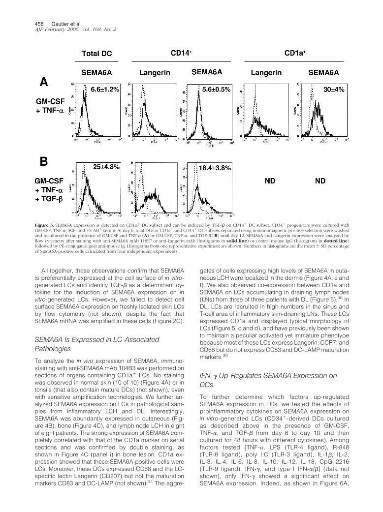
Based on the selectivity of the IFN-� effect on SEMA6Aexpression, we assessed by immunohistochemistry theexpression of IFN-� and IFN-� R1 on LCH biopsies. Al-though we could not detected significant IFN-� expres-sion, a strong expression of the IFN-� R1 was observed
on LCH cells (Figure 7), contrasting with the low expres-sion on resident skin LCs (Figure 7).
In Vitro Generated LCs Expressing SEMA6ADisplay an Immature Phenotype but Migrate inResponse to MIP-3�
IFN-� up-regulated SEMA6A expression while activatorssuch as LPS down-regulated IFN-�-induced SEMA6A ex-pression. Because DCs in LCH and DL express SEMA6Aand have been shown to possess a CD83low DCLamplow
immature phenotype, we investigated the expression ofmaturation markers such as DC-LAMP/CD208, CD86,and CD83 on LCs generated in vitro with GM-CSF, TNF-�,TGF-�, and IFN-� as described above. Interestingly, invitro-generated LCs, activated by IFN-� and expressingSEMA6A, remained immature with an absence of CD83,CD86, DC-LAMP, and CD25 markers but matured nor-mally in response to LPS activation, although lackingSEMA6A expression (Figure 8A).
However, in agreement with a previous report,46 IFN-�-stimulated LCs expressed CCR7 (Figure 8A) and re-sponded to the CCR7 ligand MIP-3�/CCL19 with a mi-
Figure 5. Expression of SEMA6A on LCs in lymph nodes from DL. Serial sections from draining lymph node of DL were stained with CD1a (a, c) or SEMA6A mAb104B3 (b, d). F denotes a B-cell follicle in c and d. c and d are higher magnifications of the boxes in a and b, respectively. Staining was detected byperoxidase-conjugated goat anti-mouse antibody using AEC as peroxidase substrate. Data are observed in three of three patients with DL. Original magnifications:�2.5 (a, b); �40 (c, d).
460 Gautier et alAJP February 2006, Vol. 168, No. 2

gration index of 10, whereas they did not migrate inresponse to the CCR6 ligand MIP-3�/CCL20 or otherchemokines such as MIG/CXCL9 or IP-10/CXCL10 (Fig-ure 8B). These observations suggest that SEMA6A ex-pression delineates a discrete stage of CCR7� immatureLCs.
Discussion
The expression of molecules primarily known for theirfunction in the nervous system has recently been shownin the immune system. SEMA6A, described in the presentstudy as selectively expressed on DCs, is a new exampleof such a molecule. Among the different semaphorinsanalyzed, SEMA6A appears to be the most DC-restrictedmember of the family. Other semaphorins expressed inDCs such as SEMA4A and 4D are also present in T andB lymphocytes, granulocytes, and monocytes, as previ-ously reported,22,23,38–40 and we also detected a broadexpression of other semaphorin mRNAs (includingSEMA4B and 4F) by lymphocytes and myeloid cells. Incontrast, mRNAs for other semaphorins such as SEMA5Awere not detected by RT-PCR analysis in DCs (data notshown). Beside semaphorins, other molecules involved in
axon guidance have been reported to be expressed inthe immune system. In particular, neuropilin-1 has beenshown to be expressed on DCs47 and Robo/Slit by Tcells48; we also recently reported the expression of sev-eral Eph proteins on DCs.25,49
Within the different subsets of DCs, SEMA6A expres-sion is restricted to LCs, as demonstrated by 1) expres-sion of the protein on the LC progeny of CD34-derivedCD1a� precursors (spontaneous differentiation); 2) ex-pression on the TGF-�-driven LC progeny of CD34�-derived CD14� precursors; 3) detection of the mRNA inLCs isolated from skin; 4) high levels of protein expres-sion on LCs from patients with cutaneous and bone LCHand with DL; and 5) absence of mRNA or protein in bloodcirculating DC subsets and moDCs. Such restricted ex-pression of SEMA6A argues for a role related to specificLC functions that remain primarily under-explored. Theprincipal characteristic of LCs is their status of residentcells with a very low turnover within pluri-stratified epithe-lia such as in the skin,50 where they form a network witha tightly controlled density. Based on the role of sema-phorins in the nervous system that control the directionalgrowth of axons through attractive or repulsive cues,7,51
it can be hypothesized that SEMA6A may play a role inthe epidermal LC network.
In normal conditions, we could not detect the proteinon skin LCs either by flow cytometry or by immunohistol-ogy. However, SEMA6A mRNA is up-regulated on TNF-�activation in skin LCs and the expression of SEMA6A onin vitro-generated LCs is strongly up-regulated on IFN-�treatment. Furthermore, IFN-�-stimulated in vitro-gener-ated LCs that express SEMA6A did not co-express DC-LAMP and CD83, markers of mature DCs and expressCCR7 and responded to the chemokine MIP-3� but not toMIP-3�. MIP-3�/CCL19 has been described to inducemigration through CCR7 of mature/activated DCs, whileMIP-3�/CCL20 induces migration of immature DCsthrough CCR6.52 These data suggest that these IFN-�-stimulated LCs were partially activated but not fully ma-ture. SEMA6A expression was only induced by IFN-� andwas down-regulated by all other tested cytokines or ac-tivators, such as LPS. Furthermore, SEMA6A expressionwas never detected in tonsil, suggesting that mature DCspresent in the tonsils either down-regulated or did notup-regulate SEMA6A expression. This suggests thatSEMA6A up-regulation on LCs was dependent on a par-ticular activation pathway produced by IFN-�. Impor-tantly, high levels of SEMA6A expression were detectedin LCs present in cutaneous and bone (eosinophilic gran-uloma) lesions from LCH patients and in lymph nodesfrom DL patients. Expression of SEMA6A completely cor-related with the LC markers CD1a and Langerin.31 TheseLCs are activated, but immature as evidenced by theabsence of the maturation markers DC-LAMP andCD83,30,31,33 in agreement with our in vitro results. LCsfrom LCH and DL can express the chemokine receptorCCR7 (data not shown),30,53 suggesting that these LCscould respond to MIP-3�/CCL19 as observed in our invitro differentiation model. MIP-3�/CCL19 responsive-ness and CCR7 expression are restricted to activatedDCs52,54,55 and have no chemotactic activity on imma-
Figure 6. IFN-� and TGF-� up-regulate SEMA6A expression on CD34�
progenitor-derived DCs. A–C: DCs derived from CD34� progenitors werecultured in the presence of GM-CSF, TNF-�, SCF, and 5% AB� serum. At day6, DCs were washed and cultured in the presence of GM-CSF, TNF-�, andTGF-� (A, C) or with GM-CSF and TNF-� (B). At day 10, DCs were washedand cultured with GM-CSF with or without IFN-� (20 ng/ml) (A–C) and withor without IL-4 (10 ng/ml), IL-10 (100 ng/ml), or LPS (25 ng/ml) for 2 days(A). SEMA6A expression was analyzed by flow cytometry after staining ofcells with anti-SEMA6A mAb 118E7 (histograms in solid line) or controlmouse IgG (histograms in dotted line) followed by PE-conjugated goatanti-mouse antibody. C: SEMA6A expression was also analyzed by RT-PCR asdescribed in Figure 1. cDNA were normalized by quantitative PCR using�-actin probe, and the absence of genomic contamination was controlled asdescribed in Figure 1. Data presented are representative of three indepen-dent experiments.
SEMA6A Expression by Human DCs 461AJP February 2006, Vol. 168, No. 2
ture DCs.55 Furthermore, in LCH lesions, IFN-� receptor(chain 1) was up-regulated compared to normal skin asdetected by immunohistochemistry (Figure 7), suggest-ing an involvement of IFN-� in this pathology. We couldhypothesize that in these pathologies, LCs are activatedby an alternative pathway, such as IFN-�, leading to anintermediate CCR7� immature state.
Induced expression on activation argues against a roleof SEMA6A in controlling the LC network during homeo-static conditions. However, the induction of SEMA6Aexpression at the cell surface during inflammation
may trigger repulsive signals between epithelial cellsand LCs, contributing together with CCR7 ligand re-sponses2,55 to the first step of emigration out of the epi-dermis. The expression of SEMA6A on immature LCs inLCH and DL, accumulating in dense aggregates at dis-tance of the epidermis (dermis, bone, lymph node), mayargue in favor of this hypothesis. Moreover, Klostermannand colleagues26 have shown that SEMA6A contains, inits intracellular domain, motifs of interaction with enabled/vasodilatator-stimulated phosphoprotein-like protein(EVL), a proline-rich protein of the Ena/VASP family im-
Figure 7. IFN-�R1 expression is up-regulated in LCH compared to normalskin. Serial sections from eosinophilic granuloma of LCH (three of threepatients) (A) and normal skin (B) were stained with anti-IFN-�R1, anti-CD1a, or anti-IgG2b isotype control antibody. Staining was detected byperoxidase-conjugated goat anti-mouse antibody using AEC as peroxi-dase substrate.
462 Gautier et alAJP February 2006, Vol. 168, No. 2

plicated in filament dynamic control and actin-based mo-tility.26,56,57 Thus, SEMA6A may participate in the controlof LC emigration through the regulation of the level ofactin polymerization and modulation of cytoskeleton re-arrangement that are involved in all steps of DCmigration.
Alternatively, SEMA6A may function as a ligand en-gaging a cell surface receptor as described for othersemaphorin members. Recently, Plexin A1 was identifiedas the major SEMA6D-binding receptor,58 but the ligand/receptor of other SEMA6 are still unknown. It would be ofinterest to understand the function of SEMA6A expressedon these activated yet immature LCs and its potential rolein regulation of immune responses such as the develop-ment of T-cell responses. Of importance, DCs with animmature phenotype have been suggested to induce ormaintain immune tolerance,1,59–61 and SEMA6A may en-gage a counter structure expressed on T cells contribut-ing to the modulation of T-cell function. In this context, in
a mouse model, IFN-� was shown to block the capacity ofLCs pulsed with tumor-associated antigens, to elicit anti-tumor immunity, showing not only the alternative activa-tion status but also the inhibition of the immune re-sponse62 as suggested here in the human LCH.
Acknowledgments
We thank Dr. S. Saeland for critically reading the manu-script; C. Massacrier (Schering-Plough) and Monique Pe-rennec (Laboratoire d’Anatomie Pathologique, HopitalNecker) for expert technical help; S. Aıt-Yahia for prepar-ing cDNA samples; C. Peronne for DNA sequencing; andDr. C. Dezutter-Dambuyant and colleagues from hospi-tals in Lyon and members of the Laboratoire d’AnatomiePathologique in Necker who provided us with biopsies,skin, and umbilical cord blood samples.
Figure 8. IFN-�-stimulated in vitro-generated LCs expressing SEMA6A maintainan immature phenotype but migrate in response to MIP-3�. Cord blood CD34�
progenitors were cultured until day 6 in the presence of GM-CSF, TNF-�, SCF,and 5% AB� serum, washed, and cultured with GM-CSF, TNF-�, and TGF-�. A:At day 10, DCs were washed and cultured for 2 days with GM-CSF or GM-CSF andLPS or GM-CSF and IFN-�. Phenotype of DCs in these three different activationconditions was determined by flow cytometry using anti-CD25, anti-CD83, anti-CD86, anti-DC-LAMP, anti-CCR7, and anti-SEMA6A 106C3 Ab. B: At day 10,CD34� DCs were washed and cultured for 2 days with GM-CSF plus IFN-�.Migration assay was performed in Transwell chambers. Chemokines werepresent in the lower chamber (1 �g/ml), and 5 � 105 DCs were seeded in theupper chamber. After 1 hour, migrating cells in the lower chamber were collectedand counted by flow cytometry. Results are representative of three independentexperiments.
SEMA6A Expression by Human DCs 463AJP February 2006, Vol. 168, No. 2

References
1. Banchereau J, Steinman RM: Dendritic cells and the control of immu-nity. Nature 1998, 392:245–252
2. Caux C, Ait-Yahia S, Chemin K, de Bouteiller O, Dieu-Nosjean MC,Homey B, Massacrier C, Vanbervliet B, Zlotnik A, Vicari A: Dendriticcell biology and regulation of dendritic cell trafficking by chemokines.Springer Semin Immunopathol 2000, 22:345–369
3. Caux C, Vanbervliet B, Massacrier C, Ait-Yahia S, Vaure C, Chemin K,Dieu-Nosjean M-C, Vicari A: Regulation of dendritic cell recruitmentby chemokines. Transplantation 2002, 73:S7–S11
4. Schall TJ, Bacon KB: Chemokines, leucocyte trafficking, and inflam-mation. Curr Opin Immunol 1994, 6:865–873
5. Cyster JG: Chemokines and cell migration in secondary lymphoidorgans. Science 1999, 286:2098–2102
6. He Z, Wang KC, Koprivica V, Ming G, Song HJ: Knowing how tonavigate: mechanisms of semaphorin signaling in the nervous sys-tem. Sci STKE 2002, RE1
7. Tessier-Lavigne M, Goodman CS: The molecular biology of axonguidance. Science 1996, 274:1123–1133
8. de Castro F: Chemotropic molecules: guides for axonal pathfindingand cell migration during CNS development. News Physiol Sci 2003,18:130–136
9. Semaphorin Nomenclature Committee. Unified nomenclature for thesemaphorins/collapsins. Cell 1999, 97:551–552
10. Spriggs MK: Shared resources between the neural and immunesystems: semaphorins join the ranks. Curr Opin Immunol 1999,11:387–391
11. Yamada T, Endo R, Gotoh M, Hirohashi S: Identification of sema-phorin E as a non-MDR drug resistance gene of human cancers. ProcNatl Acad Sci USA 1997, 94:14713–14718
12. Trusolino L, Comoglio PM: Scatter-factor and semaphorin receptors:cell signalling for invasive growth. Nat Rev Cancer 2002, 2:289–300
13. Comeau MR, Johnson R, DuBose RF, Petersen M, Gearing P, Van-denBos T, Park L, Farrah T, Buller RM, Cohen JI, Strockbine LD,Rauch C, Spriggs MK: A poxvirus-encoded semaphorin inducescytokine production from monocytes and binds to a novel cellularsemaphorin receptor, VESPR. Immunity 1998, 8:473–482
14. Tamagnone L, Artigiani S, Chen H, He Z, Ming GI, Song H, ChedotalA, Winberg ML, Goodman CS, Poo M, Tessier-Lavigne M, ComoglioPM: Plexins are a large family of receptors for transmembrane, se-creted, and GPI-anchored semaphorins in vertebrates. Cell 1999,99:71–80
15. Kumanogoh A, Kikutani H: Immune semaphorins: a new area ofsemaphorin research. J Cell Sci 2003, 116:3463–3470
16. Bismuth G, Boumsell L: Controlling the immune system throughsemaphorins. Sci STKE 2002, RE4
17. Kikutani H, Kumanogoh A: Semaphorins in interactions between Tcells and antigen-presenting cells. Nat Rev Immunol 2003,3:159–167
18. Hall KT, Boumsell L, Schultze JL, Boussiotis VA, Dorfman DM, Car-doso AA, Bensussan A, Nadler LM, Freeman GJ: Human CD100, anovel leukocyte semaphorin that promotes B-cell aggregation anddifferentiation. Proc Natl Acad Sci USA 1996, 93:11780–11785
19. Kumanogoh A, Kikutani H: The CD100-CD72 interaction: a novelmechanism of immune regulation. Trends Immunol 2001,22:670–676
20. Ishida I, Kumanogoh A, Suzuki K, Akahani S, Noda K, Kikutani H:Involvement of CD100, a lymphocyte semaphorin, in the activation ofthe human immune system via CD72: implications for the regulationof immune and inflammatory responses. Int Immunol 2003,15:1027–1034
21. Kumanogoh A, Watanabe C, Lee I, Wang X, Shi W, Araki H, Hirata H,Iwahori K, Uchida J, Yasui T, Matsumoto M, Yoshida K, Yakura H, PanC, Parnes JR, Kikutani H: Identification of CD72 as a lymphocytereceptor for the class IV semaphorin CD100: a novel mechanism forregulating B cell signaling. Immunity 2000, 13:621–631
22. Delaire S, Billard C, Tordjman R, Chedotal A, Elhabazi A, BensussanA, Boumsell L: Biological activity of soluble CD100. II. Soluble CD100,similarly to H-SemaIII, inhibits immune cell migration. J Immunol2001, 166:4348–4354
23. Kumanogoh A, Marukawa S, Suzuki K, Takegahara N, Watanabe C,Ch’ng E, Ishida I, Fujimura H, Sakoda S, Yoshida K, Kikutani H: Class
IV semaphorin Sema4A enhances T-cell activation and interacts withTim-2. Nature 2002, 419:629–633
24. Holmes S, Downs AM, Fosberry A, Hayes PD, Michalovich D, Mur-doch P, Moores K, Fox J, Deen K, Pettman G, Wattam T, Lewis C:Sema7A is a potent monocyte stimulator. Scand J Immunol 2002,56:270–275
25. de Saint-Vis B, Bouchet C, Gautier G, Valladeau J, Caux C, GarroneP: Human dendritic cells express neuronal Eph receptor tyrosinekinases: role of EphA2 in regulating adhesion to fibronectin. Blood2003, 102:4431–4440
26. Klostermann A, Lutz B, Gertler F, Behl C: The orthologous human andmurine semaphorin 6A-1 proteins (SEMA6A-1/Sema6A-1) bind to theenabled/vasodilator-stimulated phosphoprotein-like protein (EVL) viaa novel carboxyl-terminal zyxin-like domain. J Biol Chem 2000,275:39647–39653
27. Qu X, Wei H, Zhai Y, Que H, Chen Q, Tang F, Wu Y, Xing G, Zhu Y,Liu S, Fan M, He F: Identification, characterization, and functionalstudy of the two novel human members of the semaphorin genefamily. J Biol Chem 2002, 277:35574–35585
28. Correa RG, Sasahara RM, Bengtson MH, Katayama ML, Salim AC,Brentani MM, Sogayar MC, de Souza SJ, Simpson AJ: Human sema-phorin 6B [(HSA)SEMA6B], a novel human class 6 semaphorin gene:alternative splicing and all-trans-retinoic acid-dependent downregu-lation in glioblastoma cell lines. Genomics 2001, 73:343–348
29. Taniguchi M, Shimizu T: Characterization of a novel member of mu-rine semaphorin family. Biochem Biophys Res Commun 2004,314:242–248
30. Geissmann F, Dieu-Nosjean MC, Dezutter C, Valladeau J, Kayal S,Leborgne M, Brousse N, Saeland S, Davoust J: Accumulation ofimmature Langerhans cells in human lymph nodes draining chroni-cally inflamed skin. J Exp Med 2002, 196:417–430
31. Geissmann F, Lepelletier Y, Fraitag S, Valladeau J, Bodemer C,Debre M, Leborgne M, Saeland S, Brousse N: Differentiation ofLangerhans cells in Langerhans cell histiocytosis. Blood 2001,97:1241–1248
32. Egeler RM, D’Angio GJ: Langerhans cell histiocytosis. J Pediatr 1995,127:1–11
33. Laman JD, Leenen PJ, Annels NE, Hogendoorn PC, Egeler RM:Langerhans-cell histiocytosis ‘insight into DC biology.’ Trends Immu-nol 2003, 24:190–196
34. Bates EM, Fournier N, Garcia E, Valladeau J, Durand I, Pin JJ,Zurawski SM, Patel S, Abrams JS, Lebecque S, Garrone P, SaelandS: Antigen-presenting cells express DCIR, a novel C-type lectin sur-face receptor containing an immunoreceptor tyrosine-based inhibi-tory motif. J Immunol 1999, 163:1973–1983
35. Caux C, Vanbervliet B, Massacrier C, Dezutter-Dambuyant C, deSaint-Vis B, Jacquet C, Yoneda K, Imamura S, Schmitt D, BanchereauJ: CD34� hematopoietic progenitors from human cord blood differ-entiate along two independent dendritic cell pathways in response toGM-CSF�TNF-�. J Exp Med 1996, 184:695–706
36. Ray A, Schmitt D, Dezutter-Dambuyant C, Fargier MC, Thivolet J:Reappearance of CD1a antigenic sites after endocytosis on humanLangerhans cells evidenced by immunogold relabeling. J Invest Der-matol 1989, 92:217–224
37. Caux C, Dezutter-Dambuyant C, Schmitt D, Banchereau J: GM-CSFand TNF-� cooperate in the generation of dendritic Langerhans cells.Nature 1992, 360:258–261
38. Elhabazi A, Delaire S, Bensussan A, Boumsell L, Bismuth G: Biolog-ical activity of soluble CD100. I. The extracellular region of CD100 isreleased from the surface of T lymphocytes by regulated proteolysis.J Immunol 2001, 166:4341–4347
39. Shi W, Kumanogoh A, Watanabe C, Uchida J, Wang X, Yasui T,Yukawa K, Ikawa M, Okabe M, Parnes JR, Yoshida K, Kikutani H: Theclass IV semaphorin CD100 plays nonredundant roles in the immunesystem: defective B and T cell activation in CD100-deficient mice.Immunity 2000, 13:633–642
40. Wang X, Kumanogoh A, Watanabe C, Shi W, Yoshida K, Kikutani H:Functional soluble CD100/Sema4D released from activatedlymphocytes: possible role in normal and pathologic immune re-sponses. Blood 2001, 97:3498–3504
41. Caux C, Massacrier C, Vanbervliet B, Dubois B, de Saint-Vis B,Dezutter-Dambuyant C, Jacquet C, Schmitt D, Banchereau J: CD34�
hematopoietic progenitors from human cord blood differentiate alongtwo independent dendritic cell pathways in response to GM-
464 Gautier et alAJP February 2006, Vol. 168, No. 2

CSF�TNF-�. Dendritic Cells in Fundamental and Clinical Immunol-ogy. Edited by Ricciardi-Castagnoli P. New York, Plenum Press,1997, pp 21–25
42. Geissmann F, Prost C, Monnet JP, Dy M, Brousse N, Hermine O:Transforming growth factor beta 1, in the presence of granulocyte/macrophage colony-stimulating factor and interleukin 4, induces dif-ferentiation of human peripheral blood monocytes into dendriticLangerhans cells. J Exp Med 1998, 187:961–966
43. Caux C, Massacrier C, Dubois B, Valladeau J, Dezutter-DambuyantC, Schmitt D, Saeland S: Respective involvement of TGF� and IL-4 inthe development of Langerhans cells and non Langerhans dendriticcells from CD34� progenitors. J Leukocyte Biol 1999, 66:781–791
44. Borkowski TA, Letterio JJ, Farr AG, Udey MC: A role for endogenoustransforming growth factor �1 in Langerhans cell biology: the skin oftransforming growth factor �1 null mice is devoid of epidermal Lang-erhans cells. J Exp Med 1996, 184:2417–2422
45. Strobl H, Riedl E, Scheinecker C, Bello-Fernandez C, Pickl WF, Rap-persberger K, Majdic O, Knapp W: TGF-�1 promotes in vitro devel-opment of dendritic cells from CD34� hemopoietic progenitors. J Im-munol 1996, 157:1499–1507
46. Dieu-Nosjean MC, Massacrier C, Vanbervliet B, Fridman W-H, CauxC: IL-10 induces CCR6 expression during Langerhans cell develop-ment while IL-4 and IFN-g suppress it. J Immunol 2001,167:5594–5602
47. Dzionek A, Inagaki Y, Okawa K, Nagafune J, Rock JJ, Sohma Y,Winkels G, Zysk M, Yamaguchi Y, Schmitz JJ: Plasmacytoid dendriticcells: from specific surface markers to specific cellular functions(1).Hum Immunol 2002, 63:1133–1148
48. Wu JY, Feng L, Park HT, Havlioglu N, Wen L, Tang H, Bacon KB,Jiang Z, Zhang X, Rao Y: The neuronal repellent Slit inhibits leukocytechemotaxis induced by chemotactic factors. Nature 2001,410:948–952
49. Rissoan MC, Duhen T, Bridon JM, Bendriss-Vermare N, Peronne C,de Saint Vis B, Briere F, Bates EE: Subtractive hybridization revealsthe expression of immunoglobulin-like transcript 7, Eph-B1, gran-zyme B, and 3 novel transcripts in human plasmacytoid dendriticcells. Blood 2002, 100:3295–3303
50. Inaba K, Schuler G, Witmer MD, Valinksy J, Atassi B, Steinman RM:Immunologic properties of purified epidermal Langerhans cells. JExp Med 1986, 164:605–613
51. Tamagnone L, Comoglio PM: Signalling by semaphorin receptors:cell guidance and beyond. Trends Cell Biol 2000, 10:377–383
52. Dieu MC, Vanbervliet B, Vicari A, Bridon JM, Oldham E, Aıt-Yahia S,Briere F, Zlotnik A, Lebecque S, Caux C: Selective recruitment ofimmature and mature dendritic cells by distinct chemokines ex-pressed in different anatomic sites. J Exp Med 1998, 188:1–14
53. Fleming MD, Pinkus JL, Fournier MV, Alexander SW, Tam C, Loda M,Sallan SE, Nichols KE, Carpentieri DF, Pinkus GS, Rollins BJ: Coin-cident expression of the chemokine receptors CCR6 and CCR7 bypathologic Langerhans cells in Langerhans cell histiocytosis. Blood2003, 101:2473–2475
54. Rossi DL, Vicari AP, Franz-Bacon K, McClanahan TK, Zlotnik A:Identification through bioinformatics of two new macrophage proin-flammatory human chemokines: MIP-3alpha and MIP-3beta. J Immu-nol 1997, 158:1033–1036
55. Dieu-Nosjean MC, Vicari A, Lebecque S, Caux C: Regulation ofdendritic cell trafficking: a process which involves the participation ofselective chemokines. J Leukoc Biol 1999, 66:252–262
56. Gertler FB, Niebuhr K, Reinhard M, Wehland J, Soriano P: Mena, arelative of VASP and Drosophila Enabled, is implicated in the controlof microfilament dynamics. Cell 1996, 87:227–239
57. Carl UD, Pollmann M, Orr E, Gertlere FB, Chakraborty T, Wehland J:Aromatic and basic residues within the EVH1 domain of VASP specifyits interaction with proline-rich ligands. Curr Biol 1999, 9:715–718
58. Toyofuku T, Zhang H, Kumanogoh A, Takegahara N, Suto F, Kamei J,Aoki K, Yabuki M, Hori M, Fujisawa H, Kikutani H: Dual roles ofSema6D in cardiac morphogenesis through region-specific associa-tion of its receptor, Plexin-A1, with off-track and vascular endothelialgrowth factor receptor type 2. Genes Dev 2004, 18:435–447
59. Sallusto F, Lanzavecchia A: Mobilizing dendritic cells for tolerance,priming, and chronic inflammation. J Exp Med 1999, 189:611–614
60. Steinman RM, Turley S, Mellman I, Inaba K: The induction of toleranceby dendritic cells that have captured apoptotic cells. J Exp Med2000, 191:411–416
61. Bonifaz L, Bonnyay D, Mahnke K, Rivera M, Nussenzweig MC, Stein-man RM: Efficient targeting of protein antigen to the dendritic cellreceptor DEC-205 in the steady state leads to antigen presentation onmajor histocompatibility complex class I products and peripheralCD8� T cell tolerance. J Exp Med 2002, 196:1627–1638
62. Grabbe S, Bruvers S, Beissert S, Granstein RD: Interferon-gammainhibits tumor antigen presentation by epidermal antigen-presentingcells. J Leukoc Biol 1994, 55:695–701
SEMA6A Expression by Human DCs 465AJP February 2006, Vol. 168, No. 2