Protoplast isolation, transient transformation of leaf mesophyll ...
Upload
independentCategory
view
0download
0
Early local differentiation of the cell wall matrix defines the contact sitesin lobed mesophyll cells of Zea mays
E. Giannoutsou, P. Sotiriou, P. Apostolakos and B. Galatis*
Department of Botany, Faculty of Biology, University of Athens, Athens 15784, Greece* For correspondence. E-mail [email protected]
Received: 17 April 2013 Returned for revision: 20 May 2013 Accepted: 12 June 2013
† Background and Aims The morphogenesis of lobed mesophyll cells (MCs) is highly controlled and coupled withintercellular space formation. Cortical microtubule rings define the number and the position of MC isthmi. This workinvestigated early events of MC morphogenesis, especially the mechanism defining the position of contacts betweenMCs. The distributions of plasmodesmata, the hemicelluloses callose and (1 � 3,1 � 4)-b-D-glucans (MLGs) andthe pectin epitopes recognized by the 2F4, JIM5, JIM7 and LM6 antibodies were studied in the cell walls of Zea maysMCs.† Methods Matrix cell wall polysaccharides were immunolocalized in hand-made sections and in sections of materialembedded in LR White resin. Callose was also localized using aniline blue in hand-made sections. Plasmodesmatadistribution was examined by transmission electron microscopy.† Results Before reorganization of the dispersed cortical microtubules into microtubule rings, particular bands of thelongitudinal MC walls, where the MC contacts will form, locally differentiate by selective (1) deposition of calloseand the pectin epitopes recognized by the 2F4, LM6, JIM5 and JIM7 antibodies, (2) degradation of MLGs and(3) formation of secondary plasmodesmata clusterings. This cell wall matrix differentiation persists in cell contactsof mature MCs. Simultaneously, the wall bands between those of future cell contacts differentiate with (1) depositionof local cell wall thickenings including cellulose microfibrils, (2) preferential presence of MLGs, (3) absence ofcallose and (4) transient presence of the pectins identified by the JIM5 and JIM7 antibodies. The wall areasbetween cell contacts expand determinately to form the cell isthmi and the cell lobes.† Conclusions The morphogenesis of lobed MCs is characterized by the early patterned differentiation of two distinctcell wall subdomains, defining the sites of the future MC contacts and of the future MC isthmi respectively. Thispatterned cell wall differentiation precedes cortical microtubule reorganization and may define microtubule ringdisposition.
Key words: Callose, cell contacts, microtubules, mixed-linkage glucans, lobed cell morphogenesis,pectin epitopes, Zea mays.
INTRODUCTION
Morphogenesis of the lobed mesophyll cells (MCs) is an import-ant phenomenon because it is highly controlled and results inintercellular space formation (Panteris and Galatis, 2005). Thelabyrinth of intercellular spaces in mesophyll is crucial for leaffunction, since it enables photosynthesis, transpiration andrespiration (Mauseth, 1988, 1998). In MC morphogenesis, thecortical microtubules play a key role. At the onset of MCshaping, they are re-arrayed into cortical microtubule ringsarranged transverse to the longitudinal cell axis. Their assemblyis followed by deposition of cellulose microfibril rings in theadjacent cell wall regions, which become locally thickened.This differentiation enables definite regions of the longitudinalcell walls to follow a strictly controlled pattern of growth. Thecell wall areas reinforced by the cellulose microfibril rings areunable to grow in diameter, thus becoming cell isthmi, whilethe intervening cell wall areas expand significantly and formcell lobes. As a result, the cell wall areas reinforced by the cellu-lose microfibril rings start to detach from each other, initiatingschizogenous intercellular spaces (Jung and Wernicke, 1990,1991; Apostolakos et al., 1991; Wernicke and Jung, 1992;
Panteris et al., 1993a; Panteris and Galatis, 2005). The mechan-ism described above functions during morphogenesis in differ-ent types of lobed cells, even in the most asymmetrical ones(Galatis, 1988; Wernicke et al., 1993; Panteris et al., 1993a, b;Panteris and Galatis, 2005).
This work attempts to examine in MCs of the poaceous Zeamays (1) whether the pattern of microtubule reorganization is pre-ceded by another pattern that could define or affect the pattern ofmicrotubule ring disposition, and (2) the mechanism that definesthe cell wall regions that will become MC contacts. At the sitesof MC contacts of Zea mays, the plasmodesmata occur in clusters.They are of primary importance for MC function because theymediate the transfer of photosynthetic assimilates towards thesheath of the vascular bundles (Evert et al., 1977).
The local differentiation of cell wall matrix polysaccharides isrelated to (1) the establishment and maintenance of the cell con-tacts and (2) the cell wall detachment (Knox, 1992; Jarvis et al.,2003; Ordaz-Ortiz et al., 2009; Lee et al., 2011). Among otherpectins, de-esterified homogalacturonans (HGAs) linkedthrough xyloglucans contribute to the maintenance of cell adhe-sion (Willats et al., 2001; Ordaz-Ortiz et al., 2009), while the par-tially methyl-esterified HGAs and some hemicellulose epitopes
# The Author 2013. Published by Oxford University Press on behalf of the Annals of Botany Company. All rights reserved.
For Permissions, please email: [email protected]
Annals of Botany Page 1 of 15
doi:10.1093/aob/mct175, available online at www.aob.oxfordjournals.org
at University of A
thens on October 2, 2013
http://aob.oxfordjournals.org/D
ownloaded from
at U
niversity of Athens on O
ctober 2, 2013http://aob.oxfordjournals.org/
Dow
nloaded from
at University of A
thens on October 2, 2013
http://aob.oxfordjournals.org/D
ownloaded from
at U
niversity of Athens on O
ctober 2, 2013http://aob.oxfordjournals.org/
Dow
nloaded from
at University of A
thens on October 2, 2013
http://aob.oxfordjournals.org/D
ownloaded from
at U
niversity of Athens on O
ctober 2, 2013http://aob.oxfordjournals.org/
Dow
nloaded from
at University of A
thens on October 2, 2013
http://aob.oxfordjournals.org/D
ownloaded from
at U
niversity of Athens on O
ctober 2, 2013http://aob.oxfordjournals.org/
Dow
nloaded from
at University of A
thens on October 2, 2013
http://aob.oxfordjournals.org/D
ownloaded from
at U
niversity of Athens on O
ctober 2, 2013http://aob.oxfordjournals.org/
Dow
nloaded from
at University of A
thens on October 2, 2013
http://aob.oxfordjournals.org/D
ownloaded from
at U
niversity of Athens on O
ctober 2, 2013http://aob.oxfordjournals.org/
Dow
nloaded from
at University of A
thens on October 2, 2013
http://aob.oxfordjournals.org/D
ownloaded from
at U
niversity of Athens on O
ctober 2, 2013http://aob.oxfordjournals.org/
Dow
nloaded from
at University of A
thens on October 2, 2013
http://aob.oxfordjournals.org/D
ownloaded from

seem to facilitate cell wall detachment (Orfila et al., 2001;Willats et al., 2001, 2004; Ordaz-Ortiz et al., 2009).
In accordance with the above, the following phenomena wereinvestigated in differentiating MCs of Zea mays: (1) plasmodes-mata distribution along cell walls, particularly the longitudinalones, and (2) the chemical differentiation of the MC cell wallmatrix, especially in the cell wall areas resisting splittingduring intercellular space formation, where cell contacts willform. The latter phenomenon was approached by examinationof the distribution in MC walls of the hemicelluloses calloseand (1 � 3,1 � 4)-b-D-glucans (MLGs) and of the pectins car-rying the epitopes detected by 2F4, JIM5, JIM7 and LM6 anti-bodies, at different stages of MC development.
MATERIALS AND METHODS
Plant material
This study was carried out in leaves of Zea mays ‘Aris’. Seedlingswere grown in small beakers on filter paper soaked with distilledwater for 3–7 days in darkness at 25+ 1 8C or in room condi-tions for 20 d. Zea mays caryopses were kindly provided by theNational Agricultural Research Foundation, Cereal Institute,Thessaloniki, Greece.
Microtubule immunolocalization
Zea mays paradermal leaf sections were initially fixed in paraf-ormaldehyde (8 % w/v) in PME buffer (50 mM 1,4-piperazine-diethanesulfonic acid, 5 mM MgSO4, 5 mM ethylene glycoltetraacetic acid, pH 6.8) for 45 min at room temperature. Afterthorough washing with PME, the material underwent mildcell wall digestion with 1 % (w/v) cellulase (Onozuka Yakult,Honsha, Tokyo, Japan), 1 % (w/v) Macerozyme R-10 (OnozukaYakult, Honsha, Tokyo, Japan), 1 % (v/v) glucuronidase(Sigma) and 2 % (w/v) driselase (Sigma) in PME, pH 5.6, for15 min. Following rinsing with PME, the material was treatedfor 20 min with 0.5 % (v/v) Triton X-100 and 2 % (v/v) dimethylsulfoxide (DMSO) in phosphate-buffered saline (PBS), pH 7.4.The samples were washed with PBS containing 1 % (w/v)bovine serum albumin (BSA), followed by overnight incubationat room temperature with rat monoclonal anti-a-tubulin antibodyclone YOL 1/34 (Serotec, Oxford, UK) diluted 1 : 40 in PBS con-taining 1 % (w/v) BSA. After rinsing with PBS containing 1 %(w/v) BSA, the samples were incubated with fluorescein isothio-cyanate (FITC)-conjugated anti-rat immunoglobulin G (IgG)(Sigma) diluted 1 : 40 in PBS containing 1 % (w/v) BSA, for2 h at 37 8C. Following washing with PBS, the DNA wasstained for 5 min with 10 mg ml21 Hoechst 33258 (Sigma) inPBS and the samples were mounted with an anti-fade solution[2.4 mg p-phenylenediamine (Sigma) diluted in 1.5 ml of a solu-tion containing 2 : 1 glycerol : PBS].
Endoplasmic reticulum immunolocalization
For endoplasmic reticulum (ER) immunostaining, the micro-tubule labelling protocol was applied with the addition of the fol-lowing antibodies: 2E7 [(Santa Cruz Biotechnology; HDEL(2E7): sc-53472)] as first antibody and FITC-conjugated anti-mouse IgG (Sigma) as second antibody (for details seeGiannoutsou et al., 2011).
Callose localization
Callose in living Z. mays mesophyll was localized in hand-made leaf sections stained with 0.05 % (w/v) aniline blue(Sigma, C.I. 42725) in 0.07 M K2HPO4 buffer, pH 8.5 (O’Brienand McCully, 1981).
For callose immunolocalization in semi-thin sections, smallpieces of leaf were fixed in 2 % (w/v) paraformaldehyde and0.1 % (v/v) glutaraldehyde in PME at 4 8C for 1.5 h. The speci-mens were washed in the same buffer and dehydrated in a gradedethanol series (10–90 %) diluted in distilled water and threetimes in absolute ethanol, each step lasting 30 min, at 0 8C.The material was post-fixed with 0.25 % (w/v) osmium tetroxideadded to the 30 % ethanol step for 2 h. The material was infil-trated with LR White (LRW) (Sigma) acrylic resin diluted inethanol in 10 % steps to 100 % (1 h in each) at 4 8C and withpure resin overnight. The samples were embedded in gelatin cap-sules filled with LRW resin and polymerized at 60 8C for 48 h.
Semi-thin sections of material embedded in LRW resin weretransferred to glass slides and blocked with 5 % (w/v) BSA in PBSfor 5 h. After washing with PBS, anti-(1 � 3)-b-D-glucan antibody(Biosupplies Australia, Parkville, Australia) diluted 1 : 40 in PBScontaining 2 % (w/v) BSA was applied overnight at room tempera-ture. Following rinsing with PBS and blocking again with 2 % (w/v)BSA in PBS, the sections were incubated for 1 h at 37 8C in FITCanti-mouse IgG (Sigma) diluted 1 : 40 in PBS containing 2 %(w/v) BSA. After rinsing with PBS, the sections were mountedusing an anti-fade medium containing p-phenylenediamine.
Callose immunolocalization was also carried out in hand-made leaf sections. The sections were fixed in 8 % (w/v) parafor-maldehyde in PME for 45 min at room temperature, washed threetimes for 15 min with PME and treated with 1 % (w/v) cellulasein PME, pH 5.6, for 60 min. After washing with PME, the sec-tions were extracted with 0.5 % (v/v) Triton X-100 and 2 %(v/v) DMSO in PBS for 20 min and transferred to PBS containing2 % (w/v) BSA for 1 h. The sections were incubated withanti-callose antibody overnight and rinsed with PBS threetimes, each for 15 min. They were transferred to PBS containing2 % (w/v) BSA and incubated in FITC IgG as described above,washed with PBS and covered with anti-fade solution.
Callose immunogold localization was carried out in thin sec-tions of material embedded in LRW resin mounted on gold grids.They were treated with PBS for 30 min, blocked with 5 % (w/v)BSA in PBS for 3–5 h, incubated with anti-callose antibody for2.5 h at 37 8C and finally incubated with 10 nm monodispersecolloidal gold-conjugated anti-mouse IgG (Sigma) overnight.Anti-(1 � 3)-b-D-glucan antibody and gold-conjugated IgGwere diluted 1 : 40 and 1 : 10 in blocking buffer respectively.The sections were counterstained with 2 % (w/v) aqueousuranyl acetate for 20 min.
MLG localization
For immunolocalization of these mixed-linkage glucans, anantibody recognizing linear (1 � 3,1 � 4)-b-oligosaccharidesegments (Biosupplies Australia) was used, following the proto-col described for callose immunolocalization.
Homogalacturonan localization
HGAs are composed of 1,4-linked a-D-galactosyluronic acid(GalA) residues (Fry, 2011). Some of their carboxyl groups are
Giannoutsou et al. — Early establishment of cell contacts in lobed mesophyll cellsPage 2 of 15
methyl-esterified. HGAs with a low degree of methyl esterifica-tion readily form ordered structures (gels) in the presence ofcalcium ions (Fry, 1990; Michelli, 2001). JIM5 and JIM7 mono-clonal antibodies recognize different patterns of methyl esterifi-cation on HGAs; the JIM5-HGA epitope contains few or nomethyl esters, whereas the JIM7-HGA epitope is more heavilymethyl-esterified (Knox et al., 1990). The 2F4 monoclonal anti-body recognizes non-esterified HGAs that are cross-linked bycalcium (Liners et al., 1989; Douchiche et al., 2010; Eder andLutz-Meindl, 2010).
For immunolabelling of the above HGA epitopes in fixed free-hand and semi-thin leaf sections, the labelling protocol describedabove for callose localization was used by adding JIM5, JIM7and 2F4 (PlantProbes, Leeds, UK) as primary antibodies andFITC-conjugated anti-rat IgG (Sigma) as second antibody inall cases. All antibodies were diluted in PBS that contained2 % (w/v) BSA, except for 2F4 and its second antibody, whichwere diluted in T/Ca/S buffer (Tris–HCl 20 mM, pH 8.2, CaCl20.5 mM, NaCl 150 mM).
Rhamnogalacturonan localization
The rhamnogalacturonans (RGAs) are a family of closelyrelated polysaccharides containing branched and linear a-L-Arafandb-D-Galp residues (Ridley et al., 2001; Fry, 2011). LM6 anti-body (Plant Probes) recognizes 1,5-linked a-arabinans and canbind more readily to highly branched arabinans (Willats et al.,1998).
Immunolabelling of the LM6-RGA epitope was performed onfixed freehand and semi-thin leaf sections, using the labellingprotocol previously described for callose localization, addingLM6 as primary antibody and FITC-conjugated anti-rat IgG(Sigma) as second antibody (Douchiche et al., 2010; Eder andLutz-Meindl, 2010).
Transmission electron microscopy and light microscopy
Small pieces of Z. mays leaves were fixed in glutaraldehyde,post-fixed in osmium tetroxide, dehydrated in an acetone seriesand embedded in either Durcupan ACM (Fluka) or Spurr’smixture (Serva, Heidelberg, Germany). Thin sections werestained with uranyl acetate and lead citrate. Semi-thin sectionswere stained with 0.5 % (w/v) toluidine blue in 1 % (w/v)borax solution.
Observation and photography
Semi-thin and hand-made sections were examined with aZeiss Axioplan microscope equipped with a UV source, a differ-ential interference contrast (DIC) optical system, the properfilters and a Zeiss Axiocam MRc5 digital camera. Aniline blue-stained sections were examined using a filter set provided with a365 nm exciter solid glass filter and a 420 nm barrier longwavepass band filter, and immunolabelled sections were examinedwith a filter set provided with a 450–490 nm exciter pass bandfilter and a 515–565 nm barrier pass band filter. All sampleswere checked for UV autofluorescence using the above filters.Both transmission electron microscopy (TEM) and immuno-TEM specimens were examined with a Philips 300 transmissionelectron microscope.
RESULTS
General remarks
In sections vertical to the long leaf axis (transverse sections),most of the photosynthetic MCs of Z. mays are radially arrangedaround the sheath of vascular bundles (Evert et al., 1977; see alsoSupplementary Data Fig. S1). Depending on their position in themesophyll, they exhibit significant differences in shape, size andnumber of lobes. A single layer of MCs, appearing as palisade-like in transverse leaf sections, is intercalated between epidermisand bundle sheath cells on both leaf surfaces (Metcalfe, 1960; seealso Supplementary Data Fig. S1). In sections parallel to the leafsurface (paradermal sections), they appeared very elongated,were arranged with their longer axis parallel to the leaf axisand usually displayed up to six lobes (Fig. 1A). MC isthmi, ver-tical to the leaf axis, formed at the cell walls that were parallel tothe leaf axis (longitudinal cell walls) (Fig. 1A). TheMCslateral tothe vascular bundles were shorter and wider than the palisade-likeMCs. They showed deep isthmi and often exhibited cell branchescontacting one or more bundle sheath cells along relatively largecell wall areas (Fig. 1B). In leaf areas where the vascularbundles laid close to one another, one layer of MCs intervenedbetween bundle sheaths (Fig. 1C). These MCs formed one ortwo isthmi transverse to thevascularbundleaxisandexceptionallyone isthmus parallel to it (Fig. 1C).
MC initials
The MCs are generated in an acropetal fashion by a meristem-atic zone localized at the base of the leaf (Mauseth, 1988).Considering the differences in shape and size characterizingthe MCs, it seems likely that different MC initials give rise to dif-ferent MC types. Before differentiation commenced, the MCinitials underwent some division(s), during which cell wallsaligned transversely to the long leaf axis (transverse cell walls)were laid down (Fig. 2A). Then, MC initials grew in size parallelto the leaf axis, a process resulting in substantial elongation ofthe longitudinal cell walls (Fig. 2B; cf. Fig. 2A). These wallsappeared relatively thin and are interrupted by few, usuallysingle, plasmodesmata (Fig. 2I). In addition, the longitudinal MCwalls were lined by symmetrically distributed cortical microtu-bules (Fig. 2J1). At this stage, intercellular spaces were generatedat the sites of junction of three or more MC initials (Fig. 2Binset, C), which became intercellular canals along MC edges (seealso Panteris et al., 1993a).
After aniline blue staining and callose immunolabelling, fluor-escent callose spots were found along the whole surface of thelongitudinal and transverse MC walls (Figs 2C and 3A1, A2).Callose was also deposited in the walls delimiting the intercellularspaces at the junctions of the MC initials (Fig. 2C).
MLGs were found along the whole surface of the longitudinaland transverse walls of the MC initials (Figs 2D and 3A1, A2).The 2F4-HGA epitope was also localized along the wholesurface of the walls of these cells (Fig. 3A3, A4). The longitudinalwalls labelled with this antibody emitted a poor fluorescencesignal, while the transverse ones emitted a more intensesignal (Fig. 2E). The walls outlining the intercellular canals alsofluoresced.
The JIM5-HGA epitope was mainly localized in the cell wallregions delimiting the intercellular spaces generated at the
Giannoutsou et al. — Early establishment of cell contacts in lobed mesophyll cells Page 3 of 15
junctions of the MC initials (Figs 2F and 3A5, A6). Intense fluor-escence signal was also emitted by the areas where the transversewalls met the longitudinal walls (Figs 2F and 3A6). TheJIM7-HGA epitope had limited participation in the cell wallmatrix of the MC initials. In contrast, the junctions of cell wallsdelimiting the intercellular spaces among the MC initialsemitted an intense fluorescent signal (Fig. 2G). In the triangularintercellular space illustrated in the inset in Fig. 2G, the signalwas emitted by two of the cell wall junctions but not by thethird, where the cell wall detachment continued. The LM6-RGAepitope marked the walls delimiting the intercellular spacesformed at the MC junctions only (Fig. 2H).
Nascent and young MCs
The first structural signs of MC differentiation were mani-fested in the cell walls of nascent MCs. Aniline blue stainingand callose immunolabelling revealed that callose disappearedfrom most cell walls regions (Fig. 4A, B; cf. Fig. 2C). However,thispolysaccharidewas found indefinite regionsof the longitudin-al cell walls, forming two to five callose patches in each wall,2–3 mm in width (Figs 3B1, B2 and 4A, B). The cell walls ofnascent and differentiating MCs that were not stained withaniline blue or immunolabelled for callose did not emit any fluor-escence when they were observed with an epifluorescence micro-scope under the filters used for callose localization after anilineblue staining, as well as after immunodetection (SupplementaryData Fig. S2). Therefore, the callose patches do not representcell wall autofluorescence, but real callose depositions.
The callose patches were arranged at regular distances fromeach other and were usually localized in the same planes onthe two opposite longitudinal MC cell walls (Figs 3B1, B2 and4B, E). In young MCs, the distance between two successivecallose patches varied from 4.16+ 0.24 to 4.68+ 0.07 mm(Table 1). The periclinal longitudinal MC walls showed accumu-lations of distinct callose spots on the same planes as those of the
anticlinal longitudinal ones displaying callose patches (Fig. 4C,D; see also Supplementary Data Video), a finding confirmed intransverse leaf sections. Thus, in space, the callose patchesappeared in parallel cell wall bands 2–3 mm in width, trans-versely aligned to the longitudinal MC axis (Fig. 4C, D; seealso Supplementary Data Video).
The number of cell wall bands impregnated with callose wasproportional to MC length (Table 2). In MCs up to 13 mm inlength, two cell wall bands impregnated with callose weredetected. Those varying in length between 13 and 18 mm exhib-ited three callose-enriched cell wall bands, while those exceed-ing 19 mm in length exhibited four callose-enriched cell wallbands. Between neighbouring MCs the callose patches weremostly oppositely arranged (Figs 3B1, B2 and 4E; see alsoSupplementary Data Video), an observation suggesting theco-differentiation of laterally adjacent MCs. In many nascentMCs, callose patches were initially established in one of the lon-gitudinal cell walls (Fig. 4A). The callose patches seemed toappear first in the palisade-like MCs.
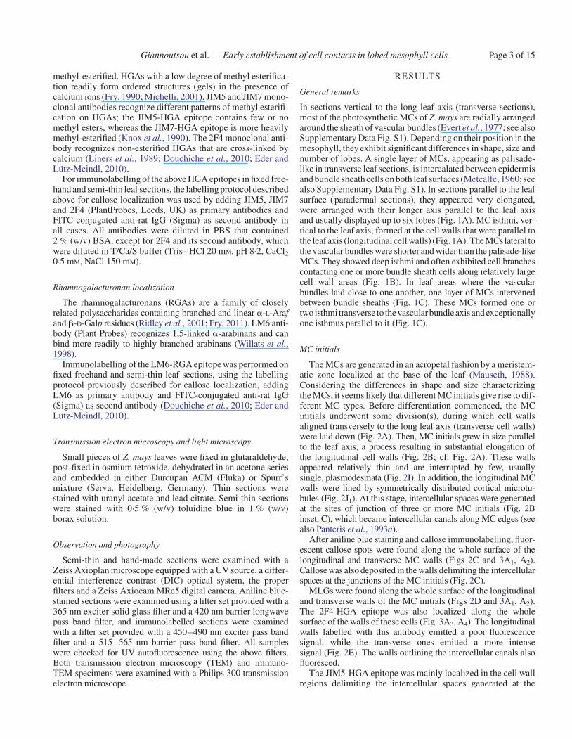
Examination of many nascent MCs by TEM revealed a shift inplasmodesmata distribution along the longitudinal cell wallsfrom the dispersed type to the clustered type. At definiteregions of these cell walls and close to pre-existing plasmodes-mata, secondary plasmodesmata were generated, formingplasmodesmata clusterings (PCs; Fig. 5A, B; see also Fig. 2J2).The cell wall regions, where PCs were initiated, appearedswollen and displayed electron-transparent materials(Fig. 5A–C). In the adjacent cortical cytoplasm, ER elementsas well as dictyosome vesicles were found, while the adjacentplasmalemma regions were convoluted (Fig. 5B, C).
During PC establishment, microtubules were scattered alongthe whole length of the longitudinal cell walls (Fig. 2J2).Often, microtubules lined the cell wall regions where PCs devel-oped (Figs 2J2 and 5C). After immunogold callose labelling, nu-merous gold grains were deposited on developing PCs (Fig. 5D).The cell wall areas between the PCs displayed very few or no gold
A B C
FI G. 1. Paradermal sections of MCs as they appear in the light microscope after staining with toluidine blue. (A) Palisade-like MCs in paradermal view. Arrows in-dicate cell contacts and arrowheads cell isthmi. Asterisks mark cell lobes in an external paradermal semi-thin section. The double arrow indicates the leaf axis in (A–C).(B) MCs were located laterally to vascular bundles. Large arrows show contacts of MCs with bundle sheath cells. Small arrows point to MC contacts and arrowheads toMC isthmi. BSC, bundle sheath cell. (C) A single layer of MCs intervenes between two adjacent vascular bundles. The large arrowhead indicates a cell isthmus parallel
to the leaf axis, while the small arrowheads show cell isthmi transverse to the leaf axis. TW, transverse wall. Scale bars ¼ 10 mm.
Giannoutsou et al. — Early establishment of cell contacts in lobed mesophyll cellsPage 4 of 15

grains. Thus, the callose patches detected in the epifluorescencemicroscope demarcate the cell wall regions where PCs develop.
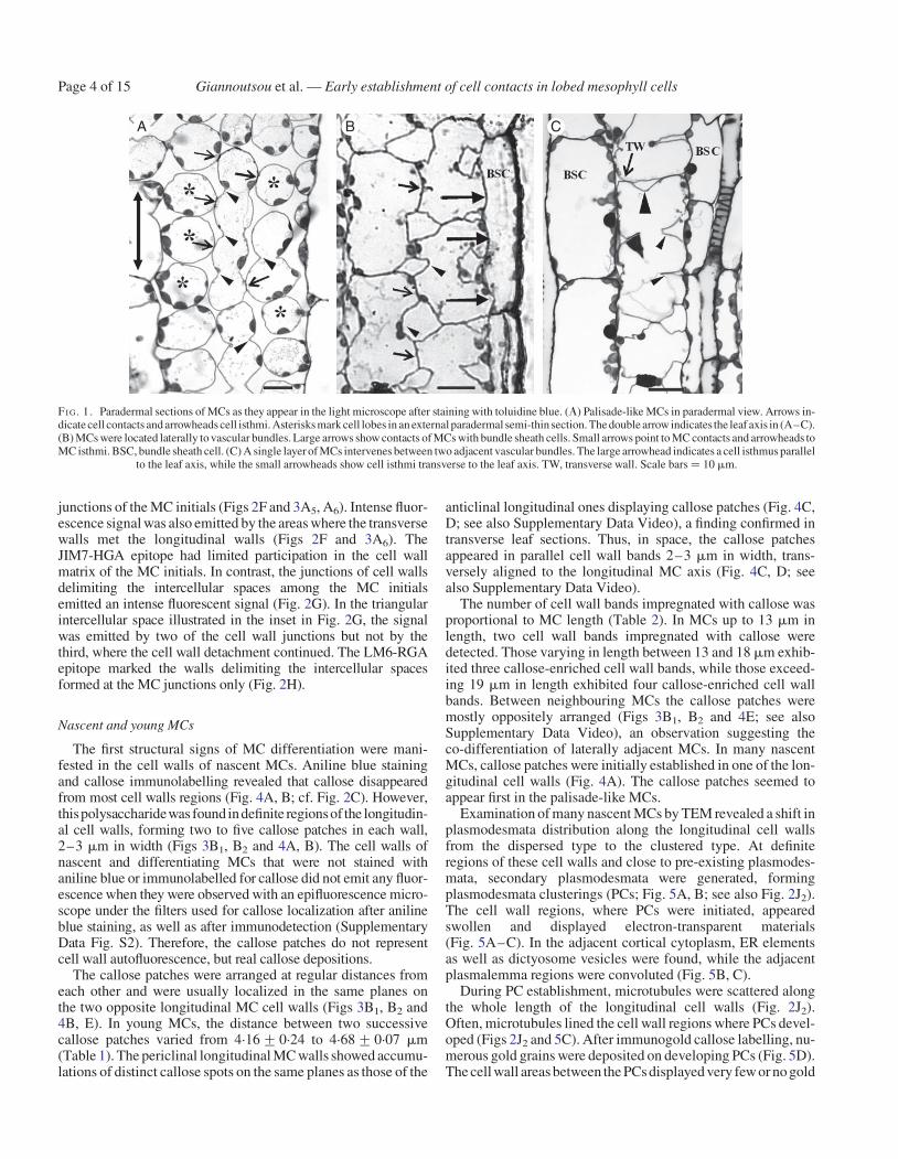
In young MCs the cortical microtubules were reorganized intomicrotubule rings (Figs 6A and Supplementary Data Fig. S3A,B, D) that control the deposition of local cell wall thickenings,which contained numerous cellulose microfibrils (SupplementaryData Fig. S3B, C). MC isthmi were formed in the region of cellwall thickenings (Supplementary Data Fig. S3A inset). Their for-mation was accompanied by the formation of schizogenous inter-cellular spaces, while in the areas between the cell isthmi celllobes were formed (Supplementary Data Fig. S3A inset, D). Inyoung MCs, the PC callose patches were prominent (Figs 3C1, C2
and 6B) and seemed to predict the sites of the future MC contacts.Callose was also deposited in the walls delimiting the intercellularspaces forming at the MC junctions and in those opening at the cellisthmi regions (Figs 3C1 and 6C, D).
The cortical microtubule rings and the cell wall bands showingPC callose patches alternated along the longitudinal walls(Fig. 6A; cf. Fig. 6B). Each local cell wall thickening lined bymicrotubule bundles always occurred between two successivePC callose patches (Fig. 6E). The cortical cytoplasm adjacentto the cell walls possessing the PC callose patches was complete-ly devoid of cortical microtubules (Fig. 6E). The repeated TEMobservation of (1) nascent MCs displaying developing PCs andmicrotubules dispersed along the whole of their longitudinalwalls (Fig. 2J2) and (2) young MCs displaying microtubulebundles between successive PCs (Fig. 6E) led to the conclusionthat microtubule reorganization follows the establishment of PCcallose patches. The measurements presented in Table 2 allow theconclusions that (1) the distance between successive microtubulerings is similar to that between successive callose patches, (2) eachmicrotubule ring forms at the mid-region between two successivecallose-enriched cell wall bands, and (3) the number of callosepatches and microtubule rings depends on cell length.
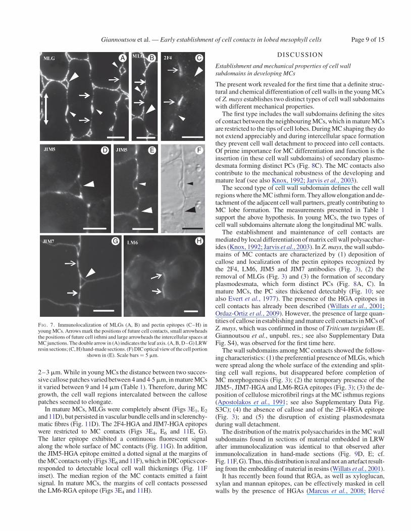
Notably, at this stage, the MLGs were removed from the sitesof the longitudinal walls, where callose had been deposited(Figs 3C2 and 7A, B). They were consistently present in the trans-verse walls (Figs 3C1, C2 and 7A), in the thickenings along thelongitudinal cell walls (Figs 3C2 and 7A) and in the cell wallregions delimiting the intercellular spaces forming at the MCisthmus regions and at the MC junctions (Figs 3C1 and 7A, B).The 2F4-HGA epitope was gradually removed from the majorpart of the cell wall surface and was restricted to the sites ofthe future MC contacts, where callose had been deposited, andto cell wall regions limiting the intercellular spaces at MC junc-tions (Figs 3C3, C4 and 7C). The JIM5-HGA epitope markedonly the junctions of the walls delimiting the intercellularspaces at MC junctions and those at the cell isthmus regions(Figs 3C5, C6 and 7D–F). The JIM7-HGA epitope enrichedthe developing local cell wall thickenings (Figs 3C6 and 7G)and the cell wall regions defining the developing intercellularspaces (Figs 3C5 and 7G). The LM6-RGA epitope was restrictedto the margins of the future cell contacts (Figs 3C3, C4 and 7H).
A
C D
E F
I J
G HB
FI G. 2. Micrographs of MC initials taken with a conventional light microscope(A, B), an epifluorescence microscope (C–H) and a TEM (I). (A, B) Paradermalsemi-thin sections of MC initials located at the meristematic leaf zone (A) and theregion just next to it (B). Arrows in (A) indicate the newly formed transverse cellwalls, while the double arrow in (B) indicates the leaf axis in (A–J). (Inset in B)Triangular intercellular space (arrowhead) generated at the junction site of threeMC initials. (C–H) Localization of callose (C), MLGs (D) and pectin epitopes(E–H) in cell walls of MC initials. Arrowheads indicate intercellular spacesformed at the junction sites of three or more MC initials. Arrow in (F) showsthe junction of a transverse with a longitudinal MC wall. (C, D, G)Immunolabelling in LRW sections. (E, F, H) Immunolabelling in hand-made
sections. (I) Longitudinal cell wall portion of an MC initial. Arrows mark plasmo-desmata. Scale bars: (A–H) ¼ 10 mm; (I) ¼ 200 nm; (B inset) ¼ 3 mm;(G inset) ¼ 5 mm. (J) Diagram showing the distribution of cortical microtubulesalonga longitudinal cell wall of a MC initial (J1) and a nascent MC (J2). Black dotsrepresent microtubules; black lines represent plasmodesmata. Arrows show
young plasmodesmata clusterings.
Giannoutsou et al. — Early establishment of cell contacts in lobed mesophyll cells Page 5 of 15

MC
initi
als
Nas
cent
MC
Youn
g M
CS
hapi
ng M
CM
atur
e M
C
CALLOSE
MLG
2F4
LM6
JIM5
JIM7
Median Plane External PlaneExternal Plane Median Plane External Plane Median Plane
A2 A3A1 A4 A5 A6
B2 B3B1 B4 B5 B6
C2 C3C1 C4 C5 C6
D2 D3D1 D4 D5 D6
E2 E3E1 E4 E5 E6
FI G. 3. Diagrams illustrating the distribution of the hemicelluloses callose and MLGs (first two columns) and pectin epitopes (remaining columns) in MC walls atsuccessive stages of MC differentiation. Cells in (A–C) are shown at higher magnification than those in (D, E).
Giannoutsou et al. — Early establishment of cell contacts in lobed mesophyll cellsPage 6 of 15

In the latter, both callose and the 2F4-HGA epitope were consist-ently found (Fig. 3C1–C4).
Shaping MCs
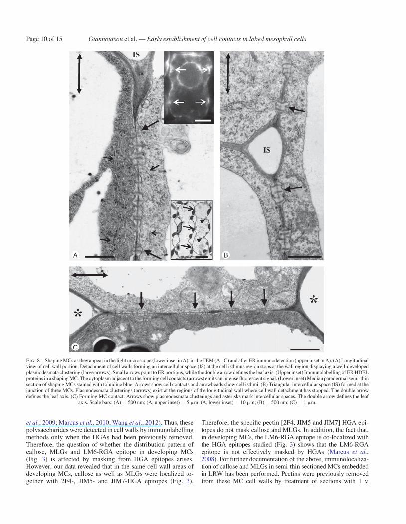
The increase in MC diameter at forming MC lobes, as well asMC elongation, was obvious in shaping MCs. This differentialMC growth seemed to create mechanical tensions that forcedfurther cell wall detachment, making the cell isthmi andcell lobes more evident (lower inset in Fig. 8A; see alsoApostolakos et al., 1991; Panteris and Galatis, 2005). The cellwall expansion occurring in developing MC lobes did notaffect PC integrity (Fig. 8A, C). Actually, MC wall detachmentstopped at the PC regions (Fig. 8A, B) that finally resided atthe tips of MC lobes, where the MCs contacted each other(Fig. 8C). In shaping MCs, the PCs occupied a greater part ofthe longitudinal walls and displayed more plasmodesmata thanthose in young MCs (Fig. 8A, C). The presence of ER in theclose vicinity of PCs was obvious in thin MC sections(Fig. 8A) as well as in MCs immunolabelled for HDEL ER pro-teins (upper inset in Fig. 8A).
In shaping MCs, intense callose depositions were found at theMC contacts (Figs 3D2 and 9A) and at the junctions of the cell
walls delimiting the intercellular spaces initiated at the MCisthmi, where wall detachment continued. The fluorescentsignal emitted by the MC contacts was either continuous orrestricted to two or more areas (Fig. 9A). The MC contactsusually displayed more than one PC (Fig. 8C). In shapingMCs, MLGs were found along the whole wall, except for theMC contacts (Figs 3D1, D2 and 9B). A more intense fluorescentsignal was emitted by the walls outlining the forming intercellu-lar spaces at the MC isthmus regions (Fig. 9B). Following immu-nolabelling with 2F4 antibody, an intense fluorescence signalwas emitted by the MC contacts (Figs 3D4 and 9C) and a veryweak signal was emitted by the detaching regions of walls deli-miting the intercellular spaces initiated at MC isthmi(Fig. 3D4). The JIM5-HGA epitope marked the MC contactsonly (Figs 3D6 and 9D). These sites emitted a dotted pattern offluorescence. The JIM7-HGA epitope was also restricted tocell contacts, but the emitted signal was continuous (Figs 3D6
and 9E). The LM6-RGA epitope was limited to the margins ofMC contacts (Figs 3D4 and 9F).
Mature MCs
The cell walls of mature MC contacts appeared relatively thickand electron-dense and were traversed by numerous plasmodes-mata (Fig. 10 inset). The increased electron density possiblyreflects the dominant presence of matrix polysaccharides. In con-trast, the cell walls delimiting the MC lobes were relativelyelectron-transparent (Fig. 10 inset). In each MC contact, theregions traversed by plasmodesmata were slightly thickened(Fig. 10; see also Evert et al., 1977). Each plasmodesma was sur-rounded by a cylinder of material more electron-transparent thanthat of the rest of the MC contact (Fig. 10). ER was prominent inthe cortical cytoplasm apposed to MC contacts (Fig. 10).
A
C D E
B
FI G. 4. Callose detection in hand-made sections of nascent MCs. (A, B) NascentMCs after aniline blue staining (A) and callose immunodetection (B). In longitu-dinal cell walls, well-defined callose patches (arrows) have been deposited in thecell wall regions where the cell contacts will be formed. The double arrow definesthe leaf axis in (A–E). (C, D) Nascent MCs after callose immunodetection inoptical sections through a median (C) and an external (D) plane. Dotted linesdefine a cell wall band impregnated with callose. (E) Callose patches (arrows)in adjacent nascent MCs display an opposite arrangement. Scale bars: (A, B) ¼
5 mm; (C–E) ¼ 10 mm.
TABLE 1. Distance between two successive callose patches inyoung and mature MCs
Longitudinal cellwalls with twocallose patches
Longitudinal cellwalls with threecallose patches
Longitudinal cellwalls with fourcallose patches
YoungMCs
4.68+0.07 mm 4.40+0.16 mm 4.16+0.24 mm
MatureMCs
10.13+1.45 mm 9.36+0.96 mm 14.12+0.99 mm
TABLE 2. Distance (mm) between two successive callose patchesand two successive microtubule (MT) rings in young MCs
Longitudinal cellwalls with twocallose patches
and one MT ring
Longitudinal cellwalls with threecallose patches
and two MT rings
Longitudinal cellwalls with fourcallose patchesand three MT
rings
Callose patches 4.68+0.07 4.40+0.16 4.16+0.24Wall length 13.36+0.82 17.52+0.96 19.68+1.35
MT rings – 4.64+0.23 5.1+0.54Wall length 12.61+0.76 16.32+0.93 18.57+1.28
Giannoutsou et al. — Early establishment of cell contacts in lobed mesophyll cells Page 7 of 15
In mature MCs, callosewas exclusively present in cell contacts(Figs 3E1, E2 and 11A, B). In median optical planes, the fluores-cent callose signal was either continuous or dotted (Figs 3E2 and11A, B), while in external and internal optical planes the signalwas emitted by distinct spherical formations (Figs 3E1 and 11C).
The length of the MC contacts was 3.5–4mm, slightly greaterthan that of the PC callose patches in young MCs, which was
A B
C D
FI G. 5. TEM views of longitudinal cell wall regions of nascent MCs. (A) Cellwall area displaying three young plasmodesmata clusterings (arrows; cf.Fig. 2I). The arrowhead points to a dictyosome. (B, C) Higher magnification ofplasmodesmata clusterings. Arrows show microtubules and arrowheads ERportions. (D) Wall area of nascent MC after immunogold callose labelling.Numerous gold grains have been deposited on the forming plasmodesmata
clusterings (arrows). Scale bars: (A, D) ¼ 500 nm; (B, C) ¼ 200 nm.
A B
C D
E
FI G. 6. Epifluorescence microscope (A–C), differential interference contrast(D) and TEM (E) views of young MCs. (A) MC tubulin immunolabelling in hand-made leaf sections. Arrowheads point to microtubule bundles formed in the cor-tical cytoplasmadjacent to the sites of future cell isthmi. Note that the microtubulebundles between the adjacent MCs have been formed on the same planes. Thedouble arrow indicates the leaf axis in (A–E). (B) Aniline blue staining ofMCs in hand-made leaf sections. Arrows mark callose patches at the positionsof future cell contacts. (C, D) External paradermal section of young MCs embed-ded in LRW after callose immunolabelling (C) and with DIC optics (D). Cellwalls outlining an intercellular space that has opened between MCs (large arrow-head) and intercellular spaces forming at future cell isthmi (small arrowheads)emit an intense fluorescent callose signal. (E) Microtubule bundle (smallarrows) lining a wall thickening (asterisk) deposited in the position of a futurecell isthmus. Plasmodesmata clusterings (PCs, large arrows) exist on both sidesof the wall thickening. Note the absence of microtubules from the cytoplasmlining the PCs (cf. Fig. 5C). (Inset) Microtubule bundle at higher magnification.
Scale bars: (A, B) ¼ 10 mm; (C, D) ¼ 5 mm; (E, E inset) ¼ 200 nm.
Giannoutsou et al. — Early establishment of cell contacts in lobed mesophyll cellsPage 8 of 15
2–3mm. While in young MCs the distance between two succes-sive callose patches varied between 4 and 4.5mm, in mature MCsit varied between 9 and 14 mm (Table 1). Therefore, during MCgrowth, the cell wall regions intercalated between the callosepatches seemed to elongate.
In mature MCs, MLGs were completely absent (Figs 3E1, E2
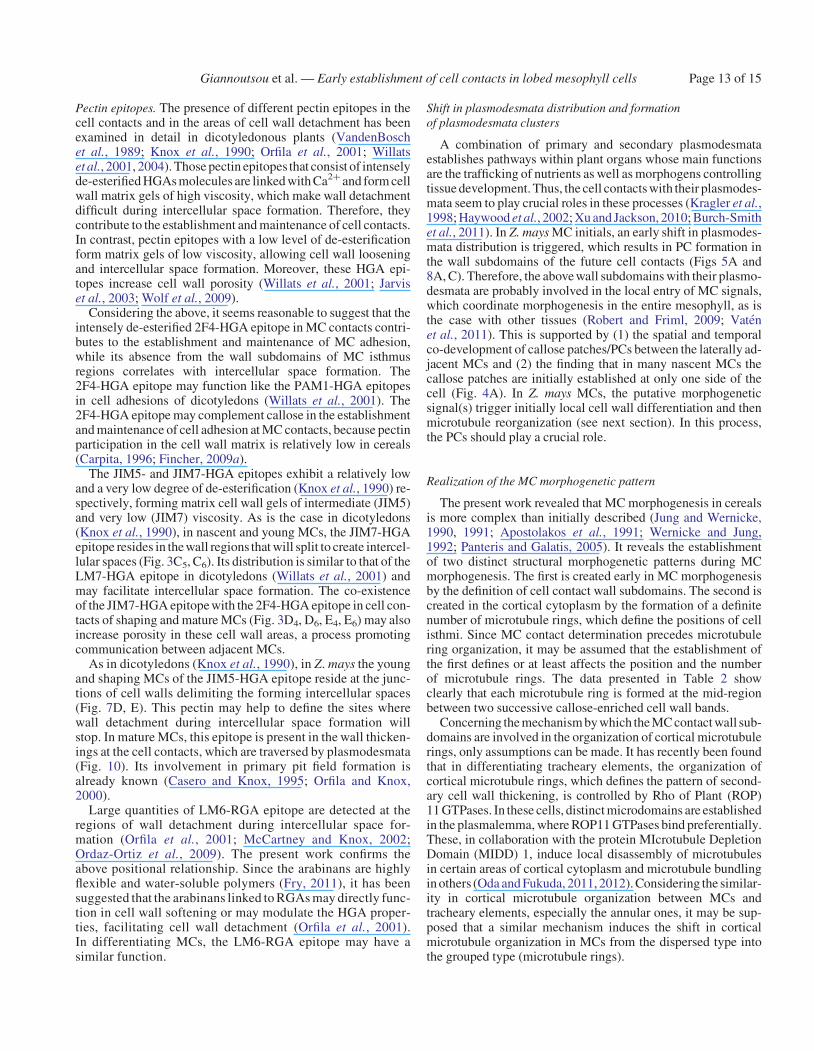
and 11D), but persisted in vascular bundle cells and in sclerenchy-matic fibres (Fig. 11D). The 2F4-HGA and JIM7-HGA epitopeswere restricted to MC contacts (Figs 3E4, E6 and 11E, G).The latter epitope exhibited a continuous fluorescent signalalong the whole surface of MC contacts (Fig. 11G). In addition,the JIM5-HGA epitope emitted a dotted signal at the margins ofthe MC contacts only (Figs 3E6 and 11F), which in DICoptics cor-responded to detectable local cell wall thickenings (Fig. 11Finset). The median region of the MC contacts emitted a faintsignal. In mature MCs, the margins of cell contacts possessedthe LM6-RGA epitope (Figs 3E4 and 11H).
DISCUSSION
Establishment and mechanical properties of cell wallsubdomains in developing MCs
The present work revealed for the first time that a definite struc-tural and chemical differentiation of cell walls in the young MCsof Z. mays establishes two distinct types of cell wall subdomainswith different mechanical properties.
The first type includes the wall subdomains defining the sitesof contact between the neighbouring MCs, which in mature MCsare restricted to the tips of cell lobes. During MC shaping they donot extend appreciably and during intercellular space formationthey prevent cell wall detachment to proceed into cell contacts.Of prime importance for MC differentiation and function is theinsertion (in these cell wall subdomains) of secondary plasmo-desmata forming distinct PCs (Fig. 8C). The MC contacts alsocontribute to the mechanical robustness of the developing andmature leaf (see also Knox, 1992; Jarvis et al., 2003).
The second type of cell wall subdomain defines the cell wallregions where the MC isthmi form. Theyallowelongation and de-tachment of the adjacent cell wall partners, greatly contributing toMC lobe formation. The measurements presented in Table 1support the above hypothesis. In young MCs, the two types ofcell wall subdomains alternate along the longitudinal MC walls.
The establishment and maintenance of cell contacts aremediated by local differentiation of matrix cell wall polysacchar-ides (Knox, 1992; Jarvis et al., 2003). In Z. mays, the wall subdo-mains of MC contacts are characterized by (1) deposition ofcallose and localization of the pectin epitopes recognized bythe 2F4, LM6, JIM5 and JIM7 antibodies (Fig. 3), (2) theremoval of MLGs (Fig. 3) and (3) the formation of secondaryplasmodesmata, which form distinct PCs (Fig. 8A, C). Inmature MCs, the PC sites thickened detectably (Fig. 10; seealso Evert et al., 1977). The presence of the HGA epitopes incell contacts has already been described (Willats et al., 2001;Ordaz-Ortiz et al., 2009). However, the presence of large quan-tities of callose in establishing and mature cell contacts in MCs ofZ. mays, which was confirmed in those of Triticum turgidum (E.Giannoutsou et al., unpubl. res.; see also Supplementary DataFig. S4), was observed for the first time here.
The wall subdomains among MC contacts showed the follow-ing characteristics: (1) the preferential presence of MLGs, whichwere spread along the whole surface of the extending and split-ting cell wall regions, but disappeared before completion ofMC morphogenesis (Fig. 3); (2) the temporary presence of theJIM5-, JIM7-HGA and LM6-RGA epitopes (Fig. 3); (3) the de-position of cellulose microfibril rings at the MC isthmus regions(Apostolakos et al., 1991; see also Supplementary Data Fig.S3C); (4) the absence of callose and of the 2F4-HGA epitope(Fig. 3); and (5) the disruption of existing plasmodesmataduring wall detachment.
The distribution of the matrix polysaccharides in the MC wallsubdomains found in sections of material embedded in LRWafter immunolocalization was identical to that observed afterimmunolocalization in hand-made sections (Fig. 9D, E; cf.Fig. 11F, G). Thus, this distribution is real and not an artefact result-ing from the embedding of material in resins (Willats et al., 2001).
It has recently been found that RGA, as well as xyloglucan,xylan and mannan epitopes, can be effectively masked in cellwalls by the presence of HGAs (Marcus et al., 2008; Herve
A B
D E F
G H
C
FI G. 7. Immunolocalization of MLGs (A, B) and pectin epitopes (C–H) inyoung MCs. Arrows mark the positions of future cell contacts, small arrowheadsthe positions of future cell isthmi and large arrowheads the intercellular spaces atMC junctions. The double arrow in (A) indicates the leaf axis. (A, B, D–G) LRWresin sections; (C, H) hand-made sections. (F) DIC optical view of the cell portion
shown in (E). Scale bars ¼ 5 mm.
Giannoutsou et al. — Early establishment of cell contacts in lobed mesophyll cells Page 9 of 15
et al., 2009; Marcus et al., 2010; Wang et al., 2012). Thus, thesepolysaccharides were detected in cell walls by immunolabellingmethods only when the HGAs had been previously removed.Therefore, the question of whether the distribution pattern ofcallose, MLGs and LM6-RGA epitope in developing MCs(Fig. 3) is affected by masking from HGA epitopes arises.However, our data revealed that in the same cell wall areas ofdeveloping MCs, callose as well as MLGs were localized to-gether with 2F4-, JIM5- and JIM7-HGA epitopes (Fig. 3).
Therefore, the specific pectin [2F4, JIM5 and JIM7] HGA epi-topes do not mask callose and MLGs. In addition, the fact that,in developing MCs, the LM6-RGA epitope is co-localized withthe HGA epitopes studied (Fig. 3) shows that the LM6-RGAepitope is not effectively masked by HGAs (Marcus et al.,2008). For further documentation of the above, immunolocaliza-tion of callose and MLGs in semi-thin sectioned MCs embeddedin LRW has been performed. Pectins were previously removedfrom these MC cell walls by treatment of sections with 1 M
A
C
B
FI G. 8. Shaping MCs as they appear in the light microscope (lower inset in A), in the TEM (A–C) and after ER immunodetection (upper inset in A). (A) Longitudinalview of cell wall portion. Detachment of cell walls forming an intercellular space (IS) at the cell isthmus region stops at the wall region displaying a well-developedplasmodesmata clustering (large arrows). Small arrows point to ER portions, while the double arrow defines the leaf axis. (Upper inset) Immunolabelling of ER HDELproteins in a shaping MC. The cytoplasm adjacent to the forming cell contacts (arrows) emits an intense fluorescent signal. (Lower inset) Median paradermal semi-thinsection of shaping MCs stained with toluidine blue. Arrows show cell contacts and arrowheads show cell isthmi. (B) Triangular intercellular space (IS) formed at thejunction of three MCs. Plasmodesmata clusterings (arrows) exist at the regions of the longitudinal wall where cell wall detachment has stopped. The double arrowdefines the leaf axis. (C) Forming MC contact. Arrows show plasmodesmata clusterings and asterisks mark intercellular spaces. The double arrow defines the leaf
axis. Scale bars: (A) ¼ 500 nm; (A, upper inset) ¼ 5 mm; (A, lower inset) ¼ 10 mm; (B) ¼ 500 nm; (C) ¼ 1 mm.
Giannoutsou et al. — Early establishment of cell contacts in lobed mesophyll cellsPage 10 of 15
KOH for 1 h at 25 8C (Marcus et al., 2010; Wang et al., 2012). InSupplementary Data Fig. S5A1–D2 it is obvious that pretreatmentwith KOH removed HGA epitopes from MC cell walls(Supplementary Data Fig. S5A1, C1; cf. Fig. S5B1, D1).However, in MCs pretreated with KOH, the distribution patternsof callose and MLGs were identical to those of control MCs(Supplementary Data Fig. S5F, H; cf. Fig. S5E, G). Therefore,masking phenomena do not seem to have affected the dataobtained in the present work.
Probable roles of matrix polysaccharides in cell wall subdomains
Callose. Callose is a unique (1 � 3)-b-D-glucan that can beformed and removed from the cell wall in a short time, and isinvolved in a variety of developmental processes, including itsdeposition in cell wall regions experiencing mechanical stress(Heslop-Harrison, 1964; Stone and Clark, 1992; Basic et al.,2009; Chen and Kim, 2009; Galatis and Apostolakos, 2010;Pirselova and Matusikova, 2012). Only hypotheses can bemade about the function(s) of callose in the wall subdomainsof MC contacts. Thus, callose, apart from its well documented in-volvement in the control of plasmodesmata permeability (Xu andJackson, 2010; Vaten et al., 2011), may serve the followingfunctions.
(1) It may serve as a cell wall-strengthening material (Parre andGeitmann, 2005), possibly making the impregnated cellwall subdomains stiff and not easily extendable. Callosewith other matrix polysaccharides deposited in the MCcontact wall subdomains may locally prevent the schizogen-ous separation of the adjacent cell walls. This callose
A B C
D E F
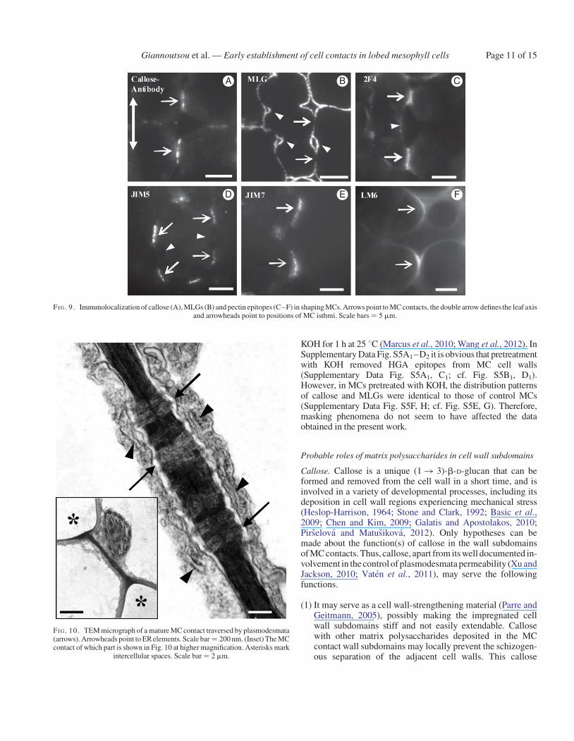
FI G. 9. Immunolocalization of callose (A), MLGs (B) and pectin epitopes (C–F) in shaping MCs. Arrows point to MC contacts, the double arrow defines the leaf axisand arrowheads point to positions of MC isthmi. Scale bars ¼ 5 mm.
FI G. 10. TEM micrograph of a mature MC contact traversed by plasmodesmata(arrows). Arrowheads point to ER elements. Scale bar ¼ 200 nm. (Inset) The MCcontact of which part is shown in Fig. 10 at higher magnification. Asterisks mark
intercellular spaces. Scale bar ¼ 2 mm.
Giannoutsou et al. — Early establishment of cell contacts in lobed mesophyll cells Page 11 of 15

property may protect the plasmodesmata from disruptionduring MC elongation.
(2) It may participate in secondary plasmodesmata formation.Such involvement has not yet been reported (Ehlers andKollmann, 2001; Xu and Jackson, 2010). In Z. mays MCs,PC development keeps pace with callose deposition, i.e. sec-ondary plasmodesmataform in acallose-enriched wall envir-onment. In this case, callose may (a) facilitate the rapid localdigestion of the cell wall, necessary for secondary plasmo-desmata formation (Ehlers and Kollmann, 2001) or (b) con-tribute to the creation of a microenvironment facilitating theduplication of the existing plasmodesmata, a fascinatingprocess recently described for secondary plasmodesmataformation (Faulkner et al., 2008). Notably, callose enrichinglocally thickened cell wall regions traversed by plasmodes-mata has been described in root cells of an Arabidopsisthaliana mutant in which genes inducing callose synthesisare over-expressed (Vaten et al., 2011).
The cell walls outlining the developing MC intercellular spacesare temporarily enriched by callose (Fig. 3). This transient calloserepresents wound callose, which is believed to protect the plasma-lemma and the adjacent cell wall regions from the mechanical
stresses generated during intercellular space initiation (Parre andGeitmann, 2005; Vaughn et al., 2007; Apostolakos et al., 2009).In contrast, the callose present in mature MC contacts is constitu-tive callose. The degree of polymerization, age and thickness ofthe callose deposits may alter the physical properties of thisglucan (Stone and Clarke, 1992).
MLGs. These hemicelluloses characterize the cell walls of Poales,to which Z. mays belongs (Carpita et al., 2001; Buckeridge et al.,2004; Fincher, 2009a) and very rarely cell walls of other vascularplants and lower land plants (Fry et al., 2008; Burton and Fincher,2009). MLGsthat are localized in the cell wall regions between thecell contacts of developing MCs (Fig. 3) may facilitate the prefer-ential and intense cell wall expansion at these areas (Table 1). Thisis consistent with the current view that in developing organs theMLGs are involved in cell wall expansion (Carpita et al., 2001;Buckeridge et al., 2004; Fincher 2009a, b). This view is reinforcedby the pattern of MLG distribution in developing MCs.The MLGsare first located throughout the surface of expanded walls of MCinitials (Fig. 3A1, A2). Then, they are removed from contact MCsites that do not expand (Fig. 3B1–D2), whereas they persist inthe cell wall regions between them (Fig. 3B1–D2), which greatlyelongate. Finally, they are removed from all cell wall regionswhen elongation of MCs has been completed (Fig. 3E1, E2).
A
E F G H
B D
C
FI G. 11. Localization of matrix cell wall polysaccharides in mature MCs. (A) Localization of callosewith aniline blue. The cell wall at the cell contacts (arrows) emitsan intense fluorescent signal. Asterisk marks an intercellular space. (B, C) Paradermal optical sections through a median plane (B) and a surface plane (C) of the sameMC lobe afteraniline blue staining.Arrows show callosedepositionsat MC contacts of anticlinal (B) and periclinal (C) longitudinal cell walls. (D) Transverse semi-thinsection of mature leaf embedded in LRW and immunolabelled for MLGs. MC walls, in contrast to walls of vascular bundle cells (arrows) and sclerenchymatic cells(arrowheads), do not fluoresce. (E–H) Immunodetection of pectin epitopes in hand-made sections of mature MCs. An intense fluorescent signal is emitted by the MCcontacts only (arrows). In (H) the section passes through the end of the cell contact. Asterisks mark intercellular spaces. (Inset in F) DIC optical view of MC contact
(arrow). Scale bars: (A–C, E–H) ¼ 5 mm; D ¼ 10 mm.
Giannoutsou et al. — Early establishment of cell contacts in lobed mesophyll cellsPage 12 of 15
Pectin epitopes. The presence of different pectin epitopes in thecell contacts and in the areas of cell wall detachment has beenexamined in detail in dicotyledonous plants (VandenBoschet al., 1989; Knox et al., 1990; Orfila et al., 2001; Willatset al., 2001, 2004). Those pectin epitopes that consist of intenselyde-esterified HGAs molecules are linked with Ca2+ and form cellwall matrix gels of high viscosity, which make wall detachmentdifficult during intercellular space formation. Therefore, theycontribute to the establishment and maintenance of cell contacts.In contrast, pectin epitopes with a low level of de-esterificationform matrix gels of low viscosity, allowing cell wall looseningand intercellular space formation. Moreover, these HGA epi-topes increase cell wall porosity (Willats et al., 2001; Jarviset al., 2003; Wolf et al., 2009).
Considering the above, it seems reasonable to suggest that theintensely de-esterified 2F4-HGA epitope in MC contacts contri-butes to the establishment and maintenance of MC adhesion,while its absence from the wall subdomains of MC isthmusregions correlates with intercellular space formation. The2F4-HGA epitope may function like the PAM1-HGA epitopesin cell adhesions of dicotyledons (Willats et al., 2001). The2F4-HGA epitope may complement callose in the establishmentand maintenance of cell adhesion at MC contacts, because pectinparticipation in the cell wall matrix is relatively low in cereals(Carpita, 1996; Fincher, 2009a).
The JIM5- and JIM7-HGA epitopes exhibit a relatively lowand a very low degree of de-esterification (Knox et al., 1990) re-spectively, forming matrix cell wall gels of intermediate (JIM5)and very low (JIM7) viscosity. As is the case in dicotyledons(Knox et al., 1990), in nascent and young MCs, the JIM7-HGAepitope resides in the wall regions that will split to create intercel-lular spaces (Fig. 3C5, C6). Its distribution is similar to that of theLM7-HGA epitope in dicotyledons (Willats et al., 2001) andmay facilitate intercellular space formation. The co-existenceof the JIM7-HGA epitope with the 2F4-HGA epitope in cell con-tacts of shaping and mature MCs (Fig. 3D4, D6, E4, E6) may alsoincrease porosity in these cell wall areas, a process promotingcommunication between adjacent MCs.
As in dicotyledons (Knox et al., 1990), in Z. mays the youngand shaping MCs of the JIM5-HGA epitope reside at the junc-tions of cell walls delimiting the forming intercellular spaces(Fig. 7D, E). This pectin may help to define the sites wherewall detachment during intercellular space formation willstop. In mature MCs, this epitope is present in the wall thicken-ings at the cell contacts, which are traversed by plasmodesmata(Fig. 10). Its involvement in primary pit field formation isalready known (Casero and Knox, 1995; Orfila and Knox,2000).
Large quantities of LM6-RGA epitope are detected at theregions of wall detachment during intercellular space for-mation (Orfila et al., 2001; McCartney and Knox, 2002;Ordaz-Ortiz et al., 2009). The present work confirms theabove positional relationship. Since the arabinans are highlyflexible and water-soluble polymers (Fry, 2011), it has beensuggested that the arabinans linked to RGAs may directly func-tion in cell wall softening or may modulate the HGA proper-ties, facilitating cell wall detachment (Orfila et al., 2001).In differentiating MCs, the LM6-RGA epitope may have asimilar function.
Shift in plasmodesmata distribution and formationof plasmodesmata clusters
A combination of primary and secondary plasmodesmataestablishes pathways within plant organs whose main functionsare the trafficking of nutrients as well as morphogens controllingtissue development. Thus, the cell contacts with their plasmodes-mata seem to play crucial roles in these processes (Kragler et al.,1998; Haywood et al., 2002; Xu and Jackson, 2010; Burch-Smithet al., 2011). In Z. mays MC initials, an early shift in plasmodes-mata distribution is triggered, which results in PC formation inthe wall subdomains of the future cell contacts (Figs 5A and8A, C). Therefore, the above wall subdomains with their plasmo-desmata are probably involved in the local entry of MC signals,which coordinate morphogenesis in the entire mesophyll, as isthe case with other tissues (Robert and Friml, 2009; Vatenet al., 2011). This is supported by (1) the spatial and temporalco-development of callose patches/PCs between the laterally ad-jacent MCs and (2) the finding that in many nascent MCs thecallose patches are initially established at only one side of thecell (Fig. 4A). In Z. mays MCs, the putative morphogeneticsignal(s) trigger initially local cell wall differentiation and thenmicrotubule reorganization (see next section). In this process,the PCs should play a crucial role.
Realization of the MC morphogenetic pattern
The present work revealed that MC morphogenesis in cerealsis more complex than initially described (Jung and Wernicke,1990, 1991; Apostolakos et al., 1991; Wernicke and Jung,1992; Panteris and Galatis, 2005). It reveals the establishmentof two distinct structural morphogenetic patterns during MCmorphogenesis. The first is created early in MC morphogenesisby the definition of cell contact wall subdomains. The second iscreated in the cortical cytoplasm by the formation of a definitenumber of microtubule rings, which define the positions of cellisthmi. Since MC contact determination precedes microtubulering organization, it may be assumed that the establishment ofthe first defines or at least affects the position and the numberof microtubule rings. The data presented in Table 2 showclearly that each microtubule ring is formed at the mid-regionbetween two successive callose-enriched cell wall bands.
Concerning the mechanism by which the MC contact wall sub-domains are involved in the organization of cortical microtubulerings, only assumptions can be made. It has recently been foundthat in differentiating tracheary elements, the organization ofcortical microtubule rings, which defines the pattern of second-ary cell wall thickening, is controlled by Rho of Plant (ROP)11 GTPases. In these cells, distinct microdomains are establishedin the plasmalemma, where ROP11 GTPases bind preferentially.These, in collaboration with the protein MIcrotubule DepletionDomain (MIDD) 1, induce local disassembly of microtubulesin certain areas of cortical cytoplasm and microtubule bundlingin others (Oda and Fukuda, 2011, 2012). Considering the similar-ity in cortical microtubule organization between MCs andtracheary elements, especially the annular ones, it may be sup-posed that a similar mechanism induces the shift in corticalmicrotubule organization in MCs from the dispersed type intothe grouped type (microtubule rings).
Giannoutsou et al. — Early establishment of cell contacts in lobed mesophyll cells Page 13 of 15

It is also known that a cell wall integrity signalling pathwaythat induces various cell activities functions in plants. Thispathway is based on the production of oligosaccharide fragmentsof the cell wall, which act as signalling molecules (Fry, 1990) andinvolves, among other signalling processes, the activation ofRho-like GTPases (reviewed by Wolf et al., 2012). Taking intoconsideration all the information given in the literature, it can beassumed that the local chemical differentiation of cell wallmatrixatMCcontact sitesmay, throughacellwall integritysignal-ling pathway, define the sites of putative ROP binding to theplasmalemma, which in turn controls the organization of micro-tubule rings. Therefore, the probable implication of ROP proteinsin MC differentiation should be examined.
SUPPLEMENTARY DATA
Supplementary data are available online at www.aob.oxford-journals.org and consist of the following. Figure S1: area oftransverse section of a mature Zea mays leaf. Figure S2:shaping MCs as they are seen in DIC optics, and by epifluores-cence microscopy under a filter set provided with exciter G365and barrier LP420 and another with exciter BP450–490 andbarrier BP 515–565. Figure S3: TEM micrographs of shapingMCs. Figure S4: young and shaping MCs of Triticum turgidumas seen byepifluorescence microscopyafteraniline blue staining.Figure S5: immunodetection of cell wall matrix polysaccharidesin sections of nascent MCs embedded in LRW. Video: nascentMCs after callose immunolocalization. The callose-enrichedcell wall bands and their opposite arrangement in adjacentMCs can be easily observed.
ACKNOWLEDGEMENTS
The authors greatly acknowledge Dr Nigel Chaffey and thereviewers for critical reading of the manuscript and theirhelpful comments, which contributed to the better presentationof the present work, and to Dr K. Karpouzis for the preparationof the video material.
LITERATURE CITED
Apostolakos P, Galatis B, PanterisE. 1991.Microtubules in cell morphogenesisand intercellular space formation in Zea mays leaf mesophyll and Pileacadierei epithem. Journal of Plant Physiology 137: 591–601.
Apostolakos P, Livanos P, Nikolakopoulou TL, Galatis B. 2009. The role ofcallose in guard cell wall differentiation and stomatal pore formation inthe fern Asplenium nidus L. Annals of Botany 104: 1373–1387.
Basic A, Fincher GB, Stone BA. 2009. Chemistry, biochemistry, and biology of(1�3)-b-glucans and related polysaccharides. Amsterdam: AcademicPress.
Buckeridge MS, Rayon C, Urbanowicz B, Tine MAS, Carpita N. 2004. Mixedlinkage (1�3)(1�4)-b-D-glucans of grasses. Cereal Chemistry 81:115–127.
Burch-Smith TM, Brunkard OJ, Choi YG, Zambruski PC. 2011.Organelle-nucleus cross talk regulates plant intercellular communicationvia plasmodesmata. Proceedings of the National Academy of Science,USA 108: E1451–E1460.
Burton RA, Fincher GB. 2009. (1,3; 1,4)-b-D-glucans in cell walls of thePoaceae, lower plants, and fungi: a tale of two linkages. Molecular Plant2: 873–882.
Carpita NC. 1996. Structure and biogenesis of the cell walls of grasses. AnnualReview of Plant Physiology and Plant Molecular Biology 47: 445–476.
Carpita NC, Defernez M, Findlay K, et al. 2001. Cell wall architecture of theelongating maize coleoptile. Plant Physiology 127: 551–565.
Casero PJ, Knox JP. 1995. The monoclonal antibody JIM5 indicates patterns ofpectin deposition in relation to pit fields at the plasma-membrane-face oftomato pericarp cell walls. Protoplasma 188: 133–137.
Chen XY, Kim JY. 2009. Callose synthesis in higher plants. Plant Signaling andBehavior 4: 489–492.
Douchiche O, Driouich A, Morvan C. 2010. Spatial regulation of cell-wallstructure in response to heavy metal stress: cadmium-induced alteration ofthe methyl-esterification pattern of homogalacturonans. Annals of Botany105: 481–491.
Eder M, Lutz-Meindl U. 2010. Analyses and localization of pectin-like carbo-hydrates in cell wall and mucilage of the green alga Netrium digitus.Protoplasma 243: 25–38.
Ehlers K, Kollmann R. 2001. Primaryand secondary plasmodesmata: structure,origin and functioning. Protoplasma 216: 1–30.
Evert RF, Eschrich W, Heyser W. 1977. Distribution and structure of plasmo-desmata in mesophyll and bundle-sheath cells of Zea mays L. Planta 136:77–89.
Faulkner C, Akman OE, Bell K, Jeffree C, Oparka K. 2008. Peeking into pitfields: a multiple twinning model of secondary plasmodesmata formation intobacco. Plant Cell 20: 1504–1518.
Fincher GB. 2009a. Revolutionary times in our understanding of cell wall bio-synthesis and remodeling in the grasses. Plant Physiology 149: 27–37.
Fincher GB. 2009b. Exploring the evolution of (1�3) (1�4)-b-D-glucans inplant cell walls: comparative genomics can help! Current Opinion inPlant Biology 12: 140–147.
Fry SC. 1990. Roles of the primary cell wall in morphogenesis. In: Nijkamp HJJ,van der Plas LHW, van Aartrijk J. eds. Progress in plant cellular and mo-lecular biology. Dordrecht: Kluwer, 504–513.
Fry SC. 2011. Cell wall polysaccharide composition and covalent crosslinking.Annual Plant Reviews 41: 1–42.
Fry SC, Nesselrode BHW, Miller JG, Newburn BR. 2008. Mixed-linkage of(1�3) (1�4)-b-D-glucan is a major hemicellulose of Equisetum (horse-tail) cell walls. New Phytologist 179: 104–115.
Galatis B. 1988. Morphogenesis of the epithem cells in hydathodes of Pileacadierei. Planta 176: 287–297.
Galatis B, Apostolakos P. 2010. A new callose function. Involvement in differ-entiation and function of fern stomatal complexes. Plant Signaling andBehavior 5: 1359–1364.
Giannoutsou E, Apostolakos P, Galatis B. 2011. Actin filament-organized localcortical endoplasmic reticulum aggregations in developing stomatal com-plexes of grasses. Protoplasma 248: 373–390.
Haywood V, Kragler F, Lucas WJ. 2002. Plasmodesmata: pathways for proteinand ribonucleoprotein signaling. Plant Cell Supplement 2002: S303–S325.
Herve C, Rogowski A, Gilbert HJ, Knox JP. 2009. Enzymatic treatments revealdifferential capacities of xylan recognition and degradation in primary andsecondary plant cell walls. Plant Journal 58: 413–422.
Heslop-Harrison J. 1964. Cell walls, cell membranes and protoplasmic connec-tions during meiosis and pollen development. In: Linskens HF. ed. Pollenphysiology and fertilization. Amsterdam: North-Holland, 29–47.
Jarvis MC, Briggs SPH, Knox JP. 2003. Intercellular adhesion and cell separ-ation in plants. Plant Cell and Environment 26: 977–989.
Jung G, Wernicke W. 1990. Cell shaping and microtubules in developing meso-phyll of wheat (Triticum aestivum L.). Protoplasma 153: 141–148.
Jung G, Wernicke W. 1991. Patterns of actin filaments during cell shaping indeveloping mesophyll of wheat (Triticum aestivum L.). European Journalof Cell Biology 56: 139–146.
Knox JP. 1992. Cell-adhesion, cell-separation and plant morphogenesis. PlantJournal 2: 137–141.
Knox JP, Linstead PJ, King J, Cooper C, Roberts K. 1990. Pectin esterificationis spatially regulated both within cell walls and between developing tissuesof root apices. Planta 181: 512–521.
Kragler F, LucasWJ, Monzer J. 1998.Plasmodesmata: dynamics, domainsandpatterning. Annals of Botany 81: 1–10.
Lee KJD, Marcus SE, Knox JP. 2011. Cell wall biology: perspectives from cellwall imaging. Molecular Plant 4: 212–219.
Liners F, Letesson JJ, Didembourg C, Van Cutsem P. 1989. Monoclonal anti-bodies against pectin. Recognition of conformation induced by calcium.Plant Physiology 91: 1419–1424.
Marcus SE, Verhertbruggen Y, Herve C, et al. 2008. Pectic homogalacturonanmasks abundant sets of xyloglucan epitopes in plant cell walls. BMC PlantBiology 8: 60.
Giannoutsou et al. — Early establishment of cell contacts in lobed mesophyll cellsPage 14 of 15
Marcus SE, Blake AW, Benians TA, et al. 2010. Restricted access of proteins tomannan polysaccharides in intact plant cell walls. Plant Journal 64:191–203.
Mauseth JD. 1988. Plant anatomy. Redwood City, CA: Benjamin/Cummings.
Mauseth JD. 1998. Botany. An introduction to plant biology Boston, MA: Jonesand Bartlett.
McCartney L, Knox JP. 2002. Regulation of pectic polysaccharide domains inrelation to cell development and cell properties in the pea testa. Journal ofExperimental Botany 53: 707–714.
Metcalfe CR. 1960. Anatomy of the monocotyledons I. Gramineae. London:Oxford University Press.
Michelli F. 2001. Pectin methylesterases: cell wall enzymes with important rolesin plant physiology. Trends in Plant Science 6: 414–419.
O’Brien TP, McCully ME. 1981. The study of plant structure: principles andselected methods. Melbourne: Termarcarphi.
Oda Y, Fukuda H. 2011. Secondary cell wall patterning during xylem differen-tiation. Current Opinion in Plant Biology 15: 1–7.
Oda Y, Fukuda H. 2012. Initiation of cell wall pattern by a Rho- andmicrotubule-driven symmetry breaking. Science 337: 1333–1336.
Ordaz-Ortiz JJ, Marcus SE, Knox JP. 2009. Cell wall microstructure analysisimplicates hemicellulose polysaccharides in cell adhesion in tomato fruitpericarp parenchyma. Molecular Plant 2: 910–921.
Orfila C, Knox JP. 2000. Spatial regulation of pectic polysaccharides in relationto pit fields in cell walls of tomato fruit pericarp. Plant Physiology 122:775–781.
Orfila C, Seymour GB, Willats WGT, et al. 2001. Altered middle lamellahomogalacturonan and disrupted deposition of (1�5)-alpha-L-arabinanin the pericarp of Cnr, a ripening mutant of tomato. Plant Physiology 126:210–221.
Panteris E, Galatis B. 2005. The morphogenesis of lobed plant cells in the meso-phyll and epidermis: organization and distinct roles of cortical microtubulesand actin filaments. New Phytologist 67: 721–732.
Panteris E, Apostolakos P, Galatis B. 1993a. Microtubule organization, meso-phyll cell morphogenesis, and intercellular space formation in Adiantumcapillus-veneris leaflets. Protoplasma 172: 97–110.
Panteris E, Apostolakos P, Galatis B. 1993b. Microtubule organization and cellmorphogenesis in two semi-lobed cell types of Adiantum capillus-venerisL. leaflets. New Phytologist 125: 509–520.
Parre E, GeitmannA. 2005.More thana leak sealant.The mechanical propertiesof callose in pollen tubes. Plant Physiology 37: 274–286.
Pirselova B, Matusıkova I. 2012. Callose: the plant cell wall polysaccharidewith multiple biological functions. Acta Physiologiae Plantarum 35:635–644.
Ridley BL, O’Neill MA, Mohnen D. 2001. Pectins: structure, biosynthesis, andoligogalacturonide-related signaling. Phytochemistry 57: 929–967.
Robert HS, Friml J. 2009. Auxin and other signals on the move in plants. NatureChemical Biology 5: 325–332.
Stone BA, Clarke AE. 1992. Chemistry and biology of (1�3)-b-glucans.Bundora, Australia: La Trobe University Press.
VandenBosch KA, Bradley DJ, Knox JP, Perotto S, Butcher GW, Brewin NJ.1989. Common components of the infection thread matrix and the intercel-lular space identified by immunocytochemical analysis of pea nodules anduninfected roots. EMBO Journal 8: 335–342.
Vaten A, Dettmer J, Wu S, Stierhof YD, et al. 2011. Callose biosynthesis reg-ulates symplastic trafficking during root development. Developmental Cell21: 1144–1155.
Vaughn KC, Talbot MJ, Offler CE, McCurdy DW. 2007. Wall ingrowths inepidermal transfer cells of Vicia faba cotelydons are modified primarywalls marked by localized accumulations of arabinogalactan proteins.Plant and Cell Physiology 48: 159–168.
Wang H-T, Liu I-H, Yeh T-F. 2012. Immunohistological study of mannan poly-saccharides in poplar stem. Cellulose Chemistry and Technology 46:149–155.
Wernicke W, Jung G. 1992. Role of cytoskeleton in cell shaping of developingmesophyll of wheat (Triticum aestivum L.). European Journal of CellBiology 57: 88–94.
Wernicke W, Gunther P, Jung G. 1993. Microtubules and cell shaping in themesophyll of Nigella damascena L. Protoplasma 173: 8–12.
Willats WGT, Marcus SE, Knox JP. 1998. Generation of a monoclonal anti-body specific to (1�5)-alpha-L-arabinan. Carbohydrate Research 308:149–152.
Willats WG, McCartney L, Mackie W, Knox JP. 2001. Pectin: cell biologyandprospects for functional analysis. Plant Molecular Biology 47: 9–27.
Willats WGT, McCartneySteele-King CG, et al. 2004. A xylogalacturonanepitope is specifically associated with plant cell detachment. Planta 218:673–681.
Wolf S, Mouille G, Pelloux J. 2009. Homogalacturonan, methyl-esterificationand plant development. Molecular Plant 2: 851–860.
Wolf S, Hematy K, Hofte H. 2012. Growth control and cell wall signaling inplants. Annual Review of Plant Biology 63: 381–407.
Xu X, Jackson D. 2010. Lights at the end of the tunnel: new views of plasmodes-mal structure and function. Current Opinion in Plant Biology 13: 684–692.
Giannoutsou et al. — Early establishment of cell contacts in lobed mesophyll cells Page 15 of 15
Copyright © 2022 FDOKUMEN