Life-Cycle Assessment of Biodiesel Production from Microalgae

Mar. Drugs 2014, 12, 1641-1675; doi:10.3390/md12031641
marine drugs ISSN 1660-3397
www.mdpi.com/journal/marinedrugs
Review
The Challenge of Ecophysiological Biodiversity for
Biotechnological Applications of Marine Microalgae
Lucia Barra †, Raghu Chandrasekaran
†, Federico Corato and Christophe Brunet *
Stazione Zoologica Anton Dohrn, Villa Comunale, Naples 80121, Italy;
E-Mails: [email protected] (L.B.); [email protected] (R.C.);
[email protected] (F.C.)
† These authors contributed equally to this work.
* Author to whom correspondence should be addressed; E-Mail: [email protected];
Tel.:+39-81-583-3243; Fax: +39-81-764-1355.
Received: 7 November 2013; in revised form: 31 January 2014 / Accepted: 12 February 2014 /
Published: 24 March 2014
Abstract: In this review, we aim to explore the potential of microalgal biodiversity and
ecology for biotechnological use. A deeper exploration of the biodiversity richness and
ecophysiological properties of microalgae is crucial for enhancing their use for applicative
purposes. After describing the actual biotechnological use of microalgae, we consider the
multiple faces of taxonomical, morphological, functional and ecophysiological biodiversity
of these organisms, and investigate how these properties could better serve the
biotechnological field. Lastly, we propose new approaches to enhancing microalgal growth,
photosynthesis, and synthesis of valuable products used in biotechnological fields, mainly
focusing on culture conditions, especially light manipulations and genetic modifications.
Keywords: biotechnology; biomass; diatoms; phytoplankton; ecophysiology; biodiversity;
functional diversity; cultures; light
1. Introduction
It is now well known that marine biotechnology (the so-called ―blue biotechnology‖) can make a
large contribution to the key societal challenges, in relation to the huge biological diversity populating
marine ecosystems. As an alternative to higher plants, microbial organisms such as microalgae are
OPEN ACCESS
Mar. Drugs 2014, 12 1642
relevant resources for applied societal purposes, due to their rich biodiversity, growth rate, and
multiple application potentials.
Microalgae, either prokaryotes or eukaryotes, are oxygenic autotrophs that populate all aquatic
ecosystems ranging from freshwater and brackish waters to oligotrophic marine waters. The microalgal
world represents rich biodiversity, characterized by different biological, ecological and functional
traits. The species number ranges from 30,000 described species to one million, and some estimates
report more than 200,000 species only for the Bacillariophyceae [1,2]. This group is the most recent
and diversified group [3], and until now, more than 8000‒10,000 diatom species have been described [4],
while only a few species have been employed for biotechnological applications [5].
For Chlorophyceae, a group which comprises of almost 2000 species, only 7–8 species are
biotechnologically active, followed by Cyanophyceae, where the number is even smaller [6,7]. Use of
microalgae for biotechnological applications has been increasing in recent years, mainly for
bioremediation, nutraceutical and pharmaceutical purposes, as well as for bioenergy production [8].
We can distinguish three kinds of algal applications (Table 1). The first application corresponds to
the property of microalgae in interacting with the environment in which they grow, such as the use of
biomass for O2 production, reduction of CO2 [9], bioremediation and bioremoval of some organic and
inorganic compounds [10]. Microalgal species or strains are selected for their efficient growth or for their
ability to harvest specific toxic compounds from the environment. The second application corresponds
to the production of primary metabolites, as for instance carotenoids [11,12], proteins [13], and lipids
such as polyunsaturated fatty acids, PUFA [14]. Some of these molecules have antioxidant
activities [15–17]. For this purpose, microalgae with high physiological plasticity such as diatoms
should be mainly used, since they are able to efficiently adapt to environmental variations [18]. The
third application is related to secondary metabolites which are generally produced in low quantities,
mainly for pharmaceutical applications [19].
Up to now, biotechnology uses around 20 species of microalgae from marine, estuarine and
freshwater (lakes, ponds) ecosystems [6], representing a miniscule percentage of the myriad of known
species. The open challenges for achieving the ―green revolution‖ with ―blue biotechnology‖ concern:
(i) the cultivation of new species; (ii) identification of new molecules that can contribute to the
development of industrial applications from marine ecosystems, and (iii) upscaling microalgal biomass
production, while keeping production costs as low as possible. To overcome the aforementioned
challenges, an extensive exploration of microalgal biodiversity (taxonomical and functional), chiefly
by focusing on morphological, ecological and physiological traits, is mandatory. These traits, resulting
from the adaptive evolution of a species to its ecological niche, determine its growth capacity in the
marine environment. Many experimental results have shown that algal growth, physiology and
biochemistry are strongly controlled by environmental variables, such as hydrology (temperature,
salinity [20,21]), chemistry (pH, macronutrients and micronutrients [22]) and light [23]. Improving
culture conditions will allow cells to be more efficient in terms of growth, photosynthesis and primary
metabolite synthesis, since growth is the result of the energy dynamics balance between production
and loss [24,25].
In this review we mainly focus on pelagic photosynthetic micro-organisms. The non-planktonic
microalgae, while ecologically relevant, have different growth properties that we are not going to
discuss in this paper (see the review by Lebeau and Robert [26]). This review aims to:
Mar. Drugs 2014, 12 1643
(i) draw a general picture on the actual use of microalgae in the biotechnological field; (ii) disseminate
the knowledge on biodiversity and richness of the microalgal world, and (iii) propose ways to improve
microalgal production in the context of biotechnological applications. For the latter, we focus
especially on the improvement of culturing conditions, in addition to the development of genetic
engineering technologies [8].
2. Biotechnological Applications of Microalgae and Cultivation
2.1. Biotechnological Applications of Microalgae
Commercial culturing of algae has a history of 440 years, starting from the cultivation of Porphyra
in the 1640s [7]. According to Hallmann [27], about 107 tons of algae are harvested each year by algal
biotechnology industries for different purposes, and almost 60 commercial companies are selling algae
or algal products worldwide. In this section, we will go through their use as bio-markers/remediation,
bio-fuel sources, and primary metabolite producers. Microalgae have gained the attention of the
scientific world, as is shown in many studies, for their biochemical and physiological capacity to
respond to organic pollutants (POPs) [28–31], polycyclic aromatic hydrocarbons (PHAs) [32–34],
polychlorinated biphenyls (PCBs) [35,36] and pesticides [37–41]. These studies have demonstrated
that macroalgae and microalgae are important tools in monitoring and controlling the presence of
heavy metals in ecosystems, due to several biochemical strategies employed to reduce toxicity of
non-essential trace metals [42–48]. Among the species resistant to heavy metal exposure, we can cite
Chlamydomonas reinhardtii [49] and some macroalgae, such as Fucus serratus, alongside the aquatic
plant Lemna minor [50].
Most organic chemicals can be naturally degraded within the aquatic environment as a result of
complex processes mediated both by auto- and heterotrophic organisms [51]. However, when the
wild-type xenobiotic detoxification systems, mainly based on metallothionein proteins [52,53], are not
sufficient to cope with pollutant biodegradation, a possible alternative route is the creation of consortia
made by microalgae and/or cyanobacteria [47,54]. A complete review of the mechanisms and solutions
for pollutant management is provided by Torres and collaborators [28].
Many reports have described microalgae as a potential oxygen producer and CO2 depository [55,56],
and as a green energy source for bio-fuels and biogases [57–62], providing feedstock for renewable
fuels such as biodiesel, methane, hydrogen and ethanol. Moreover, microalgae grow faster and reach a
higher productivity compared to conventional agricultural plants [59], and are able to grow almost
anywhere, requiring only sunlight and nutrients [63–65]. This is discussed in greater detail in the
fourth section of this review.
The wide amount of bioactive primary metabolite production is one of the key microalgal
features to be exploited in many applicative fields, such as nutraceutics [66], animal feeding
products and cosmetics [67]. Microalgal species, such as Spirulina for diet, Dunaliella salina and
Haematococcus pluvialis for carotenoid production, and several other species for aquaculture, are used
in mass culturing [68–70].
The high protein content, amino acid pools, carbohydrate digestibility, pigments, ω3 and ω6 family
lipids and the presence of nearly all essential vitamins (e.g., A, B1, B2, B6, B12, C, E, nicotinate,

Mar. Drugs 2014, 12 1644
biotin, folic acid and pantothenic acid) make microalgae an unconventional source for improving the
food nutritional state and hence the health of humans and animals [13,66,71,72]. Furthermore,
microalgae are considered to be of great interest in the biotechnological field because they are a precious
source of lipophilic pigments such as chlorophyll (0.5% to 1% of dry weight), carotenoids (0.1% to 0.2%
of dry weight on average and up to 14% of dry weight for β-carotene in some species, including
Dunaliella sp.), xanthophylls (lutein, zeaxanthin and astaxanthin) and hydrophilic pigments, such
as phycobiliproteins.
Recently, commercial forms of microalgal products are incorporated into pasta, snacks, gum, and
beverages [73,74], as sources of natural food colorants, or as nutritional supplements [69,72,75]. The
market is actually dominated by some species of chlorophytes and cyanophytes, such as Arthrospira spp.,
Chlorella spp., Dunaliella salina, Heamatococcus pluvialis and Aphanizomenon flos-aquae. More than
70 companies sell Chlorella as a source of β-1,3-glucan, which has properties of an immunosystem
stimulator, as a free radical scavenger, and as a reducer of bad blood lipids [76]. D. salina is exploited
for its β-carotene content that can reach 14% of its dry weight [77]. The chlorophyte Muriellopsis sp.
is being exploited for the production of the xanthophyll lutein, due to its high content produced under
peculiar culture conditions [78], while Heamatococcus pluvialis is used for its massive accumulation
of astaxanthin, highly synthetized under stressful conditions [79,80]. Due to its high protein and amino
acid content, Arthrospira is extensively used for human nutrition production in China and India, under
the name of Spirulina pacifica [81,82]. According to several studies, the cyanophyte A. flos-aquae can
also benefit health [83,84]. However, the diversity of usable species for aquaculture remains higher
than the species that are used for human diets [85,86], since species such as Phaeodactylum,
Chaetoceros, Skeletonema and Thalassiosira, along with Chlorella, Tetraselmis, Isochrysis, Pavlova
and Nannochloropsis, are easily ingestible and digestible by cultivable organisms, and are used in
aquaculture [87,88]. In the field of cosmetics, microalgal extracts are used for face, skin and hair care
products, as well as in sunscreen products. Genera such as Arthrospira and Chlorella are employed in
the skin care market [89]. For more information on companies selling microalgae and methods for
their cultivation, see the review by Spolaore and collaborators [67].
Last, but not least, primary metabolites from microalgae are also used in diagnostics and application
fields. In particular, phycobiliproteins, coloured proteins commonly present in cyanophytes and
cryptophytes, are extensively commercialised in clinical and immunological analysis as colorants,
fluorescent labelling molecules, and as pharmaceutical agents [90]. Until now, in the research and
diagnostics development field, phycobiliproteins have been the object of more than 200 patents [90].
2.2. Algal Cultivation Techniques
The cultivation of microalgae can be performed indoor or outdoor, using enclosed or open
systems [69]. These two systems strongly differ in shape, aeration, illumination systems, building
material and volume capacity. In the past, natural waters (lakes, lagoons, etc.) or artificial ponds were
used to grow microalgae. These outdoor open systems that use just natural light for illumination, are
inexpensive to install and maintain and present uncertainties in the success of production operation.
One of the main concerns is the uncontrolled environmental parameters, such as light, temperature, and
air humidity that can induce large variability in cell physiology, altering the growth efficiency (see

Mar. Drugs 2014, 12 1645
following sections). Excessive light or ultraviolet radiation in outdoor open systems might induce
photoinhibition or can alter the healthy state of cells. Outdoor culturing for biomass production, with
the exception of tropical areas, is strongly season dependent [91–97]. The transition periods of the year
(e.g., end of spring and autumn) also show rapid climatological changes; a relevant temperature
excursion between night and day might also affect physiological acclimation of microalgal
biomass [98]. Other drawbacks, linked to the fact that cultures are open and not axenic, are correlated
to the proliferation of bacteria, viruses, other microalgal species and predators that may decimate
microalgal cultures [70]. Closed photo-bioreactors that can be set indoor or outdoor (either naturally
or artificially illuminated), have been employed to axenically grow microalgae, such as
cyanophytes [70,99,100]. For some diatoms, both outdoor and indoor cultivation systems have been
established together with a generalised set of conditions [5]. The conditions and growth parameters can
be set more accurately in indoor culture batches than in outdoor ones. Indoor culturing techniques
prevent many environmental and biological causes of death or reduction in growth rate, and are more
efficient for maintaining mass cultivation at high production rate. Section 4 of this review focuses on
the possible ways to improve growth quantum yield of microalgal cultivation in indoor systems.
3. Exploring the Richness of Microalgal Biodiversity
Biodiversity is characterized by taxonomical metrics with distinct morphological traits, as well as
other variables related to functional diversity such as ecology, physiology, and biochemistry of the
species [101]. The cell functional traits regulate the ecophysiological requirements of the species,
thereby shaping cell performances under specific conditions or along environmental gradients, as in
nature. In this section, we aim to present the richness of microalgal biodiversity, which has been
underexploited thus far in the biotechnological field.
3.1. Taxonomical Diversity
Biodiversity is huge in the microalgal realm, with almost 64 classes (phyla) and more than
70,000 species that could represent the basin to draw for a deep investigation of the microalgal world
within a blue biotechnology aim [1]. The most recent and successful taxon group from an evolutionary
and biogeochemical point of view is the diatoms’ group [3,102] that has been estimated to have
roughly 105 species [103]. Many diatoms comprise cryptic or hidden species not easily recognizable
with classical microscopic tools [104,105]. For this reason in recent years, taxonomy has been using
molecular tools, in addition to morphological identification to better decipher the richness of the
microalgal realm [106] and the evolutionary trends of microalgae (e.g., [107–110]). Molecular
tools allow for the investigation of small cell-sized species and uncultivable ones [111], as well as
cryptic species [104,105,112]. As mentioned previously, for some taxa of phytoplankton, the known
biodiversity does not take into account the hidden and cryptic species present in the groups [113].
Indeed, with the use of ribosomal or organelle markers as LSU, SSU, ITS, 5.8 S, RbcL and COX1
associated with the ultrastructure of a diatom cell wall, the silica frustule helps resolve cryptic species
belonging to the following genera: Chaetoceros, Skeletonema and Pseudo-nitzschia [102,104,114]
thereby enlarging the possibility of choosing the most appropriate species to the ad hoc cultivation

Mar. Drugs 2014, 12 1646
system. For more information on taxonomical diversity and evolution of phytoplankton, we suggest
the recent review papers [109,110].
3.2. Functional Diversity: Morphology and Size
Morphological variations amongst species lead cells to be more or less cultivable; these variations
mostly imply changes in the surface: volume ratio, cell floatability and sinking rate. A cell’s shape can
vary from round to elongated or triangular, affecting interactions between cells and the surrounding
environment and having consequences also on the assimilation of resources [115].
The main cellular morphology includes the presence of flagella (Table 2), such as in the dinophytes,
cryptophytes and some haptophytes, in addition to the presence of spines, referred to as setae and
found mostly in diatoms. The setae can have several functions, such as cell protection against
predators, sedimentation by trapping air bubbles and generation of photosynthetic oxygen. Indeed,
setae from some Chaetoceros species contain chloroplasts that are able to move up and down,
probably in relation to the light they are exposed to [116].
Another morphological trait is represented by the possibility of forming a colony. Colony-forming
species could be considered evolutionarily favoured due to the colony-formation implication in
defence mechanism (Table 2), as well as in growth regulation and competition [117]. One example is
represented by some species of Phaeocystis (haptophyte) that can be present in both colonies and
single cells [118].
Chain-forming species (Table 2) are also widespread in an aquatic environment, and mostly belong
to the diatom genus [119]. Chain formation confers resistance to predation [120,121], enhances
nutrient fluxes [122] and can increase sexual reproduction if chains become entangled [102].
The morpho-functional traits listed above are strongly ecologically relevant, though they do not add
any gain for biotechnological purposes in relation to the current cultivation techniques that use high
nutrient content and shallowness of culture tanks.
Instead, cell size can be considered as a relevant functional trait (Table 2), which has many
implications for the ecophysiological requirement of cells and on the ecology of phytoplankton [101,123].
Cells range from 0.3 μm to several millimetres [123]. The smallest photosynthetic organisms,
belonging to picophytoplankton class (size ˂3 μm) are strongly adapted to grow and dominate in
oligotrophic waters [124,125]. The tiny cell size confers important ecological and biological properties
on picophytoplankton, such as the large surface area per unit of volume, minimal diffusion boundary
layer (i.e., higher nutrient consumption rates), low sinking rate, low package effect and efficient light
utilization [25,126,127]. Indeed, significant relationships between photosynthetic and growth rates
were obtained in different picophytoplankton species, suggesting a direct and functional link between
absorbed light [25,98] and metabolic processes. This could be related to a high physiological plasticity
and intraspecific variability, as has been revealed in picophytoplankton [24,25,128–130]. These
features might be related to a potentially higher rate of speciation in these small organisms than in
larger sized ones [128].

Mar. Drugs 2014, 12 1647
Table 1. Discrimination of the three groups of algal applications.
Application Function Requirements Algae
Biomass
production
Fluxes of matter and energy Optimization of culture conditions for growth and
photosynthesis maximization
Large-sized coastal species,
species with a high constitutive
growth and photosynthesis
Primary
metabolites
Production of interesting molecules such as
carotenoids, phycobiliproteins, proteins,
lipids, polysaccharides and antioxidants
Optimization of culture conditions for maximizing interesting
molecules production and high growth rates (photosynthetic
biotechnology through light manipulation and
metabolic engineering)
Physiologically plastic species,
such as small diatoms and
coastal species
Secondary
metabolites
Production of toxin or drugs Optimal or stressful conditions to produce this kind of molecules Selected or genetically
modified species
Table 2. Functional traits in microalgae: Ecological or physiological relevancies and interests in a biotechnological field.
* DMS: dimethylsulfide; ** DMSP: dimethylsulphoniopropionate; DCM: Deep-Chlorophyll Maximum.
Functional trait or
adaptive feature
Ecological or
physiological relevance Group of species
Interests and/or problems
in biotechnology
Multicellular life forms (chains
and colony; [117])
Influence of sinking rate,
reduced predation
Diatoms, haptophytes Little impact in shallow and
oxygenated/mixed tanks
Flagellates [131] Migration and motility Dinophytes, haptophytes
and cryptophytes
Little impact in shallow and
oxygenated/mixed tanks
Small cell size [127] Low nutrient requirements, high growth
capacity, low sinking rate
Picoeukaryotes High growth and production capacity
and acclimation
Benthic species [26,132] Growth on solid support (sediments,
leaves), highly resistant species
Some diatoms, cyanophytes Difficult to cultivate
Toxic species [133] Defence mechanisms,
highly competitive
Cyanophytes, diatoms, dinophytes Discovery and selection of new molecules
Sexual reproduction [134] Genetic recombination Some diatoms New strains selection with better fitness
Diazotrophy [135] Atmospheric N2 fixation Cyanophytes Low growth capacity
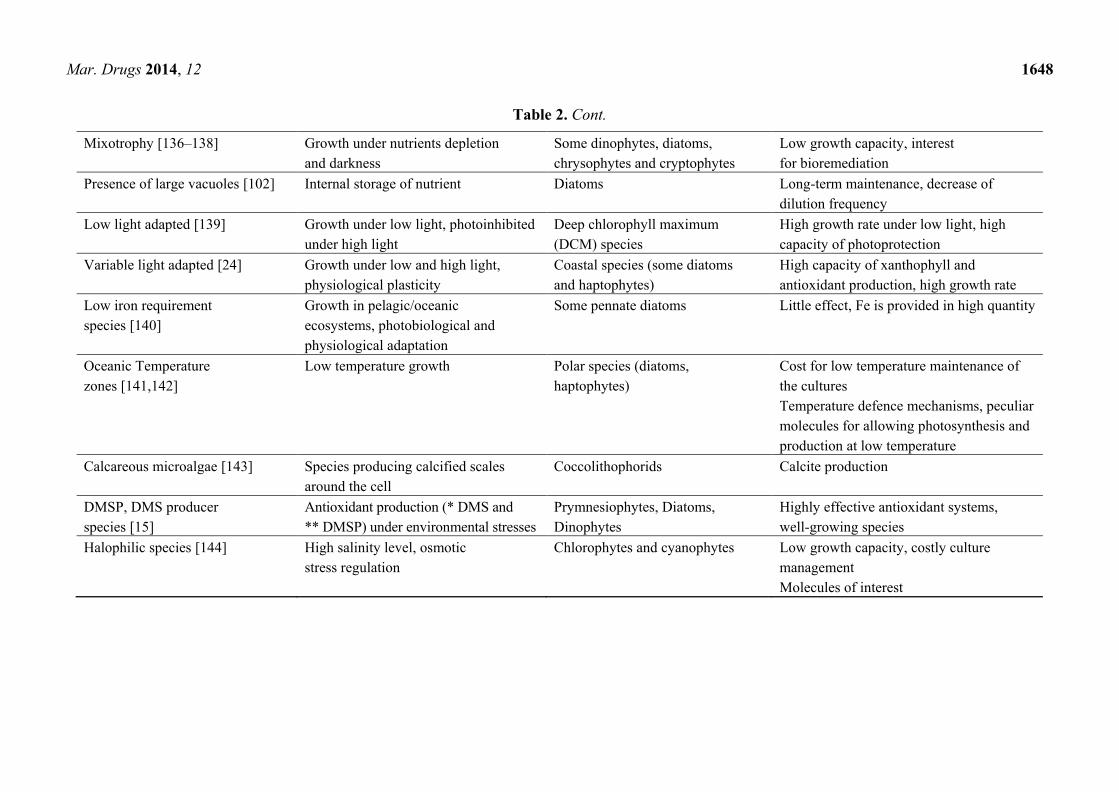
Mar. Drugs 2014, 12 1648
Table 2. Cont.
Mixotrophy [136–138] Growth under nutrients depletion
and darkness
Some dinophytes, diatoms,
chrysophytes and cryptophytes
Low growth capacity, interest
for bioremediation
Presence of large vacuoles [102] Internal storage of nutrient Diatoms Long-term maintenance, decrease of
dilution frequency
Low light adapted [139] Growth under low light, photoinhibited
under high light
Deep chlorophyll maximum
(DCM) species
High growth rate under low light, high
capacity of photoprotection
Variable light adapted [24] Growth under low and high light,
physiological plasticity
Coastal species (some diatoms
and haptophytes)
High capacity of xanthophyll and
antioxidant production, high growth rate
Low iron requirement
species [140]
Growth in pelagic/oceanic
ecosystems, photobiological and
physiological adaptation
Some pennate diatoms Little effect, Fe is provided in high quantity
Oceanic Temperature
zones [141,142]
Low temperature growth Polar species (diatoms,
haptophytes)
Cost for low temperature maintenance of
the cultures
Temperature defence mechanisms, peculiar
molecules for allowing photosynthesis and
production at low temperature
Calcareous microalgae [143] Species producing calcified scales
around the cell
Coccolithophorids Calcite production
DMSP, DMS producer
species [15]
Antioxidant production (* DMS and
** DMSP) under environmental stresses
Prymnesiophytes, Diatoms,
Dinophytes
Highly effective antioxidant systems,
well-growing species
Halophilic species [144] High salinity level, osmotic
stress regulation
Chlorophytes and cyanophytes Low growth capacity, costly culture
management
Molecules of interest

Mar. Drugs 2014, 12 1649
Although there is not much knowledge of picophytoplankton despite its high biodiversity, in
addition to the features discussed earlier, it is implied that exploring picophytoplankton for
biotechnological purposes is challenging. The low consumption together with the high growth rate and
low loss of energy between light harvesting and division processes might outweigh the scant biomass
content per cell.
3.3. Functional Diversity: Uptake of Nutritional Resources
The diversity of culture media available for growth of marine and coastal microalgae (Table 3)
reflects the physiological diversity of microalgal groups. The major natural seawater-enriched media
are: the f/2 medium, widely used for coastal and diatom species [145]; the K medium for oceanic
species; the Pro99 medium for cyanophytes and picoeukaryotes; and the MNK medium mainly used
for coccolithophorids [146].
Table 3. The most commonly used culture mediums for growth of marine microalgae
(see also [147]).
Name Microalgae Specificities
f/2 medium [148,149] Coastal microalgae, diatoms Enriched medium
K medium [146,150] Oceanic microalgae Trace metals
Pro99 [151] Prochlorococcus spp. and some picoeukaryotes High ammonia concentrations,
No vitamin requirement
MNK medium [152] Oceanic coccolithophores Enriched medium
The macronutrients required by microalgae to perform photosynthesis and growth are nitrogen
(nitrate, NO3, nitrite, NO2 or ammonium, NH4), phosphorus (phosphate, PO4) and silicate (SiO2), the
latter being required only for diatoms and silicoflagellates. As reported in many studies [101,153–155],
the variability in uptake and the efficiency in using nutrients are high among phytoplankton groups,
depending on evolutionary and ecological functional traits [156–161]. The most striking group
corresponds to the diatoms, with the presence of a large central vacuole that can store nutrients and
carbohydrates [102], which allows cells to maintain their growth during nutrient-depleted periods. This
feature provides this group a competitive advantage over other groups.
Nitrogen can be provided as nitrate, nitrite or ammonium, which is a key variable for microalgal
growth. It has been observed that different sources of nitrogen are differently metabolized, influencing
the growth capacity and protein content [162], as observed in two Chaetoceros species [163]. Evidence
from Meseck and collaborators [22] show that ammonium uptake efficiency in Tetraselmis chui
PLY 429 significantly increased when carbon dioxide was added to the culture, while it decreased with
a nitrate source. Experimental results obtained with the diatom Skeletonema marinoi grown in f/2
medium under different light conditions, revealed that after three or four days, ammonium
concentration strongly decreased, while nitrite and nitrate concentrations remained high [164]. This result
confirms that cells primarily use NH4, instead of NO3. Allen et al. [165] reported a decrease in growth
rate of Phaedactylum tricornutum grown on nitrate as the only nitrogen source. In fact, the use of
nitrate is more costly for cells than ammonium, because of the enzymes involved in its reduction into
usable form [166,167]. However, high concentrations of ammonium have been shown to inhibit
Mar. Drugs 2014, 12 1650
growth of some phytoplankton species, although the response is variable among the different
groups [168]. Eppley et al. [169] did not show any differences on the half-saturation constants for
nitrate and ammonium uptake among several coastal and oceanic species. Recently, the possible use of
the intracellular NO3 by the benthic diatom Amphora coffeaeformis as a possible survival mechanism
in darkness and anoxia has been shown [170]. The same mechanism has also been reported in the
pelagic Thalassiosira weissflogii [171].
Macronutrient limitation affects growth capacity of microalgae, as well as their physiological
state. Some of the physiological variations induced by a reduction in nutrients might be used for
biotechnological applications. Nitrogen starvation in many microalgal species is commonly linked to
an enhancement of triglyceride accumulation [61,172]. By contrast, lipid content tends to decrease
with phosphorus limitation [173]; the latter reduces the formation of phospholipids and triggers the
production of triglycerides and other neutral lipids [174]. N-limitation in addition to P-limitation
induces an increase in non-photosynthetic carotenoids, as for instance astaxanthin in Haematococcus
pluvialis [175], or β-carotene in Dunaliella bardawil upon nitrogen limitation [176]. Generally, nutrient
limitation negatively affects photosynthesis and growth rates of microalgae [177–182]. P-limitation seems
to repress the carbon-concentrating mechanism (CCM) activity and leads to lower photosynthesis [183],
while N-limitation may have a stimulatory effect on the activity of the CCM, as shown in Chlorella
emersonii [183].
Micronutrients, such as iron, manganese, zinc, cobalt, copper, molybdenum and the metalloid
selenium, are also essential for microalgal growth. They are present in insoluble forms and can be
limiting in oligotrophic waters [184,185]. To cope with this feature, offshore species have evolved
lower iron requirements to survive in iron-poor oceanic waters compared to coastal species [186]. Iron
is involved in controlling microalgal growth, as shown in the diatom Cyclotella meneghiniana, while
its requirement is nitrogen source dependent [187,188]. Manganese is needed in large amounts for
cells growing under low light conditions, as it is involved in photosynthesis [189]. Zinc is present in
proteins involved in DNA transcription and in alkaline phosphatase; it is also used as a co-factor for
carbonic anhydrase, a critical enzyme that transports and fixes CO2 [190]. Copper is also needed for
photosynthesis, being required in cytochrome oxidase and plastocyanin, even though it can be
substituted in some species by iron when cytochrome c6 is present [191]. Nickel is a co-factor of
urease, mainly required when urea is present as nitrogen source [192]. Other micronutrients, such as
selenium, cadmium and mercury, can be added to the medium, albeit carefully because of their toxic
effects. Moreover, three vitamins—vitamin B12 (cyanocobalamin), thiamine, and biotin—are also
essential for microalgal growth [193].
Peculiar functional groups of microalgae concerning resource uptake are diazotrophs and
mixotrophs (Table 2). Diazotrophs, primarily cyanophytes like the species Trichodesmium, fix
atmospheric nitrogen and are able to grow without external sources of nitrogen. Beside a potential
advantage of such strategy for biotechnological mass cultivation, the low metabolic rate efficiency of
these species limits their applications in biotechnology [135]. Mixotrophs instead are able to perform
both photosynthesis and heterotrophic grazing on particles or assimilate dissolved organic carbon
(osmotrophy) [136–138]. Cells can therefore grow under inorganic nutrient depletion and this feature
might favour the allocation of intracellular carbon into proteins [194]. This functional group can account
for up to 20% of primary production, explaining plankton blooms under extreme conditions [195].
Mar. Drugs 2014, 12 1651
However, growth rate is generally low [196] and is strongly dependent on temperature, together with
the efficiency of grazing activity. It has been recently shown that heterotrophic algae, such as
Scenedesmus spp., can be used for fatty acid production [197]. Mixotroph species might be potentially
used in the framework of the first kind of microalgae application, for pollutants bioremediation
(Table 1).
3.4. Functional Diversity: Functional Traits versus Environmental Variables
Microalgae can also be characterized by their adaptive traits to optical, physical or hydrological
variables. Since production is strictly related to the amount of energy supplied to the photosynthetic
apparatus, light is the main variable driving photosynthetic capacity, and therefore growth. In aquatic
ecosystems, light intensity is the most variable parameter, changing over different time and spatial
scales and ranging from limiting to excessive. Light limitation influences nutrient uptake [198–203]
and degree saturation of lipid [204,205]. Excessive light, which is damaging for cells, results
in photoinhibition [206], in reduction of maximum quantum yield, and CO2 uptake [207–209].
Microalgae have developed protective mechanisms, to prevent or limit irreversible photoinhibition, in
order to cope with the formation of reactive oxygen species (ROS) by synthesizing certain carotenoids
or other molecules with antioxidant activities [66]. The first response to high light is the activation of
photoprotective xanthophyll cycle [23], leading to the synthesis of zeaxanthin (in green algae) or
diatoxanthin (in many chlorophyll c-containing algae). The role of these photoprotective xanthophylls,
in countering peroxidative damage, has been reported in diatoms [210]. In Haematococcus pluvialis
high light conditions increase the astaxanthin content three-fold as compared to low light [211,212].
Two functional groups, discriminated by light responses, have been proposed: high light and
shade-acclimated species [139]. The high light group is characterized by low photosynthetic pigment
content relative to the shade-acclimated species. More recently, Dimier et al. [24] discriminated three
functional groups based on their photoregulation capacity: high light adapted, low light adapted and
variable light adapted. The latter group, composed by coastal microalgae, is characterized by efficient
and fast development of photoprotective processes such as xanthophyll cycle and non-photochemical
quenching [24].
Recently, it has been shown that the growth capacity of the toxic Ostreopsis ovata (dinophyte) is
strongly dependent on the effects of both temperature and light [21]. Temperature influences the
growth of photoautotrophs through its control of enzymatic kinetics [213,214] and changes in cellular
membrane composition [215]. Generally in microalgae, an increase in temperature leads to an
enhancement in growth [216–219], as well as in carbon content [218–222]. Indeed, it has also been
shown by Toseland and collaborators [142] that temperature strongly affects phytoplankton metabolism
in the field as nutrients and light do. Microalgal responses to temperature also vary between groups
(Table 2) [88,216,223,224]. The optimal temperature for growth capacity is usually below 10 °C for
polar species [225,226], around 10–25 °C for temperate ones [227,228], close and beyond 25 °C for
tropical species [88] and between 25 and 35 °C for desert-inhabiting microalgae [229].
Another relevant variable is salinity (Table 2). Osmoregulation is achieved by uptake of ions [230,231],
the synthesis of osmotic active compounds or the expulsion of water [76,230,232–235]. Biochemical
changes occur in cells exposed to high salinity, including an increase in ash content [236,237] and

Mar. Drugs 2014, 12 1652
protein synthesis inhibition [238]. Estuarine species are tolerant to a wide range of salinities, though
fewer species are able to grow in high salinity conditions. The success of these halophytic species is
related to enhanced synthesis of osmotic stress regulation molecules such as the zeaxanthin in
cyanophytes [239] or glycerol in the green alga Dunaliella salina [240]. The latter also accumulates
β-carotene under high salinity stress [176,241,242] while Haematococcus pluvialis accumulates
astaxanthin [175]. These species are already used in biotechnology to produce stress-response
molecules, useful in cosmetics or nutrition [144].
In a culture batch, it is also important to monitor pH since it can vary in relation to
photosynthesis. Moreover, pH variations can induce: (i) the differential availability of inorganic carbon
sources (CO2/HCO3−/CO3
2−) [243–245]; (ii) the co-precipitation of phosphate with calcium,
magnesium and carbonate at high carbonate concentrations; (iii) the low solubility and availability of
trace metals at high pH [246]; (iv) intracellular pH regulation; and (v) changes in the uptake of
essential nutrients, such as nitrate and phosphate [22,247,248]. The diatom Phaeodactylum
tricornutum presents high plasticity to pH variation being able to grow at pH > 10, making it one of the
most common contaminants in poorly buffered cultures [249,250]. This property could be
advantageous in outdoor mass cultivation.
4. Enhancing Production and Growth: “Photosynthetic Regulation Biotechnology” and
Genetic Modifications
One of the main challenges for increasing biotechnological applications of microalgae is the
enhancement of their growth and yield. The growth rate of a microalgal population is the result of the
balance between actively dividing and death cells. This ―performance index‖ mainly depends on both
cells’ acclimation to the culture conditions and the energetic cost of internal processes regulation. The
latter depends on how ecophysiological requirements match the environmental conditions and on
physiological plasticity of the cultivated strains. Two of the most promising routes to progress, in
enhancing biomass or interesting molecules production, are the ―Photosynthetic Regulation
Biotechnology‖ (Figure 1) and the genetic engineering of strains.
4.1. Light Control and the “Photosynthetic Regulation Biotechnology”
Light is the most relevant trigger for photosynthesis even though it is the most variable parameter at
sea over different temporal and spatial scales (see previous section). Many recent studies, using
physiological, biochemical or molecular approaches, have shown how light intensity, variability and
spectral composition affect the physiological state and growth of cells [24,25,251–253]. By consequence,
light manipulation might be a way for modifying microalgal productive performance [254,255],
and therefore a relevant issue for biotechnological purposes (see the review by Murchie and
collaborators [256]), in the field of ―photosynthetic regulation biotechnology‖.
Photosynthesis is a unique process that converts light energy into biochemical energy, through light
and dark reactions. In response to light intensity variations, cells modulate two stages of the
photosynthetic process by modifying the photosynthetic apparatus structure and pigments, as well as
the enzymes involved in carbon fixation [257]. Recent studies have shown that these two steps do not
respond in the same way to light changes, showing an uncoupling of their regulative processes
Mar. Drugs 2014, 12 1653
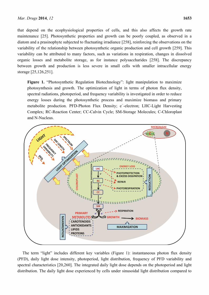
that depend on the ecophysiological properties of cells, and this also affects the growth rate
maintenance [25]. Photosynthetic properties and growth can be poorly coupled, as observed in a
diatom and a prasinophyte subjected to fluctuating irradiance [258], reinforcing the observations on the
variability of the relationship between photosynthetic organic production and cell growth [259]. This
variability can be attributed to many factors, such as variations in respiration, changes in dissolved
organic losses and metabolite storage, as for instance polysaccharides [258]. The discrepancy
between growth and production is less severe in small cells with smaller intracellular energy
storage [25,126,251].
Figure 1. ―Photosynthetic Regulation Biotechnology‖: light manipulation to maximize
photosynthesis and growth. The optimization of light in terms of photon flux density,
spectral radiations, photoperiod, and frequency variability is investigated in order to reduce
energy losses during the photosynthetic process and maximize biomass and primary
metabolite production. PFD-Photon Flux Density; e−-electron; LHC-Light Harvesting
Complex; RC-Reaction Center; CC-Calvin Cycle; SM-Storage Molecules; C-Chloroplast
and N-Nucleus.
The term ―light‖ includes different key variables (Figure 1): instantaneous photon flux density
(PFD), daily light dose intensity, photoperiod, light distribution, frequency of PFD variability and
spectral characteristics [20,260]. The integrated daily light dose depends on the photoperiod and light
distribution. The daily light dose experienced by cells under sinusoidal light distribution compared to

Mar. Drugs 2014, 12 1654
quadratic shape is around 1.9 times less, when provided using the same 12:12 h light–dark photoperiod
and maximal light value [164]. Light is generally provided following a quadratic distribution, i.e., with
an ―on/off‖ switch system. Recent studies have shown that applying a sinusoidal light regime allows
cells to activate a gradual and efficient photoregulation on the contrary of a quadratic
distribution [24,251]. It has been observed that the slow increase of light at dawn allows cells to
efficiently perform photoregulation and prepares the photosynthetic apparatus to cope with the high
light midday peak [24,251]. By contrast, a constant excessive light induces considerable damage,
increasing the biochemical costs associated with defence, protection and recovery, and decreasing the
energy fuelled towards growth. Reciprocally, when constant light is limiting for optimal photosynthesis,
cell performance will be reduced.
The photoperiod, i.e., the succession of illuminated and dark periods during the day, influences
microalgal growth and photosynthetic rate [20,261]. It is known that biomass synthesis, in terms of
produced carbon, may be higher under continuous light than alternating light–dark phases [203,262].
However, cells do not appear healthy under continuous light [263]; the light–dark succession
allows cells to recover and uncouple many biological processes, such as photosynthesis, from cell
division [20,264]. Moreover, photosystem repairing and relaxing occur during the dark period; it is
also known that during dark phytoplankton cells uptake and assimilate NH4 [265,266].
The very fast frequency of light variability (scale of milliseconds) positively influences
photosynthetic efficiency, due to the re-oxidation of the electron transporters of the photosynthetic
apparatus during dark phases [261]. Again, the frequency of the light variability, on an hour scale, is
highly relevant. The shade acclimation state of cells is strengthened by the enhancement of the light
fluctuations experienced, driving production of different carotenoids and antioxidants [251]. Light
fluctuation dynamics also strongly affect the growth capacity of cells [251,258,259,267].
The spectral radiation of light does influence growth, photoacclimation state, and cell
biochemistry [268,269]. Blue light affects many physiological processes in algae, such as
photo-morphogenesis [270], chloroplast movement [271], cell division and photosynthetic acclimation
in diatoms [252,253,270,272]. The spectral composition of light plays a key role in the ability of diatoms
to finely balance light harvesting and photoprotective capacities [273]. Indeed, red radiation mixed to
blue is necessary for activating the photoprotective pathway, such as the synthesis of xanthophyll
pigments with an antioxidant role [274,275]. The spectral properties of microalgal absorption are the
base for designing new models of photobioreactors and improving microalgal growth [276].
4.2. Genetic Transformations
Until now, biotechnological production from microalgae, such as food additives, cosmetics, animal
feed additives, pigments, polysaccharides and fatty acids, is done in a non-transgenic way. However,
genetic engineering is already being applied to microalgal research field, and selectable marker genes,
promoters, reporter genes, transformation techniques, together with other genetic tools are available
for various species. At present, about 20 species are accessible to genetic transformation [27],
and large-scale sequencing projects are in progress for several microalgae species [277]. Sequences are
available at the NCBI organelle database [278] and at the Organelle Genome Database [279].
Mar. Drugs 2014, 12 1655
The complete genome for about 30 cyanobacteria have already been sequenced; genome sequences
of the cyanophyte Synechococcus [280], the green alga Micromonas [281] and Ostreococcus [128] are
also available. For a more exhaustive list, please refer to the website from the Roscoff Culture
Collection [282,283]. The following species’ genomes (nuclear, mitochondrial and plastidial):
Alexandrium tamarense, Amphidinium operculatum, Aureococcus anophagefferens, Chlorella
vulgaris, Cyanophora paradoxa, and Dunaliella salina are being sequenced, and 37 microalgae
transcriptomic projects are currently taking place [8,284].
In the last few years, about 20 new microalgal species have been genetically modified with success:
nine green microalgae, five diatoms, three cyanobacteria, two dinophytes, two red microalgae and one
euglenoid have been successfully transformed; most of these were achieved by nuclear and stable
transformation [27]. Stable transformation of four diatom species (Phaeodactylum tricornutum,
Navicula saprophila, Cylindrotecha fusiformis and Cyclotella cryptica) has also been reported, and
62 promoters have already been tested for microalgae transformations [8]. The use of engineered
nucleases to genetically reprogram diatoms, with the aim of producing bio-fuels, has been successfully
demonstrated [285]. By targeting specific sequences within diatoms’ genomes, nucleases can be used
to accurately insert, correct, or inactivate specific genes. With the whole genome sequencing of several
diatom species, such as Thalassiosira pseudonana and Phaeodactylum tricornutum, a new era of
post-genomics research has begun, with opportunities to improve fundamental understanding of the
diatoms biology, and to build a molecular foundation for new industrial applications. Stable mutants of
some species of microalgae have been obtained in the last years such as Phaeodactylum tricornutum,
expressing a heterologous functional glucose transporter, Dunaliella salina (zea1) overproducing
zeaxanthin, and cyanobacteria-synthetizing mosquito larvacides [7,27,80,286–289]. Euglena gracilis
has been transformed at the chloroplast level [290], while Thalassiosira weissflogii has been only
transiently transformed [287]. Some groups are more difficult to be transformed by electroporation or
conjugation [291], as for instance diatoms with their silica wall, compared to species from other groups
such as Spirulina, Anabaena, or Synechocystis. Recently Synechocystis sp PCC6803 was deeply
analysed, through several growth-coupled knockouts, to identify its main metabolic properties in the
aim of biotechnological applications [292].
One of the main goals in biotechnological genetic engineering is their use as ―bioreactors‖ in
producing vaccines [293–295], specific proteins [296], or bioenergetic molecules [297]. Some
therapeutic proteins have been successfully produced using microalgae, mainly the Chlorophyta
Chlamydomonas reinhardtii, for which suitable transgenic tools and genomic data are available (for all
three genomes: nuclear, chloroplastic and mitochondrial). The chloroplast of C. reinhardtii has been
used to produce a range of recombinant proteins, including reporters such as glucuronidase (GUS),
luciferase (LUC), green fluorescent protein (GFP), industrial enzymes, vaccines and therapeutic
enzymes [298]. To date [8], 18 biopharmaceutical proteins have been expressed in C. reinhardtii and
one in Chlorella ellipsoidea [298,299]. Diatoms have not been employed for expression of any
biopharmaceutical proteins, but Hempel and collaborators [300] have recently reported the first stable
expression of a full-length human antibody and the respective antigen in P. tricornutum.
In the bio-fuel production field, the Heterokonta Nannochloropsis represents a new model with
potential for further development into an integrated photons-to-fuel production platform [301]. Several
isolates of Nannochloropsis spp. produce large quantities of triacylglycerols, related to over-representation

Mar. Drugs 2014, 12 1656
of genes involved in lipid biosynthesis together with rapid growth capacity, and industrial-scale
cultivation [301]. The genome of the high PUFA-content species Nannochloropsis oceanica has been
recently sequenced by Pan and collaborators [302]. Sequence similarity-based investigation identified
new elongase- and desaturase-encoding genes involved in the biosynthesis of PUFAs, which provide a
genetic basis for its rich eicosapentaenoic acid (EPA) content, making this species suitable for the
genetic engineering of a triacylglycerols pathway.
To conclude, we can assert that genetic transformation could be a valid tool to ―revolutionize‖ blue
biotechnology. However, cautious measures have to be taken for ecosystem bio-safety and to monitor
transgenic microorganisms in nature.
5. Conclusions
The functional biodiversity of microalgae has to be explored thoroughly, and mass culturing
conditions presently used have to be revisited in order to optimize the fitness of cultivated species and
decrease production costs. Indoor culturing systems would be preferable to outdoor systems for the
reasons already discussed on the controlled vs. uncontrolled environmental parameters. We also
suggest use of local microalgal species and seawater from which the species have been isolated, i.e.,
cultivation next to aquatic ecosystems, in the frame of what we can call the ―Km-0 mass cultivation‖
strategy. This strategy might reduce the cost of cultivation, allow for the use of freshly isolated species
and strains, and potentially also provide high flexibility in species choice that share common
ecophysiological requirements. This could also solve the potential loss of growth efficiency due to
long-term cultivation maintenance.
Furthermore, we recommend the use of ammonium, as nitrogen source, to increase biomass
production, since it is assimilated faster than nitrate, and this allows an increase in the production
efficiency (see above). We suggest providing light with an intra-diel light–dark cycle, with a sinusoidal
shape instead of quadratic distribution, for the reasons discussed in the previous sections. Turbulence,
reproducing coastal ecosystems, can be applied to coastal species cultivation, through a light variation
frequency program. We propose the use of illumination systems that allow regulation of the photon
flux density and the spectral composition to better manage photosynthetic productivity. For many
microalgal groups (e.g., diatoms or chlorophytes), green radiation may be excluded from the light
spectrum since it is not harvested, in order to increase the growth yield and reduce production
costs [276]. Since the effect of light variations on physiology and growth of autotrophs is relevant,
we suggest that genetic engineering should mainly target the photophysiological response system
(e.g., [252,253,303]), entering the ―photosynthetic regulation biotechnology.‖ The photophysiological
pathway transformation of microalgae could be useful but with a high probability of obtaining a
productive strain with a slow growth.
Diatoms that include many coastal species might be the ideal model to cultivate with
biotechnological aims, for many reasons, such as the huge biodiversity of this group and many
biological peculiarities (see previous). Many diatom species grow on benthic substrates, at least during
one step of their life cycle; therefore, their cultivation for biotechnological purposes needs a deeper
communication between ecophysiological and process engineering researchers. Moreover, they are
able to reproduce sexually [134], allowing this group frequent gene recombination processes and
Mar. Drugs 2014, 12 1657
present high physiological flexibility [274]. Due to this relevant feature, we could easily manipulate
their photophysiological responses and biochemical pathways to increase the production of targeted
molecules by varying the culture conditions.
Acknowledgments
We kindly acknowledge Adrianna Ianora and Punyasloke Bhadury for their comments on the
manuscript. We thank the three reviewers for their helpful and constructive comments. This work was
supported by Stazione Zoologica Anton Dohrn. The authors thank the Flagship RITMARE—The
Italian Research for the Sea—funded by the Italian Ministry of Education. The PhD of
Raghu Chandrasekaran is funded by Stazione Zoologica Anton Dohrn.
Author Contributions
Lucia Barra, Raghu Chandrasekaran contributed equally in writing the manuscript.
Christophe Brunet, Federico Corato conceived and designed the format of the manuscript. All the
authors contributed in critical reading and discussion on the manuscript.
Conflicts of Interest
The authors declare no conflict of interest.
References
1. Guiry, M.D. How many species of algae are there? J. Phycol. 2012, 48, 1057–1063.
2. Public Algaebase: Listing the World’s Algae. Available online: http://www.algaebase.org
(accessed on 10 October 2013).
3. Armbrust, E.V. The life of diatoms in the world’s oceans. Nature 2009, 459, 185–192.
4. Tomas, C.R. Identifying Marine Phytoplankton; Academic Press: San Diego, CA, USA,
1997; p. 858.
5. Lebeau, T.; Robert, J.M. Diatom cultivation and biotechnologically relevant products. Part II:
Current and putative products. Appl. Microbiol. Biotechnol. 2003, 60, 624–632.
6. Chu, W.-L. Biotechnological applications of microalgae. IeJSME 2012, 6, S24–S37.
7. Pulz, O.; Gross, W. Valuable products from biotechnology of microalgae. Appl. Microbiol.
Biotechnol. 2004, 65, 635–648.
8. Cadoret, J.-P.; Garnier, M.; Saint-Jean, B. Microalgae, Functional Genomics and Biotechnology.
Adv. Bot. Res. 2012, 64, 285–341.
9. Zeiler, K.G.; Heacox, D.A.; Toon, S.T.; Kadam, K.L.; Brown, L.M. The use of microalgae for
assimilation and utilization of carbon-dioxide from fossil fuel-fired power plant flue gas.
Energy Convers. Manag. 1995, 36, 707–712.
10. Wilde, E.W.; Benemann, J.R. Bioremoval of heavy-metals by the use of microalgae.
Biotechnol. Adv. 1993, 11, 781–812.
11. Guedes, A.C.; Amaro, H.M.; Malcata, F.X. Microalgae as sources of carotenoids.
Mar. Drugs 2011, 9, 625–644.

Mar. Drugs 2014, 12 1658
12. Christaki, E.; Bonos, E.; Giannenas, I.; Florou-Paneri, P. Functional properties of carotenoids
originating from algae. J. Sci. Food Agric. 2013, 93, 5–11.
13. Becker, W. Microalgae in Human and Animal Nutrition. In Handbook of Microalgal Culture;
Richmond, A., Ed.; Blackwell: Oxford, UK, 2004; pp. 312–351.
14. Sharma, K.K.; Schuhmann, H.; Schenk, P.M. High lipid induction in microalgae for biodiesel
production. Energies 2012, 5, 1532–1553.
15. Sunda, W.; Kieber, D.J.; Kiene, R.P.; Huntsman, S. An antioxidant function for DMSP and DMS
in marine algae. Nature 2002, 418, 317–320.
16. Stahl, W.; Sies, H. Antioxidant activity of carotenoids. Mol. Aspects Med. 2003, 24, 345–351.
17. Munir, N.; Sharif, N.; Naz, S.; Manzoor, F. Algae: A potent antioxidant source. Sky J. Microbiol. Res.
2013, 1, 22–31.
18. Dubinsky, Z.; Stambler, N. Photoacclimation processes in phytoplankton: Mechanisms,
consequences, and applications. Aquat. Microb. Ecol. 2009, 56, 163–176.
19. Borowitzka, M.A. Microalgae as sources of pharmaceuticals and other biologically-active
compounds. J. Appl. Phycol. 1995, 7, 3–15.
20. Grobbelaar, J.U. Microalgal biomass production: Challenges and realities. Photosynth. Res.
2010, 106, 135–144.
21. Scalco, E.; Brunet, C.; Marino, F.; Rossi, R.; Soprano, V.; Zingone, A.; Montresor, M.
Growth and toxicity responses of Mediterranean Ostreopsis cf. ovata to seasonal irradiance and
temperature conditions. Harmful Algae 2012, 17, 25–34.
22. Meseck, S.L.; Smith, B.C.; Wikfors, G.H.; Alix, J.H.; Kapareiko, D. Nutrient interactions
between phytoplankton and bacterioplankton under different carbon dioxide regimes.
J. Appl. Phycol. 2007, 19, 229–237.
23. Brunet, C.; Johnsen, G.; Lavaud, J.; Roy, S. Pigments and Photoacclimation Processes. In
Phytoplankton Pigments, Characterization, Chemotaxonomy and Applications in Oceanography;
Roy, S., Johnsen, G., Llewellyn, C., Egeland, E.S., Eds.; SCOR-UNESCO Publishing,
Cambridge University Press: Cambridge, UK, 2011; Volume 2, pp. 445–471.
24. Dimier, C.; Saviello, G.; Tramontano, F.; Brunet, C. Comparative ecophysiology of the
xanthophyll cycle in six marine phytoplanktonic species. Protist 2009, 160, 397–411.
25. Giovagnetti, V.; Cataldo, M.L.; Conversano, F.; Brunet, C. Growth and photophysiological
responses of two picoplanktonic Minutocellus species, strains RCC967 and RCC703
(Bacillariophyceae). Eur. J. Phycol. 2012, 47, 408–420.
26. Lebeau, T.; Robert, J. Biotechnology of immobilized micro algae: A Culture Technique for the
Future? In Algal Cultures, Analogues of Blooms and Applications; Rao, S., Ed.;
Science Publisher Inc.: New Hampshire, NH, USA, 2006; Volume 2, pp. 801–837.
27. Hallmann, A. Algal transgenics and biotechnology. Transgenic Plant. J. 2007, 1, 81–98.
28. Torres, M.A.; Barros, M.P.; Campos, S.C.G.; Pinto, E.; Rajamani, S.; Sayre, R.T.; Colepicolo, P.
Biochemical biomarkers in algae and marine pollution: A review. Ecotoxicol. Environ. Saf. 2008,
71, 1–15.
29. Montone, R.C.; Taniguchi, S.; Weber, R.R. Polychlorinated biphenyls in marine sediments of
Admiralty Bay, King George Island, Antarctica. Mar. Pollut. Bull. 2001, 42, 611–614.
Mar. Drugs 2014, 12 1659
30. Leitão, M.A.d.S.; Cardozo, K.H.M.; Pinto, E.; Colepicolo, P. PCB-Induced oxidative stress in
the unicellular marine dinoflagellate Lingulodinium polyedrum. Arch. Environ. Contam. Toxicol.
2003, 45, 59–65.
31. Gerofke, A.; Kömp, P.; McLachlan, M.S. Bioconcentration of persistent organic pollutants in
four species of marine phytoplankton. Environ. Toxicol. Chem. 2005, 24, 2908–2917.
32. Aksmann, A.; Tukaj, Z. The effect of anthracene and phenanthrene on the growth,
photosynthesis, and SOD activity of the green alga Scenedesmus armatus depends on the PAR
Irradiance and CO2 level. Arch. Environ. Contam. Toxicol. 2004, 47, 177–184.
33. Djomo, J.E.; Dauta, A.; Ferrier, V.; Narbonne, J.F.; Monkiedje, A.; Njine, T.; Garrigues, P.
Toxic effects of some major polyaromatic hydrocarbons found in crude oil and aquatic sediments
on Scenedesmus subspicatus. Water Res. 2004, 38, 1817–1821.
34. Lei, A.-P.; Hu, Z.-L.; Wong, Y.-S.; Tam, N.F.-Y. Removal of fluoranthene and pyrene by
different microalgal species. Bioresour. Technol. 2007, 98, 273–280.
35. Doick, K.J.; Klingelmann, E.; Burauel, P.; Jones, K.C.; Semple, K.T. Long-term fate of
polychlorinated biphenyls and polycyclic aromatic hydrocarbons in an agricultural soil.
Environ. Sci. Technol. 2005, 39, 3663–3670.
36. Borja, J.; Taleon, D.M.; Auresenia, J.; Gallardo, S. Polychlorinated biphenyls and their
biodegradation. Process Biochem. 2005, 40, 1999–2013.
37. Geoffroy, L.; Teisseire, H.; Couderchet, M.; Vernet, G. Effect of oxyfluorfen and diuron alone
and in mixture on antioxidative enzymes of Scenedesmus obliquus. Pestic. Biochem. Physiol.
2002, 72, 178–185.
38. Nyström, B.; Becker-Van Slooten, K.; Bérard, A.; Grandjean, D.; Druart, J.-C.; Leboulanger, C.
Toxic effects of Irgarol 1051 on phytoplankton and macrophytes in Lake Geneva.
Water Res. 2002, 36, 2020–2028.
39. Geoffroy, L.; Dewez, D.; Vernet, G.; Popovic, R. Oxyfluorfen toxic effect on S. obliquus
evaluated by different photosynthetic and enzymatic biomarkers. Arch. Environ. Contam. Toxicol.
2003, 45, 445–452.
40. Ma, J.; Chen, J. How to accurately assay the algal toxicity of pesticides with low water solubility.
Environ. Pollut. 2005, 136, 267–273.
41. Cai, X.; Liu, W.; Jin, M.; Lin, K. Relation of diclofop-methyl toxicity and degradation in algae
cultures. Environ. Toxicol. Chem. 2007, 26, 970–975.
42. Hall, J.L. Cellular mechanisms for heavy metal detoxification and tolerance. J. Exp. Bot. 2002,
53, 1–11.
43. Conti, M.E.; Cecchetti, G. A biomonitoring study: Trace metals in algae and molluscs from
Tyrrhenian coastal areas. Environ. Res. 2003, 93, 99–112.
44. Pinto, E.; Sigaud-kutner, T.C.S.; Leitão, M.A.S.; Okamoto, O.K.; Morse, D.; Colepicolo, P.
Heavy metal induced oxidative stress in algae. J. Phycol. 2003, 39, 1008–1018.
45. Nishikawa, K.; Yamakoshi, Y.; Uemura, I.; Tominaga, N. Ultrastructural changes in
Chlamydomonas acidophila (Chlorophyta) induced by heavy metals and polyphosphate
metabolism. FEMS Microbiol. Ecol. 2003, 44, 253–259.
46. Mallick, N. Copper-induced oxidative stress in the chlorophycean microalga Chlorella vulgaris:
Response of the antioxidant system. J. Plant Physiol. 2004, 161, 591–597.
Mar. Drugs 2014, 12 1660
47. Perales-Vela, H.V.; Peña-Castro, J.M.; Cañizares-Villanueva, R.O. Heavy metal detoxification in
eukaryotic microalgae. Chemosphere 2006, 64, 1–10.
48. Tripathi, B.N.; Mehta, S.K.; Amar, A.; Gaur, J.P. Oxidative stress in Scenedesmus sp. during
short- and long-term exposure to Cu2+
and Zn2+
. Chemosphere 2006, 62, 538–544.
49. Schroda, M.; Vallon, O.; Wollman, F.A.; Beck, C.F. A chloroplast-targeted heat shock protein 70
(HSP70) contributes to the photoprotection and repair of photosystem II during and after
photoinhibition. Plant Cell 1999, 11, 1165–1178.
50. Ireland, H.E.; Harding, S.J.; Bonwick, G.A.; Jones, M.; Smith, C.J.; Williams, J.H.H.
Evaluation of heat shock protein 70 as a biomarker of environmental stress in Fucus serratus and
Lemna minor. Biomarkers 2004, 9, 139–155.
51. Singer, A.C.; Thompson, I.P.; Bailey, M.J. The tritrophic trinity: A source of pollutant-degrading
enzymes and its implications for phytoremediation. Curr. Opin. Microbiol. 2004, 7, 239–244.
52. Lewis, S.; Donkin, M.E.; Depledge, M.H. Hsp70 expression in Enteromorpha intestinalis
(Chlorophyta) exposed to environmental stressors. Aquat. Toxicol. 2001, 51, 277–291.
53. Spijkerman, E.; Barua, D.; Gerloff-Elias, A.; Kern, J.; Gaedke, U.; Heckathorn, S.
Stress responses and metal tolerance of Chlamydomonas acidophila in metal-enriched lake water
and artificial medium. Extremophiles 2007, 11, 551–562.
54. Andrade, L.R.; Farina, M.; Amado Filho, G.M. Effects of copper on Enteromorpha flexuosa
(Chlorophyta) in vitro. Ecotoxicol. Environ. Saf. 2004, 58, 117–125.
55. Chinnasamy, S.; Bhatnagar, A.; Claxton, R.; Das, K.C. Biomass and bioenergy production
potential of microalgae consortium in open and closed bioreactors using untreated carpet industry
effluent as growth medium. Bioresour. Technol. 2010, 101, 6751–6760.
56. Sayre, R. Microalgae: The potential for carbon capture. BioScience 2010, 60, 722–727.
57. Sheehan, J.; Dunahay, T.; Benemann, J.; Roessler, P. A Look Back at the US Department of
Energy’s Aquatic Species Program.: Biodiesel from Algae; National Renewable Energy
Laboratory Golden: Golden, CO, USA, 1998; Volume 328, p. 294.
58. Tsukahara, K.; Sawayama, S. Liquid fuel production using microalgae. J. Jpn. Pet. Inst. 2005,
48, 251–259.
59. Chisti, Y. Biodiesel from microalgae. Biotechnol. Adv. 2007, 25, 294–306.
60. Li, Y.; Horsman, M.; Wu, N.; Lan, C.Q.; Dubois-Calero, N. Biofuels from microalgae.
Biotechnol. Prog. 2008, 24, 815–820.
61. Li, Y.; Horsman, M.; Wang, B.; Wu, N.; Lan, C. Effects of nitrogen sources on cell growth and
lipid accumulation of green alga Neochloris oleoabundans. Appl. Microbiol. Biotechnol. 2008,
81, 629–636.
62. Mata, T.M.; Martins, A.A.; Caetano, N.S. Microalgae for biodiesel production and other
applications: A review. Renew. Sustain. Energ. Rev. 2010, 14, 217–232.
63. Renaud, S.M.; Thinh, L.V.; Parry, D.L. The gross chemical composition and fatty acid
composition of 18 species of tropical Australian microalgae for possible use in mariculture.
Aquaculture 1999, 170, 147–159.
64. Pratoomyot, J.; Srivilas, P.; Noiraksar, T. Fatty acids composition of 10 microalgal species.
Songklanakarin J. Sci. Technol. 2005, 27, 1179–1187.
Mar. Drugs 2014, 12 1661
65. Aslan, S.; Kapdan, I.K. Batch kinetics of nitrogen and phosphorus removal from synthetic
wastewater by algae. Ecol. Eng. 2006, 28, 64–70.
66. Jin, E.; Polle, J.E.W.; Lee, H.K.; Hyun, S.M.; Chang, M. Xanthophylls in microalgae: From
biosynthesis to biotechnological mass production and application. J. Microbiol. Biotechnol.
2003, 13, 165–174.
67. Spolaore, P.; Joannis-Cassan, C.; Duran, E.; Isambert, A. Commercial applications of
microalgae. J. Biosci. Bioeng. 2006, 101, 87–96.
68. Lee, Y.-K. Commercial production of microalgae in the Asia-Pacific rim. J. Appl. Phycol. 1997,
9, 403–411.
69. Borowitzka, M.A. Commercial production of microalgae: Ponds, tanks, tubes and fermenters.
J. Biotechnol. 1999, 70, 313–321.
70. Carvalho, A.P.; Meireles, L.A.; Malcata, F.X. Microalgal reactors: A review of enclosed system
designs and performances. Biotechnol. Prog. 2006, 22, 1490–1506.
71. Cornet, J.F. Le technoscope: Les photobioréacteurs. Biofutur 1998, 176, 1–10.
72. Soletto, D.; Binaghi, L.; Lodi, A.; Carvalho, J.C.M.; Converti, A. Batch and fed-batch
cultivations of Spirulina platensis using ammonium sulphate and urea as nitrogen sources.
Aquaculture 2005, 243, 217–224.
73. Yamaguchi, K. Recent advances in microalgal bioscience in Japan, with special reference to
utilization of biomass and metabolites: A review. J. Appl. Phycol. 1996, 8, 487–502.
74. Liang, S.Z.; Liu, X.M.; Chen, F.; Chen, Z.J. Current microalgal health food R & D activities in
China. Hydrobiologia 2004, 512, 45–48.
75. Apt, K.E.; Behrens, P.W. Commercial developments in microalgal biotechnology. J. Phycol.
1999, 35, 215–226.
76. Iwamoto, K.; Shiraiwa, Y. Salt-regulated mannitol metabolism in algae. Mar. Biotechnol. 2005,
7, 407–415.
77. Metting, F.B. Biodiversity and application of microalgae. J. Ind. Microbiol. Biotechnol. 1996,
17, 477–489.
78. Del Campo, J.A.; Rodriguez, H.; Moreno, J.; Vargas, M.A.; Guerrero, M.G. Lutein production
by Muriellopsis sp. in an outdoor tubular photobioreactor. J. Biotechnol. 2001, 85, 289–295.
79. Kobayashi, M.; Kakizono, T.; Yamaguchi, K.; Nishio, N.; Nagai, S. Growth and astaxanthin
formation of Haematococcus pluvialis in heterotrophic and mixotrophic conditions. J. Ferment.
Bioeng. 1992, 74, 17–20.
80. Boussiba, S. Carotenogenesis in the green alga Haematococcus pluvialis: Cellular physiology
and stress response. Physiol. Plant. 2000, 108, 111–117.
81. Radmer, R.J. Algal diversity and commercial algal products. BioScience 1996, 46, 263–270.
82. Rangel-Yagui, C.d.O.; Danesi, E.D.G.; de Carvalho, J.C.M.; Sato, S. Chlorophyll production
from Spirulina platensis: Cultivation with urea addition by fed-batch process. Bioresour.
Technol. 2004, 92, 133–141.
83. Jensen, G.S.; Ginsberg, D.I.; Drapeau, C. Blue-green algae as an immuno-enhancer and
biomodulator. J. Am. Nutraceut. Assoc. 2001, 3, 24–30.

Mar. Drugs 2014, 12 1662
84. Benedetti, S.; Benvenuti, F.; Pagliarani, S.; Francogli, S.; Scoglio, S.; Canestrari, F.
Antioxidant properties of a novel phycocyanin extract from the blue-green alga Aphanizomenon
flos-aquae. Life Sci. 2004, 75, 2353–2362.
85. Borowitzka, M.A. Microalgae for aquaculture: Opportunities and constraints. J. Appl. Phycol.
1997, 9, 393–401.
86. Muller-Feuga, A. The role of microalgae in aquaculture: Situation and trends. J. Appl. Phycol.
2000, 12, 527–534.
87. Brown, M.R.; Mular, M.; Miller, I.; Farmer, C.; Trenerry, C. The vitamin content of microalgae
used in aquaculture. J. Appl. Phycol. 1999, 11, 247–255.
88. Renaud, S.M.; Thinh, L.V.; Lambrinidis, G.; Parry, D.L. Effect of temperature on growth,
chemical composition and fatty acid composition of tropical Australian microalgae grown in
batch cultures. Aquaculture 2002, 211, 195–214.
89. Stolz, P.; Obermayer, B. Manufacturing microalgae for skin care. Cosmet. Toilet. 2005, 120,
99–106.
90. Sekar, S.; Chandramohan, M. Phycobiliproteins as a commodity: Trends in applied research,
patents and commercialization. J. Appl. Phycol. 2008, 20, 113–136.
91. Pushparaj, B.; Pelosi, E.; Carlozzi, P.; Torzillo, G. Yield and biochemical-composition of a
marine cyanobacterium (Nodularia sp.) in outdoor culture. Aquat. Microb. Ecol. 1995, 9, 13–16.
92. Vonshak, A.; Chanawongse, L.; Bunnag, B.; Tanticharoen, M. Light acclimation and
photoinhibition in three Spirulina platensis (cyanobacteria) isolates. J. Appl. Phycol. 1996, 8,
35–40.
93. Torzillo, G.; Bernardini, P.; Masojidek, J. On-line monitoring of chlorophyll fluorescence to
assess the extent of photoinhibition of photosynthesis induced by high oxygen concentration and
low temperature and its effect on the productivity of outdoor cultures of Spirulina platensis
(Cyanobacteria). J. Phycol. 1998, 34, 504–510.
94. Molina, E.; Fernandez, J.; Acien, F.G.; Chisti, Y. Tubular photobioreactor design for algal
cultures. J. Biotechnol. 2001, 92, 113–131.
95. Roy, L.A.; Davis, D.A.; Saoud, I.P. Effects of lecithin and cholesterol supplementation to
practical diets for Litopenaeus vannamei reared in low salinity waters. Aquaculture 2006, 257,
446–452.
96. Ugwu, C.U.; Aoyagi, H.; Uchiyama, H. Influence of irradiance, dissolved oxygen concentration,
and temperature on the growth of Chlorella sorokiniana. Photosynthetica 2007, 45, 309–311.
97. Masojídek, J.; Vonshak, A.; Torzillo, G. Chlorophyll Fluorescence Applications in Microalgal
Mass Cultures. In Chlorophyll a Fluorescence in Aquatic Sciences: Methods and Applications;
Suggett, D.J., Prášil, O., Borowitzka, M.A., Eds.; Springer: Dordrecht, The Netherlands, 2010;
Volume 4, pp. 277–292.
98. Giovagnetti, V.; Brunet, C.; Conversano, F.; Tramontano, F.; Obernosterer, I.; Ridame, C.;
Guieu, C. Assessing the role of dust deposition on phytoplankton ecophysiology and succession
in a low-nutrient low-chlorophyll ecosystem: A mesocosm experiment in the Mediterranean Sea.
BioGeosciences 2013, 10, 2973–2991.
99. Pulz, O. Photobioreactors: Production systems for phototrophic microorganisms. Appl.
Microbiol. Biotechnol. 2001, 57, 287–293.

Mar. Drugs 2014, 12 1663
100. Xu, L.; Weathers, P.J.; Xiong, X.-R.; Liu, C.-Z. Microalgal bioreactors: Challenges and
opportunities. Eng. Life Sci. 2009, 9, 178–189.
101. Litchman, E.; Klausmeier, C.A. Trait-based Community Ecology of Phytoplankton. Ann. Rev.
Ecol. Evol. Syst. 2008, 39, 615–639.
102. Kooistra, W.H.C.F.; Gersonde, R.; Medlin, L.K.; Mann, D.G. The Origin and Evolution of the
Diatoms: Their Adaptation to a Planktonic Existence. In Evolution of Primary Producers in the
Sea; Paul, G.F., Andrew, H.K., Eds.; Academic Press: Burlington, Canada, 2007; pp. 207–249.
103. Mann, D.G.; Droop, S.J.M. Biodiversity, biogeography and conservation of diatoms.
Hydrobiologia 1996, 336, 19–32.
104. Amato, A.; Kooistra, W.H.C.F.; Ghiron, J.H.L.; Mann, D.G.; Proschold, T.; Montresor, M.
Reproductive isolation among sympatric cryptic species in marine diatoms. Protist 2007, 158,
193–207.
105. Amato, A.; Montresor, M. Morphology, phylogeny, and sexual cycle of Pseudo-nitzschia
mannii sp. nov. (Bacillariophyceae): A pseudo-cryptic species within the P. pseudodelicatissima
complex. Phycologia 2008, 47, 487–497.
106. Medlin, L.K.; Kooistra, W.H.C.F. Methods to estimate the diversity in the marine photosynthetic
protist community with illustrations from case studies: A review. Diversity 2010, 2, 973–1014.
107. Falkowski, P.G.; Katz, M.E.; Knoll, A.H.; Quigg, A.; Raven, J.A.; Schofield, O.; Taylor, F.J.R.
The evolution of modern eukaryotic phytoplankton. Science 2004, 305, 354–360.
108. Bowler, C.; Allen, A.E.; Badger, J.H.; Grimwood, J.; Jabbari, K.; Kuo, A.; Maheswari, U.;
Martens, C.; Maumus, F.; Otillar, R.P.; et al. The Phaeodactylum genome reveals the
evolutionary history of diatom genomes. Nature 2008, 456, 239–244.
109. Tirichine, L.; Bowler, C. Decoding algal genomes: Tracing back the history of photosynthetic
life on Earth. Plant. J. 2011, 66, 45–57.
110. Reusch, T.B.H.; Boyd, P.W. Experimental evolution meets marine phytoplankton. Evolution
2013, 67, 1849–1859.
111. Groben, R.; John, U.; Eller, G.; Lange, M.; Medlin, L.K. Using fluorescently-labelled rRNA
probes for hierarchical estimation of phytoplankton diversity—A mini-review. Nova Hedwig.
2004, 79, 313–320.
112. Orsini, L.; Procaccini, G.; Sarno, D.; Montresor, M. Multiple rDNA ITS-types within the diatom
Pseudo-nitzschia delicatissima (Bacillariophyceae) and their relative abundances across a spring
bloom in the Gulf of Naples. Mar. Ecol. Prog. Ser. 2004, 271, 87–98.
113. Massana, R.; Castresana, J.; Balagué, V.; Guillou, L.; Romari, K.; Groisillier, A.; Valentin, K.;
Pedrós-Alió, C. Phylogenetic and ecological analysis of novel marine stramenopiles.
Appl. Environ. Microbiol. 2004, 70, 3528–3534.
114. Sarno, D.; Kooistra, W.H.C.F.; Medlin, L.K.; Percopo, I.; Zingone, A. Diversity in the genus
Skeletonema (bacillariophyceae). (ii) An assessment of the taxonomy of S. costatum like species
with the description of four new species. J. Phycol. 2005, 41, 151–176.
115. Smayda, T.J. The suspension and sinking of phytoplankton in the sea. Oceanogr. Mar. Biol.
Annu. Rev. 1970, 8, 353–414.
116. Pickett-Heaps, J.D. Cell division and moprhogenesis of the centric diatom Chaetoceros decipiens
(bacillariophyceae) I. Living cells. J. Phycol. 1998, 34, 989–994.

Mar. Drugs 2014, 12 1664
117. Yokota, K.; Sterner, R.W. Trade-offs limiting the evolution of coloniality: Ecological
displacement rates used to measure small costs. Proc. Biol. Sci. 2011, 278, 458–463.
118. Rousseau, V.; Chrétiennot-Dinet, M.-J.; Jacobsen, A.; Verity, P.; Whipple, S. The life cycle of
Phaeocystis: State of knowledge and presumptive role in ecology. Biogeochemistry 2007, 83,
29–47.
119. Young, A.M.; Karp-Boss, L.; Jumars, P.A.; Landis, E.N. Quantifying diatom aspirations:
Mechanical properties of chain-forming species. Limnol. Oceanogr. 2012, 57, 1789–1801.
120. Smetacek, V.; Assmy, P.; Henjes, J. The role of grazing in structuring Southern Ocean pelagic
ecosystems and biogeochemical cycles. Antarct. Sci. 2004, 16, 541–558.
121. Bergkvist, J.; Thor, P.; Jakobsen, H.H.; Wangberg, S.-A.; Selander, E. Grazer-induced chain
length plasticity reduces grazing risk in a marine diatom. Limnol. Oceanogr. 2012, 57, 318–324.
122. Karp-Boss, L.; Boss, E.; Jumars, P.A. Nutrient fluxes to planktonic osmotrophs in the presence
of fluid motion. Oceanogr. Mar. Biol. Annu. Rev. 1996, 34, 71–107.
123. Finkel, Z.V.; Beardall, J.; Flynn, K.J.; Quigg, A.; Rees, T.A.V.; Raven, J.A. Phytoplankton in a
changing world: Cell size and elemental stoichiometry. J. Plankton Res. 2010, 32, 119–137.
124. Not, F.; Massana, R.; Latasa, M.; Marie, D.; Colson, C.; Eikrem, W.; Pedros-Alio, C.; Vaulot, D.;
Simon, N. Late summer community composition and abundance of photosynthetic picoeukaryotes
in Norwegian and Barents Seas. Limnol. Oceanogr. 2005, 50, 1677–1686.
125. Worden, A.Z.; Not, F. Ecology and Diversity of Picoeukaryotes. In Microbial Ecology of the
Oceans; Kirchman, D.L., Ed.; John Wiley & Sons, Inc.: Hoboken, NJ, USA, 2008; pp. 159–205.
126. Raven, J.A. The twelfth Tansley Lecture. Small is beautiful: The picophytoplankton. Funct. Ecol.
1998, 12, 503–513.
127. Raven, J.A.; Finkel, Z.V.; Irwin, A.J. Picophytoplankton: Bottom-up and top-down controls on
ecology and evolution. Vie Milieu Life Environ. 2005, 55, 209–215.
128. Palenik, B.; Grimwood, J.; Aerts, A.; Rouze, P.; Salamov, A.; Putnam, N.; Dupont, C.;
Jorgensen, R.; Derelle, E.; Rombauts, S.; et al. The tiny eukaryote Ostreococcus provides
genomic insights into the paradox of plankton speciation. Proc. Natl. Acad. Sci. USA 2007, 104,
7705–7710.
129. Six, C.; Finkel, Z.V.; Rodriguez, F.; Marie, D.; Partensky, F.; Campbell, D.A.
Contrasting photoacclimation costs in ecotypes of the marine eukaryotic picoplankter
Ostreococcus. Limnol. Oceanogr. 2008, 53, 255–265.
130. Six, C.; Sherrard, R.; Lionard, M.; Roy, S.; Campbell, D.A. Photosystem II and pigment
dynamics among ecotypes of the green alga Ostreococcus. Plant Physiol. 2009, 151, 379–390.
131. Cox, E.R. Phytoflagellates; Elsevier North Holland: New York, NY, USA, 1980.
132. MacIntyre, H.; Geider, R.; Miller, D. Microphytobenthos: The ecological role of the ―secret
garden‖ of unvegetated, shallow-water marine habitats. I. Distribution, abundance and primary
production. Estuaries 1996, 19, 186–201.
133. Hallegraeff, G.M. A review of harmful algal blooms and their apparent global increase.
Phycologia 1993, 32, 79–99.
134. Chepurnov, V.A.; Mann, D.G.; Sabbe, K.; Vyverman, W. Experimental Studies an Sexual
Reproduction in Diatoms. In International Review of Cytology—A Survey of Cell Biology;
Jeon, K.W., Ed.; Elsevier Academic Press: San Diego, USA, 2004; Volume 237, pp. 91–154.
Mar. Drugs 2014, 12 1665
135. Berman-Frank, I.; Quigg, A.; Finkel, Z.V.; Irwin, A.J.; Haramaty, L. Nitrogen-fixation strategies
and Fe requirements in cyanobacteria. Limnol. Oceanogr. 2007, 52, 2260–2269.
136. Flynn, K.J.; Stoecker, D.K.; Mitra, A.; Raven, J.A.; Glibert, P.M.; Hansen, P.J.; Granéli, E.;
Burkholder, J.M. Misuse of the phytoplankton–zooplankton dichotomy: The need to assign
organisms as mixotrophs within plankton functional types. J. Plankton Res. 2013, 35, 3–11.
137. Wilken, S.; Huisman, J.; Naus-Wiezer, S.; Donk, E. Mixotrophic organisms become more
heterotrophic with rising temperature. Ecol. Lett. 2013, 16, 225–233.
138. Stoecker, D.K. Conceptual models of mixotrophy in planktonic protists and some ecological and
evolutionary implications. Eur. J. Protistol. 1998, 34, 281–290.
139. Falkowski, P.G. Light-shade adaptation and vertical mixing of marine-phytoplankton—A
comparative field-study. J. Mar. Res. 1983, 41, 215–237.
140. Marchetti, A.; Maldonado, M.T.; Lane, E.S.; Harrison, P.J. Iron requirements of the pennate
diatom Pseudo-nitzschia: Comparison of oceanic (high-nitrate, low-chlorophyll waters) and
coastal species. Limnol. Oceanogr. 2006, 51, 2092–2101.
141. Hare, C.E.; Leblanc, K.; DiTullio, G.R.; Kudela, R.M.; Zhang, Y.; Lee, P.A.; Riseman, S.;
Hutchins, D.A. Consequences of increased temperature and CO2 for phytoplankton community
structure in the Bering Sea. Mar. Ecol. Prog. Ser. 2007, 352, 9–16.
142. Toseland, A.; Daines, S.J.; Clark, J.R.; Kirkham, A.; Strauss, J.; Uhlig, C.; Lenton, T.M.;
Valentin, K.; Pearson, G.A.; Moulton, V. The impact of temperature on marine phytoplankton
resource allocation and metabolism. Nat. Clim. Chang. 2013, 3, 979–984.
143. Takano, H.; Furuune, H.; Burgess, J.G.; Manabe, E.; Hirano, M.; Okazaki, M.; Matsunaga, T.
Production of ultrafine calcite particles by coccolithophorid algae grown in a biosolar reactor
supplied with sunlight. Appl. Biochem. Biotechnol. 1993, 39, 159–167.
144. DasSarma, P.; Coker, J.A.; Huse, V.; DasSarma, S. Halophiles, Industrial Applications. In
Encyclopedia of Industrial Biotechnology; John Wiley & Sons Inc.: Hoboken, NJ, USA, 2010;
pp. 1–43.
145. Berges, J.A.; Franklin, D.J.; Harrison, P.J. Evolution of an artificial seawater medium:
Improvements in enriched seawater, artificial water over the last two decades. J. Phycol. 2001,
37, 1138–1145.
146. Keller, M.D.; Selvin, R.C.; Claus, W.; Guillard, R.R.L. Media for the culture of oceanic
ultraphytoplankton. J. Phycol. 1987, 23, 633–638.
147. The National Center for Marine Algae and Microbiota (NCMA). Culturing Diversity. Available
online: https://ncma.bigelow.org (accessed on 10 October 2013).
148. Guillard, R.R.; Ryther, J.H. Studies of marine planktonic diatoms. I. Cyclotella nana Hustedt,
and Detonula confervacea (cleve) Gran. Can. J. Microbiol. 1962, 8, 229–239.
149. Guillard, R.R.L. Culture of Phytoplankton for Feeding Marine Invertebrates. In Culture of
Marine Invertebrate Animals; Smith, W.L., Chanley, M.H., Eds.; Plenum Press: New York, NY,
USA, 1975; pp. 26–60.
150. Keller, M.D.; Guillard, R.R.L. Factors Significant to Marine Diatom Culture. In Toxic
Dinoflagellates; Anderson, D.M., White, A.W., Baden, D.G., Eds.; Elsevier: New York, NY,
USA, 1985; pp. 113–116.
Mar. Drugs 2014, 12 1666
151. Moore, L.R.; Coe, A.; Zinser, E.R.; Saito, M.A.; Sullivan, M.B.; Lindell, D.; Frois-Moniz, K.;
Waterbury, J.; Chisholm, S.W. Culturing the marine cyanobacterium Prochlorococcus.
Limnol. Oceanogr. Methods 2007, 5, 353–362.
152. Noel, M.H.; Kawachi, M.; Inouye, I. Induced dimorphic life cycle of a coccolithophorid,
Calyptrosphaera sphaeroidea (Prymnesiophyceae, Haptophyta). J. Phycol. 2004, 40, 112–129.
153. Andersen, T.; Schartau, A.; Paasche, E. Quantifying external and internal nitrogen and
phosphorus pools, as well as nitrogen and phosphorus supplied through remineralization, in
coastal marine plankton by means of a dilution technique. Mar. Ecol. Prog. Ser. 1991, 69, 67–80.
154. Irwin, A.J.; Finkel, Z.V.; Schofield, O.M.E.; Falkowski, P.G. Scaling-up from nutrient
physiology to the size-structure of phytoplankton communities. J. Plankton Res. 2006, 28,
459–471.
155. Verdy, A.; Follows, M.; Flierl, G. Optimal phytoplankton cell size in an allometric model.
Mar. Ecol. Prog. Ser. 2009, 379, 1–12.
156. Shuter, B.J. Size dependence of phosphorus and nitrogen subsistence quotas in unicellular
microorganisms. Limnol. Oceanogr. 1978, 23, 1248–1255.
157. Sommer, U. The paradox of the plankton: Fluctuations of phosphorus availability maintain
diversity of phytoplankton in flow-through cultures. Limnol. Oceanogr. 1984, 29, 633–636.
158. Morel, F.M.M. Kinetics of nutrient-uptake and growth in phytoplankton. J. Phycol. 1987, 23,
137–150.
159. Smayda, T.J. Harmful algal blooms: Their ecophysiology and general relevance to phytoplankton
blooms in the sea. Limnol. Oceanogr. 1997, 42, 1137–1153.
160. Litchman, E.; Klausmeier, C.A.; Bossard, P. Phytoplankton nutrient competition under dynamic
light regimes. Limnol. Oceanogr. 2004, 49, 1457–1462.
161. Litchman, E.; Klausmeier, C.A.; Schofield, O.M.; Falkowski, P.G. The role of functional traits
and trade-offs in structuring phytoplankton communities: Scaling from cellular to ecosystem
level. Ecol. Lett. 2007, 10, 1170–1181.
162. Eppley, R.W.; Carlucci, A.F.; Holm-Hansen. O.; Kiefer, D.; McCarthy, J.J.; Venrick, E.;
Williams, P.M. Phytoplankton growth and composition in shipboard cultures supplied with
nitrate, ammonium, or urea as nitrogen source. Limnol. Oceanogr. 1971, 16, 741–751.
163. Karthikeyan, P.; Manimaran, K.; Sampathkumar, P.; Rameshkumar, L. Growth and nutrient
removal properties of the diatoms, Chaetoceros curvisetus and C. simplex under different
nitrogen sources. Appl. Water Sci. 2013, 3, 49–55.
164. Chandrasekaran, R.; Barra, L.; Caruso, T.; Corsaro, L.; Piaz F.D.; Graziani, G.; Corato, F.;
Brunet, C. Light modulation on the biochemical properties of the diatom Skeletonema marinoi:
Relevance of photosynthetic manipulation in the biotechnological field. 2014, Unpublished work.
165. Allen, A.E.; Dupont, C.L.; Obornik, M.; Horak, A.; Nunes-Nesi, A.; McCrow, J.P.; Zheng, H.;
Johnson, D.A.; Hu, H.; Fernie, A.R.; et al. Evolution and metabolic significance of the urea cycle
in photosynthetic diatoms. Nature 2011, 473, 203–207.
166. Joy, K.W.; Hageman, R.H. The purification and properties of nitrite reductase from higher
plants, and its dependence on ferredoxin. Biochem. J. 1966, 100, 263–273.
167. Flynn, K.J.; Fasham, M.J.R.; Hipkin, C.R. Modelling the interactions between ammonium and
nitrate uptake in marine phytoplankton. Phil. Trans. R Soc. B Biol. Sci. 1997, 352, 1625–1645.

Mar. Drugs 2014, 12 1667
168. Thomas, W.H.; Hastings, J.; Fujita, M. Ammonium input to the sea via large sewage
outfalls-Part 2: Effects of ammonium on growth and photosynthesis of southern California
phytoplankton cultures. Mar. Environ. Res. 1980, 3, 291–296.
169. Eppley, R.W.; Rogers, J.N.; McCarthy, J.J. Half-saturation constants for uptake of nitrate and
ammonium by marine-phytoplankton. Limnol. Oceanogr. 1969, 14, 912–920.
170. Kamp, A.; de Beer, D.; Nitsch, J.L.; Lavik, G.; Stief, P. Diatoms respire nitrate to survive dark
and anoxic conditions. Proc. Natl. Acad. Sci. USA 2011, 108, 5649–5654.
171. Kamp, A.; Stief, P.; Knappe, J.; de Beer, D. Response of the ubiquitous pelagic diatom
Thalassiosira weissflogii to darkness and anoxia. PLoS One 2013, 8, e82605.
172. Alonso, D.L.; Belarbi, E.-H.; Fernández-Sevilla, J.M.; Rodríguez-Ruiz, J.; Grima, E.M.
Acyl lipid composition variation related to culture age and nitrogen concentration in continuous
culture of the microalga Phaeodactylum tricornutum. Phytochemistry 2000, 54, 461–471.
173. Reitan, K.I.; Rainuzzo, J.R.; Olsen, Y. Effect of nutrient limitation on fatty acid and lipid content
of marine microalgae. J. Phycol. 1994, 30, 972–979.
174. Lombardi, A.T.; Wangersky, P.J. Influence of phosphorus and silicon on lipia class production
by the marine diatom Chaetoceros gracilis grown in turbidostat cage cultures. Mar. Ecol. Prog.
Ser. 1991, 77, 39–47.
175. Boussiba, S.; Vonshak, A. Astaxanthin accumulation in the green-alga Haematococcus pluvialis.
Plant Cell Physiol. 1991, 32, 1077–1082.
176. Ben-Amotz, A.; Avron, M. On the factors which determine the massive bcarotene accumulation
in the halotolerant alga Dunaliella bardawil. Plant Physiol. 1983, 72, 593–597.
177. Falkowski, P.G.; Sukenik, A.; Herzig, R. Nitrogen limitation in Isochrysis galbana
(haptophyceae). ii. Relative abundance of chloroplast proteins. J. Phycol. 1989, 25, 471–478.
178. Herrig, R.; Falkowski, P.G. Nirogen limitation in Isochrysis galbana (haptophyceae). I.
Photosynthetic energy conversion and growth efficiencies. J. Phycol. 1989, 25, 462–471.
179. Geider, R.J.; La Roche, J.; Greene, R.M.; Olaizola, M. Response of the photosynthetic apparatus
of Phaeodactylum tricornutum (bacillariophyceae) to nitrate, phosphate, or iron starvation.
J. Phycol. 1993, 29, 755–766.
180. Geider, R.J.; Macintyre, H.L.; Graziano, L.M.; McKay, R.M.L. Responses of the photosynthetic
apparatus of Dunaliella tertiolecta (Chlorophyceae) to nitrogen and phosphorus limitation.
Eur. J. Phycol. 1998, 33, 315–332.
181. Lai, J.; Yu, Z.; Song, X.; Cao, X.; Han, X. Responses of the growth and biochemical composition
of Prorocentrum donghaiense to different nitrogen and phosphorus concentrations. J. Exp. Mar.
Biol. Ecol. 2011, 405, 6–17.
182. Liu, Y.; Song, X.; Cao, X.; Yu, Z. Responses of photosynthetic characters of Skeletonema
costatum to different nutrient conditions. J. Plankton Res. 2013, 35, 165–176.
183. Beardall, J.; Johnston, A.; Raven, J. Environmental regulation of CO2 concentrating mechanisms
in microalgae. Can. J. Bot. 1998, 76, 1010–1017.
184. Martin, J.H. Iron, Liebig’s Law, and the greenhouse. Oceanography 1991, 4, 52–55.
185. Dixon, J.L. Macro and micro nutrient limitation of microbial productivity in oligotrophic
subtropical Atlantic waters. Environ. Chem. 2008, 5, 135–142.
Mar. Drugs 2014, 12 1668
186. Strzepek, R.F.; Harrison, P.J. Photosynthetic architecture differs in coastal and oceanic diatoms.
Nature 2004, 431, 689–692.
187. Raven, J.A. The iron and molybdenum use efficiencies of plant-growth with different energy,
carbon and nitrogen-sources. New Phytol. 1988, 109, 279–287.
188. Maldonado, M.T.; Price, N.M. Influence of N substrate on Fe requirements of marine centric
diatoms. Mar. Ecol. Prog. Ser. 1996, 141, 161–172.
189. Sunda, W.; Huntsman, S.A. Interrelated influence of iron, light and cell size on marine
phytoplankton growth. Nature 1997, 390, 389–392.
190. Morel, F.M.M.; Reinfelder, J.R.; Roberts, S.B.; Chamberlain, C.P.; Lee, J.G.; Yee, D. Zinc and
carbon co-limitation of marine-phytoplankton. Nature 1994, 369, 740–742.
191. Raven, J.A.; Evans, M.C.W.; Korb, R.E. The role of trace metals in photosynthetic electron
transport in O2 evolving organisms. Photosynth. Res. 1999, 60, 111–149.
192. Price, N.M.; Morel, F.M.M. Co-limitation of phytoplankton growth by nickel and nitrogen.
Limnol. Oceanogr. 1991, 36, 1071–1077.
193. Provasoli, L.; Carlucci, A.F. Vitamins and Growth Regulators. In Algal Physiology and
Biochemistry; Stewart, W.D.P., Abbott, M.R., Eds.; Blackwell Scientific: Malden, MA, USA,
1974; pp. 741–787.
194. Adolf, J.E.; Stoecker, D.K.; Harding, L.W. The balance of autotrophy and heterotrophy during
mixotrophic growth of Karlodinium micrum (Dinophyceae). J. Plankton Res. 2006, 28, 737–751.
195. Hammer, A.C.; Pitchford, J.W. The role of mixotrophy in plankton bloom dynamics, and the
consequences for productivity. ICES J. Mar. Sci. 2005, 62, 833–840.
196. Ward, B.A.; Dutkiewicz, S.; Barton, A.D.; Follows, M.J. Biophysical aspects of resource
acquisition and competition in algal mixotrophs. Am. Nat. 2011, 178, 98–112.
197. Ren, H.-Y.; Liu, B.-F.; Ma, C.; Zhao, L.; Ren, N.-Q. A new lipid-rich microalga Scenedesmus
sp. strain R-16 isolated using Nile red staining: Effects of carbon and nitrogen sources and initial
pH on the biomass and lipid production. Biotechnol. Biofuels 2013, 6, 143.
198. Ketchum, B.H. The development and restoration of deficiencies in the phosphorus and nitrogen
composition of unicellular plants. J. Cell. Comp. Physiol. 1939, 12, 373–381.
199. Syrett, P.J. Nitrogen Assimilation. In Physiology and Biochemistry of Algae; Lewin, R.A., Ed.;
Academic Press: New York, NY, USA, 1962; pp. 171–209.
200. Terry, K.L.; Hirata, J.; Laws, E.A. Light-limited growth of two strains of the marine diatom
Phaeodactylum tricornutum Bohlin: Chemical composition, carbon partitioning and the diel
peridiocity of physiological processes. J. Exp. Mar. Biol. Ecol. 1983, 68, 209–227.
201. Curtis, P.J.; Megard, R.O. Interactions among irradiance, oxygen evolution and nitrite uptake by
Chlamydomonas (chlorophyceae). J. Phycol. 1987, 23, 608–613.
202. Kerby, N.W.; Rowell, P.; Stewart, W.D.P. The Transport, Assimilation and Production of
Nitrogenous Compounds by Cyanobacteria and Microalgae. In Algal and Cyanobacterial
Biotechnology; Cresswell, R.C., Rees, T.A.V., Shah, N., Eds.; Longman Scientific & Technical:
Essex, UK, 1989; pp. 50–90.
203. Meseck, S.L.; Alix, J.H.; Wikfors, G.H. Photoperiod and light intensity effects on growth and
utilization of nutrients by the aquaculture feed microalga, Tetraselmis chui (PLY429).
Aquaculture 2005, 246, 393–404.

Mar. Drugs 2014, 12 1669
204. Fabregas, J.; Maseda, A.; Dominguez, A.; Otero, A. The cell composition of Nannochloropsis sp
changes under different irradiances in semicontinuous culture. World J. Microbiol. Biotechnol.
2004, 20, 31–35.
205. Klyachko-Gurvich, G.L.; Tsoglin, L.N.; Doucha, J.; Kopetskii, J.; Shebalina, I.B.; Semenenko, V.E.
Desaturation of fatty acids as an adaptive response to shifts in light intensity. Physiol. Plant.
1999, 107, 240–249.
206. Long, S.P.; Humphries, S.; Falkowski, P.G. Photoinhibition of photosynthesis in nature.
Annu. Rev. Plant Physiol. Plant Biol. 1994, 45, 633–662.
207. Franklin, L.A.; Osmond, C.B.; Larkum, A.W.D. Photoinhibition, UB-B and Algal
Photosynthesis. In Photosynthesis in Algae; Larkum, A.W.D., Douglas, S.E., Raven, J.A., Eds.;
Kluwer Academic Publishers: Dordrecht, The Netherlands, 2003; pp. 351–384.
208. Vonshak, A.; Torzillo, G. Environmental Stress Physiology. In Handbook of Microalgal Culture;
Richmond, A., Ed.; Blackwell: Oxford, UK, 2004; pp. 57–82.
209. Ralph, P.J.; Gademann, R. Rapid light curves: A powerful tool to assess photosynthetic activity.
Aquat. Bot. 2005, 82, 222–237.
210. Lepetit, B.; Volke, D.; Gilbert, M.; Wilhelm, C.; Goss, R. Evidence for the existence of one
antenna-associated, lipid-dissolved and two protein-bound pools of diadinoxanthin cycle
pigments in diatoms. Plant Physiol. 2010, 154, 1905–1920.
211. Fábregas, J.; Dominguez, A.; Maseda, A.; Otero, A. Interactions between irradiance and nutrient
availability during astaxanthin accumulation and degradation in Haematococcus pluvialis.
Appl. Microbiol. Biotechnol. 2003, 61, 545–551.
212. García-Malea, M.C.; Acién, F.G.; Del Río, E.; Fernández, J.M.; Cerón, M.C.; Guerrero, M.G.;
Molina-Grima, E. Production of astaxanthin by Haematococcus pluvialis: Taking the one-step
system outdoors. Biotechnol. Bioeng. 2009, 102, 651–657.
213. Raven, J.A.; Geider, R.J. Temperature and algal growth. New Phytol. 1988, 110, 441–461.
214. Davison, I.R. Environmental-effects on algal photosynthesis—Temperature. J. Phycol. 1991, 27,
2–8.
215. Fogg, G.E. Algal Adaptation to Stress–Some General Remarks. In Algal Adaptation to
Environmental Stresses: Physiological, Biochemical and Molecular Mechanisms;
Rai, L.C., Gaur, J.P., Eds.; Springer-Verlag: New York, NY, USA, 2001; pp. 1–19.
216. Eppley, R.W. Temperature and phytoplankton growth in the sea. Fish. Bull. 1972, 70, 1063–1085.
217. Goldman, J.C.; Carpenter, E.J. A kinetic approach to the effect of temperature on algal growth.
Limnol. Oceanogr. 1974, 19, 756–766.
218. Thompson, P.A.; Guo, M.; Harrison, P.J. Effects of variation in temperature. I. On the biochemical
composition of eight species of marine phytoplankton. J. Phycol. 1992, 28, 481–488.
219. Thompson, P.A.; Guo, M.; Harrison, P.J.; Whyte, J.N.C. Effects of variation in temperature.II.
On the fatty acid composition of eight species of marine phytoplankton. J. Phycol. 1992, 28,
488–497.
220. Gao, Y.; Smith, G.J.; Alberte, R.S. Temperature dependence of nitrate reductase activity in
marine phytoplankton: Biochemical analysis and ecological, implications. J. Phycol. 2000, 36,
304–313.
Mar. Drugs 2014, 12 1670
221. Lomas, M.W.; Glibert, P.M. Interactions between NH4+ and NO3
− uptake and assimilation:
Comparison of diatoms and dinoflagellates at several growth temperatures. Mar. Biol. 1999, 133,
541–551.
222. Lomas, M.W.; Glibert, P.M. Temperature regulation of nitrate uptake: A novel hypothesis about
nitrate uptake and reduction in cool-water diatoms. Limnol. Oceanogr. 1999, 44, 556–572.
223. Payer, H.D.; Chiemvichak, Y.; Hosakul, K.; Kongpanichkul, C.; Kraidej, L.; Nguitragul, M.;
Reungmanipytoon, S.; Buri, P. Temperature as an important climatic factor during mass
production of microscopic algae. In Algae Biomass; Shelef, G., Soeder, C.J., Eds.;
Elsevier/North- Holland Biomedical Press: Amsterdam, The Netherlands, 1980; pp. 389–399.
224. De Oliveira, M.; Monteiro, M.; Robbs, P.; Leite, S. Growth and chemical composition of
Spirulina maxima and Spirulina platensis biomass at different temperatures. Aquac. Int. 1999, 7,
261–275.
225. Marre, E. Temperature. In Physiology and Biochemistry of Algae; Lewin, R.A., Ed.;
Academic Press: New York, NY, USA, 1962; pp. 541–550.
226. Teoh, M.L.; Chu, W.L.; Marchant, H.; Phang, S.M. Influence of culture temperature on the
growth, biochemical composition and fatty acid profiles of six Antarctic microalgae. J. Appl.
Phycol. 2004, 16, 421–430.
227. Nelson, J.R.; Guarda, S.; Cowell, L.E.; Heffernan, P.B. Evaluation of microalgal clones for mass
culture in a subtropical greenhouse bivalve hatchery: Growth-rates and biochemical composition
at 30 °C. Aquaculture 1992, 106, 357–377.
228. Barsanti, L.; Gualtieri, P. Algae: Anatomy, Biochemistry and Biotechnology; Taylor and Francis:
Boca Raton, FL, USA, 2006.
229. Thomas, W.H.; Tornabene, T.G.; Weissman, J.C. Screening for Lipid Yielding Microalgae:
Activities for 1983; SERI/STR-231-2207 Solar Energy Research Institute: Golden,
Colorado, 1984.
230. Kirst, G.O. Coordination of ionic relations and mannitol concentrations in the euryhaline
unicellular alga, Platymonas subcordiformis (Hazen) after osmotic shocks. Planta 1977, 135,
69–75.
231. Strizh, I.; Popova, L.; Balnokin, Y.V. Physiological aspects of adaptation of the marine
microalga Tetraselmis (Platymonas) viridis to various medium salinity. Russ. J. Plant Physiol.
2004, 51, 176–182.
232. Borowitzka, L.J.; Brown, A.D. The salt relations of marine and halophilic species of the
unicellular green alga, Dunaliella. The role of glycerol as a compatible solute. Arch. Mikrobiol.
1974, 96, 37–52.
233. Kirst, G.O. Turgor Pressure Regulation in Marine Macroalgae. In Experimental Phycology;
Lobban, C.S., Chapman, D.J., Kremer, B.P., Eds.; Cambridge University Press: Cambridge, UK,
1988; pp. 203–209.
234. Reed, R.H.; Stewart, W.D.P. The Responses of Cyanobacteria to Salt Stress. In Biochemistry of
the Algae and Cyanobacteria; Rogers, L.J., Gallon, J.R., Eds.; Oxford University Press: London,
UK, 1988; pp. 217–231.
235. Kirst, G.O. Salinity tolerance of eukaryotic marine-algae. Annu. Rev. Plant Physiol. Plant Mol.
Biol. 1990, 41, 21–53.
Mar. Drugs 2014, 12 1671
236. Ben-Amotz, A.; Tornabene, T.G.; Thomas, W.H. Chemical profile of selected species of
microalgae with emphasis on lipids. J. Phycol. 1985, 21, 72–81.
237. Renaud, S.M.; Parry, D.L. Microalgae for use in tropical aquaculture II: Effect of salinity on
growth, gross chemical composition and fatty acid composition of three species of marine
microalgae. J. Appl. Phycol. 1994, 6, 347–356.
238. Ahmad, I.; Hellebust, J.A. Protein-biosynthesis in salt-shocked cells of Stichococcus bacillaris
(chlorophyceae). J. Phycol. 1993, 29, 294–300.
239. Chakraborty, P.; Acharyya, T.; Babu, P.V.R.; Bandyopadhyay, D. Impact of salinity and pH on
phytoplankton communities in a tropical freshwater system: An investigation with pigment
analysis by HPLC. J. Environ. Monit. 2011, 13, 614–620.
240. Liska, A.J.; Shevchenko, A.; Pick, U.; Katz, A. Enhanced photosynthesis and redox energy
production contribute to salinity tolerance in Dunaliella as revealed by homology-based
proteomics. Plant Physiol. 2004, 136, 2806–2817.
241. Borowitzka, M.A.; Borowitzka, L.J.; Kessly, D. Effects of salinity increase on carotenoid
accumulation in the green alga Dunaliella salina. J. Appl. Phycol. 1990, 2, 111–119.
242. Ramos, A.; Polle, J.; Tran, D.; Cushman, J.C.; Jin, E.; Valera, J. The unicellular green alga
Dunaliella salina Teod. as a model for abiotic stress tolerance: Genetic advances and future
perspectives. Algae 2011, 26, 3–20.
243. Emerson, R.; Green, L. Effect of hydrogen-ion concentration on Chlorella photosynthesis.
Plant Physiol. 1938, 13, 157–168.
244. Moss, B. The influence of environmental factors on the distribution of freshwater algae: An
experimental study. II. The role of pH and the carbon dioxide- bicarbonate system. J. Ecol. 1973,
61, 157–177.
245. Azov, Y. Effect of pH on inorganic carbon uptake in algal cultures. Appl. Environ. Microbiol.
1982, 43, 1300–1306.
246. Sunda, W.; Price, N.M.; Morel, F.M.M. Trace Metal Ion Buffers and Their Use in Culture
Studies. In Algal Culturing Techniques; Andersen, R.A., Ed.; Elsevier Academic Press: London,
UK, 2005; pp. 35–64.
247. Meseck, S.L.; Smith, B. How high pH’s can affect the chemistry in large volume cultures of
Tetraselmis chui (PLY429). J. Shellfish Res. 2004, 23, 640.
248. Smith, B.; Meseck, S. Some implications of controlling CO2 supply to cultures of Tetraselmis
chui (PLY429). J. Shellfish Res. 2004, 23, 642.
249. Goldman, J.C.; Azov, Y.; Riley, C.B.; Dennett, M.R. The effect of pH in intensive microalgal
cultures.I. Biomass regulation. J. Exp. Mar. Biol. Ecol. 1982, 57, 1–13.
250. Goldman, J.C.; Riley, C.B.; Dennett, M.R. The effect of pH in intensive microalgal cultures. II.
Species competition. J. Exp. Mar. Biol. Ecol. 1982, 57, 15–24.
251. Dimier, C.; Brunet, C.; Geider, R.; Raven, J. Growth and photoregulation dynamics of the
picoeukaryote Pelagomonas calceolata in fluctuating light. Limnol. Oceanogr. 2009, 54,
823–836.
252. Schellenberger Costa, B.; Sachse, M.; Jungandreas, A.; Bartulos, C.R.; Gruber, A.; Jakob, T.;
Kroth, P.G.; Wilhelm, C. Aureochrome 1a is involved in the photoacclimation of the diatom
Phaeodactylum tricornutum. PLoS One 2013, 8, e74451–e74451.
Mar. Drugs 2014, 12 1672
253. Schellenberger Costa, B.; Jungandreas, A.; Jakob, T.; Weisheit, W.; Mittag, M.; Wilhelm, C.
Blue light is essential for high light acclimation and photoprotection in the diatom
Phaeodactylum tricornutum. J. Exp. Bot. 2013, 64, 483–493.
254. Torkamani, S.; Wani, S.N.; Tang, Y.J.; Sureshkumar, R. Plasmon-enhanced microalgal growth in
miniphotobioreactors. Appl. Phys. Lett. 2010, 97, doi:10.1063/1.3467263.
255. Perrine, Z.; Negi, S.; Sayre, R.T. Optimization of photosynthetic light energy utilization by
microalgae. Algal Res. 2012, 1, 134–142.
256. Murchie, E.H.; Pinto, M.; Horton, P. Agriculture and the new challenges for photosynthesis
research. New Phytol. 2009, 181, 532–552.
257. Simionato, D.; Sforza, E.; Carpinelli, E.C.; Bertucco, A.; Giacometti, G.M.; Morosinotto, T.
Acclimation of Nannochloropsis gaditana to different illumination regimes: Effects on lipids
accumulation. Bioresour. Technol. 2011, 102, 6026–6032.
258. Van Leeuwe, M.A.; van Sikkelerus, B.; Gieskes, W.W.C.; Stefels, J. Taxon-specific differences
in photoacclimation to fluctuating irradiance in an Antarctic diatom and a green flagellate.
Mar. Ecol. Prog. Ser. 2005, 288, 9–19.
259. Litchman, E. Growth rates of phytoplankton under fluctuating light. Freshw. Biol. 2000, 44,
223–235.
260. Grobbelaar, J.U. Do light/dark cycles of medium frequency enhance phytoplankton productivity?
J. Appl. Phycol. 1989, 1, 333–340.
261. Sforza, E.; Simionato, D.; Giacometti, G.M.; Bertucco, A.; Morosinotto, T. Adjusted light and
dark cycles can optimize photosynthetic efficiency in algae growing in photobioreactors.
PLoS One 2012, 7, doi:10.1371/journal.pone.0038975.
262. Paasche, E. Marine plankton algae grown with light-dark cycles. II. Ditylum brightwellii and
Nitzschia turgidula. Physiol. Plant. 1968, 21, 66–77.
263. Brunet, C.; Davoult, D.; Casotti, R. Physiological reactions to a change in light regime in
cultured Skeletonema costatum (Bacillariophyta): Implications for estimation of phytoplankton
biomass. Hydrobiologia 1996, 333, 87–94.
264. Jacquet, S.; Partensky, F.; Lennon, J.F.; Vaulot, D. Diel patterns of growth and division in
phytoplankton in culture. J. Phycol. 2001, 37, 357–369.
265. Clark, D.R.; Flynn, K.J.; Owens, N.J.P. The large capacity for dark nitrate-assimilation in
diatoms may overcome nitrate limitation of growth. New Phytol. 2002, 155, 101–108.
266. Ashworth, J.; Coesel, S.; Lee, A.; Armbrust, E.V.; Orellana, M.V.; Baliga, N.S. Genome-wide
diel growth state transitions in the diatom Thalassiosira pseudonana. Proc. Natl. Acad. Sci. USA
2013, 110, 7518–7523.
267. Wagner, H.; Jakob, T.; Wilhelm, C. Balancing the energy flow from captured light to biomass
under fluctuating light conditions. New Phytol. 2006, 169, 95–108.
268. Sanchez-Saavedra, M.P.; Voltolina, D. The growth rate, biomass production and composition of
Chaetoceros sp. grown with different light sources. Aquac. Eng. 2006, 35, 161–165.
269. Marchetti, A.; Schruth, D.M.; Durkin, C.A.; Parker, M.S.; Kodner, R.B.; Berthiaume, C.T.;
Morales, R.; Allen, A.E.; Armbrust, E.V. Comparative metatranscriptomics identifies molecular
bases for the physiological responses of phytoplankton to varying iron availability. Proc. Natl.
Acad. Sci. USA 2012, 109, E317–E325.

Mar. Drugs 2014, 12 1673
270. Huysman, M.J.J.; Fortunato, A.E.; Matthijs, M.; Costa, B.S.; Vanderhaeghen, R.; van den Daele, H.;
Sachse, M.; Inze, D.; Bowler, C.; Kroth, P.G.; et al. AUREOCHROME1a-mediated induction of
the diatom-specific cyclin dsCYC2 controls the onset of cell division in diatoms (Phaeodactylum
tricornutum). Plant Cell 2013, 25, 215–228.
271. Shihira-Ishikawa, I.; Nakamura, T.; Higashi, S.-i.; Watanabe, M. Distinct responses of
chloroplasts to blue and green laser microbeam irradiations in the centric diatom
Pleurosira laevis. Photochem. Photobiol. 2007, 83, 1101–1109.
272. Cao, S.; Wang, J.; Chen, D. Settlement and cell division of diatom Navicula can be influenced by
light of various qualities and intensities. J. Basic Microbiol. 2013, 53, 884–894.
273. Brunet, C.; Chandrasekaran, R.; Barra, L.; Giovagnetti, V.; Corato, F.; Ruban, A.V. Spectral
radiation dependent photoprotective mechanism in the diatom Pseudo-nitzschia multistriata.
PLoS One 2014, 9, e87015.
274. Brunet, C.; Conversano, F.; Margiotta, F.; Dimier, C.; Polimene, L.; Tramontano, F.; Saggiomo, V.
Role of light and photophysiological properties on phytoplankton succession during the spring
bloom in the north-western Mediterranean Sea. Adv. Oceanogr. Limnol. 2013, 4, 1–19.
275. Casotti, R.; Mazza, S.; Brunet, C.; Vantrepotte, V.; Ianora, A.; Miralto, A. Growth inhibition and
toxicity of the diatom aldehyde 2-trans, 4-trans-decadienal on Thalassiosira weissflogii
(bacillariophyceae). J. Phycol. 2005, 41, 7–20.
276. Wondraczek, L.; Batentschuk, M.; Schmidt, M.A.; Borchardt, R.; Scheiner, S.; Seemann, B.;
Schweizer, P.; Brabec, C.J. Solar spectral conversion for improving the photosynthetic activity in
algae reactors. Nat. Commun. 2013, 4, doi:10.1038/ncomms3047.
277. BioProject. The National Center for Biotechnology Information (NCBI). Available online:
http://www.ncbi.nlm.nih.gov/entrez/query.fcgi?DB=genomeprj (accessed on 10 October 2013).
278. Organelle Genome Resources. The National Center for Biotechnology Information
(NCBI). Available online: http://www.ncbi.nlm.nih.gov/genomes/ORGANELLES/organelles.html
(accessed on 10 October 2013).
279. The Organelle Genome database (GOABASE). Available online: http://www.bch.umontreal.ca/
gobase/gobase.html (accessed on 10 October 2013).
280. Palenik, B.; Brahamsha, B.; Larimer, F.W.; Land, M.; Hauser, L.; Chain, P.; Lamerdin, J.;
Regala, W.; Allen, E.E.; McCarren, J.; et al. The genome of a motile marine Synechococcus.
Nature 2003, 424, 1037–1042.
281. Worden, A.Z.; Lee, J.-H.; Mock, T.; Rouzé, P.; Simmons, M.P.; Aerts, A.L.; Allen, A.E.;
Cuvelier, M.L.; Derelle, E.; Everett, M.V. Green evolution and dynamic adaptations revealed by
genomes of the marine picoeukaryotes Micromonas. Science 2009, 324, 268–272.
282. Vaulot, D.; Le Gall, F.; Marie, D.; Guillou, L.; Partensky, F. The Roscoff Culture Collection
(RCC): A collection dedicated to marine picoplankton. Nova Hedwig. 2004, 79, 49–70.
283. Roscoff Culture Collection (RCC, Biological Station, Roscoff, France). Available online:
http://www.sb-roscoff.fr/Phyto/RCC/index.php?option=com_frontpage&Itemid=1 (accessed on
10 October 2013).
284. Marine Microbial Eukaryote Transcriptome Sequencing Project. Available online:
http://marinemicroeukaryotes.org/project_organisms (accessed on 10 October 2013).
Mar. Drugs 2014, 12 1674
285. Algae Industry Magazine. Available online: http://www.algaeindustrymagazine.com (accessed
on 10 October 2013).
286. Dunahay, T.G.; Jarvis, E.E.; Roessler, P.G. Genetic transformation of the diatoms Cyclotella
cryptica and Navicula saprophila. J. Phycol. 1995, 31, 1004–1012.
287. Falciatore, A.; Casotti, R.; Leblanc, C.; Abrescia, C.; Bowler, C. Transformation of nonselectable
reporter genes in marine diatoms. Mar. Biotechnol. 1999, 1, 239–251.
288. Jin, E.; Polle, J.E.; Melis, A. Involvement of zeaxanthin and of the Cbr protein in the repair of
photosystem II from photoinhibition in the green alga Dunaliella salina. BBA Bioenerg. 2001,
1506, 244–259.
289. Zaslavskaia, L.; Lippmeier, J.; Shih, C.; Ehrhardt, D.; Grossman, A.; Apt, K. Trophic conversion
of an obligate photoautotrophic organism through metabolic engineering. Science 2001, 292,
2073–2075.
290. Doetsch, N.A.; Favreau, M.R.; Kuscuoglu, N.; Thompson, M.D.; Hallick, R.B.
Chloroplast transformation in Euglena gracilis: Splicing of a group III twintron transcribed from
a transgenic psbK operon. Curr. Genet. 2001, 39, 49–60.
291. Koksharova, O.; Wolk, C. Genetic tools for cyanobacteria. Appl. Microbiol. Biotechnol. 2002,
58, 123–137.
292. Nogales, J.; Gudmundsson, S.; Thiele, I. Toward systems metabolic engineering in
cyanobacteria: Opportunities and bottlenecks. Bioengineered 2013, 4, 158–163.
293. Sayre, R.T.; Wagner, R.E.; Siripornadulsil, S.; Farias, C. Transgenic Algae for Delivering
Antigens to an Animal. Patent US8,282,915 B2, 9 October 2012.
294. Geng, D.G.; Wang, Y.Q.; Wang, P.; Li, W.B.; Sun, Y.R. Stable expression of hepatitis B surface
antigen gene in Dunaliella salina (Chlorophyta). J. Appl. Phycol. 2003, 15, 451–456.
295. Sun, M.; Qian, K.X.; Su, N.; Chang, H.Y.; Liu, J.X.; Chen, G.F. Foot-and-mouth disease virus
VP1 protein fused with cholera toxin B subunit expressed in Chlamydomonas reinhardtii
chloroplast. Biotechnol. Lett. 2003, 25, 1087–1092.
296. Mayfield, S.P.; Franklin, S.E.; Lerner, R.A. Expression and assembly of a fully active antibody
in algae. Proc. Natl. Acad. Sci. USA 2003, 100, 438–442.
297. Melis, A.; Happe, T. Hydrogen production. Green algae as a source of energy. Plant Physiol.
2001, 127, 740–748.
298. Rasala, B.A.; Mayfield, S.P. The microalga Chlamydomonas reinhardtii as a platform for the
production of human protein therapeutics. Bioengineered 2011, 2, 50–54.
299. Chen, Y.; Wang, Y.; Sun, Y.; Zhang, L.; Li, W. Highly efficient expression of rabbit neutrophil
peptide-1 gene in Chlorella ellipsoidea cells. Curr. Genet. 2001, 39, 365–370.
300. Hempel, F.; Lau, J.; Klingl, A.; Maier, U.G. Algae as protein factories: Expression of a human
antibody and the respective antigen in the diatom Phaeodactylum tricornutum. PLoS One 2011,
6, e28424.
301. Jinkerson, R.E.; Radakovits, R.; Posewitz, M.C. Genomic insights from the oleaginous model
alga Nannochloropsis gaditana. Bioengineered 2013, 4, 37–43.
302. Pan, K.; Qin, J.; Li, S.; Dai, W.; Zhu, B.; Jin, Y.; Yu, W.; Yang, G.; Li, D. Nuclear monoploidy
and asexual propagation of Nannochloropsis oceanica (eustigmatophyceae) as revealed by its
genome sequence. J. Phycol. 2011, 47, 1425–1432.

Mar. Drugs 2014, 12 1675
303. Depauw, F.A.; Rogato, A.; d’Alcalá, M.R.; Falciatore, A. Exploring the molecular basis of
responses to light in marine diatoms. J. Exp. Bot. 2012, 63, 1575–1591.
© 2014 by the authors; licensee MDPI, Basel, Switzerland. This article is an open access article
distributed under the terms and conditions of the Creative Commons Attribution license
(http://creativecommons.org/licenses/by/3.0/).
Copyright © 2022 FDOKUMEN