The centrifugal visual system of vertebrates: A comparative analysis of its functional anatomical...
57
Review The centrifugal visual system of vertebrates: A comparative analysis of its functional anatomical organization ☆ J. Repérant a,c, ⁎ , R. Ward a,c , D. Miceli a,c , J.P. Rio b , M. Médina a , N.B. Kenigfest a,d , N.P. Vesselkin d a CNRS UMR 5166, MNHN USM 0501, Département Régulation, Développement et Diversité Moléculaire du Muséum National d'Histoire Naturelle, C. P. 32, 7 rue Cuvier, 75231 Paris cedex 05, France b INSERM U616, Hôpital de la Salpêtrière, 75013 Paris, France c Laboratoire de Neuropsychologie, Université du Québec à Trois-Rivières, Québec, Canada G9A 5H7 d Laboratory of Evolution of Neuronal Interactions, Sechenov Institute, Academy of Sciences, 194223 St. Petersburg, Russia ARTICLE INFO ABSTRACT Article history: Accepted 30 November 2005 Available online 15 February 2006 The present review is a detailed survey of our present knowledge of the centrifugal visual system (CVS) of vertebrates. Over the last 20 years, the use of experimental hodological and immunocytochemical techniques has led to a considerable augmentation of this knowledge. Contrary to long-held belief, the CVS is not a unique property of birds but a constant component of the central nervous system which appears to exist in all vertebrate groups. However, it does not form a single homogeneous entity but shows a high degree of variation from one group to the next. Thus, depending on the group in question, the somata of retinopetal neurons can be located in the septo-preoptic terminal nerve complex, the ventral or dorsal thalamus, the pretectum, the optic tectum, the mesencephalic tegmentum, the dorsal isthmus, the raphé, or other rhombencephalic areas. The centrifugal visual fibers are unmyelinated or myelinated, and their number varies by a factor of 1000 (10 or fewer in man, 10,000 or more in the chicken). They generally form divergent terminals in the retina and rarely convergent ones. Their retinal targets also vary, being primarily amacrine cells with various morphological and neurochemical properties, occasionally interplexiform cells and displaced retinal ganglion cells, and more rarely orthotopic ganglion cells and bipolar cells. The neurochemical signature of the centrifugal visual neurons also varies both between and within groups: thus, several neuroactive substances used by these neurons have been identified; GABA, glutamate, aspartate, acetylcholine, serotonin, dopamine, histamine, nitric oxide, GnRH, FMRF-amide-like peptides, Substance P, NPY and met-enkephalin. In some cases, the retinopetal neurons form part of a feedback loop, relaying information from a primary visual center back to the retina, while in other, cases they do not. The evolutionary significance of this variation remains to be elucidated, and, while many attempts have been made to explain the functional role of the CVS, opinions vary as to the manner in which retinal activity is modified by this system. © 2006 Elsevier B.V. All rights reserved. Keywords: Visual system Retinopetal pathway Anatomy Neuroactive substance Functional property Vertebrate BRAIN RESEARCH REVIEWS 52 (2006) 1 – 57 ☆ This article is dedicated to Professor Y. Galifret, who was the first person in France to carry out research on the centrifugal visual system of birds. ⁎ Corresponding author. CNRS UMR 5166, MNHN USM 0501, Muséum National d'Histoire Naturelle, Bâtiment d'Anatomie comparée, 55, rue Buffon, 75005 Paris, France. Fax: +33 1 40 79 57 54. E-mail address: [email protected] (J. Repérant). 0165-0173/$ – see front matter © 2006 Elsevier B.V. All rights reserved. doi:10.1016/j.brainresrev.2005.11.008 available at www.sciencedirect.com www.elsevier.com/locate/brainresrev
-
Upload
independent -
Category
Documents
-
view
1 -
download
0
Transcript of The centrifugal visual system of vertebrates: A comparative analysis of its functional anatomical...

B R A I N R E S E A R C H R E V I E W S 5 2 ( 2 0 0 6 ) 1 – 5 7
ava i l ab l e a t www.sc i enced i rec t . com
www.e l sev i e r. com/ loca te /b ra in res rev
Review
The centrifugal visual system of vertebrates: A comparativeanalysis of its functional anatomical organization☆
J. Repéranta,c,⁎, R. Warda,c, D. Micelia,c, J.P. Riob, M. Médinaa,N.B. Kenigfesta,d, N.P. Vesselkind
aCNRS UMR 5166, MNHN USM 0501, Département Régulation, Développement et Diversité Moléculaire du Muséum National d'HistoireNaturelle, C. P. 32, 7 rue Cuvier, 75231 Paris cedex 05, FrancebINSERM U616, Hôpital de la Salpêtrière, 75013 Paris, FrancecLaboratoire de Neuropsychologie, Université du Québec à Trois-Rivières, Québec, Canada G9A 5H7dLaboratory of Evolution of Neuronal Interactions, Sechenov Institute, Academy of Sciences, 194223 St. Petersburg, Russia
A R T I C L E I N F O
☆ This article is dedicated to Professor Y. Gsystem of birds.⁎ Corresponding author. CNRS UMR 5166, MNH
Buffon, 75005 Paris, France. Fax: +33 1 40 79E-mail address: [email protected] (J. Rep
0165-0173/$ – see front matter © 2006 Elsevidoi:10.1016/j.brainresrev.2005.11.008
A B S T R A C T
Article history:Accepted 30 November 2005Available online 15 February 2006
Thepresent review is a detailed survey of our present knowledge of the centrifugal visual system(CVS) of vertebrates. Over the last 20 years, the use of experimental hodological andimmunocytochemical techniques has led to a considerable augmentation of this knowledge.Contrary to long-heldbelief, theCVS isnotauniquepropertyofbirdsbutaconstantcomponentofthe central nervous systemwhich appears to exist in all vertebrate groups. However, it does notformasinglehomogeneousentitybutshowsahighdegreeofvariationfromonegrouptothenext.Thus,dependingonthegroup inquestion, thesomataof retinopetalneuronscanbe located inthesepto-preoptic terminal nerve complex, the ventral or dorsal thalamus, the pretectum, the optictectum, themesencephalic tegmentum, thedorsal isthmus, the raphé, orother rhombencephalicareas. The centrifugal visual fibers are unmyelinated ormyelinated, and their number varies by afactor of 1000 (10 or fewer inman, 10,000 ormore in the chicken). They generally form divergentterminals in the retina and rarely convergent ones. Their retinal targets also vary, being primarilyamacrine cells with various morphological and neurochemical properties, occasionallyinterplexiform cells and displaced retinal ganglion cells, and more rarely orthotopic ganglioncells and bipolar cells. The neurochemical signature of the centrifugal visual neurons also variesbothbetweenandwithingroups:thus,severalneuroactivesubstancesusedbytheseneuronshavebeenidentified;GABA,glutamate,aspartate,acetylcholine,serotonin,dopamine,histamine,nitricoxide,GnRH,FMRF-amide-likepeptides,SubstanceP,NPYandmet-enkephalin. Insomecases, theretinopetalneuronsformpartofafeedbackloop,relayinginformationfromaprimaryvisualcenterback to the retina,while inother, cases theydonot. Theevolutionary significanceof this variationremainstobeelucidated,and,whilemanyattemptshavebeenmadetoexplainthefunctionalroleof theCVS, opinions vary as to themanner inwhich retinal activity ismodified by this system.
© 2006 Elsevier B.V. All rights reserved.
Keywords:Visual systemRetinopetal pathwayAnatomyNeuroactive substanceFunctional propertyVertebrate
alifret, who was the first person in France to carry out research on the centrifugal visual
N USM 0501, MuséumNational d'Histoire Naturelle, Bâtiment d'Anatomie comparée, 55, rue57 54.érant).
er B.V. All rights reserved.

2 B R A I N R E S E A R C H R E V I E W S 5 2 ( 2 0 0 6 ) 1 – 5 7
Abbreviations:ACs, amacrine cellsAACs, associative amacrine cellsarGCTN, anterior retinopetalganglion cells of the terminal nerveBON, basal optic nucleusBP, bipolar cellsCVS, centrifugal visual systemECNs, ectopic centrifugal neuronsGCTN, ganglion cells of the terminalnerveHCs, horizontal cellsINL, inner nuclear layerIOT, isthmo-optic tractIPC, interplexiform cellsIPL, inner plexiform layerM5, nucleus of SchobermrGCTN, medial retinopetalganglion cells of the terminal nerveNIO, nucleus isthmo-opticusON, optic nerveOPL, outer plexiform layerOT, optic tectumprGCTN, posterior retinopetalganglion cells of the terminal nervePRN, preoptic retinopetal nucleusRGCs, retinal ganglion cellsrGCTN, retinopetal ganglion cells ofthe terminal nerveRMA, reticular mesencephalic areaTIO, tractus isthmo-opticusTNSP, terminalnerve-septo-preoptic complex
Contents
1. Introduction . . . . . . . . . . . . . . . . . . . . . . . . . . . . . . . . . . . . . . . . . . . . . . . . . . . . . . . . . . . 22. A comparative analysis . . . . . . . . . . . . . . . . . . . . . . . . . . . . . . . . . . . . . . . . . . . . . . . . . . . . . 3
2.1. Agnatha . . . . . . . . . . . . . . . . . . . . . . . . . . . . . . . . . . . . . . . . . . . . . . . . . . . . . . . . . 32.1.1. Myxiniformes . . . . . . . . . . . . . . . . . . . . . . . . . . . . . . . . . . . . . . . . . . . . . . . . . . 32.1.2. Petromyzontiformes . . . . . . . . . . . . . . . . . . . . . . . . . . . . . . . . . . . . . . . . . . . . . . 3
2.2. Gnathostomata . . . . . . . . . . . . . . . . . . . . . . . . . . . . . . . . . . . . . . . . . . . . . . . . . . . . . 72.2.1. Elasmobranchii . . . . . . . . . . . . . . . . . . . . . . . . . . . . . . . . . . . . . . . . . . . . . . . . . 72.2.2. Actinopterygii . . . . . . . . . . . . . . . . . . . . . . . . . . . . . . . . . . . . . . . . . . . . . . . . . . 92.2.3. Dipnoi and Crossopterygii . . . . . . . . . . . . . . . . . . . . . . . . . . . . . . . . . . . . . . . . . . 192.2.4. Amphibia . . . . . . . . . . . . . . . . . . . . . . . . . . . . . . . . . . . . . . . . . . . . . . . . . . . 192.2.5. Reptilia . . . . . . . . . . . . . . . . . . . . . . . . . . . . . . . . . . . . . . . . . . . . . . . . . . . . 212.2.6. Aves . . . . . . . . . . . . . . . . . . . . . . . . . . . . . . . . . . . . . . . . . . . . . . . . . . . . . . 242.2.7. Mammalia. . . . . . . . . . . . . . . . . . . . . . . . . . . . . . . . . . . . . . . . . . . . . . . . . . . 34
3. Conclusions . . . . . . . . . . . . . . . . . . . . . . . . . . . . . . . . . . . . . . . . . . . . . . . . . . . . . . . . . . 40Acknowledgments . . . . . . . . . . . . . . . . . . . . . . . . . . . . . . . . . . . . . . . . . . . . . . . . . . . . . . . . . 42References. . . . . . . . . . . . . . . . . . . . . . . . . . . . . . . . . . . . . . . . . . . . . . . . . . . . . . . . . . . . . . 42
1. Introduction
The question of the existence of a centrifugal visual pathwayin vertebrates, that is to say cerebral projections to the retina,has been the subject of extensive discussion and controversy
since the end of the 19th century. Centrifugal visual fibersterminating in the avian retina were described by Cajal (1888,1889) and Dogiel (1895) using both the silver impregnationmethod of Golgi and the intravital methylene blue methodintroduced by Ehrlich. Subsequent demonstrations of

3B R A I N R E S E A R C H R E V I E W S 5 2 ( 2 0 0 6 ) 1 – 5 7
centrifugal visual fibers in other species, based on similartechniques (lampreys (Tretjakoff, 1916); teleosts (Cajal, 1892,1893); amphibians (Rozemeyer and Stolte, 1931); mammals(Cajal, 1911; Polyak, 1957)), have been widely contested [seeRepérant et al., 1989b for review]. The electrophysiologicalevidence in favor of an efferent innervation of the retina,reviewed by Repérant et al. (1989b), is also less thancompelling. However, the introduction of experimentaltract-tracing methods in the 79s (Cowan et al., 1972;Kristensson and Olsson, 1971a,b; LaVail and LaVail, 1972,1974; Streit and Reubi, 1977), together with the developmentof fluorescence microscopy and immunohistochemical tech-niques (Björklund and Nobin, 1973; Fuxe et al., 1970;Sternberger et al., 1970), have led, particularly over the last15 years, to the accumulation of a considerable amount ofevidence obtained in different groups of vertebrates, notonly related to the existence of such projections but also tothe location and neurochemical properties of their cells oforigin and the nature of the afferent supply of the latter. Wealso note, without discussing them further, that centrifugalvisual fibers have been described in a variety of invertebratespecies (see Suzuki and Yamamoto, 2002 for review).
It should be pointed out that access to much of the dataregarding the CVS of vertebrates is, as a whole, rather difficultto obtain since, with the exception of some groups (birds,teleosts and lampreys), these findings are often reportedaccessorily and succinctly, sometimes in studies of subjectsthat are quite unrelated to that of the CVS. Consequently, theinformation is widely dispersed and must be extracted andreassembled after carefully screening through a very abun-dant literature. It is also worth noting that much of the earlyliterature is relatively inaccessible because of its age, theobscurity and unavailability of some of the publications, andthe difficulty of the language in which it was published Threeattempts were made over 15 years ago to review theaccumulated knowledge on the CVS of vertebrates (Repérantet al., 1989b; Uchiyama, 1989; Ward et al., 1991). However,given the limited and fragmentary data available, it was notpossible, in these reviews, to formulate any credible hypoth-eses regarding the evolution of this system. Even to this day,the CVS remains a subject of study that is marginal, and thiscomponent of the visual system is still relatively misunder-stood, if not neglected. Indeed, among the huge recent andextensive body of outstanding literature available on thecomparative neurology of the CNS (Butler and Hodos, 1996;Nieuwenhuys et al., 1998), data related to the CVS do notprovide an adequate overview of its organization and possibleevolution in vertebrates.
The present review constitutes a detailed survey of ourpresent knowledge of the functional anatomical organizationof the CVS of vertebrates. For each taxonomic group (seecladogram in Fig. 1), we shall consider, when data areavailable, the location of the centrifugal visual neurons, theirmode of innervation of the retina, their neurochemicalproperties, as well as their afferent supplies, and we discussthe possible function of the CVS. On the basis of these variousdata, and by means of a cladistic analysis, we propose, in asubsequent paper (Repérant et al., in preparation), someevolutionary hypotheses concerning this component of thevertebrate visual system.
2. A comparative analysis
2.1. Agnatha
The Agnatha, or jawless vertebrates, belong to the mostancient extant vertebrate group (Forey and Janvier, 1993,1994; Janvier, 1981, 1996). The living representatives of theseancestral vertebrates constitute two groups, the Myxinidae(hagfish), of which six genera containing some 32 specieshave been recognized, and the Petromyzontidae (lampreys),whose six genera contain about 41 species. In spite of theirsuperficial resemblance, hagfish and lampreys diverged earlyin vertebrate phylogeny, and lampreys are now considered tobe more closely related to Gnathostomes than to hagfish(Forey and Janvier, 1993, 1994; Janvier, 1981, 1996; Northcutt,1996) (Fig. 1).
2.1.1. MyxiniformesHagfish have been less extensively studied than lampreys, thepertinent data being provided by a single study.
2.1.1.1. Centrifugal visual neurons. Wicht and Northcutt(1990) used the carbocyanine dye DiI to retrogradely label twopopulations of centrifugal visual neurons in Eptatretus stouti.These were observed in the nucleus of the posterior commis-sure (Jansen, 1930), very likely corresponding to the nucleusM5 of lampreys, and in a population of ‘dispersed retinopetalcells’ resembling the RMA of lampreys (see 2.1.2.1). Labeling isbilateral but with a strong contralateral predominance. Dataconcerning the innervation of the retina by the axons of theseneurons, their afferent supply and immunochemical proper-ties do not appear to exist.
2.1.2. PetromyzontiformesTretjakoff's (1916) demonstration of efferent fibers to theretina of Lampetra fluviatilis, using Golgi and methylene bluetechniques, has been amply confirmed by modern experi-mental techniques. Four species have been investigated: L.fluviatilis (Fritzsch et al., 1990b; Kosareva, 1980; Kosareva et al.,1977; Repérant et al., 1980c,d, 1982a, 1985, 1989a,b; Rio, 1996;Rio et al., 1992, 1993, 1996, 1998, 2003; Vesselkin and Repérant,1985, 1987; Vesselkin et al., 1980, 1983, 1984, 1988, 1989a,b,1996), Lampetra planeri (de Miguel et al., 1990), Ichthyomyzonunicuspis (Fritzsch and Collin, 1990), and Petromyzon marinus(Anadón et al., 1998; de Miguel et al., 1989, 1990; Fritzsch andCollin, 1990; Meléndez-Ferro et al., 2002a,b; Rodicio et al.,1995).
2.1.2.1. Centrifugal visual neuronsMesencephalic retinopetal neurons. The cells of origin of the
retinopetal fibers have been identified in the mesencephalictegmentum by means of a variety of tracers: HRP eitherinjected into the eye (de Miguel et al., 1990; Fritzsch et al.,1990b; Kosareva, 1980; Kosareva et al., 1977; Repérant et al.,1981; Rodicio et al., 1995; Vesselkin et al., 1980, 1984) or appliedto the central stump of the sectioned optic nerve (Rio, 1996; Rioet al., 1993, 1996; Vesselkin and Repérant, 1985; Vesselkin etal., 1984, 1996), tritiated proline (Repérant et al., 1980c,d, 1981),tritiated glycine (Repérant et al., 1985), tritiated adenosine

Fig. 1 – A cladogram of extant vertebrates, modified from Carroll (1988), Moy-Thomas and Miles (1971), Nelson (1994), Nieuwenhuys et al. (1998) and Patterson (1982).
4BR
AIN
RESEA
RC
HR
EV
IEW
S52
(2006)
1–57

5B R A I N R E S E A R C H R E V I E W S 5 2 ( 2 0 0 6 ) 1 – 5 7
(Repérant et al., 1982a), and a variety of fluorescent tracers(Fritzsch and Collin, 1990; Vesselkin et al., 1984). In adultspecimens, the results are highly consistent and do not varyeither with the species examined or with the tracer used. Apredominantly contralateral retrograde labeling of neuronshas been observed in two principal regions of the mesence-phalic tegmentum: the nucleus M5 of Schober (Schober, 1964)and the reticular mesencephalic area (RMA). A few labeledneurons are also present in the ventro-lateral optic tectum(Vesselkin et al., 1984). A sparse ipsilateral labeling of neuronshas been observed in M5 and RMA (Vesselkin et al., 1996). Twocontrol procedures, carried out in adult specimens, have beenused to rule out the possibility of transneuronal labeling ofthese neurons or their labeling by leakage of tracer (Vesselkinand Repérant, 1985; Vesselkin et al., 1984). Intracardiacinjection of HRP does not lead to labeling of any intracerebralneurons, and if the tracer is injected after an intraocularinjection of calcium chloride, the primary visual centers arenot labeled, whereas the labeling of neurons of M5 and RMApersists. Antidromic responses to optic nerve stimulationhave also been recorded fromneurons in RMA (Vesselkin et al.,1983, 1984).
Development of retinopetal neurons. Morphology and neuro-chemical aspects. Several studies carried out in P. marinusand L. fluviatilis (Anadón et al., 1998; de Miguel et al., 1990;Meléndez-Ferro et al., 2002a,b; Rodicio et al., 1995) haveclearly shown that the centrifugal visual neurons appearvery early during development. The first retinopetal neuronsdifferentiate in the prelarval stage (Meléndez-Ferro et al.,2002b) and are situated periventricularly, in the location ofthe adult M5. Their number increases during the larvaldevelopment, to reach a total of approximately 550 contral-aterally at metamorphosis (Rodicio et al., 1995). Beginning atlarval stage 3, the centrifugal visual neurons appear in RMA,and somewhat later (larval stage 5) in the ventro-lateral optictectum (Rodicio et al., 1995), increasing in number to about230 at metamorphosis.
The temporal aspects of differentiation, distribution, anddendritic organization of the retinopetal cells are stronglysuggestive of a well-defined migration route extending fromM5 towards RMA and ventro-lateral optic tectum (Rodicio etal., 1995). Several authors (de Miguel et al., 1990; Meléndez-Ferro et al., 2002a,b; Pombal et al., 2001; Rodicio et al., 1995),using a neuromeric terminology (Puelles, 1995, 2001), haveindicated that the anlage of the retinopetal nucleus M5 arisesin the basal plate of the mesomere, while those neuronsmigrating into the RMA and ventro-lateral optic tectum aresituated in the alar plate of this neuromere. On the other hand,the study by Pombal and Puelles (1999) of the prosomeric mapof the lamprey, especially their Fig. 14, suggests that M5 issituated in the basal plate of the isthmic region.
The morphology and dendritic organization of the retino-petal neurons, particularly those in M5, change considerablyduring development. In prelarval and early larval stages, theretinopetal neurons of M5 are ovoid in shape, orientedperpendicularly to the ventricular surface, and devoid ofdendrites (Rodicio et al., 1995); some of these neurons areGABA-immunoreactive (GABA-ir) (Meléndez-Ferro et al.,2002b). In later larval stages (2–6), the centrifugal visual
neurons become bipolar, with short medial and long lateraldendrites. Initially, some of the medial dendrites cross theperiventricular layer and protrude into the ventricle; thesedisappear by larval stage 5 and form, as in the adult, part of thefibrous periventricular layer (Rodicio et al., 1995). In earlydevelopmental stages (2–3), the lateral dendrites extendtowards the meninges and then dorsally. Their lateralextensions are less pronounced at metamorphosis, by whichtime the retinopetal neurons of the RMA and ventro-lateraloptic tectum are in place.
As with M5 neurons, changes occur in the morphology andorientation of the retinopetal neurons of RMA and optictectum. In early larvae (stage 3), the neurons of RMA arebipolar and oriented perpendicularly to the ventricle; theirlateral dendrites course and branch like those of M5 neurons,while their medial dendrites pass to the nucleus M5. Insubsequent larval stages, the somata and dendritic processesbecomemore obliquely oriented in the ventro-lateral plane. Atmetamorphosis, some of the cells of RMA and ventro-lateraloptic tectum are multipolar.
Between larval stage 3 and metamorphosis, the number ofGABA-ir retinopetal neurons in M5 increases considerably,while no such cells are observed in RMA and optic tectum(Anadón et al., 1998). In striking contrast to birds, in which thenumber of retinopetal neurons in the nucleus isthmo-opticus(NIO, see Chapter 2.2.6.1.1.) declines by some 56% between themiddle of embryonic development and hatching (Clarke, 1985,1992; Clarke and Cowan, 1976), the number of retinopetalneurons in the lamprey increases steadily throughout thelarval period, and no neuronal death has been observed(Rodicio et al., 1995).
The retinopetal neurons in adults: morphology and neurochem-ical aspects. In adult lampreys, the morphologies of theperiventricular retinopetal neurons of M5 and those of themore laterally situated centrifugal visual neurons of RMA andventro-lateral optic tectum differ considerably (Fig. 2). Theneurons of M5 are large, with pyramidal somata displayingminor diameters of 8–10 μmandmajor diameters of 18–25 μm.From the apical pole, a large dendrite without spines emergesperpendicularly to the wall of the mesencephalic tegmentum;some of these reach, latero-ventrally, the tegmental accessoryoptic area. The cells of RMAand ventro-lateral optic tectumaresmaller (minor diameter 4–5 μm, major diameter 12–20 μm)and fusiform or multipolar in shape; their dendrites areoriented towards the upper tectal layers, extending occasion-ally to the superficial retinorecipient layers (Rio, 1996; Rio et al.,1996; Vesselkin et al., 1980, 1984, 1996).
The total number of retinopetal neurons varies from about230 in I. unicuspis (Fritzsch and Collin, 1990) to about 750 in L.fluviatilis (Repérant et al., 1980c, 1989b), accounting for about2% of the fibers in the optic nerve of the latter species(Repérant et al., 1989b). In Lampetra, about 560 retinopetalneurons are located in M5 (85% of which project to thecontralateral retina), the remaining 290 or so lying in RMA andventro-lateral optic tectum. It has been repeatedly shown (deMiguel et al., 1990; Repérant et al., 1989b; Rodicio et al., 1995;Vesselkin and Repérant, 1985) that the axons of the centrifugalvisual neurons regroup in the anterior mesencephalic teg-mentum, ventral to M5 and RMA, to form the axial optic tract.

Fig. 2 – Schematic representation of the retinopetal and retinofugal labeling observed in the mesencephalon of the lampreyfollowing iontophoretic deposit of horseradish peroxidase in the optic nerve (modified fromRio, 1996). Note the presence of twofeedback loops (retina→optic tectum→RMA→retina and retina→TAOA→M5→retina). Abbreviations: M5, retinopetal neurons oftheM5nucleus of Schober; RMA, retinopetal neurons of the reticularmesencephalic area; TAOA, tegmental accessory optic area(modified from Rio, 1996).
6 B R A I N R E S E A R C H R E V I E W S 5 2 ( 2 0 0 6 ) 1 – 5 7
In the adult river lamprey, the centrifugal visual neurons ofM5 and RMA and its tectal extension, identified by retrogradelabeling, have been tested for immunoreactivity to thetetrapeptide FMRF-amide (Phe-Met-Arg-Phe-NH2), the deca-peptide GnRH (gonadotropin-releasing hormone, alsodesigned as luteinizing hormone-releasing hormone, LHRH),tyrosine hydroxylase (TH, the rate-limiting enzyme of synth-esis of dopamine), serotonin (5-HT), GABA and glutamate(Crim et al., 1979a,b; Ohtomi et al., 1989; Pierre et al., 1992,1994, 1997; Pierre-Simons et al., 2002; Rio, 1996; Rio et al., 1992,1993, 1996; Vesselkin et al., 1996). Only GABA immunoreactiv-ity has been detected in about 65% of M5 centrifugal visualneurons and about 15% of RMA neurons. Rio has suggestedthat most of the GABA-immunonegative centrifugal visualneurons are glutamate-ir (Rio, 1996). Pombal et al. havedescribed an immunoreactivity to choline acetyltransferase(ChAT), the enzyme of synthesis of acetylcholine, in some ofthe neurons of M5 (Pombal et al., 2001). Nozaki and Gorbman(1986) have described neurons in M5 immunoreactive tosubstance P, and Schober et al. (1994) used NADPH-diaphorasehistochemistry to reveal nitric oxide synthase in M5. It shouldbe borne in mind, however, that M5 and RMA contain aconsiderable population of neurons which do not project tothe retina (Rio, 1996), and in the absence of double labelingstudies, it is not clear that the retinopetal neurons of M5 andRMA contain these latter substances.
Other putative sources of retinopetal neurons. Fibers immu-noreactive to GnRH and FMRF-amide-like have been describedin the chiasma and optic nerve of the lamprey (Eisthen andNorthcutt, 1996). These fibers are very likely to be retinopetal,since no retinal cells of this species are immunoreactive tothese substances [Médina and Repérant, unpublished obser-vations]; however, the origin of these fibers remains to be
determined. It has been shown that none of the retinopetalneurons of the mesencephalon show immunoreactivity toGnRH or FMRF-amide-like substances (Rio et al., 1992), and thefibers thusmost probably arise in regions of the brain in whichneurons immunoreactive to these substances are abundant,particularly the preoptic area for GnRH-ir neurons (Crim et al.,1979a,b; Eisthen and Northcutt, 1996; King et al., 1988; Tobet etal., 1995, 1996; Wright et al., 1994) and the dorsal hypothala-mus for neurons showing FMRF-amide-like immunoreactivity(Eisthen and Northcutt, 1996; Ohtomi et al., 1989). However, todate, no retinopetal neurons have been identified in theseregions by the retrograde transport of a variety of tracers afterintraocular injection, or by the iontophoretic deposit of HRPinto the optic nerve (deMiguel et al., 1990; Fritzsch et al., 1990b;Kosareva, 1980; Kosareva et al., 1977; Repérant et al., 1980c,1981, 1982a, 1985, 1993, 1996; Rodicio et al., 1995; Vesselkin andRepérant, 1985; Vesselkin et al., 1980, 1983, 1984, 1996). Thisabsence of labeling may be due, on the one hand, to aninsufficiently long survival period, or on the other, to a lack ofuptake or failure of transport of tracer by the fibers showingGnRH- or FMRF-amide-like immunoreactivity.
2.1.2.2. Retinal innervation by mesencephalic neurons. In flat-mounted retinae of L. fluviatilis, the centrifugal visual fiberslabeled by iontophoresis of HRP into in vitro preparations(Repérant et al., 1989a; Vesselkin et al., 1989a,b) have beenobserved in the same fascicles as the retinal ganglion cellaxons (RGC). These unmyelinated retinopetal fibers becomeprogressively thinner (0.2–0.3 μm) and make many bifurca-tions. In transverse sections of the retina, the fibers are seen toterminate within the most external portion of the innerplexiform layer (EIPL) that borders the inner nuclear layer(INL). The same general pattern of retinopetal fibers tracedwith HRP or dextran amine has been observed in I. unicuspis

7B R A I N R E S E A R C H R E V I E W S 5 2 ( 2 0 0 6 ) 1 – 5 7
(Fritzsch and Collin, 1990). These modern experimentalfindings confirm Tretjakoff's (Tretjakoff, 1916) initial observa-tions. The INL of the lamprey retina contains both amacrinecells (ACs) and the majority of the RGCs. Under the electronmicroscope, the centrifugal visual fibers are seen to makeasymmetrical synaptic contacts mainly with ACs and den-dritic processes and to a lesser extent with RGC dendrites(Vesselkin et al., 1988, 1989a,b).
Tretjakoff (1916) also described a second population ofcentrifugal visual axons that terminate in the outer plexiformlayer (OPL); neither Vesselkin et al. (1989a,b), Repérant et al.(1989a) nor Fritzsch and Collin (1990) have been able to confirmthis observation. However, it has recently been shown that theapical dendrites of biplexiform ganglion cells reach the OPL in L.fluviatilis (Dalil-Thiney, 1995; Rio, 1996, Rio et al., 1998), I. unicuspis(Fritzsch and Collin, 1990), and P. marinus (de Miguel et al., 1989).Under the electron microscope, these dendrites are seen totraverse the INL to enter the OPL, some extending as far as thephotoreceptor cells (Dalil-Thiney, 1995; deMiguel et al., 1989; Rio,1996, 1998). In the larval sea lamprey, Anadón et al. (1998) haveshown that the majority of terminals of the mesencephalicretinopetal neurons are GABA-ir. On the other hand, in the adultriver lamprey, only about 40% of the centrifugal axon terminalsare GABAergic (Rio et al., 1993, 2003), while the majority of theremainder are mostly glutamate-ir (Rio et al., 2003). These twotypes of terminals make synaptic contacts with GABA-ir orGABA-negative ACs, TH-ir biplexiform cells, and glutamate-irRGCs, including the biplexiform type (Dalil-Thiney, 1995; Dalil-Thiney et al., 1996; de Miguel and Wagner, 1990; Rio et al., 1993,1996). In this species, the ACs may also show immunoreactivityto glycine [Rio and Repérant, unpublished observations], 5-HT(Dalil-Thiney, 1995; Versaux-Botteri et al., 1991) or ChAT (Pombalet al., 2003). It is thus highly likely that the GABA-immunonega-tive ACs belong to one or more of these neurochemicalcategories. We note that neither 5-HT-ir retinopetal fibers(Versaux-Botteri et al., 1991) nor TH-ir retinopetal fibers (Dalil-Thiney, 1995) have been observed in the lamprey retina.Concerning the GnRH- and FMRF-amide-like-ir, presumablyretinopetal, fibers in the optic nerve of the lamprey (Eisthenand Northcutt, 1996), no attempts have been made to determinetheir mode of arborization within the retina.
2.1.2.3. Afferent supply to the mesencephalic retinopetalneurons. Double labeling techniques (HRP and immunocy-tochemistry) carried out in combination with electron micro-scopy (Rio, 1996; Rio et al., 1992, 1993, 1996; Vesselkin et al.,1988, 1989a,b, 1996) have shown that, in L. fluviatilis, thecentrifugal visual neurons are involved in two feedback loops:(1) retina→mesencephalic tegmental accessory optic area→M5→retina, and (2) retina→superficial tectal layers→RMA→retina (Fig. 2). Synapses have been observed between thelabeled dendrites of M5 neurons and the retinal terminals ofthe mesencephalic tegmental accessory optic area (Rio et al.,1993) and between the retino-tectal terminals and the distalsegments of RMA cell dendrites that penetrate the retinor-ecipient tectal layers (Rio, 1996; Rio et al., 1996; Vesselkin et al.,1996). The retinal terminals are strongly glutamate-ir andmake direct synaptic contacts with the centrifugal visualneurons, either GABA-ir or GABA-immunonegative, althoughthe possibility of intercalated interneurons cannot be ruled
out (Rio et al., 1996). On the other hand, the study of synapticmorphology shows that the neurons of RMA receive anextensive extraretinal afferent supply (accounting for 89% ofsynaptic contacts) whose origins remain to be identified.These comprise five categories of GABA-ir or -negative axonterminals that may arise from interneurons or from extra-tectal input previously described at the light microscopic level[see Rio, 1996; Rio et al., 1996 for review].
2.1.2.4. Functional considerations. No electrophysiologicalinformation is currently available to indicate the functionalrole of the lamprey tegmento-mesencephalic CVS. Through aquantitative analysis of both synaptological and neurochem-ical properties of the different components of the lampreyCVS, Rio et al. (1996) have suggested that the primary effect ofefferent neurons on the retina is inhibitory.
2.2. Gnathostomata
2.2.1. ElasmobranchiiThe cartilaginous fish appeared in the Devonian and aregenerally separated into two major divisions, the Holocephaliand the Elasmobranchii (Schaeffer andWilliams, 1977) (Fig. 1).The former, the chimeras or ratfish, are rare pelagic formsrepresented by six living genera (Carroll, 1988). The Elasmo-branchii, on the other hand, form amajor vertebrate radiationcomprising 145 genera containing over 700 species and aregenerally divided (Nelson, 1994) into the Selachimorph sharksand dogfish and the Batoidimorph skates and rays.
No data appear to exist for the Holocephali, and, in spite ofthe number and widespread dispersion of Elasmobranchianspecies, data concerning the centrifugal visual neurons ofmembers of this group are extremely fragmentary.
2.2.1.1. Centrifugal visual neurons. Luiten (1981) describedextremely sparse labeled neurons in the superficial layers ofthe contralateral optic tectum of the nurse shark Ginglymos-toma cirratum after an intraocular injection of HRP. Thepossibility cannot, however, be ruled out that these cells arelabeled by a transneuronal leakage of HRP from the retinalterminals rather than by the retrograde transport of label.
2.2.1.2. Retinal innervation. Witkovsky (1971), using areduced silver method and degeneration electron microscopetechniques, observed centrifugal visual terminals, arisingfrom thick myelinated fibers, in synaptic contact with ACsand bipolar cells (BPs) in the retina of the dogfish Musteluscanis. A highly collateralized fine 5-HT-ir retinopetal fibersystem has been described in the inner part of the IPL in thestingray Dasyatis sabina (Ritchie and Leonard, 1983), and twoskate species Raja eranicea and Raja oscellata (Schlemermeyerand Chappell, 1991). In addition, Brunken et al. (1986)described, in three elasmobranch species, the presence ofTH-ir fibers located in the nerve fiber layer, that they considerto be retinopetal since they were not connected to anyintraretinal dopaminergic somata. The cells of origin of 5-HT- or TH-ir fibers remain to be demonstrated. More recently,Demski et al. (1995, 1997) have described retinopetal GnRH-irfibers in the retina of the skate, Raja eglanteria, presumablyarising from neurons in the terminal nerve.

Table 1 – Cladogram of the Teleostei, modified from Nelson (1994), showing the distribution of the cells of origin of the different retinopetalpathways.
Note that in agreement with Fink and Weitzman (1982), we have placed the Protacanthopterygii before the Ostariophysi.Symbols: vertically striped triangle, GCTN; dotted triangle, preoptic area; white oval, ventral thalamus; black oval, dorsal thalamus; elongateddotted oval, thalamus (dorsal or ventral divisions not specified); horizontally striped oval, pretectum; cross-hatched square, optic tectum; dottedlozenge, isthmus.Abbreviations: A, Acanthopterygii; EL, Elopomorpha; O, Ostariophysi; Oste, Osteoglossomorpha; P, Protacanthopterygii; T, Teleostei.Notes: (a) only theGCTNwere studied; (b) studies not intended to show thepresence of theGCTN; (c) only the tectoretinal pathwaywas studied. (1)Also includesA. japonica; (2) also includes S. salar; (3) also includesO. kisuch, nerka, masu, tschawytscha; (4) also includes E. lineata; (5) also includes P.latippina; (6) also includes X. maculatus and an unspecified species of Xiphophorus; (7) also includes L. macrochirus; (8) also includes C. fasciatum; (9)also includes Tilapia maria; (10) also includes S. leucosticus; (11) also includes T. lucasanum; (12) also named Psetta maxima.
8 B R A I N R E S E A R C H R E V I E W S 5 2 ( 2 0 0 6 ) 1 – 5 7

9B R A I N R E S E A R C H R E V I E W S 5 2 ( 2 0 0 6 ) 1 – 5 7
2.2.2. ActinopterygiiThe ray-finned bony fish (Actinopterygii) are by far the largestimportant group of Osteichthyes. Extant actinopterygiangroups diverged from Palaeozoic Palaeniscidae; they aregenerally subdivided into four taxonomic groups, the Poly-pteriformes, Chondrostei, Holostei, and Teleostei (Lauder andLiem, 1983a,b; Moy-Thomas and Miles, 1971; Patterson, 1982;Romer, 1962, 1969), which may be considered as representa-tives of four successive stages of the Actinopterygian evolu-tion (Romer, 1962, 1969) (Figs. 1 and 3). The Polypteriformes area small group of African fresh-water fish comprising 17 extantspecies regrouped into two genera, Polypterus (the bichirs) andCalamoichthys (the ropefish). The Chondrostei are exemplifiedby the sturgeons (Acipenser) and paddlefish (Polyodon). TheHolostei are represented in their present forms by two genera,the monospecific Amia calva (the bowfin), and eight species ofthe gar Lepisosteus. Towards the end of the Mesozoic era andduring the subsequent Coenozoic, the Teleostei supplanted, bytheir expansion, the three other groups. At present, over 25,000species of these fish have been identified within four majorsubdivisions, the Osteoglossomorpha, Elopomorpha, Clupeo-morpha, andEuteleostei (Carroll, 1988; Lauder andLiem, 1983a,b; Nelson, 1994; Patterson, 1982) (Fig. 3, Table 1). NumerousTeleostean groups have been recognized and classified indifferent assemblages whose characteristics vary according toauthors and whose phyletic relationships are still debated.However, authors generally agree in recognizing two maindivisions, the primitive Euteleostei (the Protacanthopterygiiand Ostariophysi) and the advanced Euteleostei or Neoteleos-tei, containing the Acanthopterygii. Nevertheless, the phylo-genetic position of an assemblage in relation with another onevaries according to authors. In the various cladograms thathave been proposed, the localization of Protacanthopterygii, inwhich the Esociformes are included, varies in relation toOstariophysi. In the hierarchy of higher categories of Eute-leostei, we have chosen to locate the Protacanthopterygiibefore the Ostariophysi (Fig. 3, Table 1), since the firstassemblage includes the Esociformes (Nelson, 1994), generallyconsidered as the most primitive among the Euteleostei (Finkand Weitzman, 1982; Lauder and Liem, 1983a,b).
Data concerning the CVS are fragmentary for the Polypter-iformes, Chondrostei, and Holostei, and extensive forTeleostei.
2.2.2.1. PolypteriformesCentrifugal visual neurons. In Polypterus sp. (Meyer et al.,
1983) and Calamoichthys calabaricus (Malz andMeyer, 1994), twopopulations of retrogradely labeled neurons have beendescribed as the nucleus isthmo-opticus pars profunda and parssuperficialis, the first located in the periventricular region of thedorsal tegmental isthmus, the second more laterally. Bothcontain several hundreds of contralaterally labeled neuronsand a dozen or so ipsilaterally labeled cells. An isthmo-optictract (IOT) arising from these neurons passes through thetegmentum and diencephalon to reach the optic tract. Inaddition, immunoreactivity to GnRH and FMRF-amide hasbeen reported in neurons of the terminal nerve and centrifugalvisual axons in the optic nerve of two Polypteriform fish(Polypterus palmas and C. calabaricus) (Wright and Demski,1996).
We point out that the terminal nerve (TN, also designatedas the supernumerary nerve or nervus terminalis), which willoften be a subject of discussion in the present review, is aplexiform ganglionated nerve present in all Gnathostomevertebrates [see Wirsig-Wiechmann et al., 2002 for review].The ganglion cells (GCs) of the latter stemming from theolfactory placode (or preplacodal structure) migrate, depend-ing on the species, across variable distances through theprosencephalon during the course of development [seeWirsig-Wiechmann et al., 2002 for review]. In many cases(Polypteriformes, Chondrostei, Holostei, primitive Teleostei),the TN is composed of small ganglia, either close to theolfactory epithelium or at the interface of the olfactory nerveand olfactory bulb, or in both locations. In numerousEuteleostei, the ganglion cells of the terminal nerve (GCTN)are grouped more caudally in the transition zone behind thecaudo-ventral olfactory bulb and rostro-ventral telencepha-lon, forming a large ganglionic structure. Finally, in someadvanced Neoteleostei, some of the TN neurons migrate morecaudally, invading different telencephalic structures, compar-able to the medial septum and diagonal band of Broca ofterrestrial vertebrates (see Perciformes to Olfacto-retinalneurons). For the sake of convenience in the presentation ofthe data in the following text, these different cell groups aretermed, whatever their topographical localization, as GCTN;those among them that project to the retina we name asretinopetal ganglion cells of the terminal nerve (rGCTN),mainly characterized by GnRH and/or FMRF-amide-likeimmunoreactivity.
2.2.2.2. ChondrosteiCentrifugal visual neurons. The relevant studies disagree
to some extent. Hofmann et al. (1993) described a very smallpopulation of retrogradely labeled cells in the dorso-medialoptic nucleus of the thalamus ofAcipenser ruthenus, 5–7 labeledcontralaterally and 2–3 ipsilaterally. In another species,Acipenser güldenstädti, Repérant et al. (1982b) did not observeany retrogradely labeled neurons. However, in this latterspecies, the presence of retino-retinal neurons has beenrevealed by HRP tracing (Adanina and Vesselkin, 1990). Morerecently, Ito et al. (1999) have described, in Acipenser transmon-tanus, some fusiform neurons retrogradely labeled bilaterallyin the lateral portion of the ventral thalamus. Finally,immunoreactivity to GnRH has been reported in cells of theterminal nerve and retinopetal axons in the optic tract andganglion cell layer in the retina of Acipenser baeri (Leprêtre etal., 1993; Fig. 3).
2.2.2.3. HolosteiCentrifugal visual neurons. Malz and Meyer (1994)
described, in Lepisosteus osseus, a compact nucleus near theventricle of the dorsal isthmic region, in which many cells areretrogradely labeled after an intraocular injection of tracer,this labeling being bilateral. More recently, Malz et al. (1999)described, in the same species, a centrifugal FMRF-amide-like-ir projection originating from structures related to theolfactory system that likely correspond to the terminalnerve. In this structure, Münz and Claas (1987) had alreadyidentified GnRH-ir neurons. These centrifugal visual fibersterminate at the border between the IPL and INL and seem to

10 B R A I N R E S E A R C H R E V I E W S 5 2 ( 2 0 0 6 ) 1 – 5 7
establish contacts with ACs and some RGCs. Only oneinvestigation has been carried out in A. calva (Butler andNorthcutt, 1992), inwhich intraocular injections of HRP did notproduce a retrograde labeling of any intracerebral neurons(Fig. 3).
2.2.2.4. Teleostei. Investigators of the ‘classical’ period(Ward et al., 1995) provided fragmentary and contradictoryevidence in favor of the existence of a retinopetal system inTeleostei. Cajal (1892, 1893) observed axons in the IPL of theretina that appear to make synaptic contacts with ACs. Later,he argued that these arose from cell bodies in the optic tectum(Cajal, 1911). Krause (1898) observed a non-degeneratedfascicle (fasciculus fibrae tectalis nervus optici) in the optic tractof eye-enucleated fish, whose fibers terminate within theretina. Jansen (1929), however, described this fascicle as acomponent of the postoptic commissure. Catois (1901)described retinopetal fibers originating in the corpus genicula-tum thalamicus in a variety of Teleostei species and aretinopetal fascicle (tractus isthmo-opticus, IOT) arising in theisthmic nucleus that has been later described by Franz (1912).Nevertheless, these observations have not been confirmed byKudo (1923), Jansen (1929) nor by Ariëns-Kappers et al. (1936).Holmgren (1918, 1920) described four retinopetal contingentsof fibers in Teleostei: (1) the tractus recesso-opticus, arising fromthe nucleus preopticus recessi, (2) the tractus opticus posterior,arising from the nucleus preopticus pars magnocellularis, (3) thetractus olfactorius lateralis optici, and (4) the tractus olfactoriusopticus arising from the olfactory region.
Indirect evidence in favor of the existence of a retinopetalprojection was also provided by Arey (1916) who demon-strated that the retinomotor response depends on theintegrity of the optic nerve and Sandeman and Rosenthal(1974) finding that the RGCs may be excited by somestheticstimuli. Additional electrophysiological studies (Schmidt,1979; Vanegas et al., 1973) have strongly suggested that, insome teleostean species, the optic tectum contains neuronsthat project to the retina.
More recent studies have used retrograde axonal tracingtechniques (HRP, cobaltous lysine, fluorochromes) adminis-tered either by intraocular injection or iontophoresis into theoptic nerve; the latter method is generally accepted as beingthemost reliable for the demonstration of the cells of origin ofthe retinopetal fibers. These studies have produced somewhatmore consistent findings. To date, about 70 species, belongingto 34 families distributed among 14 orders, have beeninvestigated (see Table 1). It should be pointed out that noinformation concerning the CVS exists for members of severalimportant taxonomic groups such as the Clupeomorpha, alink between the primitive Teleostei (Osteoglossomorpha,Elopomorpha) and the Euteleostei or for different superorders(Stenopterygii, Cyclosquamata, Scopelomorpha, Lampridio-morpha, Polymixiomorpha, and Paracanthopterygii) of theNeoteleostei which, in order, precede the Acanthopterygii inphylogenesis (Nelson, 1994).
Centrifugal visual neuronsOsteoglossomorpha. In Pantodon buchholzi (Pantodontidae),
Münz and Claas (1987) described bilaterally distributedretinopetal cells confined to the olfactory bulb and nerve and
others in the intermediate region between the olfactory bulband ventral telencephalon corresponding to the GCTN.Retinopetal neurons have also been described in the optictract (intrachiasmatic nucleus) of Pantodon (Gerwerzhagen etal., 1982), but this observation has not been confirmed byothers (Butler and Saidel, 1991; Table 1, Fig. 2).
In Osteoglossum bicirrhosum (Osteoglossidae), retrogradelylabeled neurons, forming a compact nucleus, have beendescribed in the dorsal isthmic region that projects bilaterallyto the retina (Malz and Meyer, 1994). In the same species (vonBartheld, 1987) and in two other Osteoglossomorph fish,belonging to the Notopteridae family (Xenomystus sp., Notop-terus sp.) (Münz and Claas, 1987), GCTN GnRH-ir neurons havebeen described which project to the retina.
Elopomorpha. In eels (Anguillidae), bilaterally projectingretinopetal cells have been described in the GCTN of threespecies, Anguilla rostrata (Grober et al., 1987), Anguilla japonica(Nozaki et al., 1985; Takahashi et al., 1986), andAnguilla anguilla(Kah et al., 1989; Montero et al., 1994, 1995), a proportion ofwhich are immunoreactive to GnRH. In two other species (A.anguilla and Gymnothorax flavimarginata), retrogradely labeledcentrifugal visual neurons have been observed in the dorsalisthmic region (Malz and Meyer, 1994).
Euteleostei.ProtacanthopterygiiEsociformes. In the pike Esox lucius (Esocidae) (Malz and
Meyer, 1994; Schilling and Northcutt, 1987), retinopetalneurons are located bilaterally in the dorsal isthmic region.Retrogradely labeled GCTN have also been observed in thisspecies, after intraocular injection of HRP [Rio and Repérant,unpublished observations].
Osmeriformes. In the smelt ayu Plecoglossus altivelis(Osmeridae), Chiba et al. (1996a,b) have described retrogradelylabeled GCTN that are GnRH- and NPY-ir.
Salmoniformes. Eight species, belonging to the Salmoni-dae family, have been examined: Onchorhynchus keta, Onch-orhynchus nerka, Onchorhynchus kisutch, Onchorhynchustshawytscha, Onchorhynchus masu, Salmo salar, Salmo gairdnerior Onchorhynchus mykiss, Salmo trutta fario (Amano et al.,1991, 1998; Castro et al., 1999, 2001; Chiba, 1997; Chiba et al.,1994, 1996c; Ebbesson and Meyer, 1989; Ebbesson et al., 1991;Ekström et al., 1988; Malz and Kindermann, 1999; Nevitt etal., 1995; Östholm et al., 1990; Parhar et al., 1994, 1995;Vecino and Ekström, 1992), and a consistent finding is thepresence of retrogradely labeled GCTN projecting bilaterallyto the retina, though with a contralateral predominance.These cells are immunoreactive to GnRH (Chiba et al., 1994,1996b; Nevitt et al., 1995; Parhar et al., 1994, 1995), FMRF-amide-like (Castro et al., 2001; Chiba et al., 1996c; Ebbessonet al., 1991; Ekström et al., 1988; Malz and Kindermann, 1999;Östholm et al., 1990) or NPY (Castro et al., 1999; Chiba, 1997;Chiba et al., 1996c), and in the masu salmon theseneuropeptides are colocalized (Chiba, 1997). In S. salar andO. kisutch (Ekström et al., 1988), a population of FMRF-amide-like-ir neurons of the GCTN innervate both the retina andpineal organ. It has been shown that, during development,the GCTN neurons originate in the olfactory placode andmigrate along the olfactory pathway towards the rostraltelencephalon (Amano et al., 1998; Castro et al., 1999, 2001;Chiba et al., 1994; Parhar et al., 1994, 1995) up to the preoptic

11B R A I N R E S E A R C H R E V I E W S 5 2 ( 2 0 0 6 ) 1 – 5 7
area (Amano et al., 1991, 1998; Chiba et al., 1994; Parhar etal., 1995).
Ebbesson and Meyer (1989) have also described retro-gradely labeled neurons in the optic tectum of S. gairdneri butdid not observe such cells in the Pacific salmon species O.nerka and Onchorhynchus tschawytscha.
OstariophysiCypriniformes. This order has been extensively investi-
gated, and retrogradely labeled neurons have beendescribed in several species of Cyprinidae, among whichthe goldfish (Carassius auratus) has provided the majority ofdata. In this species, bilaterally labeled neurons (75%contralateral) have been described in the GCTN (Demski,1993; Demski and Northcutt, 1983; Kim et al., 1995;Springer, 1983; Stell et al., 1984, 1987). The GCTN are fewin number, approximately 65 on each side; they areembedded in the olfactory nerve, and some of them emitprocesses that penetrate rostrally the olfactory lamellaeand epithelium. No more than 25–30% of the GCTN neuronswere retrogradely labeled following intraocular injection ofthe tracer (Stell et al., 1987). von Bartheld and Meyer (1986)have shown that the retinopetal axons of the GCTN can beseparated into three classes on the basis of their trajec-tories to the retina. Those of the first type decussate in theregion of the anterior commissure and cross over again inthe optic chiasma before arborizing in the ipsilateral retina.Axons of the second type run through the telencephalon toreach the medial optic tract, cross in the chiasma, andterminate in the contralateral retina. The fibers of the thirdtype are of large diameter and give rise to many collateralbranches in the rostral diencephalon. Some of thesecollaterals decussate in the horizontal commissure andseveral run towards the contralateral retina, whereas otherspass through the posterior commissure; thus, a single suchrGCTN fiber is capable of both innervating the retina andprojecting bilaterally to many structures of the diencepha-lon and mesencephalon, probably including the primaryoptic centers. Stell et al. (1987) have also described somerGCTN fibers that project bilaterally to the retina, but witha contralateral bias.
The GCTN of the goldfish, including its retinopetal compo-nent, have been described as immunoreactive to numerousneuroactive substances: GnRH (Ball et al., 1989; Kah et al., 1984,1986; Kim et al., 1995; Stell et al., 1984, 1987; Walker and Stell,1986), FMRF-amide (Bonn and König, 1989a; Fujii and Kobaya-shi, 1992; Kawamata et al., 1990; Muske et al., 1987; Ohtsukaet al., 1989; Stell et al., 1984, 1987; Walker and Stell, 1986) or itshomologues F8F/A18F-amide (Kyle et al., 1995), substance P(Stell et al., 1987), and neuropeptide Y (Muske et al., 1987). Stellet al. (1986, 1987) have also observed immunoreactivity forGnRH, FMRF-amide-like peptides, and substance P which arecolocalized both in the rGCTN somata and their retinalterminals. In this same species, Kyle et al. (1995) haveshown, on the one hand, that the immunoreactivity toFMRF-amide is due to the presence of F8F/A18F-amide-likepeptides rather than to FMRF-amide itself, and, on the otherhand, that the substance P-like immunoreactivity probablyresults from a cross-reactivity with F8F/A18F-amide-likepeptides. Stell et al. (1987) have observed that one-third ofthe GCTN containing GnRH do not project to the retina.
Retrogradely labeled GCTN have also been described inBrachydanio rerio (Burrill and Easter, 1994) and, in an embryonicseries in Danio rerio, Pinelli et al. (2000) reported that theseneurons, which show FMRF-amide-like immunoreactivity,display at 60 h postfertilization, immunoreactive axons reach-ing the optic nerve and retina. In Rutilus rutilus, Behrens et al.(1993) showed that the GCTN are immunoreactive to GnRH. Inaddition, Alonso et al. (1989) reported that the GCTN of Tincatinca are immunoreactive to substance P.
Using a neurochemical approach, Lima and Urbana (1998)revealed the presence of a serotonergic retinopetal pathwayin the goldfish. A tecto-retinal projection has also beendescribed in Carassius (Schmidt, 1979), but this observationhas not been confirmed by other investigators (Demski andNorthcutt, 1983; Münz et al., 1982; Springer, 1983; Springerand Gaffney, 1981). In other cyprinid species (Alburnusalburnus, T. tinca, Scardinius erythrophthalmus, Leuciscus rutilus,R. rutilus), Peyrichoux et al. (1976, 1977) observed, afterintraocular injection of HRP, many labeled neurons in thepreoptic region and the hypothalamic nuclei of the tuber.After several control procedures, these authors concludedthat this labeling was not the result of a retrograde transportof the tracer from the retina, but an artefact caused by theuptake of the tracer into the extracellular space from thebloodstream.
Siluriformes. In Synodontis nigriventris (Mochokidae)(Ebbesson and Meyer, 1981; Meyer and Ebbesson, 1981), threepopulations of centrifugal visual neurons have been describedin two tectal layers (stratum griseum et fibrosum superficiale –SGFS – and stratum griseum centrale – SGC), pretectal complex,and dorso-medial optic nucleus of the thalamus. Ebbessonand Meyer (1981) also described a fourth group of centrifugalvisual neurons, located in the ventro-medial telencephalonthat these authors have identified as the GCTN. Retrogradelylabeled GCTN have also been described in another catfish,Clarias batrachus (Clariidae) in which species they are eitherFMRF-amide-like- or GnRH-ir (Krishna and Subhedar, 1992;Krishna et al., 1992; Subhedar and Krishna, 1988).
Gymnotiformes. Weld and Maler (1992) showed that, inthe weakly electric fish Apteronotus leptorhynchus (Apteronoti-dae), the GCTN are immunoreactive to substance P, resultsthat have been contested by Szabo et al. (1991). On the otherhand, these latter authors have observed that, in this species,as in Eigenmannia virescens (Sternopygidae) and Hypopomusartedi (Hypopomidae), the GCTN (including its retinopetalcomponent) are immunoreactive to FMRF-amide-like sub-stances. In addition, Bonn and König (1989b) have described, inEigenmannia lineata, GCTN that are immunoreactive both toGnRH and FMRF-amide-like.
AcanthopterygiiCyprinodontiformes. At least eight species, belonging to
four families, have been examined: Anablepidae (Anablepsanableps), Fundulidae (Fundulus heteroclitus), Goodeidae (Xeno-toca eisenii), and Poecilidae (Poecilia sphenops, Poecilia latipinna,Xiphophorus sp., Xiphophorus helleri, Xiphophorus maculatus)(Batten et al., 1990; Bonn and König, 1988; Cepriano andSchreibman, 1993; Halpern-Sebold and Schreibman, 1983;Halpern-Sebold et al., 1985; Magliulo-Cepriano et al., 1993;Meyer et al., 1996; Münz and Claas, 1981; Münz et al., 1981,1982; Schreibman and Margolis-Nunno, 1987; Subhedar et al.,

12 B R A I N R E S E A R C H R E V I E W S 5 2 ( 2 0 0 6 ) 1 – 5 7
1996). In all cases, retrogradely labeled centrifugal visualneurons have been observed in the GCTN and have beenshown to be immunoreactive to GnRH (Batten et al., 1990;Halpern-Sebold and Schreibman, 1983;Münz et al., 1981, 1982),FMRF-amide (Batten et al., 1990; Bonn and König, 1988;Magliulo-Cepriano et al., 1993; Meyer et al., 1996), or NPY(Cepriano and Schreibman, 1993; Subhedar et al., 1996).Halpern-Sebold et al. (1985) and Schreibman and Margolis-Nunno (1987) have reported immunoreactivity to TH in GCTNof Xiphophorus and have suggested that some of these cells arealso dopaminergic. In P. latipinna, it has been shown thatFMRF-amide and GnRH are colocalized in the GCTN (Batten etal., 1990). The processes of these retinopetal neurons enter theolfactory bulb to reach the glomeruli (Meyer et al., 1996; Münzand Claas, 1981). In addition to the retrogradely labeledneurons of the GCTN, retinopetal neurons have been detectedin the postero-ventral preoptic area and pretectum of Poeciliaand Xiphophorus (Münz et al., 1982) and in the thalamus andpretectum of Anableps (Meyer et al., 1996).
Gasterosteiformes. The stickleback Gasterosteus aculeatus(Gasterosteidae) is the sole species that has been examined inthis group (Ekström, 1984; Ekström et al., 1988). Four popula-tions of retinopetal neurons have been described in thisspecies. These are located in the (1) GCTN, (2) preoptic area, (3)nucleus commissurae posterioris of the pretectum, and (4) stratumgriseum periventriculare of the optic tectum. The retinopetalneurons of the GCTN show FMRF-amide-like immunoreactiv-ity (Ekström et al., 1988); their axon-like processes can befollowed along the ventral telencephalon and can be seen toenter the optic nerve at the chiasma before arborizing in theretina.
Scorpaeniformes. Two species, belonging to twofamilies, have been examined (Hexagrammos stelleri, Hexa-grammidae) (Uchiyama, 1989, 1990), and (Sebasticus marmor-atus, Scorpaenidae) (Ito et al., 1984). In Hexagrammos, theretinopetal neurons are located in the GCTN and pretectalarea, whereas in Sebasticus, these are localized in the GCTNand two other regions, the preoptic area and dorso-lateralthalamic nucleus. In the latter species, the GCTN fall intotwo classes; the majority (about 90) are fusiform medium-sized cells (9 × 21 μm), weakly GnRH-ir, and always FMRF-amide-like-immunonegative, and a small number (about 30)of large ellipsoidal cells (19 × 35 μm) that show intenseGnRH- and FMRF-amide-like immunoreactivity (Uchiyama,1990). Almost all large cells and about half of medium-sizedcells project onto the retina. This projection is mainlycontralateral (Uchiyama, 1990).
Perciformes. This largest order of living vertebrates com-prises 150 families. Seventeen species, representing seven ofthese families, have been investigated for the presence ofcentrifugal visual neurons. Retinopetal neurons have beendescribed in the GCTN and preoptic area; other populations ofcentrifugal visual neurons have also been observed in thethalamus, pretectum or optic tectum (Table 1).
Moronidae. Two species have been investigated. InRoccus americana, 30–50 centrifugal visual neurons arebilaterally located in the GCTN. These are medium- tolarge-sized (25–35 μm in diameter) and immunoreactive toFMRF-amide-like (Zucker and Dowling, 1987). In Dicentrarchuslabrax, Kah et al. (1991) have shown that most of GCTN are
GnRH-ir and arise from the olfactory placode (González-Martínez et al., 2002a,b, 2004).
Centrarchidae. Three species of sunfish have been exam-ined: Micropterus salmoides (Bazer and Ebbesson, 1985), Lepomismacrochirus (Münz et al., 1982), and Lepomis cyanellus (Northcuttand Butler, 1991). In all species, retrogradely labeled neuronsoccur among the GCTN. In L. macrochirus, these are GnRH-ir. InL. cyanellus, Northcutt and Butler (1991) also described retro-gradely labeled neurons in the ventral telencephalon (corre-sponding to the medial extension of the GCTN), preoptic area,thalamus, and pretectum.
Cichlidae. Eleven species have been investigated: Astron-otus ocellatus (Springer and Mednick, 1985), Cichlasoma biocella-tum, Cichlasoma nigrofasciatum (Crapon de Caprona andFritzsch, 1983; Münz and Claas, 1981; Münz et al., 1982),Sarotherodon niloticus, Sarotherodon leucostictus (Münz andClaas, 1981), Haplochromis burtoni (Crapon de Caprona andFritzsch, 1983; Fritzsch et al., 1987; Wilm and Fritzsch, 1993),Herotilapia multispinosa (Rusoff and Hapner, 1990a,b), Julidochro-mis regani (Ebbesson and Meyer, 1981), Tilapia maria, Oreochro-mis niloticus (Münz, 1999), and Aequidens pulcher (Behrens andWagner, 2004). In most cases, it has been shown that aproportion of GCTN, which varies according to species, projectbilaterally to the retinawith a contralateral predominance. Forexample, the number of retinopetal neurons in this nucleusvaries between species from 50 to 100 in Cichlasoma to 200 or soinAstronotus. In the latter species, the processes of someGCTNcan be observed to extend rostrally into the olfactory bulb(Springer and Mednick, 1985). Two types of GCTN have beenobserved in Cichlasoma and Haplochromis (Crapon de Capronaand Fritzsch, 1983). The first consists of large multipolar cells(diameter 25 μm) and the second, of much smaller (15 μm)rounded to pear-shaped cells. These latter are twice asnumerous as the first type. Immunocytochemical investiga-tions have shown that the GCTN are GnRH-ir in C. biocellatum(Münz et al., 1982) and FMRF-amide-like-ir inHerotilapia (Rusoffand Hapner, 1990a,b). In Tilapia and Oreochromis, only 10% ofrGCTN are GnRH-ir (Münz, 1999). Other populations of retro-gradely labeled neurons have been described in cichlid fish.These are located in the preoptic area in Haplochromis andCichlasoma (Crapon de Caprona and Fritzsch, 1983), the dorsalthalamus in Julidochromis (dorso-medial optic nucleus andcorpus geniculatum laterale ipsum) (Ebbesson and Meyer, 1981),the ventral thalamic nucleus thalamoretinalis in Astronotus,Haplochromis, and Herotilapia (Fritzsch et al., 1987; Rusoff andHapner, 1990a,b; Springer and Mednick, 1985), the pretectalcomplex in Cichlasoma, Sarotherodon, and Julidochromis (Ebbes-son and Meyer, 1981; Münz and Claas, 1981), and in the optictectum (SGFS and SGC) in Julidochromis (Ebbesson and Meyer,1981).
Labridae. In three different labrid fish (Thalassoma bifas-ciatum, Thalassoma lucasanum,Halichoeres bivittatus), it has beenshown (Grober and Bass, 1991) that the GCTN are denselyGnRH-ir.
Lobotidae. The single member of this family to have beenstudied (Lobotes surinamensis) (Meyer et al., 1989) is exceptionalin that no retrogradely labeled neurons have been observed inthe GCTN. Contralaterally projecting retinopetal cells arefound in a cluster extending from the preoptic area to theventral thalamic nucleus thalamoretinalis and pretectal nucleus.
13B R A I N R E S E A R C H R E V I E W S 5 2 ( 2 0 0 6 ) 1 – 5 7
Belontidae. Two species have been examined, the dwarfgourami Colisa lalia (Oka, 1992; Oka and Ichikawa, 1990, 1991,1992; Oka and Matsushima, 1993; Oka et al., 1986; Wirsig-Wiechmann and Oka, 2002; Yamamoto et al., 1995) andMacropodus opercularis (Münz et al., 1982). In Colisa, Oka et al.(1986) have described two populations of cells in Nisslpreparations of the GCTN; 6–13 giant type I cells (13 × 19μm) on each side, and 14–29 smaller (7 × 9 μm) type II cells.Following application of cobaltous lysine to the optic nerve,only type II cells are retrogradely labeled (Oka et al., 1986).However, using combined intracellular injection of biocytinand GnRH immunohistochemistry, Oka and Matsushima(1993) have shown that the type I cells also project to theretina. They have also shown that the highly collateralizedaxons of these cells project not only to the retina, but also tonumerous other targets (dorsal thalamus, hypothalamus,optic tectum, midbrain tegmentum, medulla, and rostralspinal cord). These authors also showed that rGCTN of type Iare always GnRH-ir, whereas the type II cells are alwaysGnRH-immunonegative. Wirsig-Wiechmann and Oka (2002)have recently shown that the rGCTN type I are immunor-eactive to GnRH and FMRF-amide, with axonal processesterminating in the olfactory mucosa. In Macropodus, inaddition to a population of GnRH-ir retinopetal neurons inthe GCTN, Münz et al. (1982) have described a population ofcontralaterally labeled retinopetal neurons in the pretectalnucleus.
Channidae. The retinopetal projections in Channa micro-peltes, the only member of this family for which data areavailable (von Bartheld and Meyer, 1988), arise from twopopulations of cells in the rostral telencephalon, correspond-ing to the medial extension of the GCTN, and a third in theventral thalamic nucleus thalamoretinalis. The first telencepha-lic group comprises about 80 medium-sized (15 × 20 μm)neurons lying between the olfactory bulb and the telencepha-lon, a second group of 30–40 neurons lying somewhat furthercaudally. The cells of the nucleus thalamoretinalis are morenumerous (about 140) but of similar size (14–16 μm).
Pleuronectiformes. The flatfish are characterized by anasymmetry of the visual system arising from the migration ofone eye to the other side of the cranium and by asymmetriesof the paired fins, dentition, and squamation.
Retrogradely labeled centrifugal visual neurons have beendescribed by Meyer et al. (1993) in the turbot Scophthalmusmaximus (Scophthalmidae), also named Psetta maxima (inwhich both eyes are left-sided) and the flounder Pleuronectesplatessa (Pleuronectidae, in which both eyes are right-sided).These are located in the thalamo-pretectal region and show amarked interspecific difference. In Scophthalmus, the neuronsare bilaterally distributed as a loose band throughout thediencephalon between the habenula and posterior commis-sure. Slightlymore neurons project to themigrating eye (about120) than to the stationary eye (about 110). In Pleuronectes, the100 or so centrifugal visual neurons are restricted to the right-hand side of the diencephalon. The lack of diencephalicefferents on the left-hand side of this species may possibly bedue to their involvement in the gain control of oculomotorresponses. In a third flatfish, Solea solea, Nunez-Rodriguez etal. (1985) observed GnRH immunoreactivity among the GCTN.In an immunohistochemical study of the development of the
terminal nerve of P. maxima, Prego et al. (2002) show that theGCTN (including its retinopetal component) arise from theolfactory placode and show NPY-like immunoreactivity.However, preadsorption experiments of anti-NPY antiserumwith NPY and FMRF-amide-like have revealed that theimmunoreactivity of cells observed in the GCTN is not due tothe presence of NPY, but to a molecule close or equivalent toF8F-amide and A18F-amide.
Tetraodontiformes. Three species belonging to twofamilies have been studied: Tetraodon fluviatilis (Tetraodonti-dae) (Ebbesson and Meyer, 1981; Meyer et al., 1981), Navodonmodestus (Monacanthidae) (Matsutani et al., 1986; Uchiyama,1989; Uchiyama and Ito, 1984; Uchiyama et al., 1981, 1985,1986), and Stephanolepis cirrhifer (Monacanthidae) (Uchiyamaand Ito, 1984).
In Tetraodon, five groups of retrogradely labeled centrifugalvisual neurons have been described. These are located (1) inthe GCTN, (2) in the dorso-medial optic nucleus of thethalamus, (3) in the thalamic corpus geniculatum lateraleipsum, (4) in the pretectal complex, and (5) in the SGFS andSGC of the optic tectum.
In the Monacanthid Navodon, three sources of retinopetalneurons have been identified (1) in the GCTN, (2) in the socalled ‘preoptic’ retinopetal nucleus (PRN), and (3) in thepretectal retinopetal area. The same centrifugal visual struc-tures are present in S. tephanolepis cirrhifer, except the PRN. TheGCTN of Navodon contain a population of 200–300 cells thatproject bilaterally to the retina with a contralateral predomi-nance; these are either medium-sized (12–24 μm) or large (25–50 μm). The PRN contains a considerably larger number (8000–10,000) of smaller (7–10 μm) oval-shaped neurons organized ina shell surrounding a central region of neuropil. This nucleusis elongated rostro-caudally, extending to the meso-dience-phalic junction, and the axons arising from its neuronsaccount for about 1% of the number of fibers in the opticnerve. The PRN, which has not been found in other Tetra-odontiform species (Uchiyama, 1989), is involved in theprecise relaying of topographic visual information from theoptic tectum to the retina (Uchiyama, 1989). Therefore, andbased on its localization in the brain, it is likely that itcorresponds to a ventral thalamic structure rather than to apreoptic or hypothalamic structure.
Concluding remarks. A comparative analysis (see Table 1,Fig. 3) of the data concerning the cells of origin of theretinopetal fibers of Teleostei reveal the existence, in thesefish, of at least five distinct retinopetal pathways, whichoriginate in the terminal nerve (olfacto-retinal pathway), inthe ventral or dorsal thalamus (thalamo-retinal pathway), inthe pretectum (pretecto-retinal pathway), in the optic tectum(tecto-retinal pathway), or in the isthmic region (isthmo-retinal pathway).
Olfacto-retinal neurons. This pathway, arising from theGCTN (rGCTN) has been described in most Teleostean speciesexamined, whatever the taxonomic group under investiga-tion. This corresponds to a permanent centrifugal visualcomponent in Teleostei, which seems to have appeared veryearly in the Actinopterygian radiation, since it has beenobserved in Polypteriformes, Chondrostei, and Holostei(Leprêtre et al., 1993; Malz et al., 1999; Wright and Demski,1996). These retinopetal neurons, as do all those neurons that

14 B R A I N R E S E A R C H R E V I E W S 5 2 ( 2 0 0 6 ) 1 – 5 7
compose the GCTN, arise from the olfactory placode (orpreplacodal structure) and migrate, to varying extents, in theforebrain (Amano et al., 1998; Castro et al., 1999, 2001; Craponde Caprona and Fritzsch, 1983; Dubois et al., 2002; González-Martínez et al., 2002a,b, 2004; Parhar et al., 1995; Prego et al.,2002; von Bartheld, 2004; Whitlock, 2004; Whitlock et al., 2003;Wirsig-Wiechmann and Oka, 2002). Three major patterns oforganization of rGCTN can be observed. In the first case (e.g.,Cypriniformes), these neurons located outside the brain arerostro-caudally interspersed into small ganglia of the terminalnerve close the olfactory bulb epithelium (ganglia of cribriformbone of Parhar (2002)) or at the interface of the olfactory nerveand olfactory bulb (ganglia at the rostral olfactory bulbs ofParhar), or in both locations (Parhar, 2002). In the second case,found in those teleostean brains possessing a sessile olfactorybulb (many Acanthopterygii), the majority of rGCTN form alarge ganglionic formation (nucleus olfacto-retinalis of Ebbessonand Meyer (1981)) located at the junction of the olfactory bulband telencephalon. In these two cases, we shall name theseneurons as anterior retinopetal ganglion cells of the terminalnerve (arGCTN). In the last case, found in advanced Teleostei(e.g., Perciformes), a chain of retinopetal neurons can beobserved more caudally in continuity with the nucleus olfacto-retinalis (Crapon de Caprona and Fritzsch, 1983; Northcutt andButler, 1991) in a region that has been compared to the medialseptum – diagonal band of Broca – of terrestrial vertebrates(Barry, 1979; Montero et al., 1994). We shall name theseneurons the medial retinopetal ganglion cells of the terminalnerve (mrGCTN). Most of these retinopetal neurons (arGCTNand mrGCTN) have been described as GnRH- and/or FMRF-amide-like-immunoreactive. Lastly, in several acanthoptery-gian families (Poecilidae (Münz et al., 1982), Gasterosteidae(Ekström, 1984), Scorpaenidae (Ito et al., 1984), Centrarchidae(Northcutt and Butler, 1991), Cichlidae (Crapon de Capronaand Fritzsch, 1983), and Lobotidae (Meyer et al., 1989)), theretinopetal neurons have been observed further caudally inthe preoptic area (Table 1, Fig. 3). The question can be raised asto the status of these neurons relative to the rGCTN.
Most investigators who have studied the development ofGnRH neuronal systems in vertebrates and particularly inTeleostei are in agreement in describing a continuum ofGnRH-ir neurons at the base of the prosencephalon extendingrostrally from a region adjacent anteriorly or just posteriorly tothe olfactory bulb to a more posterior region corresponding tothe preoptic area [see Wirsig-Wiechmann et al., 2002 forreview]. This group of GnRH neurons has been described byMuske (1993) as constituting the terminal nerve-septo-pre-optic complex (TNSP). It is however apparent that thepopulation of neurons constituting this complex is hetero-geneous in terms of its (1) development, (2) neurochemistry,and (3) connectivity and function. Let us consider successivelythese different points:
(1) Two distinct GnRH neuronal networks have beendefined corresponding to systems associated with theterminal nerve (STN) and preoptic area (SPA). Until now,it is far from clear whether the STN and SPA GnRHneurons are derived from a single source. Indeed, it hasbeen suggested that the GnRH neurons of the terminalnerve and those of the preoptic area have different
embryological origins, the former arising from theolfactory placode (or preplacodal structure) and thelatter, at least in advanced Teleostei, from the hypotha-lamic floor (Chiba et al., 1999; Pandolfi et al., 2002;Parhar, 1997, 2002; Parhar et al., 1998; Whitlock, 2004;Whitlock et al., 2003). In contrast, other investigatorsconsider that in both cases, these GnRH neurons are allderived from the olfactory placode (or preplacodalstructure) and are differentially programmed con-cerning the timing and distance of migration (Amanoet al., 1998; Castro et al., 1999, 2001; Dubois et al., 2002;González-Martínez et al., 2002a,b, 2004; Parhar et al.,1995; Prego et al., 2002; Wirsig-Wiechmann et al., 2002).
(2) Studies have demonstrated that, in some cases, theseneurons belong respectively to two systems which donot possess the same type of GnRH.Multiple isoforms ofthis decapeptide have been described in vertebrates andparticularly in Teleostei (see Dubois et al., 2002 forreview). All Chordates examined thus far have at leastthree forms of GnRH. The first, designated chicken II(GnRH2) being the most highly conserved, is presentexclusively in the midbrain neuronal population. Theseneurons project widely into the brain but never into theretina. The second type of GnRH (GnRH1) is morevariable since it is species-dependent and is expressedin STN and SPA neurons, at least in primitive Teleostei(Dubois, 2001; Parhar, 2002). Lastly, a third type of GnRHhas been identified in advanced Teleostei (salmonGnRHor GnRH3), which is expressed exclusively in neurons ofthe terminal nerve (Dubois et al., 2002; Oka, 2002; Parhar,2002; Wirsig-Wiechmann et al., 2002).
(3) The GnRH-immunoreactive neuronal populations of theterminal nerve and preoptic area also differ fundamen-tally with regard to their connectivity and function. Onthe one hand, the neuronal processes of GnRH-ir TNneurons project to peripheral sensory structures (e.g.,olfactory mucosa, retina) and to multiple areas of thebrain but never to the hypophysis (see Oka, 2002,Wirsig-Wiechmann et al., 2002 for review). Thus, this systemcould be considered as neurosecretory and modulatory,receiving input from the brain and modulating periph-eral sensory systems related to sensory behavior andreproduction (see Wirsig-Wiechmann et al., 2002) forreview]. On the other hand, the preoptico-GnRH neu-rons project exclusively to the pituitary gland. Thissystem is therefore involved with gonadotropin releaseand consequently its function is exclusively hypophy-siotropic (see Oka, 2002; Wirsig-Wiechmann et al., 2002;Yamamoto et al., 1998 for review).
In view of these observations, it would clearly appear thatthe retinopetal neurons described within the preoptic area innumerous modern teleosts (Crapon de Caprona and Fritzsch,1983; Ekström, 1984; Ekström et al., 1988; Meyer et al., 1989;Münz et al., 1982; Northcutt and Butler, 1991) and whichhave, moreover, been shown to be probably derived duringdevelopment from the olfactory placode (Crapon de Capronaand Fritzsch, 1983), can only belong to the TN system andnot to the preoptic system whose projections are restrictedexclusively to the hypophysis. The fact that the retinopetal

15B R A I N R E S E A R C H R E V I E W S 5 2 ( 2 0 0 6 ) 1 – 5 7
neurons of the preoptic area and those more rostrallylocated (mrGCTN, arGCTN) are in all respect comparable interms of morphology (Northcutt and Butler, 1991) constitutesan additional argument in support of this hypothesis. Itshould nevertheless be remarked that no immunocytochem-ical study has been performed to specify the neurochemicalnature of the retinopetal neurons of the preoptic area. Onecan only suppose that they are either GnRHergic or furtheryet that they contain other neuroactive substances (e.g.,FMRF-amide-like peptides, NPY, Substance P, dopamine,acetylcholine) synthesized by some neurons stemmingfrom the olfactory placode and belonging to the terminalnerve system (see von Bartheld, 2004 for review]).
In conclusion, we put forward the hypothesis that theretinopetal neurons of the preoptic area belong to the TNsystem, and that they belong to themost posterior elements ofthe rGCTN (prGCTN). In view of this, and contrary to generalbelief (Wirsig-Wiechmann et al., 2002), it would appear thatthe neurons stemming from the olfactory placode andbelonging to the TN system migrate well beyond the anteriorregion of the telencephalon, at least in advanced Teleostei.Those that are farthest removed from their site of origin, suchas the retinopetal neurons of the preoptic area would, to somedegree be masked, since they are few in number andconsequently difficult to identify among a population pre-dominantlymade up of neurons of the preoptico-hypophysealsystem. In this light, the retinopetal neurons located rostrally(in the terminal nerve proper: arGCTN), medially (in themedial septum, diagonal band of Broca: mrGCTN), andposteriorly (in the preoptic area: prGCTN) form a single entitywhich may be described either as retinopetal neurons of theTNSP or, better yet, as olfacto-retinal neurons given theircommon origin in the olfactory placode (or preplacodalstructure) and their relationship with the olfactory system(Maaswinkel and Li, 2003; Weiss and Meyer, 1988; Wirsig-Wiechmann and Oka, 2002; Wirsig-Wiechmann et al., 2002;Yamamoto and Ito, 2000).
The prGCTN and mrGCTN appear relatively few in number(Crapon de Caprona and Fritzsch, 1983; Northcutt and Butler,1991), while the number of arGCTN varies considerablyaccording to species (e.g., 20 in Carassius, 300 in Navodon).Their projection is bilateral, but with a contralateral predomi-nance. In relation to the total number of neurons of theterminal nerve, their proportion also varies according tospecies, for example, 10% in Tilapia and Oreochromis (Münz,1999), 25–30% in Carassius (Stell et al., 1987), 50% in Astronotus(Springer and Mednick, 1985). The arGCTN also display someheterogeneity, morphologically as well as neurochemically.Thus, in Carassius (Stell et al., 1984, 1987), of the 20 arGCTN,only about half are GnRH-ir, this substance being possiblycolocalized with FMRF-amide-like peptide. More particularlyin Acanthopterygii, the arGCTN are of two types, the first beinglarge-sized (L, 25–40 μm), the second medium-sized (M, 13–20μm). Proportionally, the M-type neurons are always morenumerous than the L-type (Springer and Mednick, 1985;Uchiyama, 1990; von Bartheld and Meyer, 1988). Moreover,the L-type is often described as GnRH-ir and/or FMRF-amide-like-ir and/or NPY-ir (Oka and Ichikawa, 1992; Oka andMatsushima, 1993; Uchiyama, 1990). On the other hand, theM-type neurons have been described as weakly GnRH-ir
(Uchiyama, 1990) or completely GnRH-immunonegative (Okaand Ichikawa, 1992) and FMRF-amide-like immunonegative(Uchiyama, 1990). The neurochemical specificities of thispopulation remain to be determined. Developmental studiesin Teleostei and other vertebrates have shown that neuronsderived from the olfactory placode could be dopaminergic(Schreibman and Margolis-Nunno, 1987; Verney et al., 1996) orprobably cholinergic (Schwanzel-Fukuda et al., 1986; vonBartheld, 2004; Wirsig and Getchell, 1986; Wirsig and Leonard,1986; Wirsig-Wiechmann et al., 2002). Thus, it is possible that,in Teleostei, some retinopetal neurons of the TNSP mightcontain dopamine and others acetylcholine. However, nocentrifugal visual fibers displaying immunoreactivity to any ofthese substances have been detected today in the retina ofTeleostei.
Thalamo-retinal neurons. Two retinopetal pathways havebeen described in the Actinopterygii, one arising from thedorsal thalamus, and the other from the ventral thalamus(Table 1, Fig. 3). The dorsal thalamo-retinopetal neurons havebeen described in the Chondrostei, as well as in someOstariophysi (Mochokidae) and numerous Acanthopterygii(Anablepidae, Scorpaenidae, Centrarchidae, Cichlidae, Pleur-onectidae, Scophthalmidae). The ventral thalamo-retinopetalneurons have been observed in the Chondrostei and Teleostei,exclusively in the Acanthopterygii (Anablepidae, Centrarchi-dae, Cichlidae, Lobotidae, Channidae, Pleuronectidae,Scophthalmidae, Monacanthidae). These two pathways coex-ist in someAcanthopterygii (Pleuronectidae, Scophthalmidae).The analysis of the distribution of these two thalamo-retinalpathways across the terminal taxa of the Actinopterygiancladogram indicates, following the principle of parsimony(Butler and Hodos, 1996), that they arose independently inChondrostei and Teleostei. It is also very likely that the dorsalthalamo-retinal pathway appeared in some ancient Euteleos-tei groups (possibly the stock at the origin of the Ostariophysi).On the other hand, the ventral thalamo-retinal pathwayprobably appeared later in the Neoteleostean lineage.
Pretecto-retinal neurons. These retinopetal neurons haveonly been demonstrated in Ostariophysi (Mochokidae) andAcanthopterygii (Anablepidae, Poecilidae, Gasterosteidae,Hexagrammidae, Centrarchidae, Cichlidae, Lobotidae, Belon-tidae, Pleuronectidae, Scophthalmidae, Monacanthidae, Tet-raodontidae, Table 1, Fig. 3). This retinopetal pathway appearslate in the evolution of Actinopterygii, probably in a group ofprimitive Euteleostei (again, possibly the stock at the origin ofOstariophysi).
Tecto-retinal neurons. This pathway has been described inEuteleostei (Salmonidae, Cyprinidae, Mochokidae, Gasteros-teidae, Gerreidae, Cichlidae, Tetraodontidae, Table 1, Fig. 3).Because of the lack of data in Clupeomorpha, an intermediatesubdivision between Osteoglossomorpha/Elopomorpha andEuteleostei (Fig. 3), it is difficult to specify when the tecto-retinal pathway became established. It is worth noting that itsstatus is somewhat uncertain. Several authors (Crapon deCaprona and Fritzsch, 1983; Springer, 1983; Springer andGaffney, 1981; Uchiyama, 1989; Uchiyama et al., 1981) haveargued that the labeling of tectal neurons is most likely to bethe artefactual result of transneuronal leakage from retinalterminals. However, in some cases, this labeling has beenobserved in C. auratus after a 3-day survival period following

Fig. 3 – Cladogram of extant Actinopterygii (modified from Carroll (1988), Moy-Thomas and Miles (1971), Nelson (1994),Nieuwenhuys et al. (1998) and Patterson (1982)), showing the distribution of the cells of origin of the various centrifugal visualpathways. Note that for the Teleostei, we follow Fink and Weitzman (1982), in disagreement with Nelson (1994), in placing theProtacanthopterygii before the Ostariophysi. Vertically striped triangle: GCTN. The retinopetal ganglion cells of the terminalnerve (arGCTN, mrGCTN) have been described in all Actinopterygian taxa except the Clupeomorpha, in which no studies of theCVS have been carried out (Symbol:?). Dotted triangle: the preoptic area. Retinopetal neurons in this region have only beendescribed in the Acanthopterygii (Poecilia sphenops, Xipophorus helleri, Gasterosteus aculeatus, Sebasticus marmoratus, Lepomiscyanellus, Cichlasoma biocellatum,Haplochromis burtoni, Lobotes surinamensis). These neuronsmay correspond to themost caudallylocated retinopetal ganglion cells of the terminal nerve (prGCTN). White oval: ventral thalamus. Retinopetal neurons in theventral thalamus have been described in Acipenser transmontanus (Chondrostei) and many members of the Acanthopterygii(Lepomis cyanellus, Astronotus ocellatus, Haplochromis burtoni, Heterotilapia multispinosa, Lobotes surinamensis, Pleuronectes platessa,Scophthalmus maximus). Blackened oval: dorsal thalamus. Dorsal thalamo-retinopetal neurons have been described in Acipenserruthenus (Chondrostei), Synodontis nigriventris (Ostariophysi) and many members of the Actinopterygii (Sebasticus marmoratus,Julidochromis regani, Pleuronectes platessa, Scophthalmus maximus, Tetraodon fluviatilis). Horizontally striped oval: pretectum.Retinopetal neurons in the pretectum have been observed in Synodontis nigriventris (Ostariophysi) and many members of theActinopterygii (Anableps anableps, Poecilia sphenops, Xiphophorus helleri, Gasterosteus aculeatus, Hexagrammis stelleri, Lepomismacrochirus, Lepomis cyanellus, Cichlasoma biocellatum, Julidochromis regani, Sarotherodon niloticus, Lobotes surinamensis,Macropodusopercularis, Pleuronectes platessa, Scophthalmus maximus, Navodon modestus, Stephanolepis cirrhifer, Tetraodon fluviatilis).Cross-hatchedsquare: optic tectum.Tecto-retinal neuronshavebeendescribed inSalmoniformes (Salmo gairdneri), Ostariophysi(Carassius auratus, Synodontis nigriventris), and Acanthopterygii (Gasterosteus aculeatus, Eugerres plumieri, Julidochromis regani,Tetraodon fluviatilis). Dotted lozenge: dorsal isthmus. Retinopetal neurons in this region have been described in Polypteriformes(Polypterus sp.), Holostei (Lepisosteus osseus) Osteoglossomorpha (Osteoglossum biccirhosum), Elopomorpha (Anguilla anguilla,Gymnothorax flavimarginata), and Esociformes (Esox lucius) but appear to be absent in other members of the Euteleostei.Abbreviations: arGCTN, anterior retinopetal ganglion cells of the terminal nerve (situated in the anterior terminal nerve orterminal nerve proper); mrGCTN,medial retinopetal ganglion cells of the terminal nerve (situated in the septal region); prGCTN,posterior retinopetal ganglion cells of the terminal nerve (situated in the preoptic region). See Olfacto-retinal neurons.
16 B R A I N R E S E A R C H R E V I E W S 5 2 ( 2 0 0 6 ) 1 – 5 7
injection of HRP or cobaltous lysine into the severed opticnerve (Schmidt, 1979), and some electrophysiological findingsin this species and in Eugerres plumieri have strongly suggestedthe existence of a tecto-retinal projection (Schmidt, 1979;Vanegas et al., 1973).
Isthmo-retinal neurons. This pathway has been describedin primitive bony fish (Polypteriformes and Holostei, Lepisos-teus) and in members of the most ancient teleostean subdivi-
sion Osteoglossomorpha and Elopomorpha, but has not beenfound in Euteleostei, except in the Esocidae which areconsidered to be one of the most primitive families amongthe Euteleostei (Fink and Weitzman, 1982) (Table 1, Fig. 3).Given its distribution in the actinopterygian lineage, it isobvious that this pathway appeared very early during evolu-tion, probably from the stock at the origin of Actinopterygii(Fig. 3), then disappeared at least twice, on one hand in the

17B R A I N R E S E A R C H R E V I E W S 5 2 ( 2 0 0 6 ) 1 – 5 7
chondrostean lineage, then in the Euteleostei, except in themost primitive forms (Esociformes).
Retinal innervationTerminals from olfacto-retinal neurons (rGCTN). The data con-
cern essentially the arGCTN. The mode of termination of theseneurons has been most extensively investigated in R. americana(Zucker and Dowling, 1987) and C. auratus (Ball and St. Denis,1987; Ball et al., 1989; Kah et al., 1986; Kawamata et al., 1990;Muske et al., 1987; Ohtsuka et al., 1989; Springer, 1983; Stell et al.,1984, 1986, 1987), after their identification either by HRP,fluorescent tracer, or by their immunoreactivity to GnRH orFMRF-amide-like substances (Marshak, 1992). In the goldfish(Kawamata et al., 1990), the small (0.2–1.3 μm) unmyelinatedaxons of the arGCTNemerge from the papilla and extend radiallytowards the peripheral retina, parallel to the axons of the RGCs.The retinopetal fibers abruptly change direction and, perpendi-cular to the ganglion cell fibers, penetrate the IPL, making manybifurcations; a single fiber may arborize within an entire retinalquadrant. The terminal arborizations aremainly observedwithinthe outermost S1 sublayer (Cajal, 1892) of the IPL, within whichthey are densely interwoven to forma beaded plexus (Kawamataet al., 1990). These authors have estimated that about 65 arGCTNfibers provide about 840,000 synapses on processes of retinalneurons of the IPL. In both Roccus (Zucker and Dowling, 1987) andCarassius (Kawamata et al., 1990), occasional arGCTN fibers havebeen observed to leave the plexus of the IPL and extend into thedistal retina, forming baskets around somata in the INL. Beadedsegments of arGCTN fibers have been observed to cross the OPLhorizontally (Stell et al., 1984, 1986, 1987; Zucker and Dowling,1987). Similar observations have been reported in Onchorhyncusnerka (Östholm et al., 1990). In the goldfish Carassius (Stell et al.,1987), the immunoreactivity of arGCTN terminals to either GnRHor FMRF-amide-like substances appears to depend on diurnaland seasonal factors. It is intense in dark-adapted retinae, weakin light-adapted retinae, and difficult to demonstrate in spring-living fish (Ball et al., 1989).
Under the electron microscope, the arGCTN terminals ofboth Roccus (Zucker and Dowling, 1987) and Carassius (Kawa-mata et al., 1990) are seen to contain large dense-core vesiclesand small electron-lucent synaptic vesicles. Conventionalsynapses are characterized by a cluster of small vesiclesfacing the thickened presynapticmembrane, the large vesiclesbeing observed at some distance from the synaptic mem-brane. In Roccus (Zucker and Dowling, 1987), all the largedense-core vesicles are immunolabeled by either anti-FMRF-amide or anti-GnRH antibodies, whereas the small vesicles areimmunonegative. These observations suggest that thearGCTN fibers transmit information to their target by conven-tional synaptic mechanisms involving an unidentified neuro-transmitter, whereas the neuropeptides released from thelarge vesicles may well modulate this function by acting atstructurally undifferentiated but ‘chemically addressed’ tar-gets (Stell et al., 1987; Zucker and Dowling, 1987).
The postsynaptic targets of the arGCTN fibers have beenidentified at the ultrastructural level in both Roccus (Zuckerand Dowling, 1987) and Carassius (Ball and St. Denis, 1987; Ballet al., 1989; Kawamata et al., 1990; Ohtsuka et al., 1989; Stell etal., 1987; Zucker and Dowling, 1987). The retinopetal fibersmake no synaptic contacts either in the optic fiber layer or inthe ganglion cell layer (Kawamata et al., 1990; Stell et al., 1987;
Zucker and Dowling, 1987). Synaptic contacts take placeprimarily between terminals and processes within the S1layer of the IPL and to a lesser extent in the INL and OPL;synapses are thus mainly axodendritic and seldom axoso-matic (Kawamata et al., 1990; Ohtsuka et al., 1989; Stell et al.,1987). Zucker and Dowling (1987) consider that the dopami-nergic interplexiform cells (IPCs) in Roccus are the principal, ifnot the exclusive, target of the centrifugal visual fibers.
In Carassius, on the other hand, while some synapticcontacts appear to be made with IPCs (Stell et al., 1987), mostinvestigators concur that the principal postsynaptic targetsare often GABAergic, sometimes glycinergic (Ball and St.Denis, 1987; Ball et al., 1989; Muske et al., 1987; Stell et al.,1987), or NPY-ir (Ohtsuka et al., 1989) ACs. Ohtsuka et al. (1989)have shown that the centrifugal visual terminals terminateprimarily on OFF-type ACs, identified by intracellular HRP andthat respond to light stimulation by a hyperpolarization ordecreased impulse frequency (Famiglietti et al., 1977). Finally,it is worth mentioning that rare synapses have been observedbetween arGCTN terminals and bipolar or photoreceptor cellsin Carassius (Ball et al., 1989).
Witkovsky (1971), using methylene blue staining in thecarp (Cyprinus carpio) and goldfish (C. auratus), together withelectron microscopic techniques, has described a completelydifferent type of centrifugal visual fibers. These fibers, whosenumber remains to be estimated, are myelinated and of large(≤2.4 μm not including the myelin sheath) diameter. They runin parallel with bundles of optic nerve fibers, radiating fromthe optic disk. They pass obliquely through the IPL, ramify,and take a new direction to course horizontally under the cellbodies of the INL for 0.5–0.75mm. In passage, each fibermakesseveral synapses en passant, characterized by an interruptionof the myelin sheath. In their unmyelinated terminal regions,these fibers sometimes emit small branches. The terminals ofthese fibers generally form bulbous varicosities of large size(2–3 μm), making symmetrical synaptic contact with the AC orBP perikarya or processes. The cells of origin of these fibers,together with their neurochemical properties, remain to beidentified.
Afferent supplyGanglion cells of the terminal nerve (GCTN). While data exist
on afferents to the GCTN proper (aGCTN), no precise informa-tion is available on inputs to its retinopetal neurons (arGCTN),nor to those located more caudally (mrGCTN and prGCTN).Rossi et al. (1973) have observed, in Scorpenia, a small numberof thick myelinated axons in apparent synaptic contact withaGCTN neurons. In Carassius, Fernald and Finger (1984)reported bilateral and reciprocal connections between theaGCTN and the locus coeruleus, an observation that has notbeen confirmed by von Bartheld and Meyer (1986) nor by vonBartheld et al. (1986). Degenerating terminals in the aGCTN ofNavodon have been described after lesions of the telencephalicarea dorsalis pars posterior and area ventralis pars supracommis-suralis (Matsutani et al., 1986). After injection of a variety oftracers into the aGCTN of Carassius, von Bartheld et al. (1986)observed a small number of retrogradely labeled cells in theisthmic region, rostral to the locus coeruleus that are notcatecholaminergic. It has been repeatedly observed that, inmany species, the processes (dendrites or axons) of the aGCTNneurons (including its retinopetal component) enter the

18 B R A I N R E S E A R C H R E V I E W S 5 2 ( 2 0 0 6 ) 1 – 5 7
olfactory bulb and appear to make contacts with the mitralcells of the olfactory glomeruli (Crapon de Caprona andFritzsch, 1983; Matsutani et al., 1986; Oka et al., 1986; Schreib-man and Margolis-Nunno, 1987; Springer, 1983; von Bartheldand Meyer, 1986) or with the olfactory epithelium (Chiba et al.,1996a; Stell et al., 1987; Wirsig-Wiechmann and Oka, 2002).More recently, Yamamoto and Ito (2000), in O. niloticus and C.lalia, described numerous afferent sources to the aGCTN withthe use of different techniques of axonal tracing. Theseafferents arise from the olfactory bulb and different telence-phalic areas (area dorsalis telencephali pars posterior, areaventralis telencephali pars ventralis, and supracommissuralis) andalso from the locus coeruleus and nucleus tegmento-olfactorius ofPrasada Rao and Finger (1984). These authors have suggestedthat the aGCTN receives polymodal multisensory informationcorresponding to somatosensory and visual inputs from thenucleus tegmento-olfactorius and to olfactory inputs from theolfactory bulb and telencephalic subdivisions that are sup-plied by secondary olfactory projections.
The so-called ‘preoptic’ retinopetal neurons (PRN). This largeretinopetal nucleus (8000–9000 neurons), described in thetetraodontiform Navodon, receives a discrete projection fromthe contralateral retina and a major projection from theipsilateral optic tectum (Uchiyama, 1989). The tectofugal fibersare organized into a tight bundle, their number (8000–10,000)corresponding to the number of neurons of the PRN(Uchiyama and Ito, 1984; Uchiyama et al., 1986). The axonterminals contain large, clear, rounded synaptic vesicles, andmake multiple asymmetrical synaptic contacts with the PRNneurons aswell as gap junctions (Uchiyama and Ito, 1984). Thecells of origin of these fibers have been identified by theretrograde transport of HRP injected into the PRN (Uchiyamaet al., 1986). These neurons are located in the deep layers of theoptic tectum (strata periventriculare and album centrale) andextend a long dendritic shaft arborizing into the retinoreci-pient SGFS, suggesting that the tecto-PRN neurons may wellreceive a retinal input. The horizontal extent of dendriticarborizations in the SGFS is restricted (b0.01 mm), suggestingthat the tecto-PRN neurons have restricted visual fields. Theseneurons also show horizontal dendritic branches in the outersublayer of the SGC, a layer receiving an unidentifiedextraretinal input most probably from the nucleus isthmi andtelencephalic area dorsalis (Ito et al., 1982; Murakami et al.,1983). It should be pointed out that the neurons of the nucleusisthmi in Teleostei respond to moving stimuli everywhere inthe contralateral visual field (Northmore, 1988), and that thearea dorsalis is involved in the control of eye movements(Fiebig et al., 1985).
Thus, the PRN appears to form part of two feedback loops:(1) retina→PRN→retina and (2) retina→optic tectum→PRN→retina, and this latter complex circuitry is in some respectscomparable to the retino→tecto→isthmo→retinal system ofbirds (see below). It should be pointed out that no informationexists concerning the immunocytochemical properties of theafferents to the PRN retinopetal neurons.
Other retinopetal neurons. Schilling and Northcutt (1987)did not observe a tectal projection to the isthmic retinopetalneurons in Esox, and the afferent supply to these neuronsremains to be identified. In those cases in which labeledneurons have been described in primary visual centers, if this
labeling is not the result of transneuronal labeling from theretinal terminals but indeed that of a retrograde transport, it ismost likely the case that these neurons are directly orindirectly contacted by the retinofugal fibers.
Functional considerationsThe olfacto-retinal system. As we have mentioned above,
the arGCTN axon terminals, immunoreactive to GnRH andFMRF-amide-like substances, make synaptic contacts withdopaminergic IPCs and most often upon GABAergic ACs.The finding that these olfacto-retinal fibers synapse ondopaminergic cells suggests that they may influence bipolarcells (BPs), horizontal cells (HCs), and possibly the rodphotoreceptor cells themselves, by way of a feedback loopfrom the horizontal cells (Zucker and Dowling, 1987). Sincethe BPs, ACs, and HCs contact cells of the IPL and the RGCs(Stell et al., 1987; Umino and Dowling, 1988, 1991), we mayimagine that virtually every retinal neuron in teleosts issusceptible to a centrifugal influence. Umino and Dowling(1988, 1991) have suggested that, in Roccus, this influencemay either be direct, the centrifugal visual terminals actingdirectly on the IPCs, or indirect, the centrifugal visualterminals acting on GABAergic ACs that in turn act on theIPCs.
The IPCs that receive a major afferent supply from ACs(Dowling and Ehinger, 1978) contain both GABA andenkephalin (Ehinger and Dowling, 1987). Umino and Dowl-ing (1991) have shown that the GABAergic ACs appear toinhibit the release of dopamine from the IPCs, whereas theenkephalinergic ACs stimulate its release. Through thecontrol of dopamine release by the IPCs, this retinopetalsystem appears to be able to modulate the activity of HCsand thus the strength of center-surround antagonism as afunction of adaptive state (Mangel and Dowling, 1985).Umino and Dowling (1991) and Umino et al. (1991) havealso shown that the dopamine release from the IPCs is highin the dark, light under flickering light, and low understeady illumination.
Stell et al. (1984, 1987) and Walker and Stell (1986) haveexamined the effects of GnRH and FMRF-amide-like sub-stances on the extracellularly recorded activity of RGCs insuperfused retinae. In ON- and OFF-center double-opponentcolor-sensitive cells, micromolar quantities of either peptidecause an increase in spontaneous activity in the dark, a loss oflight-induced inhibition, and a pulsating response to increasesin light intensity. Fischer and Stell (1997) have suggested thatthe release and accumulation of RF-amide-like peptides inrGCTN terminals in the retina are primarily modulated byintrinsic neurons and regulated by light. The peptide releaseappears to be inhibited tonically in the dark by GABA actingthrough GABAA receptors; light facilitates peptide release bydisinhibition due to a reduction of GABA release. Stell et al.(1984, 1987) and Walker and Stell (1986) have suggested thatsex-related olfactory stimuli may act through the arGCTN tomodulate the activity of RGCs sensitive to color contrast. Daviset al. (1988) have observed, however, that the olfacto-retinalsystem in Carassius neither enhances nor diminishes scotopicphotosensitivity for large, unfocussed stimuli, and Fujita et al.(1991), in the same species, have claimed that neurons of thearGCTN do not respond to any known olfactory stimuli, butthat their tonic activity is modified by tactile stimulation. In

19B R A I N R E S E A R C H R E V I E W S 5 2 ( 2 0 0 6 ) 1 – 5 7
contrast, Weiss and Meyer (1988) have reported, in the angelfish (Pterophyllum scalare), that the amplitude of the b-wave ofthe electroretinogram was increased after the presentation offood extracts as olfactory stimuli. A more recent study(Maaswinkel and Li, 2003) has confirmed and extended thisfinding. These authors have shown, by behavioral methodsand electroretinographic recording, that the olfactory input isinvolved in the modulation of visual sensitivity in thezebrafish; the visual sensitivity was increased by the pre-sentation of amino acids as olfactory stimuli. This effect,however, has only been observed in the early morning hours,when the circadian visual sensitivity is low in this species (Liand Dowling, 1998). The effect of olfactory influences on visionwas abolished by bulbectomy or by destruction of thedopaminergic IPCs of the retina. The authors have concludedthat the arGCTN and dopaminergic IPCs form the anatomicalsubstrate of the olfactory modulation of visual sensitivity.
Stell et al. (1987) have shown that the effects of GnRH andFMRF-amide-like substances on retinal activity vary accordingto the season, being weaker during the sexually inactiveperiod from midsummer to midwinter, as one would expect ifthe terminal nerve is involved in sexual and reproductivebehavior (Demski, 1993; Demski and Dulka, 1984; Demski andNorthcutt, 1983; Kyle and Peter, 1982; Kyle et al., 1982; Staceyand Kyle, 1983). This supposition is strengthened by thefinding that pheromonal stimulation induces spawning inmale goldfish (Yu and Peter, 1990) and a modification of theGnRH content of the olfactory bulb.
The so-called ‘preoptic’ retinopetal neurons (PRN). Whereasthis system has been extensively studied in N. modestus(Uchiyama, 1989), no functional data exist for this pathway.Uchiyama (1989) has, however, suggested that it may beinvolved in voluntary eye movements and fixation behavior.
2.2.3. Dipnoi and CrossopterygiiThe Sarcopterygii, or lobe-finned fish, within which theancestors of the tetrapods are to be found, are representedby four living genera traditionally divided (Carroll, 1988;Nelson, 1994) into the dipnoan lungfish (Neoceratodus, Lepido-siren, and Protopterus) and the crossopterygian coelacanth(Latimeria) (Fig. 1). Only one investigation has been carried out,in Protopterus dolloi, in which von Bartheld (1992) did notobserve any cerebral neurons retrogradely labeled by fluor-escent tracers.
2.2.4. AmphibiaThree orders of this group are traditionally recognized (Noble,1931): the Caudata (salamanders and newts), the Gymno-phiona (Caecilians), a highly specialized group of burrowingAmphibia restricted to moist tropical forests, and the Anura(frogs and toads) (Fig. 1). Duellman and Trueb (1986) havepointed out that the paucity of fossil records has stimulated aprolonged controversy over the relationships of these groupsto each other. After a brief review of the extensive literaturearising from this controversy, they advance the opinion thatthese three orders have a monophyletic origin, a point of viewwell supported by more recent molecular data (Canatella andHillis, 1992). Duellman and Trueb (1986) recognize more than4000 species: nine families of Caudata containing 62 generawith 352 species, six families of Gymnophiona containing 34
genera and 162 species, and 21 families of Anura containing301 genera with 3848 species. It is therefore not surprising thatconsiderably more data exist for Anura than for the other twogroups.
2.2.4.1. AnuraCentrifugal visual neurons. Larsell (1924) described, in
Golgi preparations of the frog, an IOT from which some fibersreach the retina. Maturana (1958) observed, under the electronmicroscope, 40 non-degenerated myelinated fibers in thecentral stump of the optic nerve in Bufo bufo 200 days afterits section, and interpreted these as efferent fibers of extra-retinal origin. Rubinson (1968) and Lázár (1969) both describedretinopetal degenerating fibers in the optic nerve contralateralto the lesioned optic tectum in Rana pipiens and Rana esculenta.Some physiological studies have also provided indirectevidence for the existence of retinopetal fibers in the frogand toad retina (Borchers and Ewert, 1978; Branston andFleming, 1968; Byzov and Utina, 1971; Tasaki et al., 1978).
Studies based on the active orthograde transport of axonaltracers from the retina or optic nerve have produced contra-dictory results in R. pipiens, R. esculenta, Rana catesbeiana, Bufomarinus, and Xenopus laevis. Fuller and Prior (1975) observedcobaltous lysine labeled neurons in the anterior hypothala-mus, whereas Scalia and Teitelbaum (1978), Ermakova et al.(1981), Fritzsch and Himstedt (1981) and Wirsig-Wiechmannand Basinger (1988) reported a total absence of labeling by HRPafter short survival periods, while Uchiyama et al. (1988)described some labeled neurons in the lamina terminalis, onlywhen the tracer was deposited in the optic nerve. On the otherhand, after very long survival periods, HRP labeled somatawere found within the optic tectum and other retinorecipientstructures (Ermakova et al., 1981; Hughes and Hall, 1986;Wilczynski and Zakon, 1982), a phenomenon that theseauthors have ascribed to the transcellular transport of HRPfrom the retinal terminals.
In two simultaneous publications, Wirsig-Wiechmann andBasinger (1988) and Uchiyama et al. (1988) have presentedundisputable evidence of the existence of a centrifugal visualpathway in Anura arising from the FMRF-amide-like-irneurons, or substance P-like-ir cells of the TNSP (Muske,1993; Muske and Moore, 1988).
In R. pipiens, Wirsig-Wiechmann and Basinger (1988)failed to observe retrogradely labeled intracerebral neuronsafter application of HRP or cobaltous lysine to the centralstump of the optic nerve but have been able to tracelabeled axons in the optic nerve extending rostrally as faras the lamina terminalis. In addition, they showed inanterograde tracing experiments that neurons in the rostralolfactory bulb area send varicose fibers into the opticchiasma.
In R. catesbeiana and X. laevis, Uchiyama et al. (1988)observed a few HRP-labeled neurons (2–3 per animal) in theipsilateral para-medial part of the lamina terminalis afterapplication of tracer to the stump of the optic nerve and a longsurvival period of 9–10 days. After massive injections of HRPinto this region of the brain, they were able to observe labeledfibers running caudally into the optic chiasma and ipsilateraloptic nerve, but failed to observe any labeled fibers in theretina.

20 B R A I N R E S E A R C H R E V I E W S 5 2 ( 2 0 0 6 ) 1 – 5 7
The use of immunocytochemical techniques in Anurahas led to the demonstration of the presence of centrifugalvisual fibers in the optic nerve and retina showing eitherFMRF-amide-like immunoreactivity (Uchiyama et al., 1988;Wirsig-Wiechmann and Basinger, 1988) or immunoreactiv-ity to substance P (Uchiyama et al., 1988). These authorshave been thus led to establish a precise cartography of thetwo categories of immunochemically defined neurons ofthe telencephalon, more specifically of the lamina termi-nalis, from which a proportion of retinopetal FMRF-amide-like-ir or substance P-like-ir fibers arise. The neuronsshowing FMRF-amide-like immunoreactivity, situated inthe TNSP complex, constitute an aggregation of cellsextending caudally from the GCTN (proper or anterior) tothe diagonal band of Broca and to a large labeled neuronalmass in the lamina terminalis (or septo-preoptic junction)comprising three structures: the medial septal area, the bednucleus of the anterior commissure, and the anteriorpreoptic area. The most caudal labeled neurons have beenfound in the posterior portion of the periventricularpreoptic nucleus (Uchiyama et al., 1988; Wirsig-Wiechmannand Basinger, 1988). The distribution of substance P-like-irneurons appears to be identical to that of those showingFMRF-amide-like immunoreactivity (Uchiyama et al., 1988).
While in R. catesbeiana and X.enopus laevis the rate ofsuccessful labeling of retinopetal neurons was very low,Uchiyama et al. (1988) consider that the cells of origin ofthe retinopetal FMRF-amide-like-ir and substance P-like-irfibers observed in the optic nerve and retina are exclusivelylocated in the lamina terminalis and more particularly inthe medial septal area and anterior preoptic area. Slightlydifferent results have been reported by Wirsig-Wiechmannand Basinger (1988) in R. pipiens. Following injections oftracer or lesions of the different regions of the braincontaining FMRF-amide-like-ir neurons, and a concomitantanalysis of the labeled centrifugal visual fibers in the opticnerve and retina, these authors concluded that theretinopetal fibers originate in two distinct regions; one,rostrally, corresponding to the GCTN proper or anterior, anda more caudal population of neurons located in the bed ofthe anterior commissure and preoptic region. Morerecently, Pinelli et al. (1999) have described, in the optictract and chiasma of several anuran species (R. esculenta,Pipa pipa, Hyla crucifer, Pachymedusa doemicolor), fibers show-ing FMRF-amide-like immunoreactivity, presumably centri-fugal in nature, whose cells of origin may well be located inthe septo-preoptic region.
The TNSP complex of Anura contains numerous GnRH-irneurons (Muske and Moore, 1988; Wirsig and Getchell, 1986)whose distribution closely resembles that of neurons showingFMRF-amide-like immunoreactivity (Uchiyama et al., 1988).According to these latter authors, in contrast to Teleostei,none of these GnRH-ir neurons project to the retina. Never-theless, in R. catesbeiana (Wirsig and Getchell, 1986) and X.laevis (Wirsig-Wiechmann and Wiechmann, 2002), fibersshowing GnRH immunoreactivity have been observed in theoptic chiasma and optic nerve, whose cells of origin may belocated in the TNSP complex.
It has been clearly demonstrated that in Teleostei [seeGonzález-Martínez et al., 2004 for review], as in Caudata
(Murakami et al., 1992; Northcutt and Muske, 1994), the sistergroup of the Anura, the neurons of the TNSP complex, themajority of which show FMRF-amide-like and GnRH immu-noreactivity, arise in the olfactory placode. While lessextensive data exist for the Anura, it is quite likely that thesame holds true for the members of this group (Muske andMoore, 1988, 1990). In this light, the retinopetal neuronsshowing FMRF-amide-like immunoreactivity in the TNSPcomplex of Anura can be described, as in Teleostei, asolfacto-retinal neurons. In juvenile and adult X. laevis, B.marinus, R. esculenta, the use of different tracers has revealedthe existence of retino-retinal projections (Tóth and Straz-nicky, 1989).
Retinal innervation. Rozemeyer and Stolte (1931) havedescribed, in Golgi preparations of R. esculenta, severalcentrifugal visual fibers arborizing within the IPL of theretina. This finding is considerably augmented by morerecent data provided by Wirsig-Wiechmann and Basinger(1988) and Uchiyama et al. (1988). In R. pipiens, betweeneight and ten thin unmyelinated FMRF-amide-like-ir fibersenter the retina and, near the head of the optic nerve, passin the optic fiber layer (Wirsig-Wiechmann and Basinger,1988). Further away from the optic nerve head, the fibersbranch abundantly and are observed coursing through theS1 layer of the IPL. Subsequently, they cross the IPL to runhorizontally along the proximal boundary of the amacrinecell layer, where they appear to make synaptic contacts.Twice as many fibers project to the dorsal half of the retinathan to the ventral half, and the right retina receivesslightly more fibers than the left. Very few, if any, fiberscross in the optic chiasm to ramify within the contralateralretina.
In R. catesbeiana and X. laevis (Uchiyama et al., 1988),about 30 FMRF-amide-like- or substance P-like-ir fibersemerge from the head of the optic nerve and arborize withinthe retina in a manner comparable to that described in R.pipiens (Wirsig-Wiechmann and Basinger, 1988), with twodifferences: branching of the fibers appears to be lessextensive, and fibers are more frequently observed in thecentral retina than in the periphery. Schütte and Witkovsky(1990, 1991) have described some 5-HT- and TH-ir thincentrifugal visual fibers in the retina of X. laevis that ramifyextensively among the ACs, but no information exists as tothe origin of these fibers.
Afferent supply. Relatively few data exist. On the basis ofprevious investigations (Herrick, 1909; McKibben, 1911; Wirsigand Getchell, 1986), Uchiyama et al. (1988) have advanced thehypothesis that the retinopetal neurons located in the laminaterminalis receive an afferent supply from the GnRH-ir neuronsof the terminal nerve. The anuran lamina terminalis alsoreceives afferent fibers from the hypothalamus and septalnuclei (Northcutt and Kicliter, 1980). The retinopetal neuronsmay thus be influenced by information from a number ofsources.
Functional considerations. Data are provided exclusivelyby electrophysiological studies. Branston and Fleming (1968)have found that the extracellular responses to light in OFF-and ON-OFF ganglion cells are reduced when auditory orcutaneous stimuli are presented in association with lightpulses. These investigators have also reported that the
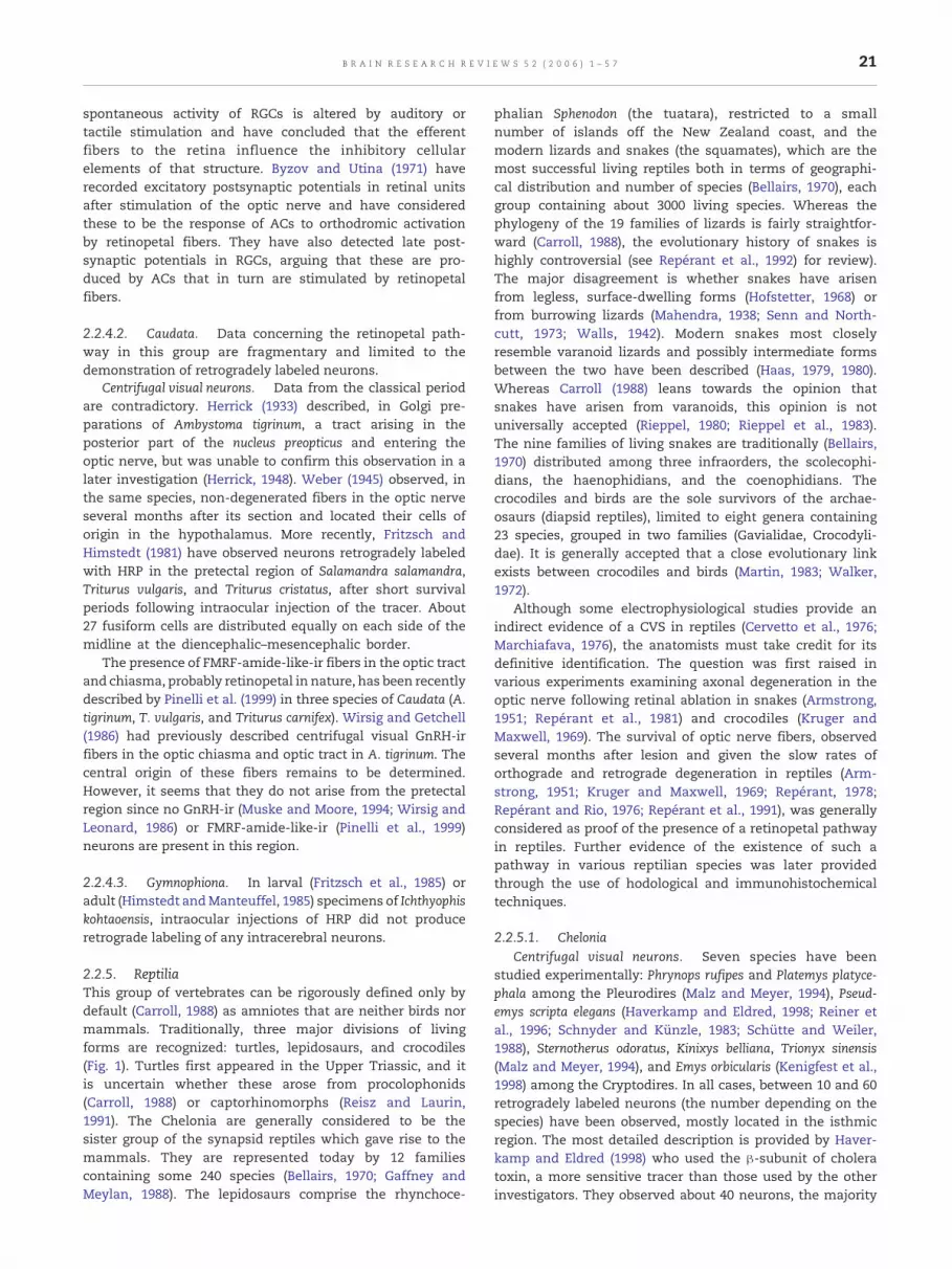
21B R A I N R E S E A R C H R E V I E W S 5 2 ( 2 0 0 6 ) 1 – 5 7
spontaneous activity of RGCs is altered by auditory ortactile stimulation and have concluded that the efferentfibers to the retina influence the inhibitory cellularelements of that structure. Byzov and Utina (1971) haverecorded excitatory postsynaptic potentials in retinal unitsafter stimulation of the optic nerve and have consideredthese to be the response of ACs to orthodromic activationby retinopetal fibers. They have also detected late post-synaptic potentials in RGCs, arguing that these are pro-duced by ACs that in turn are stimulated by retinopetalfibers.
2.2.4.2. Caudata. Data concerning the retinopetal path-way in this group are fragmentary and limited to thedemonstration of retrogradely labeled neurons.
Centrifugal visual neurons. Data from the classical periodare contradictory. Herrick (1933) described, in Golgi pre-parations of Ambystoma tigrinum, a tract arising in theposterior part of the nucleus preopticus and entering theoptic nerve, but was unable to confirm this observation in alater investigation (Herrick, 1948). Weber (1945) observed, inthe same species, non-degenerated fibers in the optic nerveseveral months after its section and located their cells oforigin in the hypothalamus. More recently, Fritzsch andHimstedt (1981) have observed neurons retrogradely labeledwith HRP in the pretectal region of Salamandra salamandra,Triturus vulgaris, and Triturus cristatus, after short survivalperiods following intraocular injection of the tracer. About27 fusiform cells are distributed equally on each side of themidline at the diencephalic–mesencephalic border.
The presence of FMRF-amide-like-ir fibers in the optic tractand chiasma, probably retinopetal in nature, has been recentlydescribed by Pinelli et al. (1999) in three species of Caudata (A.tigrinum, T. vulgaris, and Triturus carnifex). Wirsig and Getchell(1986) had previously described centrifugal visual GnRH-irfibers in the optic chiasma and optic tract in A. tigrinum. Thecentral origin of these fibers remains to be determined.However, it seems that they do not arise from the pretectalregion since no GnRH-ir (Muske and Moore, 1994; Wirsig andLeonard, 1986) or FMRF-amide-like-ir (Pinelli et al., 1999)neurons are present in this region.
2.2.4.3. Gymnophiona. In larval (Fritzsch et al., 1985) oradult (Himstedt andManteuffel, 1985) specimens of Ichthyophiskohtaoensis, intraocular injections of HRP did not produceretrograde labeling of any intracerebral neurons.
2.2.5. ReptiliaThis group of vertebrates can be rigorously defined only bydefault (Carroll, 1988) as amniotes that are neither birds normammals. Traditionally, three major divisions of livingforms are recognized: turtles, lepidosaurs, and crocodiles(Fig. 1). Turtles first appeared in the Upper Triassic, and itis uncertain whether these arose from procolophonids(Carroll, 1988) or captorhinomorphs (Reisz and Laurin,1991). The Chelonia are generally considered to be thesister group of the synapsid reptiles which gave rise to themammals. They are represented today by 12 familiescontaining some 240 species (Bellairs, 1970; Gaffney andMeylan, 1988). The lepidosaurs comprise the rhynchoce-
phalian Sphenodon (the tuatara), restricted to a smallnumber of islands off the New Zealand coast, and themodern lizards and snakes (the squamates), which are themost successful living reptiles both in terms of geographi-cal distribution and number of species (Bellairs, 1970), eachgroup containing about 3000 living species. Whereas thephylogeny of the 19 families of lizards is fairly straightfor-ward (Carroll, 1988), the evolutionary history of snakes ishighly controversial (see Repérant et al., 1992) for review).The major disagreement is whether snakes have arisenfrom legless, surface-dwelling forms (Hofstetter, 1968) orfrom burrowing lizards (Mahendra, 1938; Senn and North-cutt, 1973; Walls, 1942). Modern snakes most closelyresemble varanoid lizards and possibly intermediate formsbetween the two have been described (Haas, 1979, 1980).Whereas Carroll (1988) leans towards the opinion thatsnakes have arisen from varanoids, this opinion is notuniversally accepted (Rieppel, 1980; Rieppel et al., 1983).The nine families of living snakes are traditionally (Bellairs,1970) distributed among three infraorders, the scolecophi-dians, the haenophidians, and the coenophidians. Thecrocodiles and birds are the sole survivors of the archae-osaurs (diapsid reptiles), limited to eight genera containing23 species, grouped in two families (Gavialidae, Crocodyli-dae). It is generally accepted that a close evolutionary linkexists between crocodiles and birds (Martin, 1983; Walker,1972).
Although some electrophysiological studies provide anindirect evidence of a CVS in reptiles (Cervetto et al., 1976;Marchiafava, 1976), the anatomists must take credit for itsdefinitive identification. The question was first raised invarious experiments examining axonal degeneration in theoptic nerve following retinal ablation in snakes (Armstrong,1951; Repérant et al., 1981) and crocodiles (Kruger andMaxwell, 1969). The survival of optic nerve fibers, observedseveral months after lesion and given the slow rates oforthograde and retrograde degeneration in reptiles (Arm-strong, 1951; Kruger and Maxwell, 1969; Repérant, 1978;Repérant and Rio, 1976; Repérant et al., 1991), was generallyconsidered as proof of the presence of a retinopetal pathwayin reptiles. Further evidence of the existence of such apathway in various reptilian species was later providedthrough the use of hodological and immunohistochemicaltechniques.
2.2.5.1. CheloniaCentrifugal visual neurons. Seven species have been
studied experimentally: Phrynops rufipes and Platemys platyce-phala among the Pleurodires (Malz and Meyer, 1994), Pseud-emys scripta elegans (Haverkamp and Eldred, 1998; Reiner etal., 1996; Schnyder and Künzle, 1983; Schütte and Weiler,1988), Sternotherus odoratus, Kinixys belliana, Trionyx sinensis(Malz and Meyer, 1994), and Emys orbicularis (Kenigfest et al.,1998) among the Cryptodires. In all cases, between 10 and 60retrogradely labeled neurons (the number depending on thespecies) have been observed, mostly located in the isthmicregion. The most detailed description is provided by Haver-kamp and Eldred (1998) who used the β-subunit of choleratoxin, a more sensitive tracer than those used by the otherinvestigators. They observed about 40 neurons, the majority

22 B R A I N R E S E A R C H R E V I E W S 5 2 ( 2 0 0 6 ) 1 – 5 7
of which (36 or thereabouts) are contralateral to the injectedeye, distributed throughout a zone some 4 mm in length,primarily in and around the substantia nigra and in a morecaudal region between the locus coeruleus, lateral lemniscusand nucleus reticularis isthmi; to a lesser extent, labeledneurons have been observed in the nucleus isthmi parsparvocellularis, nucleus raphe superior, and nucleus reticularissuperior of the rostral hindbrain (Figs. 4C, D). These cells are
multipolar or bipolar, their somata ranging from 10 to 20 μmin diameter. A similar pattern of distribution has beendescribed in other species, with the exception of Kinixys(Malz and Meyer, 1994), in which labeled neurons areexclusively contralateral.
Immunocytochemical and histochemical investigationshave shown that, in Pseudemys, about 30% of the retrogradelylabeled neurons are either met-enkephalin-ir (Weiler, 1985) or

23B R A I N R E S E A R C H R E V I E W S 5 2 ( 2 0 0 6 ) 1 – 5 7
NADPH-positive and possibly ChAT-ir (Haverkamp andEldred, 1998). In addition, Schütte and Weiler (1988) haveobserved a single 5-HT-ir retrogradely labeled cell lyingbetween the locus coeruleus and the lateral lemniscus. Theafferent supply to the retinopetal neurons of turtles remainsto be identified.
Retinal innervation. Data are restricted to Pseudemys.Weiler (1985) described three to six met-enkephalin-ir fibersin the optic nerve that penetrate the retina to course towardsthe optic streak, making extensive collateral branches beforepenetrating the IPL, within which the terminal arborizationsextend over 100 μm in three to four sublayers of the plexiformlayer. Yaqub and Eldred (1991) described 4–10 stronglyaspartate-ir centrifugal visual fibers and Blute et al. (1997)about the same number of NADPH-positive, NOS-ir fibers thatmake similar arborizations within the IPL. The single 5-HT-ircentrifugal visual fiber described by Schütte andWeiler (1988),arising from a neuron in the contralateral ventral isthmicregion, emerges from the head of the optic nerve to makeextensive arborizations covering about one third of the retinalsurface in the temporal hemiretina. In all cases, these fibersare divergent. In sections of the retina, these have beenobserved to give rise to fine processes passing through the IPLto reach the border of the INL.
Functional considerations. Marchiafava (1976) observedlarge depolarizations of ACs after stimulation of the opticnerve, these having the properties of excitatory postsynapticpotentials. Yaqub and Eldred (1991) suggest that these aremediated by aspartate-containing efferent fibers, since pre-sumably enkephalin (Weiler, 1985) and serotonin (Schütteand Weiler, 1988) would have inhibitory effects on ACs.Cervetto et al. (1976) investigated the interaction between thevisual stimulation of RGCs and the stimulation of the opticnerve. They reported that when the light is paired with thestimulation of the retinopetal fibers, the response of thecentral region of the receptive field is augmented, and theyhave suggested that the centrifugal visual fibers may beimplicated in the detection of light stimuli of low intensity.Schütte and Weiler (1988) point out that the temporal retina,within which their serotonergic fiber ramifies, may beinvolved in binocular vision, and they suggest that this
Fig. 4 – Schematic representations of the distributions of centrifuspecies, labeled by retrograde transport of tracer. In all drawingsThe thalamus of Vipera aspis, modified from Repérant et al. (1980elegans, modified fromHaverkamp and Eldred (1998). (E, F) ThemiMédina et al. (2004a). The retinofugal visual fibers are indicated byNote that in the turtle and crocodile, the distributions of neuronsrelatively similar. Abbreviations: BTP, basal telencephalic pedunclongitudinalis medialis; GC, griseum centrale; GCT, griseum centGLvc, nucleus geniculatus lateralis ventralis pars cellularis; GLvmhabenula; ICO, nucleus intercollicularis; IP, nucleus interpeduncuisthmi pars parvocelularis; La, nucleus laminaris of the torus semmesencephalicus nervi trigemini; nIII, nucleus nervi oculomotornucleus suprapeduncularis; OT, optic tectum; OV, nucleus ovalis;raphes superior; Ris, nucleus reticularis superior; SCd, nucleus sventralis; SGC, stratumgriseum centrale; SGF, stratumgriseumetSN, substantia nigra; TOM, tractus opticusmarginalis; TS (or TSC,Scale bars: A, B:160 μm; C, D: 120 μm; E, F: 90 μm.
fiber may be involved, in the diving species Pseudemys, in achange from panoramic aerial vision to binocular focussingunderwater.
2.2.5.2. LepidosauromorphaRhynchocephalia. Our knowledge of the visual system of
Sphenodon is limited to a single abstract (Northcutt et al., 1974)based on an autoradiographic study which makes no mentionof retrogradely labeled centrifugal visual neurons.
LacertiliaCentrifugal visual neurons. Six species, representing four
families, have been studied experimentally by intraocularinjection of a variety of tracers: Chameleontidae, Chamaeleochameleon (El Hassni et al., 1997); Cordylidae, Cordylus cordylus(Halpern et al., 1976); Scincidae, Trachydosaurus rugosus, andMabuya peruteti (Malz and Meyer, 1994); Anguidae, Gerrhonotuscoeruleus (Halpern et al., 1976), andOphisaurus apodus (Kenigfestet al., 1986). Data are restricted to the demonstration ofretrogradely labeled neurons, and information concerningthe mode of termination of retinopetal axons, their neuro-chemical properties and afferent relations are unavailable. Inall species, with the exception of Cordylus, retrogradely labeledneurons are located in the dorsal isthmic region, the preciselocation varying between species. The projection of these cellsonto the retina is either bilateral, as in Trachydosaurus andMabuya (Malz and Meyer, 1994), or unilateral as in thechameleon (El Hassni et al., 1997). A second population ofretrogradely labeled neurons is situated in the ventral thala-mus of Chamaeleo (El Hassni et al., 1997); this population hasbeen also observed in Cordylus (Halpern et al., 1976).
OphidiaCentrifugal visual neurons. Data are available for two
haenophidian species belonging to the Boidae family (Pythonreticularis (Hoogland and Welker, 1981), Calabaria reinhardti[Repérant and Rio, unpublished observations] and fourcoenophidian species belonging to two families: the Colubri-dae (Thamnophis sirtalis, Thamnophis radix) (Halpern et al., 1976),and the Viperidae (Vipera aspis) (Repérant et al., 1980a,b, 1981)and Crotalus viridis (Schroeder, 1981). As in lizards, these arerestricted to the demonstration of retrogradely labeled neu-rons whose other properties remain to be elucidated.
gal visual neurons (indicated by large dots) in three reptilian, the right-hand side is contralateral to the injected eye. (A, B)b). (C, D) The midbrain and isthmic region of Pseudemys scriptadbrain and isthmic region of Crocodylus niloticus, modified fromdashed lines and their terminal arborizations by dotted lines.in the mesencephalic tegmentum and isthmic region arele; COTN, centrifugal optic thalamic nucleus; FLM, fasciculusralis tectalis; GLd, nucleus geniculatus lateralis pars dorsalis;, nucleus geniculatus lateralis ventralis pars molecularis; HB,laris; Ism, nucleus isthmi pars magnocellularis; ISp, nucleusicircularis; LoC; locus coeruleus; MV, nucleus
ius; nIV, nucleus nervi trochlearis; NR, nucleus raphes; NS,Prm (or nPm) nucleus profundus mesencephali; Ras, nucleusubcoeruleus pars dorsalis; SCv, nucleus subcoeruleus parsfibrosumsuperficiale; SGP, stratumgriseumperiventriculare;torus semicircularis; V, ventricle; VL, nucleus ventrolateralis.

24 B R A I N R E S E A R C H R E V I E W S 5 2 ( 2 0 0 6 ) 1 – 5 7
In Python and Calabaria, neurons labeled after injection ofHRP or fluorochromes have been observed in large number,scattered, bilaterally, widely throughout the basal telencepha-lon located rostral to the anterior commissure. Some cellshave been also observed in the lateral preoptic area [(Hooglandand Welker, 1981), Repérant and Rio, unpublished observa-tions]. In Calabaria, few retrogradely labeled neurons havebeen observed in the ventral thalamus, contralateral to the eyeinjection [Repérant and Rio, unpublished observations].
In coenophidian species (Figs. 4A, B), on the other hand,retrogradely labeled neurons are located in a ventro-lateralthalamic structure described either as the nucleus of theventral commissure (Halpern et al., 1976; Schroeder, 1981) orcentrifugal optic thalamic nucleus (Repérant et al., 1980a,b,1981). This nucleus is situated rostrally to the nucleus ovalis andextends caudally beneath the nucleus geniculatus lateralis parsventralis to the posterior thalamic boundary. In Crotalus(Schroeder, 1981), it may be divided into a rostral componentcontaining large neurons and a caudal subdivision containingsmaller neurons; in the other species, it is homogeneous.Projections are contralateral in Thamnophis (Halpern et al.,1976) and bilateral with a strong contralateral bias in Crotalus(Schroeder, 1981) and Vipera (Repérant et al., 1980a,b, 1981). Inthe latter species, about 660 bipolar retinopetal neurons exist,whose somata are between 15 and 22 μm in diameter; theaxons of these cells account for about 1% of the total numberof optic nerve fibers (Ward et al., 1989). This nucleus receivesan afferent supply from the optic tectum (Wang and Halpern,1977). Finally, it is worth noting that a few retrogradely labeledneurons have been observed in the preoptic area of the viperfollowing intraocular injection of a tracer [Repérant and Rio,unpublished observations].
2.2.5.3. CrocodiliaCentrifugal visual neurons. In the optic nerve of Alligator
mississippiensis and Caiman crocodilus (Crocodylidae), 20months following enucleation, Kruger and Maxwell (1969)found about 4000 myelinated fibers of relatively normalappearance, representing about 5% of the total population ofmyelinated axons. Considering the slow process of axonaldegeneration in those species, they concluded that thesefibers are probably retinopetal. This hypothesis has been laterconfirmed by using different retrograde axonal tracers fromthe retina.
Ferguson et al. (1978) described, in C. crocodilus, thepresence of neurons retrogradely labeled by HRP in the isthmicregion. These neurons are bilaterally distributed, but with acontralateral bias, in a single large oblong cell field borderedmedially by the trochlear nucleus and laterally by the isthmicnucleus. These authors also showed that this isthmic field,containing the retinopetal neurons, receives a strong projec-tion from the optic tectum. As in birds (see Chapter 2.2.6.3), thecentrifugal visual neurons could thus be implicated in aretino→tecto→isthmo→retinal feedback loop.
A comparable study carried out in Crocodylus niloticus(Crocodylidae) using fluorescent tracers has provided slightlydifferent results (Médina et al., 2004a). In the latter species,about 6000 bilateral retrogradely labeled neurons (70% on thecontralateral side) are distributed in seven regions, extendingfrom the mesencephalic tegmentum to the isthmus and
anterior rhombencephalon (formatio reticularis lateralis mesen-cephali, substantia nigra, griseum centralis tectalis, nucleus sub-coeruleus dorsalis, nucleus isthmi parvocellularis, nucleus locuscoeruleus, and nucleus commissura nervi trochlearis, Figs. 4E, F).None of the centrifugal visual neurons project to both retinae.Most retinopetal neurons are multipolar or bipolar in shapealthough a small number of monopolar neurons are present,resembling those of the avian NIO. Médina et al. (2004a) haveshown that about 35–38% of the retinopetal neurons areimmunoreactive respectively to ChAT and NOS, some of themcolocalizing these substances. A few retinopetal neuronsimmunoreactive to TH were also observed. fibers immunor-eactive to TH, 5-HT, NPY, and FMRF-amide-like were fre-quently observed to make intimate contacts with theretinopetal neurons. The location of these centrifugal visualneurons and their apparent relationswith the optic tectumaresimilar to those of the avian CVS (see below), findings inagreement with paleontological evidence (Martin, 1983; Mar-tin et al., 1980; Walker, 1972) for a close evolutionary relation-ship between crocodiles and birds. In Crocodylus, FMRF-amide-like-ir (Médina et al., 2004b) and GnRH-ir fibers (Médina et al.,2005), probably centrifugal, have been observed in the opticchiasma and optic nerve. It has been shown by immunohis-tochemistry that these fibers do not arise from the retinopetalneurons located in the different regions mentioned above, butprobably originate in structures localized in the TNSP complex(Médina et al., 2004b, 2005).
2.2.6. AvesThe debate whether birds arose in the Mesozoic fromthecodont (Heilmann, 1926) or from theropods (Carroll,1988; Padian, 1998) dinosaurs is likely to be resolved inlight of newly described feathered dinosaurs (Padian, 1998).This major vertebrate radiation is represented today bysome 8900 species regrouped in 27 orders containing 166families (Fig. 1).
Centrifugal visual fibers in the avian retina were describedby Cajal (1888, 1889, 1893, 1911) and Dogiel (1895), and severalinvestigators of the classical period were able to show, bymeans of retrograde degeneration, that these arose in the NIO(Cowan et al., 1961; Huber and Crosby, 1929; Jelgersma, 1896;Kosaka and Hiraiwa, 1915; Perlia, 1889). Orthograde degenera-tion, following lesions of the NIO, has confirmed thesefindings (Cowan and Powell, 1963; Galifret et al., 1971;Wallenberg, 1898). McGill (1964) and McGill et al. (1966a,b)showedwith degeneration techniques that the NIO forms partof a feedback loop: retina→optic tectum→NIO→retina. Sincethe introduction of hodological techniques based on axonaltransport and immunohistochemical methods, numerousinvestigations of the avian CVS have been carried out, forthe most part in three species: Columba livia (Columbiformes),Gallus domesticus, and Coturnix coturnix (Galliformes).
2.2.6.1. Centrifugal visual neuronsNucleus isthmo-opticus (NIO). The NIO (Ariëns-Kappers et
al., 1936; Huber and Crosby, 1929), also described as theganglion opticum dorsale (Perlia, 1889), medial optic nucleus(Wallenberg, 1898), ganglion isthmi (Edinger and Wallenberg,1899), or nucleus tractus isthmo-optici (Craigie, 1928), has beendescribed inmany avian species (see Repérant et al., 1989b) for

25B R A I N R E S E A R C H R E V I E W S 5 2 ( 2 0 0 6 ) 1 – 5 7
review) with the exception of the kiwi (Craigie, 1930), ibis(Showers and Lyons, 1968), and ostrich (Verhaart, 1971).
This nucleus, which mostly contains retinopetal neurons,is located in the dorsal isthmus, medial to the caudo-dorso-medial edge of the optic tectum, at the level of the trochlearnucleus. Its size, and the number of neurons it contains, variesfrom species to species. In Galliformes (G. domesticus), Colum-biformes (C. livia), and Passeriformes (Passer domesticus)(Repérant et al., 1989b), it is a well-developed laminarstructure containing 7000–12,000 neurons. In Anseriformes,which have a well-developed trigeminal system and generallysearch for food by poking in the mud, the nucleus is clearlyless well differentiated and contains about 3000 neurons(Repérant et al., 1989b; Sohal, 1976). In raptors (Hirschberger,1971; Repérant et al., 1989b; Shortess and Klose, 1975; Weidneret al., 1987) and in birds that ‘feed-on-the-wing’ such as theswallow (Feyerabend et al., 1994), it is even smaller and poorlydifferentiated, containing between 900 and 1400 neuronsdepending on the species.
The intraocular injection of tracer shows that the neuronsof the NIO project virtually exclusively to the contralateralretina, although in the chicken (O'Leary and Cowan, 1982) andTyto alba (Weidner et al., 1987), a very small number of neuronsproject to the ipsilateral retina. Double labeling studies(O'Leary and Cowan, 1982; Weidner et al., 1987) have shownthat bilateral projections to the retina do not exist in adults,although they may be observed in embryonic specimens(O'Leary and Cowan, 1982). Investigations of the NIO incongenitally microphthalmic chickens have produced contra-dictory results. Malz et al. (1989) have found no differencesbetween microphthalmic and normal chickens, whereasStuchbery and Ehrlich (1987) have found a greater number ofipsilaterally projecting neurons in microphthalmic speci-mens. Weidner et al. (1989), in comparing albino and normallypigmented specimens of Coturnix, found that the total numberof centrifugal visual neurons is reduced by about 40% inalbinos.
The most extensive studies of the organization of the NIOhave been carried out in pigeons and chickens. In thesespecies, it appears as a convoluted laminar structure formedby two layers of centrifugal visual cells separated by a zone ofneuropil. In the pigeon, the nucleus is about 0.7 mm longrostro-caudally, about 1 mm wide medio-laterally, and about0.7 mm deep dorso-ventrally (Wolf-Oberhollenzer, 1987). Thelaminae of neurons contain between 8000 and 11,000 cellbodies (Cowan, 1970; Hayes and Webster, 1981; O'Leary andCowan, 1982; Wolf-Oberhollenzer, 1987). Initial cytologicalinvestigations (Angaut and Repérant, 1978; Clarke and Car-anzano, 1985; Cowan, 1970; Crossland, 1979; Güntürkün, 1987;Miceli et al., 1995, 1997) have supported the notion that theretinopetal neurons form an homogeneous population offlask-shaped cells, about 15 μm in diameter, from the apicalpoles of which one to four primary dendrites arise to divideinto parallel branches directed towards the neuropil. Morerecently, using an intracellular injection of Lucifer Yellow inbrain slices maintained in vitro, Li andWang (1999) have beenable to distinguish two major categories, bipolar and multi-polar neurons each category in turn being divided into twosubgroups. The bipolar neurons, accounting for 83% of theneurons of the NIO, may be divided into B and P subtypes. The
B neurons (36%) have piriform perikarya, and long apicaldendrites that form a slender column, their axons emergingfrom the opposite pole and bearing some varicosities. The Pneurons (47%) also have piriform cell bodies, but shorterdendrites that bear some spines; the axons of P cells eitherarise from the basal pole or from one of the apical dendritesclose to the perikaryon. The multipolar cells can similarly besubdivided into M (13%) and N (4%) subtypes. The M neuronshave round or elongated perikarya, from which two to fourprimary dendrites arise, whereas the N neurons have poly-gonal somata bearing three to five primary dendritic trunks.These neurons lack axons and probably correspond to theinterneurons described by Miceli et al. (1995). Li and Wang(1999) have also been able to show that dye couplingconnections exist in about 30% of the neurons of the NIO.
Under the electron microscope, the retinopetal neuronsdisplay somata containing an eccentric nucleus with aprominent nucleolus. The cytoplasm contains well-developedGolgi cisterns and Nissl bodies evenly distributed within it(Angaut and Repérant, 1978; Crossland, 1979). The cellmembrane is smooth and occasionally shows spinous appen-dages associated with attachment plaques between theapposed membranes of neighboring cells (Angaut and Repér-ant, 1978). These attachment plaques may produce a syn-chronous electrical activation among adjacent centrifugalvisual neurons (Hu et al., 2000). In the chicken (Crossland,1979), most of the cell perimeter remains free of contacts,whereas in the pigeon (Angaut and Repérant, 1978), about 50%of the cell membrane is apposed against boutons containingeither round or pleomorphic synaptic vesicles.
Indirect evidence (Angaut and Repérant, 1978; Cowan andWenger, 1968; Cowan et al., 1961; Crossland, 1979; Holden,1968; Raffin and Repérant, 1975) has strongly suggested thatnot all neurons of the NIO project to the retina, andMiceli et al.(1995) have shown by immunofluorescence double labelingthat, in the pigeon, the nucleus contains about 200 interneur-ons. These strongly GABA-ir cells are never labeled afterintraocular injection of tracer. They are widely distributedwithin the neuropilar zone. They show all the cytologicalcharacteristics of interneurons: small (8–12 μm) roundedsomata, containing a large nucleus surrounded by a thinlayer of cytoplasm poor in organelles, bearing dendritic shaftscontaining synaptic vesicles. The dendritic processes of theseneurons, extending for distances up to 100 μm, never leave thenucleus proper.
The neuropil of the NIO is composed predominantly ofdendrites of varying caliber. The largest of these are stemdendrites abundantly endowed with Nissl bodies; spines areparticularly frequent in the smaller dendritic profiles. Bundlesof myelinated and unmyelinated fibers are scattered through-out the neuropil, and thin glial processes are numerous(Angaut andRepérant, 1978; Crossland, 1979;Miceli et al., 1995).
The profiles containing synaptic vesicles have beenextensively analyzed in both chickens (Crossland, 1979) andpigeons (Angaut and Repérant, 1978; Miceli et al., 1995). Thelatter authors have been able to distinguish eight classes ofaxon terminals on the basis of several criteria: type of synapticcontact (symmetrical or asymmetrical), shape, size, anddensity of synaptic vesicles, and positive or negative GABAimmunoreactivity. These findings suggest strongly that the

26 B R A I N R E S E A R C H R E V I E W S 5 2 ( 2 0 0 6 ) 1 – 5 7
retinopetal neurons and interneurons of the NIO receivemultiple afferent inputs.
The development of the NIO has been studied in the duck(Sohal, 1976; Sohal and Narayanan, 1974, 1975), pigeon(Bagnoli et al., 1987, 1992), andmore extensively in the chicken(Angaut and Raffin, 1981; Blaser et al., 1990; Catsicas andClarke, 1987; Catsicas et al., 1987a; Clarke, 1982, 1985, 1992;Clarke and Caranzano, 1985; Clarke and Cowan, 1976; Clarkeand Egloff, 1988; Clarke and Kraftsick, 1996; Clarke et al., 1976;Cowan and Clarke, 1976; Cowan and Wenger, 1968; Garner etal., 1996; O'Leary and Cowan, 1982; Pequignot and Clarke, 1991,1992a,b; Posada and Clarke, 1999a,b; Primi and Clarke, 1996,1997; Thanos and Dütting, 1988; Vaage, 1973; von Bartheld etal., 1991, 1994, 1996; Wizenmann and Thanos, 1990).
In the chicken, the cells of the NIO are generated betweenembryonic days 5 and 7 (E5-E7) in the ventricular zone of thecaudal isthmic alar plate. These cells migrate ventro-laterallyover some 200 μmbefore aggregating to form the anlage of thenucleus (Clarke, 1982; Clarke et al., 1976; Cowan and Wenger,1968; Vaage, 1973). The first neurons to differentiate arelocated in the ventro-lateral part of the nucleus, whereas thelast are situated in its dorso-medial aspect (Clarke, 1982). Thefirst retrogradely labeled neurons may be demonstrated atembryonic day 9 (E9), whereas by E12, all the neurons of theNIO are labeled. At E11, Cowan and Wenger (1968) observedthat the NIO contains its maximumnumber of neurons (about22,000). At this stage, the nucleus is already laminated, withpolarized neurons extending their dendrites medio-rostro-ventrally (Clarke and Caranzano, 1985; Clarke and Kraftsick,1996). Between E11 and E13, the peripheral dendrites changetheir orientation towards the center of the NIO, whereas thosein the center orient themselves ventro-medially (Blaser et al.,1990; Clarke and Caranzano, 1985). Dendritic changes continueuntil E18 (Blaser et al., 1990), and synaptogenesis andmaturation of the neuropil continue for some days afterhatching (Angaut and Raffin, 1981).
Cell death is a particular feature of the development of theNIO, in contrast to the retinopetal cells in the lamprey (M5, seeDevelopment of retinopetal neurons. Morphology and neuro-chemical aspects). In the duck (Sohal and Narayanan, 1974),about 45% of the neurons of the NIO degenerate between E16and E21, to stabilize at about 3600 cells, whereas in thechicken, the number of cells declines from its maximum of22,000 (Cowan and Wenger, 1968) at E11 to about 9500 at themoment of hatching. In embryonic specimens, the number ofipsilaterally projecting centrifugal visual neurons is greaterthan in hatchlings; between E10 and E13, O'Leary and Cowan(1982) observed about 60 such cells, this number declining toabout 10 at the moment of hatching. The same authors alsodescribed a transient bilateral retinal projection in embryonicspecimens. Wizenmann and Thanos (1990) also demon-strated, in chicken embryos between E9 and E16, transientprojections from the NIO to the optic tectum.
The number of surviving cells can be experimentallymanipulated by removing the efferent target (Catsicas andClarke, 1987; O'Leary and Cowan, 1984; Thanos and Dütting,1988) or afferent neurons (Clarke, 1985). It has been proposedthat the cell death plays an important role in refining aninitially imprecise projection of the NIO onto the retina,implying that misrouted centrifugal visual axons are elimi-
nated (Catsicas et al., 1987a; Cowan and Clarke, 1976). Thisprocess would be linked to a fast-acting death retrogradesignal initiated by calcium entry into the isthmo-opticterminals due to electrical activity (Posada and Clarke, 1999a;Primi and Clarke, 1997). Nitric oxide (NO) would play animportant role in the elicitation of a fast retrograde signal thatinfluences both the cell death and dendritic reorganization inthe NIO (Posada and Clarke, 1999b). Other studies have shownthat the survival of NIO neurons is mainly dependent on theretrograde transport (slow-acting survival signal) of a varietyof trophic substances such as Nerve Growth Factor, BrainDerived Nerve Factor, or Neurotropin 3 (Garner et al., 1996;Primi and Clarke, 1996; von Bartheld et al., 1991, 1994, 1996).Recently, in the chick embryo, Thanos (1999) has observed arobust retino-retinal projection. This appears at E6, reaches itsmaximal development at E13–E14, and gradually disappearsby E18. This author has suggested that this projection mayserve as a ‘template’ to guide centrifugal isthmo-optic axonsinto the retina.
Ectopic centrifugal visual neurons (ECNs). The existence ofretinopetal neurons outside the NIOwas first demonstrated bythe retrograde transport of HRP in chickens by Clarke andCowan (1975), who named them ectopic centrifugal neurons.These neurons have subsequently been demonstrated inhatchlings and adults of G. domesticus (Clarke and Cowan,1976; Cowan and Clarke, 1976; O'Leary and Cowan, 1982, 1984)and in other species: C. livia (Bagnoli et al., 1992; Challet et al.,1996; Hayes andWebster, 1981; Li andWang, 1999; Miceli et al.,1993, 1995, 1997; Wolf-Oberhollenzer, 1987), Coturnix japonica(Médina et al., 1998; Weidner et al., 1989) and T. alba (Weidneret al., 1987).
In adult birds, the ECNs are diffusely distributed through-out a large territory. In the pigeon, this extends rostro-caudallythrough a distance of about 5 mm [Repérant et al., unpub-lished observations]. The most caudally situated ECNs, whichare widely dispersed, lie in a region of the antero-dorsalrhombencephalon lying against the locus coeruleus. Those inthe isthmo-tegmental field around the NIO are the mostnumerous; the most rostral ECNs, few in number, extendcaudo-rostrally over 2–3 mm beyond the NIO into themesencephalic substantia grisea centralis [(Hayes and Webster,1981), Repérant and Rio, unpublished observations]. The ECNsform an heterogeneous population varying in size from 10 to20 μm, occasional larger cells (25 μm) being observed. Most aremultipolar with three or more primary dendrites that give riseto long secondary and tertiary branches, whereas some arebipolar and spindle shaped, displaying two primary dendrites(Hayes and Webster, 1981; O'Leary and Cowan, 1982; Weidneret al., 1987, 1989; Wolf-Oberhollenzer, 1987). Their axonsgenerally arise from the cell body, but occasionally from oneof the primary dendrites, and appear to follow the samecentrifugal course as those arising in the NIO (O'Leary andCowan, 1982). Ectopic neurons are fewer in number than theneurons of the NIO; the ratio of ECN/NIO neurons varies from15% in Gallus (O'Leary and Cowan, 1982), through 20–24% inCoturnix (Weidner et al., 1989) and Columba (Hayes andWebster, 1981; O'Leary and Cowan, 1982) to 29% in Tyto(Weidner et al., 1987). Projections are predominantly to thecontralateral retina, but ipsilateral projections are commonerthan those of theNIO neurons (2% inGallus, 6% in Coturnix, 13%

27B R A I N R E S E A R C H R E V I E W S 5 2 ( 2 0 0 6 ) 1 – 5 7
in Columba, and 27% in Tyto (Hayes andWebster, 1981; O'Learyand Cowan, 1982; Weidner et al., 1987, 1989). Double labelingstudies in chicken embryos have revealed an extensivecollateralization of the ECN axons.
In the chicken, the axons of ectopic neurons penetrate theretina at E9. By E11, about 3500 retrogradely labeled ECNs canbe demonstrated, of which about 140 project either ipsilat-erally or bilaterally; this number drops to about 1500 neuronsby E17, of which 30 or so project ipsilaterally (O'Leary andCowan, 1982). In adults, this bilateral projection to one eye isno longer present (Weidner et al., 1987, 1989; Wolf-Oberhol-lenzer, 1987). In contrast to the NIO neurons, the ECNs arenever affected by an intraocular injection of kainate at anyage (Catsicas and Clarke, 1987; Clarke and Kraftsick, 1996;Fritzsch et al., 1990a).
The question whether the ECN and NIO neurons have acommon embryological origin remains undecided. Severalauthors (Clarke and Cowan, 1976; Cowan and Clarke, 1976;O'Leary and Cowan, 1982) have suggested that the ECNs aremisdirected during theirmigration from the isthmic alar plate,and since the axons of both ECN and NIO neurons penetratethe retina at the same time, both share a common origin.Together with Angaut and Raffin (1981), they ascribe themorphological differences between neurons of the NIO andECN to subtle differences in the morphogenetic influences ofthe two environments. However, in the absence of anyevidence that both types of centrifugal visual neurons usethe same neurotransmitters and share a common afferentsupply and retinal target, the alternative possibility ofseparate embryological origins of the two types cannot beruled out. Furthermore, it appears that the RGCs influenced byECN and NIO neurons have different central projections(Nickla et al., 1994), a finding that tends to support thisalternative (see also Chapter 2.2.6.3.).
Neurochemical properties. Immunocytochemical studies oftheNIOandECNneuronshave givenwidely discordant results.
Serotonin (5-HT). Yamada et al. (1984) described 5-HT-irneurons in the NIO of the chicken, observations that werenot confirmed by Cozzi et al. (1991) in the quail. Doublelabeling studies, involving the retrograde labeling of retino-petal neurons by RITC combined with 5-HT immunohisto-chemistry in the quail (Médina et al., 1998) and pigeon(Challet et al., 1996), have not revealed serotonin-containingcentrifugal visual neurons in the NIO. Yamada et al. (1984)also described 5-HT-ir neurons in the region containingECNs. However, double labeling techniques (Challet et al.,1996; Médina et al., 1998) have shown that the ECNs areconsistently 5-HT-immunonegative.
Tyrosine hydroxylase (TH). In both the pigeon (Reiner et al.,1994) and quail (Médina et al., 1998), no TH-ir neurons havebeen demonstrated in the NIO, although some labeled cellshave been observed in the region containing the ECNs.However, double labeling (Médina et al., 1998) has shownthat no ECNs are TH-ir.
GABA. In the pigeon, GABA-ir neurons have beendescribed in the NIO (Miceli et al., 1995; Veenman and Reiner,1994) and among the ECNs (Miceli et al., 1995). However, theretinopetal neurons both in the NIO and ECN, retrogradelylabeled with RITC, are consistently GABA-immunonegative(Miceli et al., 1995, 1999).
Glutamate. In an electron microscopic immunocyto-chemical study, Rio (1996) has described an intense glutamateimmunoreactivity in many NIO neurons of the pigeon and aweaker glutamate immunoreactivity in GABA-ir interneurons.These findings are difficult to interpret in the absence of dataconcerning glutamate immunoreactivity in the centrifugalvisual terminals and more particularly over their synapticvesicles. Glutamate is present in most if not all cellularcompartments and is involved in a number of metabolicpathways, and several authors have suggested that theglutamate immunoreactivity reveals both the ‘metabolic’and ‘neurotransmitter’ pools of this substance (see Miceli etal., 2000; Repérant et al., 1997 for review).
Choline acetyltransferase (ChAT). The use, by severalinvestigators, of the same antibody directed against theenzyme of synthesis of acetylcholine and raised in chickensby JohnstonandEpstein (1986)hasprovideddiscordant results.No ChAT-ir NIO neurons have been observed in the chicken(Sorenson et al., 1989) or quail (Médina et al., 1998), althoughnumerous cells in the vicinity of the ECNs are ChAT-ir. In thepigeon, Bagnoli et al. (1992) described weakly immunolabeledcells in the medial pole of the NIO, as well as numerousmultipolar ChAT-ir cells outside the boundaries of the nucleus.Similar findings were reported by Miceli et al. (1999). Medinaand Reiner (1994) also reported ChAT-ir retinopetal neurons inthe pigeon but, in some specimens, the immunolabeling wasextremely weak, a phenomenon that these authors ascribe topoor fixation. They also described strongly ChAT-ir cellssurrounding the NIO that they interpreted as ECN neuronsfromwhich arise the ChAT-ir fibers observed in the optic tract.Double labeling studies in the quail (Médina et al., 1998) andpigeon (Miceli et al., 1999) have provided discordant results. Noretrogradely labeled NIO or ECNs are ChAT-ir in the quail,whereas many NIO and ECNs neurons are ChAT-ir in thepigeon. Nickla et al. (1994) have shown that, in the chicken, theantibody against the subunit α7 of the nicotinic acetylcholinereceptor labels the neurons of the NIO but not the ECNs.
Aside from the methodological difficulties associated withfixation (Medina and Reiner, 1994), a second possible explana-tion of these discordant findings may be that the retinopetalneurons of the pigeon are not truly cholinergic but contain asubstance that cross-reacts with the anti-chicken ChAT(Miceli et al., 1999).
Nitric oxide synthase (NOS) and NADPH-diaphorase. Noneurons labeled either by NOS immunohistochemistry orNADPH-d histochemistry were observed in the quail NIO byMédina et al. (1998), in contrast to the finding by Panzica et al.(1994) of rare and weakly stained NADPH-d-positive neuronsin the same species. In other species (chickens (Brüning, 1993;Xiao et al., 2000), budgerigars (Cozzi et al., 1997), and pigeons(Miceli et al., 1999)), numerous NOS-ir/NADPH-d stainedneurons have been described in the NIO. Several authorshave described an NADPH-d activity in the centrifugal visualterminals and their retinal targets (de Carvalho et al., 1996;Fischer and Stell, 1999; Morgan et al., 1994). The repeatedobservation of NOS-ir/NADPH-d activity in the ectopic area(Cozzi et al., 1997; de Carvalho et al., 1996; Médina et al., 1998;Miceli et al., 1999; Panzica et al., 1994) is offset by the findingthat, in the quail (Médina et al., 1998) and chick (Gardino et al.,2004), no ECNs retrogradely labeled with RITC are NOS-ir. On

28 B R A I N R E S E A R C H R E V I E W S 5 2 ( 2 0 0 6 ) 1 – 5 7
the other hand,Miceli et al. (1999) argue that the high degree ofoverlap of retrogradely labeled ECNs and NADPH-d positivecells may be an indirect evidence for the presence of NO in theECNs and on similar grounds suggest that NO and acetylcho-line may be colocalized in these cells.
Neuropeptides. Médina et al. (1998) carried out an immu-nohistochemical investigation of fifteen neuropeptides (angio-tensin, bradykinin, cholecystokinin, dynorphin, leu- and met-enkephalin, galanin, β-endorphin, galanin, α-neoendorphin,neurokinin, neuropeptideY, oxytocin, somatostatin, substanceP, vasopressin, and vasoactive intestinal polypeptide) in thequail. No immunoreactivity against any of these substanceswas observed in theNIO, althoughcells in theectopic regionareimmunoreactive to neuropeptide Y, dynorphin, met-enkepha-lin, galanin, and neurokinin. Double labeling with RITC,however, showed that none of these immunoreactive cellsproject to the retina.Woodson et al. (1989) in the pigeon and deLanerolle et al. (1981) in the chicken have also shown that theNIO neurons are immunonegative to numerous peptides(Substance P, enkephalin, oxytocin, NPY, CCK octapeptide).
2.2.6.2. Retinal innervationCourse of centrifugal visual fibers. The axons of both ECNs
and NIO neurons regroup to form the isthmo-optic tract(IOT) (Cowan and Powell, 1963; Galifret et al., 1971), alsodescribed as the medial or axillary optic tract (Repérant etal., 1989b). The course of this tract has been studied in detailin the pigeon (Cowan and Powell, 1963; Galifret et al., 1971)and chicken (Crossland and Hughes, 1978; Wallenberg, 1898)by degeneration methods after lesion of the NIO or IOT andby autoradiography (Crossland and Hughes, 1978). Theretinopetal fibers aggregate dorsal to the NIO to form acompact bundle with an oval cross-section. The IOT runscaudally and medially in the SGFS, progresses mediallyalong the optic tract, and penetrates the marginal optic tractrostral to the chiasma. Within the chiasma, the decussationof retinopetal fibers is observed in every interdigitationbefore finally entering the contralateral optic nerve (Galifretet al., 1971).
Extraretinal projections of the NIO to thalamic andpretectal structures have been described repeatedly in degen-eration preparations of chickens and pigeons (Cowan andPowell, 1963; Galifret et al., 1971; Wallenberg, 1898), but thesefindings have not been confirmed by autoradiographic studiesin the pigeon (Crossland and Hughes, 1978). An ultrastructuralstudy in the pigeon (Cowan, 1970) has shown that almost allretinopetal fibers are myelinated and vary from 0.5 μm to 2.5μm in diameter. In both pigeons and chickens, these fibersaccount for about 0.4% of the optic nerve fibers, this propor-tion being smaller by a factor of 10 in the raptor Buteo buteo(Repérant et al., 1989b).
Distribution of centrifugal visual fibers in the retina. Inves-tigations of the spatial distribution of centrifugal visualterminals within the retina have produced somewhat incon-sistent results. Cowan and Powell (1963) observed that, afterlesion of the NIO in pigeons, the degenerating retinopetalarborizations are mainly concentrated in the temporal retina,whereas McGill et al. (1966b) concluded, on the basis ofretrograde degeneration after ablation of different retinalregions in the same species, that the NIO projects to the
entire retina. Maturana and Frenk (1965), using the methyleneblue technique, also concluded that about 100,000 terminalarborizations are distributed over the entire retina.
More recent studies, using various methods based on theaxonal transport of tracers, are similarly somewhat incon-sistent. After injection of a radioactive tracer into the NIO,Crossland and Hughes (1978) reported that, in the chicken, theretinopetal terminals are denser near the central portion ofthe nasal half of the retina, less concentrated in the temporalhemiretina, and absent from the peripheral retina. Somewhatdifferent results were obtained by Catsicas et al. (1987a) in thechicken. After deposit of a retrograde tracer into discrete areasof the retina, these authors showed that the neurons of theNIO project only to the ventral two-thirds of the retina, withthe exception of the extreme periphery, and that thisprojection is topographically organized; the medial NIOprojects temporally, and the ventral NIO projects to the dorsalpart of the projection field. In their preparations, Catsicas etal. (1987a) also found that the ECNs project more ventrallythan the NIO neurons, that their projection fields are larger,and that the topographical organization of the projections isless precise than that of the NIO neurons. Medially locatedECNs tend to project temporally, whereas the dorsally,ventrally, and laterally located neurons tend to projectnasally.
In pigeons, Hayes and Holden (1983) observed that theretinopetal terminals, retrogradely labeled by HRP injected inlarge quantities either into the chiasma or isthmic region, aremainly restricted to an horizontal band of the retina, 3.5 mmwide and 12 mm long. Two regions of high density ofterminals have been found, one in the temporal yellow field6.5 mm from the area centralis (610 arborizations/mm2), theother in the central yellow field just above the area centralis(570 arborizations/mm2). The terminals are sparsely distrib-uted in the ventral retina and entirely absent from the dorsalthird of the retina including the red field. Comparable resultshave been presented by Woodson et al. (1995) in the samespecies; these authors have also shown that the horizontalstreak is the main terminal area of NIO neurons, whereas theinferior retina is that of ECNs. Hayes and Holden (1983)estimate that about 7000 retinopetal terminals exist in thepigeon retina, a value considerably lower than that presentedby Maturana and Frenk (1965).
Morphological and neurochemical properties of the centrifugalvisual terminals and their postsynaptic targets. Two majortypes of centrifugal visual terminals have been recognized inthe retina of different avian species, described on the onehand as restricted or convergent, and on the other as wide-spread or divergent.
Convergent terminals were described over a century agoby Cajal (1888, 1889, 1893, 1911) and Dogiel (1895) in Golgiand intravital methylene blue preparations and have beenrepeatedly demonstrated by later authors using for the mostpart hodological or immunohistochemical methods (Fischerand Stell, 1999; Fritzsch et al., 1990a; Hayes and Holden,1983; Maturana and Frenk, 1965; Nickla et al., 1994;Uchiyama and Ito, 1993; Uchiyama et al., 1995, 2004;Woodson et al., 1995).
The restricted centrifugal visual terminals arise from largemyelinated fibers (N0.5 μm) that originate exclusively in the

29B R A I N R E S E A R C H R E V I E W S 5 2 ( 2 0 0 6 ) 1 – 5 7
NIO (Fritzsch et al., 1990a; Woodson et al., 1995). These fibersrun radially from the head of the optic nerve in the optic fiberlayer, subsequently passing vertically through the ganglioncell layer and IPL to arborize in the inner third of the INL. In thechicken and pigeon, each arborization is composed of severalbranches of different lengths and thicknesses that convergeon a single target cell as a ‘synaptic nest’. Electronmicroscopicstudies (Dowling and Cowan, 1966; Hayes, 1982) have shownthat the restricted centrifugal visual terminals are larger thanmost other terminals in the IPL, being frequently over 8 μm indiameter. They contain rounded synaptic vesicles, 30–50 nmin diameter, and make synaptic contacts primarily ondendritic profiles, more rarely on somata. An alternativeconfiguration of restricted terminals may be observed as asingle (Uchiyama and Ito, 1993; Uchiyama et al., 1995), largesynaptic terminal invaginated within the basal region of thetarget perikaryon in a complex manner, so that the area ofmembrane apposition between the terminal and its target islarge with many synaptic contacts.
Some controversy exists about the nature of the post-synaptic targets of the restricted centrifugal visual terminals.Several authors (Hayes andHolden, 1983;Maturana and Frenk,1965; Woodson et al., 1995) have suggested that theseterminals make synaptic contacts with displaced RGCs andmore rarely on orthotopic RGCs (Pearlman and Hughes, 1976)or ACs (Maturana and Frenk, 1965). Others (Fischer and Stell,1999; Fritzsch et al., 1990a; Nickla et al., 1994; Uchiyama andIto, 1993; Uchiyama et al., 1995, 2004), on the other hand, agreewith Cajal (1893, 1911) that most if not all restricted terminalsmake synaptic contacts with a particular category of ACsnamed as ‘associative’ (AACs) by Cajal (1911) or type II‘proprioretinal cells’ by Catsicas et al. (1987b). These cells liein the innermost part of the INL of the ventral half of theretina. They have short dendrites and an axon. These axonsproject onto the outermost zone of the IPL in both dorsal(Catsicas et al., 1987b) and ventral halves of the retina(Uchiyama et al., 2004) in which they could establish synapticcontacts onto dendrites of ordinary ACs (Clarke et al., 1996).Catsicas et al. (1987b), confirming the observations of Cajal(1911), have also observed that, in the ventral retina, the NIOneurons send minor axon branches onto ordinary ACs.
The widespread or divergent centrifugal visual terminalswere first described by Dogiel (1895) in methylene bluepreparations, subsequently by Chmielevski et al. (1988) insilver-stained retinal whole mounts, and their existence hasrecently been confirmed by several investigators using ante-rograde tracing techniques (Fritzsch et al., 1990a; Nickla et al.,1994; Woodson et al., 1995). Each terminal forms an extensivetangential arbor in the IPL, arising from one of the numerousramifications of the axon; in the pigeon, these arbors cover amean area of 0.091 mm2, in contrast to the 0.003 mm2 of therestricted terminals (Woodson et al., 1995). The widespreadterminals arise essentially from the ECNs (Catsicas et al.,1987a; Fritzsch et al., 1990a; Nickla et al., 1994; Woodson et al.,1995) and make synaptic contacts with the displaced RGCs,that in turn project to the BON (Britto, 1983; Fite et al., 1981;Karten et al., 1977; Reiner et al., 1979; Rio, 1979).
The neurochemical properties of both restricted and wide-spread terminals and their targets are not clearly established;more data are available for restricted terminals and their
associative AC targets. The AACs areNOS-ir and can be labeledhistochemically for cytochrome oxidase (Nickla et al., 1994);these cells are GABA-immunonegative (Fischer and Stell, 1999;Nickla et al., 1994), but either glutamate- or aspartate-ir(Uchiyama et al., 1995). The suggestion that these cells maybe dopaminergic (Dos Santos and Gardino, 1998) has not beenconfirmed by others (Fischer and Stell, 1999). Histochemical orimmunohistochemical methods have shown that therestricted terminals contain nitric oxide synthase (de Carvalhoet al., 1996; Fischer and Stell, 1999; Morgan et al., 1994), and it islikely that this is colocalized with an excitatory neurotrans-mitter (Miceli et al., 1995) that may be either glutamate (Rio,1996) or acetylcholine (Miceli et al., 1999). It is also worthmentioning that the AACs are immunoreactive to antibodiesdirected either against GluR1 and GluR2/3 glutamate/AMPAreceptor subunits (Fischer and Stell, 1999) or against the α7subunit of the nicotinic acetylcholine receptor (Nickla et al.,1994). Widespread terminals are not stained by cytochromeoxidase histochemistry, nor are they labeled by antibodiesdirected against the α7 subunit of the nicotinic acetylcholinereceptor (Nickla et al., 1994). Finally, it isworthmentioning thatGnRH- and FMRF-amide-like-ir fibers in both the optic nerveand retina, respectively of the paddy-bird (Padda orizivora)(Fukuda et al., 1982/1983) and pigeon (C. livia) [Ward andMarchand, unpublished observations] have been observed,whose nature might be centrifugal.
2.2.6.3. Afferent supply. Considerably more data areavailable concerning the afferent supplies of the NIO neuronsthan for the ECNs.
Tectal afferentsNucleus isthmo-opticus. Early studies, using degeneration
methods (Cowan and Powell, 1963; McGill et al., 1966a,b;Wallenberg, 1898), have shown a dense tectal projection to theNIO by way of the tecto-isthmic tract. Electron microscopicstudies (Angaut and Repérant, 1978; Crossland, 1979) haveshown that about half of the axon terminals in the NIOdegenerate after tectal lesions, and Holden (1968) has demon-strated electrophysiologically that the NIO neurons can bemonosynaptically activated by tectal stimulation. The resultsof Cowan and Powell (1963) and McGill et al. (1966a) havesuggested that the cells of origin of the tectofugal projection tothe NIO lie in the superficial layers. The use of a variety oftracers (HRP, biocytin, Phaseolus vulgaris leuco-agglutinin,fluorochromes), injected into the NIO of chickens (Crosslandand Hughes, 1978; Marin et al., 1988; Woodson et al., 1991),pigeons (Miceli et al., 1997; Woodson et al., 1991), or quails(Uchiyama and Watanabe, 1985; Uchiyama et al., 1996) hasshown that, in all species, the labeled somata are locatedwithin a narrow band of the SGFS corresponding to Cajal's(1911) layers 9 and 10 (Fig. 5). Between 7000 and 10,000retrogradely labeled tectofugal neurons exist in the Japanesequail (Uchiyama et al., 1996) and about 12,000 in the chickenand pigeon, figures that correspond closely to the number ofNIO neurons in these species (Cowan, 1970; Hayes andWebster, 1981; O'Leary and Cowan, 1982; Uchiyama et al.,1996). In the pigeon, it has been shown (Miceli et al., 1997;Woodson et al., 1991) that the major input to the NIO arisesfrom the ventral optic tectum, which in turn is the principaltarget of the red field of the dorsal retina (Clarke and

Fig. 5 – Schematic representation of the pigeon brain,modified fromMiceli et al. (1999) illustrating the various brainregions providing afferent supplies to the NIO and ECNs.These projections arise in laminae 9 and 10 of the optictectum (OT), the zona peri-nIII (ZpnIII), the area ventralis ofTsai (AVT), the mesencephalic (MRF), and pontine (PRF)reticular formation, including the zona peri-nIV (ZpnIV) andthe nucleus linearis caudalis (LC-raphé). Possible afferents tothe tecto-NIO projection neurons which may originate in thetelencephalic hyperstriatum accessorium (HA) are indicatedby hatched lines. Abbreviations: HS, hyperstriatum superior;HD, hyperstriatum dorsale; HV, hyperstriatum ventrale; NI,neostriatum intermedium; NC, neostriatum caudale; C,cerebellum.
30 B R A I N R E S E A R C H R E V I E W S 5 2 ( 2 0 0 6 ) 1 – 5 7
Whitteridge, 1976). The close correspondence between thenumber of neurons in the NIO and the number of tectofugalneurons projecting to it has led Uchiyama et al. (1996, 1998)and Miceli et al. (1997) to suggest that each tectofugal neuronmay well make synaptic contacts with a single target neuronin the NIO, such that the tecto-isthmo-retinal system may becomposed of a number of parallel and discrete modules.
Authors disagree, however, as to the morphologicalcharacteristics of these neurons. Uchiyama and Watanabe(1985) and Uchiyama et al. (1996) have described them in theJapanese quail as an homogeneous population of round oroval cells whose dendrites extend downward into the mid-tectal layers, and Marin et al. (1988) have provided a similardescription of the tecto-NIO neurons in the chicken. Wood-son et al. (1991), however, have described them in bothchickens and pigeons as an heterogeneous population oftriangular, horizontal, pyramidal, and oval neurons, some ofwhich extend an apical dendrite into the retinorecipienttectal layers. Their description is similar to that of Miceli etal. (1997) for the tecto-NIO neurons of the pigeon. Thesedifferences have been ascribed to methodological difficultiesby Uchiyama et al. (1996), whereas Woodson et al. (1991)claim that they represent interspecific variation. Uchiyama etal. (1996) have argued that the neurons of layer 9–10 thatpossess apical dendrites do not project to the NIO but arelabeled when the tracer diffuses outside this nucleus. Theaxons of the tecto-NIO neurons of the Japanese quail give offcollaterals in the deepest lamina of the SGFS and in the SGC(Uchiyama, 1989; Uchiyama and Watanabe, 1985), whosetargets are unknown. Uchiyama (1989) has suggested that
they may contact the large multipolar neurons of the SGCthat are known to send descending projections to thepontine reticular formation, in turn known to be involvedin the control of head and eye movements (Reiner andKarten, 1982).
The neurochemical properties of the tectal neurons thatproject to the NIO are far from being clearly established.However, it has been shown that the neurons of layers 9–10are immunoreactive to several neuroactive substances or theirenzymes of synthesis: GABA (Domenici et al., 1988; Grandaand Crossland, 1989; Hunt and Künzle, 1976a,b; Veenman andReiner, 1994), ChAT (Bagnoli et al., 1992; Medina and Reiner,1994; Médina et al., 1998; Miceli et al., 1999), NADPH-d/NOS(Brüning, 1993; Médina et al., 1998; Meyer et al., 1994; Miceli etal., 1999; Xiao et al., 2000), glutamate (Rio, 1996), and also to avariety of neuropeptides; substance P (Aste et al., 1995; Bagnoliet al., 1992; Ehrlich et al., 1987; Karten et al., 1982; Médina et al.,1998), somatostatin (Fontanesi et al., 1993), enkephalin(Médina et al., 1998; Reiner et al., 1982b), and vasoactiveintestinal peptide (Aste et al., 1995; Karten et al., 1982; Médinaet al., 1998). Other studies have sought to demonstrate animmunoreactivity to these substances in the neuropil of theNIO, to which the tecto-NIO fibers project. In the Japanesequail (Médina et al., 1998), no immunoreactivity has beenobserved to any of the neuropeptides present in the optictectum. In electronmicroscope immunocytochemical studies,Miceli et al. (1995) have shown that about 50% of the axonterminals within the NIO are GABA-ir, whereas Rio (1996) andMiceli et al. (2000) have also observed many glutamate-ir axonterminals. The neuropil of the NIO also shows an immunor-eactivity to ChAT (Medina and Reiner, 1994; Médina et al.,1998), NOS/NADPH-d (Brüning, 1993; Médina et al., 1998;Panzica et al., 1994), and neuropeptide Y (Médina et al., 1998;Woodson et al., 1989).
Double labeling experiments in the pigeon (Miceli et al.,1999) have shown that many retrogradely labeled tecto-NIOneurons are GABA-immunonegative but ChAT- and NOS-ir.In a recent electrophysiological study carried out in the samespecies, Hu et al. (2001) have shown that the tecto-isthmicfibers do not use GABA and acetylcholine, but NO as atransmitter. Different data obtained by recording fieldpotentials (Holden, 1968) and postsynaptic potentials (Li etal., 1999) have clearly shown that the tecto-isthmo-opticpathway is excitatory, and recent studies strongly suggestthat the tecto-NIO neurons use glutamate as a transmitter.Thus, the type P1 terminals that account for 44% of theterminals in the neuropil of the NIO (Miceli et al., 1995) anddegenerate after lesions of the optic tectum are stronglyimmunoreactive to glutamate (Miceli et al., 2000). Further-more, Hu et al. (2001), using brain slices and microiontophor-esis, have shown that about 75% of tecto-isthmic fibers areglutamatergic and mediated by AMPA receptor. Some ofthese fibers could corelease NO.
The tecto-NIO neurons receive retinal projections, eitherdirectly onto their apical dendrites (Miceli et al., 1997;Woodson et al., 1991) or indirectly by way of interneurons(Uchiyama, 1989; Uchiyama and Watanabe, 1985; Uchiyamaet al., 1996; Woodson et al., 1991), and it is likely that theyalso receive extraretinal projections from various structures.The layers 9–12 of the optic tectum that contain descending

31B R A I N R E S E A R C H R E V I E W S 5 2 ( 2 0 0 6 ) 1 – 5 7
dendrites of tecto-NIO neurons (Uchiyama, 1989; Uchiyamaand Watanabe, 1985; Uchiyama et al., 1996; Woodson et al.,1991) overlap the terminal layers of the tectal afferents fromthe retinorecipient pretectal nuclei (nuclei pretectalis diffusus,griseus tectalis, and area pretectalis (Repérant, 1973) and otherpretectal or mesencephalic structures such as the nucleipretectalis, semilunaris, and spiriformis lateralis (Brecha, 1978;Hunt and Brecha, 1984). In the pigeon, it has been shownthat the area pretectalis and the nucleus griseus tectalis appearto be involved in visuomotor performance (Gamlin andCohen, 1988), and that the nucleus spiriformis lateralis receivesinputs from the palaeostriatum primitivum (the avian homo-logue of the mammalian globus pallidus) and sends enke-phalin-ir fibers to the optic tectum (Karten and Dubbeldam,1973; Reiner et al., 1982a,b, 1984). Bilateral lesions of thenucleus spiriformis lateralis have led to serious, permanent,impairment in the visual tracking of moving targets (Bugbeeet al., 1979).
In addition to these projections, it is possible that thetecto-NIO neurons receive inputs from two telencephalicstructures, the hyperstriatum accessorium (Fig. 5) and thearchistriatum. The tectal layers to which these structuresproject partially overlap the layers containing the dendritesof the tecto-NIO neurons (Bagnoli et al., 1982; Miceli et al.,1979, 1987; Zeier and Karten, 1971). The Wulst (Fig. 5), whichis an important source of tectal afferents (Karten et al., 1973;Miceli and Repérant, 1983; Miceli et al., 1979, 1987;Uchiyama, 1989), and which has been proposed to be thehomologue of the mammalian visual cortex (Güntürkün etal., 1993; Karten et al., 1973; Miceli et al., 1975, 1987; Repérantet al., 1974; Shimizu and Karten, 1993), receives projectionsmainly from the retinorecipient thalamic nucleus dorsolater-alis anterior (DLA). Those neurons of the Wulst that project tothe optic tectum are located in its superficial region, thehyperstriatum accessorium (Karten et al., 1973; Miceli andRepérant, 1983; Miceli et al., 1993; Reiner and Karten, 1983).Uchiyama et al. (1987) have shown, in the Japanese quail,that most neurons of the NIO are multisynaptically activatedby stimulation of the Wulst. These authors were unable todemonstrate a direct projection from the Wulst onto tecto-NIO neurons and suggest that the intrinsic tectal neuronsform part of the pathway. These findings suggest that thetecto-NIO system is not only influenced by the retino-tectalpathway but also by the retino-thalamic pathway throughthe Wulst [(Uchiyama, 1989) for review].
Functional differences between the retino-tectal andretino-thalamic pathways to the NIO may well exist, but anelectrical stimulation of the visual Wulst has been shown toelicit head movements and topographically organized visualorienting responses (Cohen and Pitts, 1967), and severalauthors have suggested that the descending pathways fromthe Wulst may be involved in the control of eye movements(Keating et al., 1983; Uchiyama, 1989).
Ectopic centrifugal visual neurons. The data are extremelyfragmentary. O'Leary and Cowan (1982), in the chicken,combined intraocular injection of a retrogradely transportedtracer with an ipsilateral intratectal injection of an ortho-gradely transported tracer. They observed individual fibers ofthe tecto-isthmic tract diverging from the main bundlepassing to the NIO and either surrounding or passing very
closely to the labeled ECNs. Woodson et al. (1991) observedsomatic labeling in the tectal layers 9–10, after an injection oftracer ventro-laterally to the NIO, in the region in which theECNs are found. An electron microscopic study is obviouslycalled for to determine whether tectofugal axons do indeedestablish synaptic contacts with the ECNs.
Extratectal afferents. Electron microscopic studies(Angaut and Repérant, 1978; Crossland, 1979) have shownthat about 40–50% of the axon terminal profiles within the NIOdo not degenerate after massive tectal lesions. Moreover, theaxon terminals of the tectal projection to the NIO containrounded synaptic vesicles (Angaut and Repérant, 1978),whereas many other types of terminals have also beenidentified within this nucleus, many containing flattened orpleomorphic synaptic vesicles (Angaut and Repérant, 1978;Crossland, 1979; Miceli et al., 1995). In contrast to the well-documented tectal supply to the NIO, the small amount ofdata concerning the extratectal inputs to this structure issomewhat inconsistent, most probably on account of meth-odological difficulties.
Using classical histological techniques, Huber and Crosby(1929) described interconnections between the NIO and thenuclei of the extraocular muscles and, in their degenerationstudy, Angaut and Repérant (1978) described projections tothe NIO from the nuclei of the abducens and trochlearnerves. Neither of these descriptions has been confirmed bymethods relying on the axonal transport of tracer (Crosslandand Hughes, 1978; Uchiyama and Watanabe, 1985). In thechicken, after an injection of HRP into the NIO, Crosslandand Hughes (1978) described retrogradely labeled somata inmany structures: the nuclei intercollicularis and mesencephalicuslateralis dorsalis, stratum griseum periventriculare of the optictectum, tegmentum medial, and ventro-medial to the NIO,central gray, and locus coeruleus. They also describedlabeling in many other diencephalic, mesencephalic, andrhombencephalic sites. Using the same method in the quail,Uchiyama and Watanabe (1985) were unable to confirmthese findings and only observed labeled cell bodies in themedial and ventro-medial tegmentum. In the pigeon, Wood-son et al. (1991) used an orthograde tracing method todemonstrate the labeling of somata of afferent fibers to theNIO, both in the contra- and ipsilateral tegmentum sur-rounding the nucleus and in the caudal tegmentum in thevicinity of the raphé.
The discrepancies between these results may well be dueto technical difficulties associated with the injection oftracer into the NIO or ECN. The NIO is very small (about0.7 mm rostro-caudally, 1 mm medio-laterally, 0.7 mmdorso-ventrally (Wolf-Oberhollenzer, 1987), and using stereo-taxic methods, it is difficult to deposit tracers accuratelywithin this deeply lying nucleus. Tracers deposited in theectopic area, within which the ECNs are widely dispersed,are likely to be taken up by a wide range of neurons thathave no relation whatsoever to the CVS, in addition to theECNs. An alternative strategy was adopted by Miceli et al.(1993); these authors initially blocked the orthograde trans-port of the fluorescent tracer (RITC) by an intraocularinjection of kainic acid, and subsequently relied on thetransneuronal retrograde transport of RITC to identify anumber of structures in the brainstem that project to the

Fig. 6 – Schematic representation of the visual feedbackcircuits in birds. Abbreviations: BOT, basal optic tract; BON,basal optic nucleus; DLA, nucleus dorsolateralis anterior;ECNs, ectopic centrifugal visual neurons; IOT, isthmo-optictract; NIO, nucleus isthmo-opticus; PS, pontine structures;TEVP, telencephalopetal visual pathway; THVP,thalamopetal visual pathway; TIT, tecto-isthmic tract; TO,tectum opticum; TOVP, tectopetal visual pathway; TSM,tractus septo-mesencephalicus.
32 B R A I N R E S E A R C H R E V I E W S 5 2 ( 2 0 0 6 ) 1 – 5 7
NIO and ECNs. The inhibition of the orthograde transport ledto no labeling of the primary visual centers in pigeons. Theseauthors described the retrograde transneuronal labeling inthe area ventralis of Tsai, nucleus linearis caudalis raphe,mesencephalic, and particularly in centers of the pontinereticular formation that are associated with oculomotormechanisms (Fig. 5). In the quail, it has been shown (Médinaet al., 1998) that these structures contain neurons immunor-eactive to a wide variety of neuropeptides (angiotensin,dynorphin, enkephalin, β-endorphin, galanin, neurokinin,neuropeptide Y, oxytocin, somatostatin, Substance P, vaso-pressin, and vasoactive intestinal polypeptide), to ChAT andNOS, and for some of them, neurons immunopositive to THand 5-HT. These authors also reported that the neuropil ofthe NIO contains elements immunoreactive to ChAT, NOS, 5-HT, and the single neuropeptide NPY. Similar observationshave been made in the pigeon (Challet et al., 1996; Miceli etal., 1999, 2002; Rio et al., 2002; Woodson et al., 1989). TheNPY- and 5-HT-ir elements are undoubtedly of extratectalorigin, since no such neurons have ever been reported in theoptic tectum of any avian species but are concentrated inthe rostral and caudal pontine reticular formation, NPY-irsomata being in addition frequent in the vicinity of thenucleus of the abducens nerve and locus coeruleus, and 5-HT-ir cell bodies frequent in the raphé (see Médina et al.,1998; Miceli et al., 2002 for review). The GABAergic andcholinergic afferents to the NIO could have an extratectalorigin (Médina et al., 1998).
In the ectopic region, the fibers and terminals areintensely immunoreactive to NPY, ChAT, 5-HT, NOS, chole-cystokinin, dynorphin, leu- and met-enkephalin, galanin,neurokinin, somatostatin, substance P, TH, and less to α-neoendorphin, oxytocin, vasoactive intestinal peptide, andvasopressin (Médina et al., 1998). The fibers are distributedthroughout the ectopic area, and, in double-labeled prepara-tions, numerous varicosities have often been observed closeto the ECNs; however, at the level of resolution of the lightmicroscope, it cannot be ascertained whether the differentimmunoreactive elements do indeed make synaptic contactswith the ECNs.
2.2.6.4. The avian CVS as a set of feedback circuits. Theretino→tecto→NIO→retina feedback loop is well known,having been demonstrated in the early 60s (Cowan, 1970;Cowan and Powell, 1963; McGill et al., 1966a,b) (Figs. 5 and 6).The early investigators have assumed that this loop isretinotopically organized in an homotopic manner, whereasmore recent studies (Crossland and Hughes, 1978; Hayes andHolden, 1983; Woodson et al., 1995) have shown clearly that itis heterotopic; the retinofugal input to the NIO arises from thedorsal retina, whereas the nucleus projects mainly to theventral retina. The excitatory terminals of the retinopetalfibers synapse on excitatory AACs that activate the RGCs ofthe ventral retina, and those of the dorsal retina by way of theretinotopically organized intraretinal axons (Catsicas et al.,1987b; Clarke et al., 1996).
Several lines of evidence suggest the existence of a second,more complex loop, retina→thalamus (nucleus dorsolateralisanterior, DLA)→telencephalon (visual Wulst)→optic tectum→NIO→retina (Fig. 6). The DLA projects to the visual Wulst that
contains the tectopetal neurons in its superficial layer, thehyperstriatum accessorium (Güntürkün et al., 1993; Miceli andRepérant, 1983; Miceli et al., 1975, 1979, 1990, 1993). Uchiyamaet al. (1987) have shown that the electrical stimulation of thehyperstriatum polysynaptically excites neurons within the NIOby way of the IOT. It should be pointed out that the retinalprojections to the DLA arise from the ventral retina (Güntür-kün et al., 1993), a zone to which the NIO projects.
The ECNsmay possibly form part of a third feedback circuit(Fig. 5) although, as we have pointed out above, the dataconcerning their afferent supplies are fragmentary. The axonsof these cells appear essentially to make synaptic contactswith the displaced RGCs, and several studies have shown thatthese never project to the optic tectum, but rather to theaccessory optic system, primarily to the BON (Brecha andKarten, 1981; Britto, 1983; Britto et al., 1988; Fite et al., 1981;Karten et al., 1977; LaVail and LaVail, 1974; Nickla et al., 1994;Reiner et al., 1979). Electrophysiological evidence has shownthat this nucleus is implicated in the optokinetic nystagmus

33B R A I N R E S E A R C H R E V I E W S 5 2 ( 2 0 0 6 ) 1 – 5 7
and in transforming visual motion signals from the retina intovestibular or oculomotor coordinates (see McKenna andWallman, 1985; Wylie et al., 1997 for review). Morphologicalstudies have demonstrated projections from the BON to thevestibulo-cerebellum, inferior olive, nucleus of Cajal andnucleus of Darkschewitsch, pontine nuclei, mesencephalicreticular formation, and area ventralis of Tsai (Brecha et al.,1980;Wylie et al., 1997), structures known to be involved in thevisual control of eye and head movements, posture, andlocomotion. Miceli et al. (1997) demonstrated projections ofseveral rhombencephalic structures onto the retinopetalneurons of the NIO and ECNs, of which the area ventralis ofTsai and the mesencephalic and pontine reticular formationmay possibly form part of a feedback loop involving thedisplaced RGCs and BON. The ECNs may receive projectionsfrom the tectal neurons of layers 9–10 (O'Leary and Cowan,1982), and neurons of these tectal layers also project to theNIO, suggesting that the loop involving the ECNs and thatinvolving the NIO may be interconnected (Fig. 6); anotherinterconnection appears possible, involving the projectionfrom the visual Wulst onto the BON: ventral retina→DLA→visual Wulst→BON→tegmental structures→retinopetalneurons (Miceli et al., 1987; Rio et al., 1983) (Fig. 6).
2.2.6.5. Functional considerationsHypotheses concerning the function of the avian CVS.
Comparative studies (Repérant et al., 1989b) have demon-strated varying levels of complexity of the NIO as mentionedpreviously (see Chapter 2.2.6.1.1.). In Columbiformes, Galli-formes, and Passeriformes, the NIO is well differentiated andcontains numerous neurons, whereas in Anseriformes, theNIO is less well differentiated and contains fewer neurons. Inraptors and insectivorous birds that feed on the wing, the NIOis even more poorly differentiated, frequently reticular inappearance, and contains even fewer neurons (Feyerabend etal., 1994; Shortess and Klose, 1975; Weidner et al., 1987). SinceColumbiformes, Galliformes, and Passeriformes feed primar-ily by pecking, while Anseriformes do so by poking or rootingin the mud, and birds in the last two categories adopt otherprey-catching strategies, several authors have suggested thatthe CVS is primarily involved in either the visual search forsmall food objects or the control of pecking behavior(Hahmann and Güntürkün, 1992; Repérant et al., 1989b;Shortess and Klose, 1977; Weidner et al., 1987). It has alsobeen suggested that the CVS is involved in the stabilization ofgaze (Woodson et al., 1995) mediated by the centrifugal inputto the displaced RGCs which project to the accessory opticsystem (Nickla et al., 1994).
Other hypotheses have proposed that the CVS is implicatedin various categories of mechanisms involved in selectivevisual attention, early detection of targets or visual search inshadowed areas (Rogers and Miles, 1972), attention duringground feeding (Hahmann and Güntürkün, 1992; Repérant etal., 1989b; Shortess and Klose, 1977; Ward et al., 1991; Weidneret al., 1987) or during search for food objects (the ‘searchlight’hypothesis) (Holden, 1990). It has been suggested that the CVSmay selectively increase the retinal sensitivity to a wide rangeof novel or biologically important stimuli, such as food objectsor an approaching predator (the ‘highlighting’ hypothesis)(Clarke et al., 1996; Miceli et al., 1995; Uchiyama, 1989). In
general, these various hypotheses assume the existence of adynamic process of selective enhancement or switching ofvisual attention to either large retinal areas or smaller, morepunctate regions of the visual field.
Possible mechanisms. It has been shown, using electro-physiological techniques, that the activation of the centrifugalvisual neurons or fibers in birds enhance the responsivenessof the RGCs to a visual input (Galifret et al., 1971; Miles, 1972;Pearlman and Hughes, 1976; Uchiyama and Barlow, 1994). Itshould be reminded that, with the use of different hodologicalmethods, two categories of centrifugal visual axon terminalshave been demonstrated in the retina. The first are conver-gent, stemming from the NIO neurons and synapse mainlyupon AACs, and, more rarely, upon ordinary ACs via minoraxonal branches. The second are divergent and originate inthe ECN neurons and make synaptic contacts upon displacedRGCs. Some authors have suggested that the facilitatory effectis mediated via the inhibitory action of the centrifugal visualfibers on the inhibitory influences of the receptive fields ofRGCs (Holden, 1978, 1982; Pearlman and Hughes, 1976). Thishypothesis must be refuted since the centrifugal visualneurons have never been shown to be immunoreactive toeither of the two principal inhibitory transmitters of the CNS,GABA, and glycine (Miceli et al., 1995; Rio, 1996). Therefore, asalready suggested byMiles (1972), it stands that the centrifugalvisual neurons, which exert an excitatory action on theirtargets, do so by way of an amino acid such as glutamate oraspartate (Rio, 1996) or acetylcholine (Bagnoli et al., 1992;Miceli et al., 1999). How then, under these conditions, can theactivation of the centrifugal visual neurons facilitate thevisual responsiveness of the RGCs? The answer appearsstraightforwardwith regard to the ECN neuronswhich directlyinnervate the displaced RGCs (Nickla et al., 1994; Woodson etal., 1995) that have been shown to be glutamatergic [Rio andRepérant, unpublished observations]. It is however morecomplexwith respect to the neurons of the NIOwhich synapsemostly upon AACs. The first hypothesis (Cajal, 1911;Uchiyama, 1989) involves the centrifugal visual fiber projec-tion (excitatory) upon AACs (inhibitory) which, in turn, inhibitthe ordinary ACs (inhibitory) which contact the RGCs, leadingto the disinhibition of RGCs [see also Clarke et al., 1996]. Thishypothesis must be considered with caution since it has beenshown that the target AACs of the convergent NIO axonterminals are probably not inhibitory since they have neverbeen found to be GABA-ir (Nickla et al., 1994), but mostprobably excitatory since they have been reported to bestrongly glutamate/aspartate-ir (Uchiyama et al., 1995). Analternative hypothesis is that the convergent centrifugalvisual fibers excite the excitatory AACs which would thenactivate sequentially two ordinary inhibitory ACs, resultingvia such a double-inhibition in an increased excitatory state ofRGCs.
Besides the latter mechanisms which have been proposedto explain the facilitatory effects on the ganglion cell activityproduced by the centrifugal cell activation, other mechan-isms have been proposed to explain how the CVS mightproduce differential effects within distinct regions of theretina (functional hypotheses related to the switching ofattention). It has been shown (Li et al., 1998) that thecentrifugal visual neurons modify the tectal activity, and

34 B R A I N R E S E A R C H R E V I E W S 5 2 ( 2 0 0 6 ) 1 – 5 7
that there is a competition between the NIO neurons withthe widely separated retinal projections (Uchiyama et al.,1998). While the anatomical basis of this competitionremains to be elucidated, one mechanism has been proposedwhich would account for the switching of attention frompecking during feeding to the detection of predators (Holden,1990). The former behavior involves the dorsal retina andmore specifically the red field, whereas the latter behavior isassociated with the ventral retina corresponding to therepresentation of the superior visual field. Such attentionalswitching might be accomplished by way of excitatoryproprioretinal ACs which directly contact the RGCs withinthe ventral retina and via long-distance tangentially project-ing axons (collaterals) to the dorsal retina which makecontacts upon ordinary inhibitory ACs that synapse on theRGCs (Clarke et al., 1996).
Another possible site mediating the differential activationof topographically separate retinal regions or loci mightoccur within the NIO itself. Retinotopically organized tecto-NIO projections are most likely excitatory and glutamatergic(Hu et al., 2001; Miceli et al., 2000; Rio, 1996). It is thuspossible that a given tectal projection may excite thecorresponding retinotopic NIO neurons and via collateralsactivate the widely branching GABAergic interneuronswithin the neuropilar zone of the nucleus and therebyinhibiting retinotopically different populations of NIO pro-jecting cells (Miceli et al., 1995; Uchiyama et al., 1998). It is yetto be clearly determined how the inhibitory mechanisms atboth the retinal and NIO levels respectively contribute to thedifferential activation of retinotopically distinct RGCs andNIO neurons.
Aside from the tectal projections, various rhombencepha-lic cell populations have been found to project upon thecentrifugal visual neurons (Miceli et al., 1997). Some of these,originating in the nucleus linearis caudalis of the midline raphésystem, may modulate the centrifugal visual activity depend-ing on the general state of arousal of the organism, otherafferents may stem from regions that control eye move-ments such as those connections stemming from the medialpontine reticular formation. The latter may explain theelectrophysiological data showing that the responses ofneurons within the NIO appear to be correlated with saccadiceye movements (Marin et al., 1990). It has been proposed thatthe oculomotor input to the centrifugal visual neurons mayserve to maintain the enhanced processing of informationwith regard to specific or relevant stimuli within the visualfield during eye movements or changes in gaze (Miceli et al.,1999).
2.2.7. MammaliaMammals appear to have diverged from the therapsid reptilesduring the Upper Triassic and Lower Jurassic (Carroll, 1988).They are represented by three living groups, the monotremes(Prototheria), six species restricted to the Australian continentand New Guinea, the 240 or so species of marsupials(Metatheria) that are additionally found in the Americancontinent, and the placental mammals (Eutheria), represent-ing 16 orders and about 1700 species, with a worldwidedistribution (Walker et al., 1968) (Fig. 1). The paucity of fossilrecords has engendered some uncertainty as to the affinities
of monotremes, but it is clearly established that the marsu-pials and placentals diverged from a common ancestor in theCretaceous (Carroll, 1988), most probably on the Americancontinent.
No data concerning a centrifugal component of the visualsystem appear to exist for monotremes and marsupials.
The existence of a centrifugal visual pathway in placen-tal mammals has long been highly controversial. WhereasCajal (1893) described centrifugal visual fibers in Golgipreparations in the retina of the dog, his observationswere seldom confirmed by later investigators using thesame technique [see Duke-Elder and Wybar, 1961; Polyak,1957; Rodieck, 1973 for review]. Similarly, the use oforthograde or retrograde degeneration techniques duringthe same period led to results that were in retrospect farfrom convincing [see Repérant et al., 1989b for review].However, a large body of electrophysiological and psycho-physical data has been interpreted by many authors(Auerbach et al., 1992; Denny et al., 1991; Eason et al.,1983; Granit, 1955; Lange et al., 1993; Molotchnikoff andTremblay, 1983, 1986; Molotchnikoff et al., 1988; Ogden andBrown, 1964; van Hasselt, 1972a,b, 1972/1973) as a sign ofcentrifugal control of the retinal activity, but this point ofview has not been universally adopted (Brindley, 1970;Brindley and Hamasaki, 1962; Mangun et al., 1986; Rodieck,1973). More recently, other methods (impregnation in toto ofthe retina, ortho- and retrograde transport of axonal tracersand immunohistochemistry) have provided additional evi-dence in favor of the existence of a centrifugal componentof the mammalian visual system, but it should be pointedout that the findings obtained by these methods differfrequently between taxonomic groups and are sometimescontradictory in the same species.
2.2.7.1. Centrifugal visual neurons. The greater part of theevidence comes from studies beginning in the late 70s, usingthe retrograde transport of various axonal tracers injectedeither into the eyeball or optic nerve, and occasionallycombining this method with immunohistochemical doublelabeling. These investigations have been carried out inembryonic or more often adult specimens of a variety ofmammalian taxa, although the majority of data have beenobtained in rats and mice.
RodentiaMuridaeRattus. Several groups of neurons, retrogradely labeled
after intraocular injection of tracer, have been described inthe rat, but these findings have not received universalacceptance.
Preoptic region and hypothalamus. After lesion of thesuprachiasmatic nucleus, Bons (1987) described, using degen-eration techniques, degenerating centrifugal visual fibers inthe chiasma and optic nerve. After intraocular injection of alabeled serotonin precursor (3H-5-hydroxytryptophan) in therat, O'Steen and Vaughan (1968) described an autoradio-graphic labeling of somata in various regions; contralaterallyin the preoptic region and lateral hypothalamic nucleus, andbilaterally in the arcuate and premammillary nuclei. Given theshort survival time used in this study, this labeling is mostprobably due to the retrograde transport either of [3H] 5-HT or

35B R A I N R E S E A R C H R E V I E W S 5 2 ( 2 0 0 6 ) 1 – 5 7
one of its metabolites from the retina into the somata ofretinopetal neurons.
Schütte (1995) injected a non-degradable analogue ofserotonin, 5,7-dihydroxytryptamine (5,7-DHT) into the vitr-eous humor of Long–Evans rats and was able to showimmunohistochemically that this marker is retrogradelytransported from the retina to many cell bodies located inthe dorsocaudal portion of the chiasma, mediolateral preopticarea, and suprachiasmatic nucleus.
It thus appears that, in the rat, in addition to aserotonergic retinopetal system arising in the raphé, asecond monoaminergic retinopetal pathway arises in thepreoptic area. Since 5,7-DHT is taken up either by indola-mine-accumulating neurons (Schütte, 1995) or by dopami-nergic neurons (Matsumoto et al., 1992), it is possible thatthis second pathway is serotonergic or dopaminergic. In thefirst case, it is probably derived from the so-called maskedserotonergic cells described in the preoptic area of the rat(Nishida et al., 1985) and in the second from the dopami-nergic neurons described in this region (Björklund andNobin, 1973).
Santacana et al. (1996) described, in Wistar rats, fibers inthe optic nerve, chiasma, and optic tract that are immuno-positive to GnRH. These fibers, that the authors assume tobe centrifugal, are also assumed to arise in the preopticarea.
The medial pretectal area (MPA), periaqueductal gray matter(PAG), dorsal raphé nucleus (DRN), and dorso-lateral tegmentalnucleus (DTN). After intraocular injection of a variety oftracers (HRP, WGA-HRP, Fast Blue, Nuclear Yellow) inSprague–Dawley albino rats, Itaya (1980) and Itaya andItaya (1985) observed labeled neurons in the MPA. Thesevary between 10 and 14 in number and are predominantlycontralateral to the injected eye, although some ipsilaterallylabeled somata have been observed. Various control proce-dures led these authors to assert that these cells are indeedretrogradely labeled. Similar findings have been laterreported in embryonic (Bunt et al., 1983) and adult rats(Kuèera et al., 1985; Villar et al., 1987).
Itaya and Itaya (1985) also observed a constant somaticlabeling of between12 and 15 cells that are scatteredwithin thecontralateral PAG, between the DRN and the mesencephalictrigeminal nucleus. Villar et al. (1987) observed from one to tenHRP-labeled neurons that are also 5-HT-ir, predominantlycontralateral to the injected eye, in the lateral part of the DRN.These data have recently been confirmed by Fite et al. (1996).After making lesions in this nucleus and measuring retinal 5-HT levels byhighperformance liquid chromatography, Villar etal. (1987) reported a 60% decrease in retinal serotonincompared to sham-operated and intact control animals. Limaand Urbana (1998) also obtained similar results after aneurochemical lesion of the DRN.
On the other hand, after intraocular injection of HRP,WGA-HRP, Fast Blue, Nuclear Yellow, True Blue, or Evans blue, otherinvestigators (Davis and McKinnon, 1982; Hoogland et al.,1985; Repérant, 1975; Repérant et al., 1981; Schnyder andKünzle, 1984; Weidner et al., 1983) have never observedretrogradely labeled neurons in the MPA of the rat. Schnyderand Künzle (1984) described the labeled cells in this region asbeing glial cells, and Weidner et al. (1983) have asserted that
the labeling of numerous somata observed in the MPA is theresult of transynaptic transport of tracer from retinal term-inals. Two days after intraocular injection of WGA-HRP,Schnyder and Künzle (1984) described labeled cells bilaterallyin the ventro-lateral mesencephalic tegmentum, ventro-medial and ventro-lateral PAG and lateral part of the dorsaltegmental nucleus. The number of labeled elements increaseswith longer survival times, leading these authors to concurwith Weidner et al. (1983) that the labeling is due totransynaptic transport, and further to assert that no centrifu-gal visual neurons exist in the rat.
However, a study carried out in Sprague–Dawley andWistar rats (Labandeira-Garcia, 1988) has both confirmed theoriginal results of Itaya (1980) and Itaya and Itaya (1985) andcalls into question the interpretation offered by Schnyder andKünzle (1984). In this investigation, the extraocular muscleswere removed from the orbit and HRP crystals deposited onthe central stump of the severed optic nerve. Forty-eight hoursafter this operation, from three to 16 labeled neurons wereobserved in a region of the ventro-lateral PAG, and four to 13labeled neurons in the MPA, only in Sprague–Dawley rats. Thenumber of labeled neurons does not increase with survivaltime, a finding difficult to reconcile with the hypothesis oftransynaptic transport. The extracellular diffusion of traceralong the optic tract can also be ruled out, since no labeledcells have been observed close to this tract; similarly, theexperimental procedure excludes the possibility of uptake byextraocular or extraretinal intraocular structures. The totalabsence of cellular labeling in Wistar rats led Labandeira-Garcia (1988) to suggest that genetic variationmay account forsome of the negative results reported by previous investiga-tors. However, it has to be noted that in the latter strain,Mikkelsen (1992) has described retinopetal neurons in thepretectum after intraocular injection of the subunit β ofcholera toxin. Labandeira-Garcia (1988) has also suggestedthat negative findings may be due to the small capacity ofuptake of tracer by fine, highly collateralized retinopetalfibers.
Lastly, it should be noted that Labandeira-Garcia et al.(1990) have described a small number of ipsilaterally HRP-labeled centrifugal visual neurons in the dorso-lateral teg-mental nucleus, after short survival periods following intrao-cular injection of the tracer.
Oculomotor nucleus (nIII). Using intraocular injection ofNuclear Yellow in six albino rat strains, Hoogland et al. (1985)detected a labeling of cell bodies in the contralateral oculo-motor nucleus. A variety of control procedures led theseauthors to conclude that this labeling is not the result ofleakage of the tracer out of the eyeball into the extraocularmuscles, and that the cells are indeed those giving rise toretinopetal fibers. In a parallel series of experiments, theseauthors deposited P. vulgaris-leucoagglutinin by iontophoresisinto the nIII and observed labeled axons of medium sizeleaving this area to penetrate the optic tract and contralateraloptic nerve, to terminate in the INL of the retina. Using thesame method, Jaeger and Benevento (1980) described neuronsof nIII that project to the eye in the macaque monkey andrabbit, but these authors put forth the hypothesis that theseneurons project to the intraocular muscles. Other authors(Itaya and Itaya, 1985; Schnyder and Künzle, 1984; Villar et al.,

36 B R A I N R E S E A R C H R E V I E W S 5 2 ( 2 0 0 6 ) 1 – 5 7
1987; Weidner et al., 1983), after a tracer injection into theeyeball, have described a labeling of cells in nIII and trochlearnucleus (nIV) but have interpreted these as the result ofextraocular leakage of tracer and its subsequent uptake byoculomotor terminals in the extraocular muscles. Finally, theresults reported by Labandeira-Garcia (1988), after uptake ofHRP by the severed optic nerve, do not support the data ofHoogland et al. (1985). Using the same method as Hoogland etal. (1985).
Contralateral eye. The use of a variety of tracers (HRP,cholera toxin conjugated with HRP, RITC) enabled Müller andHolländer (1988) to observe in adult rats about 100 of labeledRGCs in the eye contralateral to that having received anintraocular injection. Similar results have been previouslyobtained in developing rats (Bunt and Lund, 1981; Bunt et al.,1983).
Mus. Mikkelsen (1988) described 5-HT-ir fibers in theoptic nerve of the mouse and interpreted these as centrifugalvisual axons. Furthermore, Repérant et al. (2000) demon-strated, in the mouse, a serotonergic retinopetal projectionfrom the DRN demonstrated by combined [3H] 5-HT retrogradetracing and immunolabeling of endogenous 5-HT. Theirnumber is about 150 and 70% of them are located contral-aterally to the injected eye. They represent about 0.2% of fiberscontained within the optic nerve. Mikkelsen (1992) alsodescribed, in the pretectum (MPA), labeled retinopetal neuronsafter a short survival period following intraocular injection ofcholera toxin subunit β.
Gerbillidae. In the mongolian gerbil (Meriones unguicula-tus), Larsen and Møller (1985) described contralaterallylabeled neurons, 12 h after intraocular injection of HRP orWGA-HRP. These are located in the nucleus geniculatuslateralis pars dorsalis, nucleus pretectalis, and stratum griseumintermedium of the superior colliculus. Whereas the authorsdo not give precise quantitative estimates, these labeledsomata have been described as particularly numerous in therostral GLd. Given the extremely short survival time, theauthors have concluded that a transynaptic transport canbe ruled out, and that the labeled cells are centrifugal visualneurons. Mikkelsen (1992) described, in the same species,cells retrogradely labeled by cholera toxin subunit β in thepretectum (MPA) after short survival periods. Furthermore,Fite et al. (1997, 1999) described, under essentially the sameexperimental conditions, retrogradely labeled neurons inthe ipsilateral DRN and adjacent PAG. It appears that theretinopetal neurons of the DRN receive retinal afferents (Fiteet al., 1999) and are thus involved in a retina→DRN→ retinafeedback loop. Neither Mikkelsen (1992)) nor Fite et al. (1997,1999) were able to confirm the observations of Larsen andMøller (1985) concerning the presence of centrifugal visualneurons in the nucleus geniculatus lateralis or stratum griseumintermedium of the superior colliculus. In the context ofcontroversy over the existence of centrifugal visual neuronsin these latter structures, reference should be made to olderstudies in which the degeneration techniques have beenused to demonstrate a colliculo-retinal projection in the cat(Edinger, 1911), rabbit (von Monakow, 1889), and Rhesusmonkey (Noback and Mettler, 1973), and also a retinopetalpathway from the GLd in the dog (Holmes, 1901), andhumans (Wolter and Lund, 1968).
Cricetidae. In the golden hamster (Mesocricetus auratus,Mikkelsen (1992)) described retinopetal neurons labeledwith cholera toxin subunit β in the pretectal region. Onthe other hand, Reuss and Decker (1997), using the tracerfluoro-gold, reported retrogradely labeled neuronal peri-karya in the basal hypothalamus of the Hungarian hamster(Phodopus singerus).
In the prairie vole (Microtus pennsylvanicus), Wirsig-Wiech-mann and Wiechmann (2002) described, both in adult and 2-day-old neonatal specimens, GnRH-ir fibers within the opticnerve. In adult voles, these fibers penetrated for a shorterdistance into the prechiasmatic optic nerve than did those ofneonatal specimens. These authors were not able todemonstrate GnRH-ir fibers in the retinas of adults orneonates, although Southern blots after PCR revealed thepresence of GnRH receptor proteins in the retina of adultvoles. The authors consider that these GnRH-ir fibers arecentrifugal visual axons which probably arise in the TNSPcomplex.
Caviidae. Labandeira-Garcia et al. (1990) applied crystalsof HRP to the central stump of the severed optic nerve in theguinea pig (Cavia porcellus) and observed a heavy labeling ofcell bodies on both sides of the hypothalamus, with acontralateral bias. Most of the neurons are located ventrallyin the premammillary area, only a few being found slightlyrostrally and dorsally in the ventral part of the posteriorhypothalamic area and dorsal part of the dorso-medialhypothalamic area. Labeled neurons were also observed inthe DRN and bilaterally in the MPA. Various control proce-dures carried out by these authors led them to conclude thatthe labeled neurons are retinopetal.
Chiroptera. Oelschläger and Northcutt (1992) describedGnRH-ir fibers in the optic nerve of the big brown bat (Eptesicusfuscus) and assumed that these are centrifugal.
Lagomorpha. Data are limited to those obtained byLabandeira-Garcia et al. (1990) in the rabbit and are incontrast to those obtained by the same authors in rodents.HRP, applied to the sectioned optic nerve, labels neuronscontralaterally, ventral to the fasciculus longitudinalis medialisand ipsilaterally in the dorso-lateral tegmental nucleus. Asin their studies of other species, the authors relied ontheir control procedures to affirm that these cells arelabeled by retrograde transport and are centrifugal visualneurons.
CarnivoraCanidae. In the dog, after an intraocular injection of HRP
or propidium iodide, Fujisawa et al. (1983) and Terubayashi etal. (1983) both observed a single zone of somatic labeling inthe ventral hypothalamus. Between 176 and 260 labeledneurons have been observed, about 80% of these arecontralateral to the injected eye. Various control proceduresenabled these authors to rule out the diffusion of tracer alongthe optic nerve, transynaptic diffusion from retinofugalterminals, or systemic transport by the vascular system aspossible mechanisms of labeling. They conclude that theseretrogradely labeled neurons in the ventral hypothalamusare the sole source of centrifugal visual fibers innervating thecanine retina.
Felidae. After deposit of HRP onto the central stump ofthe severed optic nerve in the cat, Labandeira-Garcia et al.

37B R A I N R E S E A R C H R E V I E W S 5 2 ( 2 0 0 6 ) 1 – 5 7
(1990) observed a bilateral, but predominantly contralateral,labeling of cells in the area premammillaris, posteriorhypothalamic area, and further caudally at the mesence-phalo-metencephalic junction. In this latter region, thelabeled cells were observed in the DRN and contralateraltegmentum, just ventral to the fasciculus longitudinalismedialis. Their control procedures led them to concludethat the neurons are labeled by retrograde transport alongtheir retinopetal axons.
Mustelidae. In embryonic ferrets, Reese and Geller (1995)described, between E20 and E30, a transient population ofcentrifugal visual axons labeled with carbocyanine (DiI),arising from cells in the future anterior hypothalamus andpreoptic area. These centrifugal visual axons never reach theretina but invade the optic stalk prior to the retinofugal axons,and disappear after E33. They suggest that these centrifugalvisual axons may serve a transient guidance function for thelater developing retinofugal visual fibers.
PrimatesLemuroidae. In the prosimian Microcebus murinus, Bons
and Petter (1986) observed a somatic labeling of manyneurons after intraocular injection of a variety of tracers(Fast Blue, True Blue, RITC). These cells are located bilaterallyin two areas, the ventro-medial posterior region of thesuprachiasmatic nucleus, and the antero-medial region ofthe arcuate nucleus. Given the short (48 h) survival time, theauthors ruled out transneuronal diffusion as a possiblemechanism for this labeling and concluded that the labeledneurons project to the retina.
Cercopithecidae. Forty-eight hours after deposit of HRPonto the central stump of the severed optic nerve of Macacanemestrina, Labandeira-Garcia et al. (1990) observed bilateral,contralaterally biased, labeled neurons in several regions ofthe brain. These populations of neurons, predominantlycontralateral, are located (1) rostrally and dorsally in theventral part of the posterior hypothalamus, (2) rostrally andventrally in the premammillary area, (3) in the most caudalpart of the posterior hypothalamic area, (4) in the nucleus ofthe stria terminalis and caudal pole of the nucleus of theanterior commissure, (5) in the DRN, (6) in the mesencephalictegmental reticular formation, between the substantia nigraand the red nucleus, and (7) in the tegmental area just ventralto the dorsal tegmental nucleus. Control procedures led theseauthors to assert that these labeled neurons project to theretina.
Using the β-subunit of cholera toxin conjugated with HRP,Diano et al. (1999) described, in Cercopithecus aethiops, a distinctpopulation of 5-HT-ir neurons in the DRN, retrogradely labeledfrom the retina. Gastinger et al. (1999b) described, in the opticnerve and retina of Macaca mulatta, many histamine-ir fibersthat they assume to arise from neurons in the posteriorhypothalamus.Witkin (1987a,b) observed GnRH-ir fibers in theoptic nerve of fetal rhesus monkeys and supposed that theseare retinopetal fibers.
Hominidae. Several histopathological studies of thehuman visual system (Sacks and Lindenberg, 1969;Wolter, 1965; Wolter and Knoblich, 1965; Wolter andLund, 1968) have revealed the existence of retinopetalfibers that appear to arise in the anterior hypothalamusand GLd.
2.2.7.2. Retinal innervation. A variety of techniques (Golgi,Bielschowsky, and other reduced silver methods, orthogradeaxonal transport of tracer, immunohistochemistry) haveshown that, in a wide variety of mammalian species, twopopulations of branching axons exist in the retina. One ofthese is formed of collateral branches of RGC axons whosemain process extends to the optic disk (Dacey, 1985; Gallegoand Cruz, 1965; Marenghi, 1900; Peterson and Dacey, 1998;Usai et al., 1991), the other population being centrifugalvisual fibers whose principal characteristics are resumedbelow.
RodentiaMuridaeRattus. Using immunohistochemical methods, Gastinger
et al. (1999a) described histamine-containing axons in the ratretina. Between one and five axons, 0.6–1.3 μm in diameter,bearing varicosities, emerge from the optic disk and pass inthe optic fiber layer to the peripheral retina. They make a fewbranches in this layer before sending orthogonal branches intothe IPL within which the majority of these terminate, a fewbranches reaching the vitreal half of the INL. Some axonterminals are very closely apposed to large blood vessels. Inthe optic nerve, centrifugal histamine-ir visual fibers occa-sionally bifurcate.
After intraocular injection of an analogue of serotonin (5–7DHT), Schütte (1995) described indoleamine-accumulatingcentrifugal visual fibers visualized by an immunoreactionagainst serotonin. These fibers, arising in the preoptic area,are numerous in the optic nerve and present extremelydelicate varicosities. After leaving the optic disk, the fiberspass in the optic fiber layer and branch repeatedly towards theretinal periphery, to form a sparse plexus at the IPL/INL borderand a more extensive plexus in the outermost portion of theINL. No efferent fibers showing FMRF-amide-like immunor-eactivity have been described in the retina of the albino rat(Rusoff and Hendrickson, 1989).
Mus. Dräger et al. (1984), using an indirect immuno-fluorescence technique directed against neurofilaments,described a small number of centrifugal visual fibers inthe murine retina. These are thicker than most of theRGC axons and could be followed without difficulty overtheir trajectories in the retina after leaving the optic disk.Initially running parallel to the RGC axons, they cross theganglion cell layer and in branching they take an obliquecourse towards the IPL. Since neurofilaments do not, ingeneral, extend into the synaptic terminal, but taper out justbefore it, these authors were not able to specify the details ofthe terminations of the centrifugal visual fibers. However,their results indicate that twomodes of termination appear toexist; in themajority of cases, the fibers terminate in the IPL orat the inner edge of the INL, whereas in some cases, the fibersextend into the OPL or to the outer processes of thephotoreceptors. Dräger et al. (1984) also observed anothertype of thick centrifugal visual fiber that emerges from theoptic disk, loops around and goes back into the disk. Several ofthese loops were observed in each of the majority of theirspecimens, generally confined within the ganglion cell layerbut in some cases looping around horizontal cells or photo-receptor outer processes. These looping centrifugal visualfibers have also been described in mice by Goldberg and Galin

38 B R A I N R E S E A R C H R E V I E W S 5 2 ( 2 0 0 6 ) 1 – 5 7
(1973) in specimens subjected to a neurofibrillary stainingmethod and more recently by Gastinger et al. (1999b) in themacaque (see below). Recently, Simon et al. (2000, 2001) havedescribed TH-ir retinopetal fibers in the retina of diversemouse strains. These are thin and varicose and emerge fromthe optic nerve head and extend straight and radially overvarious distances and sometimes even to the periphery. Theysometimes branch in a centrifugal direction. They endsinuously with large varicosities or plunge into the INLwhere they are lost within the dopaminergic TH-ir plexus.The number of these fibers is low in wild-type mice(2.65 ± 0.46) and significantly higher in Weaver mice(8.30 ± 1.30). Rusoff and Hendrickson (1989) noted the absenceof FMRF-amide-like ir efferent fibers in the retinae ofpigmented or albino mice.
Gerbillidae. Larsen and Møller (1987) demonstrated, invitro, a small number of centrifugal visual fibers in the opticnerve of the mongolian gerbil (M. unguiculatus) labeled afterdeposit of HRP in the optic nerve. These thin labeled fibers,which give rise to collateral branches, were also observed inthe IPL, INL and OPL of the retina.
Caviidae. The only study to describe centrifugal visualfibers in the guinea pig retina is that of Ventura and Mathieu(1959) who used a modified Gros-Bielschowsky method toimpregnate whole-mounted retinae. Two or three fibers ofmedium thickness could be followed from the optic disk intothe IPL. These fibers give rise to many collateral branches,occasionally forming plexuses covering a radius of 2 to 3 mmwithin a given retinal quadrant.
Lagomorpha. Shkolnik-Jarros (1965) described, in Golgipreparations, several large axons that leave the optic disk tobranch and finally terminate within the IPL. In addition, Realeet al. (1971) and Luciano et al. (1971) described somecentrifugal visual fibers, showing an acetylcholinesteraseactivity, situated in the IPL adjacent to its boundary with theINL.
Carnivora
Canidae. Centrifugal visual fibers in the retina of the doghave been described by several authors, using either Golgimethods (Cajal, 1893, 1911; Shkolnik-Jarros, 1965) or reducedsilver techniques (Gallego and Ventura, 1953; Knoche, 1958;Ventura and Mathieu, 1959). In Golgi preparations, fine andweakly impregnated fibers pass vertically through the IPL toreach the INL, within which they terminate in making basket-like structures around the ACs. Shkolnik-Jarros (1965) has alsodescribed centrifugal visual fibers that arborize within theOPL. Reduced silver methods applied to whole-mountedretinae have revealed the presence of two or three largeretinopetal fibers which, after crossing the ganglion cell layer,ramify extensively within the IPL and appear to terminate atthe junction of the IPL and INL.
Felidae. Several studies, using reduced silver methods inwhole-mounted retinae or in sections (Gallego and Ventura,1953; Honrubia and Grijalbo, 1968; Knoche, 1958; Repérant etal., 1981; Stone, 1981; Ventura and Mathieu, 1959) or electronmicroscopy (Brooke et al., 1965; Wakakura and Ishikawa,1982), have clearly shown the existence of centrifugal visualfibers in the cat retina. Two or three large fibers ramifyextensively within the IPL, their branches generally terminat-ing at the border of the IPL and INL. Rusoff and Hendrickson
(1989) have noted the absence of FMRF-amide-like-ir centri-fugal visual fibers in the cat retina.
PrimatesCercopithecidae. Brooke et al. (1965) were the first to
demonstrate centrifugal visual fibers in an unspecified speciesof monkey. They described, in electron microscopic prepara-tions, the orthograde degeneration of intraretinal fibers andterminals after section of the optic nerve, the degeneratingterminals being located in the IPL. Perry et al. (1984) showedthat, after deposit of HRP in the optic nerve ofM. mulatta, a fewlabeled axons emerge from the optic disk and branchextensively before terminating at the inner boundary of theIPL. Although these fibers are few in number, Perry et al. (1984)point out that their influence is probably widespread through-out the retina, given their numerous collateral branches thatarise throughout their course close to the INL. These fibersgive off small branchlets that typically end in a knob at thesurface of the INL. The authors have also pointed out that thecentrifugal visual fibers bear no obvious relationship to thepattern of vascularization of the retina.
Many studies using reduced silver techniques (Honrubiaand Elliott, 1970; Honrubia et al., 1967; Repérant et al., 1981;Silveira and Perry, 1990, 1991; Usai et al., 1991) havedemonstrated centrifugal visual fibers in different speciesof macaque whose morphological features generally corre-spond to those described by Perry et al. (1984). Thus, Usaiet al. (1991) described a small number of large (1.5–2.5 μm)fibers that emerge from the optic disk and rapidly give offnumerous collateral branches covering a very large area ofthe retina. These numerous ramifications reach the IPL inwhich they appear to terminate in its outermost layer asan intricate spray of fine branching processes.
More recently, Marshak and Gastinger (1998) and Gastingeret al. (1999b), using immunohistochemicalmethods, describeda contingent of histaminergic centrifugal visual fibers in theretina of M. mulatta. Five to ten large (2.5 μm) fibers emergefrom the optic disk and course through the optic fiber layer tothe parafovea. These fibers do not branch until they reach thetemporal half of the retina where they give off numerouscollateral branches running in either direction around thefovea. A few of these fibers terminate with small endings inthe temporal optic fiber layer, others terminating in sublami-nae a and b of the IPL to form a broad band around the centerof this layer. Many other branches run back into the nasalretina and return to the optic disk. It is not knownwhere theseaxons terminate, but histamine-ir axons are known to exist inthe GLd and superior colliculus in the macaque (Manning etal., 1996). A second category of histamine-ir centrifugal visualfibers also exists in the macaque retina. More numerous andsmaller (less than 2 μm in diameter) than those of the previousgroup, these fibers bear varicosities and are highly collater-alized and terminate in the IPL by forming small swollenendings; no branches return to the optic disk. Small hista-mine-ir axons also interact with some of the larger bloodvessels of the optic fiber layer and IPL running alongside themand bearing varicosities. Gastinger et al. (1999b) have alsoshown that neither type of histamine-ir fiber contacts thedopaminergic ACs of the IPL. Finally, it should be noted thatthe retina of M. nemestrina is devoid of FMRF-amide-like-ircentrifugal efferents (Rusoff and Hendrickson, 1989).
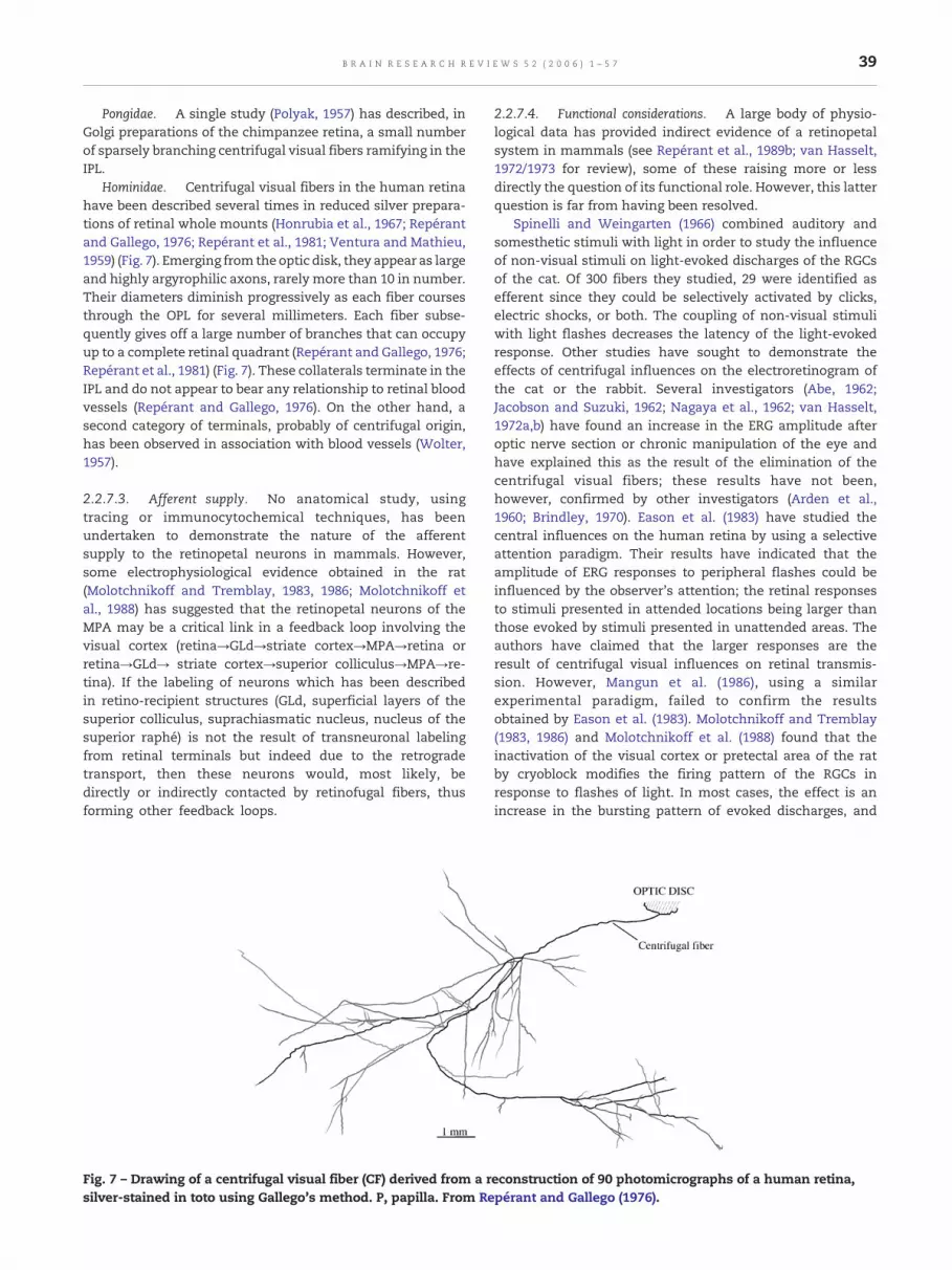
39B R A I N R E S E A R C H R E V I E W S 5 2 ( 2 0 0 6 ) 1 – 5 7
Pongidae. A single study (Polyak, 1957) has described, inGolgi preparations of the chimpanzee retina, a small numberof sparsely branching centrifugal visual fibers ramifying in theIPL.
Hominidae. Centrifugal visual fibers in the human retina
have been described several times in reduced silver prepara-tions of retinal whole mounts (Honrubia et al., 1967; Repérantand Gallego, 1976; Repérant et al., 1981; Ventura and Mathieu,1959) (Fig. 7). Emerging from the optic disk, they appear as largeand highly argyrophilic axons, rarely more than 10 in number.Their diameters diminish progressively as each fiber coursesthrough the OPL for several millimeters. Each fiber subse-quently gives off a large number of branches that can occupyup to a complete retinal quadrant (Repérant and Gallego, 1976;Repérant et al., 1981) (Fig. 7). These collaterals terminate in theIPL and do not appear to bear any relationship to retinal bloodvessels (Repérant and Gallego, 1976). On the other hand, asecond category of terminals, probably of centrifugal origin,has been observed in association with blood vessels (Wolter,1957).2.2.7.3. Afferent supply. No anatomical study, usingtracing or immunocytochemical techniques, has beenundertaken to demonstrate the nature of the afferentsupply to the retinopetal neurons in mammals. However,some electrophysiological evidence obtained in the rat(Molotchnikoff and Tremblay, 1983, 1986; Molotchnikoff etal., 1988) has suggested that the retinopetal neurons of theMPA may be a critical link in a feedback loop involving thevisual cortex (retina→GLd→striate cortex→MPA→retina orretina→GLd→ striate cortex→superior colliculus→MPA→re-tina). If the labeling of neurons which has been describedin retino-recipient structures (GLd, superficial layers of thesuperior colliculus, suprachiasmatic nucleus, nucleus of thesuperior raphé) is not the result of transneuronal labelingfrom retinal terminals but indeed due to the retrogradetransport, then these neurons would, most likely, bedirectly or indirectly contacted by retinofugal fibers, thusforming other feedback loops.
Fig. 7 – Drawing of a centrifugal visual fiber (CF) derived from a rsilver-stained in toto using Gallego's method. P, papilla. From Re
2.2.7.4. Functional considerations. A large body of physio-logical data has provided indirect evidence of a retinopetalsystem in mammals (see Repérant et al., 1989b; van Hasselt,1972/1973 for review), some of these raising more or lessdirectly the question of its functional role. However, this latterquestion is far from having been resolved.
Spinelli and Weingarten (1966) combined auditory andsomesthetic stimuli with light in order to study the influenceof non-visual stimuli on light-evoked discharges of the RGCsof the cat. Of 300 fibers they studied, 29 were identified asefferent since they could be selectively activated by clicks,electric shocks, or both. The coupling of non-visual stimuliwith light flashes decreases the latency of the light-evokedresponse. Other studies have sought to demonstrate theeffects of centrifugal influences on the electroretinogram ofthe cat or the rabbit. Several investigators (Abe, 1962;Jacobson and Suzuki, 1962; Nagaya et al., 1962; van Hasselt,1972a,b) have found an increase in the ERG amplitude afteroptic nerve section or chronic manipulation of the eye andhave explained this as the result of the elimination of thecentrifugal visual fibers; these results have not been,however, confirmed by other investigators (Arden et al.,1960; Brindley, 1970). Eason et al. (1983) have studied thecentral influences on the human retina by using a selectiveattention paradigm. Their results have indicated that theamplitude of ERG responses to peripheral flashes could beinfluenced by the observer's attention; the retinal responsesto stimuli presented in attended locations being larger thanthose evoked by stimuli presented in unattended areas. Theauthors have claimed that the larger responses are theresult of centrifugal visual influences on retinal transmis-sion. However, Mangun et al. (1986), using a similarexperimental paradigm, failed to confirm the resultsobtained by Eason et al. (1983). Molotchnikoff and Tremblay(1983, 1986) and Molotchnikoff et al. (1988) found that theinactivation of the visual cortex or pretectal area of the ratby cryoblock modifies the firing pattern of the RGCs inresponse to flashes of light. In most cases, the effect is anincrease in the bursting pattern of evoked discharges, and
econstruction of 90 photomicrographs of a human retina,pérant and Gallego (1976).

40 B R A I N R E S E A R C H R E V I E W S 5 2 ( 2 0 0 6 ) 1 – 5 7
those cells that are most affected are those lacking aconcentrically organized receptive field and predominantlygiving OFF responses. A similar effect of colliculus inactiva-tion to those of cortical blockade has also been observed(Molotchnikoff et al., 1988). These authors explain theirfindings in terms of two possible feedback loops (retino→GLd→striate cortex→MPA or retina→GLd→striate cortex→superior colliculus→MPA→retina).
More recent investigations have revealed the existence, inmammals, of a serotonergic (Repérant et al., 2000) andhistaminergic (Gastinger et al., 1999b) retinopetal system,arising in the first case from the dorsal raphé and, in thesecond, probably from the posterior hypothalamus. It is highlylikely to be the case that these retinopetal components of thevisual system have different actions from those of the cellsprojecting from the pretectum.
Retinal levels of serotonin are high at night, and thissubstance is most probably released by the centrifugal visualterminals since all immunocytochemical studies have failedto demonstrate the endogenous localization of serotonin inneuronal perikarya of the mammalian retina (Osborne, 1984).This serotonin could act as a substrate formelatonin synthesisby photoreceptors, which possess a 5-HT uptake mechanism(Redburn and Mitchell, 1989). Melatonin suppresses therelease of dopamine (Nowak, 1988) and thus counteractslight adaptation in the peripheral retina (Dowling, 1991;Witkovsky and Schütte, 1991). It has also been proposed thatthe retinopetal serotonergic system may play an importantrole in the synchronization of both eyes to circadian rhythms(Schütte, 1995).
One of the effects of histamine on the retina seems similarto that of dopamine that is released by light stimulation in themacaque retina; histamine increases the current through theGABAA receptor-activated chloride channel in isolated rat ACs(see Gastinger et al., 1999b for review). One possible inter-pretation of these findings is that dopamine and histamine actsynergistically to enhance the retinal response to rapidlychanging stimuli at high ambient light intensities (Gastingeret al., 1999b). It has been shown, furthermore, that theblockade of H1 receptors (which have an high affinity forhistamine) by antagonists increases the amplitude of the b-wave of the ERG (see Gastinger et al., 1999b) for review). Theseresults are to be compared with the earlier demonstrations ofthe effects of sectioning the centrifugal visual fibers on theERG that we havementioned above. Another possible functionof the histaminergic retinopetal systemmay be the regulationof retinal blood flow and capillary permeability (Gastinger etal., 1999b).
3. Conclusions
On the basis of the present comparative analysis, it appearsthat the CVS constitutes a permanent component of thevertebrate CNS. However, from one group to the next, thiscomponent undergoes a high degree of variation and, in asecond paper devoted to this question (Repérant et al., inpreparation), we shall attempt to address the evolutionarysignificance of this phylogenetic diversity. The variations ofthe CVS are multiple and above all concern the topographical
localization of the retinopetal neurons in the CNS (Fig. 8).Thus, the somata of these neurons can be located in the septo-preoptic terminal nerve complex (Polypteriformes, Chondros-tei, Holostei, Teleostei, Anura, Ophidia), posterior hypothala-mus (some Eutheria), ventral and/or dorsal thalamus(Chondrostei, some Ostariophysi, Acanthopterygii, Ophidia,some Eutheria), pretectum (some Ostariophysi, Acanthopter-ygii, Caudata, some Eutheria), optic tectum (Petromyzonti-formes, Elasmobranchii, many Euteleostei, some Eutheria),the mesencephalic tegmentum (Petromyzontiformes, Chelo-nia, Crocodilia, Aves, Eutheria), dorsal isthmus (Polypteri-formes, some Holostei, Osteoglossomorpha, Elopomorpha,Esociformes, Chelonia, Crocodilia, some Lacertilia, Aves),raphe (Chelonia, Eutheria), other rhombencephalic areas(Chelonia, Crocodilia, Aves, some Eutheria), and at last in thecontralateral eye (Chondrostei, Anura, some Eutheria). Thenumber of centrifugal visual axons, never collateralized inadult forms, varies according to a 1:1000 ratio (≥10 in man,≥10,000 in the chick and pigeon). These axons are either of finecaliber (≤1 μm) and often unmyelinated (Petromyzontiformes,Myxiniformes, numerous Gnathostomes) or large (1–3 μm) andmyelinated (Elasmobranchii, some Teleostei, Ophidia, Croco-dilia, Aves, Eutheria). Their mode of termination in the retinais generally of the divergent type (most vertebrates) and veryrarely (Aves) of the convergent type. Their targets can be verydifferentwithin a same group or between groups. These can beorthotopic RGCs (Holostei, Aves), displaced RGCs (Petromy-zontiformes, Aves), ACs with different morphological andneurochemical types (most vertebrates), IPC dopaminergiccells (Petromyzontiformes, Teleostei), and bipolar cells (Elas-mobranchii, Teleostei). The neurochemical signature of thesecentrifugal visual neurons also appears highly variable withina same group or between groups. Thus, several neuroactivesubstances (or their rate-limiting enzymes) used by theseneurons have been demonstrated by immunocytochemicalmethods, notably GABA (Petromyzontiformes), glutamate(Petromyzontiformes, Aves), aspartate (Chelonia), acetylcho-line (Crocodilia, Aves), Nitric Oxide (Chelonia, Crocodilia,Aves), serotonin (Elasmobranchii, Anura, Chelonia, Eutheria),dopamine (Elasmobranchii, Anura, Crocodilia, Eutheria), his-tamine (Eutheria), GnRH (Polypteriformes, Chondrostei, Tele-ostei, Crocodilia, Eutheria), FMRF-amide-like (Holostei,Teleostei, Anura, Crocodilia, substance P (Teleostei, Anura),NPY (Teleostei) and met-enkephalin (Chelonia).
These data must, however, be analyzed in the light ofseveral methodological considerations. In many cases, thelocation of the centrifugal visual neurons has been ascer-tained by means of the retrograde transport of tracer injectedinto the eyeball, and these findings must be interpreted withsome caution: neuronal labeling may be the result oftranssynaptic transport of tracer from visual terminals. Thismay occurwhen the postoperative survival time is overly long,or when substances such as lysolecithin or dimethyl sulfoxideare used to facilitate membrane penetration. Thus, it has beenclearly shown that in the frog, the somatic labeling observed inthe tectum (Ermakova et al., 1981; Hughes and Hall, 1986;Wilczynski and Zakon, 1982) is the result of transneuronaltransport from retinotectal terminals. In the same way, thetecto-retinal neurons described in many teleosts (Ebbessonand Meyer, 1981; Meyer and Ebbesson, 1981; Meyer et al., 1981;

Fig. 8 – Simplified cladogramof extant vertebrates derived from those presented in Figs. 1 and 3 showing the distribution of thecells of origin of the various centrifugal visual pathways. 1, Myxiniformes; 2, Petromyzontiformes; 3, Elasomobranchii; 4,Holocephali; 5, Polypteriformes; 6, Chondrostei; 7, Holostei; 8, Osteoglossomorpha; 9, Elapomorpha; 10, Clupeomorpha; 11,Esociformes; 12, Salmoniformes; 13, Ostariophysi; 14, Acanthopterygii; 15, Dipnoi; 16, Crossopterygii; 17, Caudata; 18, Anura;19, Prototheria; 20, Metatheria; 21, Eutheria; 22, Lacertilia; 23, Ophidia; 24, Crocodilia; 25, Aves; 26, Chelonia. Symbols: ?: no dataare available concerning the centrifugal visual neurons of these taxa. Black triangle: terminal nerve-septo-preoptic complex(arGCNT, anterior retinopetal ganglion cells of the terminal nerve; mrGCNT, medial retinopetal ganglion cells of the terminalnerve; prGCNT, posterior retinopetal ganglion cells of the terminal nerve) −5–14: arGCNT; 13, 14: arGCNT and mrGCNT; 14:arGCNT, mrGCNT, and prGCNT.White triangle: hypothalamus, white oval: ventral thalamus, Blackened oval: dorsal thalamus,horizontally striped oval: pretectum, cross-hatched rectangle: optic tectum, blackened rectangle: mesencephalic tegmentum,dotted lozenge: dorsal isthmus, horizontally striped lozenge: nuclei of the raphé, blackened lozenge: other anteriorrhombencephalic structures.
41B R A I N R E S E A R C H R E V I E W S 5 2 ( 2 0 0 6 ) 1 – 5 7
Schmidt, 1979) may be an artefact arising in the same way(Crapon de Caprona and Fritzsch, 1983; Springer, 1983;Springer and Gaffney, 1981; Uchiyama, 1989). A secondproblem arises when centrifugal fibers have been amplydemonstrated in the retina, but hodological techniques havefailed to reveal their cells of origin. This situation, apparentlydue to the failure of uptake or retrograde transport of tracer,has been observed in many cases, particularly in Amphibia(Scalia and Teitelbaum, 1978; Uchiyama et al., 1988; Wirsig-Wiechmann and Basinger, 1988), Crocodilia (Médina et al.,2004b, 2005), and Mammalia (Labandeira-Garcia, 1988; Schny-der and Künzle, 1983; Weidner et al., 1983), and its causesremain unknown. We also point out that the neurochemicalcharacteristics of the neurons of the CVS have been deter-mined mainly by immunocytochemical methods, whoseresults depend not only on the specificity of the antibodiesused, but also on the details of the appropriate controlprocedures. For example, some authors (Alonso et al., 1989)claim that substance P is a neuroactive substance present inthe neurons of the terminal nerve, while others (Kyle et al.,1995) argue that the immunolabeling of these neurons is anartefact arising from the cross-reaction of the antibody withan endogenous, non-tachykinin peptide similar to F8F-amideand A18F-amide.
It is also evident, from the analysis of the data available atpresent regarding the CVS, that there exist other uncertaintiesand voids in our knowledge. Thus, in spite of their heuristicvalue, no information exists about this system in severaltaxonomic groups (e.g., Holocephali in Chondrichthyes, Clu-peomorpha in Teleostei, Dipnoi, Crossopterygii, Gymnophionain Amphibia, Rhyncocephalia in Reptiles, Prototheria, andMetatheria inMammalia). Moreover, in several circumstances,only a partial description of the centrifugal visual neurons isavailable. Furthermore, in some cases, we know the location ofthese cells in the brain, and the details of their somaticmorphology but have no data concerning the mode ofinnervation nor the nature of their retinal targets, while inother cases, the retinopetal fibers have been described indetail, but no data are available concerning the intracerebralneuronsof their origin.Moreover, thenatureof theneuroactivesubstances for some populations of retinopetal neurons hasnot yet been identified. It should also bementioned that, apartfrom several groups (Petromyzontiformes, Teleostei, Aves),few studies have been undertaken concerning the develop-ment of the CVS. Our knowledge about the afferences to thesecentrifugal visual neurons, except in some groups (Petromy-zontiformes, Teleostei, Aves), is extremely fragmentary.Finally, few studies have been made of the function of the

42 B R A I N R E S E A R C H R E V I E W S 5 2 ( 2 0 0 6 ) 1 – 5 7
CVS, besides those on the olfacto-retinal system in Teleostei,and the isthmo-retinal system in birds.
Acknowledgments
The authors thank B. Jay for his help with computer drawings.This work was supported by CNRS (UMR 5166), MNHN (USM501), INSERM (U616), France, FQRNT (Grant no. 88454), andNSERC (Grant no. 0G0053), Canada, and the Academy ofSciences of Russia (Russian President's Grant no. 2165.3003.4,Grants no. 02-04.49576 and 03-04-49637 from the RussianFoundation for Basic Research).
R E F E R E N C E S
Abe, N., 1962. Effect of section and compression of the optic nerveon the ERG in the rabbit. Tohoku J. Exp. Med. 78, 223–227.
Adanina, V.O., Vesselkin, N.P., 1990. Axonal bifurcation of axons inthe optic chiasm of Acipenseridae. All Union Conf. onEvolutionary Physiology, Leningrad, p. 132.
Alonso, J.R., Coveñas, R., Lara, J., de León, M., Arévalo, R., Aijón, J.,1989. Substance P like immunoreactivity in the ganglion cellsof the tench terminal nerve. Neurosci. Lett. 106, 253–257.
Amano, M., Oka, Y., Aida, K., Okumoto, N., Kawashima, S.,Hasegawa, Y., 1991. Immunocytochemical demonstration ofsalmon GnRH and chicken GnRH-II in the brain of masusalmon Onchorhynchus masou. J. Comp. Neurol. 314,587–597.
Amano, M., Oka, Y., Kitamura, S., Ikuta, K., Aida, K., 1998.Ontogenetic development of salmon and chicken GnRH-IIsystems in the brain of masu salmon (Onchorhynchus masou).Cell Tissue Res. 293, 427–434.
Anadón, R., Meléndez-Ferro, M., Pérez-Costas, E., Pombal, M.A.,Rodicio, M.C., 1998. Centrifugal fibers are the only GABAergicstructures of the retina of the larval sea lamprey: animmunocytochemical study. Brain Res. 782, 297–302.
Angaut, P., Raffin, J.P., 1981. Embryonic development of thenucleus isthmo-opticus in the chick: a Golgi and electronmicroscopic study. Dev. Neurosci. 4, 1–14.
Angaut, P., Repérant, J., 1978. A light and electron microscopicstudy of the nucleus isthmo-opticus in the pigeon. Arch. Anat.Microsc. Morphol. Exp. 67, 63–78.
Arden, G., Granit, R., Ponte, F., 1960. Phase of suppressionfollowing each retinal b-wave in flicker. J. Neurophysiol. 23,305–314.
Arey, L.B., 1916. The function of the afferent fibers of the opticnerve of fishes. J. Comp. Neurol. 26, 213–245.
Ariëns-Kappers, C.U., Huber, G.C., Crosby, E.C., 1936. TheComparative Anatomy of the Nervous System of Vertebratesincluding Man. Macmillan, New York.
Armstrong, J.A., 1951. An experimental study of the visualpathways in a snake (Natrix natrix). J. Anat. 84, 275–289.
Aste, N., Viglietti-Panzica, C., Fasolo, A., Panzica, G.C., 1995.Mapping of neurochemical markers in quail central nervoussystem: VIP- and SP-like immunoreactivity. J. Chem.Neuroanat. 8, 87–102.
Auerbach, E., Dörrenhaus, A., Cavonius, C.R., 1992. Changes insensitivity of the dark-adapted eye during concurrent lightadaptation of the other eye. Vis. Neurosci. 8, 359–363.
Bagnoli, P., Francesconi, W., Magni, F., 1982. Visual Wulst-optictectum relationships in birds: a comparison with themammalian corticotectal system. Arch. Ital. Biol. 120, 212–235.
Bagnoli, P., Porciatti, V., Fontanesi, G., Sebastiani, L., 1987.
Morphological and functional changes in the retinotectalsystem of the pigeon during the early posthatching period.J. Comp. Neurol. 256, 400–411.
Bagnoli, P., Gigliola, F., Alesci, R., Erichsen, J.T., 1992.Distribution of neuropeptide Y, substance P, and cholineacetyltransferase in the developing visual system of thepigeon and effects of unilateral eye removal. J. Comp.Neurol. 318, 392–414.
Ball, A.K., St. Denis, J., 1987. LHRH-immunoreactive efferentfibres contact glycinergic and dopaminergic cells in thegoldfish retina. Supp. Invest. Ophthalmol. Visual Sci. 28,277.
Ball, A.K., Stell, W.K., Tutton, D.A., 1989. Efferent projections to thegoldfish retina. In: Weiler, R., Osborne, N.N. (Eds.),Neurobiology of the Inner Retina. NATO ASI Series. Springer,Berlin, pp. 103–116.
Barry, J., 1979. Immunohistochemistry of luteinizinghormone-releasing hormone-producing neurons of thevertebrates. Int. Rev. Cytol. 60, 179–221.
Batten, T.F., Cambre, M.L., Moons, L., Vandesande, F., 1990.Comparative distribution of neuropeptide-immunoreactivesystems in the brain of the green molly, Poecilia latipinna. J.Comp. Neurol. 302, 893–919.
Bazer, G.T., Ebbesson, S.O.E., 1985. Axosomatic retinal projectionto deep tectal neurones and retinopetal neurones inlargemouth bass (Micropterus salmoides Lacepede). Neurosci.Lett. 59, 309–312.
Behrens, U., Wagner, H.J., 2004. Terminal nerve and vision.Microsc. Res. Tech. 65, 25–32.
Behrens, U.D., Douglas, R.H., Wagner, H.J., 1993.Gonadotropin-releasing hormone, a neuropeptide of efferentprojections to the teleost retina induces light-adaptive spinuleformation on horizontal cell dendrites in dark-adaptedpreparations kept in vitro. Neurosci. Lett. 164, 59–62.
Bellairs, A., 1970. The Life of Reptiles. Universe Books, New York.Björklund, A., Nobin, A., 1973. Fluorescence histochemical and
microspectrofluorometric mapping of dopamine andnoradrenaline cell groups in the rat diencephalon. Brain Res.51, 193–205.
Blaser, P.F., Catsicas, S., Clarke, P.G., 1990. Retrograde modulationof dendritic geometry in the vertebrate brain duringdevelopment. Dev. Brain Res. 57, 139–142.
Blute, T.A., Mayer, B., Eldred, W.D., 1997. Immunocytochemicaland histochemical localization of nitric oxide synthase in theturtle retina. Vis. Neurosci. 14, 717–729.
Bonn, U., König, B., 1988. FMRFamide-like immunoreactivity inbrain and pituitary of Xenotaca eisenii: (Cyprinodontiformes,Teleostei). J. Hirnforsch. 29, 121–131.
Bonn, U., König, B., 1989a. FMRFamide immunoreactivity in thebrain and pituitary of Carassius auratus (Cyprinidae, Teleostei).J. Hirnforsch. 30, 361–370.
Bonn, U., König, B., 1989b. FMRFamide-like immunoreactivity inthe brain and pituitary of the teleost Eigenmannia lineata(Gymnotiformes). Z. Mikrosk.-Anat. Forsch. 103, 221–236.
Bons, N., 1987. Demonstration of centrifugal fibers in the opticchiasma and optic nerves in the rat after lesions of thesuprachiasmatic nucleus. C. R. Séances Soc. Biol. Fil. 181,274–281.
Bons, N., Petter, A., 1986. Retinal afferents of hypothalamic originin a prosimian primate: Microcebus murinus. Study usingretrograde fluorescent tracers. C. R. Acad. Sci. 303, 719–722.
Borchers, H.W., Ewert, J.P., 1978. Eye closure in toads (Bufo bufo L.)does not produce off response in retinal on-off ganglion cells: aquestion of efferent comments. J. Comp. Physiol. 125, 301–303.
Branston, N.M., Fleming, D.G., 1968. Efferent fibers in the frog opticnerve. Exp. Neurol. 20, 611–623.
Brecha, N., 1978. Some observations on the organization of theavian optic tectum: afferent nuclei and their tectal projections,PhD thesis, SUNY Stony Brook.

43B R A I N R E S E A R C H R E V I E W S 5 2 ( 2 0 0 6 ) 1 – 5 7
Brecha, N.C., Karten, H.J., 1981. Organization of the avian accessoryoptic system. Ann. N. Y. Acad. Sci. 374, 215–229.
Brecha, N.C., Karten, H.J., Hunt, S.P., 1980. Projections of thenucleus of the basal optic root in the pigeon: anautoradiographic and horseradish peroxidase study. J. Comp.Neurol. 189, 615–670.
Brindley, G.S., 1970. Central pathways of vision. Annu. Rev.Physiol. 32, 259–268.
Brindley, G.S., Hamasaki, D.I., 1962. Histological evidence againstthe view that the cat's optic nerve contains centrifugal fibers.J. Physiol. (London) 184, 444–449.
Britto, L.R., 1983. Retinal ganglion cells of the pigeon accessoryoptic system. Braz. J. Med. Biol. Res. 16, 357–363.
Britto, L.R., Keyser, K.T., Hamassaki, D.E., Karten, H.J., 1988.Catecholaminergic subpopulation of retinal displaced ganglioncells projects to the accessory optic nucleus in the pigeon(Columba livia). J. Comp. Neurol. 269, 109–117.
Brooke, R.N., Downer, J. de C., Powell, T.P., 1965. Centrifugal fibresto the retina in the monkey and cat. Nature 207, 1365–1367.
Brüning, G., 1993. Localization of NADPH-diaphorase in the brainof the chicken. J. Comp. Neurol. 334, 192–208.
Brunken, W.J., Witkovsky, P., Karten, H.J., 1986. Retinalneurochemistry of three elasmobranch species: animmunohistochemical approach. J. Comp. Neurol. 243, 1–12.
Bugbee, N.M., The basal ganglia-tectal pathway: its role invisually-guided behavior in the pigeon (Columba livia), PhDthesis, University of Maryland, Maryland, 1979.
Bunt, S.M., Lund, R.D., 1981. Development of a transient retino-retinal pathway in hooded and albino rats. Brain Res. 211,399–404.
Bunt, S.M., Lund, R.D., Land, P.W., 1983. Prenatal development ofthe optic projection in albino and hooded rats. Brain Res. 282,149–168.
Burrill, J.D., Easter Jr., S.S., 1994. Development of the retinofugalprojections in the embryonic and larval zebrafish (Brachydaniorerio). J. Comp. Neurol. 346, 583–600.
Butler, A.B., Hodos, W., 1996. Comparative VertebrateNeuroanatomy: Evolution and Adaptation. Wiley-Liss, NewYork.
Butler, A.B., Northcutt, R.G., 1992. Retinal projections in the bowfinAmia calva: cytoarchitectonic and experimental analysis. BrainBehav. Evol. 39, 169–194.
Butler, A.B., Saidel, W.M., 1991. Retinal projections in thefreshwater butterfly fish, Pantodon buchholzi (Osteoglossoidei): I.Cytoarchitectonic analysis and primary visual pathways. BrainBehav. Evol. 38, 127–153.
Byzov, A.L., Utina, J.A., 1971. Centrobeznije vlijanija naamacrinovije kletki setchatki liagushki. Centrifugal influenceson the amacrine cells of the frog retina.Neurofysiologi assistenten 3, 293–300 (in russian).
Cajal, S.R., 1888. Morfologia y conexiones de los elementos de laretina de las aves. Rev. Trim. Histol. Norm. Patol. 2, 1–10.
Cajal, S.R., 1889. Sur la morphologie et les connexions deséléments de la rétine des oiseaux. Anat. Anz. 4, 111–128.
Cajal, S.R., 1892. La retina de los teleosteos, Act. d. l. Soc. esp. deHistor. natur. t. II, 1892.
Cajal, S.R., 1893. La rétine des vertébrés. La Cellule 9, 17–257.Cajal, S.R., Histologie du Système Nerveux de l'Homme et des
Vertébrés, Maloine, Paris, 1911.Canatella, C., Hillis, D.M., 1992. Amphibian relationships.
Phylogenetic analysis of morphology and molecules. Herpetol.Monogr. 7, 1–7.
Carroll, R.L., 1988. Vertebrate Paleontology and Evolution. W.H.Freeman and Co, New York.
Castro, A., Beccera, M., Manso, M.J., Anadón, R., 1999. Developmentof immunoreactivity to neuropeptide Y in the brain of browntrout (Salmo trutta fario). J. Comp. Neurol. 414, 13–32.
Castro, A., Beccera, M., Anadón, R., Manso, M.J., 2001. Distributionand development of FMRFamide-like immunoreactive
neuronal systems in the brain of the brown trout, Salmo truttafario. J. Comp. Neurol. 440, 43–64.
Catois, E.H., 1901. Recherches sur l'histologie et l'anatomiemicroscopique de l'encéphale chez les poissons. Bull. Sci. Fr.Belg. 36, 1–166.
Catsicas, S., Clarke, P.G., 1987. Abrupt loss of dependence ofretinopetal neurons on their target cells, as shown byintraocular injections of kainate in chick embryos. J. Comp.Neurol. 262, 523–534.
Catsicas, S., Thanos, S., Clarke, P.G., 1987a. Major role for neuronaldeath during brain development: refinement of topographicconnections. Proc. Natl. Acad. Sci. U. S. A. 84, 8165–8168.
Catsicas, S., Catsicas, M., Clarke, P.G., 1987b. Long-distanceintraretinal connections in birds. Nature 326, 186–187.
Cepriano, L.M., Schreibman, M.P., 1993. The distribution ofneuropeptide Y and dynorphin immunoreactivity in the brainand pituitary gland of the platyfish, Xiphophorus maculatus,from birth to sexual maturity. Cell Tissue Res. 271, 87–92.
Cervetto, L., Marchiafava, P.L., Pasino, E., 1976. Influence ofefferent retinal fibres on responsiveness of ganglion cells tolight. Nature 260, 56–57.
Challet, E., Miceli, D., Pierre, J., Repérant, J., Massicotte, G., Herbin,M., Vesselkin, N.P., 1996. Distribution of serotonin-immunoreactivity in the brain of the pigeon (Columba livia).Anat. Embryol. 193, 209–227.
Chiba, A., 1997. Colocalization of gonadotropin-releasing hormone(GnRH)-, neuropeptide Y (NPY)-, and molluscancardioexcitatory tetrapeptide (FMRFamide)-likeimmunoreactivities in the ganglion cells of the terminal nerveof the masu salmon. Fish. Sci. 63, 153–154.
Chiba, A., Oka, S., Honma, Y., 1994. Ontogenetic development ofgonadotropin-releasing hormone-like immunoreactiveneurons in the brain of chum salmon, Onchorhynchus keta.Neurosci. Lett. 178, 51–54.
Chiba, A., Sohn, Y.C., Honma, Y., 1996a. Immunohistochemicaland ultrastructural characterization of the terminal nerveganglion cells of the ayu, Plecoglossus altivelis. Anat. Rec. 246,549–556.
Chiba, A., Sohn, Y.C., Honma, Y., 1996b. Distribution ofneuropeptide Y and gonadotropin-releasing hormoneimmunoreactivities in the brain and hypophysis of the ayu,Plecoglossus altivelis (Teleostei). Arch. Histol. Cytol. 59, 137–148.
Chiba, A., Oka, S., Honma, Y., 1996c. Ontogenetic changes inneuropeptide Y-like-immunoreactivity in the terminal nerveof the chum salmon and the cloudy dogfish, with specialreference to colocalization with gonadotropin-releasinghormone-immunoreactivity. Neurosci. Lett. 213, 49–52.
Chiba, A., Nakamura, H., Iwata, M., Sakuma, Y., Yamauchi, K.,Parhar, I.S., 1999. Development and differentiation ofgonadotropin hormone-releasing hormone neuronal systemsand testes in the japanese eel (Anguilla anguilla). Gen. Comp.Endocrinol. 114, 449–459.
Chmielevski, C.E., Dorado, M.E., Quesada, A., Genis-Galvez, J.M.,Prada, F.A., 1988. Centrifugal fibers in the chick retina. Amorphological study. Arch. Histol. Embryol. 17, 319–327.
Clarke, P.G., 1982. The generation and migration of the chick'sisthmic complex. J. Comp. Neurol. 207, 208–222.
Clarke, P.G., 1985. Neuronal death during development in theisthmo-optic nucleus of the chick: sustaining role of afferentsfrom the tectum. J. Comp. Neurol. 234, 365–379.
Clarke, P.G., 1992. Neuron death in the developing avianisthmo-optic nucleus, and its relation to the establishment offunctional circuitry. J. Neurobiol. 23, 1140–1158.
Clarke, P.G., Caranzano, F., 1985. Dendritic development in theisthmo-optic nucleus of chick embryos. Dev. Neurosci. 7,161–169.
Clarke, P.G., Cowan, W.M., 1975. Ectopic neurons and aberrantconnections during neural development. Proc. Natl. Acad. Sci.U. S. A. 72, 4455–4458.

44 B R A I N R E S E A R C H R E V I E W S 5 2 ( 2 0 0 6 ) 1 – 5 7
Clarke, P.G., Cowan, W.M., 1976. The development of theisthmo-optic tract in the chick, with special reference to theoccurrence and correction of developmental errors in thelocation and connections of isthmo-optic neurons. J. Comp.Neurol. 167, 143–164.
Clarke, P.G., Egloff, M., 1988. Combined effects of deafferentationand de-afferentation on isthmo-optic neurons during theperiod of their naturally occurring cell death. Anat. Embryol.179, 103–108.
Clarke, P.G., Kraftsick, R., 1996. Dendritic reorientation andcytolamination during the development of the isthmo-opticnucleus in chick embryos. J. Comp. Neurol. 365, 96–112.
Clarke, P.G., Whitteridge, D., 1976. The projection of the retina,including the ‘red area’ on to the optic tectum of the pigeon. Q.J. Exp. Physiol. Cogn. Med. Sci. 61, 351–358.
Clarke, P.G., Rogers, L.A., Cowan, W.M., 1976. The time of originand the pattern of survival of neurons in the isthmo-opticnucleus of the chick. J. Comp. Neurol. 167, 125–142.
Clarke, P.G., Gyger, M., Catsicas, S., 1996. A centrifugally controlledcircuit in the avian retina and its possible role in visualattention switching. Vis. Neurosci. 13, 1043–1048.
Cohen, D.H., Pitts, L.H., 1967. The hyperstriatal region of the avianforebrain: somatic and autonomic responses to electricalstimulation. J. Comp. Neurol. 131, 323–336.
Cowan, W.M., 1970. Centrifugal fibres to the avian retina. Br. Med.Bull. 26, 112–118.
Cowan, W.M., Clarke, P.G., 1976. The development of theisthmo-optic nucleus. Brain Behav. Evol. 13, 345–375.
Cowan, W.M., Powell, T.P., 1963. Centrifugal fibres in the avianvisual system. Proc. R. Soc., Ser. B 158, 232–252.
Cowan,W.M.,Wenger, E., 1968. The development of the nucleus oforigin of centrifugal fibers to the retina in the chick. J. Comp.Neurol. 133, 207–240.
Cowan, W.M., Adamson, L., Powell, T.P., 1961. An experimentalstudy of the avian visual system. J. Anat. (London) 95,545–563.
Cowan, W.M., Gottlieb, D.I., Hendrickson, A.E., Price, J.L., Woolsey,T.A., 1972. The autoradiographic demonstration of axonalconnections in the central nervous system. Brain Res. 37,21–51.
Cozzi, B., Viglietti-Panzica, C., Aste, N., Panzica, G.C., 1991. Theserotoninergic system in the brain of the japanese quail. Animmunohistochemical study. Cell Tissue Res. 263, 271–284.
Cozzi, B., Massa, R., Panzica, G.C., 1997. The NADPH-diaphorase-containing system in the brain of the budgerigar (Melopsittacusundulatus). Cell Tissue Res. 287, 101–112.
Craigie, E.H., 1928. Observations on the brain of the humming bird(Chrysolampis mosquitus Linn. and Chlorostilbon caribeus Lawr.).J. Comp. Neurol. 45, 377–483.
Craigie, E.H., 1930. Studies on the brain of the kiwi (Apteryxaustralis). J. Comp. Neurol. 49, 223–357.
Crapon de Caprona, M.-D., Fritzsch, B., 1983. The developmentof the retinopetal nucleus olfacto-retinalis of two cichlidfish as revealed by horseradish peroxidase. Brain Res. 11,281–301.
Crim, J.W., Urano, A., Gorbman, A., 1979a. Immunocytochemicalstudies of luteinizing-hormone-releasing hormone in brains ofagnathan fishes: I. Comparisons of adult Pacific lamprey(Entosphenus tridentata) and the Pacific hagfish (Eptatretus stouti).Gen. Comp. Endocrinol. 37, 294–305.
Crim, J.W., Urano, A., Gorbman, A., 1979b. Immunocytochemicalstudies of uteinizing hormone-releasing hormone in brains ofagnathan fishes: II. Patterns of immunoreactivity in larval andmaturing western brook lamprey (Lampetra richardsoni). Gen.Comp. Endocrinol. 38, 290–299.
Crossland, W.J., 1979. Identification of tectal synaptic terminals inthe avian isthmo-optic nucleus. In: Granda, A.M., Maxwell, J.H.(Eds.), Neural Mechanisms and Behavior in the Pigeon. PlenumPress, New York, pp. 267–285.
Crossland, W.J., Hughes, C.P., 1978. Observations on the afferentand efferent connections of the avian isthmo-optic nucleus.Brain Res. 145, 239–256.
Dacey, D.M., 1985. Wide-spreading terminal axons in the innerplexiform layer of the cat's retina: evidence for intrinsicaxon collaterals of ganglion cells. J. Comp. Neurol. 242,247–262.
Dalil-Thiney, N., 1995. Etude histologique et morphofonctionnellede la rétine de lamproie Lampetra fluviatilis: approchesultrastructurales et immunohistochimiques. Thèse deDoctorat de l'Université de Paris VII, 1995.
Dalil-Thiney, N., Versaux-Botteri, C., Nguyen-Legros, J., 1996.Electron microscopic demonstration of tyrosinehydroxylase-immunoreactive interplexiform cells in thelamprey retina. Neurosci. Lett. 207, 159–162.
Davis, J.N., McKinnon, P.N., 1982. Anterograde and transcellulartransport of a fluorescent dye, bisbenzimide, in the rat visualsystem. Neurosci. Lett. 29, 207–212.
Davis, R.E., Kyle, A., Klinger, P.D., 1988. Nervus terminalisinnervation of the goldfish retina and behavioral visualsensitivity. Neurosci. Lett. 91, 126–130.
de Carvalho, R.P., de Faria, M.H., do Nascimento, J.L., Hokoç, J.N.,1996. Development of NADPH-diaphorase in the avian retina:regulation by calcium ions and relation to nitric oxidesynthase. J. Neurochem. 67, 1063–1071.
de Lanerolle, N.C., Elde, R.P., Sparber, S.B., Frick, M., 1981.Distribution of methionine-enkephalin immunoreactivity inthe chick brain: an immunohistochemical study. J. Comp.Neurol. 199, 513–533.
de Miguel, E., Wagner, H.J., 1990. Tyrosine hydroxylaseimmunoreactive interplexiform cells in the lamprey retina.Neurosci. Lett. 113, 151–155.
de Miguel, E., Rodicio, M.C., Anadón, R., 1989. Ganglion cells andretinopetal fibers of the larval lamprey retina: an HRPultrastructural study. Neurosci. Lett. 106, 1–6.
de Miguel, E., Rodicio, M.C., Anadón, R., 1990. Organization of thevisual system in larval lampreys: an HRP study. J. Comp.Neurol. 302, 529–542.
Demski, L.S., 1993. Terminal nerve complex. Acta Anat. 148,81–95.
Demski, L.S., Dulka, J.G., 1984. Functional-anatomical studies onsperm release evoked by electrical stimulation of the olfactorytract in goldfish. Brain Res. 291, 241–247.
Demski, L.S., Northcutt, R.G., 1983. The terminal nerve: a newchemosensory system in vertebrates? Science 22, 435–437.
Demski, L.S., Sudberry, J.J., Beaver, J.A., 1995. Loss of GnRHimmunoreactivity and axonal regrowth following terminalnerve section in the clearnose skate, Raja eglanteria. Abstr.-Soc.Neurosci. 20, 115–190.
Demski, L.S., Beaver, J.A., Sudberry, J.J., Curtis, J.R., 1997. GnRHsystems in cartilaginous fishes. In: Parhar, I.S., Sakuma, Y.(Eds.), GnRHNeurons: Gene to Behavior. Brain Shuppan, Tokyo,pp. 123–143.
Denny, N., Frumkes, T.E., Barris, M.C., Eysteinsson, T., 1991. Tonicinterocular suppression and binocular summation in humanvision. J. Physiol. (London) 437, 449–460.
Diano, S., Horvath, B., Horvath, T.L., 1999. In monkeys,intraocular cholera toxin B injections reveal massive retinalprojections to different hypothalamic and forebrain nucleiand retinopetal dorsal raphe neurons. Abstr.-Soc. Neurosci.25, 1133.
Dogiel, A.S., 1895. Die Retina der Vögel. Arch. Mikrosk. Anat. 44,622–648.
Domenici, L., Waldvogel, H.J., Matute, C., Streit, P., 1988.Distribution of GABA-like immunoreactivity in the pigeonbrain. Neuroscience 25, 931–950.
Dos Santos, R.M., Gardino, P.F., 1998. Differential distribution of asecond type of tyrosine hydroxylase immunoreactiveamacrine cell in the chick retina. J. Neurocytol. 27, 33–43.

45B R A I N R E S E A R C H R E V I E W S 5 2 ( 2 0 0 6 ) 1 – 5 7
Dowling, J.E., 1991. Retinal neuromodulation: the role ofdopamine. Vis. Neurosci. 7, 87–97.
Dowling, J.E., Cowan, W.M., 1966. An electron microscope study ofnormal and degenerating centrifugal fiber terminals in thepigeon retina. Z. Zellforsch. Mikrosk. Anat. 71, 14–28.
Dowling, J.E., Ehinger, B., 1978. The interplexiform cell system: I.Synapses of the dopaminergic neurons of the goldfish retina.Proc. R. Soc. Ser., B 201, 7–26.
Dräger, U.C., Edwards, D.L., Barnstable, C.J., 1984. Antibodiesagainst filamentous components in discrete cell types of themouse retina. J. Neurosci. 4, 2025–2042.
Dubois, E.A., 2001. Development of gonadotropin-releasinghormone system in the male african catfish, Clarias gariepinus,PhD thesis, ISBN 90-393-2645-2 (2001) 13–48.
Dubois, E.A., Zandbergen, M.A., Peute, J., Goos, H.J., 2002.Evolutionary development of three gonadotropin-releasinghormone (GnRH) systems in Vertebrates. Brain Res. Bull. 57,413–418.
Duellman, W.E., Trueb, L., 1986. Biology of Amphibians.McGraw-Hill, New York.
Duke-Elder, S., Wybar, K.C., 1961. The Anatomy of the VisualSystem, vol. II. Kimpton, London.
Eason, R.G., Oakley, M., Flowers, L., 1983. Central neural influenceson the human retina during selective attention. Physiol.Psychol. 11, 18–28.
Ebbesson, S.O., Meyer, D.L., 1981. Efferents to the retina havemultiple sources in teleost fish. Science 214, 924–926.
Ebbesson, S.O., Meyer, D.L., 1989. Retinopetal cells exist in theoptic tectum of steelhead trout. Neurosci. Lett. 106, 95–98.
Ebbesson, S.O., Meyer, D.L., Malz, C.R., Bazer, G.T., 1991. Ontogenyof the olfacto retinalis projection in Coho salmon(Onchorhynchus kisutch). J. Exp. Zool. 257, 220–222.
Edinger, L., 1911. In: Vogel, F.C.W. (Ed.), Vorlesungen über den Bauder nervösen Zentralorgane. Leipzig.
Edinger, L., Wallenberg, A., 1899. Untersuchungen über das Gehirnder Tauben. Anat. Anz. 15, 245–271.
Ehinger, B., Dowling, J.E., 1987. Retinal neurocircuitry andtransmission. In: Björklund, A., Hökfelt, T., Swanson, L.W.(Eds.), Handbook of Chemical Neuroanatomy, IntegratedSystems of the CNS: Part I, vol. 5. Elsevier, Amsterdam,pp. 389–446.
Ehrlich, D., Keyser, K.T., Karten, H.J., 1987. Distribution ofsubstance P-like immunoreactive retinal ganglion cells andtheir pattern of termination in the optic tectum of chick (Gallusgallus). J. Comp. Neurol. 266, 220–233.
Eisthen, H.L., Northcutt, R.G., 1996. Silver lampreys (Ichthyomyzonunicuspis) lack a gonadotropin-releasing hormone- andFMRFamide-immunoreactive terminal nerve. J. Comp. Neurol.370, 159–172.
El Hassni, M., Repérant, J., Ward, R., Bennis, M., 1997. Theretinopetal visual system in the chameleon (Chamaeleochameleon). J. Brain Res. 38, 453–457.
Ekström, P., 1984. Central neural connections of the pineal organand retina in the teleost Gasterosteus aculeatus L. J. Comp.Neurol. 226, 321–335.
Ekström, P., Honkanen, T., Ebbesson, S.O., 1988. FMRFamide-likeimmunoreactive neurons of the nervus terminalis of teleostsinnervate both retina and pineal organ. Brain Res. 460, 68–75.
Ermakova, T.V., Kenigfest, N.B., Vesselkin, N.P., 1981.Transneuronal distribution of peroxidase in the brain of thefrog Rana temporaria. Zh. Evol. Biokhim. Fisiol. 17, 95–96 (inrussian and title in english).
Famiglietti Jr., E.V., Kaneko, A., Tachibana, M., 1977. Neuronalarchitecture of On and Off pathways to ganglion cells in carpretina. Science 198, 1267–1269.
Ferguson, J.L., Mulvanny, P.J., Brauth, S.E., 1978. Distribution ofneurons projecting to the retina of Caiman crocodilus. BrainBehav. Evol. 15, 294–306.
Fernald, R.D., Finger, T.E., 1984. Catecholaminergic neurones of
locus coeruleus project to the ganglion cells of the nervusterminalis (NT) in goldfish. Abstr.-Soc. Neurosci. 10, 50.
Feyerabend, B., Malz, C.R., Meyer, D.L., 1994. Birds thatfeed-on-the-wing have few isthmo-optic neurons. Neurosci.Lett. 182, 66–68.
Fiebig, E., Meyer, D.L., Ebbesson, S.O., 1985. Eyemovements evokedfrom telencephalic stimulation in the piranha (Serrasalmusnattereri). Comp. Biochem. Physiol., A 81, 67–70.
Fink, W.L., Weitzman, S.H., 1982. Relationships of Stomiiformesfishes (Teleostei) with a description of Diplophos. Bull. Mus.Comp. Zool. 150, 31–93.
Fischer, A.J., Stell, W.K., 1997. Light-modulated release ofRfamide-like neuropeptides from nervus terminalis axonterminals in the retina of goldfish. Neuroscience 77,585–597.
Fischer, A.J., Stell, W.K., 1999. Nitric oxide synthase-containingcells in the retina, pigmented epithelium, choroid, and sclera ofthe chick eye. J. Comp. Neurol. 405, 1–14.
Fite, K.V., Brecha, N., Karten, H.J., Hunt, S.P., 1981. Displacedganglion cells and the accessory optic system of pigeon.J. Comp. Neurol. 195, 279–288.
Fite, K.V., Foote, W., Bengston, L., Cosentino, J., 1996. Retinalafferents terminate and retinal efferents originate in the sameregion of the dorsal raphe nucleus in rats. Abstr.-Soc. Neurosci.22, 1818.
Fite, K.V., Janušonis, S., Foote, W., Bengston, L., Cosentino, J., 1997.Retinofugal and retinopetal pathways in the dorsal raphe andperiaqueductal gray of the mongolian gerbil. Abstr.-Soc.Neurosci. 23, 1819.
Fite, K.V., Janušonis, S., Foote, W., Bengston, L., 1999. Retinalafferents to the dorsal raphe nucleus in rats and mongoliangerbils. J. Comp. Neurol. 414, 469–484.
Fontanesi, G., Traina, G., Bagnoli, P., 1993. Somatostatin-likeimmunoreactivity in the pigeon visual system: developmentalexpression and effects of retina removal. Vis. Neurosci. 10,271–285.
Forey, P.L., Janvier, P., 1993. Agnathans and the origin of jawedvertebrates. Nature 361, 129–134.
Forey, P.L., Janvier, P., 1994. Evolution of the early vertebrates. Am.Sci. 82, 554–565.
Franz, V., 1912. Beiträge zur Kenntnis des Mittelhirns undZwischenhirns des Knochenfisches. Folia Neurobiol. 6,402–411.
Fritzsch, B., Collin, S.P., 1990. Dendritic distribution of twopopulations of ganglion cells and the retinopetal fibers in theretina of the silver lamprey (Ichthyomyzon unicuspis). Vis.Neurosci. 4, 533–545.
Fritzsch, B., Himstedt, W., 1981. Pretectal neurons project to thesalamander retina. Neurosci. Lett. 24, 13–17.
Fritzsch, B., Himstedt, W., Crapon de Caprona, M.D., 1985. Visualprojections in larval Ichthyophis kohtaoensis (Amphibia:Gymnophiona). Dev. Brain Res. 23, 201–210.
Fritzsch, B., Wilm, C., Crapon de Caprona, M.D., 1987. Ipsilateralretinofugal and retinopetal projections in normal andmonocular cichlid fish. Neurosci. Lett. 78, 259–264.
Fritzsch, B., Crapon de Caprona, M.D., Clarke, P.G., 1990a.Development of two morphological types of retinopetal fibersin chick embryos, as shown by the diffusion along axons of acarbocyanine dye in the fixed retina. J. Comp. Neurol. 300,405–421.
Fritzsch, B., Sonntag, R., Dubuc, R., Ohta, Y., Grillner, S., 1990b.Organization of the six motor nuclei innervating the ocularmuscles in lamprey. J. Comp. Neurol. 294, 491–506.
Fujii, K., Kobayashi, H., 1992. FMRFamide-like immunoreactivity inthe brain and pituitary of the goldfish, Carassius auratus. Anat.Anz. 174, 217–222.
Fujisawa, H., Terubayashi, U., Ibata, Y., 1983. Hypothalamo-retinalcentrifugal projection in the dog. Acta Anat. Nippon 58,251–256.

46 B R A I N R E S E A R C H R E V I E W S 5 2 ( 2 0 0 6 ) 1 – 5 7
Fujita, I., Sorensen, P.W., Stacey, N.E., Hara, T.J., 1991. The olfactorysystem, not the terminal nerve, functions as the primarychemosensory pathway mediating responses to sexpheromones in male goldfish. Brain Behav. Evol. 38, 313–321.
Fukuda, M., Ishimoto, I., Shiosaka, S., Kuwayama, Y., Shimizu,Y., Inagaki, S., Sakanaka, M., Takagi, H., Senba, E., Takatsuki,K., Umegaki, K., Tohyama, M., 1982/1983. Localization of LH-RH immunoreactivity in the avian retina. Curr. Eye Res. 2,71–74.
Fuller, P.M., Prior, D.J., 1975. Cobalt iontophoresis techniques fortracing afferent and efferent connections in the vertebrateCNS. Brain Res. 88, 211–220.
Fuxe, K., Goldstein, M., Hökfelt, T., Joh, T.H., 1970.Immunohistochemical localization of dopamine-β-hydroxylase in the peripheral and central nervous system. Res.Commun. Chem. Pathol. Pharmacol. 1, 627–636.
Gaffney, E.S., Meylan, P.A., 1988. A phylogeny of turtles. In: Benton,M.J. (Ed.), The Phylogeny and Classification of the Tetrapods.Amphibians, Reptiles, Birds, vol. 1. Clarendon Press, Oxford,pp. 157–219.
Galifret, Y., Condé-Courtine, F., Repérant, J., Servière, J., 1971.Centrifugal control in the visual system of the pigeon. VisionRes., Suppl. 3, 185–200.
Gallego, A., Cruz, J., 1965. Mammalian retina: associational nervecells in ganglion cell layer. Science 150, 1313–1314.
Gallego, A., Ventura, J., 1953. Fibras centrifugas de la retina. An.Inst. Farmacol. Esp. 2, 177–186.
Gamlin, P.D., Cohen, D.H., 1988. Projections of the retinorecipientpretectal nuclei in the pigeon (Columba livia). J. Comp. Neurol.269, 18–46.
Gardino, P.F., Schmal, A.R., da, K., Calaza, C., 2004. Identification ofneurons with acetylcholinesterase and NADPH-diaphoraseactivities in the centrifual visual system of the chick. J. Chem.Neuroanat. 27, 267–273.
Garner, A.S., Menegay, H.J., Boeshore, K.L., Xie, X.Y., Voci, J.M.,Johnson, J.E., Large, T.H., 1996. Expression of trkB receptorisoforms in the developing avian visual system. J. Neurosci. 16,1740–1752.
Gastinger, M.J., Marshak, D.W., Gardner, T.W., Barber, A.J., 1999a.Histamine containing centrifugal axons in the rat retina.Abstr.-Soc. Neurosci. 25, 135.
Gastinger, M.J., O'Brien, J.J., Larsen, N.B., Marshak, D.W., 1999b.Histamine immunoreactive axons in the macaque retina.Invest. Ophthalmol. Visual Sci. 40, 487–495.
Gerwerzhagen, K., Rickmann, M.J., Meyer, D.L., Ebbesson, S.O.,1982. Optic tract cells projecting to the retina in the teleost,Pantodon buchholzi. Cell Tissue Res. 225, 23–28.
Goldberg, S., Galin, M.A., 1973. Response of retinal ganglion cellaxons to lesions in the adult mouse retina. Invest. Ophthalmol.Visual Sci. 12, 382–385.
González-Martínez, D., Zmora, N., Zanuy, S., Sarasquete, C., Elizur,A., Kah, O., Muñoz-Cueto, J.A., 2002a. Developmentalexpression of three different prepro-GnRH (gonadotrophin-releasing hormone) messengers in the brain of the Europeansea bass (Dicentrarchus labrax). J. Chem. Neuroanat. 23, 255–267.
González-Martínez, D., Zmora, N., Mananos, E., Saligaut, D.,Zanuy, S., Zohar, Y., Elizur, A., Kah, O., Muñoz-Cueto, J.A.,2002b. Immunohistochemical localization of three differentprepro-GnRHs in the brain and pituitary of the European seabass (Dicentrarchus labrax) using antibodies to thecorresponding GnRH-associated peptides. J. Comp. Neurol. 446,95–113.
González-Martínez, D., Zmora, N., Saligaut, D., Zanuy, S., Elizur, A.,Kah, O., Muñoz-Cueto, J.A., 2004. New insights indevelopmental origins of different GnRH (gonadotrophin-releasing hormone) systems in perciform fish: animmunohistochemical study in the European sea bass(Dicentrarchus labrax). J. Chem. Neuroanat. 28, 1–15.
Granda, R.H., Crossland, W.J., 1989. GABA-like immunoreactivity
of neurons in the chicken diencephalon and mesencephalon.J. Comp. Neurol. 287, 455–469.
Granit, R., 1955. Centrifugal and antidromic effects on ganglioncells of retina. J. Neurophysiol. 18, 388–411.
Grober, M.S., Bass, A.H., 1991. Neuronal correlates of sex/rolechange in labrid fishes: LHRH-like immunoreactivity. BrainBehav. Evol. 38, 302–312.
Grober, M.S., Bass, A.H., Burd, G., Marchaterre, M.A., Segil, N.,Scholz, K., Hodgson, T., 1987. The nervus terminalis ganglion inAnguilla rostrata: an immunocytochemical and HRPhistochemical analysis. Brain Res. 436, 148–152.
Güntürkün, O., 1987. A Golgi study of the isthmic nuclei in thepigeon (Columba livia). Cell Tissue Res. 248, 439–448.
Güntürkün, O., Miceli, D., Watanabe, M., 1993. Anatomy of theavian thalamofugal pathway. In: Zeigler, H.P., Bischoff, H.J.(Eds.), Vision, Brain, and Behavior in Birds. MIT Press,Cambridge, pp. 115–135.
Haas, G., 1979. On a new snakelike reptile from the LowerCenomanian of Ein Jabrud, near Jerusalem. Bull. Mus. Natl.Hist. Nat. 1, 51–64.
Haas, G., 1980. Remarks on a new ophiomorph reptile from theLower Cenomanian of Ein Jabrud, Israel. In: Jacobs, L.L. (Ed.),Aspects of Vertebrate History. Museum of Northern ArizonaPress, Flagstaff, Arizona, pp. 177–192.
Hahmann, U., Güntürkün, O., 1992. Visual-discrimination deficitsafter lesions of the centrifugal visual system in pigeons(Columba livia). Vis. Neurosci. 9, 225–233.
Halpern, M., Wang, R.T., Colman, D.R., 1976. Centrifugal fibers tothe eye in a nonavian vertebrate: source revealed byhorseradish peroxidase studies. Science 194, 1185–1188.
Halpern-Sebold, L.R., Schreibman, M.P., 1983. Ontogeny ofcenters containing luteinizing hormone-releasing hormonein the brain of the platyfish (Xiphophorus maculatus) asdetermined by immunocytochemistry. Cell Tissue Res. 229,75–84.
Halpern-Sebold, L.R., Margolis-Kazan, H., Schreibman, M.P., Joh, T.H., 1985. Immunoreactive tyrosine hydroxylase in the brainand pituitary gland of the platyfish. Proc. Soc. Exp. Biol. Med.178, 486–489.
Haverkamp, S., Eldred, W.D., 1998. Localization of the origin ofretinal efferents in the turtle brain and the involvement ofnitric oxide synthase. J. Comp. Neurol. 393, 185–195.
Hayes, B.P., 1982. The structural organization of the pigeon retina.Prog. Retinal Res. 1, 193–221.
Hayes, B.P., Holden, A.L., 1983. The distribution of centrifugalterminals in the pigeon retina. Exp. Brain Res. 49, 189–197.
Hayes, B.P., Webster, K.E., 1981. Neurones situated outside theisthmo-optic nucleus and projecting to the eye in adult birds.Neurosci. Lett. 26, 107–112.
Heilmann, G., 1926. The Origin of Birds. Appleton, New York.Herrick, C.J., 1909. The nervus terminalis (nerve of Pinkus) in the
frog. J. Comp. Neurol. 19, 175–190.Herrick, C.J., 1933. The amphibian forebrain: VI. Necturus. J. Comp.
Neurol. 58, 1–288.Herrick, C.J., 1948. The Brain of the Tiger Salamander. University of
Chicago Press, Chicago.Himstedt, W., Manteuffel, G., 1985. Retinal projections in the
caecilian Ichthyophis kohtaoensis (Amphibia, Gymnophiona).Cell Tissue Res. 239, 689–692.
Hirschberger, W., 1971. Vergleichend experimentell-histologische Untersuchung zur retinalen Repräsentation inden primären visuellen Zentren einiger Vogelarten, Thesis,Frankfurt, 1971.
Hofmann, M.H., Piñuela, C., Meyer, D.L., 1993. Retinopetalprojections from diencephalic neurons in a primitiveactinopterygian fish, the sterlet Acipenser ruthenus. Neurosci.Lett. 161, 30–32.
Hofstetter, R., 1968. A contribution to the classification of snakes.Copeia 1, 201–213.

47B R A I N R E S E A R C H R E V I E W S 5 2 ( 2 0 0 6 ) 1 – 5 7
Holden, A.L., 1968. The centrifugal system running to the pigeonretina. J. Physiol. (London) 197, 199–219.
Holden, A.L., 1978. Centrifugal actions on pigeon retinal ganglioncells. J. Physiol. (London) 282, 8–9P.
Holden, A.L., 1982. Electrophysiology of the avian retina. Prog.Retinal Res. 1, 179–196.
Holden, A.L., 1990. Centrifugal pathway to the retina: which waydoes the 'searchlight' point? Vis. Neurosci. 4, 493–495.
Holmes, G.M., 1901. The nervous system of the dog without aforebrain. J. Physiol. (London) 27, 1–25.
Holmgren, N., 1918. Zur Kenntnis des Nervus terminalis beiTeleostiern. Folia Neurobiol. 11, 16–36.
Holmgren, N., 1920. Zur Anatomie und Histologie des Vorder- undZwischenhirnes der Knochenfische. Acta Zool. 1, 137–315.
Honrubia, F.M., Elliott, J.H., 1970. Efferent innervation of the retina:II. Morphologic study of the monkey retina. Invest.Ophthalmol. Visual Sci. 9, 971–976.
Honrubia, F.M., Grijalbo, M.P., 1968. Inervacion centrifuga de laretina del gato. Arch. Soc. Oftalmol. Hisp.-Am. 28, 744–751.
Honrubia, F.M., Grijalbo, M.P., Elliott, J.H., 1967. Fibras centrifugasen la retina de los primates. Arch. Soc. Oftalmol. Hisp.-Am. 27,561–569.
Hoogland, P.V., Welker, E., 1981. Telencephalic projections to theeye in Python reticulatus. Brain Res. 213, 173–176.
Hoogland, P.V., Vanderkrans, A., Koole, F.D., Groenewegen, H.J.,1985. A direct projection from the nucleus oculomotorius to theretina in rats. Neurosci. Lett. 56, 323–328.
Hu, J., Li, W.C., Xiao, Q., Wang, S.R., 2000. Electrical interactionbetween neurons in the pigeon isthmo-optic nucleus. BrainRes. Bull. 51, 159–163.
Hu, J., Li, S., Xiao, Q., Wang, S.R., 2001. Tecto-isthmo-optictransmission in pigeons is mediated by glutamate and nitricoxide. Brain Res. Bull. 54, 399–403.
Huber, G.C., Crosby, E.C., 1929. The nuclei and fiber paths of theavian diencephalon, with consideration of telencephalic andcertain mesencephalic centers and connections. J. Comp.Neurol. 48, 1–225.
Hughes, T.E., Hall, W.C., 1986. The transneuronal transport ofhorseradish peroxidase in the visual system of the frog, Ranapipiens. Neuroscience 17, 507–518.
Hunt, S.P., Brecha, N., 1984. The avian optic tectum: a synthesis ofmorphology and biochemistry. In: Vanegas, H. (Ed.),Comparative Neurobiology of the Optic Tectum. Plenum Press,New York, pp. 619–648.
Hunt, S.P., Künzle, H., 1976a. Observations on the projections andintrinsic organization of the pigeon optic tectum: anautoradiographic study based on anterograde and retrogradeaxonal and dendritic flow. J. Comp. Neurol. 170, 153–172.
Hunt, S.P., Künzle, H., 1976b. Selective uptake and transport oflabel within three identified neuronal systems afterinjection of 3H-GABA into the pigeon optic tectum: anautoradiographic and Golgi study. J. Comp. Neurol. 170,173–189.
Itaya, S.K., 1980. Retinal efferents from the pretectal area in the rat.Brain Res. 201, 436–441.
Itaya, S.K., Itaya, P.W., 1985. Centrifugal fibers to the rat retinafrom the medial pretectal area and the periaqueductal greymatter. Brain Res. 326, 362–365.
Ito, H., Sakamoto, N., Takatsuji, K., 1982. Cytoarchitecture, fiberconnections, and ultrastructure of nucleus isthmi in a teleost(Navodon modestus) with a special reference to degeneratingisthmic afferents from optic tectum and nucleus pretectalis.J. Comp. Neurol. 205, 299–311.
Ito, H., Vanegas, H., Murakami, T., Morita, Y., 1984. Diameters andterminal patterns of retinofugal axons in their target areas: anHRP study in two teleosts (Sebasticus and Navodon). J. Comp.Neurol. 230, 179–197.
Ito, H., Yoshimoto, M., Albert, J.S., Yamamoto, N., Sawai, N., 1999.Retinal projections and retinal ganglion cell distribution
patterns in a sturgeon (Acipenser transmontanus), a non-teleostActinopterygian fish. Brain Behav. Evol. 53, 127–141.
Jacobson, J.H., Suzuki, T.A., 1962. Effects of optic nerve section onthe ERG. Arch. Ophthalmol. 67, 791–801.
Jaeger, R.J., Benevento, L.A., 1980. A horseradish peroxidase studyof the innervation of the internal structures of the eye.Evidence for a direct pathway. Invest. Ophthalmol. Visual Sci.6, 575–583.
Jansen, J., 1929. A note on the optic tract in teleosts. Proc. K. Ned.Akad. Wet., Amsterdam 32, 1104–1117.
Jansen, J., 1930. The brain of Myxine glutinosa. J. Comp. Neurol. 49,359–507.
Janvier, P., 1981. The phylogeny of the Craniata with particularreference to the significance of fossil ‘Agnathans’. J. Vertebr.Paleontol. 1, 121–159.
Janvier, P., 1996. Early Vertebrates. Oxford Univ. Press, Oxford.Jelgersma, G., 1896. De verbindingen van de groote hersenen bij
der vogels met de oculomotoriuskern. Feestbundel iutgegevendoore Nederlandsche Vereniging 49, 241–250.
Johnston, C.D., Epstein, M.L., 1986. Monoclonal antibodies andpolyvalent antiserum to chicken choline acetyltransferase.J. Neurochem. 46, 968–976.
Kah, O., Chambolle, P., Dubourg, P., Dubois, M.P., 1984.Immunocytochemical localization of luteinizinghormone-releasing hormone in the brain of the goldfishCarassius auratus. Gen. Comp. Endocrinol. 53, 107–115.
Kah, O., Breton, B., Dulka, J.G., Nunez-Rodriguez, J., Peter, R.E.,Corrigan, A., Rivier, J.E., Vale, W.W., 1986. A reinvestigation ofthe Gn-RH (gonadotrophin-releasing hormone) systems in thegoldfish brain using antibodies to salmon Gn-RH. Cell TissueRes. 244, 327–337.
Kah, O., Dufour, S., Baloche, S., Breton, B., 1989. The GnRH systemsin the brain and pituitary of normal and hCG treated Europeansilver eels. Fish Physiol. Biochem. 6, 279–284.
Kah, O., Zanuy, S., Mañanos, E., Anglade, I., Carrillo, M., 1991.Distribution of salmon gonadotrophin releasing-hormone inthe brain and pituitary of the sea bass (Dicentrarchus labrax): animmunocytochemical and immunoenzymoassay. Cell TissueRes. 266, 129–136.
Karten, H.J., Dubbeldam, J.L., 1973. The organization andprojections of the paleostriatal complex in the pigeon (Columbalivia). J. Comp. Neurol. 148, 61–89.
Karten, H.J., Hodos, W., Nauta, W.J., Revzin, A.M., 1973. Neuralconnections of the ‘visual Wulst’ of the avian telencephalon.Experimental studies in the pigeon (Columba livia) and owl(Speotyto cunicularia). J. Comp. Neurol. 150, 253–278.
Karten, H.J., Fite, K.V., Brecha, N., 1977. Specific projections ofdisplaced retinal ganglion cells upon the accessory opticsystem in the pigeon (Columba livia). Proc. Natl. Acad. Sci. U. S. A.74, 1753–1756.
Karten, H.J., Reiner, A., Brecha, N.C., 1982. Laminarorganization and origins of neuropeptides in the avianretina and optic tectum. In: Palay, V., Palay, S.L. (Eds.),Cytochemical Methods in Neuroanatomy. Alan R. Liss, NewYork, pp. 189–204.
Kawamata, K., Ohtsuka, T., Stell, W.K., 1990. Electron microscopicstudy of immunocytochemically labeled centrifugal fibers inthe goldfish retina. J. Comp. Neurol. 293, 655–664.
Keating, E.G., Gooley, S.G., Pratt, S.E., Kelsey, J.E., 1983. Removingthe superior colliculus silences eye movements normallyevoked from stimulation of the parietal and occipital eye fields.Brain Res. 269, 145–148.
Kenigfest, N.B., Repérant, J., Vesselkin, N., 1986. Retinal projectionsin the lizard Ophisaurus apodus revealed by autoradiographicand peroxidase methods. Zh. Evol. Biokhim. Fiziol. 2, 181–187(in russian and title in english).
Kenigfest, N.B., Repérant, J., Rio, J.P., Belekhova, M.G., Ward, R.,Vesselkin, N.P., Miceli, D., Herbin, M., 1998. Retinal and corticalafferents to the dorsal lateral geniculate nucleus of the turtle,

48 B R A I N R E S E A R C H R E V I E W S 5 2 ( 2 0 0 6 ) 1 – 5 7
Emys orbicularis: a combined axonal tracing, glutamate, andGABA immunocytochemical electron microscopic study.J. Comp. Neurol. 391, 470–490.
Kim, M., Oka, Y., Amano, M., Kobayashi, M., Okuzawa, K.,Hasegawa, Y., Kawashima, S., Suzuki, Y., Aida, K., 1995.Immunocytochemical localization of sGnRH and cGnRH-II inthe brain of goldfish, Carassius auratus. J. Comp. Neurol. 356,72–82.
King, J.C., Sower, S.A., Anthony, E.L., 1988. Neuronal systemsimmunoreactive with antiserum to lampreygonadotropin-releasing hormone in the brain of Petromyzonmarinus. Cell Tissue Res. 253, 1–8.
Knoche, H., 1958. Über die Ausbreitung und Herkunft der nervösenNodulusfasern in Hypothalamus und Retina. Z. Zellforsch.Mikrosk. Anat. 48, 602–616.
Kosaka, K., Hiraiwa, K., 1915. Zur Anatomie der Sehnervenbahnenund ihrer Zentren. Folia Neurobiol. 9, 367–389.
Kosareva, A.A., 1980. Retinal projections in lampreys (Lampetrafluviatilis). J. Hirnforsch. 21, 243–256.
Kosareva, A.A., Vesselkin, N.P., Ermakova, T.V., 1977. Retinalprojections in the lamprey, Lampetra fluviatilis as revealed byhorseradish peroxidase transport along the optic nerve. Zh.Evol. Biokhim. Fiziol. 13, 405–407 (in russian and title inenglish).
Krause, K., 1898. Experimentelle Untersuchungen über dieSehbahnen des Goldkarpfens (Cyprinus auratus). Arch. Mikrosk.Anat. 51, 820–838.
Krishna, N.S., Subhedar, N.K., 1992. Distribution ofFMRFamide-like immunoreactivity in the forebrain of thecatfish, Clarias batrachus (Linn.). Peptides 13, 183–191.
Krishna, N.R.S., Subhedar, N., Schreibman, M.P., 1992.FMRF-amide-like immunoreactive nervus terminalisinnervation to the pituitary in the catfish, Clarias batrachus(Linn.): demonstration by lesion and immunocytochemicaltechniques. Gen. Comp. Neuroendocrinol. 85, 111–117.
Kristensson, K., Olsson, Y., 1971a. Uptake and retrograde axonaltransport of peroxidase in hypoglossal neurons. Electronmicroscopical localization in the neuronal perikaryon. ActaNeuropathol. 19, 1–9.
Kristensson, K., Olsson, Y., 1971b. Retrograde axonal transport ofprotein. Brain Res. 29, 363–365.
Kruger, L., Maxwell, D.S., 1969. Wallerian degeneration in the opticnerve of a reptile: an electron microscopic study. Am. J. Anat.125, 247–269.
Kuèera, P., Dolivo, M., Coulon, P., Flamand, A., 1985. Pathways ofthe early propagation of virulent and avirulent rabies strainsfrom the eye to the brain. J. Virol. 55, 158–162.
Kudo, K., 1923. Contribution to the knowledge of the brain ofbonyfishes. Proc. R. Akad. Amsterdam 26, 65–78.
Kyle, A.L., Peter, R.E., 1982. Effects of forebrain lesions onspawning behaviour in the male goldfish. Physiol. Behav. 28,1103–1109.
Kyle, A.L., Stacey, N.E., Peter, R.E., 1982. Ventral telencephaliclesions: effects on bisexual behavior, activity, and olfaction inmale goldfish. Behav. Neural Biol. 36, 229–241.
Kyle, A.L., Luo, B.G., Magnus, T.H., Stell, W.K., 1995. SubstanceP-, F8Famide-, and A18Famide-like immunoreactivity in thenervus terminalis and retina of the goldfish, Carassiusauratus. Cell Tissue Res. 280, 605–615.
Labandeira-Garcia, J.L., 1988. The retinopetal system in the rat.Neurosci. Res. 6, 88–95.
Labandeira-Garcia, J.L., Guerra-Seijas, M.J., González, F., Pérez, R.,Acuña, C., 1990. Location of neurons projecting to the retina inmammals. Neurosci. Res. 8, 291–302.
Lange, G., Frumkes, T.E., Denny, N., Schütte, M., 1993. Somesuppressive rod-cone interactions involve central visualpathways. Abstr.-Soc. Neurosci. 23, 1257.
Larsell, O., 1924. The nucleus isthmi of the frog. J. Comp. Neurol.36, 309–322.
Larsen, J.N., Møller, M., 1985. Evidence for efferent projectionsfrom the brain to the retina of the mongolian gerbil (Merionesunguiculatus). A horseradish peroxidase tracing study. ActaOphthalmol., Suppl. 173, 11–14.
Larsen, J.N., Møller, M., 1987. The presence of retinopetal fibres inthe optic nerve of the mongolian gerbil (Meriones unguiculatus):a horseradish peroxidase in vitro study. Exp. Eye Res. 45,763–768.
Lauder, G.V., Liem, K.F., 1983a. The evolution andinterrelationships of the actinopterygian fishes. Bull. Mus.Comp. Zool. 150, 95–197.
Lauder, G.V., Liem, K., 1983b. Patterns of diversity and evolutionin ray-finned fishes. In: Northcutt, R.G., Davies, R.E. (Eds.),Fish Neurobiology. The University of Michigan Press, AnnArbor, pp. 1–24.
LaVail, J.H., LaVail, M.M., 1972. Retrograde axonal transport in thecentral nervous system. Science 176, 1416–1417.
LaVail, J.H., LaVail, M.M., 1974. The retrograde intraaxonaltransport of horseradish peroxidase in the chick visual system:a light and electron microscopic study. J. Comp. Neurol. 157,303–357.
Lázár, G., 1969. Efferent pathways of the optic tectum in the frog.Acta Biol. 20, 171–183.
Leprêtre, E., Anglade, I., Williot, P., Vandesande, F., Tramu, G., Kah,O., 1993. Comparative distribution of mammalian GnRH(gonadotrophin-releasing hormone) and chicken GnRH-II inthe brain of the immature sturgeon (Acipenser baeri). J. Comp.Neurol. 337, 568–583.
Li, L., Dowling, J.E., 1998. Zebrafish visual sensitivity is regulated bya circadian clock. Vis. Neurosci. 15, 851–857.
Li, W., Wang, S., 1999. Morphology and dye-coupling of cells inthe pigeon isthmo-optic nucleus. Brain Behav. Evol. 53,67–74.
Li, J.L., Xiao, Q., Fu, Y.X., Wang, S.R., 1998. Centrifugal innervationmodulates visual activity of tectal cells in pigeons. Vis.Neurosci. 15, 411–415.
Li, W.C., Hu, J., Wang, S.R., 1999. Tectal afferentsmonosynapticallyactivate neurons in the pigeon isthmo-optic nucleus. Brain Res.Bull. 49, 203–208.
Lima, L., Urbana, M., 1998. Serotonergic projections to the retina ofrat and goldfish. Neurochem. Int. 32, 133–141.
Luciano, L., Spitznas, M., Reale, E., 1971. Morphologic evidenceof centrifugal fibers in the retina. Ophthalmology 163,281–287.
Luiten, P.G., 1981. Two visual pathways to the telencephalon in thenurse shark (Ginglymostoma cirratum): I. Retinal projections.J. Comp. Neurol. 196, 531–538.
Maaswinkel, H., Li, L., 2003. Olfactory input increases visualsensitivity in zebrafish: a possible function for the terminalnerve and dopaminergic interplexiform cells. J. Exp. Biol. 206,2201–2209.
Magliulo-Cepriano, L., Schreibman, M.P., Blüm, V., 1993. Thedistribution of immunoreactive FMRF-amide, neurotensin, andgalanin in the brain and pituitary gland of three species ofXiphophorus from birth to sexual maturity. Gen. Comp.Endocrinol. 92, 269–280.
Mahendra, B.C., 1938. Some remarks on the phylogeny of theOphidia. Anat. Anz. 86, 347–356.
Malz, C.R., Kindermann, U., 1999. Establishment of theFMRFamide-immunoreactive olfacto-retinalis pathway duringontogeny of the rainbow trout (Onchorhynchus mykiss). J. BrainRes. 39, 349–353.
Malz, C.R., Meyer, D.L., 1994. Interspecific variation of isthmo-opticprojections in poikilothermic vertebrates. Brain Res. 661,259–264.
Malz, C.R., Pritz, S., Meyer, D.L., 1989. Isthmo-optic connections incongenitallymonophthalmic chicks.CellTissueRes.257,649–652.
Malz, C.R., Jahn, H., Meyer, D.L., 1999. CentrifugalPhe-Met-Arg-Phe-NH2-like immunoreactive innervation of the

49B R A I N R E S E A R C H R E V I E W S 5 2 ( 2 0 0 6 ) 1 – 5 7
retina in a non-teleost bony fish, Lepisosteus osseus. Neurosci.Lett. 264, 33–36.
Mangel, S.C., Dowling, J.E., 1985. Responsiveness andreceptive-field size of carp horizontal cells are reduced byprolonged darkness and dopamine. Science 229, 1107–1109.
Mangun, G.R., Hansen, J.C., Hillyard, S.A., 1986. Electroretinogramsreveal no evidence for centrifugal modulation of retinal inputsduring effective attention in man. Psychophysiology 23,156–165.
Manning, K.A., Wilson, J.R., Uhlrich, D.J., 1996. Histamine-immunoreactive neurons and their innervation of visualregions in the cortex, tectum, and thalamus in the primateMacaca mulatta. J. Comp. Neurol. 373, 271–282.
Marchiafava, P.L., 1976. Centrifugal actions on amacrine andganglion cells in the retina of the turtle. J. Physiol. (London) 255,137–155.
Marenghi, G., 1900. Contributo alla fina organizzazione dellaretina. Anat. Anz. 18, 12–16.
Marin, G., Bodnarenko, S., McKenna, O.,Wallman, J., 1988. Neuronsprojecting to the retina: afferents and possible functions.Abstr.-Soc. Neurosci. 14, 992.
Marin, G., Letelier, J.C., Wallman, J., 1990. Saccade-relatedresponses of centrifugal neurons projecting to the chickenretina. Exp. Brain Res. 82, 263–270.
Marshak, D.W., 1992. Peptidergic neurons of teleost retinas. Vis.Neurosci. 8, 137–144.
Marshak, D.W., Gastinger, M.J., 1998. Centrifugal axons in themacaque retina contain immunoreactive histamine. Invest.Ophthalmol. Visual Sci. 39, S564.
Martin, L.D., 1983. The origin and early radiation of birds. In: Brush,A.H., Clarke Jr., G.A. (Eds.), Perspectives in Ornithology.Cambridge Univ. Press, Cambridge, pp. 291–338.
Martin, L.D., Stewart, J.D., Whetstone, K.N., 1980. The origin ofbirds: structure of the tarsus and teeth. Auk 97, 86–93.
Matsumoto, Y., Ueda, S., Kawata, M., 1992. Morphologicalcharacterization and distribution of indoleamine-accumulating cells in the rat retina. Acta Histochem.Cytochem. 25, 45–51.
Matsutani, S., Uchiyama, H., Ito, H., 1986. Cytoarchitecture,synaptic organization, and fiber connections of the nucleusolfactoretinalis in a teleost (Navodon modestus). Brain Res. 373,126–138.
Maturana, H.R., 1958. Efferent fibres in the optic nerve of the toadBufo bufo. J. Anat. 92, 21–27.
Maturana, H.R., Frenk, S., 1965. Synaptic connections of thecentrifugal fibers of the pigeon retina. Science 150, 359–361.
McGill, J.I., 1964. Organization within the central and centrifugalfibre pathways in the avian visual system. Nature 204,395–396.
McGill, J.I., Powell, T.P., Cowan, W.M., 1996a. The retinalrepresentation upon the optic tectum and isthmo-opticnucleus in the pigeon. J. Anat. 100, 5–33.
McGill, J.I., Powell, T.P., Cowan, W.M., 1996b. The organization ofthe projection of the centrifugal fibres to the retina in thepigeon. J. Anat. 100, 35–49.
McKenna, O.C., Wallman, J., 1985. Accessory optic system andpretectum of birds: comparisons with those of othervertebrates. Brain Behav. Evol. 26, 91–116.
McKibben, P.S., 1911. The nervus terminalis in urodele amphibia.J. Comp. Neurol. 21, 261–309.
Medina, L., Reiner, A., 1994. Distribution of cholineacetyltransferase immunoreactivity in the pigeon brain.J. Comp. Neurol. 342, 497–537.
Médina, M., Repérant, J., Miceli, D., Bertrand, C., Bennis, M., 1998.An immunohistochemical study of putative neuromodulatorsand transmitters in the centrifugal visual system of the quail(Coturnix coturnix). J. Chem. Neuroanat. 72, 75–95.
Médina, M., Repérant, J., Ward, R., Miceli, D., 2004a. Centrifugalvisual system of Crocodylus niloticus: a hodological,
histochemical, and immunocytochemical study. J. Comp.Neurol. 468, 65–85.
Médina, M., Repérant, J., Ward, R., Miceli, D., 2004b. PresumptiveFMRF-amide-like immunoreactive retinopetal fibres inCrocodylus niloticus. Brain Res. 1025, 231–236.
Médina, M., Repérant, J., Miceli, D., Ward, R., Archens, L., 2005.GnRH-immunoreactive centrifugal visual fibers in the Nilecrocodile (Crocodylus niloticus). Brain Res. 1052, 112–117.
Meléndez-Ferro, M., Pérez-Costas, E., Villar-Cheda, B., Abalo, X.M.,Rodríguez-Muñoz, R., Rodicio, M.C., Anadón, R., 2002a.Ontogeny of gamma-aminobutyric acid-immunoreactiveneuronal populations in the forebrain and midbrain of the sealamprey. J. Comp. Neurol. 446, 360–376.
Meléndez-Ferro, M., Villar-Cheda, B., Manoel Abalo, X.,Pérez-Costas, E., Rodríguez-Muñoz, R., Degrip, W.J., Yáñez, J.,Rodicio, M.C., Anadón, R., 2002b. Early development of theretina and pineal complex in the sea lamprey: comparativeimmunocytochemical study. J. Comp. Neurol. 442, 250–265.
Meyer, D.L., Ebbesson, S.O., 1981. Retinofugal and retinopetalconnections in the upside down catfish (Synodontis nigriventris).Cell Tissue Res. 218, 389–401.
Meyer, D.L., Fiebig, E., Ebbesson, S.O., 1981. A note on the reciprocalconnections between the retina and the brain in the puffer fishTetraodon fluviatilis. Neurosci. Lett. 23, 111–115.
Meyer, D.L., Gerwerzhagen, K., Fiebig, E., Ahlswede, F., Ebbesson, S.O., 1983. An isthmo-optic system in a bony fish. Cell Tissue Res.231, 129–133.
Meyer, D.L., Malz, C.R., Ebbesson, S.O., 1989. Retinopetal neuronsin the diencephalon of juvenile Lobotes surinamensis (Teleostei).Neurosci. Lett. 107, 51–54.
Meyer, D.L., Lara, J., Malz, C.R., Graf, W., 1993. Diencephalicprojections to the retinae in two species of flatfishes(Scophthalmus maximus and Pleuronectes platessa). Brain Res. 601,308–312.
Meyer, G., Bañuelos-Pineda, J., Montagnese, C., Ferrer-Meyer, C.,González-Hernandez, T., 1994. The laminar distribution andmorphology of NADPH-diaphorase containing neurons in theoptic tectum of the pigeon. J. Brain Res. 35, 445–452.
Meyer, D.L., Malz, C.R., Jadhao, A.G., 1996. Nervus terminalisprojection to the retina in the ‘four-eyed’ fish, Anablepsanableps. Neurosci. Lett. 213, 87–90.
Miceli, D., Repérant, J., 1983. Hyperstriatal-tectal projections inthe pigeon (Columba livia) as demonstrated by theretrograde double-label fluorescence technique. Brain Res.276, 147–153.
Miceli, D., Peyrichoux, J., Repérant, J., 1975. The retino-thalamo-hyperstriatal pathway in the pigeon (Columba livia). Brain Res.100, 125–131.
Miceli, D., Gioanni, H., Repérant, J., Peyrichoux, J., 1979. The avianvisual Wulst: I. An anatomical study of afferent and efferentpathways. In: Granda, A.M., Maxwell, J.H. (Eds.), NeuralMechanisms of Behavior in the Pigeon. Plenum Press, NewYork, pp. 223–236.
Miceli, D., Repérant, J., Villalobos, J., Dionne, L., 1987.Extratelencephalic projections of the visual Wulst. Aquantitative autoradioautographic study in the pigeon Columbalivia. J. Hirnforsch. 28, 45–57.
Miceli, D., Marchand, L., Repérant, J., Rio, J.P., 1990. Projections ofthe dorsolateral anterior complex and adjacent thalamicnuclei upon the visual Wulst in the pigeon. Brain Res. 518,317–323.
Miceli, D., Repérant, J., Marchand, L., Rio, J.P., 1993. Retrogradetransneuronal transport of the fluorescent dye Rhodamine βisothiocyanate from the primary and centrifugal visualsystems in the pigeon. Brain Res. 601, 289–298.
Miceli, D., Repérant, J., Rio, J.P., Médina, M., 1995. GABAimmunoreactivity in the nucleus isthmo-opticus of thecentrifugal visual system in the pigeon: a light and electronmicroscopic study. Vis. Neurosci. 12, 425–441.

50 B R A I N R E S E A R C H R E V I E W S 5 2 ( 2 0 0 6 ) 1 – 5 7
Miceli, D., Repérant, J., Bavikati, R., Rio, J.P., Volle, M., 1997.Brain-stem afferents upon retinal projecting isthmo-opticand ectopic neurons of the pigeon centrifugal visualsystem demonstrated by retrograde transneuronaltransport of rhodamine β-isothiocyanate. Vis. Neurosci. 14,213–224.
Miceli, D., Repérant, J., Bertrand, C., Rio, J.P., 1999. Functionalanatomy of the avian centrifugal visual system. Behav. BrainRes. 98, 203–210.
Miceli, D., Repérant, J., Rio, J.P., Désilets, J., Médina, M., 2000.Quantitative immunogold evidence that glutamate is aneurotransmitter in afferent synaptic terminals within theisthmo-optic nucleus of the pigeon centrifugal visual system.Brain Res. 868, 128–134.
Miceli, D., Repérant, J., Rio, J.P., Hains, P., Médina, M., 2002.Serotonin immunoreactivity in the retinal projectingisthmo-optic nucleus and evidence of brainstem rapheconnections in the pigeon. Brain Res. 958, 122–129.
Mikkelsen, J.D., 1988. The presence of serotonin-immunoreactivenerve fibers in the optic nerve of the mouse. Neurosci. Lett. 94,253–258.
Mikkelsen, J.D., 1992. Visualization of efferent retinal projectionsby immunohistochemical identification of cholera toxinsubunit β. Brain Res. Bull. 28, 619–623.
Miles, F.A., 1972. Centrifugal control of the avian retina: III. Effectsof electrical stimulation of the isthmo-optic tract on thereceptive field properties of retinal ganglion cells. Brain Res. 48,115–129.
Molotchnikoff, S., Tremblay, F., 1983. Influence of the visual cortexon responses of retinal ganglion cells in the rat. J. Neurosci. Res.10, 397–409.
Molotchnikoff, S., Tremblay, F., 1986. Visual cortex controls retinaloutput in the rat. Brain Res. Bull. 17, 21–32.
Molotchnikoff, S., Cérat, A., Casanova, C., 1988. Influences ofcortico-pretectal fibers on responses of rat pretectal neurons.Brain Res. 446, 67–76.
Montero, M., Vidal, B., King, J.A., Tramu, G., Vandesande, F.,Dufour, S., Kah, O., 1994. Immunocytochemical localization ofmammalian GnRH (gonadotropin-releasing hormone) andchicken GnRH-II in the brain of the European silver eel (Anguillaanguilla L.). J. Chem. Neuroanat. 7, 227–241.
Montero, M., LeBelle, N., King, J.A., Millar, R.P., Dufour, S., 1995.Differential regulation of the two forms of gonadotropin-releasing hormone (mGnRH and cGnRH-II) by sex steroids inthe European female silver eel (Anguilla anguilla).Neuroendocrinology 61, 525–535.
Morgan, I.G., Miethke, P., Li, Z.K., 1994. Is nitric oxide a transmitterof the centrifugal projection to the avian retina? Neurosci. Lett.168, 5–7.
Moy-Thomas, J.A., Miles, R.S., 1971. Paleozoic fishes. Chapmanand Hall, London.
Müller, M., Holländer, H., 1988. A small population of retinalganglion cells projecting to the retina of the other eye. Anexperimental study in the rat and the rabbit. Exp. Brain Res. 71,611–617.
Münz, H., 1999. GnRH-systems in the forebrain of cichlid fish. Eur.J. Morphol. 37, 100–102.
Münz, H., Claas, B., 1981. Centrifugal innervation of the retina incichlid and poecilid fishes. A horseradish peroxidase study.Neurosci. Lett. 22, 223–226.
Münz, H., Claas, B., 1987. The terminal nerve and its developmentin the teleost fishes. Ann. N. Y. Acad. Sci. 519, 50–59.
Münz, H., Stumpf, W.E., Jennes, L., 1981. LHRH systems in brain ofplatyfish. Brain Res. 221, 1–13.
Münz, H., Claas, B., Stumpf, W.E., Jennes, L., 1982. Centrifugalinnervation of the retina by luteinizing hormone releasinghormone (LHRH)-immunoreactive telencephalic neurons inteleostean fishes. Cell Tissue Res. 222, 313–323.
Murakami, S., Morita, Y., Ito, H., 1983. Extrinsic and intrinsic fiber
connections of the telencephalon in a teleost, Sebasticusmarmoratus. J. Comp. Neurol. 216, 115–131.
Murakami, S., Kikuyama, S., Arai, Y., 1992. The origin of theluteinizing hormone-releasing hormone (LHRH) neurons innewts (Cynops pyrrhogaster): the effect of olfactory placodeablation. Cell Tissue Res. 269, 21–27.
Muske, L.E., 1993. Evolution of gonadotropin-releasing hormone(GnRH) neuronal systems. Brain Behav. Evol. 42, 215–230.
Muske, L.E., Moore, F.L., 1988. The nervus terminalis inamphibians: anatomy, chemistry and relationship withhypothalamic gonadotropin-releasing hormone system. BrainBehav. Evol. 32, 141–150.
Muske, L.E., Moore, F.L., 1990. Ontogeny of immunoreactivegonadotropin-releasing hormone neuronal systems inamphibians. Brain Res. 534, 177–187.
Muske, L.E., Moore, F.L., 1994. Antibodies against different forms ofGnRH distinguish different populations of cells and axonalpathways in a urodele amphibian, Taricha granulosa. J. Comp.Neurol. 345, 139–147.
Muske, L.E., Dockgray, G.J., Chohan, K.S., Stell, W.K., 1987.Segregation of FMRFamide-immunoreactive efferent fibersfrom NPY-immunoreactive amacrine cells in goldfish retina.Cell Tissue Res. 247, 299–307.
Nagaya, T., Oishi, S., Kuno, M., 1962. The central influence uponthe electroretinogram evoked by double flashes. Arch.Ophthalmol. 68, 532–537.
Nelson, J.S., 1994. Fishes of theWorld, 3rd ed. JohnWiley and Sons,New York.
Nevitt, G.A., Grober, M.S., Marchaterre, M.A., Bass, A.H., 1995.GnRH-like immunoreactivity in the peripheral olfactorysystem and forebrain of Atlantic salmon (Salmo salar): areassessment at multiple life history stages. Brain Behav. Evol.45, 350–358.
Nickla, D.L., Gottlieb, M.D., Marin, G., Rojas, X., Britto, L.R.,Wallman, J., 1994. The retinal targets of centrifugal neuronsand the retinal neurons projecting to the accessory opticsystem. Vis. Neurosci. 11, 401–409.
Nieuwenhuys, R., ten Donkelaar, H.J., Nicholson, C., 1998. TheCentral Nervous System of Vertebrates, vols. 1–3. Springer,Berlin.
Nishida, K., Ueda, S., Sano, Y., 1985. Immunohistological studies ofmasked indoleamine cells in the area postrema of the rat.Histochemistry 82, 101–106.
Noback, C.R., Mettler, F., 1973. Centrifugal fibers in the retina in therhesus monkey. Brain Behav. Evol. 7, 382–389.
Noble, G.K., 1931. The Biology of Amphibia. McGraw-Hill,New York.
Northcutt, R.G., 1996. The Agnathan ark: the origin of craniatebrains. Brain Behav. Evol. 48, 237–247.
Northcutt, R.G., Butler, A.B., 1991. Retinofugal and retinopetalprojections in green sunfish, Lepomis cyanellus. Brain Behav.Evol. 37, 333–354.
Northcutt, R.G., Kicliter, E., 1980. Organization of the amphibiantelencephalon. In: Ebbesson, S.O. (Ed.), Comparative Neurologyof the Telencephalon. Plenum Press, New York, pp. 203–255.
Northcutt, R.G., Muske, L.E., 1994. Multiple embryonic origins ofgonadotropin-releasing hormone (GnRH) immunoreactiveneurons. Dev. Brain Res. 78, 279–290.
Northcutt, R.G., Braford Jr., M.R., Landreth, G.E., 1974. Retinalprojections in the tuatura Sphenodon punctatus: anautoradiographic study. Anat. Rec. 187, 428.
Northmore, D.P.M., 1988. Visual response properties of theteleostean nucleus isthmi. Suppl. Invest. Ophthalmol. VisualSci. 29, 330.
Nowak, J.Z., 1988. Melatonin inhibits [3H]-dopamine release fromthe rabbit retina evoked by light, potassium and electricalstimulation. Med. Sci. Res. 16, 1073–1075.
Nozaki, M., Gorbman, A., 1986. Occurrence and distribution ofsubstance P-related immunoreactivity in the brain of adult

51B R A I N R E S E A R C H R E V I E W S 5 2 ( 2 0 0 6 ) 1 – 5 7
lampreys, Petromyzon marinus and Entosphenus tridentatus. Gen.Comp. Endocrinol. 62, 217–229.
Nozaki, M., Fujita, I., Saito, N., Tsukahara, T., Kobayashi, H., Ueda,K., Oshima, K., 1985. Distribution of LHRH-likeimmunoreactivity in the brain of the japanese eel (Anguillaanguilla) with special reference to the nervus terminalis. Zool.Sci. 2, 537–547.
Nunez-Rodriguez, J., Kah, O., Breton, B., Le Menn, F., 1985.Immunocytochemical localization of GnRH (gonadotropinreleasing hormone) systems in the brain of a marine teleostfish, the sole. Experientia 41, 1574–1676.
Oelschläger, H.A., Northcutt, R.G., 1992. Immunocytochemicallocalization of luteinizing hormone-releasing hormone (LHRH)in the nervus terminalis and brain of the big brown bat,Eptesicus fuscus. J. Comp. Neurol. 315, 344–363.
Ogden, T.E., Brown, K.T., 1964. Intraretinal responses of thecynomolgus monkey to electrical stimulation of the opticnerve and retina. J. Neurophysiol. 27, 682–705.
Ohtomi, M., Fujii, K., Kobayashi, H., 1989. Distribution ofFMRFamide-like immunoreactivity in the brain andneurohypophysis of the lamprey, Lampetra japonica. Cell TissueRes. 256, 581–584.
Ohtsuka, T., Kawamata, K., Stell, W.K., 1989. Immunocytochemicalstudies of centrifugal fiber in the goldfish retina. Neurosci. Res.,Suppl. 10, S141–S150.
Oka, Y., 1992. Gonadotropin-releasing hormone (GnRH) cells of theterminal nerve as a model neuromodulator system. Neurosci.Lett. 142, 119–122.
Oka, Y., 2002. Physiology and release activity of GnRH neurons.Prog. Brain Res. 141, 259–281.
Oka, Y., Ichikawa, M., 1990. Gonadotropin-releasing hormone(GnRH) immunoreactive system in the brain of the dwarfgourami (Colisa lalia) as revealed by light microscopicimmunocytochemistry using a monoclonal antibody tocommon amino acid sequence of GnRH. J. Comp. Neurol. 300,511–522.
Oka, Y., Ichikawa, M., 1991. Ultrastructure of the ganglion cells ofthe terminal nerve in the dwarf gourami (Colisa lalia). J. Comp.Neurol. 304, 161–171.
Oka, Y., Ichikawa, M., 1992. Ultrastructural characterization ofgonadotropin-releasing hormone (GnRH)-immunoreactiveterminal nerve cells in the dwarf gourami. Neurosci. Lett. 140,200–202.
Oka, Y., Matsushima, T., 1993. Gonadotropin-releasing hormone(GnRH)-immunoreactive terminal nerve cells have intrinsicrhythmicity and project widely in the brain. J. Neurosci. 13,2161–2176.
Oka, Y., Munro, A.D., Lam, T.J., 1986. Retinopetal projections from asubpopulation of ganglion cells of the nervus terminalis in thedwarf gourami (Colisa lalia). Brain Res. 367, 341–345.
O'Leary, D.D., Cowan, W.M., 1982. Further studies on thedevelopment of the isthmo-optic nucleus with specialreference to the occurrence and fate of ectopic andipsilaterally projecting neurons. J. Comp. Neurol. 212,399–416.
O'Leary, D.D., Cowan, W.M., 1984. Survival of isthmo-opticneurons after early removal of one eye. Dev. Brain Res. 12,293–310.
Osborne, N.N., 1984. Indoleamines in the eye with specialreference to the serotoninergic neurones of the retina. In:Osborne, N.N., Chader, G. (Eds.), Progress in Retinal Research.Pergamon, Oxford, pp. 61–103.
O'Steen, W.K., Vaughan, G.M., 1968. Radioactivity in the opticpathway and hypothalamus of the rat after intraocularinjection of 5-hydroxytryptophan. Brain Res. 8, 209–212.
Östholm, T., Ekström, P., Ebbesson, S.O., 1990. Distribution ofFMRFamide-like immunoreactivity in the brain, retina andnervus terminalis of the sockeye salmon parr, Onchorhynchusnerka. Cell Tissue Res. 261, 403–418.
Padian, K., 1998. When is a bird not a bird? Nature 393,729–730.
Pandolfi, M., Parhar, I.S., Ravaglia, M.A., Meijide, F.J., Maggese,M.C., Paz, D.A., 2002. Ontogeny and distribution ofgonadotropin-releasing hormone (GnRH) neuronal systemsin the brain of cichlid fish Cichlasoma dimerus. Anat.Embryol. 205, 271–281.
Panzica, G.C., Arévalo, R., Sánchez, F., Alonso, J.R., Aste, N.,Viglietti-Panzica, C., Aijón, J., Vázquez, R., 1994. Topographicaldistribution of reduced nicotinamide adenine dinucleotidephosphate-diaphorase in the brain of the japanese quail.J. Comp. Neurol. 342, 97–114.
Parhar, I.S., 1997. GnRH in Tilapia: three genes, three origins andtheir roles. In: Parhar, I.S., Sakuma, Y. (Eds.), GnRH Neurons:Gene to BehaviorBrain Shuppan, Tokyo, pp. 99–122. chapter 5.
Parhar, I.S., 2002. Cell migration and evolutionary significance ofGnRH subtypes. Prog. Brain Res. 141, 3–17.
Parhar, I.S., Koibuchi, N., Sakai, M., Iwata, M., Yamaoka, S., 1994.Gonadotropin-releasing hormone (GnRH): expression duringsalmon migration. Neurosci. Lett. 172, 15–18.
Parhar, I.S., Iwata, M., Pfaff, D.W., Schwanzel-Fukuda, M., 1995.Embryonic development of gonadotropin-releasing hormoneneurons in the sockeye salmon. J. Comp. Neurol. 362, 256–270.
Parhar, I.S., Soga, T., Ishikawa, Y., Nagahama, Y., Sakuma, Y., 1998.Neurons synthesizing gonadotropin-releasing hormone inmRNA subtypes have multiple developmental origins in themedaka. J. Comp. Neurol. 401, 217–226.
Patterson, C., 1982. Morphology and interrelationships of primitiveactinoperygian fishes. Am. Zool. 22, 241–259.
Pearlman, A.L., Hughes, C.P., 1976. Functional role of efferents ofthe avian retina: II. Effects of reversible cooling of theisthmo-optic nucleus. J. Comp. Neurol. 166, 123–131.
Pequignot, Y., Clarke, P.G., 1991. Reduction in the death andcytolamination of developing neurons by tetrodotoxin in theaxonal target region. Dev. Brain Res. 60, 94–98.
Pequignot, Y., Clarke, P.G., 1992a. Changes in lamination andneuronal survival in the isthmo-optic nucleus following theintraocular injection of tetrodotoxin in chick embryos. J. Comp.Neurol. 321, 336–350.
Pequignot, Y., Clarke, P.G., 1992b. Maintenance of targeting errorsby isthmo-optic axons following the intraocular injection oftetrodotoxin in chick embryos. J. Comp. Neurol. 321, 351–356.
Perlia, R., 1889. Über ein neues Opticus Centrum beim Huhn. Arch.Ophthalmol. 35, 20–24.
Perry, V.H., Oehler, R., Cowey, A., 1984. Retinal ganglion cells thatproject to the dorsal lateral geniculate nucleus in the macaquemonkey. Neuroscience 12, 1101–1123.
Peterson, B.B., Dacey, D.M., 1998. Morphology of human retinalganglion cells with intraretinal axon collaterals. Vis. Neurosci.15, 377–387.
Peyrichoux, J., Weidner, C., Repérant, J., 1976. Problèmes posés parl'interprétation du marquage peroxydasique de certainsneurones chez les Cyprinidés. C. R. Acad. Sci. 283, 1591–1594.
Peyrichoux, J., Weidner, C., Repérant, J., Miceli, D., 1977. Anexperimental study of the visual system of cyprinid fish usingthe HRP method. Brain Res. 130, 531–537.
Pierre, J., Repérant, J., Ward, R., Vesselkin, N.P., Rio, J.P., Miceli, D.,Kratskin, I., 1992. The serotoninergic system of the brain of thelamprey, Lampetra fluviatilis: an evolutionary perspective.J. Chem. Neuroanat. 5, 195–219.
Pierre, J., Rio, J.P., Mahouche, M., Repérant, J., 1994.Catecholamine systems in the brain of cyclostomes, thelamprey, Lampetra fluviatilis. In: Smeets, W.J.A.J., Reiner, A.(Eds.), Phylogeny and Development of CatecholamineSystems in the CNS of Vertebrates. Cambridge Univ. Press,Cambridge, pp. 7–19.
Pierre, J., Mahouche, M., Suderevskaia, E.I., Repérant, J., Ward,R., 1997. Immunocytochemical localization of dopamineand its synthetic enzymes in the central nervous system

52 B R A I N R E S E A R C H R E V I E W S 5 2 ( 2 0 0 6 ) 1 – 5 7
of the lamprey Lampetra fluviatilis. J. Comp. Neurol. 380,119–135.
Pierre-Simons, J., Repérant, J., Mahouche, M., Ward, R., 2002. Thedevelopment of tyrosine hydroxylase-immunoreactivesystems in the brain of the larval lamprey Lampetra fluviatilis.J. Comp. Neurol. 447, 163–176.
Pinelli, C., d'Aniello, B., Fiorentino, M., Calace, P., di Meglio, M., Iela,L., Meyer, D.L., Bagnara, J.T., Rastogi, R.K., 1999. Distribution ofFMRFamide-like immunoreactivity in the amphibian brain:comparative analysis. J. Comp. Neurol. 414, 275–305.
Pinelli, C., d'Aniello, B., Sordino, P., Meyer, D.L., Fiorentino, M.,Rastogi, R.K., 2000. Comparative immunocytochemical study ofFMRFamide neuronal system in the brain of Danio rerio andAcipenser ruthenus during development. Dev. Brain Res. 119,195–208.
Polyak, S., 1957. The Vertebrate Visual System. The University ofChicago Press, Chicago.
Pombal, M.A., Puelles, L., 1999. Prosomeric map of the lampreyforebrain based on calretinin immunocytochemistry, Nisslstain, and ancillary markers. J. Comp. Neurol. 414, 391–422.
Pombal, M.A., Marin, O., González, A., 2001. Distribution of cholineacetyltransferase-immunoreactive structures in the lampreybrain. J. Comp. Neurol. 431, 105–126.
Pombal, M.A., Abolo, X.M., Rodicio, M.C., Anadón, R., González, A.,2003. Choline acetyltransferase-immunoreactive neurons inthe retina of adult and developing lampreys. Brain Res. 993,154–163.
Posada, A., Clarke, P.G., 1999a. Fast retrograde effects on neuronaldeath and dendritic organization in development: the role ofcalcium influx. Neuroscience 89, 399–408.
Posada, A., Clarke, P.G., 1999b. Role of nitric oxide in a fastretrograde signal during development. Dev. Brain Res. 114,37–42.
Prasada Rao, P.D., Finger, T.E., 1984. Asymmetry of the olfactorysystem in the brain of the winter flounder, Pseudopleuronectesamericanus. J. Comp. Neurol. 225, 492–510.
Prego, B., Doldan, M.J., Cid, P., de Miguel Villegas, E., 2002. Theterminal nerve in turbot, Psetta maxima: a developmentalimmunocytochemical study. J. Chem. Neuroanat. 24, 199–209.
Primi, M.P., Clarke, P.G., 1996. Retrograde neurotrophin-mediatedcontrol of neurone survival in the developing central nervoussystem. NeuroReport 7, 473–476.
Primi, M.P., Clarke, P.G., 1997. Presynaptic initiation by actionpotentials of retrograde signals in developing neurons.J. Neurosci. 17, 4253–4261.
Puelles, L., 1995. A segmental morphological paradigm forunderstanding vertebrate forebrains. Brain Behav. Evol. 46,319–337.
Puelles, L., 2001. Evolution of the nervous system. Brainsegmentation and forebrain development in amniotes. BrainRes. Bull. 55, 695–710.
Raffin, J.P., Repérant, J., 1975. Etude expérimentale de la spécificitédes projections visuelles d'embryons et de poussins de Gallusdomesticus L. microphtalmes et monophtalmes. Arch. Anat.Microsc. Morphol. Exp. 64, 93–111.
Reale, E., Luciano, L., Spitznas, M., 1971. The fine structurallocalization of acetylcholinesterase in the retina and opticnerve of rabbits. J. Histochem. Cytochem. 19, 85–96.
Redburn, D.A., Mitchell, C.K., 1989. Darkness stimulates rapidsynthesis and release of melatonin in rat retina. Vis. Neurosci.3, 391–403.
Reese, B.E., Geller, S.F., 1995. Precocious invasion of the optic stalkby transient retinopetal axons. J. Comp. Neurol. 353, 572–584.
Reiner, A., Karten, H.J., 1982. Laminar distribution of the cells oforigin of the descending tectofugal pathways in the pigeon(Columba livia). J. Comp. Neurol. 204, 165–187.
Reiner, A., Karten, H.J., 1983. The laminar source of efferentprojections from the avian Wulst. Brain Res. 275, 349–354.
Reiner, A., Brecha, N., Karten, H.J., 1979. A specific projection of
retinal displaced ganglion cells to the nucleus of the basal opticroot in the chicken. Neuroscience 4, 1679–1688.
Reiner, A., Brecha, N.C., Karten, H.J., 1982a. Basal gangliapathways to the tectum: the afferent and efferentconnections of the lateral spiriform nucleus of pigeon.J. Comp. Neurol. 208, 16–36.
Reiner, A., Karten, H.J., Brecha, N.C., 1982b. Enkephalin-mediatedbasal ganglia influences over the optic tectum:immunohistochemistry of the tectum and the lateral spiriformnucleus in pigeon. J. Comp. Neurol. 208, 37–53.
Reiner, A., Brauth, S.E., Karten, H.J., 1984. Evolution of the amniotebasal ganglia. Trends Neurosci. 7, 115–129.
Reiner, A., Karle, E.J., Anderson, K.D., Medina, L., 1994.Catecholaminergic perikarya and fibers in the avian nervoussystem. In: Smeets, W.J.A.J., Reiner, A. (Eds.), Phylogeny andDevelopment of Catecholamine Systems in the CNS ofVertebrates. Cambridge Univ. Press, Cambridge, pp. 135–181.
Reiner, A., Zhang, D., Eldred, W.D., 1996. Use of the sensitiveanterograde tracer cholera toxin fragment B reveals newdetails of the central retinal projections in turtles. Brain Behav.Evol. 48, 307–337.
Reisz, R.R., Laurin, M., 1991. Owenetta and the origin of turtles.Nature 349, 324–326.
Repérant, J., 1973. Nouvelles données sur les projections visuelleschez le pigeon (Columba livia). J. Hirnforsch. 14, 151–187.
Repérant, J., 1975. The orthograde transport of horseradishperoxidase in the visual system. Brain Res. 85, 307–312.
Repérant, J., 1978. Organisation Anatomique du Système Visueldes Vertébrés-Approche évolutive, Thèse de Doctorat d'Etat del'Université Paris VI, 1978.
Repérant, J., Gallego, A., 1976. Fibres centrifuges dans la rétinehumaine. Arch. Anat. Microsc. Morphol. Exp. 65, 103–120.
Repérant, J., Rio, J.P., 1976. Retinal projections in Vipera aspis. Areinvestigation using light radioautographic and electronmicroscopic degeneration techniques. Brain Res. 107,603–609.
Repérant, J., Raffin, J.P., Miceli, D., 1974. La voie rétino-thalamo-hyperstriatale chez le poussin (Gallus domesticus L.). C. R. Acad.Sci. 279, 279–280.
Repérant, J., Rio, J.P., Peyrichoux, J., Weidner, C., 1980a. Orthogradeand retrograde axonal movement of label and transneuronaltransport phenomena after intraocular injection of [3H]adenosine in a poikilotherm vertebrate (Vipera aspis). Neurosci.Lett. 16, 251–255.
Repérant, J., Peyrichoux, J., Weidner, C., Miceli, D., Rio, J.P., 1980b.The centrifugal visual system in Vipera aspis. An experimentalstudy using retrograde axonal transport of HRP and [3H]adenosine. Brain Res. 183, 435–441.
Repérant, J., Vesselkin, N.P., Ermakova, T.V., Kenigfest, N.B.,Kosareva, A.A., 1980c. Radioautographic evidence for bothorthograde and retrograde axonal transport of labeledcompounds after intraocular injection of [3H] proline in thelamprey (Lampetra fluviatilis). Brain Res. 200, 179–183.
Repérant, J., Vesselkin, N.P., Ermakova, T.V., Kenigfest, N.B.,Kosareva, A.A., 1980d. On antero- and retrograde transport of[3H] proline in the visual system of the lamprey Lampetrafluviatilis. Zh. Evol. Biokhim. Fiziol. 16, 417–418 (in russian andtitle in english).
Repérant, J., Vesselkin, N.P., Rio, J.P., Ermakova, T.V., Miceli, D.,Peyrichoux, J., Weidner, C., 1981. La voie visuelle centrifugen'existe-t-elle que chez les oiseaux? Rev. Can. Biol. 40,29–46.
Repérant, J., Vesselkin, N.P., Kenigfest, N.B., Miceli, D., Rio, J.P.,1982a. Bidirectional axonal and transcellular transport of [3H]adenosine from the lamprey retina. Brain Res. 246, 305–310.
Repérant, J., Vesselkin, N.P., Ermakova, T.V., Rustamov, E.K., Rio,J.P., Palatnikov, G.K., Peyrichoux, J., Kasimov, R.V., 1982b. Theretinofugal pathways in a primitive actinopterygian, thechondrostean Acipenser güldenstädti. An experimental study

53B R A I N R E S E A R C H R E V I E W S 5 2 ( 2 0 0 6 ) 1 – 5 7
using degeneration, radioautographic and HRP methods.Brain Res. 251, 1–23.
Repérant, J., Vesselkin, N.P., Miceli, D., Kenigfest, N.B., Rio, J.P.,1985. A comparative radioautographic study of thebidirectional axonal and transcellular transport of differentamino acids and sugars in the lamprey visual system. BrainRes. 348, 348–354.
Repérant, J., Vesselkin, N.P., Kenigfest, N.B., Rio, J.P., Shupliakov, O.V., Pierre, J., 1989a. Identification d'axones centrifuges dans larétine de la lamproie (Lampetra fluviatilis). C. R. Acad. Sci. 308,171–176.
Repérant, J., Miceli, D., Vesselkin, N.P., Molotchnikoff, S., 1989b.The centrifugal visual system of vertebrates: a century-oldsearch reviewed. Int. Rev. Cytol. 118, 115–171.
Repérant, J., Rio, J.P., Ward, R., Miceli, D., Vesselkin, N.P.,Hergueta, S., Lemire, M., 1991. Sequential events ofdegeneration and synaptic remodelling in the viper optictectum following retinal ablation. A degeneration,radioautographic and immunocytochemical study. J. Chem.Neuroanat. 4, 397–413.
Repérant, J., Rio, J.P., Ward, R., Miceli, D., Médina, M., 1992.Comparative analysis of the primary visual system of reptiles.In: Gans, C., Ulinski, P.S. (Eds.), Sensorimotor Integration,Biology of reptilia, vol. 17. The University of chicago Press,Chicago, pp. 175–240.
Repérant, J., Rio, J.P.,Ward, R.,Wasowicz, M., Miceli, D., Médina, M.,Pierre, J., 1997. Enrichment of glutamate-like immunoreactivityin the retinotectal terminals of the viper Vipera aspis: anelectron microscope quantitative immunogold study. J. Chem.Neuroanat. 12, 267–280.
Repérant, J., Araneda, S., Miceli, D., Médina, M., Rio, J.P., 2000.Serotonergic retinopetal projections from the dorsal raphenucleus in themouse by combined [3H] 5-HT retrograde tracingand immunolabeling of endogenous 5-HT. Brain Res. 878,213–217.
Reuss, S., Decker, K., 1997. Anterograde tracing ofretinohypothalamic afferents with Fluoro-Gold. Brain Res. 745,197–204.
Rieppel, O., 1980. The phylogeny of anguinomorph lizards.Denkschr. Schweiz. Nat.Forsch. Ges. 94, 1–86.
Rieppel, O., 1983. arison of the skull of Lanthanotus borneensis(Reptilia: Varanoidae) with the skull of primitive snakes. Z.Zoolog. Syst. Evol.Forsch. 21, 142–153.
Rio, J.P., 1979. The nucleus of the basal optic root in the pigeon: anelectron microscope study. Arch. Anat. Microsc. Morphol. Exp.68, 17–27.
Rio, J.P., 1996. Organisation Anatomo-Fonctionnelle et Evolutiondu Système Visuel Centrifuge des Vertébrés. Thèse de Doctoratdu Muséum National d'Histoire Naturelle, Paris, 1996.
Rio, J.P., Villalobos, J., Miceli, D., Repérant, J., 1983. Efferentprojections of the visual Wulst upon the nucleus of the basaloptic root in the pigeon. Brain Res. 271, 145–151.
Rio, J.P., Dalil, N., Kirpitchnikova, E., Vesselkin, N.P., Versaux-Botteri, C., Repérant, J., 1992. Recherche immunocytochimiquesur les médiateurs des neurones visuels centrifuges de lalamproie (Lampetra fluviatilis). C. R. Acad. Sci. 315, 505–511.
Rio, J.P., Vesselkin, N.P., Kirpitchnikova, E., Kenigfest, N.B.,Versaux-Botteri, C., Repérant, J., 1993. Presumptive GABAergiccentrifugal input to the lamprey retina: a double-labeling studywith axonal tracing and GABA immunocytochemistry. BrainRes. 600, 9–19.
Rio, J.P., Vesselkin, N.P., Repérant, J., Kenigfest, N.B., Miceli, D.,Adanina, V., 1996. Retinal and non-retinal inputs uponretinopetal RMA neurons in the lamprey: a light and electronmicroscopic study combining HRP axonal tracing and GABAimmunocytochemistry. J. Chem. Neuroanat. 12, 51–70.
Rio, J.P., Vesselkin, N.P., Repérant, J., Kenigfest, N.B., Versaux-Botteri, C., 1998. Lamprey ganglion cells contact photoreceptorcells. Neurosci. Lett. 250, 103–106.
Rio, J.P., Repérant, J., Miceli, D., Médina, M., Kenigfest-Rio, N.,2002. Serotonergic innervation of the isthmo-optic nucleus ofthe pigeon centrifugal visual system. Animmunocytochemical electron microscopic study. Brain Res.924, 127–131.
Rio, J.P., Repérant, J., Vesselin, N.P., Kenigfest-Rio, N.B., Miceli, D.,2003. Dual innervation of the lamprey retina by GABAergic andglutamatergic retinopetal fibers. A quantitative EMimmunogold study. Brain Res. 959, 336–342.
Ritchie, T.C., Leonard, R.B., 1983. Immunocytochemicaldemonstration of serotonergic neurons and processes in theretina and optic nerve of the stingray, Dasyatis sabina. BrainRes. 267, 352–356.
Rodicio, M.C., Pombal, M.A., Anadón, R., 1995. Early developmentand organization of the retinopetal system in larval sealamprey, Petromyzon marinus L. An HRP study. Anat. Embryol.192, 517–526.
Rodieck, R.W., 1973. The Vertebrate Retina: Principles of Structureand Function. W. Freeman and Co, San Francisco.
Rogers, L.J., Miles, F.A., 1972. Centrifugal control of the avianretina: V. Effects of lesions of the isthmo-optic nucleus onvisual behaviour. Brain Res. 48, 147–156.
Romer, A.S., 1962. The Vertebrate Body. Saunders, Philadelphia.Romer, A.S., 1969. Vertebrate history with special reference to
factors related to cerebellar evolution. In: Llinas, R. (Ed.),Neurobiology and Cerebellar Evolution and Development. Am.Med. Assoc, Chicago, pp. 1–18.
Rossi, A., Basile, A., Palombi, F., 1973. Speculations on the functionof the nervus terminalis system in teleosts. Riv. Biol. 65,401–409.
Rozemeyer, H.C., Stolte, J.B., 1931. Die Netzhaut des Frosches inGolgi-Fox Präparaten. Histol. Lab. Amsterdam 23, 98–118.
Rubinson, K., 1968. Projections of the tectum opticum of the frog.Brain Behav. Evol. 1, 529–560.
Rusoff, A.C., Hapner, S.J., 1990a. Organization of retinopetal axonsin the optic nerve of the cichlid fish, Herotilapia multispinosa.J. Comp. Neurol. 294, 418–430.
Rusoff, A.C., Hapner, S.J., 1990b. Development of retinopetalprojections in the cichlid fish, Herotilapia multispinosa. J. Comp.Neurol. 294, 431–442.
Rusoff, A.C., Hendrickson, A.E., 1989. Some mammalian retinaelack FMRF-amide-like immunoreactive efferents. Invest.Ophthalmol. Visual Sci. 30, 791–794.
Sacks, J.G., Lindenberg, R., 1969. Efferent nerve fibers in theanterior visual pathways in bilateral congenital cystic eyeballs.Am. J. Ophthalmol. 68, 691–695.
Sandeman, D.C., Rosenthal, N.P., 1974. Efferent axons in the fishoptic nerve and their effect on the retinal ganglion cell. BrainRes. 68, 41–54.
Santacana, M., de la Vega, A.G., Heredia, M., Valverde, F., 1996.Presence of LHRH (luteinizing hormone-releasing hormone)fibers in the optic nerve, optic chiasm and optic tract of theadult rat. Dev. Brain Res. 91, 292–299.
Scalia, F., Teitelbaum, I., 1978. Absence of efferents to the retina inthe frog and toad. Brain Res. 153, 340–341.
Schaeffer, B., Williams, M., 1977. Relationships of fossil and livingelasmobranchs. Am. Zool. 17, 293–302.
Schilling, T.F., Northcutt, R.G., 1987. Amniotes and anamniotesmay possess homoplastic retinopetal projections from theisthmic tegmentum. Abstr.-Soc. Neurosci. 13, 130.
Schlemermeyer, E., Chappell, R.L., 1991. Serotonin-likeimmunoreactivity reveals evidence for centrifugal fibers and adistinctive class of amacrine cells in the skate retina. Biol. Bull.181, 327–328.
Schmidt, J.T., 1979. The laminar organization of optic nerve fibresin the tectum of goldfish. Proc. R. Soc., B 205, 287–306.
Schnyder, H., Künzle, H., 1983. The retinopetal system in theturtle Pseudemys scripta elegans. Cell Tissue Res. 234,219–224.

54 B R A I N R E S E A R C H R E V I E W S 5 2 ( 2 0 0 6 ) 1 – 5 7
Schnyder, H., Künzle, H., 1984. Is there a retinopetal system in therat? Exp. Brain Res. 56, 502–508.
Schober, W., 1964. Vergleichend-anatomische Untersuchungenam Gehirn der Larven und adulten Tiere von Lampetra fluviatilis(Linné, 1758) und Lampetra planeri (Bloch, 1784). J. Hirnforsch. 7,107–209.
Schober, A., Malz, C.R., Schober, W., Meyer, D.L., 1994. NADPH-diaphorase in the central nervous system of the larval lamprey(Lampetra planeri). J. Comp. Neurol. 345, 94–104.
Schreibman, M.P., Margolis-Nunno, H., 1987. Reproductive biologyof the terminal nerve (nucleus olfactoretinalis) and other LHRHpathways in teleost fishes. Ann. N. Y. Acad. Sci. U. S. A. 519,60–68.
Schroeder, D.M., 1981. Retinal afferents and efferents of aninfrared sensitive snake, Crotalus viridis. J. Morphol. 170, 29–42.
Schütte, M., 1995. Centrifugal innervation of the rat retina. Vis.Neurosci. 12, 1083–1092.
Schütte, M., Weiler, R., 1988. Mesencephalic innervation of theturtle retina by a single serotonin-containing neuron. Neurosci.Lett. 91, 289–294.
Schütte, M., Witkovsky, P., 1990. Serotonin-like immunoreactivityin the retina of the clawed frog Xenopus laevis. J. Neurocytol. 19,504–518.
Schütte, M., Witkovsky, P., 1991. Dopaminergic interplexiformcells and centrifugal fibres in the Xenopus retina. J. Neurocytol.20, 195–207.
Schwanzel-Fukuda, M., Morrell, J.I., Pfaff, D.W., 1986. Localizationof choline acetyltransferase and vasoactive intestinalpolypeptide-like immunoreactivity in the nervus terminalis ofthe fetal and neonatal rat. Peptides 7, 899–906.
Senn, D.G., Northcutt, R.G., 1973. The forebrain and midbrain ofsome squamates and their bearing on the origin of snakes. J.Morphol. 140, 135–151.
Shimizu, T., Karten, H.J., 1993. The avian visual system and theevolution of neocortex. In: Zeigler, H.P., Bischoff, H.J. (Eds.),Vision, Brain, and Behavior in Birds. MIT Press, Cambridge, pp.103–135.
Shkolnik-Jarros, E.K., 1965. Nejrony i meznejronnije sviazi.Zritelnij analizator. Izdatelsvo Meditsina (in russian). Neuronsand interneuronal connections. The visual analyzer. MeditsinaPress, URSS, Leningrad, 1965. 227 pp.
Shortess, G.K., Klose, E.F., 1975. The area of the nucleus isthmo-opticus in the american kestrel (Falco sparverius) and the red-tailed hawk (Buteo jamaicensis). Brain Res. 88, 525–531.
Shortess, G.K., Klose, E.F., 1977. Effects of lesions involving efferentfibers to the retina in pigeons (Columba livia). Physiol. Behav. 18,409–414.
Showers, M.J., Lyons, P., 1968. Avian nucleus isthmi and itsrelations to hippus. J. Comp. Neurol. 132, 589–616.
Silveira, L.C., Perry, V.H., 1990. A neurofibrillar staining method forretina and skin: a simple modification for improved stainingand reliability. J. Neurosci. Methods 33, 11–21.
Silveira, L.C., Perry, V.H., 1991. The topography of magnocellularprojecting ganglion cells (M-ganglion cells) in the primateretina. Neuroscience 40, 217–237.
Simon, A., Savy, C., Martin-Martinelli, E., Douhou, A., Frédéric, F.,Verney, C., Nguyen-Legros, J., Raisman-Vozari, R., 2000.Paradoxical increase of tyrosine hydroxylase-immunoreactiveretinopetal fibers in the weaver mouse. Dev. Brain Res. 121,113–117.
Simon, A., Martin-Martinelli, E., Savy, C., Verney, C., Raisman-Vozari, R., Nguyen-Legros, J., 2001. Confirmation of theretinopetal/centrifugal nature of the tyrosine hydroxylase-immunoreactive fibers of the retina and optic nerve in theweaver mouse. Dev. Brain Res. 127, 87–93.
Sohal, G.S., 1976. Effects of deafferentation on the development ofthe isthmo-optic nucleus in the duck (Anas platyrhynchos). Exp.Neurol. 50, 161–173.
Sohal, G.S., Narayanan, C.H., 1974. The development of the
isthmo-optic nucleus in the duck (Anas platyrhynchos): I.Changes in cell number and cell size during normaldevelopment. Brain Res. 77, 243–255.
Sohal, G.S., Narayanan, C.H., 1975. Effects of optic primordiumremoval on the development of the isthmo-optic nucleus inthe duck. Exp. Neurol. 46, 521–533.
Sorenson, E.M., Parkinson, D., Dahl, J.L., Chiappinelli, V.A., 1989.Immunohistochemical localization of cholineacetyltransferase in the chicken mesencephalon. J. Comp.Neurol. 281, 641–657.
Spinelli, D.N., Weingarten, M., 1966. Afferent and efferent activityin single units of cat's optic nerve. Exp. Neurol. 15, 347–362.
Springer, A.D., 1983. Centrifugal innervation of goldfish retinafrom ganglion cells of the nervus terminalis. J. Comp. Neurol.214, 404–415.
Springer, A.D., Gaffney, J.S., 1981. Retinal projections in thegoldfish: a study using cobaltous-lysine. J. Comp. Neurol. 203,401–424.
Springer, A.D., Mednick, A.S., 1985. Retinofugal and retinopetalprojections in the cichlid fish (Astronotus ocellatus). J. Comp.Neurol. 236, 179–196.
Stacey, N.E., Kyle, A.L., 1983. Effects of olfactory tract lesions onsexual and feeding behavior in the goldfish. Physiol. Behav. 30,621–628.
Stell, W.K., Walker, S.E., Chohan, K.S., Ball, A.K., 1984. The goldfishnervus terminalis: a luteinizing hormone-releasing hormoneand molluscan cardioexcitatory peptide immunoreactiveolfactoretinal pathway. Proc. Natl. Acad. Sci. U. S. A. 81,940–944.
Stell, W.K., Ball, A.K., Chohan, K.S., Djamgoz, M.B.A., Dowling, J.E.G., Kyle, A.L., Muske, L.E., Walker, S.E., 1986. Colocalization ofneuroactive substances, and its functional significance, in thecyprinid fish retina. In: Agardh, E., Ehinger, B. (Eds.), RetinalSignal Systems: Degenerations and Transplants. ElsevierScience, New York, pp. 73–87.
Stell, W.K., Walker, S.E., Ball, A.K., 1987. Functional–anatomicalstudies on the terminal nerve projection to the retina of bonyfishes. Ann. N. Y. Acad. Sci. 519, 80–96.
Sternberger, L.A., Hardy Jr., P.H., Cuculis, J.J., Meyer, H.G., 1970. Theunlabeled antibody enzyme method ofimmunohistochemistry: preparation and properties of solubleantigen-antibody complex (horseradish peroxidase-antihorseradish peroxidase) and its use in identification ofspirochetes. J. Histochem. Cytochem. 18, 315–333.
Stone, J., 1981. The Whole Mounts Handbook. A Guide to thePreparation and Analysis of Retinal Whole Mounts. MaitlandPublications Pty. Ltd.
Streit, P., Reubi, J.C., 1977. A new and sensitive stainingmethod foraxonally transported horseradish peroxidase (HRP) in thepigeon visual system. Brain Res. 126, 530–537.
Stuchbery, J., Ehrlich, D., 1987. Retinal afferent and efferentconnections in congenitally monophthalmic chicks. BrainBehav. Evol. 30, 1–21.
Subhedar, N., Krishna, N.S., 1988. Immunocytochemicallocalization of LH-RH in the brain and pituitary of thecatfish, Clarias batrachus (Linn.). Gen. Comp. Endocrinol. 72,431–442.
Subhedar, N., Cerda, J., Wallace, R.A., 1996. Neuropeptide Y in theforebrain and retina of the killifish, Fundulus heteroclitus. CellTissue Res. 283, 313–323.
Suzuki, H., Yamamoto, T., 2002. Centrifugal neurons of theoctopus optic lobe cortex are more immunopositive forcalcitonin-gene related peptide. Neurosci. Lett. 324, 21–24.
Szabo, T., Blähser, S., Denizot, J.P., Ravaille-Véron, M., 1991. Theolfactoretinalis system=terminal nerve? NeuroReport 2, 73–76.
Takahashi, Y., Okamura, H., Terubayashi, H., Fujisawa, H., Ibata,Y., 1986. Retinopetal projection of LHRH-like immunoreactiveneurones in the eel (Anguilla anguilla) brain: combinedtechniques of retrograde axonal transport and

55B R A I N R E S E A R C H R E V I E W S 5 2 ( 2 0 0 6 ) 1 – 5 7
immunohistochemistry in the same tissue section. Proc. Int.Soc. Eye Res. 4, 62.
Tasaki, K., Tsukahara, Y., Watanabe, M., 1978. Efferent system inthe retina of the frog, Rana catesbeiana. Sens. Process 2, 396–407.
Terubayashi, H., Fujisawa, H., Itoi, M., Ibata, Y., 1983.Hypothalamo-retinal centrifugal projection in the dog.Neurosci. Lett. 40, 1–6.
Thanos, S., 1999. Genesis, neurotrophin responsiveness, andapoptosis of pronounced direct connection between the twoeyes of the chick embryo: a natural error or meaningfuldevelopmental event? J. Neurosci. 19, 3900–3917.
Thanos, S., Dütting, D., 1988. Plasticity in the developing chickvisual system: topography andmaintenance of experimentallyinduced ipsilateral projections. J. Comp. Neurol. 278, 303–311.
Tobet, S.A., Nozaki, M., Youson, J.H., Sower, S.A., 1995. Distributionof lamprey gonadotropin-releasing hormone-III (GnRH-III) inbrains of larval lampreys (Petromyzon marinus). Cell Tissue Res.279, 261–270.
Tobet, S.A., Chickering, T.W., Sower, S.A., 1996. Relationship ofgonadotropin-releasing hormone (GnRH) neurons to theolfactory system in developing lamprey (Petromyzon marinus).J. Comp. Neurol. 376, 97–111.
Tóth, P., Straznicky, C., 1989. Retino-retinal projections in threeanuran species. Neurosci. Lett. 104, 43–47.
Tretjakoff, D.K., 1916. Organy chuvstv rechnoj minogi (in russian).The Sense Organs of the Lamprey. University of Novorossijk,Odessa.
Uchiyama, H., 1989. Centrifugal pathways to the retina: influenceof the optic tectum. Vis. Neurosci. 3, 183–206.
Uchiyama, H., 1990. Immunohistochemical subpopulations ofretinopetal neurons in the nucleus olfactoretinalis in a teleost,the whitespotted greenling (Hexagrammos stelleri). J. Comp.Neurol. 293, 54–62.
Uchiyama, H., Barlow, R.B., 1994. Centrifugal inputs enhanceresponses of retinal ganglion cells in the japanese quailwithout changing their spatial coding properties. Vis. Res. 34,2189–2194.
Uchiyama, H., Ito, H., 1984. Fiber connections and synapticorganization of the preoptic retinopetal nucleus in the filefish(Balistidae, Teleostei). Brain Res. 298, 11–24.
Uchiyama, H., Ito, H., 1993. Target cells for the isthmo-optic fibersin the retina of the japanese quail. Neurosci. Lett. 154, 35–38.
Uchiyama, H., Watanabe, M., 1985. Tectal neurons projecting tothe isthmo-optic nucleus in the japanese quail. Neurosci. Lett.58, 381–385.
Uchiyama, H., Sakamoto, N., Ito, H., 1981. A retinopetal nucleus inthe preoptic area in a teleost, Navodon modestus. Brain Res. 222,119–124.
Uchiyama, H., Ito, H., Nakamura, S., 1985. Electrophysiologicalevidence for tectal efferents to the neurons projecting to theretina in a teleost fish. Exp. Brain Res. 57, 408–410.
Uchiyama, H., Matsutani, S., Ito, H., 1986. Tectal projectionneurons to the retinopetal nucleus in the filefish. Brain Res.369, 260–266.
Uchiyama, H., Matsutani, S., Watanabe, M., 1987. Activation of theisthmo-optic neurons by the visual Wulst stimulation. BrainRes. 406, 322–325.
Uchiyama, H., Reh, T.A., Stell, W.K., 1988. Immunocytochemicaland morphological evidence for a retinopetal projection inanuran amphibians. J. Comp. Neurol. 274, 48–59.
Uchiyama, H., Ito, H., Tauchi, M., 1995. Retinal neurones specificfor centrifugal modulation of vision. NeuroReport 6, 889–892.
Uchiyama, H., Yamamoto, N., Ito, H., 1996. Tectal neurons thatparticipate in centrifugal control of the quail retina: amorphological study by means of retrograde labeling withbiocytin. Vis. Neurosci. 13, 1119–1127.
Uchiyama, H., Nakamura, S., Imazono, T., 1998. Long-rangecompetition among the neurons projecting centrifugally to thequail retina. Vis. Neurosci. 15, 417–423.
Uchiyama, H., Aoki, K., Yonezawa, S., Arimura, F., Ohno, H., 2004.Retinal target cells of the centrifugal projection from theisthmo-optic nucleus. J. Comp. Neurol. 476, 146–153.
Umino, O., Dowling, J.E., 1988. The effect of LHRH and FMRFamideon horizontal cells in the white perch retina. Suppl. Invest.Ophthalmol. Visaul Sci. 29, 102.
Umino, O., Dowling, J.E., 1991. Dopamine release frominterplexiform cells in the retina: effects of GnRH, FMRFamide,bicuculline, and enkephalin on horizontal cell activity.J. Neurosci. 11, 3034–3046.
Umino, O., Lee, Y., Dowling, J.E., 1991. Effects of light stimuli on therelease of dopamine from interplexiform cells in the whiteperch retina. Vis. Neurosci. 7, 451–458.
Usai, C., Ratto, G.M., Bisti, S., 1991. Two systems of branchingaxons in monkey's retina. J. Comp. Neurol. 308, 149–161.
Vaage, S., 1973. The histogenesis of the isthmic nuclei in chickembryos (Gallus domesticus): I. A morphological study. Z. Anat.Entwicklungsgesch. 142, 283–314.
van Hasselt, P., 1972a. Effect of ablation of visual cortical areas andoptic nerve section upon double-flash electroretinogram of thecat. Ophthalmol. Resid. 3, 83–94.
van Hasselt, P., 1972b. The effects of ablation of visual corticalareas on the CFF of the electroretinogram of the cat.Ophthalmol. Resid. 3, 160–165.
van Hasselt, P., 1972/1973. The centrifugal control of retinalfunction, a review. Ophthalmol. Resid. 4, 298–320.
Vanegas, H., Amat, J., Essayag-Millan, E., 1973. Electrophysiologicalevidence of tectal efferents to the fish eye. Brain Res. 54,309–313.
Vecino, E., Ekström, P., 1992. Colocalization of neuropeptide Y(NPY)-like and FMRFamide-like immunoreactivities in thebrain of the atlantic salmon (Salmo salar). Cell Tissue Res. 270,435–442.
Veenman, C.L., Reiner, A., 1994. The distribution of GABA-containing perikarya, fibers, and terminals in the forebrainand midbrain of pigeons, with particular reference to thebasal ganglia and its projection targets. J. Comp. Neurol. 339,209–250.
Ventura, J., Mathieu, M., 1959. Exogenous fibres of the retina. Tr.Can. Ophthalmol. 22, 184–196.
Verhaart, W.J., 1971. Forebrain bundles and fibre systems in theavian brain stem. J. Hirnforsch. 13, 39–64.
Verney, C., El Amraoui, A., Zecevic, N., 1996. Comigration oftyrosine hydroxylase- and gonadotropin-releasing hormone-immunoreactive neurons in the nasal area of human embryos.Dev. Brain Res. 23, 251–259.
Versaux-Botteri, C., Dalil, N., Kenigfest, N., Repérant, J., Vesselkin,N., Nguyen-Legros, J., 1991. Immunohistochemical localizationof retinal serotonin cells in the lamprey (Lampetra fluviatilis).Vis. Neurosci. 7, 171–177.
Vesselkin, N.P., Repérant, J., 1985. Retinopetal neurons and fibersin lamprey. Neurofysiologi-assistenten 17, 723–727 (in russianand title in english).
Vesselkin, N.P., Repérant, J., 1987. Some data on centrifugalinnervation of the retina of vertebrates. Sens. Syst. (Moscow) 1,64–85 (in russian and title in english).
Vesselkin, N.P., Ermakova, T.V., Repérant, J., Kosareva, A.A.,Kenigfest, N.B., 1980. The retinofugal and retinopetal systemsin Lampetra fluviatilis. An experimental study usingradioautographic and HRP methods. Brain Res. 195, 453–460.
Vesselkin, N.P., Kenigfest, N.B., Repérant, J., 1983. Source of theretinopetal fibres in the lamprey Lampetra fluviatilis. Zh.Evol. Biokhim. Fiziol. 19, 493–499 (in russian and title inenglish).
Vesselkin, N.P., Repérant, J., Kenigfest, N.B., Miceli, D., Ermakova,T.V., Rio, J.P., 1984. An anatomical and electrophysiologicalstudy of the centrifugal system in the lamprey (Lampetrafluviatilis). Brain Res. 292, 41–56.
Vesselkin, N.P., Repérant, J., Rio, J.P., Kenigfest, N.B., Shupliakov,

56 B R A I N R E S E A R C H R E V I E W S 5 2 ( 2 0 0 6 ) 1 – 5 7
O.V., 1988. Centrifugal fibres in the retina of the lampreyLampetra fluviatilis. Zh. Evol. Biokhim. Fiziol. 24, 456–457 (inrussian and title in english).
Vesselkin, N.P., Repérant, J., Rio, J.P., Kenigfest, N.B., Shupliakov,O.V., 1989a. Light microscopic, electron microscopic andelectrophysiological investigation of centrifugal fibres inlamprey retina. Zh. Evol. Biokhim. Fiziol. 26, 514–536 (inrussian and title in english).
Vesselkin, N.P., Repérant, J., Kenigfest, N.B., Rio, J.P., Miceli, D.,Shupliakov, O.V., 1989b. Centrifugal innervation of the lampreyretina. Light- and electron microscopic andelectrophysiological investigations. Brain Res. 493, 51–65.
Vesselkin, N.P., Rio, J.P., Repérant, J., Kenigfest, N.B., Adanina, V.O.,1996. Retinopetal projections in lampreys. Brain Behav. Evol.48, 277–286.
Villar, M.J., Vitale, M.L., Parisi, M.N., 1987. Dorsal rapheserotonergic projection to the retina. A combinedperoxidase tracing-neurochemical/high-performance liquidchromatography study in the rat. Neuroscience 22,681–686.
Von Bartheld, C.S., 1987. Central connections of the terminal nervein ray-finned fishes. Ann. N. Y. Acad. Sci. 519, 393–410.
Von Bartheld, C.S., 1992. Oculomotor and sensory mesencephalictrigeminal neurons in lungfishes: phylogenetic implications.Brain Behav. Evol. 39, 247–263.
Von Bartheld, C.S., 2004. The terminal nerve and its relation withextrabulbar ‘olfactory’ projections: lessons from lampreys andlungfishes. Micr. Res.Tech. 65, 13–24.
von Bartheld, C.S., Meyer, D.L., 1986. Tracing of single fibers of thenervus terminalis in the goldfish brain. Cell Tissue Res. 245,143–158.
Von Bartheld, C.S., Meyer, D.L., 1988. Retinofugal and retinopetalprojections in the teleost (Channa micropeltes) (Channiformes).Cell Tissue Res. 251, 651–653.
von Bartheld, C.S., Rickmann, M.J., Meyer, D.L., 1986. A light- andelectron-microscopic study of mesencephalic neuronsprojecting to the ganglion of the nervus terminalis in thegoldfish. Cell Tissue Res. 246, 63–70.
von Bartheld, C.S., Heuer, J.G., Bothwell, M., 1991. Expression ofnerve growth factor (NGF) receptors in the brain and retina ofchick embryos: comparison with cholinergic development.J. Comp. Neurol. 310, 103–129.
von Bartheld, C.S., Kinoshita, Y., Prevette, D., Yin, Q.W.,Oppenheim, R.W., Bothwell, M., 1994. Positive and negativeeffects of neurotrophins on the isthmo-optic nucleus in chickembryos. Neuron 12, 639–654.
von Bartheld, C.S., Williams, R., Lefcort, F., Clary, D.O., Reichardt, L.F., Bothwell, M., 1996. Retrograde transport of neurotrophinsfrom the eye to the brain in chick embryos: roles of the p75NTRand trkB receptors. J. Neurosci. 16, 2995–3008.
von Monakow, C., 1889. Experimentelle undpathologisch-anatomische Untersuchungen über dieoptischen Centren und Bahnen (neue Folge). Arch. Psychiatr.Nervenkr. 20, 717–787.
Wakakura, M., Ishikawa, S., 1982. Ultrastructural study oncentrifugal fibers in the feline retina. Jpn. J. Ophthalmol. 26,282–291.
Walker, A.D., 1972. New light on the origin of birds and crocodiles.Nature 137, 257–263.
Walker, S.E., Stell, W.K., 1986. Gonadotropin-releasing hormone(GnRF), molluscan cardioexcitatory peptide (FMRFamide),enkephalin and related neuropeptides affect goldfish retinalganglion cell activity. Brain Res. 384, 262–273.
Walker, E.P., Warwick, F., Hamlet, S.E., Lange, K.I., Davis, M.S.,Vible, H.E., Wright, P.F., 1968. Mammals of the World, 2nd ed.,vols. 1 and 2 . The John Hopkins Press, Baltimore. Revised by J.L.Paradiso.
Wallenberg, A., 1898. Das mediale opticus Bundel der Taube.Neurol. Zbl. 17, 532–537.
Walls, G.L., 1942. The Vertebrate Eye and its Adaptative Radiation.Cranbrook Inst. of Science, Bloomfield Hills.
Wang, R.T., Halpern, M., 1977. Afferent and efferent connections ofthalamic nuclei of the visual system of garter snake. Anat. Rec.187, 741–742.
Ward, R., Repérant, J., Rio, J.P., Peyrichoux, J., Lemire, M., 1989. Theoptic nerve of the viper, Vipera aspis. J. Hirnforsch. 30, 565–576.
Ward, R., Repérant, J., Miceli, D., 1991. The centrifugal visualsystem: what can comparative anatomy tell us about itsevolution and possible function? In: Bagnoli, P., Hodos, W.(Eds.), The Changing Visual System. Plenum Press, New York,pp. 61–76.
Ward, R., Repérant, J., Hergueta, S., Miceli, D., Lemire, M., 1995.Ipsilateral visual projections in non-eutherian species: randomvariation in the central nervous system? Brain Res. Rev. 20,155–170.
Weber, A., 1945. Fibres centrifuges dans le nerf optique de l'axolotl.Schweiz. Med. Wochenschr. 17, 532–537.
Weidner, C., Miceli, D., Repérant, J., 1983. Orthograde axonaland transcellular transport of different fluorescent tracers inthe primary visual system of the rat. Brain Res. 272,129–136.
Weidner, C., Repérant, J., Desroches, A.M., Miceli, D., Vesselkin, N.P., 1987. Nuclear origin of the centrifugal visual pathways inbirds of prey. Brain Res. 436, 153–160.
Weidner, C., Desroches, A.M., Repérant, J., Kirpitchnikova, E.,Miceli, D., 1989. Comparative study of the centrifugal visualsystem in the pigmented and glaucomatous albino quail. Biol.Struct. Morphog. 2, 89–93.
Weiler, R., 1985. Mesencephalic pathway to the retina exhibitsenkephalin-like immunoreactivity. Neurosci. Lett. 55, 11–16.
Weiss, O., Meyer, D.L., 1988. Odor stimuli modulate retinalexcitability in fish. Neurosci. Lett. 93, 209–213.
Weld, M.M., Maler, L., 1992. Substance P-like immunoreactivityin the brain of the gymnotiform fish Apteronotusleptorhynchus: presence of sex differences. J. Chem.Neuroanat. 5, 107–129.
Whitlock, K.E., 2004. Development of the nervus terminalis: originand migration. Micr. Res. Tech. 65, 2–12.
Whitlock, K.E., Wolf, C.D., Boyce, M.L., 2003. Gonadotropin-releasing hormone (GnRH) cells arise from cranial neural crestand adenohypophyseal regions of the neural plate in thezebrafish, Danio rerio. Dev. Biol. 257, 140–152.
Wicht, H., Northcutt, R.G., 1990. Retinofugal and retinopetalprojections in the Pacific hagfish, Eptatretus burgeri(Myxinoidae). Brain Behav. Evol. 36, 315–328.
Wilczynski, W., Zakon, H., 1982. Transcellular transfer of HRP inthe amphibian visual system. Brain Res. 239, 29–40.
Wilm, C., Fritzsch, B., 1993. Ipsilateral retinopetal projection of thenucleus olfactoretinalis (NOR) during development andregeneration: a DiI study in a cichlid fish. J. Neurobiol. 24, 70–79.
Wirsig, C.R., Getchell, T.V., 1986. Amphibian terminal nerve:distribution revealed by LHRH and AChE markers. Brain Res.385, 10–21.
Wirsig, C.R., Leonard, C.M., 1986. Acetylcholinesterase andluteinizing hormone-releasing hormone distinguish separatepopulations of terminal nerve neurons. Neuroscience 19,719–740.
Wirsig-Wiechmann, C.R., Basinger, S.F., 1988. FMRFamide-immunoreactive retinopetal fibers in the frog (Rana pipiens):demonstration by lesion and immunocytochemicaltechniques. Brain Res. 449, 116–134.
Wirsig-Wiechmann, C.R., Oka, Y., 2002. The terminal nerveganglion cells project to the olfactory mucosa in the dwarfgourami. Neurosci. Res. 44, 337–341.
Wirsig-Wiechmann, C.R., Wiechmann, A.F., 2002. Vole retina is atarget for gonadotropin-releasing hormone. Brain Res. 950,210–217.
Wirsig-Wiechmann, C.R., Wiechmann, A.F., Eisthen, H.L., 2002.

57B R A I N R E S E A R C H R E V I E W S 5 2 ( 2 0 0 6 ) 1 – 5 7
What defines the nervus terminalis? Neurochemical,developmental, and anatomical criteria. Prog. Brain Res. 141,45–58.
Witkin, J.W., 1987a. Nervus terminalis, olfactory nerve, and opticnerve representation of luteinizing hormone-releasinghormone in primates. Ann. N. Y. Acad. Sci. 519, 174–183.
Witkin, J.W., 1987b. Immunocytochemical demonstration ofluteinizing hormone-releasing hormone in optic nerve andnasal region of fetal rhesus macaque. Neurosci. Lett. 79,73–77.
Witkovsky, P., 1971. Synapses made by myelinated fibers runningto teleost and elasmobranch retinas. J. Comp. Neurol. 142,205–221.
Witkovsky, P., Schütte, M., 1991. The organization ofdopaminergic neurons in vertebrate retinas. Vis. Neurosci. 7,113–124.
Wizenmann, A., Thanos, S., 1990. The developing chick isthmo-optic nucleus forms a transient efferent projection to the optictectum. Neurosci. Lett. 113, 241–246.
Wolf-Oberhollenzer, F., 1987. A study of the centrifugal projectionsto the pigeon retina using two fluorescent markers. Neurosci.Lett. 73, 16–20.
Wolter, J.R., 1957. Über Endigungen zentrifugaler Nervenfasern andem Blutgefassern der menschlichen Netzhaut. Arch.Ophthalmol. Graefe's 6, 158.
Wolter, J.R., 1965. The centrifugal nerves in human optic tract,chiasm, optic nerve and retina. Trans. Am. Ophthalmol. Soc.63, 678–707.
Wolter, J.R., Knoblich, R.R., 1965. Pathways of centrifugal fibres inthe human optic nerve, chiasm, and tract. Br. J. Ophthalmol. 49,246–250.
Wolter, J.R., Lund, O.E., 1968. Reaction of centrifugal nerves in thehuman retina two weeks after photocoagulation. Trans. Am.Ophthalmol. Soc. 66, 173–195.
Woodson, W., Shimizu, T., Karten, H.J., 1989. Transmitter andpeptide content of the isthmo-optic nucleus in the pigeon(Columba livia): a study of non-tectal afferents. Abstr.-Soc.Neurosci. 15, 459.
Woodson, W., Reiner, A., Anderson, K., Karten, H.J., 1991.Distribution, laminar location, and morphometry of tectalneurons projecting to the isthmo-optic nucleus and nucleusisthmi, pars parvocellularis in the pigeon (Columba livia) andchick (Gallus domesticus): a retrograde labelling study. J. Comp.Neurol. 305, 470–488.
Woodson, W., Shimizu, T., Wild, J.M., Schimke, J., Cox, K., Karten,
H.J., 1995. Centrifugal projections upon the retina: ananterograde tracing study in the pigeon (Columba livia). J. Comp.Neurol. 362, 489–509.
Wright, D.E., Demski, L.S., 1996. Organization of GnRH andFMRF-amide systems in two primitive bony fishes (orderPolypteriformes). Brain Behav. Evol. 47, 267–278.
Wright, G.M., McBurney, K.M., Youson, J.H., Sower, S.A., 1994.Distribution of lamprey gonadotropin-releasing hormone inthe brain and pituitary gland of larval metamorphic andadult sea lampreys, Petromyzon marinus. Can. J. Zool. 72,48–53.
Wylie, D.R., Linkenhoker, B., Lau, K.L., 1997. Projections of thenucleus of the basal optic root in pigeons (Columba livia)revealedwith biotinylated dextran amine. J. Comp. Neurol. 384,517–536.
Xiao, Q., Xu, H.Y., Wang, S.R., Lázár, G., 2000. Developmentalchanges of NADPH-diaphorase positive structures in theisthmic nuclei of the chick. Anat. Embryol. 201, 509–519.
Yamada, H., Takeuchi, Y., Sano, Y., 1984. Immunohistochemicalstudies on the serotonin neuron system in the brain of thechicken (Gallus domesticus): I. The distribution of the neuronalsomata. Biog. Amines 1, 83–94.
Yamamoto, N., Ito, H., 2000. Afferent sources to the ganglion ofthe terminal nerve in teleosts. J. Comp. Neurol. 428,355–375.
Yamamoto, N., Oka, Y., Amano, M., Aida, K., Hasegawa, Y.,Kawashima, S., 1995. Multiple gonadotropin-releasinghormone (GnRH)-immunoreactive systems in the brain of thedwarf gourami, Colisa lalia: immunohistochemistry andradioimmunoassay. J. Comp. Neurol. 355, 354–368.
Yamamoto, N., Parhar, I.S., Sawai, N., Oka, Y., Ito, H., 1998. Preopticgonadotropin-releasing hormone (GnRH) neurons innervatethe pituitary in teleosts. Neurosci. Res. 31, 31–38.
Yaqub, A., Eldred, W.D., 1991. Localization of aspartate-likeimmunoreactivity in the retina of the turtle (Pseudemys scripta).J. Comp. Neurol. 312, 584–598.
Yu, K.L., Peter, R.E., 1990. Dopaminergic regulation of braingonadotropin-releasing hormone in male goldfish duringspawning behavior. Neuroendocrinology 52, 276–283.
Zeier, H., Karten, H.J., 1971. The archistriatum of the pigeon:organization of afferent and efferent connections. Brain Res.31, 313–326.
Zucker, C.L., Dowling, J.E., 1987. Centrifugal fibres synapse ondopaminergic interplexiform cells in the teleost retina. Nature330, 166–168.