The cell-autonomous mechanisms underlying the activity of ...
6
The cell-autonomous mechanisms underlying the activity of metformin as an anticancer drug Francesca Sacco 1 , Alberto Calderone 2 , Luisa Castagnoli 3 and Gianni Cesareni * ,3 1 Department of Biochemistry, Max Plank Institute, Martinsried (Munich) 82152, Germany; 2 IBBE-CNR at the Bioinformatics and Computational Biology Unit, Department of Biology, University of Rome Tor Vergata, Rome 00133, Italy and 3 Department of Biology, University of Rome Tor Vergata, Rome 00133, Italy The biguanide drug metformin profoundly affects cell metabolism, causing an impairment of the cell energy balance and triggering a plethora of pleiotropic effects that vary depending on the cellular or environmental context. Interestingly, a decade ago, it was observed that metformin-treated diabetic patients have a significantly lower cancer risk. Although a variety of in vivo and in vitro observations emphasising the role of metformin as anticancer drug have been reported, the underlying mechanisms are still poorly understood. Here, we discuss our current understanding of the molecular mechanisms that are perturbed by metformin treatment and that might be relevant to understand its antitumour activities. We focus on the cell-autonomous mechanisms modulating growth and death of cancer cells. Metformin is the first-line pharmacological treatment for type 2 diabetes (T2D) as its oral administration lowers blood glucose levels and improves insulin sensitivity of diabetic patients (Kirpichnikov et al, 2002). The antihyperglycaemic effect is mediated by different systemic adaptations including inhibition of gluconeogenesis in the liver and of glucose adsorption in the intestine combined with an increase in hexose uptake by adipose tissue and skeletal muscle (Sokolovska et al, 2010; Li et al, 2011). The observation that reduced oxygen consumption by mitochon- dria accompanies the hypoglycaemic effect suggested that this organelle is a metformin target (Davidoff, 1971). Indeed, in a variety of systems, metformin inhibits complex I in the mitochondrial respiratory chain (El-Mir et al, 2000; Owen et al, 2000), causing a decrease of the ATP/AMP ratio (Viollet et al, 2012). This primary perturbation, causing an impairment of the cell energy balance, triggers a plethora of pleiotropic effects that vary depending on the cellular or environmental context. The complexity of the organismal response to metformin is pictured in the four-word clouds in Figure 1 representing the co-occurrence in PubMed abstracts of the string ‘metformin’ with different terms referring to biological processes, gene names, cell types and diseases. This analysis clearly highlights the role of metformin as caloric restriction mimetics, being associated with biological processes such as glycolysis, autophagy, insulin resistance as well as with master regulators of nutrient response, such as LKB1, mTOR and AMPK1/2 (Figure 1). In agreement with its role as a caloric restriction mimetics, metformin improves lifespan in nematodes, fruit flies and mice (Anisimov, 2013). Consistently with its antiageing effect, metformin treatment promotes neuro- genesis and enhances spatial memory formation by activating an atypical PKC-CBP pathway (Wang et al, 2012). In skeletal muscle, metformin attenuates damage by counteracting calcium influx triggered by cardiotoxin (Langone et al, 2014). The clinical applications of metformin have been extended from treatment of T2D to polycystic ovary syndrome, where the biguanide treatment induces a reduction of inflammatory markers (Morin-Papunen et al, 2003). METFORMIN AS AN ANTITUMOUR DRUG In 2005, it was reported that metformin-treated diabetic patients have a significantly lower cancer risk (Evans et al, 2005). These initial observations prompted many clinical trials in recent years. *Correspondence: Dr G Cesareni; E-mail: [email protected] Received 29 July 2016; revised 12 October 2016; accepted 23 October 2016; published online 22 November 2016 & 2016 Cancer Research UK. All rights reserved 0007 – 0920/16 MINIREVIEW Keywords: metformin; signalling; metabolic perturbations; AMPK; diabetes; systems biology British Journal of Cancer (2016) 115, 1451–1456 | doi: 10.1038/bjc.2016.385 www.bjcancer.com | DOI:10.1038/bjc.2016.385 1451
-
Upload
khangminh22 -
Category
Documents
-
view
1 -
download
0
Transcript of The cell-autonomous mechanisms underlying the activity of ...
The cell-autonomous mechanisms underlyingthe activity of metformin as an anticancerdrugFrancesca Sacco1, Alberto Calderone2, Luisa Castagnoli3 and Gianni Cesareni*,3
1Department of Biochemistry, Max Plank Institute, Martinsried (Munich) 82152, Germany; 2IBBE-CNR at the Bioinformatics andComputational Biology Unit, Department of Biology, University of Rome Tor Vergata, Rome 00133, Italy and 3Department ofBiology, University of Rome Tor Vergata, Rome 00133, Italy
The biguanide drug metformin profoundly affects cell metabolism, causing an impairment of the cell energy balance andtriggering a plethora of pleiotropic effects that vary depending on the cellular or environmental context. Interestingly, a decadeago, it was observed that metformin-treated diabetic patients have a significantly lower cancer risk. Although a variety of in vivoand in vitro observations emphasising the role of metformin as anticancer drug have been reported, the underlying mechanismsare still poorly understood. Here, we discuss our current understanding of the molecular mechanisms that are perturbed bymetformin treatment and that might be relevant to understand its antitumour activities. We focus on the cell-autonomousmechanisms modulating growth and death of cancer cells.
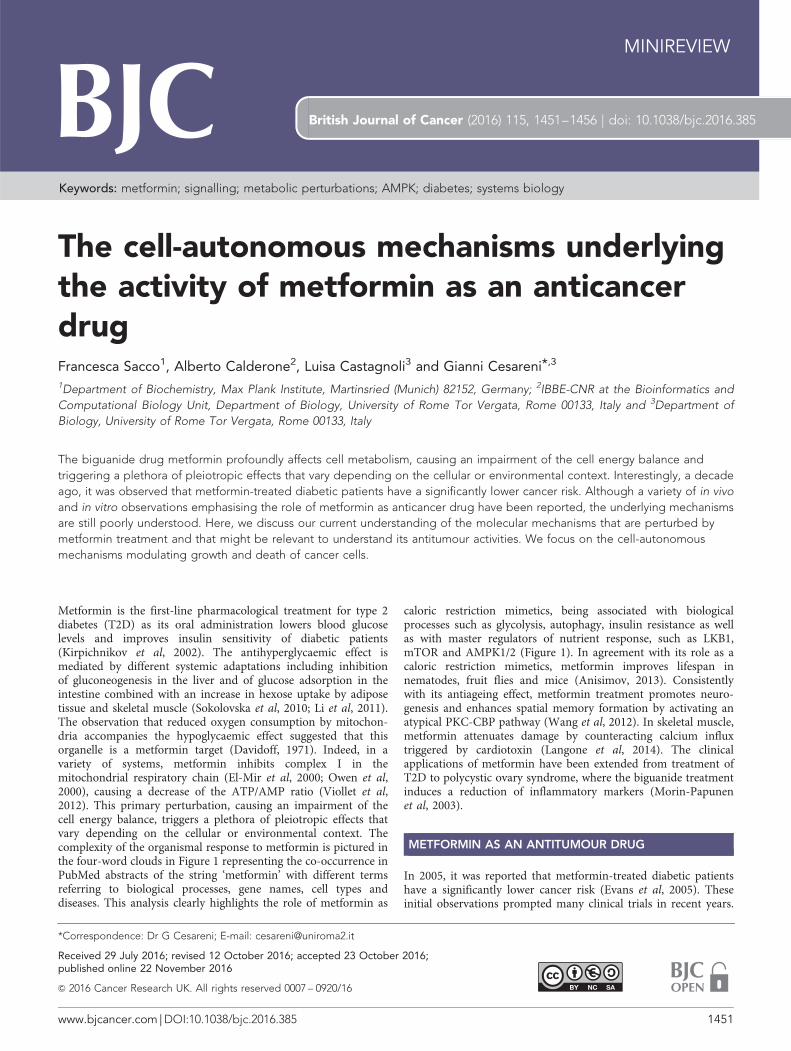
Metformin is the first-line pharmacological treatment for type 2diabetes (T2D) as its oral administration lowers blood glucoselevels and improves insulin sensitivity of diabetic patients(Kirpichnikov et al, 2002). The antihyperglycaemic effect ismediated by different systemic adaptations including inhibitionof gluconeogenesis in the liver and of glucose adsorption in theintestine combined with an increase in hexose uptake by adiposetissue and skeletal muscle (Sokolovska et al, 2010; Li et al, 2011).The observation that reduced oxygen consumption by mitochon-dria accompanies the hypoglycaemic effect suggested that thisorganelle is a metformin target (Davidoff, 1971). Indeed, in avariety of systems, metformin inhibits complex I in themitochondrial respiratory chain (El-Mir et al, 2000; Owen et al,2000), causing a decrease of the ATP/AMP ratio (Viollet et al,2012). This primary perturbation, causing an impairment of thecell energy balance, triggers a plethora of pleiotropic effects thatvary depending on the cellular or environmental context. Thecomplexity of the organismal response to metformin is pictured inthe four-word clouds in Figure 1 representing the co-occurrence inPubMed abstracts of the string ‘metformin’ with different termsreferring to biological processes, gene names, cell types anddiseases. This analysis clearly highlights the role of metformin as
caloric restriction mimetics, being associated with biologicalprocesses such as glycolysis, autophagy, insulin resistance as wellas with master regulators of nutrient response, such as LKB1,mTOR and AMPK1/2 (Figure 1). In agreement with its role as acaloric restriction mimetics, metformin improves lifespan innematodes, fruit flies and mice (Anisimov, 2013). Consistentlywith its antiageing effect, metformin treatment promotes neuro-genesis and enhances spatial memory formation by activating anatypical PKC-CBP pathway (Wang et al, 2012). In skeletal muscle,metformin attenuates damage by counteracting calcium influxtriggered by cardiotoxin (Langone et al, 2014). The clinicalapplications of metformin have been extended from treatment ofT2D to polycystic ovary syndrome, where the biguanide treatmentinduces a reduction of inflammatory markers (Morin-Papunenet al, 2003).
METFORMIN AS AN ANTITUMOUR DRUG
In 2005, it was reported that metformin-treated diabetic patientshave a significantly lower cancer risk (Evans et al, 2005). Theseinitial observations prompted many clinical trials in recent years.
*Correspondence: Dr G Cesareni; E-mail: [email protected]
Received 29 July 2016; revised 12 October 2016; accepted 23 October 2016;published online 22 November 2016
& 2016 Cancer Research UK. All rights reserved 0007 – 0920/16
MINIREVIEW
Keywords: metformin; signalling; metabolic perturbations; AMPK; diabetes; systems biology
British Journal of Cancer (2016) 115, 1451–1456 | doi: 10.1038/bjc.2016.385
www.bjcancer.com |DOI:10.1038/bjc.2016.385 1451
The results of most of them supported the evidence that metformindecreases the risk to develop a variety of tumours. However, somestudies reached conflicting conclusions. Overall, a variety of in vivoand in vitro observations place an emphasis on metformintreatment as a promising therapeutic strategy for cancer preventionand therapy (Sui et al, 2015). Metformin treatment exerts itsanticancer role by triggering different processes: it decreases theamount of the Ki67 proliferation marker in biopsy samples ofnondiabetic women with breast cancer (DeCensi et al, 2014); itsuppresses metastasis formation by preferentially killing stem cellsof different cancer types (Hirsch et al, 2009; Vazquez-Martin et al,2011; Mohammed et al, 2013). The potential of the clinical use ofmetformin for cancer prevention and treatment have beendiscussed in recent reviews (Kourelis and Siegel, 2012; Pernicovaand Korbonits, 2014; Morales and Morris, 2015). Here, we considerour current understanding of the molecular mechanisms under-lying the anticancer activity of metformin, focussing on thecell-autonomous mechanisms modulating growth and death ofcancer cells.
SYSTEMIC EFFECTS OF METFORMIN ON CANCERPROGRESSION
Hyperinsulinaemia and hyperglycaemia have been associated withtumourigenesis risk and poor prognosis in several cancers(Anandakumar et al, 2009; Algire et al, 2010; Nakamura et al,2012). However, it is not clear whether reduced levels of glucoseand insulin are sufficient to explain the antineoplastic effect ofmetformin. For instance, Eikawa et al (2015) proposed that thechemotherapeutic property of metformin stands on its ability toboost the immune system against neoplastic proliferation. Asidefrom the direct effect on tumourigenesis, glucose levels also affecttumour cell sensitivity to metformin treatment (Menendez et al,2012; Wahdan-Alaswad et al, 2013; Zordoky et al, 2014). Cellsgrown in low glucose rely on increased mitochondrial metabolismand are therefore more sensitive to metformin inhibition of
mitochondrial metabolism (Andrzejewski et al, 2014). In addition,apoptosis induced by metformin in triple-negative and ER-high orHER-high breast cancer cell lines depends on AMPK but issuppressed by high concentration of glucose (25mM) and aminoacids (Silvestri et al, 2015). These observations stress theimportance of cell environmental and metabolic context onresponse to metformin treatment. However, our understandingof the systemic anticancer effects of metformin remains limited.
CELL-AUTONOMOUS MECHANISMS MEDIATING THEANTICANCER PROPERTIES OF METFORMIN
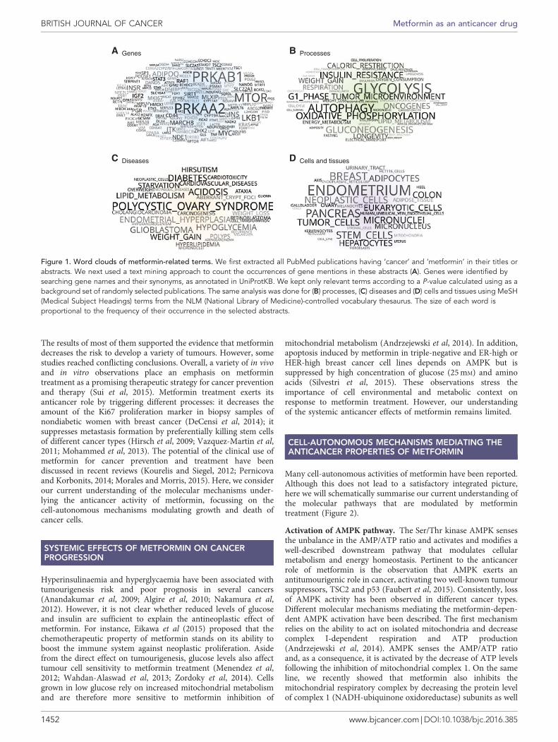
Many cell-autonomous activities of metformin have been reported.Although this does not lead to a satisfactory integrated picture,here we will schematically summarise our current understanding ofthe molecular pathways that are modulated by metformintreatment (Figure 2).
Activation of AMPK pathway. The Ser/Thr kinase AMPK sensesthe unbalance in the AMP/ATP ratio and activates and modifies awell-described downstream pathway that modulates cellularmetabolism and energy homeostasis. Pertinent to the anticancerrole of metformin is the observation that AMPK exerts anantitumourigenic role in cancer, activating two well-known tumoursuppressors, TSC2 and p53 (Faubert et al, 2015). Consistently, lossof AMPK activity has been observed in different cancer types.Different molecular mechanisms mediating the metformin-depen-dent AMPK activation have been described. The first mechanismrelies on the ability to act on isolated mitochondria and decreasecomplex I-dependent respiration and ATP production(Andrzejewski et al, 2014). AMPK senses the AMP/ATP ratioand, as a consequence, it is activated by the decrease of ATP levelsfollowing the inhibition of mitochondrial complex 1. On the sameline, we recently showed that metformin also inhibits themitochondrial respiratory complex by decreasing the protein levelof complex 1 (NADH-ubiquinone oxidoreductase) subunits as well
GenesA ProcessesB
DiseasesC Cells and tissuesD
Figure 1. Word clouds of metformin-related terms. We first extracted all PubMed publications having ‘cancer’ and ‘metformin’ in their titles orabstracts. We next used a text mining approach to count the occurrences of gene mentions in these abstracts (A). Genes were identified bysearching gene names and their synonyms, as annotated in UniProtKB. We kept only relevant terms according to a P-value calculated using as abackground set of randomly selected publications. The same analysis was done for (B) processes, (C) diseases and (D) cells and tissues using MeSH(Medical Subject Headings) terms from the NLM (National Library of Medicine)-controlled vocabulary thesaurus. The size of each word isproportional to the frequency of their occurrence in the selected abstracts.
BRITISH JOURNAL OF CANCER Metformin as an anticancer drug
1452 www.bjcancer.com |DOI:10.1038/bjc.2016.385
as five components of complex 3 (cytochrome bc1 complex; Saccoet al, 2016). As a second mechanism, it was proposed thatmetformin activates AMPK by triggering the activation of theATM–LKB1–AMPK axis (GoDarts et al, 2011). In a genome-wideassociation study, the authors show that inhibition of ATM in a ratcancer cell line weakens metformin-mediated activation of themetabolic enzyme AMPK.
Interestingly, after metformin treatment, the phosphoproteomeprofile of breast cancer cells was significantly enriched for thesubstrate motif of ATM, supporting the involvement of theactivation of this kinase in the cell response to metformin (Sacco
et al, 2016). In addition to these two mechanisms, we recentlyshowed that metformin significantly increases the concentration ofthe regulatory subunit b of AMPK (PRKAB1), whose interactionwith the catalytic subunit triggers the enzyme kinase activity. Thislast observation suggests that full activation of AMPK requires denovo transcription and translation that is consistent with the factthat metformin requires a long-term treatment for completeAMPK activation.
Besides the AMPK-dependent regulation of proteins involvedin lipid and glucose metabolism (ACACA and GFPT1), themetformin-mediated activation of AMPK results in the
Metformin
Complex I and III
ATP/AMP
LKB1
ATM
PRKAB1
GFPT1
ACC
CRTC2
MAPK modulation PTPRJ
ERK2ERK1
JNK2
JNK1
CDK1
PLK1MYC
SP1
CCND1 VHLCDKN1A
Cell cycle inhibition
Activation
Inhibited by metforminActivated by metformin
Inhibition
Not modulated by metformin
SP1 degradation
p38
ULK
IRS1
PIK3CA
REDDRAG
TSC1TSC2
TORC1
MTORDEPTOR
RPTOR
RPS6KB2 p70S6K
rpS6
PPP2CA
PPP2R5D
AKT1
PRKAA1AMPK
activation
AKT-mTORmodulation
INSRAmino acids
Figure 2. Schematic representation of the pathways affected by metformin treatment. The proteins mentioned in this review were linked in agraph representation by using causal relationships extracted from the SIGNOR database (Perfetto et al, 2015). Red hammerheads and blue arrowsrepresent activation and inactivation relationships, respectively. Proteins that are activated or inactivated by metformin are coloured in green andred, respectively.
Metformin as an anticancer drug BRITISH JOURNAL OF CANCER
www.bjcancer.com |DOI:10.1038/bjc.2016.385 1453

downmodulation of cell proliferation-related proteins, such asRAF1 and TBC1D1 (Sacco et al, 2016).
Inhibition of AKT–mTORC1 pathway. The impairment of targetof rapamycin (mTOR) signalling pathway is one of the hallmarksof metformin treatment. The mTOR plays a key role in regulationof cell growth and metabolism, and thus the correlation betweenthe activation of mTOR and tumour progression is not surprising.The activation of the mTOR pathway is involved in the regulationof many cancer hallmarks, including tumour growth, angiogenesisand metastasis (Hanahan and Weinberg, 2011). The mTORC1activation is limited by metformin in several ways: by increasingthe mTOR inhibitor REDD1 (regulated in development and DNAdamage responses) (1); it inhibits the Ragulator complex thatpositively regulates mTOR (2) and it mimics amino acid depletioncausing loss of colocalisation between mTOR and its activatorRheb (3) (Kalender et al, 2010; Pierotti et al, 2013). Themetformin-mediated mTORC1 complex inhibition exerts a strongimpact on key physiological processes, such as autophagy, proteinsynthesis and cell proliferation.
A deep MS-based phosphoproteomics approach revealed thatmetformin does not induce a mere inhibition of the whole AKT–mTOR axis, but rather rewires the whole pathway. Analysing theAKT–mTORC1 pathway in breast cancer cells MCF7 treated withmetformin, we found that metformin decreases phosphorylation oftwo mTOR substrates, p70S6K and rpS6 but, contrary toexpectation, this is not mediated by a diminished mTOR activity.We find that, in these conditions, the activity of the upstreaminducer of mTORC1 complex, the AKT kinase, is increased bymetformin. As a consequence, the mTOR inhibitor TSC2 isphosphorylated and therefore inhibited. There are two main logicaldisconnections in this pathway when perturbed by metformin: themTOR repressor, TSC2, is inhibited but mTOR remains steadilyactivated, whereas its downstream effector S6K is inhibited. On thesame line, in MCF7 cells treated with metformin, addition ofinsulin-like growth factor (IGF) triggers IGF1R and AKTphosphorylation, whereas p70S6K substrates (rpS6, IRS1) are notactivated, remaining in a constitutive inactive form. We proposethat IGF stimulation fails to activate p70S6K because metformintriggers the assembly and activation of the PP2A phosphatase byincreasing phosphorylation of the PPP2R5C regulatory subunit(Sacco et al, 2016).
Inhibition of cell cycle progression. Cell cycle progression isoften altered in cancer cells, leading to an aberrant regulation ofcell division and DNA replication. Many reports demonstrated thatthe anticancer property of metformin is a consequence of cell cycleperturbation. Specifically, exposure to metformin induced cell cyclearrest in G0/G1 and G2/M phases in different cancer types,including breast cancer cells and oesophageal squamous cancercells. The molecular mechanisms underlying the metformin-induced cell cycle arrest have been extensively described andinvolve the upregulation of cyclin-dependent kinase inhibitors(CDKIs), such as p21 or p27 proteins. Although in breast cancercells (ER positive and negative, erbB2-overexpressing and erbB2-normal) metformin treatment does not affect the expression levelsof p21 e p27 CDKIs (Alimova et al, 2009), other authors also reporta cyclin D1 reduction with a concomitant increase of p53, p21, Baxand cyclin E expression in metformin-treated MCF7 (Malki andYoussef, 2011). On the same line, in a metformin-treatedoesophageal squamous cells carcinoma xenograft model, theauthors observed the upregulation of p53, p21 and p27 and thedownregulation of cyclin D1 (Cai et al, 2015).
Metformin treatment also affects the activity of cyclin-dependent kinases, such as CDK1 in MCF7 cells as well asCDK2, CDK4 and CDK6 in head and neck squamous cellcarcinoma (Sikka et al, 2012; Sacco et al, 2016).
MAPK regulation. The mitogen-activated protein kinases(MAPKs), including the ERK1/2, p38 and JNK kinase branches,control fundamental biological processes, including proliferation,differentiation, survival and death. Although there is a generalconsensus about the metformin-dependent inhibition of ERK1/2kinases in different cancer types (Memmott et al, 2010; Gou et al,2013; Sacco et al, 2016), contrasting evidences have been reportedabout the modulation of p38 and JNK pathways. This incongruitycan be explained by the pro-apoptotic or pro-proliferative rolesthat these stress-activated kinases may play depending on thecellular context. In lung cancer cells, metformin treatmentdecreases the activity of JNK and p38 that, on the contrary, ishyperactivated in metformin-treated MCF7 cells (Tseng et al, 2013;Queiroz et al, 2014; Sacco et al, 2016). The molecular mechanismsunderlying the metformin-mediated modulation of the MAPKcascades are still poorly understood. We suggest that metforminmodulates these pathways by acting on the protein level of theupstream regulatory kinases and phosphatases. Metforminincreases the concentration of the tyrosine phosphatase PTPRJ, anegative regulator of ERK1/2 (Sacco et al, 2009, 2012) andconcomitantly it decreases the expression level of the upstreamkinase ARAF. On the same line, after metformin treatment, themRNA and protein levels of the MAP3K3 and MAP3K7 kinasesare upregulated, resulting in increased phosphorylation of both p38and the JNK substrate, JunD. Our hypothesis is that metforminputs MCF7 breast cancer cells in a pro-apoptotic state triggering asustained activation of JNK and p38. If MCF7 cells are treated withmetformin they become more sensitive to apoptotic stimuli such asthe ones given by staurosporin or other chemotherapeutic drugs(Sacco et al, 2016).
SP1 transcription factor regulation. Metformin treatmentinduces a radical remodelling of the transcriptome and proteome,and it is not surprising to observe modulation of the activity ofdifferent transcription factors. Recent evidences demonstrated thatmetformin inhibits the SP transcription factor family membersin vivo and in different cancer cell lines, such as pancreatic tumourcells or breast cancer cells (Sacco et al, 2016).
Safe and colleagues (Nair et al, 2013) demonstrated that inpancreatic cancer L3.6pL and Panc28 cells, metformin inducedproteasome-dependent degradation of Sp1, Sp3 and Sp4 transcrip-tion factors, resulting in the downregulation of several pro-oncogenic Sp-regulated genes including bcl-2, survivin, cyclin D1,vascular endothelial growth factor with its receptor and fatty acidsynthase. Interestingly, the molecular mechanism through whichmetformin inhibits Sp transcription factors is context dependent.The authors reported that in a different pancreatic cancer cells,Panc, metformin decreased microRNA-27a and induced the Sprepressor, ZBTB10 (Nair et al, 2013).
The analysis of metformin-induced transcriptome and pro-teome profiles in breast cancer cells enabled us to identify the SP1transcription factor as the most significantly modulated, as it isequally depleted at both transcriptome and proteome levels. Ourdata, combined with the metformin-dependent phosphoproteomeprofile, are consistent with a model where metformin induces theSP1 degradation by increasing the phosphorylation of Ser51 thatdestabilises the transcription factor. We inferred that p38 kinase isresponsible of the metformin-mediated SP1 phosphorylation. Thedecrease in the levels of SP1 is mirrored by a significant reductionof many SP1-controlled cancer and metabolic pathways. Theexpression of Von Hippel Lindau protein (VHL) is also diminishedby metformin. The VHL is SP1 controlled and, besides its role asan E3 ligase of HIF-a in normoxia, it can repress the promoteractivity of IGF1R by sequestering SP1 (Yuen et al, 2007). The SP1also controls the expression of pyruvate kinase that is over-expressed in many tumours. We observe a downregulation of theoncogenic isoform pyruvate kinase M2 in MCF7 and SKBR3 breast
BRITISH JOURNAL OF CANCER Metformin as an anticancer drug
1454 www.bjcancer.com |DOI:10.1038/bjc.2016.385

cancer cells treated with metformin when grown in normogly-caemic condition. On the contrary, if glucose is increased fourfold,metformin does not affect the expression levels of PKM2 (Silvestriet al, 2015).
The unfolded protein response (UPR). The UPR is triggered by apathological increase of unfolded or misfolded proteins that issensed by three receptors on the ER membrane. When thesesensors dissociate from the ER chaperon GRP78, they activatesignal transductions to stop protein synthesis, to increaseproteasomal degradation and, ultimately, undergo apoptosis byoverexpressing CHOP. Metformin was found to cause an AMPK-dependent downregulation of GRP78 while increasing the levels ofunfolded proteins in the ER lumen. As a consequence, the cellbecomes unable to adequately respond to ER stress and dies (Saitoet al, 2009).
In prostate cancer cells, metformin also induces CHOP-dependent apoptosis by regulating the expression of numerousmiRNAs. Among them, the tumour suppressor miR-708-5p isstrongly upregulated. This miR collaborates in increasing the ERstress by suppressing the ER membrane protein neuronatin,NNAT, that controls calcium homeostasis (Yang et al, 2015). It isnoteworthy that in cardiomyocytes, metformin activates the UPRbut, in spite of a strong upregulation of the pro-apoptotic CHOPprotein, cells are not driven to death (Quentin et al, 2012), stressingonce more that metformin modulates cell death and survival in acell-specific manner.
CONCLUSIONS
The observation that metformin reduces cancer risk in diabeticpatients has raised considerable interest and has stimulated avariety of studies. Despite the wealth of information, we are still farfrom a clear consistent picture that could support rationalstrategies for cancer prevention and treatment. Metformin has aprofound impact on the organism energy balance and metabolism,and the cells in the different organs, by sensing these signals,respond differently, depending on the molecular and cellularcontext. Tumour cell growth is affected by both changes in theenvironmental cues and a direct action of metformin on themolecular pathways that regulate cell growth and death. Here wehave discussed the cell-autonomous mechanisms that are affectedby metformin treatment combining the results of a high-contentgenome-wide study on a breast cancer cell line with more focussedreports on different tumour systems. After metformin treatment,the transcriptome and proteome of MCF7 breast cancer cells areremodelled and the signalling pathways are rewired. Transcription,translation and post-translational modifications are profoundlyaffected. The resulting picture is revealing and complex at the sametime and partly explains why metformin has such pleiotropiceffects depending on context (Figure 2). These studies have openedthe path to a systems understanding of the molecular mechanismsunderlying metformin effects, but at the same time call for moresuch studies in different systems and in different conditions. Onlysuch a genome-wide understanding of the subtle differences in theresponse of different systems in different conditions will eventuallyoffer a rational basis for designing new strategies to potentiate theaction of metformin by combining it with different antitumourtreatments.
ACKNOWLEDGEMENTS
This work was supported by the DEPTH project of the EuropeanResearch Council (Grant Agreement 322749) by the ItalianAssociation for Cancer Research Grant (N.14135) to GC.
CONFLICT OF INTEREST
The authors declare no conflict of interest.
REFERENCES
Algire C, Amrein L, Zakikhani M, Panasci L, Pollak M (2010) Metforminblocks the stimulative effect of a high-energy diet on colon carcinomagrowth in vivo and is associated with reduced expression of fatty acidsynthase. Endocr Relat Cancer 17(2): 351–360.
Alimova IN, Liu B, Fan Z, Edgerton SM, Dillon T, Lind SE, Thor AD (2009)Metformin inhibits breast cancer cell growth, colony formation andinduces cell cycle arrest in vitro. Cell Cycle 8(6): 909–915.
Anandakumar P, Kamaraj S, Jagan S, Ramakrishnan G, Devaki T (2009) Effectof capsaicin on glucose metabolism studied in experimental lungcarcinogenesis. Nat Prod Res 23(8): 763–774.
Andrzejewski S, Gravel SP, Pollak M, St-Pierre J (2014) Metformin directlyacts on mitochondria to alter cellular bioenergetics. Cancer Metabol 2: 12.
Anisimov VN (2013) Metformin: do we finally have an anti-aging drug? CellCycle 12(22): 3483–3489.
Cai X, Hu X, Tan X, Cheng W, Wang Q, Chen X, Guan Y, Chen C, Jing X(2015) Metformin induced AMPK activation, G0/G1 phase cell cyclearrest and the inhibition of growth of esophageal squamous cellcarcinomas in vitro and in vivo. PLoS One 10(7): e0133349.
Davidoff F (1971) Effects of guanidine derivatives on mitochondrial function.3. The mechanism of phenethylbiguanide accumulation and itsrelationship to in vitro respiratory inhibition. J Biol Chem 246(12):4017–4027.
DeCensi A, Puntoni M, Gandini S, Guerrieri-Gonzaga A, Johansson HA,Cazzaniga M, Pruneri G, Serrano D, Schwab M, Hofmann U, Mora S,Aristarco V, Macis D, Bassi F, Luini A, Lazzeroni M, Bonanni B, PollakMN (2014) Differential effects of metformin on breast cancer proliferationaccording to markers of insulin resistance and tumor subtype in arandomized presurgical trial. Breast Cancer Res Treat 148(1): 81–90.
Eikawa S, Nishida M, Mizukami S, Yamazaki C, Nakayama E, Udono H(2015) Immune-mediated antitumor effect by type 2 diabetes drug,metformin. Proc Natl Acad Sci USA 112(6): 1809–1814.
El-Mir MY, Nogueira V, Fontaine E, Averet N, Rigoulet M, Leverve X (2000)Dimethylbiguanide inhibits cell respiration via an indirect effect targetedon the respiratory chain complex I. J Biol Chem 275(1): 223–228.
Evans JM, Donnelly LA, Emslie-Smith AM, Alessi DR, Morris AD (2005)Metformin and reduced risk of cancer in diabetic patients. BMJ 330(7503):1304–1305.
Faubert B, Vincent EE, Poffenberger MC, Jones RG (2015) The AMP-activated protein kinase (AMPK) and cancer: many faces of a metabolicregulator. Cancer Lett 356(2 Pt A): 165–170.
GoDARTS and UKPDS Diabetes Pharmacogenetics Study Group, WellcomeTrust Case Control Consortium, Zhou K, Bellenguez C, Spencer CC,Bennett AJ, Coleman RL, Tavendale R, Hawley SA, Donnelly LA,Schofield C, Groves CJ, Burch L, Carr F, Strange A, Freeman C, BlackwellJM, Bramon E, Brown MA, Casas JP, Corvin A, Craddock N, Deloukas P,Dronov S, Duncanson A, Edkins S, Gray E, Hunt S, Jankowski J, LangfordC, Markus HS, Mathew CG, Plomin R, Rautanen A, Sawcer SJ, Samani NJ,Trembath R, Viswanathan AC, Wood NW, investigators M, Harries LW,Hattersley AT, Doney AS, Colhoun H, Morris AD, Sutherland C,Hardie DG, Peltonen L, McCarthy MI, Holman RR, Palmer CN,Donnelly P, Pearson ER (2011) Common variants near ATM areassociated with glycemic response to metformin in type 2 diabetes. NatGenet 43(2): 117–120.
Gou S, Cui P, Li X, Shi P, Liu T, Wang C (2013) Low concentrations ofmetformin selectively inhibit CD133(þ ) cell proliferation in pancreaticcancer and have anticancer action. PLoS One 8(5): e63969.
Hanahan D, Weinberg RA (2011) Hallmarks of cancer: the next generation.Cell 144(5): 646–674.
Hirsch HA, Iliopoulos D, Tsichlis PN, Struhl K (2009) Metformin selectivelytargets cancer stem cells, and acts together with chemotherapy to blocktumor growth and prolong remission. Cancer Res 69(19): 7507–7511.
Kalender A, Selvaraj A, Kim SY, Gulati P, Brule S, Viollet B, Kemp BE,Bardeesy N, Dennis P, Schlager JJ, Marette A, Kozma SC, Thomas G(2010) Metformin, independent of AMPK, inhibits mTORC1 in a ragGTPase-dependent manner. Cell Metabol 11(5): 390–401.
Metformin as an anticancer drug BRITISH JOURNAL OF CANCER
www.bjcancer.com |DOI:10.1038/bjc.2016.385 1455
Kirpichnikov D, McFarlane SI, Sowers JR (2002) Metformin: an update. AnnIntern Med 137(1): 25–33.
Kourelis TV, Siegel RD (2012) Metformin and cancer: new applications for anold drug. Med Oncol 29(2): 1314–1327.
Langone F, Cannata S, Fuoco C, Lettieri Barbato D, Testa S, Nardozza AP,Ciriolo MR, Castagnoli L, Gargioli C, Cesareni G (2014) Metforminprotects skeletal muscle from cardiotoxin induced degeneration. PLoS One9(12): e114018.
Li Y, Xu S, Mihaylova MM, Zheng B, Hou X, Jiang B, Park O, Luo Z, Lefai E,Shyy JY, Gao B, Wierzbicki M, Verbeuren TJ, Shaw RJ, Cohen RA, Zang M(2011) AMPK phosphorylates and inhibits SREBP activity to attenuatehepatic steatosis and atherosclerosis in diet-induced insulin-resistant mice.Cell Metab 13(4): 376–388.
Malki A, Youssef A (2011) Antidiabetic drug metformin induces apoptosis inhuman MCF breast cancer via targeting ERK signaling. Oncol Res 19(6):275–285.
Memmott RM, Mercado JR, Maier CR, Kawabata S, Fox SD, Dennis PA(2010) Metformin prevents tobacco carcinogen–induced lungtumorigenesis. Cancer Prev Res 3(9): 1066–1076.
Menendez JA, Oliveras-Ferraros C, Cufi S, Corominas-Faja B, Joven J,Martin-Castillo B, Vazquez-Martin A (2012) Metformin is syntheticallylethal with glucose withdrawal in cancer cells. Cell cycle 11(15):2782–2792.
Mohammed A, Janakiram NB, Brewer M, Ritchie RL, Marya A, Lightfoot S,Steele VE, Rao CV (2013) Antidiabetic drug metformin preventsprogression of pancreatic cancer by targeting in part cancer stem cells andmTOR signaling. Transl Oncol 6(6): 649–659.
Morales DR, Morris AD (2015) Metformin in cancer treatment andprevention. Annu Rev Med 66: 17–29.
Morin-Papunen L, Rautio K, Ruokonen A, Hedberg P, Puukka M, Tapanainen JS(2003) Metformin reduces serum C-reactive protein levels in womenwith polycystic ovary syndrome. J Clin Endocrinol Metabol 88(10):4649–4654.
Nair V, Pathi S, Jutooru I, Sreevalsan S, Basha R, Abdelrahim M, Samudio I,Safe S (2013) Metformin inhibits pancreatic cancer cell and tumor growthand downregulates Sp transcription factors. Carcinogenesis 34(12): 2870–2879.
Nakamura A, Tajima K, Zolzaya K, Sato K, Inoue R, Yoneda M, Fujita K,Nozaki Y, Kubota KC, Haga H, Kubota N, Nagashima Y, Nakajima A,Maeda S, Kadowaki T, Terauchi Y (2012) Protection from non-alcoholicsteatohepatitis and liver tumourigenesis in high fat-fed insulin receptorsubstrate-1-knockout mice despite insulin resistance. Diabetologia 55(12):3382–3391.
Owen MR, Doran E, Halestrap AP (2000) Evidence that metformin exerts itsanti-diabetic effects through inhibition of complex 1 of the mitochondrialrespiratory chain. Biochem J 348(Pt 3): 607–614.
Perfetto L, Briganti L, Calderone A, Perpetuini AC, Iannuccelli M, Langone F,Licata L, Marinkovic M, Mattioni A, Pavlidou T, Peluso D, Petrilli LL,Pirro S, Posca D, Santonico E, Silvestri A, Spada F, Castagnoli L, Cesareni G(2015) SIGNOR: a database of causal relationships between biologicalentities. Nucleic Acids Res 44: D548–D554.
Pernicova I, Korbonits M (2014) Metformin–mode of action and clinicalimplications for diabetes and cancer. Nat Rev Endocrinol 10(3):143–156.
Pierotti MA, Berrino F, Gariboldi M, Melani C, Mogavero A, Negri T, Pasanisi P,Pilotti S (2013) Targeting metabolism for cancer treatment andprevention: metformin, an old drug with multi-faceted effects. Oncogene32(12): 1475–1487.
Queiroz EA, Puukila S, Eichler R, Sampaio SC, Forsyth HL, Lees SJ, Barbosa AM,Dekker RF, Fortes ZB, Khaper N (2014) Metformin induces apoptosis andcell cycle arrest mediated by oxidative stress, AMPK and FOXO3a inMCF-7 breast cancer cells. PLoS One 9(5): e98207.
Quentin T, Steinmetz M, Poppe A, Thoms S (2012) Metformin differentiallyactivates ER stress signaling pathways without inducing apoptosis. DisModel Mech 5(2): 259–269.
Sacco F, Gherardini PF, Paoluzi S, Saez-Rodriguez J, Helmer-Citterich M,Ragnini-Wilson A, Castagnoli L, Cesareni G (2012) Mapping the humanphosphatome on growth pathways. Mol Syst Biol 8: 603.
Sacco F, Silvestri A, Posca D, Pirro S, Gherardini P, Castagnoli L, Mann M,Cesareni G (2016) Deep proteomics of breast cancer cells reveals thatmetformin rewires signaling networks away from a pro-growth state. CellSyst 3: 2–13.
Sacco F, Tinti M, Palma A, Ferrari E, Nardozza AP, Hooft van Huijsduijnen R,Takahashi T, Castagnoli L, Cesareni G (2009) Tumor suppressor density-enhanced phosphatase-1 (DEP-1) inhibits the RAS pathway by directdephosphorylation of ERK1/2 kinases. J Biol Chem 284(33): 22048–22058.
Saito S, Furuno A, Sakurai J, Sakamoto A, Park HR, Shin-Ya K, Tsuruo T,Tomida A (2009) Chemical genomics identifies the unfolded proteinresponse as a target for selective cancer cell killing during glucosedeprivation. Cancer Res 69(10): 4225–4234.
Sikka A, Kaur M, Agarwal C, Deep G, Agarwal R (2012) Metforminsuppresses growth of human head and neck squamous cell carcinoma viaglobal inhibition of protein translation. Cell Cycle 11(7): 1374–1382.
Silvestri A, Palumbo F, Rasi I, Posca D, Pavlidou T, Paoluzi S, Castagnoli L,Cesareni G (2015) Metformin induces apoptosis and downregulatespyruvate kinase M2 in breast cancer cells only when grown in nutrient-poor conditions. PLoS One 10(8): e0136250.
Sokolovska J, Isajevs S, Sugoka O, Sharipova J, Lauberte L, Svirina D, Rostoka E,Sjakste T, Kalvinsh I, Sjakste N (2010) Influence of metformin on GLUT1gene and protein expression in rat streptozotocin diabetes mellitus model.Arch Physiol Biochem 116(3): 137–145.
Sui X, Xu Y, Wang X, Han W, Pan H, Xiao M (2015) Metformin: a novel butcontroversial drug in cancer prevention and treatment.Mol Pharm 12(11):3783–3791.
Tseng SC, Huang YC, Chen HJ, Chiu HC, Huang YJ, Wo TY, Weng SH,Lin YW (2013) Metformin-mediated downregulation of p38 mitogen-activated protein kinase-dependent excision repair cross-complementing 1decreases DNA repair capacity and sensitizes human lung cancer cells topaclitaxel. Biochem Pharmacol 85(4): 583–594.
Vazquez-Martin A, Oliveras-Ferraros C, Cufi S, Del Barco S, Martin-Castillo B,Lopez-Bonet E, Menendez JA (2011) The anti-diabetic drug metforminsuppresses the metastasis-associated protein CD24 in MDA-MB-468triple-negative breast cancer cells. Oncol Rep 25(1): 135–140.
Viollet B, Guigas B, Sanz Garcia N, Leclerc J, Foretz M, Andreelli F (2012)Cellular and molecular mechanisms of metformin: an overview. Clin Sci122(6): 253–270.
Wahdan-Alaswad R, Fan Z, Edgerton SM, Liu B, Deng XS, Arnadottir SS,Richer JK, Anderson SM, Thor AD (2013) Glucose promotes breast canceraggression and reduces metformin efficacy. Cell Cycle 12(24): 3759–3769.
Wang J, Gallagher D, DeVito LM, Cancino GI, Tsui D, He L, Keller GM,Frankland PW, Kaplan DR, Miller FD (2012) Metformin activates anatypical PKC-CBP pathway to promote neurogenesis and enhance spatialmemory formation. Cell Stem Cell 11(1): 23–35.
Yang J, Wei J, Wu Y, Wang Z, Guo Y, Lee P, Li X (2015) Metformin inducesER stress-dependent apoptosis through miR-708-5p/NNAT pathway inprostate cancer. Oncogenesis 4: e158.
Yuen JS, Cockman ME, Sullivan M, Protheroe A, Turner GD, Roberts IS,Pugh CW, Werner H, Macaulay VM (2007) The VHL tumor suppressorinhibits expression of the IGF1R and its loss induces IGF1R upregulationin human clear cell renal carcinoma. Oncogene 26(45): 6499–6508.
Zordoky BN, Bark D, Soltys CL, Sung MM, Dyck JR (2014) The anti-proliferative effect of metformin in triple-negative MDA-MB-231 breastcancer cells is highly dependent on glucose concentration: implications forcancer therapy and prevention. Biochim Biophys Acta 1840(6): 1943–1957.
This work is licensed under the Creative CommonsAttribution-Non-Commercial-Share Alike 4.0 Inter-
national License. To view a copy of this license, visit http://creativecommons.org/licenses/by-nc-sa/4.0/
BRITISH JOURNAL OF CANCER Metformin as an anticancer drug
1456 www.bjcancer.com |DOI:10.1038/bjc.2016.385