Neural Circuit Mechanisms Underlying the Exacerbation of ...
84
Neural Circuit Mechanisms Underlying the Exacerbation of Alzheimer’s Disease by Chronic Stress Citation Mason, Xenos. 2015. Neural Circuit Mechanisms Underlying the Exacerbation of Alzheimer’s Disease by Chronic Stress. Doctoral dissertation, Harvard Medical School. Permanent link http://nrs.harvard.edu/urn-3:HUL.InstRepos:15821598 Terms of Use This article was downloaded from Harvard University’s DASH repository, and is made available under the terms and conditions applicable to Other Posted Material, as set forth at http:// nrs.harvard.edu/urn-3:HUL.InstRepos:dash.current.terms-of-use#LAA Share Your Story The Harvard community has made this article openly available. Please share how this access benefits you. Submit a story . Accessibility
-
Upload
khangminh22 -
Category
Documents
-
view
0 -
download
0
Transcript of Neural Circuit Mechanisms Underlying the Exacerbation of ...

Neural Circuit Mechanisms Underlying the Exacerbation of Alzheimer’s Disease by Chronic Stress
CitationMason, Xenos. 2015. Neural Circuit Mechanisms Underlying the Exacerbation of Alzheimer’s Disease by Chronic Stress. Doctoral dissertation, Harvard Medical School.
Permanent linkhttp://nrs.harvard.edu/urn-3:HUL.InstRepos:15821598
Terms of UseThis article was downloaded from Harvard University’s DASH repository, and is made available under the terms and conditions applicable to Other Posted Material, as set forth at http://nrs.harvard.edu/urn-3:HUL.InstRepos:dash.current.terms-of-use#LAA
Share Your StoryThe Harvard community has made this article openly available.Please share how this access benefits you. Submit a story .
Accessibility

な
Acknowledgements 2
Glossary 3
Abstract 4
Introduction 5
Methods 30
Results 38
Discussion 46
Summary 57
References 58
Figures 73
Figure Legends 80

に
Firstly, I wish to thank the labs of William Klein (Northwestern University) and Karl Deisseroth
(Stanford University) for their scientific collaboration; thanks also to Ivana Dellale (Boston
University), Matthew Frosch (Massachusetts General Hospital), the Neuropathology Core of the
Massachusetts Alzheimer Disease Research Center (P50 AG005134), and the Neuropathology
Service of the Massachusetts General Hospital for their help in obtaining and processing human
tissue samples. Though not included in this thesis, data from these experiments will likely contribute
to any future manuscripts detailing the research herein.
Second, I wish to thank HHMI for facilitating and funding an exciting, rewarding, and formative
one-year research fellowship. Special thanks to Melanie Daub and William R. Galey for their truly
tireless work on behalf of the HHMI Medical Fellows.
Lastly and most importantly, to the Tsai lab – thank you for accepting me into a dynamic, creative,
and incredibly productive group of scientists, and friends. I’ve found unending support and
encouragement from all of you, but a few people deserve particular mention.
Li-Huei Tsai - your expectation for excellence was always balanced by guidance, and a solid
confidence in my work. Through your advocacy, I never felt like anything less than a full member of
the lab – an honor in itself. Thank you for giving me the opportunity to join your team.
Adam Bero – I often learned more from our coffee breaks than from hours of reading. Thank you
for sharing your scholarship and principled approach to science.
Matthew Dobbin, Ram Magabushi, and Becky Canter – thank you all for your close instruction and
guidance, and of course for all the humour.
Damien Rei – your mentorship and friendship has been paramount to any and all of my success in
the Tsai lab. I hope that my year short with you has fulfilled the old proverb, that “shared joy is a
double joy; shared sorrow is half a sorrow”.
ぬ
AAV5: Adeno-associated virus, serotype 5
Aȕ: Amyloid-beta
AD: Alzheimer’s disease
APP: Amyloid precursor protein
BLA: Basolateral amygdala (basolateral nuclear group)
CaMKII: Calcium/calmodulin-dependent protein kinase
ChR2: Channelrhodopsin-2
CNO: Clozapine-N-oxide
CRF: Corticotropin releasing factor
CRS: Chronic restraint stress
CSF: Cerebrospinal fluid
DG: Dentate gyrus
eYFP: Enhanced yellow fluorescent protein
GiDREADD: Inhibitory G-protein-coupled “designer receptors exclusively activated by
designer drugs”
GFAP: Glial fibrillary acidic protein
GR: Glucocorticoid receptor
HDAC2: Histone deacetylase 2
JNK: c-Jun N-terminal-kinase
HPA: Hypothalamic pituitary adrenal (axis)
MCI: Mild cognitive impairment
MR: Mineralocorticoid receptor
NOL: Novel object location
NOR: Novel object recognition
PS1: Presenilin-1
PS2: Presenelin-2
PVN: Paraventricular nucleus of the hypothalamus
RFS: Repetitive foot-shock
RIP: Regulated intramembrane proteolysis

ね
Both epidemiological and animal studies have demonstrated a strong association between
Alzheimer’s disease (AD), neuropsychiatric symptoms such as depression and anxiety, and chronic
psychological stress. The neurophysiological basis of fear, anxiety, and stress has been well studied
and is thought to involve the basolateral amygdala (BLA) – a structure of the anterior temporal lobe,
which interprets fearful stimuli and outputs a behavioral fear response. Similarly, dysfunction and
maladaptation within limbic circuits involving the BLA is thought to be a common etiological factor
of otherwise distinct neuropsychiatric disorders such as Major Depression, and Generalized Anxiety.
To determine if increased BLA activity could act to accelerate the progression of AD, we
manipulated a direct BLA-to-hippocampus circuit using optogenetic (ChR2) and pharmacogenetic
(GiDREADD) technologies, and subsequently examined hippocampal AD-related pathology,
synaptic density, histone-deacetylase-2 expression, and hippocampus-dependent learning and
memory abilities. We found that in wild-type mice, activation of glutamatergic BLA neurons was
both necessary and sufficient to produce the molecular and cognitive effects of chronic stress.
Terminal photostimulation of direct BLA afferents within the hippocampus was also sufficient,
suggesting that the effects of chronic stress throughout the brain are mediated at least in part by
direct excitatory projections originating in the BLA. Chronic activation of BLA glutamatergic
neurons in the 5xFAD model of AD accelerated the neuropathological and cognitive AD-like
phenotype, while chronic BLA inactivation had opposite effects. Overall our results suggest that
neuropsychiatric disease and chronic stress may act through enhanced BLA activation to accelerate
the progression of AD.

の
A ’ D N D
Alzheimer’s disease (AD) is a neurodegenerative disease of the cortical grey matter. It is the most
common dementia in the United States (60-80 percent of all dementia cases, with a prevalence of
4.7-5.4 million), and is the sixth leading cause of death (1; 2). One in nine people older than age 65,
and about one third of those older than 85 suffer from AD; as the population above age 85 increases
through to 2050, the prevalence of AD is expected to climb to 13.8-16 million, accompanied by
rising healthcare costs and public burden of disease (1; 2). Unfortunately there are currently few
effective disease modifying treatments, and no available preventative therapy.
Although AD is a pathological diagnosis, the Diagnostic and Statistical Manual of Mental Disorders V
outlines diagnostic clinical criteria for “probable” AD: (i) presence of a neurocognitive disorder,
defined as decline in a cognitive domain (attention, executive functioning, learning and declarative
memory, language, perceptual-motor, or social cognition) which interferes with independent
functioning, and (ii) progressive gradual decline specifically in learning and declarative memory, or
evidence of a Alzheimer’s disease genetic mutation (3). Neuropsychiatric symptoms and affective
dysregulation are also included in the symptom profile of AD (4).
New diagnostic guidelines from the National Institutes of Aging highlight the importance of
biomarkers for the diagnosis of disease, which has lead to both a shift in research focus, and in
perceptions of AD. “Pre-clinical AD” has been defined as a biomarker-positive but symptom-free
stage, while precursory “mild-cognitive impairment” defines the earliest stages of cognitive decline

は
before frank dementia can be diagnosed (5). The symptomatic phase of AD is now thought to be the
final result of years – and perhaps decades – of biochemical and neurophysiological derangement.
These earlier changes produce the neuropathological hallmarks of this disease – Amyloid-Beta (Aが)
neuritic plaques and neurofibrillary tangles of hyperphosphorylated Tau protein (6). These two
hallmarks also serve as the triggers of definitive diagnosis on autopsy, since clinical diagnosis is
generally based on the exclusion of other causes of dementia (3). Although Aが and Tau are well
studied, the mechanisms of their toxicity, and thus the pathophysiology of AD, is largely
unexplained. Two hypotheses have dominated the scientific literature over the past half-century: the
cholinergic hypothesis, and the amyloid-cascade hypothesis (7) (8) (9).
P A ’ D :
T C A C H
Early hypotheses on the etiology of AD focused on the similarity with Parkinson’s disease, in which
neurodegeneration of the substantia nigra pars compacta leads to dopamine depletion and basal
ganglia dysfunction. In the case of AD, another neuromodulatory neurotransmitter – acetylcholine –
was postulated to be involved (9). The cholinergic nuclei project very broadly throughout the cortex
and subcortical limbic structures, providing a powerful substrate for disease should these projections
degenerate. Although cholinesterase inhibitors are indicated for the symptomatic treatment of AD
(10; 11), metanalysis has demonstrated that these drugs have no effect on the rate of conversion
from MCI to AD (12). Though circuit level dysfunction of the cholinergic system may contribute
to attention and memory deficits in AD, the greater majority of evidence weighs in favor of another
process being the cause of this disease.

ば
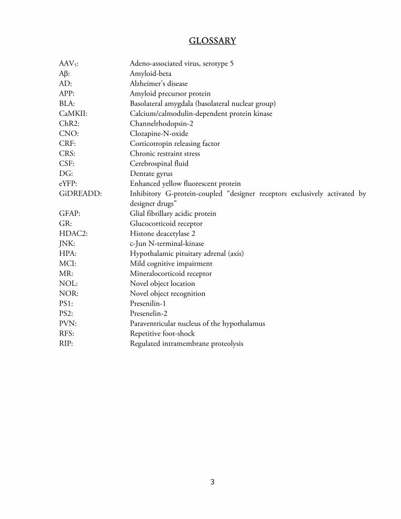
The dominant hypothesis of AD pathophysiology is termed the “amyloid cascade”. Briefly, cleavage
of amyloid precursor protein (APP) – a ubiquitous transmembrane protein of unknown function –
by membrane proteases produces short peptide fragments known as amyloid-beta (Aが), which are
thought to aggregate into neurotoxic oligomers and fibrillary polymers in the extracellular space (7).
Aが generation begins with transport of APP to the cell membrane and axon terminal (13), where it
associates closely with three membrane proteases – ゎ-secretase, が-secretase (also known as が-amyloid
cleaving enzyme, or BACE-1) , and ぐ-secretase (a multi-subunit complex composed of one of
presenilin-1 (PS-1) or presenilin-2 (PS-2), and three accessory proteins) (14). Two divergent
catabolic pathways of “regulated intramembrane proteolysis” (RIP) then proceed from this point. In
the first pathway, APP is cleaved at the cell membrane first by ゎ-secretase and then by ぐ-secretase,
generating entirely soluble, “non-amyloidogenic” APP fragments (14). In the “amyloidogenic”
pathway, APP is internalized to the endosomal fraction where it is cleaved first by が-secretase, then ぐ-secretase (15; 16). Exocytosis releases soluble fragments of either 40 (Aが40) or 42 (Aが42) amino
acids in length, which self assemble into oligomers, protofibrils, and insoluble fibrillar plaques (15).
ぱ
(13)
The physiological function of APP remains unknown; APP knockout mice show no immediate
phenotype, but may develop learning and memory deficits with age (17). Experiments in vivo and in
vitro suggest that APP may act as a growth factor - regulating synaptic pruning, neuronal migration,

ひ
and neuron survival in development (18; 19). More recent studies offer more support to a
hypothesized function in synaptogenesis and maintenance of synaptic density, but also provide
evidence that APP may participate in calcium homeostasis, synaptic transmission, and epigenetic
regulation of gene expression (through interactions with the histone acetyltransferase Tip60) (20).
There is also work to suggest that Aが fragments themselves may have a physiological function in
decreasing activity at highly active synapses and facilitating LTP at low picomolar concentration, but
most research on Aが fragments has focused on direct neurotoxicity (21).
Evidence for toxicity of Aが fibrils and oligomers is voluminous. Early work showed that a peptide
fragment generated from APP and released into the extracellular medium was toxic to cultured
hippocampal neurons (22). The neurotoxic effect of Aが is increased in the presence of Tau protein,
but the causality likely goes only one way: Aが exposure alone leads to Tau hyper-phosphorylation,
degeneration of the microtubule cytoskeleton, and neurite death (23; 24). Aが exposure can also
induce microglial activation, cause oxidative damage via mitochondrial dysfunction, generate
apoptosis, or activate activity-dependent enzymes such as Cdk5 and Fyn-kinase, all of which may
contribute to neuronal atrophy in AD (13; 25; 26). More recently, Aが oligomers have been shown
to bind with high affinity to cellular prion-protein at the post synaptic membrane (27; 26). This
oligomer-prion-protein complex is capable of binding metabotropic receptors and directly activating
intracellular kinases; blockage of this cascade ameliorates molecular and behavioral AD-like
pathology in a mouse model (26). Furthermore, Aが dimers isolated directly from post-mortem
human tissue have been shown to impair long-term-potentiation, enhance long-term-depression,
and cause loss of dendritic spines in hippocampal slice preparations (28).

など
The amyloid cascade hypothesis is also supported by genetic studies of early-onset familial AD,
which is inherited in an autosomal-dominant fashion. 200 mutations in the genes encoding APP,
PS1, and PS2 – all proteins in the APP catabolic pathway – have been associated with familial AD
(29). Further genetic evidence for the role of APP and Aが comes from patients with Down’s
syndrome (Trisomy 21), who carry an extra copy of the APP gene on the duplicate chromosome,
and almost always develop AD-like pathology after age 40 (30). Deep sequencing studies of single-
nucleotide polymorphisms on the APP gene has also identified a neuroprotective mutation that is
found more frequently in the cognitively normal elderly population. This alanine-to-threonine
substitution adjacent to the が-secretase cleavage site reduces the production of both Aが40 and Aが42,
possibly facilitating the maintenance of normal cognition, and reducing AD incidence in carriers
(31). Genome wide association studies have also implicated a number of proteins that may be
involved in Aが production, aggregation, and clearance through induction of inflammation (29).
Finally, whole-genome sequencing studies have recently identified specific variants of two proteins -
TREM2 and Phospholipase D3 - that are both associated with AD and also tied to Aが processing
(32–34). TREM2 is a surface receptor found on microglia, intimately tied to the regulation of
neuroinflammation secondary to Aが plaque formation, while Phospholipase D3 is a membrane-
bound enzyme highly expressed by neurons of the hippocampus and limbic cortices, involved in
APP processing and extracellular Aが accumulation (34; 35).
Various mouse models have been developed based on the studies of AD genetics. The 5XFAD
mouse carries a triply-mutated APP, and a human presenilin gene with two additional mutations.

なな
This model uniquely develops dense Aが42 aggregates by two months of age with significant cognitive
decline at six months (36). Because these mice lack abundant neurofibrillary tangle formation,
neuronal atrophy, and cell death, the 5xFAD mouse is an especially useful model for preclinical AD.
As no animal model has yet to recapitulate all aspects of AD pathology, the 5xFAD mouse is also a
well-utilized model of AD in general (see methods for additional details).
Although the Aが hypothesis has withstood twenty years of critical research, important questions
remain. A significant number of patients without any cognitive decline show prominent dementia
neuropathology, including amyloid angiopathy, lewy-bodies, and importantly Aが plaques and
neurofibrillary tangles throughout the hippocampus and cerebral cortex (37). Not surprisingly, the
onset of cognitive decline correlates best with cerebral atrophy and synaptic loss, and the association
between AD neuropathology and cognitive decline actually weakens as patients age, suggesting that
plaque and tangle pathology may be a consequence (perhaps inevitable) of human brain aging (38).
Recent metanalysis of 3500 independent cohort subjects with normal cognition has combined
analysis of neuropathology, Aが-ligand analysis by positron emission tomography, and CSF Aが
assays, concluding that the degree of amyloid burden accounts for approximately 12 percent of the
variation in episodic memory, and 19 percent of the variation in global functioning. Although not
diagnostically useful, this study supports the hypothesis that AD is a late result of a long-standing Aが
load. Indeed, one recent study (using a large cohort of patients with dominantly-inherited AD)
examined the delay between appearance of biomarker changes in subjects and the age of AD onset in
subject’s parents. These authors found that declining CSF Aが42, and Aが deposition in the precuneus
(superior parietal lobe) are the earliest significant changes, detected 25 and 15 years respectively prior

なに
to symptom onset (in the parents), followed by increased concentrations of CSF Tau and cerebral
atrophy after an average delay of 5 years (see below) (39). The extended time frame of biochemical
and neuropathological change following Aが deposition suggests that early modulation of pathology
– years or even decades before cognitive decline – could have symptomatic benefit.
(39)
Previous studies from our lab have suggested that Aが exposure may increase the expression of
histone-deacetylase-2 (HDAC2) – a chromatin remodeling enzyme which is upregulated in both AD
patients and AD mouse models, and likely contributes to disease pathogenesis and cognitive
impairment in advanced AD (40). In otherwise healthy mice, over-expression of HDAC2 in neurons
has been shown to impair spatial memory formation, reduce synaptic density, impair synaptic
plasticity, and alter expression of a number of synapse associated genes (e.g. synaptophysin) (41). In
vitro, exposure to Aが increases HDAC2 expression by increasing glucocorticoid-receptor (GR)

なぬ
phosphorylation, which enhances binding to the GR element on the HDAC2 promoter (40). Thus
stress may be intimately tied to AD pathogenesis, and indeed there is much literature to support this
hypothesis.
(40)
N S , S : E H S
Epidemiology linking neuropsychiatric symptoms with AD is controversial. Although
neuropsychiatric symptoms are commonly observed in AD, it has been difficult to determine if these
are features of late-stage disease, prodromic symptoms, or genuine risk factors. Many cross sectional
studies have concluded that neuropsychiatric symptoms – depression, apathy, agitation, and anxiety
なね
for example – are common features of AD, and in fact have an overall prevalence approaching 90
percent on 5-year follow-up (42–44). Prospective studies using a measure called “distress proneness”
(derived from the NEO Five-Factor Inventory of neuroticism (45)) were likewise able to
demonstrate that patients who are prone to psychological distress are 2.4 times more likely to
develop AD, and this trend persisted when patients with depressive symptoms were excluded (46).
Subsequent research has extended this result to show that distress proneness also increases the risk of
developing MCI (47)(48). Indeed, a comprehensive review of 27 longitudinal and cross sectional
studies estimates the prevalence of neuropsychiatric symptoms in MCI to 35-85%, and highlights
the high frequency of depression, anxiety and irritability in these patients (49). Multiple groups have
also conducted analyses to show that neuropsychiatric symptoms are not simply a characteristic of
dementia – their presence is a risk factor for progression from MCI to AD (50–52)(53). Symptoms
of anxiety were specifically implicated by one study (51), while two suggested that depression and
apathy – features of major depression – were especially tied to progression (50)(52).
Consistent with the data on neuropsychiatric symptoms in general, comorbid Major Depression has
been found to accelerate the rate of cognitive decline after the diagnosis of AD (54), and metanalysis
has also shown that depression is a significant risk factor for the development of AD (odds ratio=
2.02) (55). The most recent cohort study found that the presence of Major Depression did not
predispose a cognitively normal individual to developing MCI, but did find that once MCI was
diagnosed, comorbid Major Depression increased rates of progression to AD (53). Post-mortem

なの
neuropathological studies have also shown that patients with a history of major depression show
increased plaque and tangle pathology (54).
Upwards of 65% of patients with major depressive disorders do not respond to the dexamethasone
suppression test – that is, administration of a synthetic cortisol analogue fails to exert negative
feedback on the hypothalamic-pituitary-adrenal (HPA) axis (56). This profound dysregulation of the
HPA axis can also be observed in AD. AD patients frequently fail in the dexamethasone suppression
test (57), show elevated salivary cortisol concentrations upon waking (58), and have an enhanced
stress response following skin incision during surgery, as measured by plasma cortisol and
epinephrine (59). Elevated cortisol may further exacerbate disease, as patients with elevated plasma
cortisol experience more rapid cognitive decline (60), and a randomized controlled trial examining
use of prednisone in AD showed a significantly more rapid cognitive decline with prednisone
compared to placebo (61). Post-mortem studies of AD patients also demonstrate increased
expression of Corticotropin Releasing Factor (CRF) – the trigger for cortisol release (see below) – in
the hypothalamic paraventricular nucleus, suggesting that the central nervous system contributes to
HPA overactivation in AD (62).
Thus data from both large populations and small samples of human patients demonstrate that
distress, neuropsychiatric disturbance, and physical dysregulation of the stress response are all
intimately tied to AD: these factors can predict the development of MCI, increase the likelihood of
converting from MCI to AD, and accelerate the progression of AD once cognitive dysfunction has
been established. The influence of prolonged stress on AD pathogenesis has not been directly
なは
examined in human populations. However, results from animal models strongly support the
hypothesis that stress has a profound effect on AD.
S : A M
Data from animal models has shown that chronic stress has the capacity to worsen disease-related
pathology. The first study to demonstrate an effect of stress on AD pathology showed that social
isolation reduced proliferation of neurons in the dentate gyrus of APP mutant mice (63). In double
transgenic mice harboring human mutations in the APP and PS1 proteins, social isolation was found
to increase Aが production and impair memory through enhanced activation of the enzyme Cdk5
(64). Likewise, exposing 3xTg AD-model mice to altered social groups by mixing cage composition
worsens the accumulation of neuritic plaques (65). In mice carrying an additional mutation in the
Tau protein, intraperitoneal dexamethasone administration is able to increase Aȕ accumulation,
Tau-hyperphosphorylation and BACE-1 expression in a dose-dependent manner (66). This central
effect is confusing given that dexamethsone has limited penetration through the blood brain barrier,
and acts at the pituitary to suppress release of adrenocorticotropic hormone (67). Dexamethasone
would however suppress the release of CRF, which then might carry forward to have central effects.
Indeed, by using microdialysis to measure rapid changes in Aȕ production, one recent study was able
to determine that stress acted to exacerbate AD not through cortisol, but rather by increasing release
of CRF which enhanced Aȕ monomer production (68). Consistent with these observations, crosses
of APP-transgenic mice with mice bred to overexpress CRF in the forebrain produces offspring with
accelerated Aȕ production and impaired cognition relative to APP-transgenics (69).

なば
T N S M
Stress is known to have a parabolic effect on cognitive abilities – acute stress can enhance memory
and cognitive abilities while chronic unremitting stress has the opposite effect (70). Research into the
effects of stress has focused on (1) the effects of glucocorticoids on CNS structures, in particular the
hippocampus, and (2) direct effects of activating limbic circuits. Before discussing these two factors,
it is necessary to introduce two major players: the hippocampus, and the amygdala.
is perhaps the best-characterized structure in the mammalian brain, very likely
because of the well-defined anatomical borders and histology, prominent size, and conservation
across multiple mammalian species, all of which make it an easy target of curiosity. Anatomically, it
is a structure of the medial temporal lobes bordered by the lateral ventricle superiorly, and
continuous with the entorhinal cortex inferiorly (71). It can be functionally divided into the dentate
gyrus (DG), subiculum, and hippocampus proper (also called ammon’s horn in english, or cornu
ammonus in latin) (71). The hippocampus proper can then be subdivided into 4 cornu ammonus
(CA) fields in humans: CA1, CA2, CA3 and CA4 (72). The hippocampus proper can also be
subdivided into various layers, or strata – one of which contains exclusively cell bodies (stratum
pyramidale), while the others contain differing proportions of axons and dendrites, depending on the
CA subfield (72)
The canonical circuit flow of information through the hippocampus begins with inputs from the
entorhinal cortex, which project to cells of the dentate gyrus through the perforant pathway. These
cells then project to the CA3 region via the “mossy fiber” axons, and the CA3 in turn projects to

なぱ
CA1 via the “Shaffer collateral” pathway. CA1 cells project their axons to the subiculum, and these
fibers coalesce to form the hippocampal fornix – the major output tract (72).
The study of the hippocampus in memory formation began with Henry Molaison, who had
undergone bilateral medial temporal lobectomy which left him with a permanent anterograde and
partial retrograde amnesia of declarative memory (73). The first characterization of long-term-
potentiation, believed to be the neural correlate of memory formation, was also conducted in the
hippocampus (74), and subsequent work has demonstrated a role for the hippocampus in the
formation of episodic memory (75), and subsequent memory retrieval (76). The hippocampus is
especially important in encoding spatial memory, and particular cells in the hippocampus (known as
“place cells”) will fire when an animal enters a defined location within a known environment (77).
In addition to its role in memory formation, the hippocampus also functions as a limbic structure
through its connections with the amygdala, cingulate gyrus and thalamus (71).
named for its almond-like shape from the greek amygdálē– is a cluster of
subcortical grey matter in the anterior temporal lobe. At the highest anatomical level, the amygdala
can be divided into three divisions: the basolateral nuclear group (BLA), the centromedial nuclear
group (CMA), and the superficial cortex-like nuclear group (SCLA) (78). These divisions are not
only anatomical: the corticomedial group has a developmental history distinct from that of the
basolateral group, and has even been relegated to status of “extended amygdala” by some
classifications (79). In vivo diffusion tensor imaging of the human amygdala can also divide the
structure along similar boundaries, suggesting that the organization of fiber tracts within the nuclei

なひ
(perhaps corresponding to dominant targets) segregates along with the three divisions that have been
previously established (80). Lastly, fMRI analysis based on whole-brain connectivity and co-
activation also divides the amygdala into highly similar groupings, suggesting that the previous
cytoarchitectonic designations also accurately reflect separate functions (81). The basolateral nuclear
group, (BLA) consists of three sub-nuclei: the lateral nucleus, basolateral nucleus (note to be confused
with the basolateral nuclear group (BLA), which again includes all three sub-nuclei), and basomedial
nucleus (78).
The response to stress involves both activation of the HPA, and of emotional circuits that facilitate
the recognition of fearful stimuli, and a behavioral response.
HPA activation begins with release of the neuropeptide corticotropin-releasing-factor (CRF) by
neurons in the paraventricular nucleus (PVN) of the hypothalamus (82). Although the canonical
function of CRF is to promote the release of adrenocorticotropic hormone from the anterior
pituitary, it is also centrally active as an excitatory neuropeptide, binding various G-protein coupled
CRF receptors to increase cellular excitability (decrease threshold of excitability), and circuit
activation (decreased seizure threshold) (83). CRF receptors are found throughout the limbic system
including the hippocampus, and are especially dense throughout the BLA (84). BLA activation
increases CRF release through excitatory connections to the PVN via the central and medial nuclei,
suggesting a positive feedback loop (82) (85). The hippocampus, on the other hand, dampens the
release of CRF through its connections with relay inhibitory neurons of the extended amygdala (85).
Scattered and clustered CRF-positive neurons have also been found in the central nuclei of the
にど
amygdala and in the CA1, CA3 and DG regions of the hippocampus, suggesting further capacity for
circuit level regulation of CRF release by limbic structures such as the BLA and hippocampus (83).
The final downstream effect of CRF release from PVN neurons is cortisol release from the adrenal
gland. Peripheral cortisol release has systemic effects that facilitate the stress response (ie, liberation
of liver glycogen), but it also has effects on brain structures. Cortisol binds to both the high-affinity
mineralocorticoid receptor (MR) and the lower affinity glucocorticoid receptor (GR), both of which
are distributed in high density throughout the inhibitory feedback circuits of the limbic system,
including the hippocampus (70). Cortisol binding has a “sweet spot” – both too little and too much
can lead to dendritic debranching and impair cognition (70; 86).

にな
(87)
Circuits controlling the behavioral response to stress have been well-characterized using the fear-
conditioning model, in which a subject (usually a rodent) learns to associate a particular
environment with an aversive stimulus, like an electric shock (88). There is also good concordance
between animal studies and neuroimaging studies in humans, suggesting that circuits controlling
fear, anxiety and stress are well conserved (89).
にに
Through this extremely expansive breadth of studies in both humans and animals, the amygdala has
emerged as the most important structure in recognizing, responding to, and remembering fearful
stimuli. Lesioning the BLA by injecting NMDA eliminates the expression of fear behavior in
response to aversive stimulus (90). In humans exposed to a fearful stimulus, the right amygdala
becomes activated only when a physiological response to the stimulus occurs, suggesting a function
in association and expression of fear (91). Amygdala activation in response to fearful faces is also
greater in healthy patients with high-anxiety states, suggesting an additional role for the amygdala in
modifying and adapting fear behavior in response to emotional state (92). Amygdala activation is
also important in the formation of emotional memory: when human subjects viewed emotional film
clips, glucose metabolism in the right amygdala correlated with the subsequent memory of detail
from the clips (93). This is consistent with animal studies showing that even long after exposure to a
fearful stimulus, lesioning the amygdala reduces the recall of the fear memory (94). Finally, the
amygdala is also a hub for the downstream activation of multiple limbic and cortical structures,
including the anterior cingulate cortex, insular cortex, and hippocampus (95).
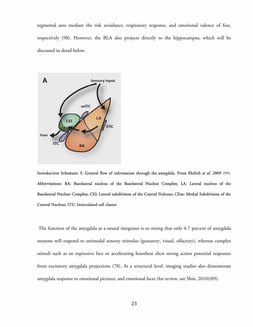
The flow of information through the amygdala has been well characterized. The lateral nucleus of
the BLA receives multimodal external and internal sensory input, which are then integrated in the
basolateral nucleus (For review, see Ledoux, 2000)(88). Neurons in the BLA then project directly
and indirectly to the output nuclei of the CMA, and thus BLA activity can both promote and
suppress anxiety, depending on the specific cell populations that become activated (96). The central
nucleus of the amygdala then outputs to various regions to drive the behavioral response to fearful
stimuli (97). For example, specific projections to the hypothalamus, parabrachial nucleus and ventral
にぬ
tegmental area mediate the risk avoidance, respiratory response, and emotional valence of fear,
respectively (98). However, the BLA also projects directly to the hippocampus, which will be
discussed in detail below.
(99)
The function of the amygdala as a neural integrator is so strong that only 4-7 percent of amygdala
neurons will respond to unimodal sensory stimulus (gustatory, visual, olfactory), whereas complex
stimuli such as an expressive face or accelerating heartbeat elicit strong action potential responses
from excitatory amygdala projections (78). At a structural level, imaging studies also demonstrate
amygdala response to emotional pictures, and emotional faces (for review, see Shin, 2010)(89).

にね
In states of acute stress, the BLA enhances memory formation through connections with memory
structures such as the hippocampus (100; 101). For example, infusion of the noradrenergic agonist
clenbuterol into the BLA during behavioral training enhanced memory recall and hippocampal
expression of the immediate-early-gene Arc, suggesting the BLA can modulate hippocampal activity
(102). Lesions of the BLA block the memory facilitation induced by glucocorticoid infusion into the
hippocampus, and likewise block the memory impairment induced by either adrenalectomy or intra-
hippocampal infusions of a GR antagonist (100).
While acute stress generally enhances memory formation, chronic stress has the opposite effect (70;
103). At a cellular level, chronic stress leads to contrasting patterns of dendritic remodelling in the
BLA and hippocampus: elaboration of dendritic arborisation occurs in excitatory projection neurons
of the BLA, while debranching and regression of apical dendrites occurs in CA3 pyramidal neurons
following chronic stress (70; 104; 105). Indeed, this dendritic remodelling of the BLA following
chronic stress has been shown to increase baseline excitability of regional neurons, suggesting that
BLA output may be increased in states of chronic stress (105).
Thus many studies suggest that the BLA participates in neurophysiological changes associated with
stress. Unknown however, is whether the direct circuit connectivity between the BLA and associated
structures (such as the hippocampus) facilitates these changes, or rather if some secondary factor such
as HPA axis activation is responsible.

にの
C L S C
The hippocampus is both physically adjacent to the BLA, and functionally integrated in providing
emotional content to memory. Even considering the confusing, often impenetrably dense, and
sometimes contradictory nomenclature of amygdala anatomy, trends emerge from studies in both
rodents and non human primates that sketches an extensive network of monosynaptic connections
between these two structures.
There is extensive reciprocal innervation of the BLA (lateral, basolateral and basomedial subnuclei),
and all hippocampal subfields, except notably the DG which neither receives nor sends any
projections to the BLA (106–108). First, in the rat (Rattus norvegicus) all three subnuclei of the BLA
send projections most densely to the ventral (“temporal”) two-thirds of the hippocampus. In the rat,
projections to CA3 originate in the basolateral subnuclei exclusively; projections to hippocampal
CA1 and the subiculum seem to be segregated by cell layer: the strata molecular and lacunosum
(internal axonal layers) receive input from the basomedial nucleus, while the strata oriens and
radiatum (external, dendritic layers) receive from the basolateral nucleus (109). In rodents the
temporal CA1 was observed to receive more connections from all BLA subnuclei, and is the only
hippocampal subfield to send reciprocal afferents back to the BLA (108). The interconnections
between the BLA and entorhinal and perirhinal cortexes are interesting in their capacity to modify
and modulate hippocampal output, but are beyond the scope of this discussion.
In the rhesus monkey (Macaca mulatta) as in the rat, BLA afferents to the hippocampus consistently
originate in only the basolateral and basomedial subnuclei of the BLA, and their projections form a

には
more dense “continuous band” throughout the hippocampal subfields (106; 107). In contrast,
projections from the lateral subnuclei of the BLA are sparse, as are those from all of the centromedial
and cortical nuclei (106). CA4 through CA1 receive inputs from the basomedial nucleus, while the
prosubiculum and subiculum receive their projections from the parvocellular division of the
basolateral subnucleus (107). Unlike in the rat, amygdala projections in primates terminate most
often in the dorsal (“septal”) hippocampus (106). Reciprocal connections from the hippocampus
originate most often in dorsal CA1 – these do not travel via the fornix, but instead via a direct tract
through the angular gyrus known as the “angular bundle” (106). Reciprocal hippocampal afferents
terminate in all BLA subnuclei according to Aggleton (1986), or exclusively in the basolateral and
basomedial subnuclei according to Saunders (1988).
These studies show interesting commonalities – that the BLA, and perhaps specifically the
basolateral and basomedial subnuclei, are especially reciprocally connected with the hippocampus
CA1. This suggests a conserved organization to amygdala-hippocampus connectivity, which can be
manipulated experimentally or therapeutically to examine the effect of stress on memory and AD.
A D
Psychiatric disease suffers from a symptom-based classification system that bypasses structural,
chemical, and genetic etiologies. Thus, multiple psychiatric conditions can share contributing
pathophysiology. For example in both Major Depressive disorder (110), and Anxiety-disorders
(111), there is enhanced activation of the amygdala in response to presentation of a fearful face.
Treatment of depression with Citalopram (a selective serotonin reuptake inhibitor) normalizes the

にば
response of the amygdala to fearful faces after only seven days – an interesting result considering that
full symptomatic relief generally occurs only after weeks or months of treatment (112). Beyond these
changes in amygdala activation, psychiatric disease also features changes in the functional
connectivity of the amygdala with other brain regions. As measured by correlations in metabolic
activity (Fludeoxyglucose (F18) Positron Emission Tomography [FDG-PET]), patients with bipolar
disorder demonstrate stronger connectivity with the prefrontal cortex and hippocampus; in unipolar
depression there is also a stronger, though negative, connectivity with prefrontal cortex (113).
Dysfunction of amygdalar circuitry might thus contribute to multiple classes of psychiatric disorders.
In AD, epidemiological literature suggests that neuropsychiatric symptoms are present before
memory disturbance, and contribute to disease progression (see above). However, it is possible that
instead, amygdalar changes are secondary to AD-related degeneration, and themselves lead to limbic
dysfunction and neuropsychiatric symptoms. Indeed the amygdala has been shown to lose functional
connectivity with the both the hippocampus and various frontal cortical regions in AD (by resting
state MRI analysis), and these changes correlate with clinical assessments of cognitive status (114).
There is also morphometric evidence to suggest that amygdalar degeneration correlates with
cognitive decline in AD. For example, one recent structural MRI study demonstrated a correlation
between recall of emotionally charged words and gray matter volume of the amygdala and
hippocampus (after normalization for overall cortical volume), while another found a similar
association between emotional memory abilities and amygdalar (but not hippocampal) volumes
(115; 116). Regarding neuropsychiatric symptoms in particular, although one study found a trend
suggesting that AD patients who experienced clinical anxiety showed relative preservation of the
にぱ
amygdala from degeneration (117), the bulk of evidence speaks against a specific correlation between
amygdalar degeneration or preservation in AD, and presence of neuropsychiatric symptoms (118).
Overall these studies suggest that although amygdalar dysfunction contributes to cognitive decline in
AD, it does not obviously relate to the presence or absence of neuropsychiatric symptoms. However,
the importance of chronic amygdala activation to overall disease progression (as is suggested by the
epidemiological link between AD and long standing “distress”) has not yet been addressed.
F
To summarize, AD is a disease in which emotional dysregulation and neuropsychiatric symptoms are
common. The pathophysiology of AD is well understood to involve Aが and Tau-induced
derangements in neuronal function, and eventually neuronal atrophy and death. Animal models
have established a link between chronic stress, and exacerbation of these pathological hallmarks of
disease; the epidemiological literature has provided an equally strong link between stress,
neuropsychiatric symptoms, and accelerated AD progression or increased incidence. The BLA is
known to underlie many of the effects of stress, and also known to be dysfunctional in multiple
neuropsychiatric disorders. Perhaps then, multiple neuropsychiatric symptoms converge to act on a
common structure – the BLA – and increased activity of this structure accelerates AD. The
experiments outlined below seek to assess two hypotheses: (1) that direct BLA-to-hippocampus
circuit activity underlies the cognitive and molecular effects of chronic stress, at least in the domain
of spatial memory and (2) that directly manipulating this sensitive circuit can alter pathological
progression in the 5xFAD mouse model of AD.

にひ
To address these hypotheses, we used optogenetic and pharmacogenetic strategies to
specifically manipulate excitatory projection neurons of the BLA. Intraparenchymal injection of
adeno-associated virus (AAV5) encoding channelrhodopsin-2 (ChR2) allowed us to examine
structural activation (96; 119; 120), while injection of virus encoding GiDREADD enabled us to
examine structural inactivation (121–123) (see fig. 1 for a summary of experimental paradigms).
Briefly, we found that the activity of glutamatergic cells of the BLA was (1) necessary for chronic-
stress induced molecular and cognitive changes (2) sufficient to replicate these changes in the
absence of any stressor. We also found that direct and selective activation of only BLA terminals
arriving in the dorsal hippocampus was sufficient to replicate these effects. In the 5xFAD model of
AD, we replicated previous findings that chronic stress exacerbated AD pathology. Applying our
earlier results to the 5xFAD model, we found that chronic BLA activation could worsen AD
pathology and impair cognition, while chronic inactivation slowed pathological progression, and
may improve cognitive function.

ぬど
All mouse work was approved by the Committee for Animal Care of the Division of Comparative
Medicine at the Massachusetts Institute of Technology. Adult male Swiss Webster mice (Taconic,
Charles River) were caged in groups of 4 to 5 on a normal light dark schedule and given access to
food and water ad libitum. 5xFAD homozygote mice in a B6SJL genetic background were obtained
from The Jackson Laboratory. These mice express human APP harboring the Swedish, Florida, and
London Familial-AD mutations, and human Presenilin-1 harboring the M146L and L286V
Familial-AD mutations, all driven by the neuron-specific promoter Thy1. In the 5xFAD mouse,
extraneuronal amyloid deposition begins at 2 months (preceded by intraneuronal accumulation),
with significant loss of synaptic density and layer-5 cortical pyramidal neurons by 9 months (trend at
4 months), and increase p25 by 9 months (36). All mice were used at an age of 3 months for the
RFS treatment, 10 weeks for the BLA-cell body viral injection (to allow 3 weeks for viral expression)
and 8 weeks for the BLA-terminal viral injection (to allow 6 weeks for viral expression).
Mice were placed in fear conditioning chambers (TSE systems) for 1 hour between 10 AM and 6
PM. Freezing and behavioral activity was automatically measured using TSE fear conditioning
software. Mice received 10-foot shocks at an intensity of 0.8mA and at random intervals during a
one-hour session. Control mice were placed in the chamber for 1 hour but did not receive foot
shocks. This procedure was repeated for 7 days.
ぬな
Each CRS session began at a variable time, between 10:00 AM and 6:00 PM. Plastic DecapiCones
(Braintree Scientific) were prepared by cutting 5 mm from the small open end to allow for easier
breathing, and a large hole near the tail for excreta. The cone was secured around the tail using
elastic bands or labeling tape. Mice were arranged in the bottom of a large rat cage on a lab bench,
and inclined to allow for urine and feces to clear from the bags. CRS sessions lasted 2 hours, after
which mice were quickly returned to their home cage. Control mice were brought to the lab bench
but left in their home cages for 2 hours.
All surgeries were performed under aseptic conditions (with stereotaxic guidance for intracranial
injections). Mice were anesthetized using 1 to 2% isoflurane and Ketamine/Xylazine. All coordinates
are relative to bregma in mm and defined according to the Paxinos sterotaxic atlas (124).
Gi All mice received bilateral injections of adeno-associated virus (AAV5) virus (1-
5*1012) expressing Gi-DREADD under the CaMKIIゎ promoter
(http://genetherapy.unc.edu/services.htm#AAV). Virus was provided by the UNC vector core.
Swiss-Webster mice received a bilateral injection of 0.5 づl at a rate of 0.05μl/min using 10μl
elongated glass capillary (Wiretrol Drummond) and a microinjector (Quintessential Steroetaxic
Injector, #53311); 5xFAD mice received a bilateral injection of 0.3 づl at a rate of 0.05μl/min using
the same equipment. Stereotaxic coordinates for BLA injections in Swiss Webster mice were (-1.34
mm AP, ±3.35 mm ML, -3.9 mm DV) and in 5xFAD mice were (-1.14 mm AP ±3.51 mm ML, -
3.9mmDV). After injection, the capillary was left in place for an additional 5-10 minutes to allow

ぬに
for virus diffusion, and then was slowly withdrawn. Animals were kept on a 37° heating pad during
the surgery, and until recovery from anesthesia.
The Gi-DREADD activating drug Clozapine N-oxide (Sigma Aldrich, C0832) was diluted in 0.9%
sterile saline (Aqualite System) and injected i.p. (2.5mg/kg) 30 minutes prior to starting the RFS
treatment. For osmotic pump delivery, CNO was diluted in 0.9% sterile saline to a concentration of
5mg/mL. Approximately 240 づL of CNO solution was loaded into the Alzet® model 2004 pump.
The pump was implanted subcutaneously using a 1cm dorsal incision made at the base of the neck
between the scapulae.
Intracranial injections were performed as described in the Gi section. Control of
BLA glutamatergic projection neurons was achieved using an AAV5 vector carrying hChR2(H134R)-
eYFP , or control virus carrying eYFP only, both driven by the CaMKIIゎ promoter (viruses provided
by Karl Deisseroth, maps and clones available at http:// www.optogenetics.org). All mice were
bilaterally implanted with self assembled implantable optical fibers using the protocol from the
synthetic neurobiology website (http://syntheticneurobiology.org/protocols/protocoldetail/35/9),
with the only difference being the use of metal ferrules with an internal diameter of 330 μm
(precision fiber products) and 300 μm fiber optic, 0.37 NA (Thorlabs). For ChR2-cell body mice
the implants were placed 0.5mm dorsal to the BLA (Swiss Webster: (-1.34 mm AP, ±3.35 mm ML,
-3.4 mm DV) 5xFAD: (-1.14 mm AP, ±3.51, -3.4 mm DV)). For the ChR2-terminal experiments,
implants were placed 0.5mm dorsal to the hippocampus CA3 in the dorsal hippocampus (-2.18 mm
AP, ±2.67 mm ML, -1.37 mm DV). Two self-tapping bone screws (Bioanalytical Systems) were
screwed into the scull to hold the implant in place. One layer of adhesive cement (C&B metabond;

ぬぬ
Parkell, Edgewood, NY) followed by cranioplastic cement (Dental cement; Stoelting, Wood Dale,
IL) was applied to the implants, skull, and screws to secure these elements to the skull. Virus was
allowed to express for 3 or 6 weeks for the cell body or terminal experiments respectively, before
beginning the laser stimulation paradigms.
A fiber-optic cable was used to connect a 473 nm laser diode (OEM Laser Systems, East Lansing,
MI) through an FC/PC adapter to a rotatory joint (Doric Lenses) mounted inside the fear-
conditioning chamber (TSE Systems). Laser output was controlled using a Master-8 pulse stimulator
(A.M.P.I., Jerusalem, Israel). All included animals had the center of the viral injection located in the
BLA, though there was occasional leak to neighboring regions or along the needle tract. Specificity of
the stimulation was dependent on fiber optic implant location, and was histologically confirmed in
all cases. The skull-mounted optical fibers were plugged to a branching fiberoptic patchcord (Doric
Lenses) to split the laser output, providing bilateral light delivery. This patchcord was connected to
the external fiber optic cable through the rotatory joint inside the fear-conditioning chamber. Once
connected, mice were allowed 3 min to acclimatize before starting the laser stimulation. BLA-cell
body photostimulation in Swiss-Webster mice consisted of pairing 4.5-kHz tone pips (1 Hz; 250 ms
on/ 750 ms off) for 20 s with 2 s of laser stimulation (20 Hz; 10 ms pulse-width; 473 nm; 3-5 mW;
~57 mW/mm2), which coterminated with the tone, occurring 16 times during a 20 min period at
variable interval, repeated daily for 7 days. Terminal photostimulation in Swiss Webster mice
consisted of either an identical paradigm to that for cell body stimulation, or 20s of constant laser
stimulation at 7-8 mW (~106 mW/ mm2 at the tip of the fiber) occurring 10 times during a 20 min
session at random intervals, repeated daily for 7 days. BLA-cell body photostimulation in 5xFAD
ぬね
mice consisted of 20 sec of laser stimulation (20 Hz; 5 ms pulse-width; 473 nm; 1-3 mW;
~34mW/mm2) occurring 10 times during a 20 min session, repeated daily for 14 days.
Behavioral arenas consisted of 4
rectangular rat cages with no bedding, arranged in a 2x2 rectangle, separated by four opaque walls. A
triangle or three vertical lines of red labeling tape were placed onto the two opposing short walls for
spatial cues. For novel object recognition and novel object location, mice were placed into the
arenas on day 5 and 6 of the RFS or laser-stimulation paradigm, and allowed to explore and
familiarize for one 10 min session per day. On day 7, the novel object location protocol started with
the training phase, wherein two objects were placed along one short side of each arena (100ml Pyrex
glass bottles, positioned in the corners, 3 cm away from the walls). The mice were placed facing the
middle of the opposite short wall at its midsection. Mice were allowed to familiarize with the objects
for two 10 min training sessions, separated by an inter-trial interval of 1h. 75 min after the second
training session, one of the objects was moved to the opposite corner and mice were again placed
into the arenas. Immediately following this testing period, the novel object recognition procedure
started with the training phase, in which the two objects were placed in their original location and
mice were allowed 10 additional min of free exploration. 24h later, one of the glass bottles was
substituted for a plastic 55ml bottle and the mice were again placed into the arena. For both tests,
time spent with each object (old and new locations or old and new objects) was recorded during a 5
min testing period. During each step of the training and testing, video of the performing mice was
acquired using a Sony Camcorder fixed to the ceiling above the arenas. Videos were analyzed by an
experimenter blind to the experimental treatment. Mice were scored as exploring an object if they

ぬの
showed obvious signs of directed attention: climbing, sniffing, or prolonged observation (if within
approximately 2 cm of the object). Time spent on top of the objects was not counted, unless the
mice were simultaneously directing attention to the object.
Mice were allowed to habituate to the fear-conditioning room for at least 30
minutes prior to starting the procedure. The fear conditioning chambers (TSE) were scented using a
paper towel saturated with natural vanilla extract. Background noise was set to 0.1 dB. Mice were
given 3 minutes to habituate to the box. Two 0.8 mA shocks were administered, each preceded by a
30 seconds of 4.5 kHz tone pips. 15 seconds after the final shock, mice were returned to the home
cage. Boxes were disinfected with quatricide between mice, and the paper towel was re-saturated
with vanilla. 24 hours later, mice were returned to the unaltered chamber and freezing was assessed
for 3 minutes (no tone, no shock). After contextual testing, the boxes were cleaned and modified: the
vanilla extract was replaced with 2 % Acetic Acid, the metal shock bars were covered with a grey
plastic flooring, the walls of the box were covered with yellow paper, and the background noise was
removed. Mice were then returned to the box for cued testing and freezing was assessed during 3
minutes of constant 4.5 kHz tone pips.
Mice were perfused with 10% paraformaldehyde under deep anaesthesia (ketamine, xylazine) and
their brains sectioned at 40 づm thickness using a vibratome (Leica). Slices were permeabilized with
0.1% Triton X-100, blocked (10% donkey serum), and incubated overnight with 0.1% Triton X-
100/10% donkey serum in PBS containing primary antibodies: rabbit HDAC2 (Abcam; 1:1000),
Mouse synaptophysin (Sigma; 1:1000), Rabbit GFAP (Abcam; 1:10,000), Mouse 4G8 (Covance,
1:500), and Mouse Nu-1 (William Klein, Northwestern University; 1:3600). Primary antibodies
ぬは
were visualized with Alexa-Fluor 488, Cy3 and Cy5 antibodies (Molecular Probes), and neuronal
nuclei visualized with Hoechst 33342 (Invitrogen). Images were acquired using a confocal
microscope (LSM 510, Zeiss; LSM 710, Zeiss) at identical settings and focal plane for each of the
conditions. Images were quantified using ImageJ 1.42q by an experimenter blind to the experimental
treatment. For HDAC2 and SYP quantification, sections at bregma -1.70 were selected and 5 images
per section were acquired. For HDAC2 40-60 representative cells were analyzed, while for SYP, the
stratum radiatum region of the CA1 subfield was selected in each picture and the mean intensity was
quantified. For GFAP, a similar protocol was used with the exception that all strata were analyzed.
For 4G8 plaque load analysis, two sections at bregma -2.18 were analyzed under epifluorescence by a
blinded experimenter. 4G8-positive plaques were counted in the DG, and CA1. Aggregates were
determined to be individual plaques if their total size was larger than a nuclei, and if they could be
resolved from adjacent aggregates. For Nu-1 analysis, 2 sections at bregma -2.18 were selected. A
single image of the DG and septal CA1 was acquired for each hemisection with identical settings and
focal plane. Images were exported to ImageJ 1.42q, thresholded for intensity, converted to binary,
and finally the percent coverage of all strata the stratum pyramidale was calculated based on a
constant threshold intensity.
Following the fifth session of RFS/optogenetic stimulation or the final session of CRS, mice were
scruffed, and the facial vein was punctured within 5-10 seconds using a 5 mm Goldenrod Animal
Lancet (MEDIpoint). 5 drops of blood were collected on ice and heparinized with 10 づL of
1000U/mL heparin. Samples were centrifuged at 4000 RPM for 10 minutes at 4°C, the plasma
removed, and frozen for later use. A corticosterone ELISA kit (Enzo Life Sciences) was used to

ぬば
measure concentration of these samples. The provided steroid displacement reagent was mixed with
assay buffer, and this mixture was used to dilute the plasma samples to the suggested ratio of 1:50,
and such that 2.5 parts steroid displacement reagent were present for every 97.5 parts undiluted
sample. The rest of the protocol was conducted as per the manufacturer’s instructions. Absorbance at
405 nm was read using a multiplate reader (EnSpire; PerkinElmer), blanked to blank-wells.
Concentration was calculated from the standard-curve provided by standard concentration in each
plate.
Statistical analyses were performed using GraphPad Prism 5. One-way analyses of variance followed
by Tukey post-hoc tests, one-tailed Student’s t-tests, and the Pearson product-moment correlation
coefficient were used unless otherwise indicated. All data are represented as mean±s.e.m. Statistical
significance was set at P≤0.05.
ぬぱ
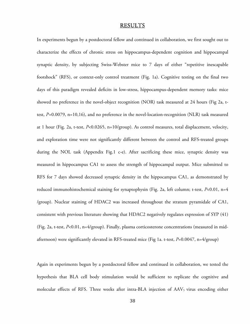
In experiments begun by a postdoctoral fellow and continued in collaboration, we first sought out to
characterize the effects of chronic stress on hippocampus-dependent cognition and hippocampal
synaptic density, by subjecting Swiss-Webster mice to 7 days of either “repetitive inescapable
footshock” (RFS), or context-only control treatment (Fig. 1a). Cognitive testing on the final two
days of this paradigm revealed deficits in low-stress, hippocampus-dependent memory tasks: mice
showed no preference in the novel-object recognition (NOR) task measured at 24 hours (Fig 2a, t-
test, P=0.0079, n=10,16), and no preference in the novel-location-recognition (NLR) task measured
at 1 hour (Fig. 2a, t-test, P<0.0265, n=10/group). As control measures, total displacement, velocity,
and exploration time were not significantly different between the control and RFS-treated groups
during the NOL task (Appendix Fig.1 c-e). After sacrificing these mice, synaptic density was
measured in hippocampus CA1 to assess the strength of hippocampal output. Mice submitted to
RFS for 7 days showed decreased synaptic density in the hippocampus CA1, as demonstrated by
reduced immunohistochemical staining for synaptophysin (Fig. 2a, left column; t-test, P<0.01, n=4
/group). Nuclear staining of HDAC2 was increased throughout the stratum pyramidale of CA1,
consistent with previous literature showing that HDAC2 negatively regulates expression of SYP (41)
(Fig. 2a, t-test, P<0.01, n=4/group). Finally, plasma corticosterone concentrations (measured in mid-
afternoon) were significantly elevated in RFS-treated mice (Fig 1a. t-test, P=0.0047, n=4/group)
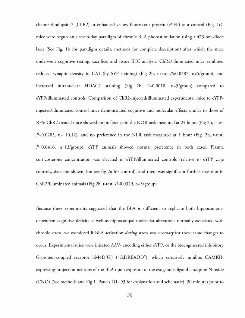
Again in experiments begun by a postdoctoral fellow and continued in collaboration, we tested the
hypothesis that BLA cell body stimulation would be sufficient to replicate the cognitive and
molecular effects of RFS. Three weeks after intra-BLA injection of AAV5 virus encoding either
ぬひ
channelrhodopsin-2 (ChR2) or enhanced-yellow-fluorescent protein (eYFP) as a control (Fig. 1c),
mice were begun on a seven-day paradigm of chronic BLA photostimulation using a 473 nm diode
laser (See Fig. 1b for paradigm details; methods for complete description) after which the mice
underwent cognitive testing, sacrifice, and tissue IHC analysis. ChR2/illuminated mice exhibited
reduced synaptic density in CA1 (by SYP staining) (Fig 2b, t-test, P=0.0487, n=5/group), and
increased intranuclear HDAC2 staining (Fig 2b, P=0.0018, n=5/group) compared to
eYFP/illuminated controls. Comparison of ChR2-injected/illuminated experimental mice to eYFP-
injected/illuminated control mice demonstrated cognitive and molecular effects similar to those of
RFS: ChR2 treated mice showed no preference in the NOR task measured at 24 hours (Fig 2b, t-test
P=0.0285, n= 10,12), and no preference in the NLR task measured at 1 hour (Fig. 2b, t-test,
P=0.0416, n=12/group). eYFP animals showed normal preference in both cases. Plasma
corticosterone concentration was elevated in eYFP/illuminated controls (relative to eYFP cage
controls, data not shown, but see fig 2a for control), and there was significant further elevation in
ChR2/illuminated animals (Fig 2b, t-test, P=0.0329, n=5/group)
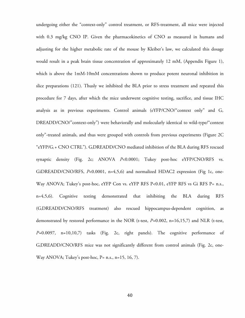
Because these experiments suggested that the BLA is sufficient to replicate both hippocampus-
dependent cognitive deficits as well as hippocampal molecular alterations normally associated with
chronic stress, we wondered if BLA activation during stress was necessary for these same changes to
occur. Experimental mice were injected AAV5 encoding either eYFP, or the bioengineered inhibitory
G-protein-coupled receptor hM4D(Gi) (“GiDREADD”), which selectively inhibits CAMKII-
expressing projection neurons of the BLA upon exposure to the exogenous ligand clozapine-N-oxide
(CNO) (See methods and Fig 1. Panels D1-D3 for explanation and schematic). 30 minutes prior to
ねど
undergoing either the “context-only” control treatment, or RFS-treatment, all mice were injected
with 0.3 mg/kg CNO IP. Given the pharmacokinetics of CNO as measured in humans and
adjusting for the higher metabolic rate of the mouse by Kleiber’s law, we calculated this dosage
would result in a peak brain tissue concentration of approximately 12 mM, (Appendix Figure 1),
which is above the 1mM-10mM concentrations shown to produce potent neuronal inhibition in
slice preparations (121). Thusly we inhibited the BLA prior to stress treatment and repeated this
procedure for 7 days, after which the mice underwent cognitive testing, sacrifice, and tissue IHC
analysis as in previous experiments. Control animals (eYFP/CNO/“context only” and Gi-
DREADD/CNO/”context-only”) were behaviorally and molecularly identical to wild-type/“context
only”-treated animals, and thus were grouped with controls from previous experiments (Figure 2C
“eYFP/Gi + CNO CTRL”). GiDREADD/CNO mediated inhibition of the BLA during RFS rescued
synaptic density (Fig. 2c; ANOVA P<0.0001; Tukey post-hoc eYFP/CNO/RFS vs.
GiDREADD/CNO/RFS, P<0.0001, n=4,5,6) and normalized HDAC2 expression (Fig 1c, one-
Way ANOVA; Tukey’s post-hoc, eYFP Con vs. eYFP RFS P<0.01, eYFP RFS vs Gi RFS P= n.s.,
n=4,5,6). Cognitive testing demonstrated that inhibiting the BLA during RFS
(GiDREADD/CNO/RFS treatment) also rescued hippocampus-dependent cognition, as
demonstrated by restored performance in the NOR (t-test, P=0.002, n=16,15,7) and NLR (t-test,
P=0.0097, n=10,10,7) tasks (Fig. 2c, right panels). The cognitive performance of
GiDREADD/CNO/RFS mice was not significantly different from control animals (Fig. 2c, one-
Way ANOVA; Tukey’s post-hoc, P= n.s., n=15, 16, 7).

ねな
Although these experiments suggest that glutamatergic cell activity in the BLA is somehow
responsible for the effects of chronic stress, these neurons project broadly to many downstream
targets. In order to determine if the direct BLA-to-hippocampus circuit was responsible for our
observations, a postdoctoral fellow and I worked to photostimulate the ChR2-positive axon
terminals of BLA afferent projections to the hippocampus. Having observed that BLA projection
fibers arrived most densely to the stratum oriens, lucidum and radiatum of the CA3 field in our mice,
we injected ChR2 into the BLA and implanted optical fibers 0.5 mm dorsal to CA3 in the dorsal
hippocampus (bregma -2.18mm anteroposterior) (fig. 3a). Thereby we selectively modulated the
activity of a direct BLA-to-hippocampus circuit.
The ChR2 or eYFP AAV5 viral constructs were allowed 6 weeks for expression in order to increase
channel density at the axon terminals (Fig. 3a). eYFP- and ChR2-injected mice were then begun on
a seven-day photo-stimulation paradigm. In most cases, this paradigm was identical to that used for
stimulation of the cell bodies (Fig. 1b); some animals received 20 sec bursts of constant illumination
but after analysis were found to be molecularly and behaviorally identical to animals receiving 2 sec
bursts (data not shown). ChR2-injected/terminal-illuminated mice again exhibited the same
molecular changes as were observed in RFS-treated animals: reduced synaptic density in CA1 (by
SYP staining) (Fig 3b, t-test, P=0.0069), and increased intranuclear HDAC2 staining (Fig 3b, t-test,
P=0.0019). Photostimulation of BLA afferents in CA3 also produced a cognitive deficit in ChR2-
injected/terminal-illumination animals, again demonstrated by lack of preference on the NOR (Fig.
3c, t-test, P=0.0057) and NLR (Figure 3c, t-test, P=0.0092) tasks compared to eYFP-
injected/terminal-illumination treated controls. Plasma corticosterone concentration was not

ねに
elevated in ChR2-injected/terminal-illumination animals relative to eYFP-injected/terminal-
illumination controls (Fig. 3d. ANOVA,P=0.087; Tukey post-hoc eYFP vs. ChR2, P>0.05), though
both were elevated compared to eYFP-injected cage controls (Tukey post-hoc P<0.05, P<0.05). Also
of note is that although overall concentrations did not differ between the experimental groups, they
were high compared to previous experiments (Fig. 3d, Fig. 2a, Fig. 2b).
Having established that activation of a specific BLA circuit can replicate the effects of chronic-stress
on the circuit target (in this case, the hippocampus), and given the myriad connections between
chronic-stress and AD, I wondered if modulating BLA activity could alter the progression of
hippocampal pathology in the 5xFAD murine model of AD.
I first sought to examine whether previous results that linked chronic stress and exacerbated AD
pathology could be recapitulated in a mouse model of AD used in our lab. 5xFAD mice exhibited
behavior suggesting they were resistant to stress in the RFS paradigm (Appendix Fig. 1c), and so I
used 14 days of chronic restraint stress (CRS) to model chronic stress in 4 month-old 5xFAD
animals (See Fig 1e and methods for complete description). CRS seemed to be an effective stressor in
5xFAD mice, as demonstrated by both increased plasma corticosterone concentration after a single
CRS session (Fig.4a, t-test, P=0.0013, n=6,16), and decreased body mass after 14 days in CRS-
treated animals compared to a net gain in controls (Fig. 4b, t-test, P=0.0008, n=6,10). CRS-treated
animals had a higher number of 4G8-positive Aが plaques throughout their hippocampus (Fig. 4c, t-
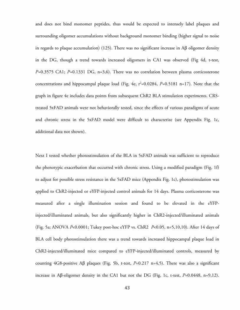
test, P=0.0115 n=10,10). The Aが-oligomer specific antibody Nu-1 was also used to assess Aが
oligomer density. This antibody is highly selective for Aが oligomers (trimer, tetramer, 12-24mer)
ねぬ
and does not bind monomer peptides, thus would be expected to intensely label plaques and
surrounding oligomer accumulations without background monomer binding (higher signal to noise
in regards to plaque accumulation) (125). There was no significant increase in Aが oligomer density
in the DG, though a trend towards increased oligomers in CA1 was observed (Fig 4d, t-test,
P=0.3575 CA1; P=0.1331 DG, n=3,6). There was no correlation between plasma corticosterone
concentrations and hippocampal plaque load (Fig. 4e, r2=0.0284, P=0.5181 n=17). Note that the
graph in figure 4e includes data points from subsequent ChR2 BLA stimulation experiments. CRS-
treated 5xFAD animals were not behaviorally tested, since the effects of various paradigms of acute
and chronic stress in the 5xFAD model were difficult to characterize (see Appendix Fig. 1c,
additional data not shown).
Next I tested whether photostimulation of the BLA in 5xFAD animals was sufficient to reproduce
the phenotypic exacerbation that occurred with chronic stress. Using a modified paradigm (Fig. 1f)
to adjust for possible stress resistance in the 5xFAD mice (Appendix Fig. 1c), photostimulation was
applied to ChR2-injected or eYFP-injected control animals for 14 days. Plasma corticosterone was
measured after a single illumination session and found to be elevated in the eYFP-
injected/illuminated animals, but also significantly higher in ChR2-injected/illuminated animals
(Fig. 5a; ANOVA P<0.0001; Tukey post-hoc eYFP vs. ChR2 P<0.05, n=5,10,10). After 14 days of
BLA cell body photostimulation there was a trend towards increased hippocampal plaque load in
ChR2-injected/illuminated mice compared to eYFP-injected/illuminated controls, measured by
counting 4G8-positive Aが plaques (Fig. 5b, t-test, P=0.217 n=4,5). There was also a significant
increase in Aが-oligomer density in the CA1 but not the DG (Fig. 1c, t-test, P=0.0448, n=9,12).

ねね
Despite this increase in Aが staining, there was no increase in astrocyte activation/gliosis, as measured
by the optical density of glial-fibrillary acidic protein (GFAP) in all strata of CA1 (Fig. 5d, t-test,
P=0.835, n=5,5). Because 5xFAD mice show cognitive impairments in NOR and NOL tasks at 4
months (data not shown), I measured freezing levels as an approximation of contextual-fear-
memory. ChR2-injected mice showed reduced contextual freezing levels after 14 days of BLA
photostimulation, compared to eYFP-injected/illuminated controls (Fig. 5e. t-test, P=.0419,
n=8,12). There was however no difference in cued freezing, suggesting normal
amygdalohippocampal connectivity (Fig 5f, t-test, P=0.43, n=8,12)
Since BLA activation seemed to worsen AD pathology in 5xFAD mice, I wondered if chronically
inactivating the BLA could slow progression of AD pathology, even absent any stressful experimental
treatment. Three weeks after intra-BLA injection of AAV5 virus encoding either GiDREADD or
eYFP (using the same procedure as in previous experiments, Fig. d1-d3), I implanted subdermal
osmotic pumps to deliver systemic CNO at a constant rate over 28 days (Fig. 6a). Using the same
pharmacokinetic data as for previous calculations (See Appendix Fig. 1b) and the volume/flow rate
parameters of our pumps, I calculated that osmotic pump delivery would sustain brain tissue CNO
concentrations of 500nM over this 28 day experiment. Following this treatment, I observed a
significant decrease in the density of Aが oligomers in hippocampus CA1 of GiDREADD-
injected/CNO-treated animals compared to eYFP-injected/CNO-treated controls (Fig 6b. t-test,
P=0.0043, n=2,3). This same effect was not observed in the DG (Fig. 6b, t-test, P=0.63, n=2,3).
(4G8 immunostaining was attempted but adequate experimental preparations were not achieved.) I
again measured freezing levels as an approximation of hippocampal-cognition, and observed only a
ねの
slight trend towards increased freezing in GiDREADD-injected/CNO-treated animals compared to
eYFP-injected/CNO-treated controls (Fig 6c, t-test, P=0.26, n=5,6). There was no trend suggested
in cued freezing, suggesting normal amygdalohippocampal function (Fig. 6d. t-test, P=0.93, n=5,6).
Because BLA inactivation was chronic in this paradigm, it was felt that measurement of plasma
corticosterone would not be informative, and therefore this was not performed.

ねは
These results can be divided into two sections, each with their own implications. This first series of
experiments (Figures 2, and 3) strongly suggest that activation of direct BLA projections can produce
consistent molecular changes in the projection target (in this case the hippocampus), and also impair
cognition that depends on that projection target. The second set (Figures 4, 5 and 6) suggests that
modulating BLA activity in an AD model mouse may alter the progression of AD pathology in a
BLA projection target, and also alter target-dependent cognition.
A U M
C E C S
Previous studies probing the effect of acute stress on cognitive function have found that inactivating
the amygdala is sufficient to block both the behavioral and cellular effects of acute stress (126; 127).
Specifically, electrolytic lesioning or pharmacological inactivation prevented alterations in
hippocampal long term potentiation, and spatial memory deficits, both of which were induced by
restraint or tailshock stress administered just prior to training (126; 127). Because our methods of
activation and inactivation were highly specific, we can now assert that (1) excitatory activity of the
BLA is both necessary and sufficient to produce the molecular effects of chronic stress on associated
brain structures such as the hippocampus, and resultant cognitive effects, and (2) direct circuit
connections are powerful and potentially causal contributions to the negative influences of
psychological stress.

ねば
It is important to consider these results in the context of our corticosterone measurements, from
which two related conclusions can be drawn. The first is that surgery and illumination increase
plasma corticosterone concentrations significantly (Fig 2, Fig 5), but that photostimulation of the
BLA results in even greater corticosterone release (Fig. 2, Fig. 5). The second is that terminal
stimulation does not appear to elevate plasma corticosterone concentrations (Fig 3). Of note, the
corticosterone concentrations of both eYFP and ChR2 injected mice were high in the terminal-
illumination experiment. This might have been due to differences in surgical trauma (4 craniotomies
instead of 2), handling, or sample collection.
Overall, these corticosterone results suggest that the changes in cognition, synaptic density, and
HDAC2 staining we observed in terminal stimulation experiments occurred in the absence of
increased peripheral corticosterone concentrations, which are known to act biphasically on the
hippocampus and are often implicated in the effects of stress (70). They also suggest that in the
terminal photostimulation experiments, antidromic propagation from the terminals to the cell body
did not occur, or if so that the resultant depolarization was not sufficient to activate the soma of BLA
neurons and drive further corticosterone release. We did not conduct experiments to silence the BLA
pharmacologically while activating BLA terminals, and so we cannot claim that terminal stimulation
did not result in antidromic propagation to the BLA. However, previous experiments using
optogenetic stimulation of brain-slices revealed that antidromically driven activation of the somata
occurred in only 5% of recordings, and in this experiment the terminals and cell bodies were
separated by 300 づm, compared to many mm in our in vivo experiments (96).
ねぱ
However, our results do not absolve corticosterone and GR. Instead they demand an explanation
that does not involve increased glucocorticoid concentration.
One possibility is that increased neuronal activity leads to phosphorylation of GR and altered
downstream gene expression. Activity-dependent Ca2+ influx leads to increased activity of the tau-
kinase protein Cdk5, through calpain-mediated cleavage of its activator protein p35 to the more
stable p25 (128). Cdk5 can phosphorylate GR at multiple serine residues including S211, which
alters recruitment of cofactors and can modify GR transcriptional activity (129). GR
phosphorylation at S211 has also been found to increase nuclear recruitment, and modify
transcriptional activation in a gene dependent manner (130). Thus without changes in
corticosterone/cortisol concentration, driving neuronal activity through circuit activation could
activate Cdk5, increasing GR phosphorylation and altering gene expression.
In our paradigm, ChR2 simulation of pyramidal neurons would induce significant elevation of
hippocampal glutamate – previously observed using microdialysis following chronic stress (131).
Imaging data from our experiments (not shown) suggested that in our mice, most BLA terminals
arrive in the stratum oriens, lucidum and radiatum of the CA3 field. Hence BLA activity would lead
to excitation of CA3, activation of Schaffer collaterals, and increased glutamate release in CA1.
Coupled with even a relatively small excitatory drive from direct BLA afferents to CA1, this could
lead to significant activation of the CA1 and potential allostatic or excitotoxic damage from excess
glutamate release (132). Normally, this connectivity may allow the BLA-hippocampus axis to act in
limbic-autoregulation by responding to both changes in corticosteroid concentration (through
ねひ
hippocampal GR and MR binding), and to altered GR phosphorylation state via the activity of
excitatory BLA afferents. However in states of chronic stress, increased glutamate release with altered
transcriptional programming could lead to hippocampal maladaptation, and potentially even
degeneration. Indeed the lifetime duration of major depression, (a disease known to involve
amygdala hyperactivation and provide significant psychosocial stress) negatively correlates with
hippocampal size (133).
Our results are the first to demonstrate that HDAC2 can be regulated by direct modulation of
neural circuitry. As discussed above, this result may also be underlain by activity-dependent
phosphorylation of GR, which our lab has previously shown to upregulate HDAC2 expression (40).
Another recent study has suggested that HDAC2 regulation of glial-derived-neurotrophic-factor
expression in the striatum is one determinant of the behavioral response to stress (134). Although
our data do not allow us to claim that HDAC2 upregulation causes the molecular or cognitive
deficits we observed, they do provide evidence for an epigenetic link between the pathophysiology of
stress, and of AD.
E P 5X M
Our results demonstrate that chronic BLA activation
impairs spatial memory in 5xFAD mice, as measured by contextual freezing levels (Fig. 5). We also
observed a maintenance of cued fear memory (Fig. 5, 6), which suggests that BLA function and
amygdalohippocampal connectivity were preserved throughout these experiments (135). Taken
together, these results suggest that enhanced BLA activity impairs hippocampus dependent cognition

のど
in the 5xFAD murine model of AD. Our data also suggest that chronic inactivation has the potential
to delay memory impairment in a preclinical AD model. A greater sample size is first needed to
determine if these results (Fig. 6) are statistically significant.
It is important to note that our experiments cannot determine if impaired spatial memory in 5xFAD
animals is due to the chronic-stress-like effect of BLA photostimulation (as in Figures 2 and 3), or
truly accelerated AD-related cognitive decline. In future experiments, delaying cognitive testing of
BLA-photostimulated 5xFAD mice for days to weeks would allow the effects of stress to wear off,
while cognitive effects from accelerated disease progression would remain. Our chronic inhibition
experiments demonstrate that even in the absence of chronic stress, modulating BLA activity can
change AD pathology progression and the rate of cognitive decline. Taken together, these results
would support the hypothesis that BLA activation alone is sufficient to accelerate AD-related
cognitive decline.
Ⱦ Although overall Aが plaque accumulation increased
following chronic stress, consistent with previous literature, (figure 4), data from both activation and
inactivation experiments suggested that Aが load (both plaques and oligomers) could be altered most
strongly in CA1, and was not changed in DG. (Note that analysis of CA1 was prioritized in the
study of our 5xFAD mice – in contrast to the Swiss-Webster strain analyzed in figures 2 and 3 –
since we did not directly examine the site of BLA afferents in the 5xFAD mouse, and classically the
CA3 subfield receives fewer efferent BLA projections in the rodent and sends no reciprocal
projections to the BLA in either rodent or primate (see introduction); further analysis would be
のな
appropriate). The observations in CA1 are consistent with data from tracing studies in which CA1 is
shown to receive excitatory projections from the BLA and also supply a strong reciprocal connection,
while the DG neither receives nor returns excitatory BLA projections (106; 107; 109). Thus the
distribution of Aが plaque accumulation in our experiments suggests activity-dependent generation of
Aが – a hypothesis for which a wealth of literature offers support.
Much of the recent interest in activity-driven Aが generation has stemmed from the discovery of the
brain’s “default network”. Defined through fMRI to including prefrontal cortex, posterior cingulate
cortex, inferior parietal lobule and hippocampal formation, the default network is active in
introspective memory, self-reflection, and other forms of “internal cognition”, and is perhaps the
most active network across the human lifespan (136). Early in AD there is hypometabolism of these
default network regions, and network dysfunction progresses more severely in AD than in normal
aging (136). Moreover regional amyloid deposition and atrophy overlaps prominently with these
default network regions (137).
In support of imaging data that links structural activation with Aȕ accumulation, two lines of
evidence can be drawn to a molecular underpinning between neuronal activity and Aȕ production.
Firstly, rapid changes in Aが production occur in response to modulating neuronal activity. In vitro,
suppression of neuronal activity in brain slice preparations by either tetrodotoxin or high-Mg2+ can
decrease APP processing and Aȕ production, while neuronal activation by picrotoxin has the
opposite effect (21). Immediate increases in Aȕ production in response to neuronal activity can also

のに
be observed in vivo: regional Aȕ production correlates well with regional neuronal metabolism, and
pharmacological modulation of neuronal activity can increase (with activation) or decrease (with
inactivation) Aȕ production and release into the interstitial fluid (ISF) as measured by in vivo
microdialysis (138)(139). At a circuit level, experimental sensory deprivation can decrease Aȕ plaque
deposition specifically in the sensory cortex (138). Furthermore, the physiological activity
modulation that occurs naturally during the sleep-wake cycle decreases Aȕ production and release
(140).
The cellular mechanism of activity-dependent Aȕ production has also been elucidated. Neuronal
activity and synaptic vesicle release lead to increase rates of membrane recycling through endocytosis
at the presynaptic terminal. Transmembrane APP is thus internalized in endocytosed vescicles and
cleaved by endosomal BACE-1, resulting in release of the Aȕ fragment upon subsequent exocytosis
(141)(142). Both basal and activity-regulated Aȕ generation have been shown to depend on
functional endocytosis (143), further suggesting that activity-driven changes are mediated through
the synaptic release machinery.
As a second line of evidence, neuronal activity can also drive changes in expression of genes related to
APP processing. The expression of BACE-1 and its enzymatic activity are both increased in response
to oxidative stress, which might occur following intense neuronal activation. After oxidative stress,
this increase in BACE-1 expression is mediated by phosphorylation of the immediate-early-gene
product c-Jun by c-Jun N-terminal-kinase (JNK)(144; 145). There is also evidence that both JNK
enzymatic activity and c-Jun phosphorylation increase after physiological neuronal activation,

のぬ
following exposure to a novel environment (146), or after seizure induction by kainate
administration (147). JNK mRNA expression is also regulated by sleep-wake cycle, suggesting that
circadian rhythms can regulate the APP processing machinery (148) in addition to altering synaptic
Aȕ release. Another immediate early gene – Arc – is also involved in the trafficking of APP and
association of APP with its processing machinery in the endosomes (149).
Thus neuronal activity can increase Aȕ production in two ways: (1) through accelerated synaptic
vesicle recycling, and (2) through genetic regulation of APP processing, via activation of immediate-
early gene products.
T A
Key enzymes in the biosynthetic or catabolic pathways for acetylcholine are found in high density in
the BLA. Choline-acetyltransferase (ChAT), acetylcholinesterase, and high-affinity choline
transporters are often used as control markers to delineate the basolateral subnucleus of the BLA (78;
79), because the density of these markers is higher in the BLA than in any other forebrain structure
(150). In normal aging, there is very little depletion of cholinergic innervations to the BLA, while in
AD there is marked depletion of cholinergic markers in the neuropil, with high loss in the lateral
nucleus, and relatively little loss in the basolateral nucleus (150). Thus the sensitivity of the
cholinergic system to degeneration may feed forward to exacerbate BLA dysregulation in AD.

のね
T P
Studies of deep brain stimulation (DBS) in AD are ongoing. Pilot studies in 5 patients have
demonstrated that stimulation of the hippocampal fornix can slow cognitive decline and normalize
metabolic derangements in multiple neural networks throughout the brain (151; 152). The
stimulators in these trials are inserted to lie parallel to the vertical portion of the hippocampal fornix,
2 mm dorsal to the optic tract at their ventral most extent, 5 mm from the midline. This would
place these stimulators in the hypothalamus, and adjacent to the PVN – an important structure in
regulation of the HPA axis. Although not mentioned by these authors, modulation of the limbic-
stress axis might contribute to the effect of stimulation in these human patients.
Early intervention is crucial to a disease where the first symptoms arise decades after initial metabolic
derangements occur. Should DBS prove therapeutically valuable in AD, there may be great promise
in modulation of the stress axis. Suppressing the activity of the BLA through DBS may prove to have
benefits in patients with MCI and a history of psychosocial distress, or longstanding
neuropsychiatric disease.
A E L
A major limitation of this study was leakage of AAV5 virus into the central nucleus or along the
injection tract. In all cases we analyzed amygdala expression of viral constructs to exclude mice with
inaccurate injection, or excessive viral diffusion to surrounding regions. The vast majority of neurons
in the centromedial amygdala (>95%) (including projection neurons) are GABAergic medium spiny
neurons (153), which would not express our CaMKII-driven viral constructs. Additionally, cFos

のの
data from immunohistochemical experiments demonstrated that there was no increase in activation
of the central nucleus beyond that of eYFP controls, even when viral expression extended into the
central nuclei (data not shown). This may have been due to fiber optic placement, or feed-forward
inhibition from the centrolateral nucleus to the centromedial nucleus upon BLA activation (96).
Fear conditioning testing was used in 5xFAD mice because deficits were identified in more low-stress
tasks (NOR, NLR) in 4-month old untreated mice. Nevertheless, fear conditioning is a stressful
cognitive test in an experiment designed to examine the effects of chronic stress. Because fear-
memory is potentiated by stress, we expected increased freezing in BLA photostimulated mice. Since
cued freezing was unchanged and contextual freezing was in fact reduced in experimentally treated
5xFAD mice, we believe these measures accurately reflect cognitive abilities.
Our study used exclusively male 5xFAD mice. There are many known differences between sexes in
both central (especially limbic), peripheral, and behavioral effects of chronic stress (154) . Although
this study focused on specific activation of direct circuit projections and sought to exclude the
influence of the peripheral stress axis, it is possible there are also differences in central circuits and
structural connectivity in male and female mice (and by extension primates) that affect the
generalizability of this study.
Finally, our experiments assume that 500 nM concentration of CNO is sufficient to suppress
neuronal activity. Although significant results in the opposite direction as BLA activation suggest
のは
effective chronic BLA suppression did occur, this concentration is somewhat lower than used in
previous literature (121)(123), and so should be tested in slice preparation.
C
In brief, we believe that these results suggest that activation of BLA circuits can lead to molecular
dysfunction in directly connected regions, and impair target-dependent cognition. Activity-
dependent perturbations in neuronal function, and alterations in glucocorticoid signaling may
underlie these effects. Our results also characterize HDAC2 as an epigenetic link between chronic
stress and AD, and suggest that processes involved in maladaptation to chronic stress in an otherwise
healthy brain may also accelerate progression of disease in the AD brain. Finally, our experiments
suggest that modulating BLA activity can alter the progression of pathology and cognitive decline in
a mouse model of AD.

のば
(1) In Swiss-Webster mice, chronic stress (RFS, 7 days) acts to decrease synaptic density in the
hippocampus and increase expression of HDAC2. Hippocampus-dependent cognition is also
impaired, as measured by novel-object and novel-location discrimination abilities.
(2) Activity of glutamatergic projections neurons in the basolateral nuclear group of the
amygdala (BLA) is both necessary and sufficient to produce the molecular and cognitive effects of
chronic stress on the hippocampus. Furthermore, activation of direct BLA projections within the
hippocampus also leads to decreased synaptic density, increased HDAC2 expression, and impaired
hippocampus-dependent cognition, implicating the direct BLA-to-hippocampus circuit in the effects
of chronic stress.
(3) Chronic-restraint-stress is an effective model of chronic stress in 5xFAD mice. Like in other
mouse models of AD, stress worsens AD-like neuropathology in 5xFAD mice.
(4) Chronic ChR2-mediated BLA photostimulation (14 days) is sufficient to increase AD-like
neuropathology in 5xFAD mice, and impair cognition.
(5) Chronic GiDREADD-mediated BLA inactivation (28 days) slows the development of AD-
like neuropathology in 5xFAD mice, and may improve cognition relative to untreated 5xFAD
controls.

のぱ
1. Association A. Alzheimer’s disease facts and figures. Alzheimer’s Dement. 2014;10(2)
2. Hebert LE, Weuve J, Scherr P, Evans D. Alzheimer disease in the United States (2010-2050)
estimated using the 2010 census. [Internet]. Neurology 2013 Feb;[cited 2013 Mar 5 ]
Available from: http://www.ncbi.nlm.nih.gov/pubmed/23390181
3. American Psychiatric Association. Diagnostic and Statistical Manual of Mental Disorders,
Fifth Edition. 5th ed. Washington, DC: Author; 2013.
4. Devanand D, Jacobs D, Tang M-X, Del Castillo-Castaneda C, Sano M, Marder K, Bell K,
Bylsma F, Brandt J, Albers M, Stern Y. The course of psychopathologic features in mild to
moderate Alzheimer’s disease. Arch. Gen. Psychiatry 1997;54(3):257 –263.
5. Jack CR, Albert MS, Knopman DS, McKhann GM, Sperling R a, Carrillo MC, Thies B,
Phelps CH. Introduction to the recommendations from the National Institute on Aging-
Alzheimer’s Association workgroups on diagnostic guidelines for Alzheimer's disease.
[Internet]. Alzheimers. Dement. 2011 May;7(3):257–62.[cited 2013 Mar 2 ] Available from:
http://www.pubmedcentral.nih.gov/articlerender.fcgi?artid=3096735&tool=pmcentrez&rend
ertype=abstract
6. Braak H, Braak E. Acta H ’ pathologica Neuropathological stageing of Alzheimer-related
changes. 1991;239–259.
7. Hardy JA, Higgins GA. The Amyloid Cascade Hypothesis. Science (80-. ).
1992;256(5054):184–185.
8. Davies P, Maloney AJF. Selective Loss of Central Cholinergic Neurons in Alzheimer’s
Disease. Lancet 1976;2(8000):1403.
9. Francis PT, Palmer M, Snape M, Wilcock GK. The cholinergic hypothesis of Alzheimer’s
disease: a review of progress [Internet]. J. Neurol. Neurosurg. Psychiatry 1999
Feb;66(2):137–147.Available from: http://jnnp.bmj.com/cgi/doi/10.1136/jnnp.66.2.137
10. Knapp MJ, Knopman DS, Solomon PR, William W, Davis CS, Gracon SI. A 30-Week
Randomized Controlled Trial of High-Dose Tacrine in Patients With Alzheimer ’ s Disease.
J. Am. Med. Assoc. 1994;271(13):985–991.
11. Rösler M, Anand R, Cicin-sain A, Gauthier S, Agid Y, Dal-bianco P. Efficacy and safety of
rivastigmine in patients with Alzheimer ’ s disease: international randomised controlled trial.
Br. Med. J. 1999;518:633–640.
12. Raschetti R, Albanese E, Vanacore N, Maggini M. Cholinesterase inhibitors in mild cognitive
impairment: a systematic review of randomised trials. [Internet]. PLoS Med. 2007
Nov;4(11):e338.[cited 2013 Apr 15 ] Available from:
http://www.pubmedcentral.nih.gov/articlerender.fcgi?artid=2082649&tool=pmcentrez&rend
ertype=abstract
13. O’Brien RJ, Wong PC. Amyloid precursor protein processing and Alzheimer’s disease.
[Internet]. Annu. Rev. Neurosci. 2011 Jan;34:185–204.[cited 2013 Mar 5 ] Available from:
http://www.pubmedcentral.nih.gov/articlerender.fcgi?artid=3174086&tool=pmcentrez&rend
ertype=abstract
のひ
14. Haass C, Selkoe DJ. Soluble protein oligomers in neurodegeneration: lessons from the
Alzheimer’s amyloid beta-peptide. [Internet]. Nat. Rev. Mol. Cell Biol. 2007 Feb;8(2):101–
12.[cited 2013 Feb 28 ] Available from: http://www.ncbi.nlm.nih.gov/pubmed/17245412
15. Haass C. Take five--BACE and the gamma-secretase quartet conduct Alzheimer’s amyloid
beta-peptide generation. [Internet]. EMBO J. 2004 Feb;23(3):483–8.[cited 2013 Feb 27 ]
Available from:
http://www.pubmedcentral.nih.gov/articlerender.fcgi?artid=1271800&tool=pmcentrez&rend
ertype=abstract
16. Luo Y, Bolon B, Kahn S, Bennett BD, Babu-khan S, Denis P, Fan W, Kha H, Zhang J,
Gong Y, Martin L, Louis J, Yan Q, Richards WG, Citron M, Vassar R. Mice deficient in
BACE1 , the Alzheimer ’ s gamma-secretase , have normal phenotype and abolished �-
amyloid generation. 2001;3–4.
17. Senechal Y, Kelly PH, Dev KK. Amyloid precursor protein knockout mice show age-
dependent deficits in passive avoidance learning. [Internet]. Behav. Brain Res. 2008
Jan;186(1):126–32.[cited 2013 Apr 16 ] Available from:
http://www.ncbi.nlm.nih.gov/pubmed/17884188
18. Young-Pearse TL, Bai J, Chang R, Zheng JB, LoTurco JJ, Selkoe DJ. A critical function for
beta-amyloid precursor protein in neuronal migration revealed by in utero RNA interference.
[Internet]. J. Neurosci. 2007 Dec;27(52):14459–69.[cited 2013 Mar 10 ] Available from:
http://www.ncbi.nlm.nih.gov/pubmed/18160654
19. Nikolaev A, McLaughlin T, O’Leary DDM, Tessier-Lavigne M. APP binds DR6 to trigger
axon pruning and neuron death via distinct caspases. [Internet]. Nature 2009
Feb;457(7232):981–9.[cited 2013 Mar 5 ] Available from:
http://www.pubmedcentral.nih.gov/articlerender.fcgi?artid=2677572&tool=pmcentrez&rend
ertype=abstract
20. Nalivaeva NN, Turner AJ. The amyloid precursor protein: A biochemical enigma in brain
development, function and disease [Internet]. FEBS Lett. 2013;587(13):2046–2054.Available
from: http://dx.doi.org/10.1016/j.febslet.2013.05.010
21. Kamenetz F, Tomita T, Hsieh H, Seabrook G, Borchelt D, Iwatsubo T, Sisodia S, Malinow
R. APP Processing and Synaptic Function. 2003;37:925–937.
22. Yankner B a, Dawes LR, Fisher S, Villa-Komaroff L, Oster-Granite ML, Neve RL.
Neurotoxicity of a fragment of the amyloid precursor associated with Alzheimer’s disease.
[Internet]. Science 1989 Jul;245(4916):417–20.Available from:
http://www.ncbi.nlm.nih.gov/pubmed/2474201
23. Jin M, Shepardson N, Yang T, Chen G, Walsh D, Selkoe DJ. Soluble amyloid beta-protein
dimers isolated from Alzheimer cortex directly induce Tau hyperphosphorylation and neuritic
degeneration. [Internet]. Proc. Natl. Acad. Sci. U. S. A. 2011 Apr;108(14):5819–24.[cited
2013 Mar 1 ] Available from:
http://www.pubmedcentral.nih.gov/articlerender.fcgi?artid=3078381&tool=pmcentrez&rend
ertype=abstract
24. De Felice FG, Wu D, Lambert MP, Fernandez SJ, Velasco PT, Lacor PN, Bigio EH, Jerecic
J, Acton PJ, Shughrue PJ, Chen-Dodson E, Kinney GG, Klein WL. Alzheimer’s disease-type
neuronal tau hyperphosphorylation induced by A beta oligomers. [Internet]. Neurobiol.

はど
Aging 2008 Sep;29(9):1334–47.[cited 2012 Nov 13 ] Available from:
http://www.pubmedcentral.nih.gov/articlerender.fcgi?artid=3142933&tool=pmcentrez&rend
ertype=abstract
25. Lee MS, Kwon YT, Li M, Peng J, Friedlander RM, Tsai LH. Neurotoxicity induces cleavage
of p35 to p25 by calpain. [Internet]. Nature 2000 May;405(6784):360–4.Available from:
http://www.ncbi.nlm.nih.gov/pubmed/10830966
26. Um J, Kaufman A, Kostylev M, Heiss J, Stagi M, Takahashi H, Kerrisk M, Vortmeyer A,
Wisniewski T, Koleske A, Gunther E, Nygaard H, Strittmatter S. Metabotropic Glutamate
Receptor 5 Is a Coreceptor for Alzheimer A Oligomer Bound to Cellular Prion Protein
[Internet]. Neuron 2013;79(5):887–902.Available from:
http://dx.doi.org/10.1016/j.neuron.2013.06.036
27. Rushworth J V., Griffiths HH, Watt NT, Hooper NM. Prion protein-mediated toxicity of
amyloid-beta oligomers requires lipid rafts and the transmembrane LRP1. J. Biol. Chem.
2013;288(13):8935–8951.
28. Shankar GM, Li S, Mehta TH, Garcia-Munoz A, Shepardson NE, Smith I, Brett FM, Farrell
M a, Rowan MJ, Lemere C a, Regan CM, Walsh DM, Sabatini BL, Selkoe DJ. Amyloid-beta
protein dimers isolated directly from Alzheimer’s brains impair synaptic plasticity and
memory. [Internet]. Nat. Med. 2008 Aug;14(8):837–42.[cited 2013 Feb 27 ] Available from:
http://www.pubmedcentral.nih.gov/articlerender.fcgi?artid=2772133&tool=pmcentrez&rend
ertype=abstract
29. Bertram L, Lill CM, Tanzi RE. The genetics of Alzheimer disease: back to the future.
[Internet]. Neuron 2010 Oct;68(2):270–81.[cited 2013 Feb 27 ] Available from:
http://www.ncbi.nlm.nih.gov/pubmed/20955934
30. Rumble B, Retallack R, Hilbich C, Simms G, Multhaup G, Martins R, Hockey A,
Montgomery P, Beyreuther K, Masters C. Amyloid A4 Protein and its Precursor in Down’s
Syndrome and Alzheimer's Disease. N. Engl. J. Med. 1989;320(22):1446–1452.
31. Jonsson T, Atwal JK, Steinberg S, Snaedal J, Jonsson P V, Bjornsson S, Stefansson H, Sulem
P, Gudbjartsson D, Maloney J, Hoyte K, Gustafson A, Liu Y, Lu Y, Bhangale T, Graham
RR, Huttenlocher J, Bjornsdottir G, Andreassen O a, Jönsson EG, Palotie A, Behrens TW,
Magnusson OT, Kong A, Thorsteinsdottir U, Watts RJ, Stefansson K. A mutation in APP
protects against Alzheimer’s disease and age-related cognitive decline. [Internet]. Nature 2012
Aug;488(7409):96–9.[cited 2013 Feb 27 ] Available from:
http://www.ncbi.nlm.nih.gov/pubmed/22801501
32. Jonsson T, Stefansson H, Ph.D. SS, Jonsdottir I, Jonsson P V., Snaedal J, Bjornsson S,
Huttenlocher J, Levey AI, Lah JJ, Rujescu D, Hampel H, Giegling I, Andreassen O a.,
Engedal K, Ulstein I, Djurovic S, Ibrahim-Verbaas C, Hofman A, Ikram MA, van Duijn
CM, Thorsteinsdottir U, Kong A, Stefansson K. Variant of TREM2 Associated with the Risk
of Alzheimer’s Disease [Internet]. N. Engl. J. Med. 2012 Nov;121114152813005.[cited 2012
Nov 15 ] Available from: http://www.nejm.org/doi/abs/10.1056/NEJMoa1211103
33. Guerreiro R, Wojtas A, Bras J, Carrasquillo M, Rogaeva E, Majounie E, Cruchaga C, Sassi C,
Kauwe JSK, Younkin S, Hazrati L, Collinge J, Pocock J, Lashley T, Williams J, Lambert J-C,
Amouyel P, Goate A, Rademakers R, Morgan K, Powell J, St. George-Hyslop P, Singleton A,
Hardy J. TREM2 Variants in Alzheimer’s Disease [Internet]. N. Engl. J. Med. 2012
はな
Nov;121114171407007.[cited 2012 Nov 15 ] Available from:
http://www.nejm.org/doi/abs/10.1056/NEJMoa1211851
34. Cruchaga C, Karch CM, Jin SC, Benitez B a, Cai Y, Guerreiro R, Harari O, Norton J, Budde
J, Bertelsen S, Jeng AT, Cooper B, Skorupa T, Carrell D, Levitch D, Hsu S, Choi J, Ryten
M, Hardy J, Trabzuni D, Weale ME, Ramasamy A, Smith C, Sassi C, Bras J, Gibbs JR,
Hernandez DG, Lupton MK, Powell J, Forabosco P, Ridge PG, Corcoran CD, Tschanz JT,
Norton MC, Munger RG, Schmutz C, Leary M, Demirci FY, Bamne MN, Wang X, Lopez
OL, Ganguli M, Medway C, Turton J, Lord J, Braae A, Barber I, Brown K, Passmore P,
Craig D, Johnston J, McGuinness B, Todd S, Heun R, Kölsch H, Kehoe PG, Hooper NM,
Vardy ERLC, Mann DM, Pickering-Brown S, Kalsheker N, Lowe J, Morgan K, David Smith
a, Wilcock G, Warden D, Holmes C, Pastor P, Lorenzo-Betancor O, Brkanac Z, Scott E,
Topol E, Rogaeva E, Singleton AB, Kamboh MI, St George-Hyslop P, Cairns N, Morris JC,
Kauwe JSK, Goate AM. Rare coding variants in the phospholipase D3 gene confer risk for
Alzheimer’s disease. [Internet]. Nature 2014;505(7484):550–4.Available from:
http://www.ncbi.nlm.nih.gov/pubmed/24336208
35. Melchior B, Garcia AE, Hsiung B-K, Lo KM, Doose JM, Thrash JC, Stalder AK, Staufenbiel
M, Neumann H, Carson MJ. Dual induction of TREM2 and tolerance-related transcript,
Tmem176b, in amyloid transgenic mice: implications for vaccine-based therapies for
Alzheimer’s disease. [Internet]. ASN Neuro 2010 Jan;2(3):e00037.[cited 2013 Apr 1 ]
Available from:
http://www.pubmedcentral.nih.gov/articlerender.fcgi?artid=2905103&tool=pmcentrez&rend
ertype=abstract
36. Oakley H, Cole SL, Logan S, Maus E, Shao P, Craft J, Guillozet-Bongaarts A, Ohno M,
Disterhoft J, Van Eldik L, Berry R, Vassar R. Intraneuronal beta-amyloid aggregates,
neurodegeneration, and neuron loss in transgenic mice with five familial Alzheimer’s disease
mutations: potential factors in amyloid plaque formation. [Internet]. J. Neurosci. 2006
Oct;26(40):10129–40.[cited 2013 Mar 1 ] Available from:
http://www.ncbi.nlm.nih.gov/pubmed/17021169
37. Davis DG, Schmitt FA, Wekstein DR, Markesbery WR. Alzheimer Neuropathologic
Alterations in Aged Cognitively Normal Subjects. 1999;376–388.
38. Savva GM, Wharton SB, Ince PG, Forster G, Matthews FE, Brayne C. Age, Neuropathology,
and Dementia. 2009;
39. Bateman RJ, Xiong C, Benzinger TLS, Fagan AM, Goate A, Fox NC, Marcus DS, Cairns
NJ, Xie X, Blazey TM, Holtzman DM, Santacruz A, Buckles V, Oliver A, Moulder K, Aisen
PS, Ghetti B, Klunk WE, McDade E, Martins RN, Masters CL, Mayeux R, Ringman JM,
Rossor MN, Schofield PR, Sperling R a, Salloway S, Morris JC. Clinical and biomarker
changes in dominantly inherited Alzheimer’s disease. [Internet]. N. Engl. J. Med. 2012
Aug;367(9):795–804.[cited 2013 Feb 27 ] Available from:
http://www.ncbi.nlm.nih.gov/pubmed/22784036
40. Gräff J, Rei D, Guan J-S, Wang W-Y, Seo J, Hennig KM, Nieland TJF, Fass DM, Kao PF,
Kahn M, Su SC, Samiei A, Joseph N, Haggarty SJ, Delalle I, Tsai L-H. An epigenetic
blockade of cognitive functions in the neurodegenerating brain. [Internet]. Nature 2012
Mar;483(7388):222–6.[cited 2013 Feb 27 ] Available from:
はに
http://www.pubmedcentral.nih.gov/articlerender.fcgi?artid=3498952&tool=pmcentrez&rend
ertype=abstract
41. Guan J-S, Haggarty SJ, Giacometti E, Dannenberg J-H, Joseph N, Gao J, Nieland TJF,
Zhou Y, Wang X, Mazitschek R, Bradner JE, DePinho R a, Jaenisch R, Tsai L-H. HDAC2
negatively regulates memory formation and synaptic plasticity. [Internet]. Nature 2009
May;459(7243):55–60.[cited 2013 Feb 27 ] Available from:
http://www.pubmedcentral.nih.gov/articlerender.fcgi?artid=3498958&tool=pmcentrez&rend
ertype=abstract
42. Geda YE, Roberts RO, Knopman DS, Petersen RC, Christianson TJH, Pankratz VS, Smith
GE, Boeve BF. Prevalence of Neuropsychiatric Symptoms in Mild Cognitive Impairment and
Normal Cognitive Aging. Arch. Gen. Psychiatry 2008;65(10):1193–1198.
43. Lyketsos CG, Steinberg M, Tschanz JT, Norton MC, Steffens DC, Breitner JC. Mental and
behavioral disturbances in dementia: findings from the Cache County Study on Memory in
Aging. [Internet]. Am. J. Psychiatry 2000 May;157(5):708–14.Available from:
http://www.ncbi.nlm.nih.gov/pubmed/10784462
44. Steinberg M, Sheppard J, Tschanz JT, Ph D, Norton MC, Steffens DC, Breitner JCS,
Lyketsos CG. and Behavioral Disturbances in Dementia: The Cache County Study. J.
Neuropsychiatry Clin. Neurosci. 2003;15(3):340–345.
45. Costa PT, McCrae RR. NEO Five-Factor Inventory (NEO-FFI). 2003;
46. Wilson RS, Barnes LL, Bennett D a, Li Y, Bienias JL, Mendes de Leon CF, Evans D a.
Proneness to psychological distress and risk of Alzheimer disease in a biracial community.
[Internet]. Neurology 2005 Jan;64(2):380–2.Available from:
http://www.ncbi.nlm.nih.gov/pubmed/15668449
47. Wilson RS, Schneider J, Boyle P, Arnold SE, Tang Y, Bennett D a. Chronic distress and
incidence of mild cognitive impairment. [Internet]. Neurology 2007 Jun;68(24):2085–
92.[cited 2013 Apr 18 ] Available from: http://www.ncbi.nlm.nih.gov/pubmed/17562829
48. Fischer P, Jungwirth S, Zehetmayer S, Weissgram S, Hoenigschnabl S, Gelpi E, Krampla W,
Tragl KH. Conversion from subtypes of mild cognitive impairment to Alzheimer dementia.
[Internet]. Neurology 2007 Jan;68(4):288–91.[cited 2012 Sep 16 ] Available from:
http://www.ncbi.nlm.nih.gov/pubmed/17242334
49. Monastero R, Mangialasche F, Camarda C, Ercolani S, Camarda R. A systematic review of
neuropsychiatric symptoms in mild cognitive impairment. [Internet]. J. Alzheimers. Dis.
2009 Jan;18(1):11–30.[cited 2012 Sep 16 ] Available from:
http://www.ncbi.nlm.nih.gov/pubmed/19542627
50. Chan WC, Lam LCW, Tam CWC, Lui VWC, Leung GTY, Lee ATC, Chan SSM, Fung
AWT, Chiu HFK, Chan WM. Neuropsychiatric symptoms are associated with increased risks
of progression to dementia: a 2-year prospective study of 321 Chinese older persons with mild
cognitive impairment. [Internet]. Age Ageing 2011 Jan;40(1):30–5.[cited 2013 Mar 19 ]
Available from: http://www.ncbi.nlm.nih.gov/pubmed/21106558
51. Palmer K, Berger a K, Monastero R, Winblad B, Bäckman L, Fratiglioni L. Predictors of
progression from mild cognitive impairment to Alzheimer disease. [Internet]. Neurology
2007 May;68(19):1596–602.[cited 2013 Apr 21 ] Available from:
http://www.ncbi.nlm.nih.gov/pubmed/17485646

はぬ
52. Teng E, Lu PH, Cummings JL. Neuropsychiatric symptoms are associated with progression
from mild cognitive impairment to Alzheimer’s disease. [Internet]. Dement. Geriatr. Cogn.
Disord. 2007 Jan;24(4):253–9.[cited 2013 Mar 17 ] Available from:
http://www.ncbi.nlm.nih.gov/pubmed/17700021
53. Richard E, Reitz C, Honig LH, Schupf N, Tang MX, Manly JJ, Mayeux R, Devanand D,
Luchsinger J a. Late-Life Depression, Mild Cognitive Impairment, and Dementia. [Internet].
Arch. Neurol. 2012 Dec;70(3):1–7.[cited 2013 Mar 6 ] Available from:
http://www.ncbi.nlm.nih.gov/pubmed/23277390
54. Rapp M a, Schnaider-Beeri M, Grossman HT, Sano M, Perl DP, Purohit DP, Gorman JM,
Haroutunian V. Increased hippocampal plaques and tangles in patients with Alzheimer
disease with a lifetime history of major depression. [Internet]. Arch. Gen. Psychiatry 2006
Feb;63(2):161–7.Available from: http://www.ncbi.nlm.nih.gov/pubmed/16461859
55. Ownby RL, Crocco E, Acevedo A, John V, Loewenstein D. Depression and the risk of
Alzheimer disease [Internet]. Arch. Gen. Psychiatry 2006;63:530–538.[cited 2012 Sep 16 ]
Available from:
http://journals.lww.com/epidem/Abstract/2005/03000/Depression_and_the_Risk_of_Alzhei
mer_Disease.13.aspx
56. Carroll B, Feinberg M, Greden J, Tarika J, Albala A, Haskett R, James N, Kronfol Z, Lohr
N, Steiner M, de Vigne J, Young E. A specific laboratory test for the diagnosis of
melancholia. Standardization, validation, and clinical utility. Achives Gen. Psychiatry [date
unknown];38(1):15–22.
57. Greenwald B, Mathé A, Mohs R, Levy M, Johns C, Davis K. Cortisol and Alzheimer’s
disease, II: Dexamethasone suppression, dementia severity, and affective symptoms. Am. J.
Psychiatry 1986;143(4):442–6.
58. Wahbeh H, Kishiyama SS, Zajdel D, Oken BS. Salivary Cortisol Awakening Response in
Mild Alzheimer. 2008;22(2):181–183.
59. Kudoh a, Ishihara H, Matsuki a. Response to surgical stress in elderly patients and
Alzheimer’s disease. [Internet]. Can. J. Anaesth. 1999 Mar;46(3):247–52.Available from:
http://www.ncbi.nlm.nih.gov/pubmed/10210049
60. Csernansky JG, Dong H, Fagan AM, Wang L, Xiong C, Holtzman DM, Morris JC. Plasma
cortisol and progression of dementia in subjects with Alzheimer-type dementia. [Internet].
Am. J. Psychiatry 2006 Dec;163(12):2164–9.Available from:
http://www.pubmedcentral.nih.gov/articlerender.fcgi?artid=1780275&tool=pmcentrez&rend
ertype=abstract
61. Aisen PS, Davis KL, Berg JD, Schafer K, Campbell K, Thomas RG, Weiner MF, Farlow MR,
Sano M, Grundman M, Thal LJ. A randomized controlled trial of prednisone in Alzheimer’s
disease [Internet]. Neurology 2000 Feb;54(3):588–588.[cited 2013 Apr 21 ] Available from:
http://www.neurology.org/cgi/doi/10.1212/WNL.54.3.588
62. Raadsheer F, van Heerikhuize J, Lucassen P, Hoogendijk W, Tilders F, Swaab D.
Corticotropin-Releasing Hormone mRNA Levels in the Paraventricular Nucleus of Patients
With Alzheimer’s Disease and Depression. Am. J. Psychiatry 1995;152:1372–1376.
63. Dong H, Goico B, Martin M, Csernansky C a, Bertchume a, Csernansky JG. Modulation of
hippocampal cell proliferation, memory, and amyloid plaque deposition in APPsw (Tg2576)

はね
mutant mice by isolation stress. [Internet]. Neuroscience 2004 Jan;127(3):601–9.[cited 2013
Mar 28 ] Available from: http://www.ncbi.nlm.nih.gov/pubmed/15283960
64. Hsiao Y-H, Chen PS, Chen S-H, Gean P-W. The involvement of Cdk5 activator p35 in
social isolation-triggered onset of early Alzheimer’s disease-related cognitive deficit in the
transgenic mice. [Internet]. Neuropsychopharmacology 2011 Aug;36(9):1848–58.[cited 2012
Sep 16 ] Available from:
http://www.pubmedcentral.nih.gov/articlerender.fcgi?artid=3154103&tool=pmcentrez&rend
ertype=abstract
65. Rothman SM, Herdener N, Camandola S, Texel SJ, Mughal MR, Cong W-N, Martin B,
Mattson MP. 3xTgAD mice exhibit altered behavior and elevated Aȕ after chronic mild social
stress. [Internet]. Neurobiol. Aging 2012 Apr;33(4):830.e1–12.[cited 2012 Sep 3 ] Available
from: http://www.ncbi.nlm.nih.gov/pubmed/21855175
66. Green KN, Billings LM, Roozendaal B, McGaugh JL, LaFerla FM. Glucocorticoids increase
amyloid-beta and tau pathology in a mouse model of Alzheimer’s disease. [Internet]. J.
Neurosci. 2006 Aug;26(35):9047–56.[cited 2013 Jan 30 ] Available from:
http://www.ncbi.nlm.nih.gov/pubmed/16943563
67. Cole M a, Kim PJ, Kalman B a, Spencer RL. Dexamethasone suppression of corticosteroid
secretion: evaluation of the site of action by receptor measures and functional studies.
[Internet]. Psychoneuroendocrinology 2000 Feb;25(2):151–67.Available from:
http://www.ncbi.nlm.nih.gov/pubmed/10674279
68. Kang J-E, Cirrito JR, Dong H, Csernansky JG, Holtzman DM. Acute stress increases
interstitial fluid amyloid-beta via corticotropin-releasing factor and neuronal activity.
[Internet]. Proc. Natl. Acad. Sci. U. S. A. 2007 Jun;104(25):10673–8.Available from:
http://www.pubmedcentral.nih.gov/articlerender.fcgi?artid=1965571&tool=pmcentrez&rend
ertype=abstract
69. Dong H, Murphy KM, Meng L, Montalvo-Ortiz J, Zeng Z, Kolber BJ, Zhang S, Muglia LJ,
Csernansky JG. Corticotrophin releasing factor accelerates neuropathology and cognitive
decline in a mouse model of Alzheimer’s disease. [Internet]. J. Alzheimers. Dis. 2012
Jan;28(3):579–92.[cited 2013 May 16 ] Available from:
http://www.pubmedcentral.nih.gov/articlerender.fcgi?artid=3494090&tool=pmcentrez&rend
ertype=abstract
70. De Kloet ER, Joëls M, Holsboer F. Stress and the brain: from adaptation to disease.
[Internet]. Nat. Rev. Neurosci. 2005 Jun;6(6):463–75.[cited 2013 Mar 3 ] Available from:
http://www.ncbi.nlm.nih.gov/pubmed/15891777
71. Nolte J. The Human Brain: An Introduction to its Functional Neuroanatomy. 6th ed.
Philadelphia: Mosby Elselvier; 2006.
72. Anderson P, Morris R, Amaral D, Bliss T, O’Keefe J. The Hippocampus Book. New York:
Oxford University Press; 2007.
73. Scoviille WB, Milner B. LOSS OF RECENT MEMORY AFTER BILATERAL
HIPPOCAMPAL LESIONS. J. Neurol. Neurosurg. Psychiatry 1957;20:11–21.
74. Bliss T, Lomo T. Long lasting potentiation of synaptic transmission in the dentate area of the
anaesthetized rabbit following stimulation of the perforant path. J. Physiol. 1973;232:331–
356.

はの
75. Vargha-Khadem F, Gadian DG, Watkins KE, Connelly a, Van Paesschen W, Mishkin M.
Differential effects of early hippocampal pathology on episodic and semantic memory.
[Internet]. Science 1997 Jul;277(5324):376–80.Available from:
http://www.ncbi.nlm.nih.gov/pubmed/9219696
76. Eldridge LL, Knowlton BJ, Furmanski CS, Bookheimer SY, Engel S a. Remembering
episodes: a selective role for the hippocampus during retrieval. [Internet]. Nat. Neurosci.
2000 Nov;3(11):1149–52.Available from: http://www.ncbi.nlm.nih.gov/pubmed/11036273
77. O’Keefe J, Dostrovsky J. The hippocampus as a spatial map. Preliminary evidence from unit
activity in the freely-moving rat. Brain Res. 1971;34(1):171–175.
78. Yilmazer-Hanke DM. Amygdala. In: Paxinos G, Mai J, editors. The Human Nervous System.
Elselvier; 2012
79. Heimer L, De Olmos J, Alheid G, Pearson J, Sakamoto N, Shinoda K, Marsteiner J, Switcer
R. The Human Basal Forebrain, Part II. Part 3. In: Bloom F, Bjorklund A, Hokfelt T,
editors. Handbook of Chemical Neuroanatomy: The Primate Nervous System, Volume 15.
Elselvier; 1999 p. 57–226.
80. Solano-Castiella E, Anwander A, Lohmann G, Weiss M, Docherty C, Geyer S, Reimer E,
Friederici AD, Turner R. Diffusion tensor imaging segments the human amygdala in vivo.
[Internet]. Neuroimage 2010 Feb;49(4):2958–65.[cited 2013 Feb 28 ] Available from:
http://www.ncbi.nlm.nih.gov/pubmed/19931398
81. Bzdok D, Laird AR, Zilles K, Fox PT, Eickhoff SB. An investigation of the structural,
connectional, and functional subspecialization in the human amygdala. [Internet]. Hum.
Brain Mapp. 2012 Jul;000(November 2011)[cited 2013 Mar 5 ] Available from:
http://www.ncbi.nlm.nih.gov/pubmed/22806915
82. Herman JP, Cullinan WE. Neurocircuitry of stress: central control of the hypothalamo-
pituitary-adrenocortical axis. [Internet]. Trends Neurosci. 1997 Mar;20(2):78–84.Available
from: http://www.ncbi.nlm.nih.gov/pubmed/9023876
83. Baram TZ, Hatalski CG. Neuropeptide-mediated excitability: a key triggering mechanism for
seizure generation in the developing brain. [Internet]. Trends Neurosci. 1998
Nov;21(11):471–6.Available from:
http://www.pubmedcentral.nih.gov/articlerender.fcgi?artid=3372323&tool=pmcentrez&rend
ertype=abstract
84. Chen Y, Brunson KL, Müller MB, Cariaga W, Baram TZ. Immunocytochemical distribution
of corticotropin-releasing hormone receptor type-1 (CRF(1))-like immunoreactivity in the
mouse brain: light microscopy analysis using an antibody directed against the C-terminus.
[Internet]. J. Comp. Neurol. 2000 May;420(3):305–23.Available from:
http://www.pubmedcentral.nih.gov/articlerender.fcgi?artid=3119344&tool=pmcentrez&rend
ertype=abstract
85. Herman JP. Stress Responseௗ: Neural and Feedback Regulation of the HPA Axis. 2009;505–
510.
86. Morales-Medina JC, Sanchez F, Flores G, Dumont Y, Quirion R. Morphological
reorganization after repeated corticosterone administration in the hippocampus, nucleus
accumbens and amygdala in the rat. [Internet]. J. Chem. Neuroanat. 2009 Dec;38(4):266–
72.[cited 2013 Apr 2 ] Available from: http://www.ncbi.nlm.nih.gov/pubmed/19505571

はは
87. Erdmann G, Berger S, Schütz G. Genetic dissection of glucocorticoid receptor function in the
mouse brain. [Internet]. J. Neuroendocrinol. 2008 Jun;20(6):655–9.[cited 2013 May 21 ]
Available from: http://www.ncbi.nlm.nih.gov/pubmed/18513206
88. Ledoux JE. Emotion Circuits in the Brain. Annu. Rev. Neurosci. 2000;23:155–184.
89. Shin LM, Liberzon I. The neurocircuitry of fear, stress, and anxiety disorders. [Internet].
Neuropsychopharmacology 2010 Jan;35(1):169–91.[cited 2013 Mar 1 ] Available from:
http://www.pubmedcentral.nih.gov/articlerender.fcgi?artid=3055419&tool=pmcentrez&rend
ertype=abstract
90. Sananes CB, Davis M. N-methyl-D-aspartate lesions of the lateral and basolateral nuclei of
the amygdala block fear-potentiated startle and shock sensitization of startle. [Internet].
Behav. Neurosci. 1992 Feb;106(1):72–80.Available from:
http://www.ncbi.nlm.nih.gov/pubmed/1554439
91. Cheng DT, Knight DC, Smith CN, Helmstetter FJ. Human amygdala activity during the
expression of fear responses. [Internet]. Behav. Neurosci. 2006 Dec;120(6):1187–95.[cited
2013 Mar 11 ] Available from: http://www.ncbi.nlm.nih.gov/pubmed/17201461
92. Bishop SJ, Duncan J, Lawrence AD. State anxiety modulation of the amygdala response to
unattended threat-related stimuli. [Internet]. J. Neurosci. 2004 Nov;24(46):10364–8.[cited
2013 Mar 4 ] Available from: http://www.ncbi.nlm.nih.gov/pubmed/15548650
93. Cahill L, Haier RJ, Fallon J, Alkire MT, Tang C, Keator D, Wu J, McGaugh JL. Amygdala
activity at encoding correlated with long-term, free recall of emotional information.
[Internet]. Proc. Natl. Acad. Sci. U. S. A. 1996 Jul;93(15):8016–21.Available from:
http://www.pubmedcentral.nih.gov/articlerender.fcgi?artid=38867&tool=pmcentrez&rendert
ype=abstract
94. Fanselow M, Gale G. The Amygdala, Fear, and Memory. Ann. N. Y. Acad. Sci.
2003;985:125–134.
95. Alvarez RP, Biggs A, Chen G, Pine DS, Grillon C. Contextual fear conditioning in humans:
cortical-hippocampal and amygdala contributions. [Internet]. J. Neurosci. 2008
Jun;28(24):6211–9.[cited 2013 Mar 20 ] Available from:
http://www.pubmedcentral.nih.gov/articlerender.fcgi?artid=2475649&tool=pmcentrez&rend
ertype=abstract
96. Tye KM, Prakash R, Kim S-Y, Fenno LE, Grosenick L, Zarabi H, Thompson KR, Gradinaru
V, Ramakrishnan C, Deisseroth K. Amygdala circuitry mediating reversible and bidirectional
control of anxiety. [Internet]. Nature 2011 Mar;471(7338):358–62.[cited 2012 Jul 17 ]
Available from:
http://www.pubmedcentral.nih.gov/articlerender.fcgi?artid=3154022&tool=pmcentrez&rend
ertype=abstract
97. LeDoux JE, Iwata J, Cicchetti P, Reis DJ. Different projections of the central amygdaloid
nucleus mediate autonomic and behavioral correlates of conditioned fear. [Internet]. J.
Neurosci. 1988 Jul;8(7):2517–29.Available from:
http://www.ncbi.nlm.nih.gov/pubmed/2854842
98. Kim S-Y, Adhikari A, Lee SY, Marshel JH, Kim CK, Mallory CS, Lo M, Pak S, Mattis J, Lim
BK, Malenka RC, Warden MR, Neve R, Tye KM, Deisseroth K. Diverging neural pathways
assemble a behavioural state from separable features in anxiety. [Internet]. Nature 2013

はば
Apr;496(7444):219–23.[cited 2013 May 21 ] Available from:
http://www.ncbi.nlm.nih.gov/pubmed/23515158
99. Ehrlich I, Humeau Y, Grenier F, Ciocchi S, Herry C, Lüthi A. Amygdala inhibitory circuits
and the control of fear memory. [Internet]. Neuron 2009 Jun;62(6):757–71.[cited 2013 May
21 ] Available from: http://www.ncbi.nlm.nih.gov/pubmed/19555645
100. McGaugh JL. The amygdala modulates the consolidation of memories of emotionally
arousing experiences. [Internet]. Annu. Rev. Neurosci. 2004 Jan;27:1–28.[cited 2012 Jul 16 ]
Available from: http://www.ncbi.nlm.nih.gov/pubmed/15217324
101. Roozendaal B, McEwen BS, Chattarji S. Stress, memory and the amygdala. [Internet]. Nat.
Rev. Neurosci. 2009 Jun;10(6):423–33.[cited 2012 Jul 24 ] Available from:
http://www.ncbi.nlm.nih.gov/pubmed/19469026
102. McIntyre CK, Miyashita T, Setlow B, Marjon KD, Steward O, Guzowski JF, McGaugh JL.
Memory-influencing intra-basolateral amygdala drug infusions modulate expression of Arc
protein in the hippocampus. [Internet]. Proc. Natl. Acad. Sci. U. S. A. 2005
Jul;102(30):10718–23.[cited 2013 May 4 ] Available from:
http://www.pubmedcentral.nih.gov/articlerender.fcgi?artid=1175582&tool=pmcentrez&rend
ertype=abstract
103. Luine V, Villegas M, Martinez C, McEwen BS. Repeated stress causes reversible impairments
of spatial memory performance. [Internet]. Brain Res. 1994 Mar;639(1):167–70.Available
from: http://www.ncbi.nlm.nih.gov/pubmed/8180832
104. Vyas A, Mitra R, Shankaranarayana Rao BS, Chattarji S. Chronic stress induces contrasting
patterns of dendritic remodeling in hippocampal and amygdaloid neurons. [Internet]. J.
Neurosci. 2002 Aug;22(15):6810–8.Available from:
http://www.ncbi.nlm.nih.gov/pubmed/12151561
105. Padival M, Quinette D, Rosenkranz JA. Effects of repeated stress on excitatory drive of basal
amygdala neurons in vivo. [Internet]. Neuropsychopharmacology 2013;38(9):1748–
62.Available from: http://www.ncbi.nlm.nih.gov/pubmed/23535779
106. Aggleton JP. A description of the amygdalo-hippocampal interconnections in the macaque
monkey. Exp. Brain Res. 1986;64:515–526.
107. Saunders RC, Rosene DL, Van Hoesen GW. Comparison of the efferents of the amygdala
and the hippocampal formation in the rhesus monkey: II. Reciprocal and non-reciprocal
connections. [Internet]. J. Comp. Neurol. 1988 May;271(2):185–207.Available from:
http://www.ncbi.nlm.nih.gov/pubmed/2454247
108. Pitkänen a, Pikkarainen M, Nurminen N, Ylinen a. Reciprocal connections between the
amygdala and the hippocampal formation, perirhinal cortex, and postrhinal cortex in rat. A
review. [Internet]. Ann. N. Y. Acad. Sci. 2000 Jun;911:369–91.Available from:
http://www.ncbi.nlm.nih.gov/pubmed/10911886
109. Pikkarainen M, Rönkkö S, Savander V, Insausti R, Pitkänen a. Projections from the lateral,
basal, and accessory basal nuclei of the amygdala to the hippocampal formation in rat.
[Internet]. J. Comp. Neurol. 1999 Jan;403(2):229–60.Available from:
http://www.ncbi.nlm.nih.gov/pubmed/9886046
110. Sheline YI, Barch DM, Donnelly JM, Ollinger JM, Snyder a Z, Mintun M a. Increased
amygdala response to masked emotional faces in depressed subjects resolves with

はぱ
antidepressant treatment: an fMRI study. [Internet]. Biol. Psychiatry 2001 Nov;50(9):651–
8.Available from: http://www.ncbi.nlm.nih.gov/pubmed/11704071
111. Rauch SL, Whalen PJ, Shin LM, McInerney SC, Macklin ML, Lasko NB, Orr SP, Pitman
RK. Exaggerated amygdala response to masked facial stimuli in posttraumatic stress disorder:
a functional MRI study. [Internet]. Biol. Psychiatry 2000 May;47(9):769–76.Available from:
http://www.ncbi.nlm.nih.gov/pubmed/10812035
112. Harmer CJ, Mackay CE, Reid CB, Cowen PJ, Goodwin GM. Antidepressant drug treatment
modifies the neural processing of nonconscious threat cues. [Internet]. Biol. Psychiatry 2006
May;59(9):816–20.[cited 2013 Mar 10 ] Available from:
http://www.ncbi.nlm.nih.gov/pubmed/16460693
113. Benson BE, Willis MW, Ketter T a., Speer A, Kimbrell T a., Herscovitch P, George MS, Post
RM. Differential abnormalities of functional connectivity of the amygdala and hippocampus
in unipolar and bipolar affective disorders [Internet]. J. Affect. Disord. 2014;168:243–
253.Available from: http://dx.doi.org/10.1016/j.jad.2014.05.045
114. Yao H, Liu Y, Zhou B, Zhang Z, An N, Wang P, Wang L, Zhang X, Jiang T. Decreased
functional connectivity of the amygdala in Alzheimer’s disease revealed by resting-state fMRI
[Internet]. Eur. J. Radiol. 2013;82(9):1531–1538.Available from:
http://dx.doi.org/10.1016/j.ejrad.2013.03.019
115. Mistridis P, Taylor KI, Kissler JM, Monsch AU, Kressig RW, Kivisaari SL. Distinct neural
systems underlying reduced emotional enhancement for positive and negative stimuli in early
Alzheimer’s disease. [Internet]. Front. Hum. Neurosci. 2013;7(January):939.Available from:
http://www.pubmedcentral.nih.gov/articlerender.fcgi?artid=3895803&tool=pmcentrez&rend
ertype=abstract
116. Mori E, Ikeda M, Nobutsugu H, Kitagaki H, Imamura T, Shimomura T. Amygdalar
Volume and Emotional Memory in Alzheimer ’ s Disease. Am. J. Psychiatry
1999;156(2):216–222.
117. Poulin SP, Dautoff R, Morris JC, Barrett LF, Dickerson BC. Amygdala atrophy is prominent
in early Alzheimer’s disease and relates to symptom severity. [Internet]. Psychiatry Res. 2011
Oct;194(1):7–13.[cited 2013 Apr 1 ] Available from:
http://www.pubmedcentral.nih.gov/articlerender.fcgi?artid=3185127&tool=pmcentrez&rend
ertype=abstract
118. Horínek D, Petrovický P, Hort J, Krásenský J, Brabec J, Bojar M, Vanecková M, Seidl Z.
Amygdalar volume and psychiatric symptoms in Alzheimer’s disease: an MRI analysis.
[Internet]. Acta Neurol. Scand. 2006 Jan;113(1):40–5.[cited 2013 Apr 1 ] Available from:
http://www.ncbi.nlm.nih.gov/pubmed/16367898
119. Zhang F, Gradinaru V, Adamantidis AR, Durand R, Airan RD, de Lecea L, Deisseroth K.
Optogenetic interrogation of neural circuits: technology for probing mammalian brain
structures. [Internet]. Nat. Protoc. 2010 Mar;5(3):439–56.[cited 2012 Jul 17 ] Available
from: http://www.ncbi.nlm.nih.gov/pubmed/20203662
120. Cardin J a, Carlén M, Meletis K, Knoblich U, Zhang F, Deisseroth K, Tsai L-H, Moore CI.
Targeted optogenetic stimulation and recording of neurons in vivo using cell-type-specific
expression of Channelrhodopsin-2. [Internet]. Nat. Protoc. 2010 Feb;5(2):247–54.[cited
2012 Jul 16 ] Available from: http://www.ncbi.nlm.nih.gov/pubmed/20134425

はひ
121. Armbruster BN, Li X, Pausch MH, Herlitze S, Roth BL. Evolving the lock to fit the key to
create a family of G protein-coupled receptors potently activated by an inert ligand.
[Internet]. Proc. Natl. Acad. Sci. U. S. A. 2007 Mar;104(12):5163–8.Available from:
http://www.pubmedcentral.nih.gov/articlerender.fcgi?artid=1829280&tool=pmcentrez&rend
ertype=abstract
122. Dong S, Rogan SC, Roth BL. Directed molecular evolution of DREADDs: a generic
approach to creating next-generation RASSLs. [Internet]. Nat. Protoc. 2010 Mar;5(3):561–
73.[cited 2012 Oct 9 ] Available from: http://www.ncbi.nlm.nih.gov/pubmed/20203671
123. Krashes MJ, Koda S, Ye C, Rogan SC, Adams AC, Cusher DS, Maratos-flier E, Roth BL,
Lowell BB. Brief report Rapid , reversible activation of AgRP neurons drives feeding behavior
in mice. 2011;121(4):2–6.
124. Paxinos G, Franklin K. The mouse brain in stereotaxis coordinates. San Diego: Elselvier
Science; 2001.
125. Lambert MP, Velasco PT, Chang L, Viola KL, Fernandez S, Lacor PN, Khuon D, Gong Y,
Bigio EH, Shaw P, De Felice FG, Krafft G a, Klein WL. Monoclonal antibodies that target
pathological assemblies of Abeta. [Internet]. J. Neurochem. 2007 Jan;100(1):23–35.[cited
2012 Nov 12 ] Available from: http://www.ncbi.nlm.nih.gov/pubmed/17116235
126. Kim JJ, Koo JW, Lee HJ, Han J-S. Amygdalar inactivation blocks stress-induced impairments
in hippocampal long-term potentiation and spatial memory. [Internet]. J. Neurosci. 2005
Feb;25(6):1532–9.[cited 2012 Nov 9 ] Available from:
http://www.ncbi.nlm.nih.gov/pubmed/15703407
127. Kim JJ, Lee HJ, Han JS, Packard MG. Amygdala is critical for stress-induced modulation of
hippocampal long-term potentiation and learning. [Internet]. J. Neurosci. 2001
Jul;21(14):5222–8.Available from: http://www.ncbi.nlm.nih.gov/pubmed/11438597
128. Su SC, Tsai L-H. Cyclin-dependent kinases in brain development and disease. [Internet].
Annu. Rev. Cell Dev. Biol. 2011 Jan;27:465–91.[cited 2013 Apr 12 ] Available from:
http://www.ncbi.nlm.nih.gov/pubmed/21740229
129. Kino T, Ichijo T, Amin ND, Kesavapany S, Wang Y, Kim N, Rao S, Player A, Zheng Y-L,
Garabedian MJ, Kawasaki E, Pant HC, Chrousos GP. Cyclin-dependent kinase 5
differentially regulates the transcriptional activity of the glucocorticoid receptor through
phosphorylation: clinical implications for the nervous system response to glucocorticoids and
stress. [Internet]. Mol. Endocrinol. 2007 Jul;21(7):1552–68.[cited 2013 Apr 10 ] Available
from: http://www.ncbi.nlm.nih.gov/pubmed/17440046
130. Chen W, Dang T, Blind RD, Wang Z, Cavasotto CN, Hittelman AB, Rogatsky I, Logan SK,
Garabedian MJ. Glucocorticoid receptor phosphorylation differentially affects target gene
expression. [Internet]. Mol. Endocrinol. 2008 Aug;22(8):1754–66.[cited 2012 Nov 5 ]
Available from:
http://www.pubmedcentral.nih.gov/articlerender.fcgi?artid=2725771&tool=pmcentrez&rend
ertype=abstract
131. Moghaddam B. Stress preferentially increases extraneuronal levels of excitatory amino acids in
the prefrontal cortex: comparison to hippocampus and basal ganglia. [Internet]. J.
Neurochem. 1993 May;60(5):1650–7.Available from:
http://www.ncbi.nlm.nih.gov/pubmed/8097232

ばど
132. McEwan B, Stellar E. Stress and the Individual. Arch. Intern. Med. 1993;153:2093–2101.
133. Sheline YI, Sanghavi M, Mintun M a, Gado MH. Depression duration but not age predicts
hippocampal volume loss in medically healthy women with recurrent major depression.
[Internet]. J. Neurosci. 1999 Jun;19(12):5034–43.Available from:
http://www.ncbi.nlm.nih.gov/pubmed/10366636
134. Uchida S, Hara K, Kobayashi A, Otsuki K, Yamagata H, Hobara T, Suzuki T, Miyata N,
Watanabe Y. Epigenetic status of Gdnf in the ventral striatum determines susceptibility and
adaptation to daily stressful events. [Internet]. Neuron 2011 Jan;69(2):359–72.[cited 2013
Mar 16 ] Available from: http://www.ncbi.nlm.nih.gov/pubmed/21262472
135. Phillips RG, LeDoux JE. Differential contribution of amygdala and hippocampus to cued
and contextual fear conditioning. [Internet]. Behav. Neurosci. 1992 Apr;106(2):274–
85.Available from: http://www.ncbi.nlm.nih.gov/pubmed/1590953
136. Buckner RL, Andrews-Hanna JR, Schacter DL. The brain’s default network: anatomy,
function, and relevance to disease. [Internet]. Ann. N. Y. Acad. Sci. 2008 Mar;1124:1–
38.[cited 2012 Jul 12 ] Available from: http://www.ncbi.nlm.nih.gov/pubmed/18400922
137. Buckner RL, Snyder AZ, Shannon BJ, LaRossa G, Sachs R, Fotenos AF, Sheline YI, Klunk
WE, Mathis C a, Morris JC, Mintun M a. Molecular, structural, and functional
characterization of Alzheimer’s disease: evidence for a relationship between default activity,
amyloid, and memory. [Internet]. J. Neurosci. 2005 Aug;25(34):7709–17.[cited 2013 Mar 1
] Available from: http://www.ncbi.nlm.nih.gov/pubmed/16120771
138. Bero AW, Yan P, Roh JH, Cirrito JR, Stewart FR, Raichle ME, Lee J-M, Holtzman DM.
Neuronal activity regulates the regional vulnerability to amyloid-ȕ deposition. [Internet]. Nat.
Neurosci. 2011 Jun;14(6):750–6.[cited 2013 Feb 27 ] Available from:
http://www.pubmedcentral.nih.gov/articlerender.fcgi?artid=3102784&tool=pmcentrez&rend
ertype=abstract
139. Cirrito JR, Yamada K a, Finn MB, Sloviter RS, Bales KR, May PC, Schoepp DD, Paul SM,
Mennerick S, Holtzman DM. Synaptic activity regulates interstitial fluid amyloid-beta levels
in vivo. [Internet]. Neuron 2005 Dec;48(6):913–22.[cited 2013 Feb 27 ] Available from:
http://www.ncbi.nlm.nih.gov/pubmed/16364896
140. Kang J-E, Lim MM, Bateman RJ, Lee JJ, Smyth LP, Cirrito JR, Fujiki N, Nishino S,
Holtzman DM. Amyloid-beta dynamics are regulated by orexin and the sleep-wake cycle.
[Internet]. Science 2009 Nov;326(5955):1005–7.[cited 2013 Mar 7 ] Available from:
http://www.pubmedcentral.nih.gov/articlerender.fcgi?artid=2789838&tool=pmcentrez&rend
ertype=abstract
141. O’Brien RJ, Wong PC. Amyloid precursor protein processing and Alzheimer’s disease.
[Internet]. Annu. Rev. Neurosci. 2011 Jan;34:185–204.[cited 2012 Jul 13 ] Available from:
http://www.pubmedcentral.nih.gov/articlerender.fcgi?artid=3174086&tool=pmcentrez&rend
ertype=abstract
142. Vassar R. Beta-Secretase Cleavage of Alzheimer’s Amyloid Precursor Protein by the
Transmembrane Aspartic Protease BACE [Internet]. Science (80-. ). 1999
Oct;286(5440):735–741.[cited 2013 Mar 16 ] Available from:
http://www.sciencemag.org/cgi/doi/10.1126/science.286.5440.735
ばな
143. Cirrito JR, Kang J-E, Lee J, Stewart FR, Verges DK, Silverio LM, Bu G, Mennerick S,
Holtzman DM. Endocytosis is required for synaptic activity-dependent release of amyloid-
beta in vivo. [Internet]. Neuron 2008 Apr;58(1):42–51.[cited 2013 Feb 27 ] Available from:
http://www.pubmedcentral.nih.gov/articlerender.fcgi?artid=2390913&tool=pmcentrez&rend
ertype=abstract
144. Tamagno E, Guglielmotto M, Aragno M, Borghi R, Autelli R, Giliberto L, Muraca G, Danni
O, Zhu X, Smith M a, Perry G, Jo D-G, Mattson MP, Tabaton M. Oxidative stress activates
a positive feedback between the gamma- and beta-secretase cleavages of the beta-amyloid
precursor protein. [Internet]. J. Neurochem. 2008 Feb;104(3):683–95.[cited 2013 Feb 28 ]
Available from:
http://www.pubmedcentral.nih.gov/articlerender.fcgi?artid=2220052&tool=pmcentrez&rend
ertype=abstract
145. Tamagno E, Parola M, Bardini P, Piccini A, Borghi R, Guglielmotto M, Santoro G, Davit A,
Danni O, Smith M a, Perry G, Tabaton M. Beta-site APP cleaving enzyme up-regulation
induced by 4-hydroxynonenal is mediated by stress-activated protein kinases pathways.
[Internet]. J. Neurochem. 2005 Feb;92(3):628–36.[cited 2013 Mar 16 ] Available from:
http://www.ncbi.nlm.nih.gov/pubmed/15659232
146. Xu X, Raber J, Yang D, Su B, Mucke L. Dynamic regulation of c-Jun N-terminal kinase
activity in mouse brain by environmental stimuli. [Internet]. Proc. Natl. Acad. Sci. U. S. A.
1997 Nov;94(23):12655–60.Available from:
http://www.pubmedcentral.nih.gov/articlerender.fcgi?artid=25073&tool=pmcentrez&rendert
ype=abstract
147. Mielke K, Brecht S, Dorst a, Herdegen T. Activity and expression of JNK1, p38 and ERK
kinases, c-Jun N-terminal phosphorylation, and c-jun promoter binding in the adult rat brain
following kainate-induced seizures. [Internet]. Neuroscience 1999 Jan;91(2):471–
83.Available from: http://www.ncbi.nlm.nih.gov/pubmed/10366004
148. Cirelli C, Tononi G. Gene expression in the brain across the sleep-waking cycle. [Internet].
Brain Res. 2000 Dec;885(2):303–21.Available from:
http://www.ncbi.nlm.nih.gov/pubmed/11102586
149. Wu J, Petralia RS, Kurushima H, Patel H, Jung M, Volk L, Chowdhury S, Shepherd JD,
Dehoff M, Li Y, Kuhl D, Huganir RL, Price DL, Scannevin R, Troncoso JC, Wong PC,
Worley PF. Arc/Arg3.1 regulates an endosomal pathway essential for activity-dependent ȕ-
amyloid generation. [Internet]. Cell 2011 Oct;147(3):615–28.[cited 2012 Jul 30 ] Available
from: http://www.ncbi.nlm.nih.gov/pubmed/22036569
150. Emre M, Heckers S, Mash DC, Geula C, Mesulam MM. Cholinergic innervation of the
amygdaloid complex in the human brain and its alterations in old age and Alzheimer’s
disease. [Internet]. J. Comp. Neurol. 1993 Oct;336(1):117–34.Available from:
http://www.ncbi.nlm.nih.gov/pubmed/8254109
151. Smith GS, Laxton AW, Tang-Wai DF, McAndrews MP, Diaconescu AO, Workman CI,
Lozano AM. Increased cerebral metabolism after 1 year of deep brain stimulation in
Alzheimer disease. [Internet]. Arch. Neurol. 2012 Sep;69(9):1141–8.[cited 2012 Nov 8 ]
Available from: http://www.ncbi.nlm.nih.gov/pubmed/22566505

ばに
152. Laxton AW, Tang-Wai DF, McAndrews MP, Zumsteg D, Wennberg R, Keren R, Wherrett
J, Naglie G, Hamani C, Smith GS, Lozano AM. A phase I trial of deep brain stimulation of
memory circuits in Alzheimer’s disease. [Internet]. Ann. Neurol. 2010 Oct;68(4):521–
34.[cited 2012 Nov 7 ] Available from: http://www.ncbi.nlm.nih.gov/pubmed/20687206
153. McDonald A. Cytoarchitecture of the central amygdaloid nucleus of the rat. J. Comp.
Neurol. 1982;208(4):401–18.
154. Mclaughlin K, Baran SE, Conrad CE. Chronic Stress- and Sex-Specific Neuromorphological
and Functional Changes in Limbic Structures. Mol. Neurobiol. 2009;40(2):166–82.

ü éáü éá
AChClozapine-N-Oxide
(CNO)
M4-AChR “hM4D(Gi)”
ü éá
GIRK
éá
K+
Hyperpolarization; Reduced neuronal excitability
D1
D2
D3
Basolateral
Amygdala
Bilateral injection
Muscarinic acetylcholine receptor mutation
AAV5 IRES HA PCAMKII hM4D(Gi) mCitrine
AAV5 PCAMKII mCitrine Bilateral virus injection
and laser stimulation
AP: -1.34mm
PCAMKII eYFPChR2
C
= 1h session, 10x1s foot shocks, 0.8mA at random intervals
day 1 day 7day 6day 5day 4day 3day 2
day 7day 6
NOL
training
NOL
testing
NOR
training
NOR
testing>->->-h42+h1+
= 2s 20Hz 10ms pulse laser stimulation,
16x at random intervals over 20 minutes
day 1 day 7day 6day 5day 4day 3day 2
day 7day 6
NOL
training
NOL
testing
NOR
training
NOR
testing>->->-
h42+h1+
===
= 20s 20Hz 5 ms pulse laser stimulation,
10x at random intervals over 20 minutes
day 1 day 14day 13day 12day 3day 2
day 14day 13
Fear-Conditioning
Training
===
Contextual
Testing
Cued
Testing >-h42+
>-h6+
day 1 day 14day 13day 12day 3day 2
= 2h session of chronic restraint, variable start time
A B
E F
PCAMKII eYFPAAV
AAV
5
5
Figure 1

0
20
40
60
80
100 *
Me
an
SY
P
op
tica
l de
nsi
ty (
a.u
.)
0
1000
2000
3000**
Me
an
HD
AC
2
op
tica
l de
nsi
ty (
a.u
.)
0
20
40
60
80
100
120
140***
Me
an
HD
AC
2
op
tica
l de
nsi
ty (
a.u
.)
0
20
40
60
80
100**
Me
an
SY
P
op
tica
l de
nsi
ty (
a.u
.)
eYFP+Stress
SYPHDAC2
Stress+GiDREADD+CNO
CTRL
eYFP+Light ChR2+Light
Stress+eYFP+CNO
SYPHDAC2
SYPHDAC2
Stress CTRL
Stress
0.0
0.1
0.2
0.3
0.4
0.5
0.6
0.7
Dis
cri
min
atio
n In
de
x
*
0.0
0.1
0.2
0.3
0.4
0.5
0.6
0.7
Dis
cri
min
atio
n In
de
x
**
Str
ess
BL
A E
xcit
ati
on
(Ch
R2
)
BL
A In
hib
itio
n
(GiD
RE
AD
D)
Hippocampus CA1
Immunohistochemical Staining
Novel Object
Recognition
Novel Location
Recognition
0.0
0.1
0.2
0.3
0.4
0.5
0.6
0.7
0.8
Dis
cri
min
atio
n in
de
x
*eYFP + laser
ChR2 + laser
0.0
0.1
0.2
0.3
0.4
0.5
0.6
0.7
0.8
Dis
cri
min
atio
n In
de
x
*
IHC
Quant.
A
B
C
CTRL+eYFP/Gi+CNO 0
50
100
150
200
n.s.
***
*
0.0
0.2
0.4
0.6
0.8
1.0
1.2
n.s.
Me
an
HD
AC
2
op
tica
l de
nsi
ty (
a.u
.)
Me
an
SY
P o
pti
cal d
en
sity
(no
rma
lize
d t
o c
on
tro
l)
CTRL
+Stress
+Stress
eYFP/Gi +CNO
eYFP +CNO
Gi +CNO
0.0
0.1
0.2
0.3
0.4
0.5
0.6
0.7
Dis
cri
min
atio
n In
de
xn.s. n.s.
0.0
0.1
0.2
0.3
0.4
0.5
0.6
0.7
Dis
cri
min
atio
n In
de
x
**
Plasma
Corticosterone
Co
nc
en
tra
tio
n (
ng
/ml)
0
50
100
150
200
250 **
0
50
100
150
200
Co
ncen
trati
on
(n
g/m
l)
*
Figure 2

**
1500
2000
2500
0
500
1000
Novel Object Recognition noitacoL tcejbO levoN
CA1eYFP-Dorsal ChR2-Dorsal
SYPHDAC2
0.0
0.1
0.2
0.3
0.4
0.5
0.6
0.7
Dis
cri
min
atio
n in
de
x
**
0.0
0.1
0.2
0.3
0.4
0.5
0.6
0.7
Dis
cri
min
atio
n in
de
x
**
Bilateral virus injection and
laser stimulation of ChR2-positive axon-terminals
in Dorsal Hippocampus
Me
an
SY
P
op
tica
l de
nsi
ty (
a.u
.)
Me
an
HD
AC
2
op
tica
l de
nsi
ty (
a.u
.)
0
200
400
600
800
1000
**
AP: -2.18mm
A
B
C
PCAMKII eYFPChR2
PCAMKII eYFPAAV
AAV
5
5
eYFP + laser
ChR2 + laser
0
50
100
150
200
Co
ncen
trati
on
(n
g/m
l)
250 n.s.
eYFP cage control
Plasma
CorticosteroneD
Figure 3

5XFAD+Stress 5XFAD+Stress
0
50
100
150 **
6 16
Co
nce
ntr
ati
on
(n
M)
5XFAD
5XFAD
+CRS
Plasma Corticosterone
(Late Afternoon)
-2.0
-1.5
-1.0
-0.5
0.0
0.5
1.0
1.5***
6
10
Bo
dy
We
igh
t (g
)
5XFAD
+CRS
5XFAD
Change in Body Weight
Over 14 Days
0
10
20
30
10 10
*
5XFAD
5XFAD
+Stre
ss
4G
8-P
osi
tiv
e P
laq
ue
s (C
ou
nt)
Hippocampal Plaque Load
0 50 100 1500
5
10
15
20
25
30
35
40
45
Serum Corticosterone vs.
Hippocampal Plaque Load
4G
8 P
osi
tiv
e P
laq
ue
s (C
ou
nt)
Corticosterone Conc. (ng/mL)
r2=0.0284
5XFAD
5XFAD
5XFAD + Stress
5XFAD + Stress
Detail
Detail
De
nta
te G
yru
sC
A1
5XFAD 5XFAD+Stress
De
nta
te G
yru
sC
A1
0.0
0.5
1.0
1.5
2.0
2.5
5XFAD
5XFAD
+Stre
ss
Nu
-1 P
osi
tiv
e C
ov
era
ge
(%
)
CA1
Oligomer Coverage
0.0
0.5
1.0
1.5
2.0
2.5
3.0
3.5
4.0
4.5
Nu
-1 P
osi
tiv
e C
ov
era
ge
(%
)
Dentate Gyrus
Oligomer Coverage
5XFAD
5XFAD
+Stre
ss
A C
D E
4G8
Nu-1
3 6
3 6
5XFAD 5XFAD+Stress
B
Figure 4

Fre
ezi
ng
(%
)
Contextual Fear Memory
4 Months of age
Hippocampal Plaque Load
0
10
20
30
4G
8-P
osi
tiv
e P
laq
ue
s
(Co
un
t)
3 6
5XFAD+EYFP+Light
5XFAD+EYFP+Light
5XFAD+ChR2+Light
5XFAD+ChR2+Light
De
nta
te G
yru
sC
A1
5XFAD+EYFP+Light 5XFAD+ChR2+Light
De
nta
te G
yru
sC
A1
Nu
-1 P
osi
tiv
e C
ov
era
ge
(%
)
CA1
Oligomer Coverage
A
Nu-1
4G8
5XFAD+EYFP+Light
5XFAD+EYFP+Light
5XFAD+ChR2+Light
5XFAD+ChR2+Light
0
100
200 ****
Plasma Corticosterone
Post-Illumination (Late Afternoon) C
on
cen
tra
tio
n (
ng
/mL
)
105 10
0
10
20
30
40
50
8 12
*
9 12
Nu
-1 P
osi
tiv
e C
ov
era
ge
(%
)
Dentate Gyrus
Oligomer Coverage
9 12
5X
FAD
+E
YF
P+
Lig
ht
5X
FAD
+C
hR
2+
Lig
ht
GFAP GFAP+DAPI
0
5
10
15
Hippocampal Gliosis
(CA1)
GFA
P O
pti
cal D
en
sity
(a.u
.)
5 5
B
C D
E
*
5xFAD + eYFP + light
5xFAD + ChR2 + light
cage control
Fre
ezi
ng
(%
)
Cued Fear Memory
4 Months of age
0
10
20
30
40
50
8 12
F60
Figure 5
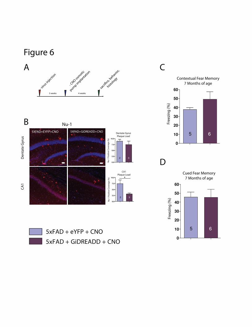
5XFAD+EYFP+CNO 5XFAD+GiDREADD+CNO
De
nta
te G
yru
sC
A1
0.0
2.5
5.0
7.5
10.0
Dentate Gyrus
Plaque Load
2 3
Nu
-1 P
osi
tiv
e C
ov
era
ge
(%
)
0.0
2.5
5.0
7.5
10.0
CA1
Plaque Load
Nu
-1 P
osi
tiv
e C
ov
era
ge
(%
)
2 3
Nu-1
3 weeks 4 weeks
- viru
s inje
ction
- CNO
osm
otic
pump im
planta
tion
- sacr
i�ce
, behavio
r,
histolo
gy
A
B
C
0
10
20
30
40
50
60
5 6
Fre
ezi
ng
(%
)
Contextual Fear Memory
7 Months of age
*
5xFAD + eYFP + CNO
5xFAD + GiDREADD + CNO
D
0
10
20
30
40
50
60
5 6
Fre
ezi
ng
(%
)Cued Fear Memory
7 Months of age
Figure 6
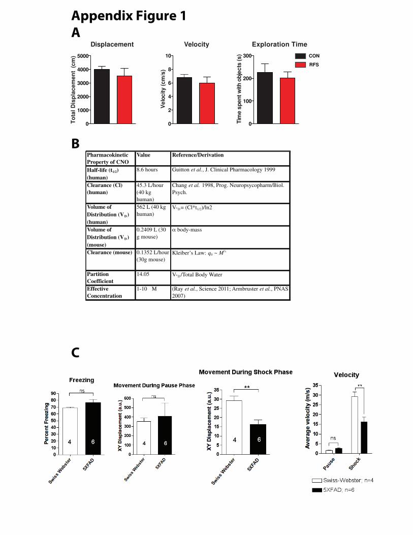
Pharmacokinetic
Property of CNO
Value Reference/Derivation
Half-life (t1/2)
(human)
8.6 hours Guitton et al., J. Clinical Pharmacology 1999
Clearance (Cl)
(human)
45.3 L/hour
(40 kg
human)
Chang et al. 1998, Prog. Neuropsycopharm/Biol.
Psych.
Volume of
Distribution (VD)
(human)
562 L (40 kg
human)
V-D= (Cl*t1/2)/ln2
Volume of
Distribution (VD)
(mouse)
0.2409 L (30
g mouse)
body-mass
Clearance (mouse) 0.1352 L/hour
(30g mouse)
Kleiber’s Law: q0 ~ M
Partition
Coefficient
14.05 V-D/Total Body Water
Effective
Concentration
1-10 μ (Ray et al., Science 2011; Armbruster et al., PNAS
2007)
Displacement
0
1000
2000
3000
4000
5000
To
tal
Dis
pla
ce
me
nt
(c
m) CON
RFS
Velocity
0
2
4
6
8
10
Ve
loc
ity
(c
m/s
)
Exploration Time
0
100
200
300
Tim
e s
pe
nt w
ith
ob
jec
ts (s
)
C
B
A
Appendix Figure 1

80
(A) Paradigm for repetitive-footshock treatment in Swiss-Webster mice.
Inset shows behavioral testing schedule. (B) Optical stimulation paradigm for Swiss-Webster mice undergoing
BLA cell body photostimulation. Inset shows behavioral testing schedule. (C) Paradigm for intracranial viral
injection of eYFP (top) or ChR2-eYFP (bottom) viral constructs, and schematic of location of viral
expression, fiber optic implantation, and illumination. (D) GiDREADD “hM4D(Gi)” generation by ligand
binding site mutation (Armbruster, 2007) Schematic illustrating eYFP or GiDREADD viral constructs,
and territory for viral expression Upon CNO binding to GiDREADD, normal G-protein dissociation
occurs, leading to activation of G protein-coupled inwardly-rectifying potassium channels (GIRKs) and
neuronal inhibition. (E) Paradigm for chronic-restraint stress treatment of 5xFAD mice. (F) Modified optical
stimulation paradigm for 5xFAD mice undergoing BLA cell body photostimulation. Inset shows behavioral
testing schedule.
(A) :
Representative confocal immunohistochemical microscopy demonstrating labelling of synaptophysin and
HDAC2 in hippocampus CA1 (leftmost), and densitometric quantification (left; n=4 per group); RFS mice
showed no discrimination between old and novel objects or object-locations (discrimination index=0.5
(chance)) in NOR (n=10,16) and NLR (n=10/group); ELISA measurement of mid-afternoon plasma
corticosterone demonstrated significant elevation after RFS (n=5/group). (B)
: representative confocal microscopy in hippocampus CA1
with densitometric quantification (left, n=5/group) displays reduction in SYP and elevation in HDAC2
similar to that seen after RFS. ChR2/photostimulated mice showed no discrimination between old and novel
objects or object-locations (discrimination index=0.5 (chance)) in NOR (n=10,12) and NLR (n=12/group);
ELISA measurement of mid-afternoon plasma corticosterone demonstrated significant elevation after RFS

81
(n=5/group). (C) Gi :
representative confocal microscopy in hippocampus CA1 with densitometric quantification (left, n=4,5,6)
displays rescue of SYP and HDAC2 after BLA inactivation during RFS by GiDREADD/CNO treatment.
Both novel-object and novel-location discrimination abilities are rescued in RFS mice by GiDREADD/CNO
treatment - NOR (n= 16, 15, 7) and NLR (n=10, 10 and 7).
(A)
Constructs for eYFP-control (top) or ChR2-eYFP (bottom) virus used in bilateral intra-BLA injection;
Paradigm demonstrating location of viral expression, optical fiber implantation, and illumination. (B)
Representative confocal immunohistochemical microscopy demonstrating labelling of synaptophysin and
HDAC2 in hippocampus CA1, and densitometric quantification (right; n=5 per group)(C) Cognitive testing
using hippocampus-dependent paradigms demonstrated that ChR2/terminal photostimulated mice showed
no discrimination between old and novel objects or object-locations (Discrimination index=0.5 (chance)) in
NOR (n=16,10) and NLR (n=16/10). (D) ELISA measurement of mid-afternoon plasma corticosterone
(n=5/group).
(A) ELISA measurement of mid-afternoon plasma corticosterone after 1 session of CRS or control
treatment. (B) Change in body weight after 14 daily sessions of CRS or control treatment. (C) Representative
confocal microscopy demonstrating increased overall hippocampal Aé plaque burden by 4G8 antibody
staining, including digital-zoom image showing individual plaques (left panels, representative images; right
panel, quantification by counts under epifluorescence). (D) Representative confocal microscopy

82
demonstrating increased overall hippocampal Aé oligomer coverage by Nu-1 antibody (left panels,
representative images; right panels, quantification). (E) Plasma corticosterone concentration following a
single stress-treatment does not correlate with hippocampal plaque load (pearson correlation coefficient =
0.0284)
(A) ELISA measurement of mid-afternoon plasma corticosterone in undisturbed
cage controls, or after 1 session of BLA photostimulation of eYFP or ChR2-injected mice. (B) Representative
confocal microscopy demonstrating increased overall hippocampal Aé plaque burden by 4G8 antibody
staining, including quantification by counts under epifluorescence. (C) Representative confocal microscopy
demonstrating increased Aé oligomer coverage by Nu-1 antibody staining in the hippocampus CA1
(rightmost panels – quantification by binary intensity-thresholded percent area). (D) Representative confocal
microscopy demonstrating no change in GFAP intensity in the hippocampus CA1 (rightmost panels –
quantification by overall intensity. ( ) 14 days of BLA photostimulation leads to a deficit in contextual fear
memory (E), but no difference in cued fear memory (by presentation of the stimulus tone)(F).
i
A. Experimental paradigm for genetic-pharmacological BLA
inactivation by GiDREADD (B) Representative confocal microscopy demonstrating reduced hippocampal Aé
oligomers in hippocampus CA1 by Nu-1 staining (right panels, quantification by binary intensity-thresholded
percent area). ( ) 4 weeks of BLA inactivation may produce a slight improvement in contextual fear
memory ( not significant), but did not lead to a change in fear memory cued by presentation of the

83
stimulus tone (D).
(A) Computerized tracking during NOR testing demonstrates no difference in various
control behavioral measures between RFS and context-only (control) treated mice (n=10,16). (B) Table of
pharmacokinetic parameters used to calculate approximate tissue-concentrations of CNO. (C) Computerized
tracking demonstrates that Swiss-Webster and 5xFAD mice respond differently to shocks in the RFS
paradigm, but freeze identically. These results were considered sufficient to justify adjusting the stress
paradigm in 5xFAD mice.