Revisiting the conversion of muscle into meat and the underlying mechanisms
15
Revisiting the conversion of muscle into meat and the underlying mechanisms Ahmed Ouali a, * , Carlos Hernan Herrera-Mendez a , Gerald Coulis a , Samira Becila b , Abdelghani Boudjellal b , Laurent Aubry a , Miguel Angel Sentandreu a,1 a QuaPA, BPM, INRA de Clermont Ferrand–Theix, 63122 Saint Gene `s Champanelle, France b INATAA, Universite ´ de Constantine, Route de Aı ¨n El Bey, 25000 Constantine, Algeria Received 24 February 2006; received in revised form 27 April 2006; accepted 8 May 2006 Abstract The conversion of muscle into meat is a complex process in which all mechanisms responsible for the development of meat qualities are very likely interdependent. Colour and flavour are thus both dependent on oxidative mechanisms. Oxidation and proteolysis are probably two processes involved in the development of meat tenderness. This paper reviewed the consequences of programmed cell death or apoptosis on muscle cells structure and biochemistry and on meat qualities as well. We therefore look at different new hypothesis susceptible to highlight the meat science field and provide new supports for a more dynamic meat research. One of them which would have appeared evident for our purpose since a decade, deals with the fact that, after animal bleeding, muscle cells have no other alter- native to only enter the programmed cell death procedure or apoptosis. If we introduce an early phase corresponding to apoptosis, taking place before the rigor onset and overlapping it, we will see that the known consequences of that process bring forward possible answers to still unexplained observations. After an overview of the actual state-of-the-art in meat science, we will introduce the programmed cell death and its underlying mechanisms. We then described the strong analogies between the known consequences of apoptosis and the postmortem changes affecting a set of different muscle characteristics. Ó 2006 Elsevier Ltd. All rights reserved. Keywords: Meat qualities; Muscle; pH; Calcium; Caspases; Apoptosis; Cell death 1. Introduction Storage of meat at low temperature is a prerequisite for the development of the major eating qualities including tenderness and flavour. Whether increasing storage length will be profitable for tenderness and flavour, it will have a rather deleterious effect on juiciness and colour. Hence, it will be necessary to find out the best compromise between these adverse time effects and/or to apply storage technologies enabling a preservation of these qualities. For decades, consumers considered tenderness as the most important quality attribute of meat. In a review of factors influencing consumption, selection and acceptabil- ity of meat purchases, Jeremiah (1982) concluded that the most common cause of unacceptability in beef was tough- ness and that this was a common problem in pork and lamb. In contrast to mammals, fish flesh often undergoes very rapid softening rendering it unappealing to consumers (Ashie & Simpson, 1997; Crapo, Himelbloom, Pfutzenre- uter, & Chong, 1999; Jiang, 2000). Despite some progress, this is still an acute problem for meat industry and fisheries. Actually, the meat tenderising process is unanimously rec- ognized to be enzymatic in nature and the most studied proteolytic systems were cathepsins, calpains and, although more recently, the 20S proteasome (Sentandreu, Coulis, & Ouali, 2002). However, the major peptidases of concern are 0309-1740/$ - see front matter Ó 2006 Elsevier Ltd. All rights reserved. doi:10.1016/j.meatsci.2006.05.010 * Corresponding author. Tel.: +33 4 73 62 41 63; fax: +33 4 73 62 42 68. E-mail address: [email protected] (A. Ouali). 1 Present address: Department of Food Science, Instituto de Agroqui- mica y Tecnologia de Alimentos (C.S.I.C.), Apt. 73, 46100 Burjassot, Valencia, Spain. www.elsevier.com/locate/meatsci Meat Science 74 (2006) 44–58 MEAT SCIENCE
-
Upload
independent -
Category
Documents
-
view
0 -
download
0
Transcript of Revisiting the conversion of muscle into meat and the underlying mechanisms

www.elsevier.com/locate/meatsci
Meat Science 74 (2006) 44–58
MEATSCIENCE
Revisiting the conversion of muscle into meat and theunderlying mechanisms
Ahmed Ouali a,*, Carlos Hernan Herrera-Mendez a, Gerald Coulis a, Samira Becila b,Abdelghani Boudjellal b, Laurent Aubry a, Miguel Angel Sentandreu a,1
a QuaPA, BPM, INRA de Clermont Ferrand–Theix, 63122 Saint Genes Champanelle, Franceb INATAA, Universite de Constantine, Route de Aın El Bey, 25000 Constantine, Algeria
Received 24 February 2006; received in revised form 27 April 2006; accepted 8 May 2006
Abstract
The conversion of muscle into meat is a complex process in which all mechanisms responsible for the development of meat qualitiesare very likely interdependent. Colour and flavour are thus both dependent on oxidative mechanisms. Oxidation and proteolysis areprobably two processes involved in the development of meat tenderness. This paper reviewed the consequences of programmed cell deathor apoptosis on muscle cells structure and biochemistry and on meat qualities as well. We therefore look at different new hypothesissusceptible to highlight the meat science field and provide new supports for a more dynamic meat research. One of them which wouldhave appeared evident for our purpose since a decade, deals with the fact that, after animal bleeding, muscle cells have no other alter-native to only enter the programmed cell death procedure or apoptosis. If we introduce an early phase corresponding to apoptosis, takingplace before the rigor onset and overlapping it, we will see that the known consequences of that process bring forward possible answers tostill unexplained observations. After an overview of the actual state-of-the-art in meat science, we will introduce the programmed celldeath and its underlying mechanisms. We then described the strong analogies between the known consequences of apoptosis and thepostmortem changes affecting a set of different muscle characteristics.� 2006 Elsevier Ltd. All rights reserved.
Keywords: Meat qualities; Muscle; pH; Calcium; Caspases; Apoptosis; Cell death
1. Introduction
Storage of meat at low temperature is a prerequisite forthe development of the major eating qualities includingtenderness and flavour. Whether increasing storage lengthwill be profitable for tenderness and flavour, it will havea rather deleterious effect on juiciness and colour. Hence,it will be necessary to find out the best compromisebetween these adverse time effects and/or to apply storagetechnologies enabling a preservation of these qualities.
0309-1740/$ - see front matter � 2006 Elsevier Ltd. All rights reserved.
doi:10.1016/j.meatsci.2006.05.010
* Corresponding author. Tel.: +33 4 73 62 41 63; fax: +33 4 73 62 42 68.E-mail address: [email protected] (A. Ouali).
1 Present address: Department of Food Science, Instituto de Agroqui-mica y Tecnologia de Alimentos (C.S.I.C.), Apt. 73, 46100 Burjassot,Valencia, Spain.
For decades, consumers considered tenderness as themost important quality attribute of meat. In a review offactors influencing consumption, selection and acceptabil-ity of meat purchases, Jeremiah (1982) concluded that themost common cause of unacceptability in beef was tough-ness and that this was a common problem in pork andlamb. In contrast to mammals, fish flesh often undergoesvery rapid softening rendering it unappealing to consumers(Ashie & Simpson, 1997; Crapo, Himelbloom, Pfutzenre-uter, & Chong, 1999; Jiang, 2000). Despite some progress,this is still an acute problem for meat industry and fisheries.Actually, the meat tenderising process is unanimously rec-ognized to be enzymatic in nature and the most studiedproteolytic systems were cathepsins, calpains and, althoughmore recently, the 20S proteasome (Sentandreu, Coulis, &Ouali, 2002). However, the major peptidases of concern are

A. Ouali et al. / Meat Science 74 (2006) 44–58 45
not identified yet in an unquestionable way and this ques-tion is still strongly debated.
Besides meat tenderness, juiciness and flavour are twoother organoleptic qualities the consumers are lookingfor when eating meat especially red meats.
Despite its close relationship to overall meat desirability,juiciness has received limited research attention. Variabilityin juiciness is commonly recognized in various meat animalspecies but, to our knowledge, there is still no clear expla-nation for this phenomenon. Meat juiciness is assumed tobe directly related to the progressive intracellular waterefflux occurring during the acidification of postmortemmuscle, a change in good agreement with the parallelincrease in the extra-cellular space (Guignot, Vignon, &Monin, 1993; Monin & Ouali, 1991; Offer & Knight,1988a, 1988b). Attempts to find out muscle characteristicssusceptible to explain this biological variability led tohighly controversial findings and much remains to be learntabout meat juiciness and muscles water holding capacity aswell, two variables probably inter-related (Ouali, 1991).
Flavour is an important part of the eating quality offoods, including meat, and complaints of blandness areoften levelled against modern lean meat from most animalspecies. Attempts to improve this quality with enhancednutritional characteristics were therefore carried out (Molo-ney, Mooney, Kerry, & Troy, 2001; Wood et al., 1999). Fla-vour compounds are highly variable in nature. Peptides(Sentandreu et al., 2003), amino acids, lipids derivativesare indeed important determinants of meat flavour. Onthe other hand, radical reactions are known to constitutea central process in the oxidation of these compounds evenif the nature and the origin of the radicals remain stillunclear (Renerre, 1999). In addition, cooking conditionsare essential in the flavour development through the Mail-lard reaction taking place, upon heating, between carbohy-drates and proteins and their derivatives (Mottram, 1992).Apart from the management of its intensity through modu-lating the end-point temperature or the heating length, con-trol of the Maillard reaction is difficult to achieve.
Meat colour is the first quality taken into account byconsumers when purchasing the meat, brown colour beingsynonymous of bad hygienic quality for consumers. Meatcolour is defined by the extent of myoglobin oxygenationand the oxidative status of the haem iron. Oxygenated myo-globin gives an appealing light red coloured meat and adark red colour in the absence of oxygen. Oxidation ofthe haem iron from a ferrous (Fe2+) to a ferric (Fe3+) stategive rise to the brown colour and is often associated to therelease of oxygen radical (Satoh & Shikama, 1981; Wallace,Houtchens, Maxwell, & Caughey, 1982). The concentrationof residual oxygen and the radical formation are believed tobe the major cause of colour deterioration in stored meatbut the underlying mechanisms are still unclear.
The conversion of muscle into meat as a whole and thepostmortem development of the eating quality are far frombeing understood. Improvement of our knowledge aboutthe underlying mechanisms is particularly faced to the large
biological variability of these qualities and to the non-iden-tification of their major determinants.
Since about a decade, intensive researches have beendeveloped on programmed cell death (PCD) (Kerr, Wyllie,& Currie, 1972) in relation with important pathologies likecancers (cells never died), neurodegenerative disordersincluding Alzheimer disease (cells died before their com-plete differentiation), etc. (Hengartner, 2000; Majino &Jons, 1995; Nicholson, 2000; Sandri & Carraro, 1999;Shi, 2002; Tews, 2005). The major form of PCD is apopto-sis (‘‘suicide’’ program), a process finely regulated and ini-tiated by either the central nervous system or by the targetcell itself. After animal bleeding, all cells will be in anoxiaand will receive no more nutriments. In such conditions,each cell can decide to die by initiating the apoptotic pro-cess. Up to date, the conversion of muscle into meat isassumed to occur through three steps: the pre-rigor stepwhich is not well defined yet, the rigor step and the tende-rising step. If we assumed that postmortem muscle cells willcommit to suicide, the apoptotic process will start immedi-ately and progress as long as the enzymes of concernremain active. Apoptosis induced a series of biochemicaland structural changes in dying cells which will be verylikely found in postmortem muscle. The extent of thesechanges will be however more limited than in vivo wherethe cell content totally disappear, since the dynamic ofthe process will be faced to unfavourable environmentalmodifications (pH, ionic strength, low energy availability,etc.).
In this review, we revisited the conversion of muscle intomeat after the integration of an additional phase before therigor process corresponding to the apoptotic phase. After arapid overview of the state-of-the-art, we will describe themechanisms and the consequences of the programmed celldeath process and, in the last part of this review paper, wewill try to see how the known effects of cell death on thecellular structures and proteins can constitute a newapproach to the muscle conversion into meat and providepossible original answers to some still unexplained observa-tions reported in postmortem muscle.
2. Current concept of the conversion of muscle into meat
Animal are slaughtered, dressed, deboned and musclesthen stored at refrigerated temperature for one week ormore depending on the current national practice and/orregulations before selling. Storage of muscles for a reason-able length of time is a perquisite for the development ofthe organoleptic qualities of the final product namely meat.
2.1. Tenderness
Postmortem improvement in meat tenderness resultsfrom a softening of the myofibrillar structure by endoge-nous peptidases (Ouali, 1992; Sentandreu et al., 2002).For several decades, attention of meat scientists has beenmainly focused on the two best known enzymatic systems,
46 A. Ouali et al. / Meat Science 74 (2006) 44–58
i.e. cathepsins and calpains. Three currents ways of think-ing about that process however exist:
– those who are prone to think that calpains are the onlyproteases responsible for meat tenderisation,
– those who suggest that the two quoted systems take partin this process,
– a third group of researchers, to which we belong, whopropose a multienzymatic process implying these systemsand also probably others which function, in postmortemmuscle, is less clear (proteasomes, caspases, etc.).
Actually, the meat tenderising process is unanimouslyrecognized to be enzymatic in nature and the most studiedproteolytic systems are:
– the cathepsins, a system discovered in the years 1950 byDe Duve, Pressman, Gianetto, Wattiaux, and Appel-mans (1955).
– the calpains which are calcium-dependent peptidaseshighlighted for the first time in rat brain by Guroff(1964).
– the proteasomes discovered more recently by Wilk andOrlowski (1980).
The cathepsins were the first enzymatic system consid-ered in the studies focusing on the mechanisms of meattenderisation. Later, calpains received much more atten-tion than cathepsins mainly because of their ability to alterthe Z-line density, a modification often observed postmor-tem, even if this change is not correlated with tenderness(Taylor, Geesink, Thompson, Koohmaraie, & Goll,1995). More recently, several sets of evidence supported apotential role of the 20S proteasome in this process. Basedon different approaches, all results reported clearly showedthat proteasome could contribute to tenderisation of storedmeat (Dutaud, 1998; Lamare, Taylor, Farouta, Briand, &Briand, 2002; Matsuishi & Okitani, 1997; Otsuka et al.,1998; Ouali, 1999; Thomas et al., 2004).
The most current actual concept suggested a major roleof calpains and more specially of calpain 1 or l-calpain(Veiseth and Koohmaraie, 2005). However, this statementhas never been definitely proven and the main reason isvery likely our very limited knowledge about calpains, theirin situ regulation and their exact biological functions. Ascompared to proteasomes, a proteolytic complex discov-ered much more recently, we can affirm that our knowledgeof the biological function and the operating mode of thecalpain system in the cell is much less advanced. This state-ment is supported by recent findings indicating that calpain1 is concentrated on the N1 and N2 line region of titin andthis might constitute a reservoir of calpain 1 for the cell(Fernandez et al., 2005).This further suggest a special reg-ulation of the release of calpain 1 from its binding sites andthe existence of a finely regulated equilibrium between theamounts of free and bound enzyme. On the other hand,calpastatin, the specific inhibitor of ubiquitous calpains,
is often considered as a single protein. In fact, as alreadysuggested in the 1990s (Ouali & Talmant, 1990), this cal-pain inhibitor is a family of at least four different isoforms(Raynaud et al., 2005) some of which being differentlyexpressed in slow-twitch and fast-twitch skeletal muscles(Ouali & Talmant, 1990). Their properties and tissueexpression are still unknown. Therefore, it must be empha-sized that our understanding of the calpain system is actu-ally very restricted and most remains to be learned aboutthe peptidases themselves, their activity regulation andtheir specific inhibitor.
Calpains are inhibited by calpastatin, a highly polymor-phic protein. Cathepsins have their own inhibitors designedcystatins (Dubin, 2005; Sentandreu et al., 2002). The term‘‘cystatin’’ refers to a group of homologous and evolution-ary related cysteine peptidase inhibitors (Barrett, 1987;Muller-Esterl et al., 1985; Turk et al., 1986) which are inac-tive against other classes of peptidases (serine-, aspartyl-and metallopeptidases). On the basis of their primary struc-ture, there are four distinct families of cysteine peptidaseinhibitors, which are recognized as belonging to the cysta-tin superfamily (Rawlings & Barrett, 1990).
– Family 1 cystatins, also designated stefins, are lowmolecular weight- proteins (10–14 kDa) containing onecopy of the basic inhibitory structure but no intramolec-ular disulfide bridges. The most well characterised mem-bers of this family are stefin A and stefin B. As stefinslack the signal sequence present in secreted proteins,they are generally found intracellularly.
– Family 2 cystatins, also designated cystatins, are lowmolecular weight proteins (10–14 kDa) containing onecopy of the basic inhibitory structure and at least oneintramolecular disulfide bridge. Cystatins are predomi-nantly extracellular. Cystatin C and chicken cystatinare the most representative of this cystatin family.
– Family 3 cystatins, also designated kininogens, are highMr cystatins (>50 kDa) containing generally three cop-ies of the basic inhibitory structure and several disulfidebonds. The kininogen family comprises three subclassesreferred to as low Mr (LMW), high Mr (HMW) and T-kininogens differing by their molecular masses and theirspecificity towards cysteine peptidases. They are gener-ally present in body fluids.
– Family 4 cystatins are glycosylated protein inhibitors ofintermediate Mr containing two copies of the basicinhibitory structure and several intramolecular disulfidebridges.
All members of the cystatin superfamily inhibit cysteinepeptidases such as papain and the major lysosomal pepti-dases including cathepsins B, H and L. In addition, kinino-gens have been shown to inhibit the ubiquitous calpains,which are calcium dependent cysteine peptidases (Ishiguroet al., 1987).
In muscle tissue, several low Mr cystatin-like proteinshave been described (Bige, Ouali, & Valin, 1985; Schwartz
A. Ouali et al. / Meat Science 74 (2006) 44–58 47
& Bird, 1997) but as no sequence was available, it was andis still impossible to certify that these are really members ofthe cystatin superfamily. Indeed, their identification ascystatin was only based on their closely similar molecularmass and inhibitory properties. This statement is true formost of the other cysteine peptidase inhibitors isolatedfrom skeletal muscles of various animal species (Oualiet al., 1995; Zeece, Woods, Keen, & Reville, 1992). Impor-tance of cystatins in postmortem and living muscles is sup-ported by their identification as potent predictor of meattenderness (Barnier, 1995; Shackelford et al., 1991) andtheir possible use as biological markers of different pathol-ogies in humans (Dubin, 2005; Kos & Lah, 1998; Strojanet al., 2000).
Each one of us will be therefore able to note that, arapid and objective analysis of the literature published todate in this field clearly indicates that the process of meattenderisation results probably from the synergistic actionof several endogenous enzymatic systems, even if the majorpeptidases of concern are not identified yet in an unques-tionable way.
2.2. Juiciness
When meat is tender, juiciness is the next quality of inter-est for consumers especially for red meats, whiter meat cutsbeing generally considered as dry. Decrease in juiciness wasoften associated with water holding (WHC) capacity ofmuscles. However, the ultimate pH which is one of themajor determinants of WHC seems to have negligible effecton meat juiciness (Bouton, Carroll, Fisher, Harris, & Shor-those, 1973). Relationship of juiciness with various biolog-ical parameters is unclear and data reported often highlycontroversial. Valin, Touraille, Vigneron, and Ashmore(1982) thus noted a significant correlation between muscletype and meat juiciness confirming the empiric thought thatred meats are more juicy than white meats. No valuable bio-logical determinants were however identified so far. A cor-relation with muscle fat content was also mentioned (Owens& Gardner, 1999) but, since years this point led to contro-versial findings as already analysed in the 1970s by Purchasand Davies (1974). Nevertheless, all scientists agree thatjuiciness strongly depends on the movement of intracellularwater towards the extracellular space assuming that, as thepH becomes more acidic and closer to the pI of myofibrillarproteins, these will release their bound water (Bertram,Schafer, Rosenvolda, & Andersen, 2004; Lawson, 2004;Offer & Knight, 1988a, 1988b).
By analogy with pork where a mutation of the PRKAG3gene encoding the c3 subunit of the AMP dependent kinase(AMPK) was responsible for the PSE character of the meat,one can thought that mutations of the same gene could beresponsible for the biological variability in drip loss. TheAMPK, comprising three subunits (a, b,c), has been pointedout as one of the main actors in the regulation of intracellu-lar energy metabolism (Carling, 2004). As AMPK is acti-vated by a drop in the energy status, it is usually
considered as a cellular fuel gauge. Its activation switch offenergy-using pathways and switch on energy-generatingpathways, thus helping to restore the energy balance withinthe cell. Numerous mechanisms of AMPK action on lipidand carbohydrate metabolism have been proposed (Ferre,Azzout-Marniche, & Foufelle, 2003; Hardie, Scott, Pan, &Hudson, 2003). The corresponding cattle gene has beenrecently sequenced and was shown to be highly polymorphand to exhibit several mutations leading to different possiblealleles (Roux et al., 2006). The consequences of these muta-tions on muscle WHC, meat juiciness and drip loss are how-ever still unknown. Investigation on the AMPK genefunction and its polymorphic allelic expression would bean alternative way to provide answers to carcasses, musclesand meat exudation. In a longer term, these studies wouldprobably be also very helpful for the genetic selection of ani-mals expressing the most suitable isoform of the corre-sponding AMPK subunit. In addition, this gene is verylikely common to all meat animal species and findingsobtained can be therefore extended to any species.
2.3. Meat flavour and colour
Although important for the consumers, our purpose willnot be to overview in detail these qualities, their biologicalvariability and the underlying mechanisms. Our objectivewill be just to emphasize the importance of peroxidationin the time course evolution of these characteristics.
Major contributors to meat flavour are lipids peroxida-tion together with amino-acids and peptides generated byproteolysis (Campo et al., 2006; Gorbatov & Lyaskovs-kaya, 1980; Sentandreu et al., 2003). Similarly, colour deg-radation during storage related to metmyoglobinformation also results from oxidative processes. Metmyo-globin indeed is generated by a spontaneous oxidation ofthe haem iron from the ferrous state (Mb–Fe2+) to the fer-ric state (Mb–Fe3+) which cannot bind O2 (reviewed byBekhit & Faustman, 2005).
For both meat qualities and tenderness as well (Warner,Dunshea, Ponnampalam, & Cottrell, 2005), oxidation inmuscle systems is initiated as a free-radical autocatalyticchain mechanism in which prooxidants, especially oxygenand related radicals will generate free radicals ensuringthe autocatalytic propagation of the oxidative chain (Kan-ner, 1994).
3. Programmed cell death or apoptosis
The concept of programmed cell death or apoptosis ismuch older than suggested in most reports. Indeed, accord-ing to Clarke and Clarke (1995), naturally occurring celldeath, including the concept of apoptosis, was a flourishingsubject at the end of the 19th century. The first report thatcells die naturally in development was published by CarlVogt in 1842 (cited by Clarke & Clarke, 1995).
Programmed cell death is a physiological mechanismnaturally occurring in living organisms that eliminates
48 A. Ouali et al. / Meat Science 74 (2006) 44–58
excessive, damaged or potentially dangerous cells from anorganism without damaging surrounding cells (Fidzianska,Kaminska, & Glinka, 1991; Kerr et al., 1972). Such system-atic cell clearance is necessary for both the normal develop-ment of a multicellular organism during embryogenesis andthe maintenance of tissue homeostasis in adults (Dirks &Leeuwenburgh, 2005; Meier, Finch, & Evan, 2000).
Selective elimination of the cells is ensured by a processcalled apoptosis. The word apoptosis refers to the pro-grammed fall of leaves at autumn time: apo for distanceand ptosis for fall. The very strict regulation of this pro-gram is essential to make sure that it is activated only inthe cell concerned and at the right time. Deregulation ofthe apoptotic process is associated with various pathologiessuch as cancer, auto-immune and degenerative diseases(Hengartner, 2000; Kerr et al., 1972; Majino & Jons,1995; Nicholson, 2000; Sandri & Carraro, 1999; Shi,2002; Tews, 2005).
Apoptosis is usually mediated by a complex machinerywhich is becoming well-known in humans (Fuentes-Prior& Salvesen, 2004; Green & Amarante-Mendes, 1998; Hen-gartner, 2000; Zimmermann, Bonzon, & Green, 2001; Zim-mermann & Green, 2001). On the other hand, althoughresearchers agree that programmed cell death also existsin plants, relatively little is known about mediators of theprocess in plants. However, its role in plant developmentand morphogenesis is regarded as essential. It is thusadmitted that deregulation of apoptosis is very often asso-ciated with diverse disturbances in development but alsowith a strong lethality (Sanmartın, Jaroszewski, Raikhel,& Rojo, 2005). Apoptosis is mediated by a particular groupof cysteine peptidases called caspases.
Apoptosis is an evolutionary highly conserved processfrom monocellular organisms to mammals (Driscoll,1996; Yuan, 1996). To illustrate this fact, we must keepin mind that the biological function of caspases, the mainenzymes responsible for programmed cell death, was dis-covered in the nematode Caenorhabditis elegans (Yuan,Shaham, Ledoux, Ellis, & Horvitz, 1993). It is in this nem-atode that the first gene responsible for cellular death, thegene encoding for CED3, was identified: CED3 is a cys-teine peptidase very homologous with ICE (Interleukin-1Converting Enzyme), an enzyme discovered in humansshortly before (Cerretti et al., 1992; Thornberry et al.,1992). These last findings constitute the starting point ofmany works developed thereafter on these peptidases andtheir regulators. These extensive investigations led veryquickly to a better understanding of the apoptotic process.
3.1. Necrosis and apoptosis
The concept of apoptosis was introduced in 1972 byKerr et al. to indicate a form of cellular death totally differ-ent from necrosis, from both the morphological and bio-chemical points of view. Comparative analysis of musclecell death led to the same conclusion (Fidzianska et al.,1991).
Necrosis is regarded as a ‘‘disordered’’ cellular death.During necrosis, cells swell up with water to the pointwhere lysis of their plasmic membrane occurs. It is a genu-ine cellular explosion which leads to the release of the cyto-plasmic contents in the surrounding medium. Cellularorganites also inflate and are emptied of their contents.The nuclear ADN is degraded in a ‘‘random’’ way by endo-nucleases activated in particular by serine peptidases. Thesize of the DNA fragments generated is very heteroge-neous. Necrosis of a cell affects other cells by the actionof the released intracellular enzymes and of blood leuco-cytes which come in reinforcement to clear the site (localinflammation). The result is that a whole area of an organwill have to be regenerated after total destruction of thedamaged cells (Buja, Eigenbrodt, & Eigenbrodt, 1993; Fid-zianska et al., 1991; Majino & Jons, 1995).
In opposition to necrosis, apoptosis is regarded as an‘‘ordered’’ cellular death, proceeding through variousphases (Bratton et al., 1997; Buja et al., 1993; Gavrieli,Sherman, & Ben-Sasson, 1992; Majino & Jons, 1995; Mar-tin et al., 1995; Matsura et al., 2005; Wyllie, 1980; Youle &Karbowski, 2005)
� first of all, cells in apoptosis are isolated by loss of con-tacts with neighbouring cells;� an important condensation of the nucleus and of the
cytoplasm induces a significant reduction in cellularvolume;� mitochondria of the apoptotic cell undergo several
major modifications: release of cytochrome c in the cyto-plasm, reduction in the membrane potential and deteri-oration of the membrane permeability with opening ofspecialized pores and diffusion of diverse proapoptoticproteins;� after condensation of the nucleus, chromatin is cleaved in
regular fragments of approximately 180 pairs of bases;� sometimes, the plasmic membrane buds and forms
apoptotic bodies, containing some of the cell cytoplasm;� in order to facilitate recognition of the apoptotic bodies
by phagocytes, the cell signals its apoptotic state by achange of localisation of phosphatidylserines molecules,from a cytoplasmic orientation to an extracellularorientation.
Programmed cell death is a rapid process (from a fewminutes to a few hours) (Green, 2005). Compared to necro-sis, one of the key properties of apoptosis is that the plas-mic membrane is not entirely destructed during the process,thereby avoiding discharge of the cellular contents andconsequent damage inflicted to neighbouring cells.
3.2. The caspases
Because caspases (structure and functions) and apopto-tic regulation will be only succinctly overviewed hereafter,readers must refer to the review of Fuentes-Prior andSalvesen (2004) for more information.
A. Ouali et al. / Meat Science 74 (2006) 44–58 49
3.2.1. Caspases, a new family of peptidases
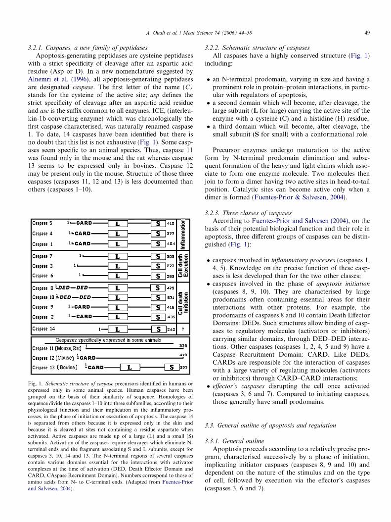
Apoptosis-generating peptidases are cysteine peptidaseswith a strict specificity of cleavage after an aspartic acidresidue (Asp or D). In a new nomenclature suggested byAlnemri et al. (1996), all apoptosis-generating peptidasesare designated caspase. The first letter of the name (C)stands for the cysteine of the active site; asp defines thestrict specificity of cleavage after an aspartic acid residueand ase is the suffix common to all enzymes. ICE, (interleu-kin-1b-converting enzyme) which was chronologically thefirst caspase characterised, was naturally renamed caspase1. To date, 14 caspases have been identified but there isno doubt that this list is not exhaustive (Fig. 1). Some casp-ases seem specific to an animal species. Thus, caspase 11was found only in the mouse and the rat whereas caspase13 seems to be expressed only in bovines. Caspase 12may be present only in the mouse. Structure of those threecaspases (caspases 11, 12 and 13) is less documented thanothers (caspases 1–10).
Fig. 1. Schematic structure of caspase precursors identified in humans orexpressed only in some animal species. Human caspases have beengrouped on the basis of their similarity of sequence. Homologies ofsequence divide the caspases 1–10 into three subfamilies, according to theirphysiological function and their implication in the inflammatory pro-cesses, in the phase of initiation or execution of apoptosis. The caspase 14is separated from others because it is expressed only in the skin andbecause it is cleaved at sites not containing a residue aspartate whenactivated. Active caspases are made up of a large (L) and a small (S)subunits. Activation of the caspases require cleavages which eliminate N-terminal ends and the fragment associating S and L subunits, except forcaspases 3, 10, 14 and 13. The N-terminal regions of several caspasescontain various domains essential for the interactions with activatorcomplexes at the time of activation (DED, Death Effector Domain andCARD, CAspase Recruitment Domain). Numbers correspond to those ofamino acids from N- to C-terminal ends. (Adapted from Fuentes-Priorand Salvesen, 2004).
3.2.2. Schematic structure of caspases
All caspases have a highly conserved structure (Fig. 1)including:
� an N-terminal prodomain, varying in size and having aprominent role in protein–protein interactions, in partic-ular with regulators of apoptosis,� a second domain which will become, after cleavage, the
large subunit (L for large) carrying the active site of theenzyme with a cysteine (C) and a histidine (H) residue,� a third domain which will become, after cleavage, the
small subunit (S for small) with a conformational role.
Precursor enzymes undergo maturation to the activeform by N-terminal prodomain elimination and subse-quent formation of the heavy and light chains which asso-ciate to form one enzyme molecule. Two molecules thenjoin to form a dimer having two active sites in head-to-tailposition. Catalytic sites can become active only when adimer is formed (Fuentes-Prior & Salvesen, 2004).
3.2.3. Three classes of caspasesAccording to Fuentes-Prior and Salvesen (2004), on the
basis of their potential biological function and their role inapoptosis, three different groups of caspases can be distin-guished (Fig. 1):
� caspases involved in inflammatory processes (caspases 1,4, 5). Knowledge on the precise function of these casp-ases is less developed than for the two other classes;� caspases involved in the phase of apoptosis initiation
(caspases 8, 9, 10). They are characterised by largeprodomains often containing essential areas for theirinteractions with other proteins. For example, theprodomains of caspases 8 and 10 contain Death EffectorDomains: DEDs. Such structures allow binding of casp-ases to regulatory molecules (activators or inhibitors)carrying similar domains, through DED–DED interac-tions. Other caspases (caspases 1, 2, 4, 5 and 9) have aCaspase Recruitment Domain: CARD. Like DEDs,CARDs are responsible for the interaction of caspaseswith a large variety of regulating molecules (activatorsor inhibitors) through CARD–CARD interactions;� effector’s caspases disrupting the cell once activated
(caspases 3, 6 and 7). Compared to initiating caspases,those generally have small prodomains.
3.3. General outline of apoptosis and regulation
3.3.1. General outline
Apoptosis proceeds according to a relatively precise pro-gram, characterised successively by a phase of initiation,implicating initiator caspases (caspases 8, 9 and 10) anddependent on the nature of the stimulus and on the typeof cell, followed by execution via the effector’s caspases(caspases 3, 6 and 7).
50 A. Ouali et al. / Meat Science 74 (2006) 44–58
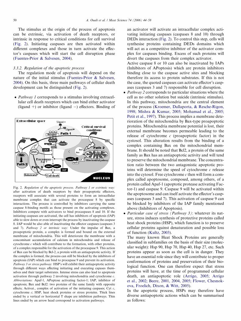
The stimulus at the origin of the process of apoptosiscan be extrinsic, via activation of death receptors, orintrinsic in response to critical conditions for cell survival(Fig. 2). Initiating caspases are then activated withindifferent complexes and those in turn activate the effec-tor’s caspases which will start the cell disruption phase(Fuentes-Prior & Salvesen, 2004).
3.3.2. Regulation of the apoptotic process
The regulation mode of apoptosis will depend on thenature of the initial stimulus (Fuentes-Prior & Salvesen,2004). On this basis, three main pathways of cellular deathdevelopment can be distinguished (Fig. 2).
� Pathway 1 corresponds to a stimulus involving extracel-lular cell death receptors which can bind either activator(ligand +) or inhibitor (ligand �) effectors. Binding of
Fig. 2. Regulation of the apoptotic process. Pathway 1 or extrinsic way:
after activation of death receptors by their proapoptotic effectors,receptors will associate with several proteins to form an intracellularmembrane complex that can activate the procaspase 8 by specificinteractions. The process is controlled by inhibitors carrying the samecaspase 8-binding motifs as those present on the activating complexes.Inhibitors compete with activators to bind procaspases 8 and 10. If theinitiating caspases are activated, the cell has inhibitors of apoptosis (IAP)able to slow down or even interrupt the process by inactivating the caspase8. IAP would be also able of inactivating the effector caspases (caspases 3and 7). Pathway 2 or intrinsic way: Under the impulse of Bax, aproapoptotic protein, a complex is formed and bound on the externalmembrane of mitochondria. This will deteriorate the membrane with aconcomitant accumulation of calcium in mitochondria and release ofcytochrome c which will contribute to the formation, with other proteins,of a complex responsible for the activation of the procaspase 9. This actionof Bax can be blocked by Bcl-2, a protein with an antiapoptotic activity. Ifthe complex is formed, the process can still be blocked by the inhibitors ofapoptosis (IAP) which can bind to procapase 9 and prevent its activation.Pathway 3 or stress pathway: HSP’s will exhibit their antiapoptotic activitythrough different ways affecting initiating and executing capases them-selves and their target substrates. Intense stress can also lead to apoptosisactivation through pathway 2 involving mitochondria and cytochrome c.Abbreviations: Apaf-1, APoptose activating factor-1; IAP, inhibitor ofapoptosis; Bax and Bcl2: two proteins of the same family with oppositeeffects; Activat., complex of activation of the initiating caspases; Cyt c,cytochrome c; HSP, heat shock proteins or stress proteins. Thick linesended by a vertical or horizontal T shape are inhibition pathways. Thinlines ended by an arrow head correspond to activation pathways.
an activator will activate an intracellular complex acti-vating initiating caspases (caspases 8 and 10) throughDEDs interaction (Fig. 2). To control this step, cells willsynthesise proteins containing DEDs domains whichwill act as a competitive inhibitor of the activator com-plex for caspases binding. Excess of such proteins willdivert the caspases from their complex activator.Active caspase 8 or 10 can also be inactivated by IAPs(Inhibitors of APoptosis) which are protein inhibitorsbinding close to the caspase active sites and blockingtherefore its access to protein substrates. If this is notthe case, the quoted caspases can activate effector’s casp-ases (caspases 3 and 7) responsible for cell disruption.� Pathway 2 corresponds to particular situations where the
cell as no other solution but suicide (intrinsic stimulus).In this pathway, mitochondria are the central elementof the process (Kroemer, Dallaporta, & Resche-Rigon,1998; Mishra & Kumar, 2005; Mohamad et al., 2005;Petit et al., 1997). This process implies a membrane dete-rioration of the mitochondria by Bax-type proapoptoticproteins. Mitochondria membrane potential is lost, theirexternal membrane becomes permeable leading to therelease of cytochrome c (proapoptotic factor) in thecystosol. This alteration results from the binding of acomplex containing Bax on the mitochondrial mem-brane. It should be noted that Bcl2, a protein of the samefamily as Bax has an antiapoptotic activity and will tendto preserve the mitochondrial membrane. The concentra-tion ratio between the two antagonistic apoptotic pro-teins will determine the speed of cytochrome c releaseinto the cytosol. Free cytochrome c then will form a com-plex called apoptosome, composed, among others, of aprotein called Apaf-1 (apoptotic protease activating Fac-tor-1) and caspase 9. Caspase 9 will be activated withinthe apoptosome and can itself activate the effector’s casp-ases (caspases 3 and 7). This activation of caspase 9 canbe blocked by inhibitors of the IAP family mentionedabove (Inhibitors of Apoptosis).� Particular case of stress (Pathway 3): whatever its nat-
ure, stress induces synthesis of protective proteins calledheat shock proteins (HSP), which function is to preservecellular proteins against denaturation and possible lossof function (Kultz, 2003).The many known Heat Shock Proteins are generallyclassified in subfamilies on the basis of their size (molec-ular weight): Hsp 90, Hsp 70, Hsp 40, Hsp 27, etc. Suchproteins appear as soon as the cell is in danger. Theyhave an essential role since they will contribute to properconformation of proteins and preservation of their bio-logical function. One can therefore expect that stressproteins will have, at the time of programmed cellulardeath, an antiapoptotic role (Arrigo, 2005; Arrigoet al., 2002; Beere, 2001, 2004, 2005; Flower, Chesnok-ova, Froelich, Dixon, & Witt, 2005).In the apoptotic process, HSPs may therefore havediverse antiapoptotic actions which can be summarisedas follows:
A. Ouali et al. / Meat Science 74 (2006) 44–58 51
– Formation of a complex with active caspases (initia-tors or effectors) thus hindering their function.
– Protection of target proteins (substrates) of effectorcaspases preventing or delaying their degradationby these enzymes.
– Attempt to re-established the initial and active struc-ture of proteins having undergone structural damagefollowing either the stress itself or the initiation ofapoptosis.
Through HSPs, stress will therefore generate actions ofan antiapoptotic nature. In the case of intense stress, how-ever, it can induce cellular death by the mitochondrialpathway (pathway 2 previously described).
4. Cell death and meat qualities
As for most other aspects of cell biology, skeletal muscletissue is far from being a current model for apoptosis inves-tigations. In humans, apoptosis in muscle tissue have beenmainly studied in relation with either pathologies includingneuromuscular disorders, myosistis or muscle atrophy(Leeuwenburgh, 2003; Liu & Ahearn, 2001; Primeau,Adhihetty, & Hood, 2002; Sandri & Carraro, 1999; Sandri,2002; Tews, 2002; Tews, 2005; Yuan, Wang, & Murrell,2003).
In all meat animal species and whatever the technologyof stunning used, the last phase of the slaughter process isbleeding. Consequently, all cells and tissues will be irrevers-ibly deprived of nutrients and oxygen. Under these veryharmful environmental conditions, muscular cells will haveno alternative but engage towards ‘‘suicide’’, with all theconsequences described above. Under similar conditions,such cell behaviour is currently observed in living organ-isms. Hence why this would not be the case in postmortemtissues and cells?
All following considerations relate mostly to cattle, but,because of the ubiquitous character of apoptosis, thereported observations can be extended to all meat animalspecies. The objective will be to consider the main cellularchanges associated with apoptosis and to establish a linkwith modifications observed in muscle during meat ageing,in relation to organoleptic qualities and more particularlytenderness, the major quality attribute for the consumer.
4.1. Inversion of the membrane polarity
4.1.1. Rapid overview of the process in vivo
In vivo, cellular membranes have a well defined polaritydependent on the distribution of phospholipids. The elec-tronegative phosphatidylserine groups, are on the innerleaflet of the cellular plasmatic membrane whereas the elec-tropositive phosphatidylcholine and phosphatidylethanol-amine groups, are on the outer leaflet. When the processof apoptosis begins, an inversion of the phospholipids dis-tribution occurs: phosphatidylserine switching to the exter-
nal leaflet of the membrane by a well-known flip-flopprocess, while the reverse happens for the other phospho-lipids (Martin et al., 1995). This change isolates the apop-totic cell from surrounding cells and signals its suicidestatus. In apoptotic cells, the membrane remains howeverimpermeable to avoid diffusion of the intracellular compo-nents in the extracellular environment. Transfer of phos-phatidylserine groups to the external leaflet of themembrane constitutes also a sign of recognition by macro-phages which will, in vivo, take part in the degradation ofdying cells. The phospholipids translocation is ensured bydifferent types of translocases which can be divided intothree classes: bidirectional ‘‘scramblases’’ and energy-dependent transporters that move phospholipids toward(‘‘flippases’’) or away (‘‘floppases’’) from the inner surfaceof the membrane (Bevers, Comfurius, & Zwaal, 1996).Among them, the most important active class of translo-cases in postmortem muscle will be more likely scramblaseswhich are bidirectional translocases with a wide specificity.Scramblases are calcium but not energy dependent (Brat-ton et al., 1997).
Cell death is not coordinated meaning that, each indi-vidual cell can initiate apoptosis independently. That isparticularly true since, within a muscle, fibres are very het-erogeneous (Pette & Staron, 1990).
4.1.2. Consequences on postmortem muscle pH fall
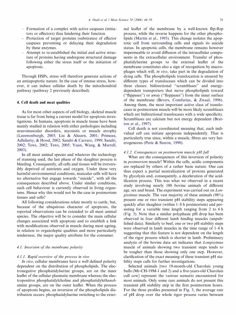
What are the consequences of this inversion of polarityin postmortem muscle? Within the cells, acidic componentsare replaced by others of a rather basic nature. One canthus expect a partial neutralization of protons generatedby glycolysis and, consequently, a deceleration of the acid-ification process. This was what we observed in a recentstudy involving nearly 180 bovine animals of differentage, sex and breed. The experiment was carried out on Lon-
gissimus muscle. The vast majority of these animals (98%)present one or two transient pH stability steps appearingquickly after slaughter (within 1–8 h postmortem) and per-sisting for a variable time length ranging from 2 to 6 h(Fig. 3). Note that a similar polyphasic pH drop has beenobserved in four different lamb hindleg muscles (unpub-lished data). Similarly to beef, one or two pH stability stepswere observed in lamb muscles in the time range of 1–6 hsuggesting that this feature is not dependent on the lengthof the rigor process which is shorter in lamb. Preliminaryanalysis of the bovine data set indicates that Longissimusmuscle of animals showing two transient steps tends tobe tougher than those showing only one step. However,clarification of the exact meaning of these transient pH sta-bility steps calls for further investigations.
Selected animals [two 19-month-old Charolais youngbulls (Mr-CH-19M-1 and 2) and a five-years-old Charolaiscull cow] represent the various scenario encountered formost animals. Only some rare animals do not present thistransient pH stability step in the first postmortem hours.For the three profiles presented in Fig. 3, the average rateof pH drop over the whole rigor process varies between

Fig. 3. pH evolution in the Longissimus muscle of three animals(charolais). The three animals are two 19-month-old young bulls (Mr.-CH-19M-1 and -2) and a 54 month-old cull cow (F-CH-54M). pH wasmeasured every hour during the first 8 h after slaughter then at 24, 48 and72 h. The pattern is presented for each animal with the average rate of pHfall indicated on each curve and expressed in Unit pH per hour(UpH h�1). In the insert, evolution of this parameter during the first 8 hpost-slaughter is presented. Each point is the mean of three independentdeterminations. For each point, the coefficient of variation is generallylower than 1%.
52 A. Ouali et al. / Meat Science 74 (2006) 44–58
0.065 and 0.21 pHunit/hour (UpH h�1), the Charolais cullcow exhibiting the fastest pH fall. Refined analysis of thepH drop profile during the first 8 h following slaughter(insert Fig. 3) shows a discontinuity in the pH fall, withthe presence of one (F-CH-54M, Mr.-CH-19M-2) or twopH stability steps (Mr.-CH-19M-1). The time length ofthese steps varied between 2 and 5 h. For the first Charolaisyoung bull (Mr.-CH-19M-1), a first pH stability step isobserved between 1 and 3 h followed by a second between4 and 8 h. For the second Charolais young bull (Mr.-CH-19M-2), only one pH stability step appears between 4and 6 h. For the Charolais cull cow, only one pH stabilitystep is observed between 1 and 3 h. Note the very fast pHdrop in this first phase (0–3 h). Unfortunately, in the avail-able literature, it was difficult to find out a detailed analysisof the pH drop in the first hours postmortem which limitthe discussion and the extension of our conclusions to theresults so far published.
Postmortem, when phosphocreatine stores are exhausted,the required energy is mainly produced through degradationof glycogen by glycolysis. The rate of the process depends onthe type of muscle considered but, in all cases, it persists aslong as enzymes are not inhibited by acidic pH. Therefore,the discontinuity in pH fall observed here cannot beexplained by a transient reduction in the activity of phospho-creatine kinase and other enzymes of the glycolytic pathwaybut rather by a modification of either the buffering capacityand/or the charge distribution within muscle cells. Replace-ment of acidic components (phosphatidylserine) by basiccomponents (phosphatidylcholine and phosphatidyletha-nolamine) in the intracellular compartment, accompaniedby a redistribution of ions, could explain the existence ofthese transient pH stability steps. Because such transientpH stability occurs between 1 and 8 h postmortem, inversion
of polarity of the plasmic membrane probably take placeduring the first 8 h postmortem when pH ranged between6.4 and approximately 6.8. This observation is comfortedby the change observed, in the same pH range, in the conduc-tivity of muscle tissue assessed by impedancemetry (Bertramet al., 2004; Damez, Lepetit, Desneux, Clerjon, & Favier,2002, in press).
4.1.3. Consequences on muscle thrombin activation
The large heterogeneity of muscle tissue reflects its highdegree of functional specialization and is the basis of itsfunctional plasticity and adaptability (Pette & Staron,1990). This plasticity concerned to a similar extent the neu-romuscular junction since the synapse must be constantlyadapted to the contractile properties and the electricalactivity of each fibre (Glazner et al., 1997). In the 1990s,extravascular cellular functions mediated by thrombin inthe process of neural development have been also identi-fied. Since then, thrombin, a serine peptidase extensivelystudied in the vascular system where the enzyme is knownto play a key role in the maintenance of haemostasis (Fen-ton, 1986), the suspected new extravascular functions ofthis peptidase have received much attention. In musclecells, thrombin has been shown to be involved in synapseelimination and remodelling at the neuromuscular junction(Liu, Fields, Festoff, & Nelson, 1994; Zoubine, Ma, Smir-nova, Citron, & Festoff, 1996) as well as in inflammatorypathologies affecting skeletal muscles (Akaaboune et al.,1998; Mbebi, Hantai, Jandrot-Perrus, Doyennette, & Ver-diere-Sahuque, 1999). The recruitment of activated throm-bin from blood in the absence of vascular injury duringthese events is unlikely. In support of this scenario, theexpression of prothrombin mRNA has been demonstratedin brain and neural cell lines (Dihanich, Kaser, Reinhard,Cunningham, & Monard, 1991; Weinstein, Gold, Cunning-ham, & Gall, 1995), as well as in rodent skeletal muscle andprimary skeletal muscle cultures (Glazner et al., 1997; Zou-bine et al., 1996). Thrombin has been thus found to belocated at the neuromuscular junction in the vicinity ofthe plasma membrane (Akaaboune et al., 1998; Mbebiet al., 1999). Muscle cells expressed thrombin may be acti-vated to act locally (Citron, Smirnova, Zoubine, & Festoff,1997; Kim, Buonanno, & Nelson, 1998). As shown inFig. 4, activation of thrombin is facilitated by flip-flopexposure of phosphatidylserine on the outer membraneleaflet (Boon, Lambert, Sisson, Davis, & Smith, 2003).The membrane itself is not sufficient for thrombin activa-tion suggesting that the presence of phosphatidylserine atthe surface of the membrane is an absolute requirementfor its activation (Majumder, Weinreb, & Lentz, 2005).Postmortem apoptotic induced phosphatidylserine expo-sure at the surface of the cell membrane would thereforelead to thrombin activation at the neuromuscular junction.The consequence will be a rapid alteration of the synapseand of the electrical conduction towards the cell. This willprobably affect also the nerves themselves (Grand, Grab-ham, Gallimore, & Gallimore, 1989; Gurwitz & Cunning-

Fig. 4. Thrombin activation needs the translocation of phosphatidylserineto the outer leaflet of the membrane (adapted from Boon et al., 2003).
A. Ouali et al. / Meat Science 74 (2006) 44–58 53
ham, 1988; Jalink & Moolenaar, 1992; Suidan, Stone,Hemmings, & Monard, 1992; Tews, 2002).
Does this can explain the rapid time dependent efficiencyloss of low voltage electrical stimulation which essentiallyuses the nervous system for the electrical field conduction?It is indeed well recognized that low voltage electrical stim-ulation of carcasses is efficient only if applied within fewminutes following animal stunning and bleeding. As illus-trated in Fig. 5, decrease in the rate of pH decline is signif-icantly reduced when low voltage stimulation (100 V,2 min) is applied 15 min postmortem as compared to theeffect of similar stimulation applied 2 min postmortem.After 2 min, the kinetic of the time course pH drop iswholly comparable to the curve obtained upon high volt-age stimulation (750 V, 2 min) applied 30 min postmortem.
4.2. Calcium and meat ageing
Since the 1960s, we know that injection of calcium inmeat accelerates the tenderising process (Khan & Kim,
Fig. 5. pH drop profile in the Longissimus muscle friesen cull cows. Opencircles, control animals; close circles, high-voltage (HV) stimulated animals;curves 2, 10 and 15 correspond to low-voltage (LV) stimulated animals 2, 10and 15 min postmortem. Sampling times for LV stimulated animals are thesame than for the control and the HV stimulated animals. Each point is themean for three animals (5–6-years-old friesan cull cows). The horizontaldashed line corresponds to the pH limit for prevention of cold shortening,i.e. pH 6. The current voltage was 750 and 100 V for HV and LVstimulation, respectively. (adapted from Valin, 1986).
1975). The action of calcium is generally attributed to anactivation of calpains, the calcium-dependent peptidases.Because of the numerous roles of this cation in cell signal-ling pathways, other potential functions of calcium havereceived very few attentions from meat scientists. However,if we now consider that, after slaughter, cells have no otheralternative but engage towards suicide or apoptosis, wehave to reconsider some of these functions. Calcium isindeed a crucial effector for triggering and controllingapoptosis (Orrenius, Zhivotovsky, & Nicotera, 2003; Sza-badkai & Rizzuto, 2004). In postmortem muscle, calciumconcentration increases gradually in the cytoplasm duringthe rigor mortis onset while the sarcoplasmic reticulum isemptied of its contents (Vignon, Beaulaton, & Ouali, 1989).
We now know that this cation is a central element of theapoptotic process, inducing swelling and extensive alter-ation of mitochondria. It contributes also to the releaseof cytochrome c together with other proapoptotic proteins.This process ends by the activation of caspase 9 which inturn, will activate the effector caspases. Since the processof apoptosis is irreversible, once engaged, it continues dur-ing all the shelf-life of refrigerated meat. As for other intra-cellular peptidases active at neutral pH, one of the majorlimitations to the activity of caspases will be the acidicpH. Unfortunately, as no data are so far available on thepH effect on caspases activity, this point remains to beanalysed.
4.3. Variation of intra- and extracellular spaces in
postmortem muscle
Over the past decades, much work was devoted to thepostmortem evolution of intra- and extracellular spaces inrelation with water movements in the muscle and withwater holding capacity (see reviews of Offer & Knight,1988a, 1988b). It was generally recognized that the maincause of these changes was the distribution, between thetwo compartments, of water which accounts in weight forapproximately 75% of muscle tissue. Acidification of mus-cle decreases protein charges and increase their hydropho-bicity, thereby reducing water retention. This is confirmedby the very high correlation observed between the increasein extracellular space and muscle pH (Guignot et al., 1993).The only point which remained unexplained was the earlyincrease in extracellular space, starting immediately afterslaughter, whereas pH was still very close to neutrality.Events associated with cell death provide an explanationsince a cell entering in apoptosis is dissociated from othersand ‘‘shrinks’’. The consequence will be a reduction inintracellular space and a parallel increase in extracellularspace.
We have shown above that postmortem pH profile waspolyphasic and presented one or two steps of relative sta-bility in the first 8 h following slaughter. By analogy, Guig-not et al. (1993) showed that extracellular space reached itsmaximum value approximately 10 h postmortem. Thusretraction of cells related to cellular death coincides with
54 A. Ouali et al. / Meat Science 74 (2006) 44–58
the period of polyphasic pH decrease and with progressiveincrease in extracellular space. All these results would sug-gest that cellular retraction and probably also inversion ofmembranes polarity, two major consequences of cellulardeath, reached their ultimate point approximately 8–10 hpost-slaughter.
4.4. Deterioration of mitochondria and cellular oxidation
Mitochondria are a central element in the apoptotic pro-cess and this explains the large number of interesting scien-tific reviews published over the last decade on the role ofthis cellular organelle in cell death (Bras, Queenan, &Susin, 2005; Gottlieb, 2000; Granville & Gottlieb, 2002;Green & Amarante-Mendes, 1998; Gulbins, Dreschers, &Bock, 2003; Haeberlein, 2004; Parone, James, & Martinou,2002; Ravagnan, Roumier, & Kroemer, 2002). This is par-ticularly true as postmortem, the stimulus does not consistin an activation of cellular death receptors but rather fromthe cell itself in response in the harmful environmental con-ditions. In addition to the respiratory chain losing itscapacity to oxidize molecular oxygen, the mitochondrialexternal membrane becomes permeable to all protein com-pounds localised in the intermembrane space, includingcytochrome c, a central caspase 9 activator. Other proteinswith proapoptotic activity are also released in the cytosol(Youle & Karbowski, 2005). In parallel, calcium from theendoplasmic reticulum is transferred to the mitochondria.Mitochondria become overloaded with calcium causingan irreversible alteration of their internal membrane.Molecular oxygen, not oxidized anymore by the respira-tory chain, will form free oxygen radicals able to oxidizeall cellular compounds (lipids, proteins, etc.).
As apoptosis will start within few minutes after death,the first oxygen radicals will be generated by mitochondriaand this will initiate the autocatalytic process which will goon over the whole storage period even at low temperatureincluding freezing. This essential initiating phase must betaken into account in studies dealing with the identificationof the major determinants of meat flavour and colourevolution.
4.5. Stress and apoptosis
It is well recognized that stress impairs the meat ageingprocess, leading generally to tougher meat. Probably thebest illustration of this phenomenon is the case of exsuda-tive pig meat.
Confronted to any type of stress, living organisms reactby the emission of signals directed to the cells, the firstchronological signals being hormones. If the stress is par-ticularly intense (e.g. oxidative stress), the cells receiveapoptosis-inducing signals via the receptors of cellulardeath. If the stress is not as severe, cells prepare theirdefence as quickly as possible. Among available means,the most described is the synthesis of various protectiveproteins known as HSPs (heat shock proteins). These pro-
teins help in the protection of intracellular components andstructures against hazards associated with loss of their bio-logical functions.
In relation to the subject of cellular death, we saw thatHSPs have an anti-apoptotic activity (Beere, 2004). Theywill consequently slow down the process of cellular deathand will constitute an obstacle to good meat ageing. Onceagain, the concept of apoptosis being the first stage of meatageing provides an answer to the question of the relation-ship nature between animal stress and unacceptable meattenderisation.
4.6. Peptidases and proteolysis
Tenderisation of meats results very likely from the soft-ening of the myofibrillar structure by the synergistic actionof endogenous peptidases including mainly cathepsins, cal-pains and proteasome (Sentandreu et al., 2002).
Up to date, calpains are considered as the major prote-olytic system responsible for meat tenderisation and mostresearchers are prone to think that this system explainedmost changes in postmortem muscle (Veiseth & Koohma-raie, 2005).
If we reconsider meat tenderisation through introduc-tion of programmed cell death, the first active peptidasesafter animal bleeding would be undoubtedly caspases.These peptidases are in a better position than others toalter cellular structures since this is their primary functionin vivo (Creagh & Martin, 2001). The common occurrenceof sequences that match the preferred inherent substratespecificity of caspases in intracellular proteins would sug-gest a multitude of substrates in vivo – somewhere in theorder of several hundred. Indeed, the list of proteins thatare cleaved by caspases either in vivo or in vitro is evergrowing (for detailed analysis of caspase substrates, seeEarnshaw, Martins, & Kaufmann, 1999; Fischer, Janicke,& Schulze-Osthoff, 2003; Nicholson, 1999). However, onlya few of these proteins have been rigorously established asbiologically relevant and many of them may represent just‘innocent bystanders (Denault & Salvesen, 2002). Thesefindings are comparable to those reported for calpainsfor which a great number of potential muscle protein sub-strates have been reported for calpains (Goll, Thompson,Li, Wei, & Cong, 2003). Because capases are specializedin cell destructuration, they probably first degrade thekey proteins involved in the complex spatial organisationof myofibrils within muscle cells and the hydrolysis of cel-lular components and organelles will then go on with theprobable contribution of other proteolytic systems includ-ing very likely cathepsins, calpains, proteasomes and else.The same process will very likely take place similarly inpostmortem muscle and, because of the changes in physi-cochemical conditions occurring in muscle after animalbleeding, the extent of cellular deterioration will be lessextensive.
Caspases are neutral cysteine peptidases and their activ-ity will be affected by muscle acidification to a similar
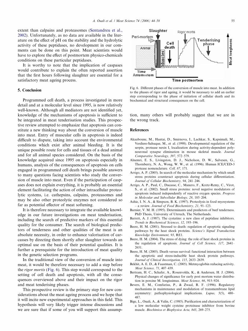
Fig. 6. Different phases of the conversion of muscle into meat. In additionto the phases of rigor and ageing, it would be necessary to add an earlierstep corresponding to the phase of initiation of cellular death and itsbiochemical and structural consequences on the cell.
A. Ouali et al. / Meat Science 74 (2006) 44–58 55
extent than calpains and proteasomes (Sentandreu et al.,2002). Unfortunately, as no data are available in the liter-ature on the effect of pH on the stability and the hydrolyticactivity of these peptidases, no development in our com-ments can be done on this point. Meat scientists wouldhave to explore the effect of postmortem physico-chemicalsconditions on these particular peptidases.
It is worthy to note that the implication of caspaseswould contribute to explain the often reported assertionthat the first hours following slaughter are essential for asatisfactory meat ageing process.
5. Conclusion
Programmed cell death, a process investigated in moredetail and at a molecular level since 1995, is now relativelywell-known. Although all mediators are not identified yet,knowledge of the mechanisms of apoptosis is sufficient tobe integrated in meat tenderisation studies. This prospec-tive review attempted to emphasize that apoptosis can con-stitute a new thinking way about the conversion of muscleinto meat. Entry of muscular cells in apoptosis is indeeddifficult to dispute, taking into account the environmentalconditions which exist after animal bleeding. It is theunique possible route for cells and tissues of a dead animaland for all animal species considered. On the basis of theknowledge acquired since 1995 on apoptosis especially inhumans, analysis of the consequences of apoptosis on cellsengaged in programmed cell death brings possible answersto many questions facing scientists who study the conver-sion of muscle into meat. Even if the participation of casp-ases does not explain everything, it is probably an essentialelement facilitating the action of other intracellular proteo-lytic systems, i.e. cathepsins, calpains, proteasomes andmay be also other proteolytic enzymes not considered sofar as potential effector of meat softening.
It is therefore necessary to integrate the available knowl-edge in our future investigations on meat tenderisation,including the search of predictive markers of this essentialquality for the consumer. The search of biological predic-tors of tenderness and other qualities of the meat is anabsolute necessity, in order to enhance valorisation of car-casses by directing them shortly after slaughter towards anoptimal use on the basis of their potential qualities. It isfurther a prerequisite for the introduction of meat qualityin the genetic selection programs.
In the traditional view of the conversion of muscle intomeat, it would be therefore necessary to add a step beforethe rigor mortis (Fig. 6). This step would correspond to thesetting of cell death and apoptosis, with all the conse-quences overviewed above and their impact on the rigorand meat tenderising phases.
This prospective review is the primary step for new con-siderations about the meat ageing process and we hope thatit will incite new experimental approaches in this field. Thishypothesis will very likely trigger intense discussions andwe are sure that if some of you will support this assump-
tion, many others will probably suggest that we are inthe wrong track.
References
Akaaboune, M., Hantai, D., Smirnova, I., Lachkar, S., Kapsimali, M.,Verdiere-Sahuque, M., et al. (1998). Developmental regulation of theserpin, protease nexin I, localization during activity-dependent poly-neuronal synapse elimination in mouse skeletal muscle. Journal
Comparative Neurology, 397, 572–579.Alnemri, E. S., Livingston, D. J., Nicholson, D. W., Salvesen, G.,
Thornberry, N. A., Wong, W. W., et al. (1996). Human ICE/CED-3protease nomenclature. Cell, 87, 171.
Arrigo, A. P. (2005). In search of the molecular mechanism by which smallstress proteins counteract apoptosis during cellular differentiation.Journal of Cellular Biochemistry, 94, 241–246.
Arrigo, A. P., Paul, C., Ducasse, C., Manero, F., Kretz-Remy, C., Virot,S., et al. (2002). Small stress proteins: novel negative modulators ofapoptosis induced independently of reactive oxygen species. Progress
in Molecular and Subcellular Biology, 28, 185–204.Ashie, I. N. A., & Simpson, B. K. (1997). Proteolysis in food myosystems
– a review. Journal of Food Biochemistry, 21, 91–123.Barnier, V. M. H. (1995). Determinants and predictors of beef tenderness.
PhD Thesis, University of Utrrech, The Netherlands.Barrett, A. J. (1987). The cystatins: a new class of peptidase inhibitors.
Trends in Biochemical Sciences, 12, 193–196.Beere, H. M. (2001). Stressed to death: regulation of apoptotic signaling
pathways by the heat shock proteins. Science’s Signal Transduction
Knowledge Environment, 93, RE1.Beere, H. M. (2004). The stress of dying: the role of heat shock proteins in
the regulation of apoptosis. Journal of Cell Science, 117, 2641–2651.
Beere, H. M. (2005). Death versus survival: functional interaction betweenthe apoptotic and stress-inducible heat shock protein pathways.Journal of Clinical Investigation, 115, 2633–2639.
Bekhit, A. E. D., & Faustman, C. (2005). Metmyoglobin reducing activity.Meat Science, 71, 407–439.
Bertram, H. C., Schafer, A., Rosenvolda, K., & Andersen, H. J. (2004).Physical changes of significance for early post mortem water distribu-tion in porcine M. longissimus. Meat Science, 66, 915–924.
Bevers, E. M., Comfurius, P., & Zwaal, R. F. (1996). Regulatorymechanisms in maintenance and modulation of transmembrane lipidasymmetry: pathophysiological implications. Lupus, 5(5), 480–487.
Bige, L., Ouali, A., & Valin, C. (1985). Purification and characterization ofa low molecular weight cysteine proteinase inhibitor from bovinemuscle. Biochimica et Biophysica Acta, 843, 269–275.
56 A. Ouali et al. / Meat Science 74 (2006) 44–58
Boon, J. M., Lambert, T. N., Sisson, A. L., Davis, A. P., & Smith, B. D.(2003). Facilitated phosphatidylserine (PS) flip-flop and thrombinactivation using a synthetic PS scramblase. Journal of the American
Chemical Society, 125, 8195–8201.Bouton, P. E., Carroll, F. D., Fisher, A. L., Harris, P. V., & Shorthose, W.
R. (1973). Effect of altering ultimate pH on bovine muscle tenderness.Journal of Food Science, 38, 816–820.
Bras, M., Queenan, B., & Susin, S. A. (2005). Programmed cell death viamitochondria: different modes of dying. Biochemistry (Moscow), 70,231–239.
Bratton, D. L., Fadok, V. A., Richter, D. A., Kailey, J. M., Guthrie, L.A., & Henson, P. M. (1997). Appearance of phosphatidylserine onapoptotic cells requires calcium-mediated nonspecific flip-flop and isenhanced by loss of the aminophospholipid translocase. Journal of
Biological Chemistry, 272, 26159–26165.Buja, L. M., Eigenbrodt, M. L., & Eigenbrodt, E. H. (1993). Apoptosis
and necrosis. Basic types and mechanisms of cell death. Archives of
Pathology and Laboratory Medicine, 117, 1208–1214.Campo, M. M., Nute, G. R., Hughes, S. I., Enser, M., Wood, J. D., &
Richardson, R. I. (2006). Flavour perception of oxidation in beef.Meat Science, 72, 303–311.
Carling, D. (2004). The AMP-activated protein kinase cascade—aunifying system for energy control. Trends in Biochemical Science,
29, 18–24.Cerretti, D. P., Kozlosky, C. J., Mosley, B., Nelson, N., Van Ness, K.,
Greenstreet, T. A., et al. (1992). Molecular cloning of the interleukin-1�converting enzyme. Science, 256, 97–100.
Citron, B. A., Smirnova, I. V., Zoubine, M. N., & Festoff, B. W. (1997).Quantitative PCR analysis reveals novel expression of prothrombinmRNA and regulation of its level in developing mouse muscle.Thrombosis Research, 87, 303–313.
Clarke, P. G. H., & Clarke, S. (1995). Historic apoptosis. Nature, 378, 230.Crapo, C., Himelbloom, B., Pfutzenreuter, R., & Chong, L. (1999). Causes
for soft flesh in giant grenadier (Albatrossia pectoralis) fillets. Journal
of Aquatic Food Production Technology, 8, 55–68.Creagh, E. M., & Martin, S. J. (2001). Caspases: cellular demolition
experts. Biochemical Society Transactions, 29, 696–702.Damez, J. L., Clerjon, S., & Abouelkaram, S., (in press). Mesostructure
assessed by alternating current spectroscopy during meat ageing. In51st international congress of meat science and technology, 2005,Baltimore, August 7–12.
Damez, J. L., Lepetit, J., Desneux, L., Clerjon, S., & Favier, R. (2002).Non destructive assessment of meat ageing using electrical impedanceanisotropy. 48th ICOMST. Rome, Italie, 1, 802–803.
De Duve, C., Pressman, B. C., Gianetto, R., Wattiaux, R., & Appelmans,F. (1955). Tissue fractionation studies. 6. Intracellular distributionpatterns of enzymes in rat-liver tissue. Biochemical Journal, 60,604–617.
Denault, J. B., & Salvesen, G. S. (2002). Caspases: keys in the ignition ofcell death. Chemical Reviews, 102, 4489–4500.
Dihanich, M., Kaser, M., Reinhard, E., Cunningham, D., & Monard, D.(1991). Prothrombin mRNA is expressed by cells of the nervoussystem. Neuron, 6, 575–581.
Dirks, A. J., & Leeuwenburgh, C. (2005). The role of apoptosis in age-related skeletal muscle atrophy. Sports Medicine, 35, 473–483.
Driscoll, M. (1996). Cell death in C. elegans: molecular insights intomechanisms conserved between nematodes and mammals. Brain
Pathology, 6, 411–425.Dubin, G. (2005). Proteinaceous cysteine protease inhibitors. Cellular and
Molecular Life Sciences, 62, 653–669.Dutaud, D. (1998). Quantification et caracterisation du proteasome 20 S
de muscle de bovin en relation avec l’attendrissage de la viande bovine.PhD Thesis, Blaise Pascal University, Clermont-Ferrand, France.
Earnshaw, W. C., Martins, L. M., & Kaufmann, S. H. (1999). Mammaliancaspases: structure, activation, substrates, and functions duringapoptosis. Annual Review of Biochemistry, 68, 383–424.
Fenton, J. W. II, (1986). Thrombin. Annals of the New York Academy of
Sciences, 485, 5–15.
Fernandez, E., Raynaud, F., Coulis, G., Aubry, L., Vignon, X., Bleimling,N., et al. (2005). Calpain 1–titin interactions concentrate calpain 1 inthe Z-band edges and in the N2-line region within the skeletalmyofibril. FEBS Journal, 272, 2578–2590.
Ferre, P., Azzout-Marniche, D., & Foufelle, F. (2003). AMP activatedprotein kinase and hepatic genes involved in glucose metabolism.Biochemical Society Transactions, 31, 220–223.
Fidzianska, A., Kaminska, A., & Glinka, Z. (1991). Muscle cell death.Ultrastructural differences between muscle cell necrosis and apoptosis.Neuropatologia Polska, 29, 19–28.
Fischer, U., Janicke, R. U., & Schulze-Osthoff, K. (2003). Many cuts toruin: a comprehensive update of caspase substrates. Cell Death
Differentiation, 10, 76–100.Flower, T. R., Chesnokova, L. S., Froelich, C. A., Dixon, C., & Witt, S.
N. (2005). Heat shock prevents alpha-synuclein-induced apoptosis in ayeast model of Parkinson’s disease. Molecular Biology, 351,1081–1100.
Fuentes-Prior, P., & Salvesen, G. S. (2004). The protein structures thatshape caspase-activity, specificity, activation, and inhibition. Biochem-
ical Journal, 384, 201–232.Gavrieli, Y., Sherman, Y., & Ben-Sasson, S. A. (1992). Identification of
programmed cell death in situ via specific labeling of nuclear DNAfragmentation. Journal of Cell Biology, 119, 493–501.
Glazner, G. W., Yadav, K., Fitzgerald, S., Coven, E., Brenneman, D. E.,& Nelson, P. G. (1997). Cholinergic stimulation increases thrombinactivity and gene expression in cultured mouse muscle. Developmental
Brain Research, 99, 148–154.Goll, D. E., Thompson, V. F., Li, H., Wei, W., & Cong, J. (2003). The
calpain system. Physiological Reviews, 83, 731–801.Gorbatov, V. M., & Lyaskovskaya, Y. N. (1980). Review of the flavour-
contributing volatiles and water-soluble non-volatiles in pork meatand derived products. Meat Science, 4, 209–225.
Gottlieb, R. A. (2000). Role of mitochondria in apoptosis. Critical
Reviews in Eukaryotic Gene Expression, 10, 231–239.Grand, R. J., Grabham, P. W., Gallimore, M. J., & Gallimore, P. H.
(1989). Modulation of morphological differentiation of human neuro-epithelial cells by serine proteases: independence from blood coagu-lation. EMBO Journal, 8, 2209–2215.
Granville, D. J., & Gottlieb, R. A. (2002). Mitochondria: regulators of celldeath and survival. Scientific World Journal, 2, 1569–1578.
Green, D. R. (2005). Apoptotic pathways: ten minutes to dead. Cell, 121,671–674.
Green, D. R., & Amarante-Mendes, G. P. (1998). The point of no return:mitochondria, caspases, and the commitment to cell death. Results and
Problems in Cell Differentiation, 24, 45–61.Guignot, F., Vignon, X., & Monin, G. (1993). Post mortem evolution of
myofilament spacing and extracellular space in veal muscle. Meat
Science, 33, 333–347.Gulbins, E., Dreschers, S., & Bock, J. (2003). Role of mitochondria in
apoptosis. Experimental Physiology, 88, 85–90.Guroff, G. (1964). A neutral, calcium-activated proteinase from the
soluble fraction of rat brain. Journal of Biological Chemistry,
239(January), 149–155.Gurwitz, D., & Cunningham, D. D. (1988). Thrombin modulates and
reverses neuroblastoma neurite outgrowth. Proceedings of the National
Academy of Sciences of the United States of America, 3440–3444.Haeberlein, S. L. (2004). Mitochondrial function in apoptotic neuronal
cell death. Neurochemical Research, 29, 521–530.Hardie, D. G., Scott, J. W., Pan, D. A., & Hudson, E. R. (2003).
Management of cellular energy by the AMP-activated protein kinasesystem. FEBS Letters, 546, 113–120.
Hengartner, M. O. (2000). The biochemistry of apoptosis. Nature,
407(6805), 770–776 (Review).Ishiguro, H., Higashiyama, S., Namikawa, C., Kunimatsu, M., Takano,
E., Tanaka, K., et al. (1987). Interaction of human calpains I and IIwith high molecular weight and low molecular weight kininogens andtheir heavy chain: mechanism of interaction and the role of divalentcations. Biochemistry, 26, 2863–2870.
A. Ouali et al. / Meat Science 74 (2006) 44–58 57
Jalink, K., & Moolenaar, W. H. (1992). Thrombin receptor activationcauses rapid neural cell rounding and neurite retraction independent ofclassic second messengers. Journal of Cell Biology, 118, 411–419.
Jeremiah, L. E. (1982). Factors affecting consumption, selection andacceptability of meat purchases. Journal of Consumer Studies and
Home Economics, 6, 137–154.Jiang, S. T. (2000). Effect of proteinases on the meat texture and seafood
quality. Food Science and Agricultural Chemistry, 2, 55–74.Kanner, J. (1994). Oxidative processes in meat and meat products: quality
implications. Meat Science, 36, 169–189.Kerr, J. F., Wyllie, A. H., & Currie, A. R. (1972). Apoptosis: a basic
biological phenomenon with wide-ranging implications in tissuekinetics. British Journal of Cancer, 26, 239–257.
Khan, A. W., & Kim, Y. K. (1975). Effect of calcium on isometric tension,glycolysis and tenderness of poultry breast meat. Journal Food Science,
40, 1119–1121.Kim, S., Buonanno, A., & Nelson, P. G. (1998). Regulation of
prothrombin, thrombin receptor, and protease nexin-1 expressionduring development and after denervation in muscle. Journal of
Neuroscience Research, 53, 304–311.Kos, J., & Lah, T. T. (1998). Cysteine proteinases and their endogenous
inhibitors: target proteins for prognosis, diagnosis and therapy incancer. Oncology Reports, 5, 1349–1361.
Kroemer, G., Dallaporta, B., & Resche-Rigon, M. (1998). The mitochon-drial death/life regulator in apoptosis and necrosis. Annual Review of
Physiology, 60, 619–642.Kultz, D. (2003). Evolution of the cellular stress proteome: from
monophyletic origin to ubiquitous function. Journal of Experimental
Biology, 206, 3119–3124.Lamare, M., Taylor, R. G., Farouta, L., Briand, Y., & Briand, M. (2002).
Changes in proteasome activity during postmortem aging of bovinemuscle. Meat Science, 61, 199–204.
Lawson, M. A. (2004). The role of integrin degradation in post-mortemdrip loss in pork. Meat Science, 68, 559–566.
Leeuwenburgh, C. (2003). Role of apoptosis in sarcopenia. Journal of
Gerontology A. Biological Sciences and Medical Sciences, 58, 999–1001.Liu, C. C., & Ahearn, J. M. (2001). Apoptosis of skeletal muscle cells and
the pathogenesis of myositis: a perspective. Current Rheumatology
Reports, 3, 325–333.Liu, Y., Fields, R. D., Festoff, B. W., & Nelson, P. G. (1994). Proteolytic
action of thrombin is required for electrical activity-dependent synapsereduction. Proceedings of the National Academy of Sciences of the
United States of America, 91, 10300–10304.Majino, G., & Jons, I. (1995). Apoptosis, oncosis and necrosis. An
overview of cell death. American Journal of Pathology, 146, 3–15.Majumder, R., Weinreb, G., & Lentz, B. R. (2005). Efficient thrombin
generation requires molecular phosphatidylserine, not a membranesurface. Biochemistry, 44(51), 16998–17006.
Martin, S. J., Reutelingsperger, C. P., McGahon, A. J., Rader, J. A., vanSchie, R. C., LaFace, D. M., & Green, D. R. (1995). Earlyredistribution of plasma membrane phosphatidylserine is a generalfeature of apoptosis regardless of the initiating stimulus: inhibition byoverexpression of Bcl-2 and Abl. Journal of Experimental Medicine,
182, 1545–1556.Matsuishi, M., & Okitani, A. (1997). Proteasome from rabbit skeletal
muscle: some properties and effects on muscle proteins. Meat Science,
45, 451–462.Matsura, T., Togawa, A., Kai, M., Nishida, T., Nakada, J., Ishibe, Y.,
et al. (2005). The presence of oxidized phosphatidylserine on Fas-mediated apoptotic cell surface. Biochimica et Biophysica Acta, 1736,181–188.
Mbebi, C., Hantai, D., Jandrot-Perrus, M., Doyennette, M. A., &Verdiere-Sahuque, M. (1999). Protease nexin I expression is up-regulated in human skeletal muscle by injury-related factors. Journal of
Cellular Physiology, 179, 305–314.Meier, P., Finch, A., & Evan, G. (2000). Apoptosis in development.
Nature, 407, 796–801.
Mishra, N. C., & Kumar, S. (2005). Apoptosis: a mitochondrialperspective on cell death. Indian Journal of Experimental Biology, 43,25–34.
Mohamad, N., Gutierrez, A., Nunez, M., Cocca, C., Martin, G., Cricco,G., et al. (2005). Mitochondrial apoptotic pathways. Biocell, 29,149–161.
Moloney, A. P., Mooney, M. T., Kerry, J. P., & Troy, D. J. (2001).Producing tender and flavoursome beef with enhanced nutritionalcharacteristics. Proceedings of the Nutrition Society, 60, 221–229.
Monin, G., & Ouali, A. (1991). Muscle differentiation and meat quality.Developments in Meat Science, 5, 89–157.
Mottram, D. S. (1992). Meat flavour. Meat Focus International, June
issue, 81–92.Muller-Esterl, W., Fritz, H., Kellermann, J., Lottspeich, F., Machleidt,
W., & Turk, V. (1985). Genealogy of mammalian cysteine proteinaseinhibitors. Common evolutionary origin of stefins, cystatins andkininogens. FEBS Letters, 191, 221–226.
Nicholson, D. W. (2000). From bench to clinic with apoptosis-basedtherapeutic agents. Nature, 407, 810–816.
Nicholson, D. W. (1999). Caspase structure, proteolytic substrates, andfunction during apoptotic cell death. Cell Death Differentiation, 6,1028–1042.
Offer, G., & Knight, P. (1988a). The structural basis of water-holding inmeat. I. General principles and water uptake in meat processing.Developments in Meat Science, 4, 63–171.
Offer, G., & Knight, P. (1988b). The structural basis of water-holding inmeat. II. Drip losses. Developments in Meat Science, 4, 173–243.
Orrenius, S., Zhivotovsky, B., & Nicotera, P. (2003). Regulation of celldeath: the calcium-apoptosis link. Nature Reviews Molecular Cell
Biology, 4, 552–565.Otsuka, Y., Homma, N., Shiga, K., Ushiki, J., Ikeuchib, Y., & Suzukib,
A. (1998). Purification and properties of rabbit muscle proteasome,and its effect on myofibrillar structure. Meat Science, 49, 365–378.
Ouali, A. (1992). Proteolytic and physicochemical mechanisms involved inmeat texture development. Biochimie, 74, 251–265.
Ouali, A. (1991). Sensory quality of meat as affected by musclebiochemistry and modern technologies. In L. O. Fiems, B. G. Cottyn,& D. I. Demeyer (Eds.), Animal biotechnology and the quality of meat
production (pp. 85–105). Amsterdam: Elsevier Science Publishers B.V..Ouali, A. (1999). Structure and biochemistry of muscle as related to meat
texture. In Proc. 14th European symposium on the quality of poultry
meat, Bologna, (Vol. 1, pp. 91–121).Ouali, A., & Talmant, A. (1990). Calpains and calpastatin distribution in
bovine, porcine and ovine skeletal muscles. Meat Science, 28, 331–348.Ouali, A., Rouchon, P., Zabari, M., Berri, M., Zamora, F., & Tassy, C.
(1995). Natural serine and cysteine proteinase inhibitors in bovineskeletal muscle. In A. Ouali, D. Demeyer, & F. J. M. Smulders (Eds.),Expression of tissue proteinases and regulation of protein degradation as
related to meat quality (pp. 173–198). Utrecht, NL: ECCEAMST.Owens, F. N., & Gardner, B. A. (1999). Ruminent nutrition and meat
quality. Proceedings of the Annual Reciprocal Meat Conference, 52,25–36.
Parone, P. A., James, D., & Martinou, J. C. (2002). Mitochondria:regulating the inevitable. Biochimie, 84, 105–111.
Petit, P. X., Zamzami, N., Vayssiere, J. L., Mignotte, B., Kroemer, G., &Castedo, M. (1997). Implication of mitochondria in apoptosis.Molecular and Cellular Biochemistry, 174, 185–188.
Pette, D., & Staron, R. S. (1990). Cellular and molecular diversities ofmammalian skeletal muscle fibers. Reviews of Physiology Biochemistry
and Pharmacology, 116, 1–76.Primeau, A. J., Adhihetty, P. J., & Hood, D. A. (2002). Apoptosis in heart
and skeletal muscle. Canadian Journal of Applied Physiology, 27,349–395.
Purchas, R. W., & Davies, L. H. (1974). Meat production of Friesiansteers: the effect of intramuscular fat on palatability and the effect ofgrowth rates on composition changes. Australian Journal of Agricul-
tural Research, 25, 667–677.
58 A. Ouali et al. / Meat Science 74 (2006) 44–58
Ravagnan, L., Roumier, T., & Kroemer, G. (2002). Mitochondria, thekiller organelles and their weapons. Journal of Cellular Physiology,
192(2), 131–137.Rawlings, N. D., & Barrett, A. J. (1990). Evolution of proteins of the
cystatin superfamily. Journal of Molecular Evolution, 30, 60–71.Raynaud, P., Gillard, M., Parr, T., Bardsley, R., Amarger, V., & Leveziel,
H. (2005). Correlation between bovine calpastatin mRNA transcriptsand protein isoforms. Archives of Biochemistry and Biophysics, 440,46–53.
Renerre, M. (1999). Biochemical basis of fresh meat colour. Proceedings of
the 45th ICoMST. Yokohama, Japan, 2, 344–353.Roux, M., Nizou, A., Forestier, L., Ouali, A., Leveziel, H., & Amarger, V.
(2006). Characterization of the bovine PRKAG3 gene: structure,polymorphism, and alternative transcripts. Mammalian Genome, 17,83–92.
Sandri, M., & Carraro, U. (1999). Apoptosis of skeletal muscles duringdevelopment and disease. International Journal of Biochemistry and
Cell Biology, 31, 1373–1390.Sandri, M. (2002). Apoptotic signaling in skeletal muscle fibers during
atrophy. Current Opinion in Clinical Nutrition and Metabolic Care, 5,249–253.
Sanmartın, M., Jaroszewski, L., Raikhel, N. V., & Rojo, E. (2005).Caspases. Regulating death since the origin of life. Plant Physiology,
137, 841–847.Satoh, Y., & Shikama, K. (1981). Autoxidation of oxymyoglobin. A
nucleophilic displacement mechanism. Journal of Biological Science,
256, 10272–10275.Schwartz, W., & Bird, J. W. C. (1997). Degradation of myofibrillar
proteins by cathepsins B and D. Biochemical Journal, 167, 811–820.Sentandreu, M. A., Coulis, G., & Ouali, A. (2002). Role of muscle
endopeptidases and their inhibitors in meat tenderness. Trends in Food
Science and Technology, 13(12), 400–421.Sentandreu, M. A., Stoeva, S., Aristoy, M. C., Laib, K., Voelter, W., &
Toldra, F. (2003). Identification of small peptides generated in Spanishdry-cured ham. Journal of Food Science, 68, 64–69.
Shackelford, S. D., Koohmaraie, M., Whipple, G., Wheeler, T. L., Miller,M. F., Crouse, J. D., et al. (1991). Predictors of beef tenderness:development and verification. Journal of Food Science, 56, 1130–1135.
Shi, Y. (2002). Mechanisms of caspase activation and inhibition duringapoptosis. Molecular Cell, 9, 459–470.
Strojan, P., Budihna, M., Smid, L., Svetic, B., Vrhovec, I., Kos, J., &Skrk, J. (2000). Prognostic significance of cysteine proteinases cathep-sins B and L and their endogenous inhibitors stefins A and B inpatients with squamous cell carcinoma of the head and neck. Clinical
Cancer Research, 6, 1052–1062.Suidan, H. S., Stone, S. R., Hemmings, B. A., & Monard, D. (1992).
Thrombin causes neurite retraction in neuronal cells through activa-tion of cell surface receptors. Neuron, 8, 363–375.
Szabadkai, G., & Rizzuto, R. (2004). Participation of endoplasmicreticulum and mitochondrial calcium handling in apoptosis: morethan just neighbourhood. FEBS Letters, 567, 111–115.
Taylor, R. G., Geesink, G. H., Thompson, V. F., Koohmaraie, M., &Goll, D. E. (1995). Is Z-disk degradation responsible for postmortemtenderization? Journal of Animal Science, 73, 1351–1367.
Tews, D. S. (2002). Apoptosis and muscle fibre loss in neuromusculardisorders. Neuromuscular Disorders, 12, 613–622.
Tews, D. S. (2005). Muscle-fiber apoptosis in neuromuscular diseases.Muscle and Nerve, 32, 443–458.
Thomas, A. R., Gondoza, H., Hoffman, L. C., Oosthuizen, V., Ryno, J., &Naude, A. (2004). The roles of the proteasome, and cathepsins B, L, Hand D, in ostrich meat tenderisation. Meat Science, 67, 113–120.
Thornberry, N. A., Bull, H. G., Calaycay, J. R., Chapman, K. T.,Howard, A. D., Kostura, M. J., et al. (1992). A novel heterodimericcysteine protease is required for interleukin-1� processing in monoc-tes. Nature, 356, 768–774.
Turk, V., Brzin, J., Kotnik, M., Lenarcic, B., Popovic, T., Ritonja, A.,et al. (1986). Human cysteine proteinases and their protein inhibitorsstefins, cystatins an kininogens. Biomedica Biochimica Acta, 45,1375–1384.
Valin, C., Touraille, C., Vigneron, P., & Ashmore, R. (1982). Prediction oflamb meat quality traits based on muscle biopsy fibre typing. Meat
Science, 6, 257–263.Valin, C. (1986). Caracteristiques qualitatives et technologiques des viands
bovines: influence des conditions d’abattage et de la technologie. In D.Micol (Ed.). Production de Viande Bovine (pp. 85–97). Paris: INRA.
Veiseth, E. & Koohmaraie, M. (2005). Beef tenderness: significance of thecalpain proteolytic system (pp. 111–126). In Hocquette, J. F., Gigli S.,(Eds.), EAAP Publ. 112, Wageningen, The Netherlands: WageningenAcademic Publishers.
Vignon, X., Beaulaton, J., & Ouali, A. (1989). Ultrastructural localizationof calcium in post-mortem bovine muscle: a cytochemical and X-raymicroanalytical study. Histochemical Journal, 21, 403–411.
Wallace, W. J., Houtchens, R. A., Maxwell, J. C., & Caughey, W. S.(1982). Mechanism of autooxidation for hemoglobins and myoglobins.Promotion of superoxide production by protons and anions. Journal of
Biological Science, 257, 4966–4977.Warner, R. D., Dunshea, F. R., Ponnampalam, E. N., & Cottrell, J. J.
(2005). Effects of nitric oxide and oxidation in vivo and post-mortemon meat tenderness. Meat Science, 71, 205–217.
Weinstein, J. R., Gold, S. J., Cunningham, D. D., & Gall, C. M. (1995).Cellular localization of thrombin receptor mRNA in rat brain:expression by mesencephalic dopaminergic neurons and codistributionwith prothrombin mRNA. Journal of Neuroscience, 15, 2906–2919.
Wilk, S., & Orlowski, M. (1980). Cation-sensitive neutral endopeptidase:isolation and specificity of the bovine pituitary enzyme. Journal of
Neurochemistry, 35(5), 1172–1182.Wood, J. D., Enser, M., Fisher, A. V., Nute, G. R., Richardson, R. I., &
Sheard, P. R. (1999). Manipulating meat quality and composition.Proceedings of the Nutrition Society, 58, 363–370.
Wyllie, A. H. (1980). Glucocorticoid-induced thymocyte apoptosis isassociated with endogenous endonuclease activation. Nature, 284,555–556.
Youle, R. J., & Karbowski, M. (2005). Mitochondrial fission in apoptosis.Nature Reviews Molecular Cell Biology, 6, 657–663.
Yuan, J., Wang, M. X., & Murrell, G. A. (2003). Cell death andtendinopathy. Clinical Sports Medicine, 22, 693–701.
Yuan, J. (1996). Evolutionary conservation of a genetic pathway ofprogrammed cell death. Journal of Cellular Biochemistry, 60, 4–11.
Yuan, J., Shaham, S., Ledoux, S., Ellis, H. M., & Horvitz, H. M. (1993).The C. elegans cell death gene ced-3 encodes a protein similar tomammalian interleukin1-converting enzyme. Cell, 75, 641–652.
Zeece, M. G., Woods, T. L., Keen, M. A., & Reville, W. J. (1992). Role ofproteinases and inhibitors in postmortem muscle protein degradation.Proceedings of the Annual Reciprocal Meat Conference, 45, 51–61.
Zimmermann, K. C., Bonzon, C., & Green, D. R. (2001). The machineryof programmed cell death. Pharmacology Therapy, 92, 57–70.
Zimmermann, K. C., & Green, D. R. (2001). How cells die: apoptosispathways. Journal of Allergy and Clinical Iummunology, 108(Suppl. 4),S99–S103.
Zoubine, M. N., Ma, J. Y., Smirnova, I. V., Citron, B. A., & Festoff, B.W. (1996). A molecular mechanism for synapse elimination: novelinhibition of locally generated thrombin delays synapse loss inneonatal mouse muscle. Developmental Biology, 179, 447–457.