The Bicoid Stability Factor Controls Polyadenylation and Expression of Specific Mitochondrial mRNAs...
14
The Bicoid Stability Factor Controls Polyadenylation and Expression of Specific Mitochondrial mRNAs in Drosophila melanogaster Ana Bratic 1,2. , Anna Wredenberg 2. , Sebastian Gro ¨ nke 2 , James B. Stewart 2 , Arnaud Mourier 2 , Benedetta Ruzzenente 2 , Christian Kukat 2 , Rolf Wibom 1 , Bianca Habermann 2 , Linda Partridge 2 , Nils-Go ¨ ran Larsson 1,2 * 1 Department of Laboratory Medicine, Karolinska Institutet, Solna, Sweden, 2 Max Planck Institute for Biology of Ageing, Cologne, Germany Abstract The bicoid stability factor (BSF) of Drosophila melanogaster has been reported to be present in the cytoplasm, where it stabilizes the maternally contributed bicoid mRNA and binds mRNAs expressed from early zygotic genes. BSF may also have other roles, as it is ubiquitously expressed and essential for survival of adult flies. We have performed immunofluorescence and cell fractionation analyses and show here that BSF is mainly a mitochondrial protein. We studied two independent RNAi knockdown fly lines and report that reduced BSF protein levels lead to a severe respiratory deficiency and delayed development at the late larvae stage. Ubiquitous knockdown of BSF results in a severe reduction of the polyadenylation tail lengths of specific mitochondrial mRNAs, accompanied by an enrichment of unprocessed polycistronic RNA intermediates. Furthermore, we observed a significant reduction in mRNA steady state levels, despite increased de novo transcription. Surprisingly, mitochondrial de novo translation is increased and abnormal mitochondrial translation products are present in knockdown flies, suggesting that BSF also has a role in coordinating the mitochondrial translation in addition to its role in mRNA maturation and stability. We thus report a novel function of BSF in flies and demonstrate that it has an important intra-mitochondrial role, which is essential for maintaining mtDNA gene expression and oxidative phosphorylation. Citation: Bratic A, Wredenberg A, Gro ¨ nke S, Stewart JB, Mourier A, et al. (2011) The Bicoid Stability Factor Controls Polyadenylation and Expression of Specific Mitochondrial mRNAs in Drosophila melanogaster. PLoS Genet 7(10): e1002324. doi:10.1371/journal.pgen.1002324 Editor: Gregory S. Barsh, Stanford University School of Medicine, United States of America Received April 14, 2011; Accepted August 4, 2011; Published October 13, 2011 Copyright: ß 2011 Bratic et al. This is an open-access article distributed under the terms of the Creative Commons Attribution License, which permits unrestricted use, distribution, and reproduction in any medium, provided the original author and source are credited. Funding: The study was supported by grants from the Swedish Research Council (2010-2766) (http://www.vr.se) and an ERC Advanced Investigator grant (http:// erc.europa.eu/) to N-GL. AW was funded by a FEBS Long-Term Fellowship (http://www.febs.org). The funders had no role in study design, data collection and analysis, decision to publish, or preparation of the manuscript. Competing Interests: The authors have declared that no competing interests exist. * E-mail: [email protected] . These authors contributed equally to this work. Introduction The maternal to zygotic transition, during which control of development shifts from maternally contributed mRNAs to genes expressed in the zygote, is of considerable interest. The maternally contributed bicoid mRNA, encoding a protein important for formation of anterior body patterning, is dependent on regulatory mechanisms controlling the cytoplasmic stability and localization of the mRNA in the zygote. The bicoid stability factor (BSF) is thought to be involved in this process as it binds the 39UTR of the bicoid mRNA [1]. Mutation of the BSF binding site leads to reduced abundance of bicoid mRNA, whereas a P element insertion mutation that leads to a drastic reduction in BSF protein levels does not affect the abundance or distribution of endogenous bicoid mRNA [1]. In another study, BSF was reported to have a role in regulation of early zygotic genes by binding a short consensus sequence in the 59UTR of genes expressed in the early zygote [2]. It was also reported that BSF is essential, as a P element insertion in the bsf open reading frame is lethal in the homozygous form [2]. However, homozygous bicoid mutant flies are viable and BSF therefore is likely to have an additional role besides regulating bicoid expression [2]. Characterization of global gene expression patterns in flies have shown that BSF is ubiquitously expressed in adults, further indicating that the role of BSF may not be limited to embryogenesis [3]. Studies of the subcellular localization of BSF have shown that it is present in cytoplasmic particles in oocytes and surrounding nurse cells [1] and in the cytoplasm and nucleus in early embryos [2]. Bioinformatics analyses suggest that BSF has some homology to the mammalian LRPPRC protein [1,4], which belongs to the pentatricopeptide repeat class of proteins [5]. The LRPPRC protein has been reported to have roles in cytoplasmic RNA transport [6–8] and nuclear transcription [9,10], but its main localization is in the mitochondrial matrix [4,10,11], where it has been suggested to stabilize mitochondrial transcripts [12]. The fly mtDNA is a small compact, circular molecule, encoding 13 essential polypeptides, which are components of the mitochon- drial oxidative phosphorylation system, and RNA components of the mitochondrial translational system, which include 22 transfer RNAs and 2 ribosomal RNAs [13]. Mitochondrial transcription generates large polycistronic transcripts, which are processed by endonucleolytic cleavage to generate individual mRNAs [14,15] These mRNAs are subsequently polyadenylated [16], in a process PLoS Genetics | www.plosgenetics.org 1 October 2011 | Volume 7 | Issue 10 | e1002324
-
Upload
biochem-mpg -
Category
Documents
-
view
0 -
download
0
Transcript of The Bicoid Stability Factor Controls Polyadenylation and Expression of Specific Mitochondrial mRNAs...

The Bicoid Stability Factor Controls Polyadenylation andExpression of Specific Mitochondrial mRNAs inDrosophila melanogasterAna Bratic1,2., Anna Wredenberg2., Sebastian Gronke2, James B. Stewart2, Arnaud Mourier2, Benedetta
Ruzzenente2, Christian Kukat2, Rolf Wibom1, Bianca Habermann2, Linda Partridge2, Nils-Goran
Larsson1,2*
1 Department of Laboratory Medicine, Karolinska Institutet, Solna, Sweden, 2 Max Planck Institute for Biology of Ageing, Cologne, Germany
Abstract
The bicoid stability factor (BSF) of Drosophila melanogaster has been reported to be present in the cytoplasm, where itstabilizes the maternally contributed bicoid mRNA and binds mRNAs expressed from early zygotic genes. BSF may also haveother roles, as it is ubiquitously expressed and essential for survival of adult flies. We have performed immunofluorescenceand cell fractionation analyses and show here that BSF is mainly a mitochondrial protein. We studied two independent RNAiknockdown fly lines and report that reduced BSF protein levels lead to a severe respiratory deficiency and delayeddevelopment at the late larvae stage. Ubiquitous knockdown of BSF results in a severe reduction of the polyadenylation taillengths of specific mitochondrial mRNAs, accompanied by an enrichment of unprocessed polycistronic RNA intermediates.Furthermore, we observed a significant reduction in mRNA steady state levels, despite increased de novo transcription.Surprisingly, mitochondrial de novo translation is increased and abnormal mitochondrial translation products are present inknockdown flies, suggesting that BSF also has a role in coordinating the mitochondrial translation in addition to its role inmRNA maturation and stability. We thus report a novel function of BSF in flies and demonstrate that it has an importantintra-mitochondrial role, which is essential for maintaining mtDNA gene expression and oxidative phosphorylation.
Citation: Bratic A, Wredenberg A, Gronke S, Stewart JB, Mourier A, et al. (2011) The Bicoid Stability Factor Controls Polyadenylation and Expression of SpecificMitochondrial mRNAs in Drosophila melanogaster. PLoS Genet 7(10): e1002324. doi:10.1371/journal.pgen.1002324
Editor: Gregory S. Barsh, Stanford University School of Medicine, United States of America
Received April 14, 2011; Accepted August 4, 2011; Published October 13, 2011
Copyright: � 2011 Bratic et al. This is an open-access article distributed under the terms of the Creative Commons Attribution License, which permitsunrestricted use, distribution, and reproduction in any medium, provided the original author and source are credited.
Funding: The study was supported by grants from the Swedish Research Council (2010-2766) (http://www.vr.se) and an ERC Advanced Investigator grant (http://erc.europa.eu/) to N-GL. AW was funded by a FEBS Long-Term Fellowship (http://www.febs.org). The funders had no role in study design, data collection andanalysis, decision to publish, or preparation of the manuscript.
Competing Interests: The authors have declared that no competing interests exist.
* E-mail: [email protected]
. These authors contributed equally to this work.
Introduction
The maternal to zygotic transition, during which control of
development shifts from maternally contributed mRNAs to genes
expressed in the zygote, is of considerable interest. The maternally
contributed bicoid mRNA, encoding a protein important for
formation of anterior body patterning, is dependent on regulatory
mechanisms controlling the cytoplasmic stability and localization
of the mRNA in the zygote. The bicoid stability factor (BSF) is
thought to be involved in this process as it binds the 39UTR of the
bicoid mRNA [1]. Mutation of the BSF binding site leads to
reduced abundance of bicoid mRNA, whereas a P element
insertion mutation that leads to a drastic reduction in BSF protein
levels does not affect the abundance or distribution of endogenous
bicoid mRNA [1]. In another study, BSF was reported to have a
role in regulation of early zygotic genes by binding a short
consensus sequence in the 59UTR of genes expressed in the early
zygote [2]. It was also reported that BSF is essential, as a P element
insertion in the bsf open reading frame is lethal in the homozygous
form [2]. However, homozygous bicoid mutant flies are viable and
BSF therefore is likely to have an additional role besides regulating
bicoid expression [2]. Characterization of global gene expression
patterns in flies have shown that BSF is ubiquitously expressed in
adults, further indicating that the role of BSF may not be limited to
embryogenesis [3]. Studies of the subcellular localization of BSF
have shown that it is present in cytoplasmic particles in oocytes
and surrounding nurse cells [1] and in the cytoplasm and nucleus
in early embryos [2]. Bioinformatics analyses suggest that BSF has
some homology to the mammalian LRPPRC protein [1,4], which
belongs to the pentatricopeptide repeat class of proteins [5]. The
LRPPRC protein has been reported to have roles in cytoplasmic
RNA transport [6–8] and nuclear transcription [9,10], but its
main localization is in the mitochondrial matrix [4,10,11], where it
has been suggested to stabilize mitochondrial transcripts [12].
The fly mtDNA is a small compact, circular molecule, encoding
13 essential polypeptides, which are components of the mitochon-
drial oxidative phosphorylation system, and RNA components of
the mitochondrial translational system, which include 22 transfer
RNAs and 2 ribosomal RNAs [13]. Mitochondrial transcription
generates large polycistronic transcripts, which are processed by
endonucleolytic cleavage to generate individual mRNAs [14,15]
These mRNAs are subsequently polyadenylated [16], in a process
PLoS Genetics | www.plosgenetics.org 1 October 2011 | Volume 7 | Issue 10 | e1002324

believed to take place in two steps. First, oligoadenylated
transcripts are generated by addition of a short adenine tail to
the 3‘ends of most mitochondrial mRNAs. The enzyme necessary
for this oligoadenylation has not yet been identified. Second, the
poly A polymerase enzyme will add up to ,50 adenines to the
oligoadenylated mRNAs to create the long polyadenylated tail
[17].
The vast majority of the ,103 mitochondrial proteins are
encoded by nuclear genes, including the majority of the
respiratory chain subunits, all proteins involved in replication
and transcription of mtDNA and all proteins of the mitochondrial
ribosome. The regulation of oxidative phosphorylation capacity is
thus dependent on a crosstalk between two genomes and nuclear
genes play a key role in this process as they regulate mtDNA
expression at many different levels. The mitochondrial genomes of
flies and mammals have the same gene content although there are
substantial differences in gene order. The high level of conserva-
tion of mtDNA and important nuclear-encoded regulators of
mtDNA expression suggests that key regulatory processes are
similar in insects and mammals. We therefore hypothesized that
BSF might have a role in fly mitochondria, in addition to its
suggested regulatory roles in developmental processes. We report
here that BSF is a bona fide mitochondrial protein involved in
regulating mtDNA gene expression. Contrary to previous reports,
our data demonstrate that BSF is involved in the maturation and
polyadenylation of mitochondrial mRNAs and coordinates
mitochondrial translation. We have thus identified an essential
role for BSF in maintaining mtDNA gene expression and oxidative
phosphorylation in flies.
Results
BSF is a mitochondrial protein and the fly homologue ofLRPPRC
We previously identified two possible fly homologues for the
mammalian LRPPRC protein [4]. Here we present additional
phylogenetic analyses showing that BSF is the most closely related
homologue to LRPPRC in flies (Figure 1A). Previous experiments
have demonstrated a punctuated cytoplasmic localization of BSF
in oocytes and a cytoplasmic and nuclear localization in early
embryos [1,2]. We further addressed the subcellular localization
of BSF by transfecting Schneider (S2R+) and HeLa cells with a
BSF-FLAG-GFP fusion construct. There was a perfect overlay
between Mitotracker Deep Red and BSF-FLAG-GFP fluores-
cence with a co-localization rate of 9161% in Schneider cells
(N = 5) and 9463% in HeLa cells (N = 7) (Figure 1B), thus
indicating that BSF is localized to mitochondria. We also
performed subcellular fractionation experiments of tissues from
adult flies and found that BSF was present in the mitochondrial
fraction (Figure 1C). Our results thus show that BSF is mainly
localized to mitochondria, which is in good agreement with the
known main localization of its mammalian homolog LRPPRC
[4].
Efficient RNAi induced knockdown of BSF in vivoIn order to analyze the in vivo function of BSF, we induced gene
silencing by using the UAS-GAL4 system and two independent bsf
RNAi-knock down fly lines, that target different, non-overlapping
regions of the bsf transcript. In each case, the performed fly crosses
generated the bsf knockdown lines wDahT;+;UAS-bsf-RNAi#1/
daGAL4 or wDahT;UAS-bsf-RNAi#2/+;daGAL4/+ (w;;UAS-
bsfRNAi#1/daGAL4 or w;UAS-bsfRNAi#2/+;daGAL4/+) and
two control lines wDahT;+;daGAL4/+ (w;;daGAL4/+), and
wDahT;+;UAS-bsfRNAi#1/+;+ or wDahT;UAS-bsfRNAi#2/
+;+ (w;;UAS-bsfRNAi#1/+; or w;UAS-bsfRNAi#2/+), which
all were analyzed in parallel in the experiments. Ubiquitous
knockdown (KD), using the daughterless-GAL4 (daGAL4) driver
line, resulted in an up to 80% down-regulation of bsf transcript
levels both in third-instar larvae and in adult flies (Figure 2A).
Western blot analyses revealed that the BSF protein was
undetectable in third-instar KD larvae (Figure 2B) and KD flies
(Figure S1A), demonstrating a highly efficient KD of BSF protein
expression in both bsf-RNAi lines.
BSF deficiency affects climbing ability, fecundity, and lifespan in flies
Ubiquitous BSF KD caused a delay in pupal development, with
the majority of pupae from line 1 hatching, and only 15% from
line 2 managing to complete their eclosure (Figure 2C). KD flies
had a significantly reduced climbing ability, suggesting muscle
weakness (Figure 2D). Female flies from both KD lines weighed
less (Figure 2E), whereas there was no consistent weight change in
the males (Figure 2F). Continuous bsf KD male/female crossings
resulted in no offspring, and KD flies laid significantly fewer eggs
in comparison with controls (Figure 2G). Dissecting ovaries from
adult bsf KD flies revealed a significant reduction in size in
comparison with controls (45621% v. 100610%)(Figure 2H and
Figure S1B), possibly contributing to the lower body weight of the
female KD flies. Flies of both bsf KD lines had a drastically
reduced life span compared with controls (Figure 2I), with most
flies surviving less than 30 days. Together, these results identify
BSF as an essential protein for fly survival.
BSF deficiency leads to a respiratory chain dysfunctionand increased lactate levels
To investigate the biochemical consequence of reduced BSF
expression levels we measured respiratory chain enzyme
activities in isolated mitochondria from larval tissue. We found
that all respiratory chain complexes containing mitochondrial-
ly-encoded subunits had reduced enzyme activities. Complex I
was the most affected, but complex I+III, complex II+III and
complex IV activities were also profoundly reduced in bsf KD
Author Summary
The majority of the cellular energy currency ATP is formedin a tubular network, termed mitochondria, present withinvirtually all eukaryotic cells. The mitochondria are uniqueamong cellular organelles in that they contain their owngenome, which encodes critical proteins necessary forcellular energy production. However, the vast majority ofmitochondrial proteins are encoded in the nucleus andimported into mitochondria. Gene expression thus needsto be coordinated between the two genomes to ensureefficient mitochondrial function and sufficient adaptationto different physiological demands. The regulation of themitochondrial genome is poorly understood, with many ofthe basic regulators not yet being characterized. We usedRNAi in the fruit fly to study the in vivo function of thebicoid stability factor (BSF), previously thought to be acytoplasmic and nuclear protein important for fly devel-opment. We show here that BSF is mainly localized tomitochondria, where it is essential for mtDNA geneexpression, regulating the polyadenylation and maturationof specific mRNAs. Furthermore, BSF coordinates thetranslation and assembly of mitochondrial peptides inthe inner mitochondrial membrane.
BSF in Drosophila melanogaster
PLoS Genetics | www.plosgenetics.org 2 October 2011 | Volume 7 | Issue 10 | e1002324

Figure 1. Phylogenetic analysis and subcellular localization of BSF. (A) Phylogenetic tree of the LRPPRC family of proteins. Phylogeneticanalyses show two arthropod families related to LRPPRC, i.e. the Bicoid stability factor (BSF) family and the CG14786 family. The BSF family isorthologous to deuterostomian LRPPRC proteins. Dots indicate stable branches by non-parametric bootstrap analysis (found in at least 450 of500 replicates). Orthologous sequences in nematodes are divergent, leading to long branches. Abbreviations and accession numbers are asfollows: H. sapiens, Homo sapiens, NP_573566; M. musculus, Mus musculus, NP_082509; M. domesticus, Monodelphis domesticus, XP_001382190,G. gallus, Gallus gallus, XP_001234903; X. tropicalis, Xenopus tropicalis, NP_001039203; D. rerio, Danio rerio, NP_001136064; S. kowalevskii,
BSF in Drosophila melanogaster
PLoS Genetics | www.plosgenetics.org 3 October 2011 | Volume 7 | Issue 10 | e1002324

flies (Figure 3A). Complex II, containing exclusively nuclear-
encoded subunits, exhibited normal enzyme activity (Figure 3A),
strongly supporting the notion that loss of BSF has specific
effects on mtDNA gene expression. We also analyzed
mitochondrial respiratory capacity by monitoring oxygen
consumption in permeabilized tissues extracted from third-
instar larvae or thoraces of flies. Maximal oxygen consumption
levels were significantly reduced in mitochondria from third-
instar bsf KD larvae and flies in the presence of substrates
entering the respiratory chain at the level of complex I (CPI),
but not with substrates (SUCC-G3P) that deliver electrons to
complex II or glycerol-3-phosphate dehydrogenase, which both
are upstream of complex III (Figure 3B). There was significant
reduction of oxygen consumption with combined substrates
Saccoglossus kowaleskii, XP_002734047; S. purpuratus, Strongylocentrotus purpuratus, XP_001200846; B. floridae, Branchiostoma floridae,XP_002613352; A. pisum, Acyrthosiphon pisum, BSF: XP_001944507; D. melanogaster, Drosophila melanogaster, BSF: NP_523596, CG14786:NP_569913; A. gambiae, Anopheles gambiae, BSF: XP_557938, CG14786: XP_321013; C. quinquefasciatus, Culex quinquefasciatus, BSF:XP_001846253; A. aegypti, Aedes aegypti, BSF: XP_001658446, CG14786: XP_001657384; T. castaneum, Tribolium castaneum, BSF: XP_975329. C.elegans, Caenorhabditis elegans, C04E6.11: NP_504540, C14C10.4: NP_506151; B. malayi, Brugia malayi, 52970: XP_001902062, 51715:XP_001901811. (B) S2R+ cells expressing a GFP-tagged BSF-FLAG fusion protein (BSF-FLAG-GFP), counterstained with Mitotracker Deep Red(upper panels), scale bar size 10 mm and HeLa cells expressing a GFP-tagged BSF-FLAG fusion protein (BSF-FLAG-GFP), counterstained withMitotracker Deep Red (lower panels), scale bar size 10 mm. (C) Western blot analyses of nuclear, mitochondrial and cytoplasmic fractions todetermine the subcellular localization of BSF. Antibodies against subunits NDUFS3 of mitochondrial complex I, tubulin and histone H3 wereused to assess the purity of the fractions.doi:10.1371/journal.pgen.1002324.g001
Figure 2. Ubiquitous bsf KD negatively affects hatching rate, climbing ability, body weight, and life span of Drosophila. (A) QRT-PCRof BSF transcript levels in third-instar control (white and grey bars) and third-instar bsf KD (black and purple bars) larvae or six-day old flies (*p,0.05;**p,0.01; ***p,0.001, n = 4-5). (B) Western blot analyses were performed on mitochondrial protein preparations from third-instar larvae. Proteinextracts, 10–20 mg, were separated by standard SDS-PAGE followed by Western blot analysis with antibodies against BSF. Coomassie staining of themembrane was used to assess loading. (C) The relative hatching rate in control lines (white and grey bars) and bsf KD lines (black bars). Hatching ratesare shown relative to controls. ***p,0.001, Student-t-test. (D) bsf KD impairs climbing abilities of four-day old flies. ***p,0.001, Mann-Whitney U test.(E, F) Body weight of three-day-old female and male flies. ***p,0.001, Student-t–test. (G) Reduced fecundity upon ubiquitous bsf KD. **p,0.01,Student-t-test. (H) Ovaries from six-day old bsf KD females are significantly smaller (small arrow) than control ovaries (large arrow), scale bar size1 mm. (I) bsf KD flies are short lived. Survival curves of bsf KD flies and control flies on the standard food.doi:10.1371/journal.pgen.1002324.g002
BSF in Drosophila melanogaster
PLoS Genetics | www.plosgenetics.org 4 October 2011 | Volume 7 | Issue 10 | e1002324
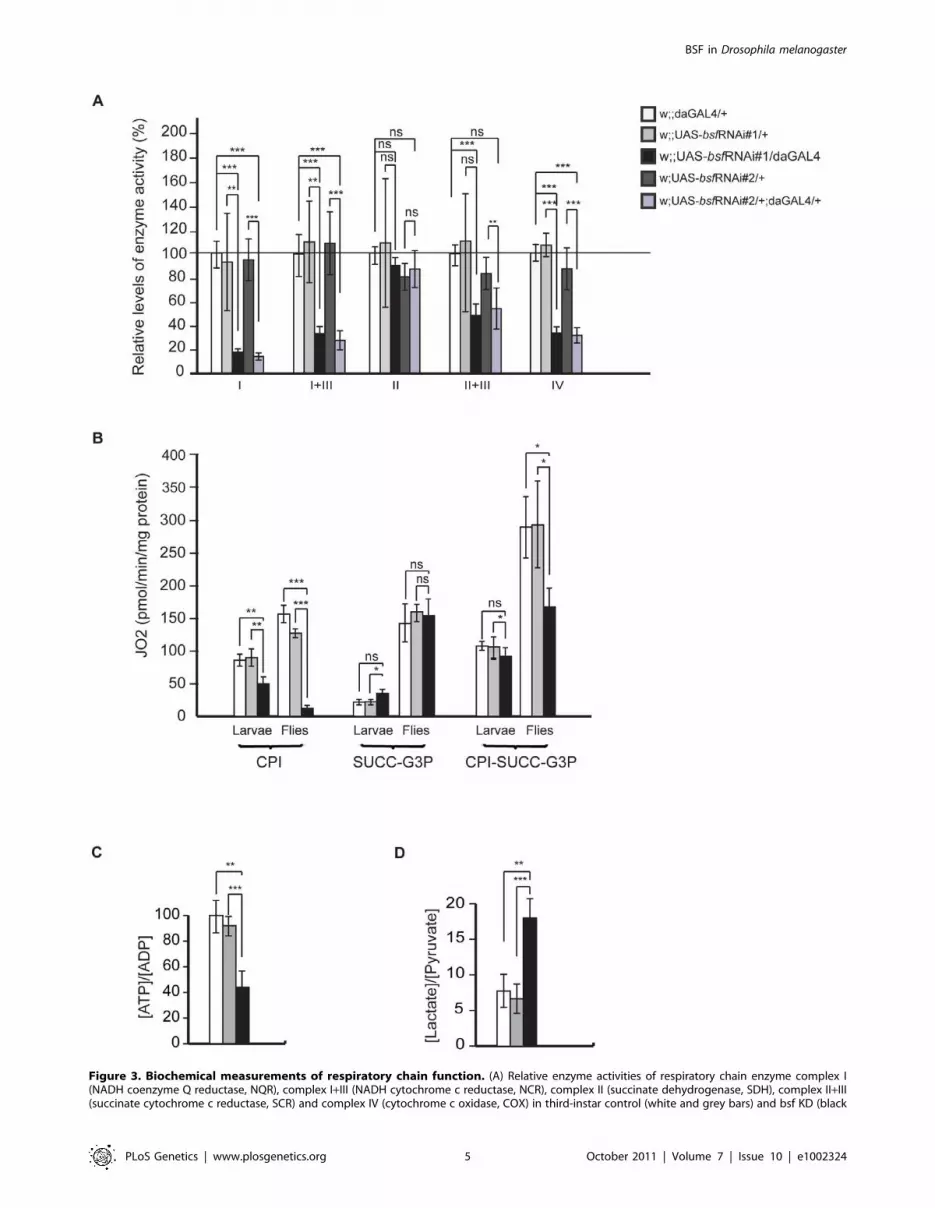
Figure 3. Biochemical measurements of respiratory chain function. (A) Relative enzyme activities of respiratory chain enzyme complex I(NADH coenzyme Q reductase, NQR), complex I+III (NADH cytochrome c reductase, NCR), complex II (succinate dehydrogenase, SDH), complex II+III(succinate cytochrome c reductase, SCR) and complex IV (cytochrome c oxidase, COX) in third-instar control (white and grey bars) and bsf KD (black
BSF in Drosophila melanogaster
PLoS Genetics | www.plosgenetics.org 5 October 2011 | Volume 7 | Issue 10 | e1002324

(CPI-SUCC-G3P) in adult fly mitochondria (Figure 3B). We
also observed a progression of the decrease in the uncoupled
oxygen consumption with complex I substrates, when compar-
ing third-instar larvae and flies (Figure 3B). In summary, the
results from measurements of enzyme activities (Figure 3A) and
oxygen consumption (Figure 3B) show that complex I is the
most affected of the oxidative phosphorylation complexes,
perhaps due to its high number of mtDNA-encoded subunits.
As a result of the respiratory chain deficiency the [ATP]/
[ADP] ratio in adult bsf KD flies was reduced to 44% of ratio in
controls (Figure 3C). Furthermore, in adult bsf KD flies the
[lactate]/[pyruvate] ratio was considerably increased
(Figure 3D), most likely as a consequence of the compromised
respiratory function, which leads to increased lactic acid
fermentation in order to maintain the reduction-oxidation
homeostasis.
BSF affects steady-state levels of mitochondrialtranscripts
We performed a detailed study on steady-state levels of
mitochondrial transcripts in bsf KD lines by using both QRT-
PCR and northern blot analyses. The levels of all analyzed
mitochondrial mRNAs were reduced in third-instar bsf KD larvae
(Figure 4A, 4C, 4D and Figure S1C) and flies (Figure 4B), with the
exception of ND6, which was significantly reduced only in adult
flies. QRT-PCR demonstrated slightly increased mtDNA levels in
third-instar bsf KD larvae (Figure 4E), showing that the reduced
mitochondrial mRNA levels cannot be explained by mtDNA
depletion. In contrast, the mitochondrially encoded small
ribosomal subunit rRNA (12S rRNA) was significantly increased,
while the large ribosomal subunit rRNA (16S rRNA) was slightly
decreased (Figure 4C and 4D). Additionally, there was a
substantial increase in levels of all analyzed mitochondrial tRNAs
and purple bars) larvae are shown. Bars indicate mean 6 SD (*p,0.05; **p,0.01; ***p,0.001; n = 6-7). (B) Oxygen consumption normalized to proteincontent in third-instar control (white and grey bars) and bsf KD (black bars) larvae and six-day old flies. Oxygen consumption was assessed inpermeabilized larvae and thoraces from six-day old flies by using substrates entering at the level of complex I (CPI), complex II (SUCC), glycerol-3-phosphate dehydrogenase, (G3P) and complex I, II and glycerol-3-phosphate dehydrogenase (CPI-SUCC-G3P), (C) Relative ATP/ADP levels in six-dayold bsf KD flies. (D) Lactate/pyruvate ratios in six-day old bsf KD flies.doi:10.1371/journal.pgen.1002324.g003
Figure 4. Steady-state levels of mtDNA and mitochondrial transcripts. (A-B) QRT-PCR analysis of relative levels of mitochondrial mRNAs incomparison with the nuclear ribosomal protein 49 transcript in (A) third-instar larvae and (B) six-day old flies. Northern Blot analyses (C) andquantification (D) of mitochondrial transcripts normalized to nuclear ribosomal protein 49 transcript in third-instar larvae. (E) QRT-PCR analysis ofmtDNA levels in third-instar larvae. Northern blot analyses (F) and quantification (G) of mitochondrial tRNA levels in third-instar larvae. (*p,0.05;**p,0.01; ***p,0.001. n = 5).doi:10.1371/journal.pgen.1002324.g004
BSF in Drosophila melanogaster
PLoS Genetics | www.plosgenetics.org 6 October 2011 | Volume 7 | Issue 10 | e1002324

in third-instar bsf KD larvae (Figure 4F and 4G). There was no
clear correlation between the level of a particular tRNA and the
location of its gene in fly mtDNA.
Reduced BSF level leads to increased de novomitochondrial transcription and aberrant translation
The presence of increased steady-state levels of tRNAs makes it
unlikely that the reduction in levels of mRNAs are explained by
reduced transcription as both types of mature transcripts are
produced by processing of polycistronic precursor transcripts. We
nevertheless assessed mitochondrial de novo transcription by
performing in organello labeling experiments. Both third-instar bsf
KD larvae (Figure 5A and Figure S2B) and bsf KD flies (Figure
S2A) had a dramatic increase in de novo transcription, showing
that the reduced mRNA levels must be explained by increased
degradation. The increased mitochondrial de novo transcription
and increased mtDNA copy number are likely parts of a
compensatory mitochondrial biogenesis response induced by the
respiratory chain deficiency, which, in turn, is caused by defective
post-transcriptional regulation of mitochondrial mRNA stability in
the absence of BSF. We further investigated whether the decrease
in mRNA steady-state levels resulted in decreased mitochondrial
translation by assessing de novo translation in isolated mitochon-
dria. Surprisingly, third-instar bsf KD larvae demonstrated a
selective increase in the synthesis of subunits of complex I (ND1-6
and ND4L) and complex IV (COXI-III), whereas the synthesis of
subunits of complex III (Cyt b) and complex V (ATP6) remained
unchanged (Figure 5B). We observed an exceptionally large
increase in levels of the COXII subunit of complex IV (Figure 5B,
asterisk). We also found an unidentified translation product
migrating above the ND1 subunit of complex I (Figure 5B,
arrow). Interestingly, both of these aberrant translation products
were almost invisible after a 3-hour chase with cold methionine
(Figure 5B, left panel), suggesting that they are subjected to an
increased degradation shortly after synthesis.
BSF is required for polyadenylation of mitochondrialmRNAs
Decreased mitochondrial mRNA steady-state levels, in combi-
nation with increased de novo transcription and translation of
specific mitochondrial polypeptides (ND1-6 and COX1-3) led us
to investigate the nature of the mature mitochondrial mRNAs. We
Figure 5. De novo mitochondrial transcription, de novo mitochondrial translation in bsf KD larvae, and polyadenylation profiles.(A) De novo transcription of mtDNA as determined by a32P-UTP incorporation. Isolated mitochondria were incubated with a32P-UTP and labeledtranscripts were separated on MOPS/formaldehyde agarose gels and normalized to the total mitochondrial protein content per ml of sample (lowerpanel). (B) Analysis of de novo mitochondrial translation as determined by S35-methionine incorporation, normalized to the total mitochondrialprotein content per ml of sample (Figure 5A, lower panel) The asterisk indicates the COXII subunit. The arrow indicates an unidentified proteinmigration just above the ND1 subunit. (C) Examples of electropherograms showing the polyadenylation status of the COXII mRNA in mitochondria ofcontrol (w;;daGAL4/+) and bsf KD (w;;UAS-bsfRNAi/daGAL4) larvae. (D) A summary of the polyadenylation status of mRNAs in mitochondria of third-instar controls (w;;daGAL4/+) and bsf KD (w;;UAS-bsfRNAi/daGAL4) larvae. AAAA = 40-60 adenines, AA = ,20 adenines. Polycistronic transcript ofCOXIII-ATP6/8 ({), antisense tRNA-Threonine ({).doi:10.1371/journal.pgen.1002324.g005
BSF in Drosophila melanogaster
PLoS Genetics | www.plosgenetics.org 7 October 2011 | Volume 7 | Issue 10 | e1002324

analyzed the 59-and 39-ends of mitochondrial transcripts, using
RNA circularization, followed by reverse transcription and direct
sequencing or cloning and subsequent sequencing (see Materials
and Methods). The 59 and 39 ends of the 12S and 16S rRNA were
identical in bsf KD and control larvae. However, we observed
severely reduced poly A tail lengths of all mitochondrial mRNAs
except Cytb and ATP6/8 (Figure 5C and 5D). Interestingly, those
mRNAs with reduced poly A tail length encoded the polypeptides
that showed increased levels of de novo synthesis. We further
analyzed the 59 and 39ends of two polycistronic transcripts each
containing an mRNA with a retained tRNA at its 39 end (ND3
plus tRNA-Ala and COXII plus tRNA-Lys). In control samples
these polycistronic transcripts were polyadenylated at their 39-
ends, showing that the polyadenylation process is not specific for
fully processed mRNAs, but rather can occur at any free 39end. In
the bsf KD larvae, these polycistronic RNAs had severely reduced
lengths of their polyA tail, thus suggesting that the lack of
polyadenylation is a direct consequence of the loss of BSF and not
a secondary effect of impaired translation. In the bsf KD larvae, we
consistently failed to recover the mature COX III mRNA and only
observed COXIII as part of a large polycistronic RNA. The 59part
of this RNA consisted of ATP6/8 and the 3‘part of COXIII. This
RNA had undergone a correct processing at the 39 end of COXIII
but lacked polyadenylation. The lack of mature polyadenylated
COXIII transcripts in the bsf KD larvae is thus likely explained by
a combination of defective RNA processing and defective
polyadenylation. Cloning and subsequent sequencing of the
ND4/ND4L PCR product revealed several different RNA species
in the bsf KD samples, indicating both polyadenylation and
processing differences in comparison with control samples
(Figure 5D). In summary, sequencing of RNA 59 and 39-ends in
third-instar bsf KD larvae revealed a severe mRNA maturation
defect with reduced poly A tail lengths and an enrichment of
unprocessed polycistronic RNA intermediates containing COXIII
and ND4/ND4L sequences.
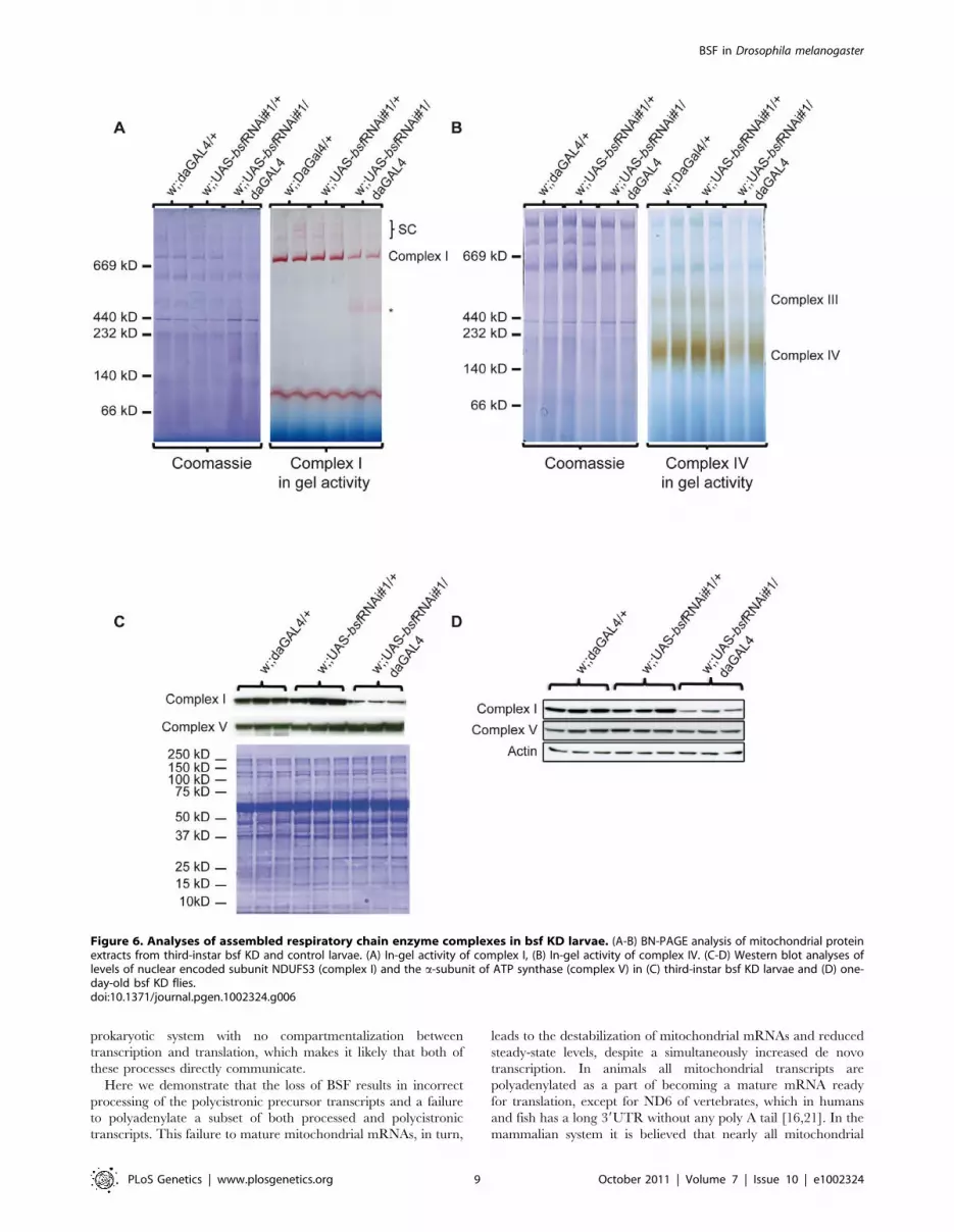
The biochemical defect in bsf KD larvae is caused byreduced levels of assembled complex I, III, and IV
Despite the observed compensatory increase of de novo
transcription and translation, third-instar bsf KD larvae show a
severe respiratory chain dysfunction presumably causing delayed
hatching and reduced lifespan. Sequencing of the mitochondrial
mRNAs showed reduced length of poly A tails and processing
defects, which could affect translational initiation or lead to the
production of abnormal polypeptides. Subunits of complex I and
IV were translated at increased rates, but a subset of the newly
synthesized subunits were nevertheless preferentially degraded
(Figure 5B), suggesting that they fail to assemble into mature
complexes. We therefore assessed the levels of assembled
respiratory chain enzyme complexes by using Blue-Native
polyacrylamide gel electrophoresis (BN-PAGE). We observed
reduced levels of assembled complex I, complex III, complex IV
and supercomplexes in third-instar bsf KD larvae (Figure 6A and
6B) and flies (Figure S2C and S2D). The levels of assembled
complex V (ATP synthase) were unaffected. This reduction in
steady-state levels of assembled complexes was accompanied by
reduced in-gel activity of complex I (Figure 6A, Figure S2C) and
complex IV (Figure 6B, Figure S2D). The BN-PAGE and complex
I in-gel activity analyses showed reduced steady-state levels of
supercomplexes and the presence of a smaller, partially assembled,
form of complex I in third-instar bsf KD larvae (Figure 6A,
asterisk). Western blot analyses showed reduced levels of a nuclear
encoded subunit of complex I (NDUFS3 subunit), indicating and
supporting the notion that there is a severe reduction in steady-
state levels of assembled complex I in third-instar bsf KD larvae
(Figure 6C, Figure S2E) and flies (Figure 6D). There was no major
reduction in steady-state levels of the nuclear-encoded complex V
subunits (a-subunit of ATP synthase) in third-instar bsf KD larvae
(Figure 6C, Figure S1B) and flies (Figure 6D), suggesting normal
assembly. Together, these results show that third-instar bsf KD
larvae and flies fail to assemble sufficient levels of complex I, III
and IV as well as supercomplexes consisting of these complexes,
which explains the observed profound reduction in oxidative
phosphorylation capacity.
Discussion
BSF has previously been suggested to stabilize cytoplasmic
mRNAs in oocytes and early zygotic cells during the first few hours
of fly embryogenesis [1,2]. However, the ubiquitous expression of
the BSF RNA [3] and the punctuate cytoplasmic localization in
flies has prompted us to re-investigate the function of BSF in the
fly. Surprisingly, we were able to demonstrate that the BSF protein
is mainly localized to mitochondria, where it controls polyadenyl-
ation of specific mitochondrial mRNAs. Further, BSF plays a key
role in the regulation of mtDNA gene expression, by coordinating
mitochondrial translation in flies.
GFP-tagged BSF localized to mitochondria in transfected tissue
culture cells and cell fractionation experiments showed that BSF is
mainly present in mitochondria of adult flies. Interestingly, BSF
was not detectable in the cytoplasmic and nuclear fractions of
adult flies. It is important to note that the mammalian homolog of
BSF, LRPPRC, has also been reported to have roles in regulation
of cytoplasmic mRNA transport [6-8] and in regulation of nuclear
transcription [9,10]. However, convincing evidence proposes that
the main proportion of LRPPRC is localized to mitochondria
[4,10,11]. In one study of tissue culture cells, endogenously
expressed LRPPRC was not detectable in highly purified nuclei
lacking mitochondrial contamination [4]. These findings do not
exclude that there is a small fraction of BSF and LRPPRC
localized in other subcellular compartments besides mitochondria.
However, the proposed extramitochondrial functions of BSF and
LRPPRC should be revisited in a new set of experiments focusing
on avoiding mitochondrial contamination of isolated nuclei and
cytoplasmic extracts.
Ubiquitous RNAi-induced reduction of endogenous bsf levels in
flies causes mitochondrial dysfunction with decreased respiratory
chain enzyme activities and reduced levels of assembled complex I,
III and IV. This mitochondrial dysfunction leads to severe
phenotypes, including delayed and incomplete eclosure, reduced
fecundity, sterility and shortened life span.
Mammalian cell lines with reduced LRPPRC levels have
reduced mitochondrial mRNA steady-state levels and reduced
mitochondrial translation [12,18]. Interestingly, downregulation of
BSF protein levels in flies also leads to decreased steady-state levels
of mitochondrial mRNAs but, in contrast with the results in
mammals, translation is increased and aberrant translation
products are generated. Some of the mitochondrial translation
products that are synthesized at increased rates in flies are subject
to increased degradation, suggesting that BSF might also act as a
translational coordinator. Regulation of mitochondrial gene
expression in response to different metabolic demands in animal
cells is largely unknown, and most likely this regulation occurs at
several different levels. The basic mtDNA transcription machinery
has been defined [19], but it is unclear how it is regulated.
Processing of mRNAs is required for correct translation [20], but
the coupling between transcript processing and translation is
poorly understood. Furthermore, mitochondria are in essence a
BSF in Drosophila melanogaster
PLoS Genetics | www.plosgenetics.org 8 October 2011 | Volume 7 | Issue 10 | e1002324
prokaryotic system with no compartmentalization between
transcription and translation, which makes it likely that both of
these processes directly communicate.
Here we demonstrate that the loss of BSF results in incorrect
processing of the polycistronic precursor transcripts and a failure
to polyadenylate a subset of both processed and polycistronic
transcripts. This failure to mature mitochondrial mRNAs, in turn,
leads to the destabilization of mitochondrial mRNAs and reduced
steady-state levels, despite a simultaneously increased de novo
transcription. In animals all mitochondrial transcripts are
polyadenylated as a part of becoming a mature mRNA ready
for translation, except for ND6 of vertebrates, which in humans
and fish has a long 39UTR without any poly A tail [16,21]. In the
mammalian system it is believed that nearly all mitochondrial
Figure 6. Analyses of assembled respiratory chain enzyme complexes in bsf KD larvae. (A-B) BN-PAGE analysis of mitochondrial proteinextracts from third-instar bsf KD and control larvae. (A) In-gel activity of complex I, (B) In-gel activity of complex IV. (C-D) Western blot analyses oflevels of nuclear encoded subunit NDUFS3 (complex I) and the a-subunit of ATP synthase (complex V) in (C) third-instar bsf KD larvae and (D) one-day-old bsf KD flies.doi:10.1371/journal.pgen.1002324.g006
BSF in Drosophila melanogaster
PLoS Genetics | www.plosgenetics.org 9 October 2011 | Volume 7 | Issue 10 | e1002324

RNAs are oligoadenylated during transcription by a yet unknown
enzyme, followed by the polyadenylation to approximately 50
adenines by the mitochondrial poly A polymerase (mtPAP) [22]. A
number of mitochondrial transcripts require polyadenylation to
generate functional stop codons, but otherwise the exact function
of mRNA polyadenylation in mitochondria is unknown. While in
bacteria and chloroplasts the poly A tail seems to promote
transcript degradation, eukaryotes seem to polyadenylate nearly
every fully processed cytosolic mRNA at the 39 end, resulting in
increased mRNA stability, increased translational efficiency, and
promotion of transport of the processed mRNA from the nucleus
to the cytoplasm [23]. A primary role of BSF in the maturation of
mitochondrial mRNAs is supported by the recent observation that
PPR proteins in trypanosomes affect polyadenylation of mito-
chondrial mRNAs. In this unicellular protozoon, the PPR proteins
KPAF1 and KPAF2 associate with the mtPAP, stimulating mRNA
polyadenylation and thereby coordinate stability and translation of
mRNA [24]. Our results suggest that BSF is involved in the actual
mRNA maturation process by controlling polyadenylation of
specific transcripts.
The mammalian BSF homolog LRPPRC has been shown to be
stabilized by a second RNA-binding protein called SLIRP, in a
direct interaction [12]. A SLIRP homolog has also been suggested
to exist in flies, raising the possibility that such an interaction with
a second RNA binding protein is also required in fly mitochondria.
Interestingly, bioinformatics analyses identified an additional
LRPPRC homolog, besides BSF, in Drosphila melanogaster [4],
suggesting that both homologs work together to control the
polyadenylation and translation of different sets of mitochondrial
mRNAs. This is supported by our observation that several
mitochondrial transcripts, such as cytb and ATP6/8, are still
polyadenylated in the bsf KD lines.
Loss of BSF also results in increased and aberrant translation,
and reduced levels of assembled RC complex I, III and IV,
suggesting that BSF coordinates mitochondrial translation and RC
complex assembly. The coupling of transcription to translation is
supported by studies of the yeast homologue of BSF and
LRPPRC, PET309 [25]. This factor is implicated in activation
of translation by binding to the 59UTR of yeast mitochondrial
mRNAs, thereby tethering mitochondrial polysomes to the inner
mitochondrial membrane, which, in turn, is thought to ensure co-
translational insertion of newly synthesized polypeptides into the
assembling respiratory chain complexes [26,27]. Mitochondrial
mRNAs without these 59-UTRs are translated at normal levels
followed by rapid degradation, suggesting that targeting to the
inner mitochondrial membrane is necessary for stability of the
newly translated peptides [28]. It is important to point out that
most mRNAs encoded by metazoan mtDNA lack 59UTRs and
targeting of translation to the inner membrane of animal
mitochondria, if this occurs, must therefore involve, at least partly,
different regulatory mechanisms.
Cytoplasmic mRNAs have several different types of destabiliz-
ing elements and it seems that translation is coupled to
degradation of some classes of mRNAs [29]. If a similar
mechanism exists in mitochondria, it would suggest that the
increased de novo translation in BSF KD larvae results in
increased degradation of the mitochondrial mRNAs. Some
support for this proposed mechanism comes from characterization
of a patient with a microdeletion between the genes for ATP6/8
and COXIII, which has been reported to cause incorrect
processing of the corresponding mitochondrial mRNAs and
severely reduced steady-state levels of the ATP6/8 transcript due
to translationally induced deadenylation [30]. Additional support
comes from the observation that human cells treated with the
mitochondrial translation inhibitor thiamphenicol have increased
steady-state levels of mitochondrial mRNAs [31]. However,
decreased translation does not always increase mRNA steady-
state levels as exemplified by the conditional mouse knockout for
TFB1M, which abolishes mitochondrial translation, but does not
affect the levels of most mitochondrial mRNAs despite activation
of de novo transcription [32] .
In conclusion, we present here the unexpected result that the
BSF protein mainly is localized to mitochondria, where it controls
the polyadenylation of specific mitochondrial mRNAs. In
addition, our results suggests that BSF has a novel role in
coordinating mitochondrial translation as loss of BSF leads to
increased and uncoordinated translation with increase synthesis of
unstable translation products. BSF thus has an essential role in
regulating mitochondrial function in the fly.
Materials and Methods
Drosophila stocks and maintenanceFor in vivo KD studies two independent non-overlapping UAS-
bsf RNAi lines were used. w;;UAS-bsfRNAi#1 (#10302R-I) was
obtained from the National Institute of Genetics (Japan) and
w;UAS-bsfRNAi#2 (#22839) was obtained from the Vienna
Drosophila RNAi Center (VDRC). Ubiquitous bsf knock down
was achieved by crossing UAS-bsf RNAi lines to a daughterless-
GAL4 (da-GAL4) driver line. UAS-RNAi lines and daGAL4
driver lines were backcrossed for at least 6 generations into the
white Dahomey background (wDahT). All fly stocks were free
from the endosymbiontic bacterium Wolbachia. Flies were
propagated and experiments were conducted at 25uC on a
12 h:12 h light:dark cycle at constant humidity on a standard
sugar-agar-yeast medium.
BioinformaticsHomologs of LRPPRC were collected using PSI-BLAST [33]
against the RefSeq protein database at NCBI and aligned using
ClustalX [34] with the BLOSUM matrix. Multiple sequence
alignments were trimmed using the GBLOCKS server [35] with
relaxed settings. The trimmed alignment was submitted to PhyML
[36] using standard parameters and non-parametric bootstrap
analysis with 500 replicates. The resulting tree was displayed using
Dendroscope [37] and prepared for publication in Illustrator.
Colocalization studiesFull-length bsf cDNA was obtained from the Drosophila
Genomics Resource Center (SD10676, AY058795). Two amino
acid changing substitutions were identified in the bsf cDNA in
comparison with the reference sequence (FBtr0081087). The
corresponding mutations at nucleotide positions 415 and 710 were
changed by site-directed mutagenesis of the cDNA, using the
QuickChange II XL Site-Directed Mutagenesis Kit (Agilent
Technologies). A cDNA encoding FLAG-tagged BSF was cloned
into the plasmid pAcGFP1-N2 (Clontech, Mountain View, USA)
to generate the vector pbsfFLAG-AcGFP1, which encodes a fusion
protein consisting of BSF-FLAG with an in-frame addition of
green fluorescent protein (GFP) to its carboxy-terminus (BSF-
FLAG-GFP).
Schneider 2R+ and HeLa cells were transfected in microscopy
dishes (m-Dish, ibidi, Martinsried, Germany) with pbsfFLAG-
AcGFP1 using the FuGENE HD Transfection Reagent (Roche
Diagnostics, Mannheim, Germany). The mitochondrial counter-
staining was achieved by 100 nm MitoTracker Deep Red FM
(Invitrogen, Darmstadt, Germany). Live cell image acquisition was
BSF in Drosophila melanogaster
PLoS Genetics | www.plosgenetics.org 10 October 2011 | Volume 7 | Issue 10 | e1002324

performed with a Leica TCS SP5-X confocal microscope (Leica
Microsystems, Wetzlar, Germany).
The colocalization rate was determined by the software LAS AF
(Leica Microsystems, Wetzlar, Germany) under the following
conditions and calculations: threshold 30%, background 20%,
colocalization rate [%] = colocalization area/area foreground,
and area foreground = area image 2 area background.
Subcellular fractionation and Western blot analysisAll fractions were isolated from adult wDahT flies. Mitochon-
drial, cytoplasmic and nuclear fractions were isolated by
differential centrifugation as previously described [38]. The used
primary antibodies were: HISTONE H3 (Santa Cruz Biotechnol-
ogy, dilution 1:200), Complex I-subunit NDUFS3 (Mitoscience
MS112, dilution 1:1000), tubulin (Sigma, dilution 1:1000) and
polyclonal rat antisera raised against BSF (kindly provided by
Professor MacDonald PM, Stanford University, dilution 1:1000).
Protein bands were visualized with ECL western blotting reagents
(Bio-Rad).
Western blot analyses were performed using whole fly or
mitochondrial protein extracts according to the Cell Signaling
Technology protocol (CellSignaling). Additional primary antibod-
ies used were: complex V (Mitoscience MS504, dilution 1:5000),
VDAC (Mitoscience MSAO3, dilution 1:2000), actin (Sigma,
dilution 1:1000).
Hatching rate, lifespan, fecundity, and climbing assays offlies
For adult hatching rate measurements eggs were collected
during a 3 hours time window and transferred to vials (80 eggs/
vial) to ensure standard larval density. Hatching of adult flies was
monitored in regular intervals. After hatching, virgin females and
males were collected and mated for 2 days.
For lifespan analyses, 50–100 females per genotype were used at
a density of 10 flies per vial. Flies were transferred to new vials
every two to three days and dead flies were counted. For fecundity
assays, 100 females were equally distributed in ten vials,
transferred to new vials every day and the number of eggs was
counted.
Climbing assays were conducted and performance index was
calculated as described [39]. 100 three-day-old males per genotype
were tested for their climbing ability.
DNA isolation and quantitative RT–PCRDNA of third-instar larvae was extracted using the DNAeasy
Kit (Qiagen). Mitochondrial DNA levels were determined by
quantitative real-time PCR (QRT-PCR) on a 7900HT Real Time
PCR system (Applied Biosystems), using SYBR green master mix
(Invitrogen). Reactions were carried out in triplicates per sample in
a final volume of 20 ml with 5 ng of DNA and 10 pmol of specific
primers (primers are listed in Table S1).
RNA isolation, QRT-PCR, and Northern blot analysisTotal RNA from third-instar larvae or adult flies was extracted
using the Totally RNA KIT (Ambion). Reverse transcription and
QRT-PCR was performed using the High capacity RNA-to-
cDNA kit (Applied Biosystems) and the Taqman 2x Universal
PCR mastermix, No Amperase UNG (Applied Biosystems),
respectively. Custom-made TaqMan probes against Drosophila
mitochondrial transcripts were obtained from Applied Biosystems
and are listed in Table S1.
For Northern blot analyses, RNA was fractionated on 1.2%
agarose gels and blotted to Hybond-N+ membranes (Amersham
Biosciences). Membranes were hybridized with 32P-labeled probes
and afterwards exposed to PhosphoImager Screens and/or X-ray
films. Labeling of mitochondrial double-stranded DNA probes and
oligonucleotides was performed as described [40]. Primers and
oligonucleotides used for Northern blot are listed in Table S1.
Biochemical evaluation of respiratory chain functionIsolation of mitochondria from third-instar larvae was per-
formed as described [41] with modifications in buffer composition.
Briefly, third-instar larvae were washed and gently homogenized
in ice-cold MSB buffer (210 mM mannitol, 70 mM sucrose,
10 mM EDTA, 50 mM Tris, pH 7.5) using 15 ml Dounce
homogenizers. Protein concentration was determined using
Bradford assay and aliquots corresponding to 10 mg mitochondrial
proteins were pelleted and resuspended in resuspension buffer
(250 mM sucrose, 15 mM K2HPO4, 2 mM MgAc2, 0.5 mM
EDTA and 0.5 g/L HSA, pH 7.2). Biochemical activities of
respiratory chain complexes were determined as described [42].
Respiratory ratesThird-instar larvae (n = 10) or thoraces from adult flies (n = 5)
were dissected in PBS and resuspended in 2 ml of respiratory
buffer (120 mM sucrose, 50 mM KCl, 20 mM Tris-HCl, 4 mM
KH2PO4, 2 mM MgCl2, 1 mM EGTA, 0.01% digitonin,
pH 7.2). Oxygen consumption was measured at 25uC using an
oxygraph chamber (OROBOROS). Complex I-dependent respi-
ration was assessed by adding the substrates proline (10 mM),
pyruvate (10 mM), malate (5 mM) and glutamate (5 mM).
Succinate and glycerol-3-phosphate dehydrogenase activities were
measured using 20 mM succinate (SUCC) and 15 mM glycerol-3-
phosphate (G3P), respectively. Mitochondrial quality of each
sample was assessed by measuring the respiratory control rate
(RCR), using 1 mM ADP (state 3) or 1 mM ADP and 2.5 mg/ml
oligomycin (pseudo state 4). Permeabilized control mitochondria
consistently had RCR values between 4 and 7 with complex I
substrates.
The respiration was uncoupled by the addition of 400 mM
CCCP and the rotenone-sensitive flux was measured in the
presence of 200 mM rotenone. Finally, the protein content was
determined by the Bradford method (BioRad) in order to
normalize the oxygen consumption flux to mitochondrial protein
content.
ATP/ADP and lactate/pyruvate ratiosFlies (n = 10) were snap frozen directly in liquid nitrogen and
kept at -80uC. Acidic extraction (PCA 7%) was performed,
samples were centrifuged (16000 g, 10 min), the supernatant was
neutralized with 2N KOH, 10 mM MOPS and metabolites were
quantified. ADP and ATP levels were assessed as previously
described [43,44]. Briefly, ATP was quantified by using the
ATPlite one step kit (PerkinElmer). For ADP levels, samples were
incubated for 10 min at 37uC in 75 mM KCl, 8 mM MgSO4,
10 mg/ml pyruvate kinase and 2 mM phosphoenolpyruvate.
Lactate and pyruvate concentrations were determined after 1 h
incubation with horse radish peroxydase 5 U/ml, Amplex red
20 mM, 0.1 M phosphate, pH 7.2, supplemented with lactate
oxidase or pyruvate oxidase, followed by fluorimetric analysis
(ex:560 nm, em:590 nm), using an Infinite 200 Pro fluorimeter
(Tecan).
In organello transcription and translation assaysFor the preparation of mitochondria, third-instar larvae or flies
were homogenized in ice-cold isolation buffer STE+BSA (250 mM
BSF in Drosophila melanogaster
PLoS Genetics | www.plosgenetics.org 11 October 2011 | Volume 7 | Issue 10 | e1002324

sucrose, 5 mM Tris, 2 mM EGTA, 1% (w/v) BSA, pH 7.4) using
a 15 ml Dounce homogenizer. Cellular debris were pelleted at
1000 g for 5 min and supernatants were transferred to new tubes.
Mitochondria were washed two times and final mitochondrial
pellets were resuspend in 1 ml STE buffer in the presence of
200 mg/ml emetine (Sigma) and 100 mg/ml cycloheximide
(Sigma) to inhibit cytoplasmic translation. Protein concentrations
were determined using the Bradford assay.
In organello transcription assays were performed as described [45]
using 200 mg mitochondria/sample and a modified transcription
buffer (25 mM sucrose, 75 mM sorbitol, 100 mM KCl, 10 mM
K2HPO4, 50 mM EDTA, 5 mM MgCl2, 1 mM ADP, 10 mM
glutamate, 2.5 mM malate, 10 mM Tris-HCl (pH 7.4) and 1%
(w/v) BSA). In short, after labeling, mitochondrial RNA was
isolated using Totally RNA kit (Ambion). Mitochondrial RNA was
fractionated on 1.2% agarose gels and blotted to Hybond-N+membranes (Amersham Biosciences).
In vitro assays to study mitochondrial de novo translation with
[35S]-methionine were performed as described [46]. Equal amounts
of total mitochondrial protein were loaded on 15% SDS-PAGE
gels. Gels were fixed in isopropanol-acetic solution, stained with
Coomassie, destained in ethanol-acetic acid solution and treated
with Amplify Solution (GE Healthcare). Afterwards gels were dried
and [35S]–methionine-labelled proteins were visualized by autora-
diography. The mitochondrial translation profile was compared to
previously published profiles in Schneider cell lines [47], addition-
ally ND2 and ATP6 were identified by endopeptidase fingerprinting
in the second dimension (data not shown) [48].
RNA circularization and RT–PCRAn RNA circularization protocol was modified from [49,50].
Approximately 6 ng total mitochondrial RNA was circularized
with 5 U T4 RNA ligase in 200 ml at 16uC for at least 16 h in
manufacturer-supplied buffer (NEB). The circularized RNA was
precipitated with an equal volume of isopropanol, incubated at -
20uC for at least 4 h, and centrifuged for 20 min at top speed in a
bench top centrifuge. The entire precipitate was used for
complementary DNA synthesis with gene specific primers using
GeneAmp RNA PCR kit (Applied Biosystems). PCR products
were purified using ExoSAP-IT (Affymetrix) and sequenced.
Selected PCR products were cloned into pCR-II (Invitrogen)
and sequenced in order to confirm the results from direct
sequencing. Primer sequences for RT-PCR and subsequent PCR
are contained within Table S2.
Blue native-polyacrylamide gel electrophoresis(BN-PAGE) and in-gel histochemistry
For BN-PAGE, 75 mg of mitochondria were pelleted and lyzed
in 50 ml ice-cold digitonin buffer (1% digitonin, 20 mM Tris
pH 7.4, 0.1 mM EDTA, 50 mM NaCl, 10% glycerol, 1 mM
PMSF). After 15 min of incubation on ice, unsolubilized material
was removed by centrifugation at 4uC. The supernatant was
mixed with 5 ml of 10 x loading dye (5% (w/v) Coomassie Brilliant
Blue G-250, 100 mM Tris pH 7, 500 mM 6-aminocaproic acid)
and loaded on 4–10% gradient BN-PAGE gels [51,52]. In gel
complex I activity was determined by incubating the BN-PAGE
gels in 2 mM Tris-HCl pH 7.4, 0.1 mg/ml NADH (Roche) and
2.5 mg/ml iodonitrozolium (Sigma). In gel complex IV activity
was determined by incubating the BN-PAGE gels in 50 ml of
0.05 mM phosphate buffer pH 7.4, 25 mg 3.39-diamidobenzidine
tetrahydrochloride (DAB), 50 mg cytochrome c, 3.75 g sucrose
and 1 mg catalase. All stainings were carried out at room
temperature.
Statistical analysisData were presented as mean 6 SD. The Mann-Whitney test
was used to analyze climbing index and the log-rank test was used
to analyze lifespan. Unpaired t-test was used to analyze all other
data statistically.
Supporting Information
Figure S1 Phenotypic analysis and steady state level of
mitochondrial transcripts in bsf KD flies. (A) Western blot analyses
with antibodies against BSF were performed on mitochondrial
protein preparations from six-day old bsf KD and control flies.
Antibodies against actin were used to assess loading. (B)
Quantification of ovary sizes in six-day old bsf KD and control
flies. (C) QRT-PCR analysis of relative levels of mitochondrial
mRNAs in comparison with the nuclear ribosomal protein 49
transcript in third-instar bsfRNAi#2 KD and control flies.
(TIF)
Figure S2 Level of de novo mitochondrial transcription and
analyses of assembled respiratory chain complexes in bsf KD
larvae and flies. (A and B) De novo transcription of mtDNA
determined by a32P–UTP incorporation. Isolated mitochondria
were incubated with a32P–UTP and labeled transcripts were
separated on MOPS/formaldehyde agarose gels. (A) six-day old
bsf KD and control flies. (B) third-instar bsf KD and control
larvae. De novo transcription (left panel), and probing the same
membrane to detect 16S rRNA for size comparison (right panel).
(C) BN-PAGE analysis of mitochondrial protein extracts from six-
day old bsf KD and control flies. The assembled respiratory chain
complexes and supercomplexes are shown in the left panel. The
right panel shows in-gel activity of complex I. (D) BN-PAGE
analysis of mitochondrial protein extracts from six-day old bsf KD
flies. The assembled respiratory chain complexes and super-
complexes are shown in the left panel. The right panel shows in-
gel activity of complex IV. (E) Western blot analyses of levels of
nuclear encoded subunit NDUFS3 (complex I) and the a-subunit
of ATP synthase (complex V) in third-instar bsf KD larvae.
Antibodies against VDAC were used to assess loading.
(TIF)
Table S1 List of oligonucleotide sequences and Taqman probes
used for cloning of the BSF-FLAG-GFP construct and quantifica-
tion of steady-state levels of mtDNA, mt-tRNAs and mt-mRNAs.
(TIF)
Table S2 List of oligonucleotide sequences used for RT PCR and
subsequent PCR to determine the polyadenylation profile of mt
transcripts. RT-PCR was done using the forward primer (F) and
subsequent PCR for sequencing was done using the forward primer in
combination with the reverse (R) primer. Forward primer for COXI
and additional primer sequences used for polyadenylation sequencing
were designed according to Stewart and Beckenbach, 2009 [50].
(TIF)
Acknowledgments
We thank Ewa Gustafsson for technical assistance with the assays of the
respiratory chain enzyme activities.
Author Contributions
Conceived and designed the experiments: AB AW SG JBS BR LP N-GL.
Performed the experiments: AB AW AM BR CK RW BH. Analyzed the
data: AB AW JBS AM BR RW BH LP N-GL. Contributed reagents/
materials/analysis tools: BH SG CK AM. Wrote the paper: AB AW LP N-
GL.
BSF in Drosophila melanogaster
PLoS Genetics | www.plosgenetics.org 12 October 2011 | Volume 7 | Issue 10 | e1002324
References
1. Mancebo R, Zhou X, Shillinglaw W, Henzel W, Macdonald PM (2001) BSFbinds specifically to the bicoid mRNA 39 untranslated region and contributes to
stabilization of bicoid mRNA. Mol Cell Biol 21: 3462–3471. doi:10.1128/MCB.21.10.3462-3471.2001.
2. De Renzis S, Elemento O, Tavazoie S, Wieschaus EF (2007) Unmasking
activation of the zygotic genome using chromosomal deletions in the Drosophilaembryo. PLoS Biol 5: e117. doi:10.1371/journal.pbio.0050117.
3. Chintapalli VR, Wang J, Dow JAT (2007) Using FlyAtlas to identify better
Drosophila melanogaster models of human disease. Nat Genet 39: 715–720.doi:10.1038/ng2049.
4. Sterky FH, Ruzzenente B, Gustafsson CM, Samuelsson T, Larsson N-G (2010)
LRPPRC is a mitochondrial matrix protein that is conserved in metazoans.Biochemical and Biophysical Research Communications 398: 759–764.
doi:10.1016/j.bbrc.2010.07.019.
5. Lurin C, Andres C, Aubourg S, Bellaoui M, Bitton F, et al. (2004) Genome-wideanalysis of Arabidopsis pentatricopeptide repeat proteins reveals their essential
role in organelle biogenesis. Plant Cell 16: 2089–2103. doi:10.1105/
tpc.104.022236.
6. Mili S, Shu HJ, Zhao Y, Pinol-Roma S (2001) Distinct RNP complexes of
shuttling hnRNP proteins with pre-mRNA and mRNA: candidate intermediates
in formation and export of mRNA. Mol Cell Biol 21: 7307–7319. doi:10.1128/MCB.21.21.7307-7319.2001.
7. Tsuchiya N, Fukuda H, Nakashima K, Nagao M, Sugimura T, et al. (2004)
LRP130, a single-stranded DNA/RNA-binding protein, localizes at the outernuclear and endoplasmic reticulum membrane, and interacts with mRNA in
vivo. Biochemical and Biophysical Research Communications 317: 736–743.doi:10.1016/j.bbrc.2004.03.103.
8. Topisirovic I, Siddiqui N, Lapointe VL, Trost M, Thibault P, et al. (2009)
Molecular dissection of the eukaryotic initiation factor 4E (eIF4E) export-competent RNP. EMBO J 28: 1087–1098. doi:10.1038/emboj.2009.53.
9. Labialle S, Dayan G, Gayet L, Rigal D, Gambrelle J, et al. (2004) New
invMED1 element cis-activates human multidrug-related MDR1 and MVPgenes, involving the LRP130 protein. Nucleic Acids Res 32: 3864–3876.
doi:10.1093/nar/gkh722.
10. Cooper MP, Qu L, Rohas LM, Lin J, Yang W, et al. (2006) Defects in energyhomeostasis in Leigh syndrome French Canadian variant through PGC-1alpha/
LRP130 complex. Genes Dev 20: 2996–3009. doi:10.1101/gad.1483906.
11. Xu F, Morin C, Mitchell G, Ackerley C, Robinson BH (2004) The role of theLRPPRC (leucine-rich pentatricopeptide repeat cassette) gene in cytochrome
oxidase assembly: mutation causes lowered levels of COX (cytochrome coxidase) I and COX III mRNA. Biochem J 382: 331–336. doi:10.1042/
BJ20040469.
12. Sasarman F, Brunel-Guitton C, Antonicka H, Wai T, Shoubridge EA, et al.(2010) LRPPRC and SLIRP interact in a ribonucleoprotein complex that
regulates posttranscriptional gene expression in mitochondria. Mol Biol Cell 21:
1315–1323. doi:10.1091/mbc.E10-01-0047.
13. Boore JL (1999) Animal mitochondrial genomes. Nucleic Acids Res 27:
1767–1780.
14. Montoya J, Ojala D, Attardi G (1981) Distinctive features of the 59-terminalsequences of the human mitochondrial mRNAs. Nature 290: 465–470.
15. Ojala D, Montoya J, Attardi G (1981) tRNA punctuation model of RNA
processing in human mitochondria. Nature 290: 470–474.
16. Temperley RJ, Wydro M, Lightowlers RN, Chrzanowska-Lightowlers ZM(2010) Human mitochondrial mRNAs--like members of all families, similar but
different. Biochim. Biophys. Acta 1797: 1081–1085. doi:10.1016/j.bbabio.2010.02.036.
17. Tomecki R, Dmochowska A, Gewartowski K, Dziembowski A, Stepien PP
(2004) Identification of a novel human nuclear-encoded mitochondrial poly(A)polymerase. Nucleic Acids Res 32: 6001–6014. doi:10.1093/nar/gkh923.
18. Gohil VM, Nilsson R, Belcher-Timme CA, Luo B, Root DE, et al. (2010)
Mitochondrial and nuclear genomic responses to loss of LRPPRC expression.Journal of Biological Chemistry 285: 13742–13747. doi:10.1074/
jbc.M109.098400.
19. Falkenberg M, Larsson N-G, Gustafsson CM (2007) DNA replication andtranscription in mammalian mitochondria. Annu Rev Biochem 76: 679–699.
doi:10.1146/annurev.biochem.76.060305.152028.
20. Gagliardi D, Stepien PP, Temperley RJ, Lightowlers RN, Chrzanowska-Lightowlers ZMA (2004) Messenger RNA stability in mitochondria: different
means to an end. Trends Genet 20: 260–267. doi:10.1016/j.tig.2004.04.006.
21. Coucheron DH, Nymark M, Breines R, Karlsen BO, Andreassen M, et al.(2011) Characterization of mitochondrial mRNAs in codfish reveals unique
features compared to mammals. Curr Genet 57: 213–222. doi:10.1007/s00294-011-0338-2.
22. Bobrowicz AJ, Lightowlers RN, Chrzanowska-Lightowlers Z (2008) Polyade-
nylation and degradation of mRNA in mammalian mitochondria: a missinglink? Biochem. Soc Trans 36: 517–519. doi:10.1042/BST0360517.
23. Colgan DF, Manley JL (1997) Mechanism and regulation of mRNA
polyadenylation. Genes Dev 11: 2755–2766.
24. Aphasizheva I, Maslov D, Wang X, Huang L, Aphasizhev R (2011)Pentatricopeptide repeat proteins stimulate mRNA adenylation/uridylation to
activate mitochondrial translation in trypanosomes. Mol Cell 42: 106–117.doi:10.1016/j.molcel.2011.02.021.
25. Mootha VK, Lepage P, Miller K, Bunkenborg J, Reich M, et al. (2003)Identification of a gene causing human cytochrome c oxidase deficiency by
integrative genomics. Proc Natl Acad Sci USA 100: 605–610. doi:10.1073/pnas.242716699.
26. Manthey GM, McEwen JE (1995) The product of the nuclear gene PET309 is
required for translation of mature mRNA and stability or production of intron-containing RNAs derived from the mitochondrial COX1 locus of Saccharomy-
ces cerevisiae. EMBO J 14: 4031–4043.
27. Manthey GM, Przybyla-Zawislak BD, McEwen JE (1998) The Saccharomycescerevisiae Pet309 protein is embedded in the mitochondrial inner membrane.
Eur J Biochem 255: 156–161.
28. Sanchirico ME, Fox TD, Mason TL (1998) Accumulation of mitochondrially
synthesized Saccharomyces cerevisiae Cox2p and Cox3p depends on targeting
information in untranslated portions of their mRNAs. EMBO J 17: 5796–5804.doi:10.1093/emboj/17.19.5796.
29. Sachs AB (1993) Messenger RNA degradation in eukaryotes. Cell 74: 413–421.
30. Temperley RJ, Seneca SH, Tonska K, Bartnik E, Bindoff LA, et al. (2003)Investigation of a pathogenic mtDNA microdeletion reveals a translation-
dependent deadenylation decay pathway in human mitochondria. Hum MolGenet 12: 2341–2348. doi:10.1093/hmg/ddg238.
31. Chrzanowska-Lightowlers ZM, Preiss T, Lightowlers RN (1994) Inhibition of
mitochondrial protein synthesis promotes increased stability of nuclear-encodedrespiratory gene transcripts. J Biol Chem 269: 27322–27328.
32. Metodiev MD, Lesko N, Park CB, Camara Y, Shi Y, et al. (2009) Methylation of
12S rRNA is necessary for in vivo stability of the small subunit of themammalian mitochondrial ribosome. Cell Metab 9: 386–397. doi:10.1016/
j.cmet.2009.03.001.
33. Altschul SF, Madden TL, Schaffer AA, Zhang J, Zhang Z, et al. (1997) GappedBLAST and PSI-BLAST: a new generation of protein database search
programs. Nucleic Acids Res 25: 3389–3402.
34. Chenna R, Sugawara H, Koike T, Lopez R, Gibson TJ, et al. (2003) Multiple
sequence alignment with the Clustal series of programs. Nucleic Acids Res 31:
3497–3500.
35. Talavera G, Castresana J (2007) Improvement of phylogenies after removing
divergent and ambiguously aligned blocks from protein sequence alignments.
Syst Biol 56: 564–577. doi:10.1080/10635150701472164.
36. Guindon S, Delsuc F, Dufayard J-F, Gascuel O (2009) Estimating maximum
likelihood phylogenies with PhyML. Methods Mol Biol 537: 113–137.doi:10.1007/978-1-59745-251-9_6.
37. Huson DH, Richter DC, Rausch C, Dezulian T, Franz M, et al. (2007)
Dendroscope: An interactive viewer for large phylogenetic trees. BMCBioinformatics 8: 460. doi:10.1186/1471-2105-8-460.
38. Cox B, Emili A (2006) Tissue subcellular fractionation and protein extraction for
use in mass-spectrometry-based proteomics. Nat Protoc 1: 1872–1878.doi:10.1038/nprot.2006.273.
39. Greene JC, Whitworth AJ, Kuo I, Andrews LA, Feany MB, et al. (2003)
Mitochondrial pathology and apoptotic muscle degeneration in Drosophilaparkin mutants. Proc Natl Acad Sci USA 100: 4078–4083. doi:10.1073/
pnas.0737556100.
40. Freyer C, Park CB, Ekstrand MI, Shi Y, Khvorostova J, et al. (2010)
Maintenance of respiratory chain function in mouse hearts with severely
impaired mtDNA transcription. Nucleic Acids Res 38: 6577–6588. doi:10.1093/nar/gkq527.
41. Fernandez-Moreno MA, Farr CL, Kaguni LS, Garesse R (2007) Drosophilamelanogaster as a model system to study mitochondrial biology. Methods Mol
Biol 372: 33–49.
42. Wredenberg A, Wibom R, Wilhelmsson H, Graff C, Wiener HH, et al. (2002)Increased mitochondrial mass in mitochondrial myopathy mice. Proc Natl Acad
Sci USA 99: 15066–15071. doi:10.1073/pnas.232591499.
43. Kimmich GA, Randles J, Brand JS (1975) Assay of picomole amounts of ATP,ADP, and AMP using the luciferase enzyme system. Anal Biochem 69: 187–206.
44. Mourier A, Devin A, Rigoulet M (2010) Active proton leak in mitochondria: a
new way to regulate substrate oxidation. Biochim Biophys Acta 1797: 255–261.doi:10.1016/j.bbabio.2009.10.011.
45. Enrıquez JA, Perez-Martos A, Lopez-Perez MJ, Montoya J (1996) In organelloRNA synthesis system from mammalian liver and brain. Meth Enzymol 264:
50–57.
46. Cote C, Poirier J, Boulet D (1989) Expression of the mammalian mitochondrialgenome. Stability of mitochondrial translation products as a function of
membrane potential. J Biol Chem 264: 8487–8490.
47. Roberti M, Bruni F, Loguercio Polosa P, Manzari C, Gadaleta MN, et al. (2006)MTERF3, the most conserved member of the mTERF-family, is a modular
factor involved in mitochondrial protein synthesis. Biochim Biophys Acta 1757:1199–1206. doi:10.1016/j.bbabio.2006.04.026.
48. Sasarman F, Antonicka H, Shoubridge EA (2008) The A3243G tRNA-
Leu(UUR) MELAS mutation causes amino acid misincorporation and acombined respiratory chain assembly defect partially suppressed by overexpres-
sion of EFTu and EFG2. Hum Mol Genet 17: 3697–3707. doi:10.1093/hmg/ddn265.
49. Couttet P, Fromont-Racine M, Steel D, Pictet R, Grange T (1997) Messenger
RNA deadenylylation precedes decapping in mammalian cells. Proc Natl AcadSci USA 94: 5628–5633.
BSF in Drosophila melanogaster
PLoS Genetics | www.plosgenetics.org 13 October 2011 | Volume 7 | Issue 10 | e1002324

50. Stewart JB, Beckenbach AT (2009) Characterization of mature mitochondrial
transcripts in Drosophila, and the implications for the tRNA punctuation modelin arthropods. Gene 445: 49–57. doi:10.1016/j.gene.2009.06.006.
51. Schagger H, Jagow von G (1991) Blue native electrophoresis for isolation of
membrane protein complexes in enzymatically active form. Anal Biochem 199:223–231.
52. Dekker PJ, Muller H, Rassow J, Pfanner N (1996) Characterization of the
preprotein translocase of the outer mitochondrial membrane by blue native
electrophoresis. Biol Chem 377: 535–538.
BSF in Drosophila melanogaster
PLoS Genetics | www.plosgenetics.org 14 October 2011 | Volume 7 | Issue 10 | e1002324