MOOD DISORDERS IN STROKE PATIENTS: IMPORTANCE OF LOCATION OF LESION
© 2015 Terroni et al. This work is published by Dove Medical Press Limited, and licensed under Creative Commons Attribution – Non Commercial (unported, v3.0) License. The full terms of the License are available at http://creativecommons.org/licenses/by-nc/3.0/. Non-commercial uses of the work are permitted without any further
permission from Dove Medical Press Limited, provided the work is properly attributed. Permissions beyond the scope of the License are administered by Dove Medical Press Limited. Information on how to request permission may be found at: http://www.dovepress.com/permissions.php
Neuropsychiatric Disease and Treatment 2015:11 233–242
Neuropsychiatric Disease and Treatment Dovepress
submit your manuscript | www.dovepress.com
Dovepress 233
O r i g i N a l r e s e a r c h
open access to scientific and medical research
Open access Full Text article
http://dx.doi.org/10.2147/NDT.S73722
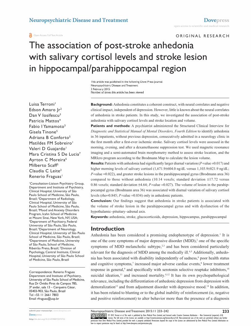
The association of post-stroke anhedonia with salivary cortisol levels and stroke lesion in hippocampal/parahippocampal region
luisa Terroni1
edson amaro Jr2
Dan V iosifescu3
Patricia Mattos4
Fabio i Yamamoto5
gisela Tinone5
adriana B conforto5
Matildes FM sobreiro1
Valeri D guajardo1
Mara cristina s De lucia7
ayrton c Moreira6
Milberto scaff5
claudia c leite2
renerio Fraguas1
1consultation-liaison Psychiatry group, Department and institute of Psychiatry, clinical hospital, University of são Paulo school of Medicine, são Paulo, Brazil; 2Department of radiology, clinical hospital, University of são Paulo school of Medicine, são Paulo, Brazil; 3Mood and anxiety Disorders Program, icahn school of Medicine at Mount sinai, New York, NY, Usa; 4Department of Psychiatry, Federal University of são Paulo, são Paulo, Brazil; 5Department of Neurology, clinical hospital, University of são Paulo school of Medicine, são Paulo, Brazil; 6Department of Medicine, University of são Paulo, school of Medicine, ribeirão Preto, Brazil; 7Division of Psychology, central institute, clinical hospital, University of são Paulo school of Medicine, são Paulo, Brazil
Background: Anhedonia constitutes a coherent construct, with neural correlates and negative
clinical impact, independent of depression. However, little is known about the neural correlates
of anhedonia in stroke patients. In this study, we investigated the association of post-stroke
anhedonia with salivary cortisol levels and stroke location and volume.
Patients and methods: A psychiatrist administered the Structured Clinical Interview for
Diagnostic and Statistical Manual of Mental Disorders, Fourth Edition to identify anhedonia
in 36 inpatients, without previous depression, consecutively admitted in a neurology clinic in
the first month after a first-ever ischemic stroke. Salivary cortisol levels were assessed in the
morning, evening, and after a dexamethasone suppression test. We used magnetic resonance
imaging and a semi-automated brain morphometry method to assess stroke location, and the
MRIcro program according to the Brodmann Map to calculate the lesion volume.
Results: Patients with anhedonia had significantly larger diurnal variation (P-value =0.017) and
higher morning levels of salivary cortisol (1,671.9±604.0 ng/dL versus 1,103.9±821.9 ng/dL;
P-value =0.022), and greater stroke lesions in the parahippocampal gyrus (Brodmann area 36)
compared to those without anhedonia (10.14 voxels; standard deviation ±17.72 versus
0.86 voxels; standard deviation ±4.64; P-value =0.027). The volume of lesion in the parahip-
pocampal gyrus (Brodmann area 36) was associated with diurnal variation of salivary cortisol
levels (rho=0.845; P-value =0.034) only in anhedonic patients.
Conclusion: Our findings suggest that anhedonia in stroke patients is associated with
the volume of stroke lesion in the parahippocampal gyrus and with dysfunction of the
hypothalamic–pituitary–adrenal axis.
Keywords: anhedonia, stroke, glucocorticoids, depression, hippocampus, parahippocampal
IntroductionAnhedonia has been considered a promising endophenotype of depression.1 It is
one of the core symptoms of major depressive disorder (MDD),2 one of the specific
symptoms of MDD melancholic subtype,2,3 and has been considered particularly
relevant for the diagnosis of MDD among the medically ill.4,5 Additionally, anhedo-
nia has been associated with disability independently of sadness,6 poor health status
and cognitive symptoms,7 increased major adverse cardiac events,8 lower treatment
response in general,9 and specifically with serotonin selective reuptake inhibitors,10
suicidal ideation,11 and increased mortality.8,12 It has its own psychopathological
relevance, including the differentiation of anhedonic depression from depression with
demoralization13 and from adjustment disorder with depressive mood.14 In addition,
it has been related to blunting or to the global inability of reinforcement (ie, negative
and positive reinforcement) to alter behavior more than the presence of a diagnosis
correspondence: renerio FraguasDepartment and institute of Psychiatry,University of são Paulo school of Medicine, rua Dr Ovidio Pires de campos 785, 3º andar, sala 13 – cerqueira césar, 05403-903, são Paulo, BrazilTel +55 11 2661 7803email [email protected]
Journal name: Neuropsychiatric Disease and TreatmentArticle Designation: Original ResearchYear: 2015Volume: 11Running head verso: Terroni et alRunning head recto: The association of post-stroke anhedonia with salivary cortisol levels and stroke lesionDOI: http://dx.doi.org/10.2147/NDT.S73722
Neuropsychiatric Disease and Treatment 2015:11submit your manuscript | www.dovepress.com
Dovepress
Dovepress
234
Terroni et al
of depression.15 Its unique importance has also been reported
in psychiatric disorders other than depression, including
obsessive compulsive disorder,16 eating disorders,17,18 social
anhedonia in schizophrenia,19,20 and as a possible predictor
for stimulant misuse.21 Anhedonia constitutes a coherent
construct, consistent with the new Research Domain Criteria
proposed by the National Institutes of Mental Health.22 The
Research Domain Criteria aim to identify neurobiological
mechanisms underlying clusters of psychiatric symptoms
or “domains” of mental function (eg, anhedonia and cogni-
tive deficits) across the traditional psychiatric diagnoses.22,23
Clinical data have indicated that post-stroke depression has
imprecise boundaries and better understanding of its psycho-
pathological profile is necessary.24 This approach enhances
the relevance of studies investigating the brain morphology
and endocrine correlates of anhedonia.25
Neurophysiologically, anhedonia has been associated
with reduced connectivity between pregenual anterior cingu-
late cortex and caudate nucleus, reduced neuronal activity in
ventromedial prefrontal cortex, amygdala/ventral striatum,26
and reduced activity in the nucleus accumbens.27 Anhedonia
has been associated with vascular depression28 and neuro-
anatomical alterations that have been related to anhedonia
include a greater volume of white matter hyperintensities,29
reduced caudate volume,30 deep white matter lesions, lacu-
nar infarcts in deep white matter, and cortical atrophy.31
Regarding neurotransmitters, antidepressants acting on
noradrenaline and not only on serotonin neurotransmission
may be relevant to treat anhedonia.32 Neuro-endocrinologi-
cally, anhedonia has been related to a dysfunctional feedback
between the hypothalamic–pituitary–adrenal (HPA) axis and
specific brain regions.33
Of note, increased cortisol levels have been consistently
associated with melancholic depression, an MDD subtype
where anhedonia is a cardinal marker.34 Clinically, increased
cortisol levels have been associated with poor outcomes,
increased severity, increased risk for depression,35 poor
response to treatment,36,37 and higher relapse of depres-
sive episodes.34,38–40 Neuroanatomical correlates of cortisol
that have been reported include reduced hippocampal
volume,41 deficits in hippocampus-related neuropsychologi-
cal functions including verbal and visuospatial memory,42
and dysfunctional hippocampal response to cortisol in
depressed women.43 Additionally, depressed patients who are
dexamethasone/CRH non-suppressors (a marker of HPA dys-
function) showed metabolic changes in the medial prefrontal
cortex (mPFC) and in the hippocampal/parahippocampal
brain regions after treatment with antidepressants.44 Recently,
the diurnal slope of salivary cortisol has been negatively
associated with resting neural activity in the mPFC, hip-
pocampus and parahippocampal gyrus in healthy volunteers;
in contrast, anhedonic subjects failed to present an association
of salivary cortisol with mPFC activity.33 It should be noted
that distorted daily cortisol rhythms may lead to diverse
effects on emotional functions depending upon the cellular
pathway or part of the network it is disturbing.45 Such effect
is influenced by individual regional differences in the sensi-
tivity of the brain determined by differential local epigenetic
events.45 In aggregate, these data support the association
between anhedonia, HPA dysfunction, and abnormal func-
tion in specific brain areas involved in emotional regulation
(eg, mPFC and hippocampus/parahippocampal area).
In stroke patients, anhedonia has been associated with
increased levels of depression at hospital discharge,46 and cog-
nitive deficits47 including executive dysfunction.48 In contrast
with depressive and anxiety symptoms, anhedonia in post-
stroke patients has been related to aging and not to a previous
history of mood disorders or to the functional status (Barthel
Index), supporting its particular brain correlates.49 In rats,
anhedonia induced by ischemic stroke has been associated
with decreased protein expression of 5-hydroxytryptamine 1A
receptors and messenger ribonucleic acid levels in the dentate
hippocampal gyrus.50 These changes could be associated with
decreased neurogenesis and were reversed by citalopram.50,51
Notwithstanding the abovementioned relevance of anhedonia,
it is not known whether its neuroendocrine correlates in stroke
patients match the pattern described in MDD.
In post-stroke depression, hypercortisolemia has been
associated with lesions in frontal brain areas52–57 and with
poor clinical outcome of stroke.55,56,58 Post-stroke hyper-
cortisolemia was a predictor of later depression in some54
but not all studies.53 A recent literature review reported that
elevated post-stroke cortisol levels have been associated with
dependency, morbidity, and mortality.59 Early studies were
conflicting regarding stroke location and depression.60–62
However, a possible explanation for these discrepancies
is that depression in acute and inpatients is related to left
side lesions, and depression occurring later than 3 months
after stroke and in community patients is related to right
side lesions.63 Recent studies have attributed the occur-
rence of depression to disruption of prefrontal subcorti-
cal circuits by the stroke lesion.64,65 Recently, post-stroke
major depression episode (MDE) has also been associated
with stroke lesions in the limbic–cortical–striato–pallido–
thalamic41,66,67 circuit including the mPFC, cingulate cortex,
hippocampal/parahippocampal region, and amygdala.68
Neuropsychiatric Disease and Treatment 2015:11 submit your manuscript | www.dovepress.com
Dovepress
Dovepress
235
The association of post-stroke anhedonia with salivary cortisol levels and stroke lesion
Based on these findings and those relating anhedonia with
neuroendocrine and neural dysfunction33 we hypothesized
that anhedonia in stroke patients would be associated with
cortisol levels and with lesions in mPFC and areas in the
hippocampal/parahippocampal region. Therefore, the goal
of this study was to investigate the association of anhedonia
with salivary cortisol levels and with stroke lesion in the
mPFC and hippocampus/parahippocampal areas.
Material and methodsPatientsWe screened 326 male and female inpatients, 18 years of
age or older, consecutively admitted to the neurology unit
of a university hospital with a diagnosis of ischemic stroke
between August 2002 and May 2008. Details of the protocol
have been previously described.68,69 In short, the diagnosis
of stroke was made by a neurologist in accordance with the
World Health Organization criteria70 and confirmed by mag-
netic resonance imaging (MRI). At screening we interviewed
patients using the modules for mood episodes, psychotic
symptoms, and substance use disorders of the Structured Clin-
ical Interview Patient Version axis I (SCID-I/P) for Diagnostic
and Statistical Manual of Mental Disorders, Fourth Edition
(DSM-1V) to investigate past and current mood disorders.71
A psychiatrist performed the interview with the patient and
a family member/caregiver. A neurologist assessed the daily
living activities using the Barthel Index,72 and the stroke
severity using the National Institutes of Health Stroke Scale
(NIHSS).73 The Barthel Index scores range from 0 (completely
dependent) to 100 (completely independent).72 The NIHSS is
a graded neurological examination assessing consciousness,
eye movements, visual fields, motor and sensory impairments,
ataxia, speech, cognition, and inattention and its scores range
from 0 to 42 (more severe).73 Both, NIHSS and Barthel Index
Brazilian versions have been validated.74
We excluded 253 patients for the following criteria:
a) previous history of stroke, infratentorial stroke, greater
than 3 weeks interval between stroke occurrence and screen-
ing interview, or hemorrhagic transformation of stroke
(n=89); b) drug/alcohol dependence, psychoses, delirium,
history of MDE, current MDE with pre-stroke onset, dysthy-
mia, or bipolar disorder (n=54); c) aphasia that impeded the
interview (n=37); d) neurological diseases or severe clinical
condition that impeded the interview (n=22); e) problems dur-
ing the MRI acquisition (n=19); f) other reasons (n=32).68,69
Of 73 eligible patients, five declined to participate and for
32 we lacked proper cortisol samples, leaving 36 patients
for the current analysis.
The 36 patients were evaluated on average within
11.9 days after stroke (standard deviation [SD] ±4.7; range
5–22 days). Patients were free from corticoids, oral con-
traceptives, and antidepressants. Patients with MDD were
only those whose depression had started after the stroke. All
patients with MDD were oriented and referred to treatment
after the evaluation.
The institutional review board of the Clinics Hospital of
the University of São Paulo, School of Medicine, approved
the study protocol. Written informed consent was obtained
from all participating patients after explanation about the
procedures and study.
anhedonia assessmentAnhedonia, defined as diminished interest or pleasure in
response to stimuli previously perceived as rewarding dur-
ing the premorbid state, was diagnosed by a psychiatrist
administering the SCID-I/P for DSM-1V.71 The psychiatrist
was blinded for the cortisol and radiological results.
cortisol measuresCortisol samples were collected with the supervision of a
member of the nursing team or by one of the researchers. Sali-
vary samples (1 mL) were collected in a plastic tube by direct
spitting during a 15 minutes period at 9 am and 11 pm. After
collecting basal morning and evening samples, 1 mg of dex-
amethasone was administered orally at 11 pm. The next morn-
ing at 9 am, we collected a salivary sample to investigate the
inhibitory effect of the dexamethasone. Salivary samples were
stored at 4°C and analyses were performed at the neuroen-
docrine laboratory of the University of Sao Paulo, Ribeirão
Preto School of Medicine. After centrifugation at 2,000 rpm,
the supernatants were stored at -20°C until assayed. Sali-
vary cortisol measurements were performed by a previously
described radioimmunoassay method on 25 μL samples of
saliva without previous extraction or chromatography. This
method previously demonstrated a good correlation (r=50.95)
with plasma free cortisol levels determined by equilibrium
dialysis.75 The assay sensitivity was 60 ng/dL.75 The mean
intra-assay coefficient of variation was 5.5%. All samples
obtained from each subject were analyzed in duplicate in the
same assay. The technicians performing cortisol assays were
blind to the clinical characteristics of the patients.
Mri protocol, stroke location and volumeAll MRIs were acquired on a 1.5-Tesla system (GE-Horizon
LX; General Electric Healthcare, WI, USA), using a specific
previously described imaging protocol.68 Lesion location
Neuropsychiatric Disease and Treatment 2015:11submit your manuscript | www.dovepress.com
Dovepress
Dovepress
236
Terroni et al
and volume quantification were determined using a semi-
automated method. Initially, spoiled gradient recalled acqui-
sition in steady state and axial fluid attenuated inversion
recovery (FLAIR) acquisitions were both normalized to the
Montreal Neurological Institute template.76 We used linear
transformation with 12 degrees of freedom and 15 nonlinear
interactions implemented in Statistical Parametric Mapping
(SPM5, Wellcome Trust for Neuroimaging, London, http://
www.fil.ion.ucl.ac.uk/spm/), and based on coordinates
referenced in the Talairach and Tornoux Atlas.77 During
this process, all images were sampled to 2.3×2.3×2.6 mm.
Lesion tracing was performed by a trained psychiatrist (LT),
blinded for neurological and cortisol data, using a mouse
device to trace the ischemic lesion and analyzing all slices
of each FLAIR image using the MRIcro Software (http://
www.sph.sc.edu/comd/rorden/mricro.html). All lesions’
tracings from all patients were reviewed by a neuroradiolo-
gist, blinded for clinical data and psychiatric diagnoses. The
regions of interest were then analyzed automatically using
the Brodmann Cytoarchitectonic Atlas placed in the same
spaces,78 in order to count the number of voxels within each
Brodmann area (BA). The total lesion volume was obtained
by multiplying the number of voxels by voxel size in nor-
malized images.
statistical analysisAnhedonia was analyzed as a categorical variable (yes/no)
according to the specific item on the SCID-I/P for DSM-1V.71
Salivary cortisol levels were analyzed as a continuous variable
and by comparing tertiles, as it has been done previously.39
Patients in the highest tertile of cortisol levels were compared
to those in the lower two tertiles combined. Patients with cor-
tisol levels below the detection limit of the assay (ie, below
60 ng/dL) were assigned to the inferior tertile. The cut-off for
the upper tertile of morning salivary cortisol levels (increased
level) was .1.515 ng/dL and for the evening salivary cortisol
levels was .575 ng/dL; the cut-off for the upper tertile of
morning salivary cortisol levels after dexamethasone supres-
sion test was .600 ng/dL. Lesion volume was obtained by
determining the voxel-based lesion morphometry in each BA
of interest. Lesion volumes were expressed as mean number
of voxels and SD (FLAIR voxel size 2.3×2.3×2.6 mm). The
a priori defined brain areas for investigation were the mPFC
and the BAs 28, 34, 35, and 36 from the hippocampus/
parahippocampal region. We estimated the mPFC area by
summing the BAs 10, 11, 12, 13, 14, 24, 25, 32, and 47 as
previously done.68
Statistical analyses were performed using the Statistical
Package for the Social Sciences, version 14 (SPSS Inc.,
Chicago, IL, USA). Chi-square tests or Fisher’s exact tests
were used for categorical data. If not otherwise specified,
Student’s t-tests were used for continuous variables; alterna-
tively, the non-parametric Mann–Whitney U-test was used
when data did not follow normal distribution according to
the Kolmogorov–Smirnov test. The Wilcoxon Signed Rank
test was used to compare morning cortisol levels (9 am)
with evening cortisol levels (11 pm). We used Spearman
correlations to investigate the relationship between diurnal
reduction (from morning to evening) in cortisol levels with
stroke volumes in the a priori determined brain areas (mPFC
and hippocampus/parahippocampal areas), separately in
patients with and without anhedonia, in accordance to
previous report.33 Results are presented as frequencies or
mean ± SD. All statistical tests were two-tailed and alpha
was set at 5%.
ResultsWe identified anhedonia in seven (19.4%) patients and it
was significantly associated with new onset post-stroke
MDE (Table 1). Salivary cortisol levels were significantly
higher in the morning compared to the evening in both
anhedonic and non-anhedonic patients (Table 1). Patients
with anhedonia had significantly higher mean morning
salivary cortisol levels than non-anhedonic patients (Table 1
and Figure 1). Consistent with this, 71.4% of the anhedonic
patients had morning salivary cortisol levels in the upper
tertile, a rate significantly higher than in non-anhedonic
patients (24.1%) (Table 1).
Anhedonic patients experienced a larger diurnal decrease
in cortisol levels compared to non-anhedonic patients
(P-value =0.017, Mann–Whitney U-test).
Anhedonic patients had larger stroke lesion volumes
in the parahippocampal gyrus (BA 36) compared to non-
anhedonic patients (Table 1; Figure 2). We found no other
differences between anhedonic and non-anhedonic patients
regarding stroke lesion volumes in the other a priori deter-
mined brain areas.
Stroke lesion volumes in the parahippocampal gyrus
(BA 36) were correlated with a larger diurnal decrease of
salivary cortisol levels (rho=0.845; P-value =0.034) among
anhedonic patients but not among the non-anhedonic ones
(rho=0.045; P-value =0.834). No other BAs showed correla-
tion with diurnal variation of salivary cortisol levels.
DiscussionWe found that patients experiencing anhedonia within the
first month after a first ever ischemic stroke had higher levels
of morning salivary cortisol and larger diurnal decrement of
Neuropsychiatric Disease and Treatment 2015:11 submit your manuscript | www.dovepress.com
Dovepress
Dovepress
237
The association of post-stroke anhedonia with salivary cortisol levels and stroke lesion
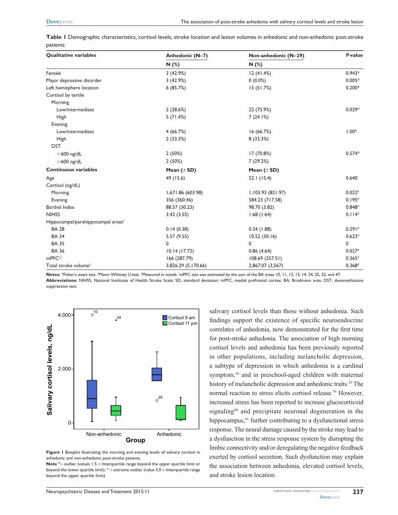
Table 1 Demographic characteristics, cortisol levels, stroke location and lesion volumes in anhedonic and non-anhedonic post-stroke patients
Qualitative variables Anhedonic (N=7) Non-anhedonic (N=29) P-value
N (%) N (%)
Female 3 (42.9%) 12 (41.4%) 0.943*Major depressive disorder 3 (42.9%) 0 (0.0%) 0.005*left hemisphere location 6 (85.7%) 15 (51.7%) 0.200*cortisol by tertile
Morninglow/intermediate 2 (28.6%) 22 (75.9%) 0.029*high 5 (71.4%) 7 (24.1%)
eveninglow/intermediate 4 (66.7%) 16 (66.7%) 1.00*high 2 (33.3%) 8 (33.3%)
DsT,600 ng/dl 2 (50%) 17 (70.8%) 0.574*
.600 ng/dl 2 (50%) 7 (29.2%)
Continuous variables Mean (± SD) Mean (± SD)age 49 (15.6) 52.1 (15.4) 0.640cortisol (ng/dl)
Morning 1,671.86 (603.98) 1,103.93 (821.97) 0.022#
evening 356 (360.46) 584.23 (717.58) 0.195#
Barthel index 88.57 (30.23) 98.70 (3.82) 0.848#
Nihss 3.42 (3.55) 1.68 (1.64) 0.114#
hippocampal/parahippocampal areas†
Ba 28 0.14 (0.38) 0.34 (1.88) 0.291#
Ba 34 5.57 (9.55) 10.52 (30.16) 0.623#
Ba 35 0 0 0Ba 36 10.14 (17.72) 0.86 (4.64) 0.027#
mPFc†,∫ 166 (287.79) 108.69 (257.51) 0.365#
Total stroke volume† 3,826.29 (5,170.66) 2,867.07 (3,567) 0.368#
Notes: *Fisher’s exact test. #Mann–Whitney U-test. †Measured in voxels. ∫mPFc size was estimated by the sum of the Ba areas 10, 11, 12, 13, 14, 24, 25, 32, and 47.Abbreviations: Nihss, National institutes of health stroke scale; sD, standard deviation; mPFc, medial prefrontal cortex; Ba, Brodmann area; DsT, dexamethasone suppression test.
4.000 Cortisol 9 amCortisol 11 pm
2.000
0
Non-anhedonic AnhedonicGroup
Saliv
ary
cort
isol
leve
ls, n
g/dL
26
34*
10
Figure 1 Boxplot illustrating the morning and evening levels of salivary cortisol in anhedonic and non-anhedonic post-stroke patients.Note °= outlier (values 1.5 × interquartile range beyond the upper quartile limit or beyond the lower quartile limit); * = extreme outlier (value 3.0 × interquartile range beyond the upper quartile limit).
salivary cortisol levels than those without anhedonia. Such
findings support the existence of specific neuroendocrine
correlates of anhedonia, now demonstrated for the first time
for post-stroke anhedonia. The association of high morning
cortisol levels and anhedonia has been previously reported
in other populations, including melancholic depression,
a subtype of depression in which anhedonia is a cardinal
symptom,34 and in preschool-aged children with maternal
history of melancholic depression and anhedonic traits.35 The
normal reaction to stress elicits cortisol release.79 However,
increased stress has been reported to increase glucocorticoid
signaling80 and precipitate neuronal degeneration in the
hippocampus,81 further contributing to a dysfunctional stress
response. The neural damage caused by the stroke may lead to
a dysfunction in the stress response system by disrupting the
limbic connectivity and/or deregulating the negative feedback
exerted by cortisol secretion. Such dysfunction may explain
the association between anhedonia, elevated cortisol levels,
and stroke lesion location.
Neuropsychiatric Disease and Treatment 2015:11submit your manuscript | www.dovepress.com
Dovepress
Dovepress
238
Terroni et al
A B
C
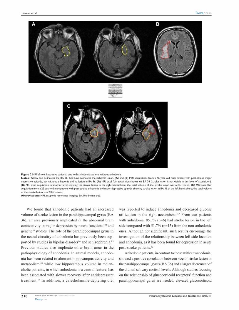
Figure 2 Mri of two illustrative patients, one with anhedonia and one without anhedonia.Notes: Yellow line delineates the Ba 36. red line delineates the ischemic lesion. (A) and (B) Mri acquisitions from a 46 year old male patient with post-stroke major depressive episode, but without anhedonia and no lesion in Ba 36. (A) MRI axial flair acquisition shown left BA 36 (stroke lesion is not visible in this level of acquisition). (B) Mri axial acquisition in another level showing the stroke lesion in the right hemisphere; the total volume of the stroke lesion was 6,273 voxels. (C) MRI axial flair acquisition from a 22 year old male patient with post-stroke anhedonia and major depressive episode showing stroke lesion in Ba 36 of the left hemisphere; the total volume of the stroke lesion was 2,032 voxels.Abbreviations: Mri, magnetic resonance imaging; Ba, Brodmann area.
We found that anhedonic patients had an increased
volume of stroke lesion in the parahippocampal gyrus (BA
36), an area previously implicated in the abnormal brain
connectivity in major depression by neuro functional82 and
genetic83 studies. The role of the parahippocampal gyrus in
the neural circuitry of anhedonia has previously been sup-
ported by studies in bipolar disorder84 and schizophrenia.85
Previous studies also implicate other brain areas in the
pathophysiology of anhedonia. In animal models, anhedo-
nia has been related to aberrant hippocampus activity and
metabolism,86 while low hippocampus volume in melan-
cholic patients, in which anhedonia is a central feature, has
been associated with slower recovery after antidepressant
treatment.87 In addition, a catecholamine-depleting diet
was reported to induce anhedonia and decreased glucose
utilization in the right accumbens.67 From our patients
with anhedonia, 85.7% (n=6) had stroke lesion in the left
side compared with 51.7% (n=15) from the non-anhedonic
ones. Although not significant, such results encourage the
investigation of the relationship between left side location
and anhedonia, as it has been found for depression in acute
post-stroke patients.63
Anhedonic patients, in contrast to those without anhedonia,
showed a positive correlation between size of stroke lesion in
the parahippocampal gyrus (BA 36) and a larger decrement of
the diurnal salivary cortisol levels. Although studies focusing
on the relationship of glucocorticoid receptors’ function and
parahippocampal gyrus are needed, elevated glucocorticoid

Neuropsychiatric Disease and Treatment 2015:11 submit your manuscript | www.dovepress.com
Dovepress
Dovepress
239
The association of post-stroke anhedonia with salivary cortisol levels and stroke lesion
levels have been associated with parahippocampal blood
flow reduction.88 Hence, it is possible that stroke lesion in the
parahippocampal gyrus could impair glucocorticoid receptors’
function leading to an increased morning cortisol level as a
compensatory mechanism,89 and consequently a larger decre-
ment of the diurnal levels. Anhedonia could be related to an
increased vulnerability to disrupt the modulation of the HPA
axis feedback via lesions in the parahippocampal gyrus. Putnam
et al reported that health subjects with anhedonia have a dys-
functional feedback between the mPFC and the neuroendocrine
HPA axis.33 Using dense-array resting electroencephalography,
they found a negative correlation between diurnal salivary corti-
sol slope and current density in the hippocampal/parahippocam-
pal region for both subjects with and without anhedonia.33 In
addition, they found a correlation of diurnal cortisol slope with
current density in the mPFC in subjects without anhedonia,
but not in subjects with anhedonia. Accordingly, patients with
previous vulnerability to anhedonia would have a dysfunctional
feedback between mPFC and neuroendocrine regulation. Con-
sequently, it is possible that in anhedonic subjects the HPA axis
would depend on feedback from other brain areas, including the
hippocampal/parahippocampal region. Thus, stroke lesions in
the parahippocampal gyrus (BA 36) in patients with anhedonia
would disrupt the feedback of this region to the neuroendocrine
HPA axis. Supporting this hypothesis, in our sample, the cor-
relation of salivary cortisol diurnal decrease with the size of
lesion in the parahippocampal gyrus (BA 36) was found only
in anhedonic patients. Complementary to this, the absence of
correlation between size of lesion in parahippocampal gyrus
(BA 36) and the decrease in diurnal salivary cortisol levels in
patients without anhedonia may suggest that these patients
could have the brain HPA axis feedback maintained by the
modulation of other brain regions in particular the mPFC as
suggested by previous studies.33 In conclusion, our findings
indicate that subjects with post-stroke anhedonia may have
a dysfunctional feedback between parahippocampal gyrus
(BA 36) and the HPA axis.
A consistent line of research has investigated the correlates
of apathy in stroke patients. Apathy has a wide array of defi-
nitions and conceptualizations usually including diminished
interest and motivation among other symptoms.90 The item
of the SCID-I/P we have used to assess anhedonia includes
two aspects: decrease in motivation and/or reduction in expe-
rienced pleasure. Consequently, the symptom “decrease in
motivation/interest” included in the definition of anhedonia
that we have used, overlaps with some definitions of apathy.
Stroke lesions of patients with apathetic depression often did
not overlap with lesion of patients with affective depression;91
reinforcing that apathy has a distinct pathophysiology and
should not be considered only as a symptom of depression.
A recent review concluded that it was not possible to identify
a specific stroke lesion location for apathy.90 Actually, post-
stroke apathy has been associated with fronto subcortical,92
pontine,93 brainstem and bilateral striatum infarcts.91 The use
of diverse definitions of apathy, capturing its distinct aspects
may in part explain the diversity of results regarding its stroke
location. Similarly, the motivational and consummatory (hedo-
nic response) components of anhedonia have been reported
to relay on separate neural systems.25 Thus, assessing specific
components of apathy and anhedonia is necessary to address
their neural correlates.
Some limitations of our study should be considered. We
did not assess the plasma corticotrophin hormone levels.
Corticotrophin hormone assessment and several cortisol
measurements could give a more detailed and reliable
appraisal of HPA function.34 The cross-sectional design of
our study restricts conclusions about direction of causality
in the association between increased cortisol levels and
anhedonia. Although the neuroanatomical areas of inter-
est were determined a priori and literature data support the
relationship of the parahippocampal gyrus with anhedonia in
non-stroke subjects33 and its deactivation has been reported
in paradigms eliciting the reward circuit,94 we did not correct
for multiple comparisons. We did not find an association of
anhedonia with the mPFC26 and other brain areas previously
reported to be involved in the neural basis of anhedonia such
as the anterior cingulate cortex and nucleus accumbens.27,95
Similarly to apathy, as commented above, fatigue is common
after stroke. Defined as a feeling of lack of energy, weariness,
and aversion to effort,96 assessing fatigue in addition to apathy
could give a more specific and integrated view of anhedonia.
Finally, our sample is relatively young and because of the
inclusion and exclusion criteria, patients with more severe
stroke have been excluded. Hence, considering the existence
of multiple comparisons, the small sample size and small
power, our results are better understood as preliminary.
Future confirmatory studies with greater sample size allow-
ing the performance of multivariate analysis investigating
the influence of total stroke volume, left side location, stroke
severity, restriction of daily living activities, the presence of
old patients, and more severe stroke cases are warranted.
ConclusionIn conclusion, our data suggest the existence of a relationship
between anhedonia with stroke size in the parahippocampal
gyrus (area BA 36), with increased morning salivary cortisol
Neuropsychiatric Disease and Treatment 2015:11submit your manuscript | www.dovepress.com
Dovepress
Dovepress
240
Terroni et al
levels and increased diurnal cortisol decrement in stroke
patients. Our findings contribute to the understanding of post-
stroke anhedonia as a result of disruption of neural circuits
caused by the stroke lesion and by dysfunctional neuroen-
docrine feedback of HPA axis associated with that lesion;
suggesting that increased morning levels and a larger decre-
ment of diurnal salivary cortisol levels may be a biomarker
of anhedonia in stroke patients. Considering the small sample
size and absence of correction for multiple comparisons,
future studies are necessary to confirm our findings.
AcknowledgmentsWe thank Mr Bernardo Santos for the statistical analysis,
Mr João Ricardo Sato for the essential assistance in the lesion
computational analysis of neuroimaging acquisitions, and
our colleagues from the Consultation-Liaison Group for the
valuable comments and suggestions.
DisclosureThe authors declare that there is no conflict of interest regard-
ing the publication of this paper.
References 1. Pizzagalli DA. Depression, stress, and anhedonia: toward a synthesis
and integrated model. Annu Rev Clin Psychol. 2014;10:393–423. 2. American Psychiatric Association. Diagnostic and Statistical Manual
of Mental Disorders Fifth Edition. Arlington, VA, USA: American Psychiatric Publishing; 2013.
3. Hyett MP, Parker GB, Proudfoot J, Fletcher K. Examining age effects on prototypic melancholic symptoms as a strategy for refining definition of melancholia. J Affect Disord. 2008;109(1–2):193–197.
4. Sibitz I, Berger P, Freidl M, et al. ICD-10 or DSM-IV? Anhedonia, fatigue and depressed mood as screening symptoms for diagnosing a current depressive episode in physically ill patients in general hospital. J Affect Disord. 2010;126(1–2):245–251.
5. Fraguas R Jr, Alves TC. Depressão no Hospital Geral: estudo de 136 casos [Depression in General Hospital: a study of 136 cases]. Rev Assoc Méd Bras. 2002;48(3):225–230. Portuguese.
6. Covinsky KE, Cenzer IS, Yaffe K, O’Brien S, Blazer DG. Dysphoria and Anhedonia as Risk Factors for Disability or Death in Older Persons: Implications for the Assessment of Geriatric Depression. Am J Geriatr Psychiatry. 2014;22(6):606–613.
7. Pelle AJ, Pedersen SS, Erdman RA, et al. Anhedonia is associated with poor health status and more somatic and cognitive symptoms in patients with coronary artery disease. Qual Life Res. 2011;20(5):643–651.
8. Davidson KW, Burg MM, Kronish IM, et al. Association of anhedonia with recurrent major adverse cardiac events and mortality 1 year after acute coronary syndrome. Arch Gen Psychiatry. 2010;67(5):480–488.
9. Spijker J, Bijl RV, de Graaf R, Nolen WA. Determinants of poor 1-year outcome of DSM-III-R major depression in the general population: results of the Netherlands Mental Health Survey and Incidence Study (NEMESIS). Acta Psychiatr Scand. 2001;103(2):122–130.
10. Shelton RC, Tomarken AJ. Can recovery from depression be achieved? Psychiatr Serv. 2001;52(11):1469–1478.
11. Pfeiffer PN, Brandfon S, Garcia E, et al. Predictors of suicidal ideation among depressed Veterans and the interpersonal theory of suicide. J Affect Disord. 2014;152(154):277–281.
12. Furlanetto LM, von Ammon Cavanaugh S, Bueno JR, Creech SD, Powell LH. Association between depressive symptoms and mortality in medical inpatients. Psychosomatics. 2000;41(5):426–432.
13. Clarke DM, issane DW, Trauer T, Smith GC. Demoralization, anhedonia and grief in patients with severe physical illness. World Psychiatry. 2005;4(2):96–105.
14. Zimmerman M, Martinez JH, Dalrymple K, Chelminski I, Young D. “Subthreshold” depression: is the distinction between depressive disorder not otherwise specified and adjustment disorder valid? J Clin Psychiatry. 2013;74(5):470–476.
15. Chase HW, Frank MJ, Michael A, Bullmore ET, Sahakian BJ, Robbins TW. Approach and avoidance learning in patients with major depression and healthy controls: relation to anhedonia. Psychol Med. 2010;40(3):433–440.
16. Abramovitch A, Pizzagalli DA, Reuman L, Wilhelm S. Anhedonia in obsessive-compulsive disorder: Beyond comorbid depression. Psychia-try Res. 2014;216(2):223–229.
17. Tchanturia K, Davies H, Harrison A, Fox JR, Treasure J, Schmidt U. Altered social hedonic processing in eating disorders. Int J Eat Disord. 2012;45(8):962–969.
18. Keating C, Tilbrook AJ, Rossell SL, Enticott PG, Fitzgerald PB. Reward processing in anorexia nervosa. Neuropsychologia. 2012; 50(5):567–575.
19. Velthorst E, Meijer C, Investigators G.R.O.U.P. The association between social anhedonia, withdrawal and psychotic experiences in general and high-risk populations. Schizophr Res. 2012;138(2–3):290–294.
20. Goldstein KE, Hazlett EA, New AS, et al. Smaller superior temporal gyrus volume specificity in schizotypal personality disorder. Schizophr Res. 2009;112(1–3):14–23.
21. Leventhal AM, Kahler CW, Ray LA, et al. Anhedonia and amotivation in psychiatric outpatients with fully remitted stimulant use disorder. Am J Addict. 2008;17(3):218–223.
22. Insel TR. Translating scientific opportunity into public health impact: a strategic plan for research on mental illness. Arch Gen Psychiatry. 2009;66(2):128–133.
23. Miller G Psychiatry, Beyond DSM: seeking a brain-based classification of mental illness. Science. 2010;327(5972):1437.
24. da Rocha e Silva CE, Alves Brasil MA, Matos do Nascimento E, de Braganca Pereira B, Andre C. Is poststroke depression a major depression? Cerebrovasc Dis. 2013;35(4):385–391.
25. Treadway MT, Zald DH. Reconsidering anhedonia in depression: les-sons from translational neuroscience. Neurosci Biobehav Rev. 2011; 35(3):537–555.
26. Keedwell PA, Andrew C, Williams SC, Brammer MJ, Phillips ML. The neural correlates of anhedonia in major depressive disorder. Biol Psychiatry. 2005;58(11):843–853.
27. Wacker J, Dillon DG, Pizzagalli DA. The role of the nucleus accum-bens and rostral anterior cingulate cortex in anhedonia: integration of resting EEG, fMRI, and volumetric techniques. Neuroimage. 2009; 46(1):327–337.
28. Krishnan KR, Hays JC, Blazer DG. MRI-defined vascular depression. Am J Psychiatry. 1997;154(4):497–501.
29. Lavretsky H, Zheng L, Weiner MW, et al. The MRI brain correlates of depressed mood, anhedonia, apathy, and anergia in older adults with and without cognitive impairment or dementia. Int J Geriatr Psychiatry. 2008;23(10):1040–1050.
30. Pizzagalli DA, Holmes AJ, Dillon DG, et al. Reduced caudate and nucleus accumbens response to rewards in unmedicated individu-als with major depressive disorder. Am J Psychiatry. 2009;166(6): 702–710.
31. Grool AM, van der Graaf Y, Mali WP, et al. Location and progression of cerebral small-vessel disease and atrophy, and depressive symptom profiles: the Second Manifestations of ARTerial disease (SMART)-Medea study. Psychol Med. 2012;42(2):359–370.
32. Pringle A, McCabe C, Cowen PJ, Harmer CJ. Antidepressant treatment and emotional processing: can we dissociate the roles of serotonin and noradrenaline? J Psychopharmacol. 2013;27(8):719–731.
Neuropsychiatric Disease and Treatment 2015:11 submit your manuscript | www.dovepress.com
Dovepress
Dovepress
241
The association of post-stroke anhedonia with salivary cortisol levels and stroke lesion
33. Putnam KM, Pizzagalli DA, Gooding DC, Kalin NH, Davidson RJ. Neural activity and diurnal variation of cortisol: evidence from brain electrical tomography analysis and relevance to anhedonia. Psy-chophysiology. 2008;45(6):886–895.
34. Stetler C, Miller GE. Depression and hypothalamic-pituitary-adrenal activation: a quantitative summary of four decades of research. Psy-chosom Med. 2011;73(2):114–126.
35. Dougherty LR, Klein DN, Olino TM, Dyson M, Rose S. Increased waking salivary cortisol and depression risk in preschoolers: the role of maternal history of melancholic depression and early child tempera-ment. J Child Psychol Psychiatry. 2009;50(12):1495–1503.
36. Martiny K, Lunde M, Unden M, Dam H, Bech P. High cortisol awaken-ing response is associated with an impairment of the effect of bright light therapy. Acta Psychiatr Scand. 2009;120(3):196–202.
37. Juruena MF, Pariante CM, Papadopoulos AS, Poon L, Lightman S, Cleare AJ. The role of mineralocorticoid receptor function in treatment-resistant depression. J Psychopharmacol. 2013;27(12):1169–1179.
38. Appelhof BC, Huyser J, Verweij M, et al. Glucocorticoids and relapse of major depression (dexamethasone/corticotropin-releasing hormone test in relation to relapse of major depression). Biol Psychiatry. 2006; 59(8):696–701.
39. Brouwer JP, Appelhof BC, van Rossum EF, et al. Prediction of treatment response by HPA-axis and glucocorticoid receptor poly-morphisms in major depression. Psychoneuroendocrinology. 2006; 31(10):1154–1163.
40. Turkcapar MH, Akdemir A, Orsel SD, et al. The validity of diagnosis of melancholic depression according to different diagnostic systems. J Affect Disord. 1999;54(1–2):101–107.
41. Sheline YI. 3D MRI studies of neuroanatomic changes in unipolar major depression: the role of stress and medical comorbidity. Biol Psychiatry. 2000;48(8):791–800.
42. Hinkelmann K, Moritz S, Botzenhardt J, et al. Cognitive impairment in major depression: association with salivary cortisol. Biol Psychiatry. 2009;66(9):879–885.
43. Abercrombie HC, Jahn AL, Davidson RJ, Kern S, Kirschbaum C, Halverson J. Cortisol’s effects on hippocampal activation in depressed patients are related to alterations in memory formation. J Pychiatr Res. 2011;45(1):15–23.
44. Aihara M, Ida I, Yuuki N, et al. HPA axis dysfunction in unmedicated major depressive disorder and its normalization by pharmacotherapy correlates with alteration of neural activity in prefrontal cortex and limbic/paralimbic regions. Psychiatry Res. 2007;155(3):245–256.
45. Herbert J. Cortisol and depression: three questions for psychiatry. Psychol Med. 2013;43(3):449–469.
46. Sibon I, Lassalle-Lagadec S, Renou P, Swendsen J. Evolution of depres-sion symptoms following stroke: a prospective study using computer-ized ambulatory monitoring. Cerebrovasc Dis. 2012;33(3):280–285.
47. Farner L, Wagle J, Flekkoy K, et al. Factor analysis of the Montgomery Aasberg Depression Rating Scale in an elderly stroke population. Int J Geriatr Psychiatry. 2009;24(11):1209–1216.
48. Piamarta F, Iurlaro S, Isella V, et al. Unconventional affective symp-toms and executive functions after stroke in the elderly. Arch Gerontol Geriatr Suppl. 2004;(9):315–323.
49. Quaranta D, Marra C, Gainotti G. Post-stroke depression: Main phe-nomenological clusters and their relationships with clinical measures. Behav Neurol. 2012;25(4):303–310.
50. Wang SH, Zhang ZJ, Guo YJ, Teng GJ, Chen BA. Decreased expres-sion of serotonin 1A receptor in the dentate gyrus in association with chronic mild stress: a rat model of post-stroke depression. Psychiatry Res. 2009;170(2–3):245–251.
51. Wang SH, Zhang ZJ, Guo YJ, Sui YX, Sun Y. Involvement of sero-tonin neurotransmission in hippocampal neurogenesis and behavioral responses in a rat model of post-stroke depression. Pharmacol Biochem Behav. 2010;95(1):129–137.
52. Johansson A, Olsson T, Carlberg B, Karlsson K, Fagerlund M. Hyper-cortisolism after stroke – partly cytokine-mediated? J Neurol Sci. 1997;147(1):43–47.
53. Olsson T, Astrom M, Eriksson S, Forssell A. Hypercortisolism revealed by the dexamethasone suppression test in patients [corrected] with acute ischemic stroke. Stroke. 1989;20(12):1685–1690.
54. Astrom M, Olsson T, Asplund K. Different linkage of depression to hypercortisolism early versus late after stroke. A 3-year longitudinal study. Stroke. 1993;24(1):52–57.
55. Feibel JH, Hardy PM, Campbell RG, Goldstein MN, Joynt RJ. Prognos-tic value of the stress response following stroke. JAMA. 1977;238(13): 1374–1376.
56. Murros K, Fogelholm R, Kettunen S, Vuorela AL. Serum cortisol and out-come of ischemic brain infarction. J Neurol Sci. 1993;116(1):12–17.
57. Fassbender K, Schmidt R, Mossner R, Daffertshofer M, Hennerici M. Pattern of activation of the hypothalamic-pituitary-adrenal axis in acute stroke. Relation to acute confusional state, extent of brain damage, and clinical outcome. Stroke. 1994;25(6):1105–1108.
58. Olsson T, Marklund N, Gustafson Y, Nasman B. Abnormalities at different levels of the hypothalamic-pituitary-adrenocortical axis early after stroke. Stroke. 1992;23(11):1573–1576.
59. Barugh AJ, Gray P, Shenkin SD, Maclullich AM, Mead GE. Cortisol levels and the severity and outcomes of acute stroke: a systematic review. J Neurol. 2014;261(3):533–545.
60. Carson AJ, MacHale S, Allen K, et al. Depression after stroke and lesion location: a systematic review. Lancet. 2000;356(9224):122–126.
61. Narushima K, Kosier JT, Robinson RG. A reappraisal of poststroke depression, intra- and inter-hemispheric lesion location using meta-analysis. J Neuropsychiatry Clin Neurosci. 2003;15(4):422–430.
62. Yu L, Liu CK, Chen JW, Wang SY, Wu YH, Yu SH. Relationship between post-stroke depression and lesion location: a meta-analysis. Kaohsiung J Med Sci. 2004;20(8):372–380.
63. Bhogal SK, Teasell R, Foley N, Speechley M. Lesion location and poststroke depression: systematic review of the methodological limita-tions in the literature. Stroke. 2004;35(3):794–802.
64. Vataja R, Pohjasvaara T, Leppavuori A, et al. Magnetic resonance imag-ing correlates of depression after ischemic stroke. Arch Gen Psychiatry. 2001;58(10):925–931.
65. Vataja R, Leppavuori A, Pohjasvaara T, et al. Poststroke depression and lesion location revisited. J Neuropsychiatry Clin Neurosci. 2004;16(2): 156–162.
66. Drevets WC, Price JL, Furey ML. Brain structural and functional abnormalities in mood disorders: implications for neurocircuitry models of depression. Brain Struct Funct. 2008;213(1–2):93–118.
67. Hasler G, Fromm S, Carlson PJ, et al. Neural response to catecholamine depletion in unmedicated subjects with major depressive disorder in remission and healthy subjects. Arch Gen Psychiatry. 2008;65(5): 521–531.
68. Terroni L, Amaro E, Iosifescu DV, et al. Stroke lesion in cortical neural circuits and post-stroke incidence of major depressive episode: a 4-month prospective study. World J Biol Psychiatry. 2011;12(7):539–548.
69. Terroni L, Fraguas R, Lucia M, et al. Importance of retardation and fatigue/interest domains for the diagnosis of major depressive episode after stroke: a four months prospective study. Rev Bras Psiquiatr. 2009; 31(3):202–207.
70. [No authors listed]. Stroke – 1989. Recommendations on stroke preven-tion, diagnosis, and therapy. Report of the WHO Task Force on Stroke and other Cerebrovascular Disorders. Stroke. 1989;20(10):1407–1431.
71. First MB SR, Gibbson M, Williams JBW. Structured clinical inter-view for axis I DSM-IV disorders (Version 2.0)-patient edition. New York:Biometrics Research Department, New York State Psychiatric Institute; 1995.
72. Mahoney FI, Barthel DW. Functional Evaluation: The Barthel Index. Md State Med J. 1965;14:61–65.
73. Brott T, Adams HP Jr, Olinger CP, et al. Measurements of acute cerebral infarction: a clinical examination scale. Stroke. 1989;20(7):864–870.
74. Cincura C, Pontes-Neto OM, Neville IS, et al. Validation of the National Institutes of Health Stroke Scale, modified Rankin Scale and Barthel Index in Brazil: the role of cultural adaptation and structured interview-ing. Cerebrovasc Dis. 2009;27(2):119–122.
Neuropsychiatric Disease and Treatment
Publish your work in this journal
Submit your manuscript here: http://www.dovepress.com/neuropsychiatric-disease-and-treatment-journal
Neuropsychiatric Disease and Treatment is an international, peer-reviewed journal of clinical therapeutics and pharmacology focusing on concise rapid reporting of clinical or pre-clinical studies on a range of neuropsychiatric and neurological disorders. This journal is indexed on PubMed Central, the ‘PsycINFO’ database and CAS,
and is the official journal of The International Neuropsychiatric Association (INA). The manuscript management system is completely online and includes a very quick and fair peer-review system, which is all easy to use. Visit http://www.dovepress.com/testimonials.php to read real quotes from published authors.
Neuropsychiatric Disease and Treatment 2015:11submit your manuscript | www.dovepress.com
Dovepress
Dovepress
Dovepress
242
Terroni et al
75. Castro M, Elias PC, Quidute AR, Halah FP, Moreira AC. Out-patient screening for Cushing’s syndrome: the sensitivity of the combination of circadian rhythm and overnight dexamethasone suppression salivary cortisol tests. J Clin Endocrinol Metab. 1999;84(3):878–882.
76. Evans AC, Collins DL, Mills SR, Brown ED, Kelly RL, Peters TM. 3D statistical neuroanatomical models from 305 MRI volumes. Proceedings of IEEE-Nuclear Science Symposium and Medical Imaging Conference, October 31 1993-November 6 1993. San Francisco, CA, USA. 1993:1813–1817.
77. Talairach J, Tornoux P. Co-planar stereotaxic atlas of the human brain. New York: Thieme Medical Publishers Inc.; 1988.
78. Van Essen DC, Drury HA, Joshi S, Miller MI. Functional and structural mapping of human cerebral cortex: solutions are in the surfaces. Proc Natl Acad Sci U S A. 1998;95(3):788–795.
79. Vaisvaser S, Lin T, Admon R, et al. Neural traces of stress: cortisol related sustained enhancement of amygdala-hippocampal functional connectivity. Front Hum Neurosci. 2013;7:313.
80. Holsboer F. The corticosteroid receptor hypothesis of depression. Neuropsychopharmacology. 2000;23(5):477–501.
81. Sapolsky RM. Glucocorticoids and hippocampal atrophy in neuropsy-chiatric disorders. Arch Gen Psychiatry. 2000;57(10):925–935.
82. Monkul ES, Silva LA, Narayana S, et al. Abnormal resting state corticolimbic blood flow in depressed unmedicated patients with major depression: a (15)O-H(2)O PET study. Human brain mapping. 2012;33(2):272–279.
83. Montag C, Weber B, Fliessbach K, Elger C, Reuter M. The BDNF Val66Met polymorphism impacts parahippocampal and amygdala volume in healthy humans: incremental support for a genetic risk factor for depression. Psychological medicine. 2009;39.(11):1831–1839.
84. Almeida JR, Mechelli A, Hassel S, Versace A, Kupfer DJ, Phillips ML. Abnormally increased effective connectivity between parahippocampal gyrus and ventromedial prefrontal regions during emotion labeling in bipolar disorder. Psychiatry Res. 2009;174(3):195–201.
85. Crespo-Facorro B, Paradiso S, Andreasen NC, et al. Neural mechanisms of anhedonia in schizophrenia: a PET study of response to unpleasant and pleasant odors. JAMA. 2001;286(4):427–435.
86. Delgado y Palacios R, Campo A, Henningsen K, et al. Magnetic resonance imaging and spectroscopy reveal differential hippocampal changes in anhedonic and resilient subtypes of the chronic mild stress rat model. Biological Psychiatry. 2011;70(5):449–457.
87. Soriano-Mas C, Hernandez-Ribas R, Pujol J, et al. Cross-sectional and longitudinal assessment of structural brain alterations in melancholic depression. Biol Psychiatry. 2011;69(4):318–325.
88. de Quervain DJ, Henke K, Aerni A, et al. Glucocorticoid-induced impairment of declarative memory retrieval is associated with reduced blood flow in the medial temporal lobe. Eur J Neurosci. 2003;17(6): 1296–1302.
89. Pariante CM. Risk factors for development of depression and psychosis. Glucocorticoid receptors and pituitary implications for treatment with antidepressant and glucocorticoids. Ann N Y Acad Sci. 2009;1179: 144–152.
90. van Dalen JW, Moll van Charante EP, Nederkoorn PJ, van Gool WA, Richard E. Poststroke apathy. Stroke. 2013;44(3):851–860.
91. Murakami T, Hama S, Yamashita H, et al. Neuroanatomic pathways associated with poststroke affective and apathetic depression. Am J Geriatr Psychiatry. 2013;21(9):840–847.
92. Brodaty H, Sachdev PS, Withall A, Altendorf A, Valenzuela MJ, Lorentz L. Frequency and clinical, neuropsychological and neuroimag-ing correlates of apathy following stroke – the Sydney Stroke Study. Psychol Med. 2005;35(12):1707–1716.
93. Tang WK, Chen YK, Liang HJ, et al. Location of infarcts and apathy in ischemic stroke. Cerebrovasc Dis. 2013;35(6):566–571.
94. Papoiu AD, Nattkemper LA, Sanders KM, et al. Brain’s reward circuits mediate itch relief a functional MRI study of active scratching. PloS One. 2013;8(12):e82389.
95. Gabbay V, Mao X, Klein RG, et al. Anterior cingulate cortex γ-aminobutyric acid in depressed adolescents: relationship to anhedonia. Arch Gen Psychiatry. 2012;69(2):139–149.
96. Mead G, Lynch J, Greig C, Young A, Lewis S, Sharpe M. Evaluation of fatigue scales in stroke patients. Stroke. 2007;38(7):2090–2095.
Copyright © 2022 FDOKUMEN