The association between fetal position at the onset of labour ...
190
THE ASSOCIATION BETWEEN FETAL POSITION AT THE ONSET OF LABOUR AND BIRTH OUTCOMES by Aishah Ahmad (Nee Bibi) A thesis submitted to: The University of Birmingham For the degree of DOCTOR OF PHILOSOPHY Department of Public Health and Epidemiology Medical School The University of Birmingham August 2011
-
Upload
khangminh22 -
Category
Documents
-
view
5 -
download
0
Transcript of The association between fetal position at the onset of labour ...
THE ASSOCIATION BETWEEN FETAL POSITION AT THE ONSET OF LABOUR AND BIRTH OUTCOMES
by
Aishah Ahmad (Nee Bibi)
A thesis submitted to: The University of Birmingham For the degree of DOCTOR OF PHILOSOPHY
Department of Public Health and Epidemiology Medical School The University of Birmingham
August 2011

University of Birmingham Research Archive
e-theses repository This unpublished thesis/dissertation is copyright of the author and/or third parties. The intellectual property rights of the author or third parties in respect of this work are as defined by The Copyright Designs and Patents Act 1988 or as modified by any successor legislation. Any use made of information contained in this thesis/dissertation must be in accordance with that legislation and must be properly acknowledged. Further distribution or reproduction in any format is prohibited without the permission of the copyright holder.
i
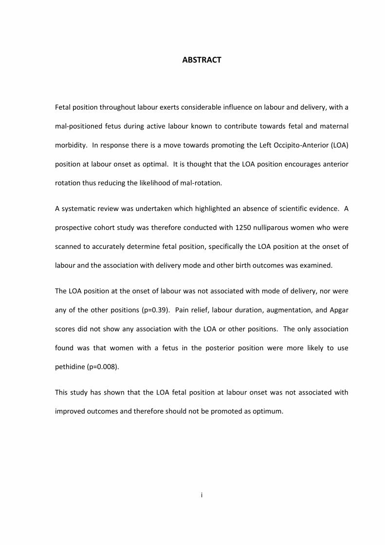
ABSTRACT
Fetal position throughout labour exerts considerable influence on labour and delivery, with a
mal-positioned fetus during active labour known to contribute towards fetal and maternal
morbidity. In response there is a move towards promoting the Left Occipito-Anterior (LOA)
position at labour onset as optimal. It is thought that the LOA position encourages anterior
rotation thus reducing the likelihood of mal-rotation.
A systematic review was undertaken which highlighted an absence of scientific evidence. A
prospective cohort study was therefore conducted with 1250 nulliparous women who were
scanned to accurately determine fetal position, specifically the LOA position at the onset of
labour and the association with delivery mode and other birth outcomes was examined.
The LOA position at the onset of labour was not associated with mode of delivery, nor were
any of the other positions (p=0.39). Pain relief, labour duration, augmentation, and Apgar
scores did not show any association with the LOA or other positions. The only association
found was that women with a fetus in the posterior position were more likely to use
pethidine (p=0.008).
This study has shown that the LOA fetal position at labour onset was not associated with
improved outcomes and therefore should not be promoted as optimum.

ii
DEDICATION
Dedicated to Dr Heather Winter (1959-2007) whose inspiration remained with me throughout my time spent on this work.
Dr Heather Winter supervised this work as my second supervisor for almost 3 years and provided invaluable advice and support. Dr Winter died in November 2007 from ovarian cancer, which had been diagnosed 10 years ago. During the period Dr Winter spent supporting my research I was unaware of her illness, highlighting the generosity she portrayed towards the support of others.
Dr Winter’s knowledge in obstetrics, gynaecology and public health guided my research and was closely linked to her own work of interest, that being improving the health of mothers in particular those living in adverse circumstances.
For me, Dr Winter was a realist, and during the period when my research was not achieving its planned targets, it was the words and realism that such was not good enough that drove the successful completion of what is, ‘our’ work.
Thank you.
iii
ACKNOWLEDGEMENTS
With thanks to my supervisors Professor Christine MacArthur, Professor Khalid Khan and Dr
Heather Winter for their patience, persistence and their invaluable support. Thanks to Alice
Sitch for providing me with her time and statistical support which made data analysis
possible.
Sincere appreciation to the sonographers at the Birmingham Women’s Hospital who helped
train and assess the midwives involved. The Royal College of Midwives, Birmingham
Women’s NHS Foundation Trust, West Midlands NWAHP and Mothercare for their support
with funding.
Jenny Henry who made changes happen and her continued support and belief in the
importance of midwifery research. Sara Webb, Bernadette Early and Salma Hussain whose
friendships and support proved invaluable. Angela McBennett, Stephanie Caves and
Bernadette Early for their persistence and hard work in achieving the required scans. To all
the women and midwives for their participation and for whom this study was done.
And finally, my husband Shafiq Ahmad who supported me patiently and to whom I have a lot
of lost time to make up.
Thank you.

iv
TABLE OF CONTENTS
Chapter 1. Background
1.0. Introduction............................................................................................................................. 1
1.1. Normal Obstetric Labour.......................................................................................................... 3
1.1.1. Descent ...................................................................................................................... 4
1.1.2. Flexion ........................................................................................................................ 5
1.1.3. Internal rotation ......................................................................................................... 6
1.1.4. Extension.................................................................................................................... 6
1.1.5. External rotation ........................................................................................................ 7
1.2. Fetal Mal-position .................................................................................................................... 8
1.3. Maternal and Fetal Outcomes Associated with Occipito-Posterior Mal-position ..................... 11
1.4. The Rising Rates of Intervention............................................................................................. 21
1.5. Jean Suttons ‘Optimal Fetal Positioning’ ................................................................................ 23
1.6. Evidence Underpinning OFP Theory ....................................................................................... 27
1.7. OFP Theory and Acceptance .................................................................................................. 29
1.8. The Apollo Study .................................................................................................................... 31
1.9. Summary ............................................................................................................................... 33
Chapter 2. Systematic Review
2.1. Introduction........................................................................................................................... 34
2.2. The Review Question: In pregnant woman, with a singleton fetus at term gestation, does fetal position at the onset of labour influence mode of delivery? .................................................. 35
2.3. Inclusion Criteria .................................................................................................................... 35
2.4. Search Strategy ...................................................................................................................... 35
2.5. Expert Contact ....................................................................................................................... 36
2.6. Electronic Search Strategy ..................................................................................................... 36
2.7. Study Selection/Quality Assessment ...................................................................................... 37

v
2.8. Results ................................................................................................................................... 39
2.9. Discussion .............................................................................................................................. 43
Chapter 3. Methodology
3.1. Study Design .......................................................................................................................... 46
3.2. Composition of Sample .......................................................................................................... 47
3.3. Method of measure ............................................................................................................... 48
3.4. Point of measure ................................................................................................................... 54
3.5. Definition of labour onset and period of measure ................................................................. 55
3.6. Follow-up .............................................................................................................................. 57
3.7. Pilot Study ............................................................................................................................. 58
Chapter 4. Methods
4.1. Aim of the Study .................................................................................................................... 62
4.1.1. Objectives ................................................................................................................ 62
4.2. Study Design .......................................................................................................................... 63
4.3. Ethics ..................................................................................................................................... 63
4.4. Setting ................................................................................................................................... 64
4.5. Participants and Inclusion Criteria ......................................................................................... 64
4.6. Recruitment ........................................................................................................................... 64
4.7. Determining Fetal Occiput and Fetal Spine Position by Trans-Abdominal Supra-Pubic Ultrasound ............................................................................................................................ 67
4.8. Fetal Position: Occiput ........................................................................................................... 68
4.9. Fetal Position: Spine .............................................................................................................. 69
4.10. Description of Ultrasound Technique Used to Determine Fetal Position ................................ 71
4.11. Study Implementation ........................................................................................................... 74
4.12. Obtaining a ‘Valid’ Scan ......................................................................................................... 75
4.13. Outcome Measures ............................................................................................................... 78
4.13.1. Primary Outcome ................................................................................................... 78

vi
4.13.2. Secondary Outcomes .............................................................................................. 79
4.14. Data Collection ...................................................................................................................... 81
4.15. Data Verification .................................................................................................................... 82
4.16. Sample Size ........................................................................................................................... 82
4.17. Data Analysis ......................................................................................................................... 84
Chapter 5. Results
5.1. Assembly of Study Cohort ...................................................................................................... 86
5.2. Maternal Characteristics ........................................................................................................ 88
5.3. Neonatal Characteristics ........................................................................................................ 91
5.4. Maternal Outcomes ............................................................................................................... 92
5.5. Neonatal Outcomes ............................................................................................................... 94
5.6. Distribution of Fetal Occiput Position ..................................................................................... 95
5.7. Distribution of Fetal Spine Position ........................................................................................ 96
5.8. Distribution of Combined Fetal Occiput and Spine Position .................................................... 97
5.9. Fetal Position and Mode of Delivery..................................................................................... 101
5.9.1. Mode of delivery with LOA or non-LOA fetal occiput position .................................... 102
5.9.2. Mode of delivery with fetal occiput cavity position.................................................... 103
5.9.3. Mode of delivery with fetal spine position................................................................. 105
5.9.4. Mode of delivery with LspA and non-LspA fetal spine position .................................. 107
5.9.5. Logistic regression analysis (fetal occiput position and mode of delivery) .................. 109
5.10. Labour Augmentation .......................................................................................................... 111
5.10.1. Labour augmentation with LOA and non-LOA fetal occiput position ..................... 113
5.11. Pain Relief ........................................................................................................................... 114
5.12. Labour Duration .................................................................................................................. 117
5.12.1. Duration of second stage ...................................................................................... 119
5.12.2. Duration of membrane rupture ............................................................................ 120
5.13. Neonatal Condition at Birth ................................................................................................. 121
5.13.1. Fetal occiput position and Apgar score at 1 minute .............................................. 121

vii
5.13.2. Apgar score at 5 minutes .................................................................................... 123
Chapter 6. Discussion and Conclusion
6.1. Main Results ........................................................................................................................ 125
6.2. Strengths and Weaknesses of Study ..................................................................................... 127
6.3. Interpretation of Findings .................................................................................................... 132
6.4. Implication for Practice ....................................................................................................... 142
6.5. Generalisabilty ..................................................................................................................... 143
6.6. Recommendations for Further Research .............................................................................. 145
Conclusion ..................................................................................................................................... 147
Appendix 1..................................................................................................................................... 149
Appendix 2..................................................................................................................................... 152
Appendix 3..................................................................................................................................... 155
Appendix 4..................................................................................................................................... 160
Appendix 5..................................................................................................................................... 164
Appendix 6..................................................................................................................................... 166
References .................................................................................................................................... 167

vii
TABLE OF ILLUSTRATIONS
Chapter 1.
Figure 1.1 Decent and engagement in the transverse position .................................................... 5
Figure 1.2 Complete flexion of fetal head .................................................................................... 5
Figure 1.3 Complete internal rotation to anterior posterior position ........................................... 6
Figure 1.4 Extension and delivery of fetal head ........................................................................... 7
Figure 1.5 Dextro-rotation of the lower uterine segment .......................................................... 25
Figure 1.6 The Apollo Study Objectives...................................................................................... 32
Chapter 2.
Figure 2.1 Process of systematic review and results .................................................................. 38
Chapter 4.
Figure 4.1 Classification of fetal occiput position within the 360° pelvic cavity........................... 69
Figure 4.2 Classification of fetal spine position within the 360° abdominal cavity ...................... 70
Figure 4.3 Occiput identification................................................................................................ 71
Figure 4.4 Recording fetal occiput position ................................................................................ 72
Figure 4.5 Spine identification ................................................................................................... 73
Figure 4.6 Recording fetal spine position ................................................................................... 73
Figure 4.7 Study flow following admission ................................................................................. 77
Chapter 5.
Figure 5.1 Recruitment of study sample .................................................................................... 87
Figure 5.2 Distribution of score used to calculate Kappa Statistic ............................................ 100
Chapter 6.
Figure 6.1 Degree of occiput rotation ...................................................................................... 140
viii
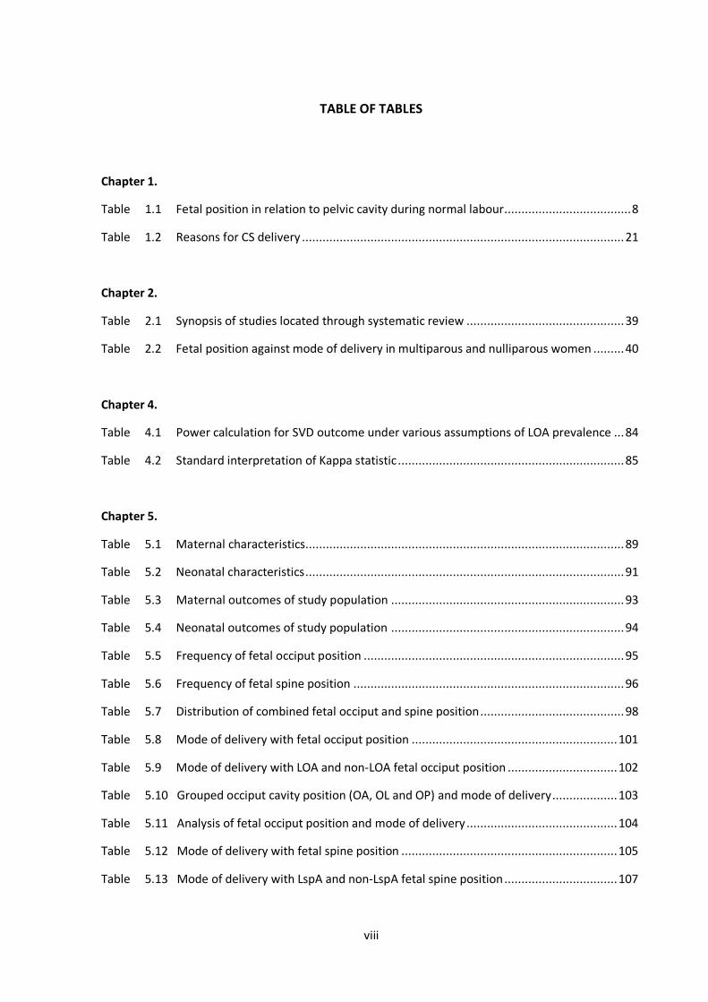
TABLE OF TABLES
Chapter 1.
Table 1.1 Fetal position in relation to pelvic cavity during normal labour ..................................... 8
Table 1.2 Reasons for CS delivery .............................................................................................. 21
Chapter 2.
Table 2.1 Synopsis of studies located through systematic review .............................................. 39
Table 2.2 Fetal position against mode of delivery in multiparous and nulliparous women ......... 40
Chapter 4.
Table 4.1 Power calculation for SVD outcome under various assumptions of LOA prevalence ... 84
Table 4.2 Standard interpretation of Kappa statistic .................................................................. 85
Chapter 5.
Table 5.1 Maternal characteristics............................................................................................. 89
Table 5.2 Neonatal characteristics ............................................................................................. 91
Table 5.3 Maternal outcomes of study population .................................................................... 93
Table 5.4 Neonatal outcomes of study population .................................................................... 94
Table 5.5 Frequency of fetal occiput position ............................................................................ 95
Table 5.6 Frequency of fetal spine position ............................................................................... 96
Table 5.7 Distribution of combined fetal occiput and spine position .......................................... 98
Table 5.8 Mode of delivery with fetal occiput position ............................................................ 101
Table 5.9 Mode of delivery with LOA and non-LOA fetal occiput position ................................ 102
Table 5.10 Grouped occiput cavity position (OA, OL and OP) and mode of delivery ................... 103
Table 5.11 Analysis of fetal occiput position and mode of delivery ............................................ 104
Table 5.12 Mode of delivery with fetal spine position ............................................................... 105
Table 5.13 Mode of delivery with LspA and non-LspA fetal spine position ................................. 107

ix
Table 5.14 Analysis of fetal spine position and mode of delivery .............................................. 108
Table 5.15 Logistic regression analysis (SVD and fetal occiput position) .................................... 109
Table 5.16 Regression Analysis- Modelling against trends of known association ....................... 111
Table 5.17 Labour augmentation and fetal occiput position ..................................................... 112
Table 5.18 Labour augmentation with LOA and non-LOA fetal occiput position ........................ 113
Table 5.19 Pain relief and fetal occiput position ....................................................................... 114
Table 5.20 Chi-squared analysis of individual forms of pain relief against all fetal positions ..... 116
Table 5.21 Grouped cavity fetal occiput position (OA, OL, and OP) and pethidine use .............. 116
Table 5.22 Pethidine with LOA against non-LOA fetal occiput position ..................................... 117
Table 5.23 Duration of active first stage and fetal occiput position ........................................... 118
Table 5.24 Duration of second stage of labour and fetal occiput position ................................. 119
Table 5.25 Duration of membrane rupture and fetal occiput position ...................................... 120
Table 5.26 Apgar score at 1 minute and fetal occiput position.................................................. 121
Table 5.27 Apgar score at 1 minute with cavity grouped fetal positions ................................... 122
Table 5.28 Apgar score at 5 minutes and fetal occiput position ................................................ 123
Table 5.29 Apgar score at 5 minutes with cavity grouped fetal position ................................... 124
Chapter 6.
Table 6.1 Study results on prevalence of grouped fetal positions and point of measure. ......... 135
Table 6.2 Study results on prevalence of individual occiput positions ...................................... 138
Table 6.3 Study results on prevalence of cavity grouped left and right fetal positions .............. 139

x
LIST OF ABBREVIATIONS
APOLLO Analysis of fetal Position at the Onset of Labour and Labour Outcomes
ARM Artificial Rupture of Membranes
BMI Body Mass Index
BWHCT Birmingham Women’s NHS Foundation Trust
CI Confidence Intervals
CS Caesarean Section
DOA Direct Occipito-Anterior
DOH Department of Health
DOP Direct Occipito-Posterior
DspA Direct spine Anterior
DspP Direct spine Posterior
FD Forceps Delivery
GA General Anaesthetic
Hrs Hours
IUD Intra-Uterine Death
Kg Kilograms
LOA Left Occipito-Anterior
LOL Left Occipito-Lateral
LOP Left Occipito-Posterior
LspA Left spine Anterior
LspL Left spine Lateral
LspP Left spine Posterior
MeSH Medical Subject Headings

xi
Mins Minutes
NCT National Childbirth Trust
NICE National Institute for Health and Clinical Excellence
OA Occipito-Anterior
OFP Optimal Fetal Positioning
OL Occipito-Lateral
OP Occipito-Posterior
OR Odds Ratio
OT Occipito-Transverse
POP Persistent Occipito-Posterior
R/LOT Right/Left Occipito-Transverse
ROA Right Occipito-Anterior
ROL Right Occipito-Lateral
ROP Right Occipito-Posterior
RR Relative Risk
RspA Right spine Anterior
RspL Right spine Lateral
RspP Right spine Posterior
SD (sd) Standard Deviation
SVD Spontaneous Vaginal Delivery
TENS Transcutaneous Electrical Nerve Stimulation
UK United Kingdom
US United Stated
VD Ventouse Delivery
WHO World Health Organisation
WM NMAHP West Midlands Nursing, Midwifery and Allied Health Professionals
xii
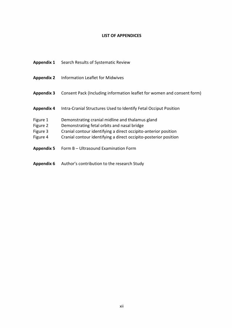
LIST OF APPENDICES
Appendix 1 Search Results of Systematic Review
Appendix 2 Information Leaflet for Midwives
Appendix 3 Consent Pack (Including information leaflet for women and consent form)
Appendix 4 Intra-Cranial Structures Used to Identify Fetal Occiput Position Figure 1 Demonstrating cranial midline and thalamus gland Figure 2 Demonstrating fetal orbits and nasal bridge Figure 3 Cranial contour identifying a direct occipito-anterior position Figure 4 Cranial contour identifying a direct occipito-posterior position Appendix 5 Form B – Ultrasound Examination Form
Appendix 6 Author’s contribution to the research Study
1
INTRODUCTION
There has been over the past few decades a decline in the rate of spontaneous vaginal birth
and an increase in the inability of women to give birth without medical management and
intervention (Savage, 2007). This has resulted in an increase in both operative vaginal and
abdominal delivery rates. In an attempt to limit such interventions and associated morbidity
various approaches to the management of care are being implemented. One such approach
proposed by Jean Sutton which is gaining acceptance amongst maternity associated
organisations is fetal positioning at labour onset, this is thought to increase the rates of
Spontaneous Vaginal Delivery (SVD). The underlying relationship however, between fetal
position at labour onset and associated outcomes is not properly understood. The author
met with Sutton at a ‘normal birth’ conference, and considered that the theory should be
further investigated. The author then studied the theory in depth and attended many of the
conferences and presentations performed by Sutton; at times the author presented Sutton’s
theory at alternative conferences in order to debate the plausibility of the theory. The
general opinion and criticism was clearly based on the lack of scientific evidence.
This thesis therefore examined the association between fetal position at labour onset and
mode of delivery to asses specifically the theory of Optimal Fetal Positioning (OFP) as
devised by Jean Sutton. OFP advocates the use of maternal exercises to manipulate the
fetus into the Left Occipito-Anterior (LOA) position as the fetal position considered optimum
for achieving SVD (Sutton, 2007). Sutton considers that this provides an explanation to the

2
decreasing rates of SVD and offers OFP as a solution to the observed problems (Sutton,
2001).
A large prospective cohort study was performed to investigate the influence of fetal position
at the onset of labour on delivery outcomes supported by a systematic review of the
literature.
3
CHAPTER 1
BACKGROUND
1.1. Normal Obstetric Labour
The complete process of labour and delivery in the main defines the physiological transition
from intra-uterine to extra-uterine life for the fetus, and from pregnant women to mother
for the labouring female. The knowledge of normal labour as a physiological event is
primarily based on the 18th century contributions of Fielding Old, William Smellie and Solares
de Renhac (Dietze, 2001). This is largely due to the ethical issues associated with replicating
the work they undertook, and has consequently resulted in our understanding of labour
mechanism as we know it today. Mechanism of labour as understood and depicted in many
textbooks is described as essentially a mechanical process, whereby the fetus is expelled
from the uterus with the aid of uterine contractions (Tiran, 2008, McCormick, 2008). The
fetus has always been described as an active participant that must undergo a series of
intricate movements as it descends into the maternal pelvis.
The process of birth is underpinned by the position of the fetus and the movements it
adopts in order to birth spontaneously. Fetal position is defined as the relationship of a
particular landmark of the fetus, which in the normal instance is the fetal occiput, in relation
to the landmarks of the female pelvis (Akmal and Paterson-Brown, 2009). Labour
4
mechanism is mapped according to this relationship and is how the process of normal labour
is described. The processes of labour mechanism is however complex and may give rise to
many points at which deviation from the norm may arise.
To give birth spontaneously the fetus must undergo five distinct movements during the
process of labour and delivery as it passes through the maternal pelvis (Caruthers, 2000).
The five movements (McCormick, 2008, Caruthers, 2000, Cunningham et al., 2008) are
recognised as:
Descent
Flexion
Internal rotation
Extension
External rotation
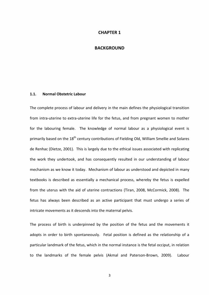
1.1.1. Descent
The fetus begins its journey with descent in order to achieve engagement of the presenting
part into the true pelvis which commences at the pelvic brim or pelvic inlet (Selman and
Johnston, 2010). The description of mechanism of labour is depicted with engagement
taking place when the transverse diameter of the fetal head has passed through the
transverse diameter of the pelvic brim (McCormick, 2008, Tiran, 2008). This suggests that
engagement only takes place in the transverse plane as it is where the widest pelvic
diameter is available for the widest part of the fetal head (Fig. 1.1).
5
Figure 1.1. Decent and engagement in the transverse position (Faulkner, 2009).
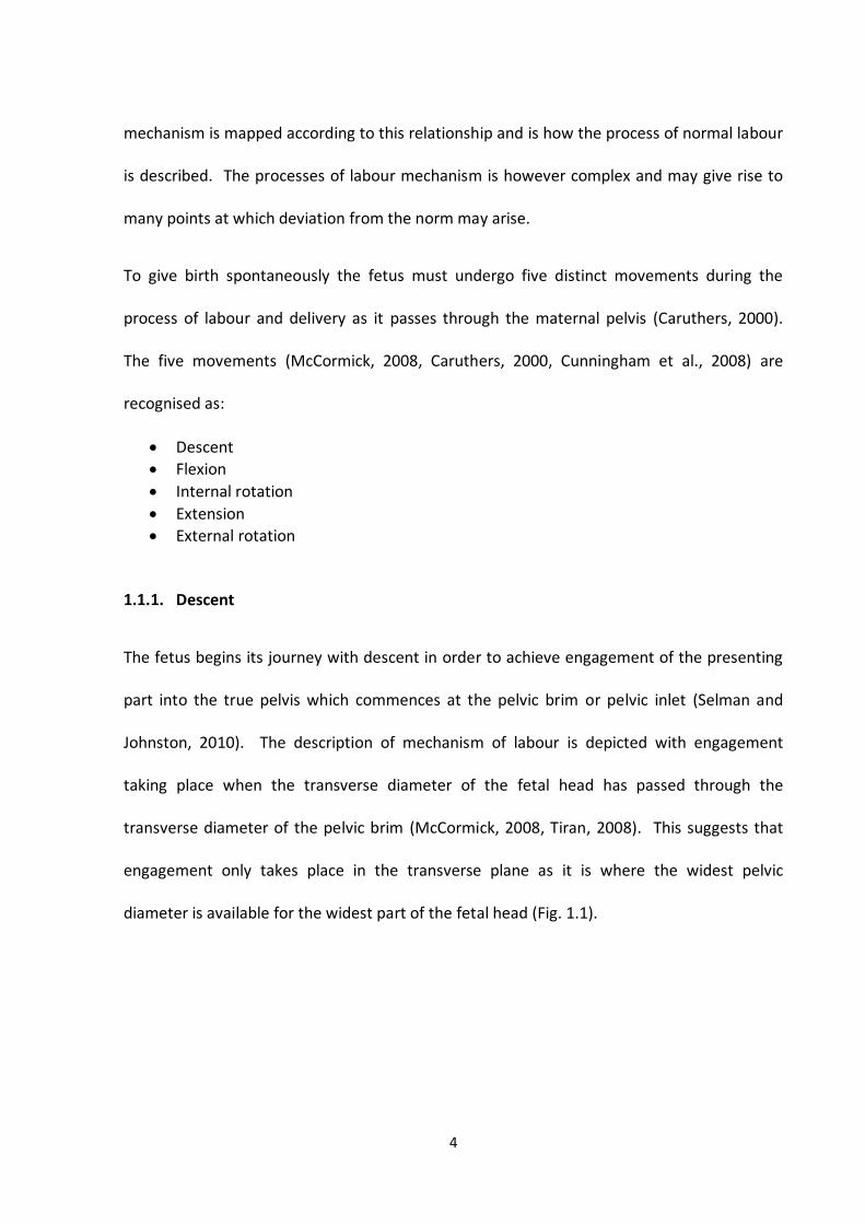
1.1.2. Flexion
This is followed by flexion, which takes place whilst decent into the mid-cavity of the pelvis is
occurring (Caruthers, 2000). Complete flexion is achieved when the fetal chin is resting on
the fetal chest. By achieving complete flexion, the fetal occiput becomes the leading part
ensuring that the smallest diameter of the fetal head is leading (Selman and Johnston, 2010).
The position of the fetal head remains in the transverse plane during the process of descent
and flexion (Fig 1.2).
Figure 1.2. Complete flexion of fetal head (Mathai et al., 2000)
6
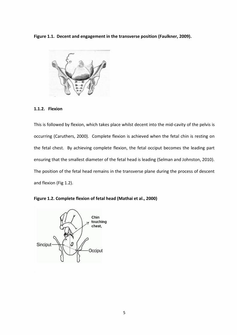
1.1.3. Internal rotation
Flexion is then followed by 90° internal rotation of the fetal head, which takes place
between the mid-cavity and pelvic floor (McCormick, 2008). During this stage the fetal head
assumes an oblique or in the majority of cases an anterior posterior position, ready for the
expulsive phase of labour (Fig. 1.3) (Dietze, 2001).
Figure 1.3. Complete internal rotation to anterior posterior position (Faulkner, 2009).
1.1.4. Extension
As the fetal head reaches the pelvic floor, extension of the fetal head commences along the
sacral curve (McCormick, 2008, Caruthers, 2000). The fetal chin no longer rests on the fetal
chest and instead it extends as movement along the perineal body achieves delivery of the
fetal head (Fig. 1.4).
7
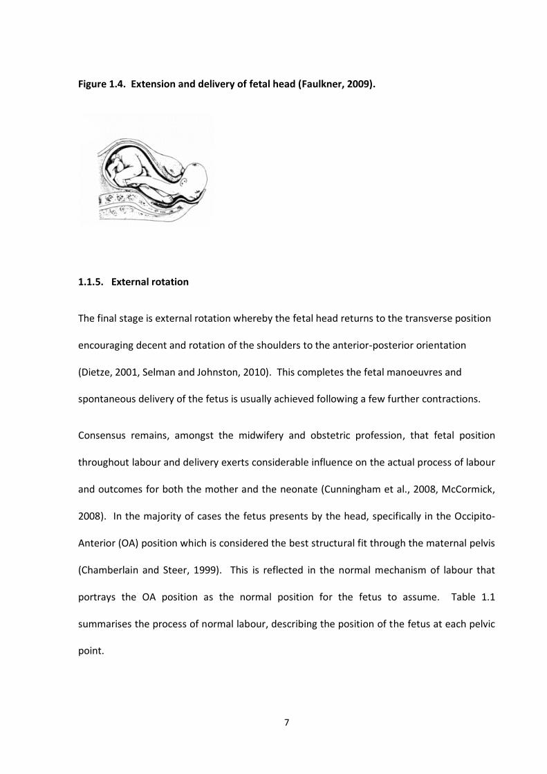
Figure 1.4. Extension and delivery of fetal head (Faulkner, 2009).
1.1.5. External rotation
The final stage is external rotation whereby the fetal head returns to the transverse position
encouraging decent and rotation of the shoulders to the anterior-posterior orientation
(Dietze, 2001, Selman and Johnston, 2010). This completes the fetal manoeuvres and
spontaneous delivery of the fetus is usually achieved following a few further contractions.
Consensus remains, amongst the midwifery and obstetric profession, that fetal position
throughout labour and delivery exerts considerable influence on the actual process of labour
and outcomes for both the mother and the neonate (Cunningham et al., 2008, McCormick,
2008). In the majority of cases the fetus presents by the head, specifically in the Occipito-
Anterior (OA) position which is considered the best structural fit through the maternal pelvis
(Chamberlain and Steer, 1999). This is reflected in the normal mechanism of labour that
portrays the OA position as the normal position for the fetus to assume. Table 1.1
summarises the process of normal labour, describing the position of the fetus at each pelvic
point.
8
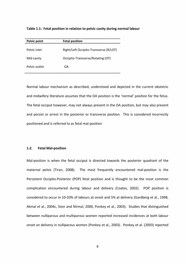
Table 1.1: Fetal position in relation to pelvic cavity during normal labour
Pelvic point Fetal position
Pelvic inlet
Right/Left Occipito-Transverse (R/LOT)
Mid-cavity Occipito-Transverse/Rotating (OT)
Pelvic outlet
OA
Normal labour mechanism as described, understood and depicted in the current obstetric
and midwifery literature assumes that the OA position is the ‘normal’ position for the fetus.
The fetal occiput however, may not always present in the OA position, but may also present
and persist or arrest in the posterior or transverse position. This is considered incorrectly
positioned and is referred to as fetal mal-position
1.2. Fetal Mal-position
Mal-position is when the fetal occiput is directed towards the posterior quadrant of the
maternal pelvis (Tiran, 2008). The most frequently encountered mal-position is the
Persistent Occipito-Posterior (POP) fetal position and is thought to be the most common
complication encountered during labour and delivery (Coates, 2002). POP position is
considered to occur in 10-20% of labours at onset and 5% at delivery (Gardberg et al., 1998,
Akmal et al., 2004c, Sizer and Nirmal, 2000, Ponkey et al., 2003). Studies that distinguished
between nulliparous and multiparous women reported increased incidences at both labour
onset an delivery in nulliparous women (Ponkey et al., 2003). Ponkey et al. (2003) reported
9
that the incidence of Occipito-Posterior (OP) deliveries in nulliparous women (7.2%) were
almost double compared to multiparous women (4.0%).
The cause of fetal mal-position remains unclear with the most plausible explanations being
physical inhibitors and mechanistic deviation. It is thought that pelvic types may predispose
fetuses to adopt a posterior fetal position, particularly those that have a narrowed fore-
pelvis or a flat sacrum such as the android and anthropoid pelvises (Sweet, 1997). It is also
argued that a de-flexed fetal head may cause mal-position (Coates, 2002). Some studies
have shown that an anterior positioned placenta may contribute towards a posterior fetal
position, although other studies have not reported similar findings (Gardberg and
Tuppurainen, 1994). The most common rationale is that of mechanical deviation, where
mal-rotation is thought to be the primary cause of mal-position or a POP fetal position
(Chamberlain and Steer, 1999). The reasons as to why some fetuses adopt the posterior
position remains unclear and it maybe that no single cause explains the reason for mal-
position with varying characteristics predisposing fetuses to the posterior position.
The posterior fetal position is understood to be the less favoured position as the process of
labour and delivery is often challenged with varying clinical complications. Once
engagement has taken place in the posterior rather than the OA position, labour mechanism
is altered from the point of labour onset (Gardberg et al., 1998). For the posterior fetus to
engage it must do so with a deflexed head, unlike the OA position which engages in the
flexed position. In the OP position the maternal spine acts as an inhibitor preventing the
head from flexing sufficiently to allow the chin to rest on the chest (Selman and Johnston,
2010). The OA position achieves this with ease and ensures that the smallest diameter of
10
the fetal head becomes the leading part. In the OP position the lack of flexion encourages
the largest diameter of the fetal head to present and descend as the presenting part (Faber,
1998). The deflexed head with the larger diameter does not fit the pelvis well or position
itself centrally over the cervix (Biancuzzo, 1993). Since the deflexed head of the OP fetus
rests anteriorly over the cervix rather than resting centrally, contractions are not effectively
stimulated. In turn this causes poor uterine activity that leads to both delayed descent and
uneven and slow cervical dilatation (Chamberlain and Steer, 1999, Selman and Johnston,
2010).
Eventually, when cervical dilation is achieved the increased diameter of the presenting part
is 2.5cm larger and to achieve vaginal delivery the fetal head must mould significantly and
the perineum must stretch more than is necessary with an OA positioned fetus (Coates,
2008). This then increases the need for operative delivery and causes greater perineal
trauma (Benavides et al., 2005).
The mechanism of the OP fetus is based on theory as to what ‘may’ happen when the
anatomy and physiology of the OP fetus is considered. No scientific evidence exists on the
posterior mechanism of labour. Scientific evidence does however exist in relation to both
the maternal and fetal morbidity associated with a posterior fetal mal-position compared to
the favoured OA fetal position. It is thought that morbidity is associated with the
interventions that become necessary in order to salvage the process and achieve a safe
delivery when the fetus persists in the OP position (Ridley, 2007).
11
1.3. Maternal and Fetal Outcomes Associated with Occipito-Posterior Mal-position
The process of labour and delivery involves the fetus adopting intricate movements as it
descends the maternal pelvis. The movements the fetus adopts are vital if SVD is to be
achieved, in the OP fetus the direction of movement is altered giving rinse to mal-rotation
resulting in less favoured outcomes. A number of studies have investigated maternal and
fetal outcomes associated with the OP position.
Sizer and Nirmal (2000) conducted a study of 16,781 nulliparous women at term gestation,
with singleton cephalic fetuses during the period of 1990-1998. The study was a
retrospective review of health records to determine if there were differences in obstetric
outcomes between the OP and the OA deliveries, the main outcomes being mode of delivery
and Apgar scores at 5 minutes. Secondary outcomes were maternal age, induction, epidural
use, augmentation by oxytocic drugs and neonatal birth weight (Sizer and Nirmal, 2000). OP
was defined as delivery of ‘face-to-pubes’ and delivery where rotation of an OP to an OA
position was undertaken prior to delivery. The frequency of the OP position at delivery was
4.7%. Mode of delivery associated with fetal position was significantly different, the rate of
SVD for women who delivered an OA fetus was 61.8% and 14.6% for those who delivered an
OP fetus (p=0.001). The instrumental delivery rate was 24.4% for the OA group and 43.7%
for the OP group (p=0.001) and the emergency CS rate for the OA group was 13.7%
compared to 41.7% for the OP group (p=0.001). Sizer and Nirmal (2000) also found an
increased association between the use of epidural analgesia in labour and oxytocic
augmentation of the OP fetus, apgar scores at 5 minutes (<8) were not significantly different
and nor were increasing maternal age, induction of labour and gestational age. Sizer and
12
Nirmal’s (2000) study suggested that delivery in the OP position was associated with
increased morbidity for the mother although not for the neonate.
Pearl et al. (1993) conducted a study in the United States (US) to investigate the association
between OP delivery and maternal and fetal morbidity using a larger number of neonatal
outcomes than the previous study (Pearl et al., 1993). The study was retrospective and
compared 564 vaginal OP deliveries with 1068 OA controls matched for race, parity, delivery
method and neonatal birth weight. Computerised patient medical databases were used to
obtain data on maternal and neonatal outcomes, including delivery mode. Pearl et al. (1993)
did not outline specifically how position of the fetus was determined but since retrospective
review of medical records were used it is presumed that position documented by the
attending clinician at delivery was used. The OP position at delivery was found to be
associated with a longer second stage of labour despite fetuses being matched for
equivalent birth weight (p<0.05) and an increased incidence of episiotomy and severe
perineal lacerations that extended to include third and fourth degree perineal lacerations
(p<0.05). Blood loss following delivery was also increased, and women who had OP
deliveries had longer hospital stays compared to women who had OA deliveries (p<0.05)
(Pearl et al., 1993). Neonates who had operative deliveries in the OP position were more
likely to suffer from facial nerve and Erb’s palsy (p<0.05). The overall rates of facial nerve
and Erb’s palsy were 1.3 and 0.3 per 1000 live births, but OP fetuses were examined 12.4
and 5.3 per 1,000 live births. Despite this and the OP fetuses having a higher frequency of
fetal distress, Apgar scores, cord gas pH and neonatal intensive care admissions were no
different from the OA group (Pearl et al., 1993).
13
The study concluded that delivery of the OP position per se was associated with increased
maternal morbidity. Increased fetal morbidity for the OP position was associated with
operative OP delivery (Pearl et al., 1993).
The study highlighted important information relating to fetal malposition, however the
generalisability of the study to the population in the United Kingdom (UK) is uncertain since
the medical system and practices in the US vary considerably from those in the UK. Certain
findings that were reported may be linked or at the very least influenced by the obstetric
practices in the US, rather than sole association with fetal OP positioning.
The increased incidence of episiotomy and severe perineal trauma in the form of anal
sphincter and rectal tissue damage may be related to the preferred choice of instrument in
the study population which was said to be the forceps (Pearl et al., 1993). 76% of the
instrumental deliveries in the study were forceps deliveries with only 24% of deliveries being
ventouse. Forceps deliveries are generally associated with increased perineal trauma, need
for episiotomy and neonatal injury comparative to other forms of vaginal delivery (Johanson
and Menon, 2009). Comparatively in the UK, ventouse is now the favoured choice of
instrument rather than the forceps (Johanson and Menon, 2009). The second difference is
the type of episiotomy performed, which in the UK is usually medio-lateral which are less
likely to extend and cause further trauma (Downe, 2008). In Pearl et al.’s (1993) study the
vast majority of women had median episiotomies which are more likely to extend and cause
third and fourth degree tears (Downe, 2008). It is, therefore difficult to draw firm
conclusions and generalise the study findings to the UK population.
14
Fitzpatrick et al. (2001) undertook an observational study in Ireland comparing outcomes of
246 women with POP positions at delivery with 13,543 vaginal deliveries of OA positioned
fetuses. They found that the incidence of the OP position at delivery was 1.8% overall, 2.4%
in nulliparous and 1.3% in multiparous women. Position was defined as that observed at
delivery or that diagnosed on vaginal examination prior to delivery by the attending clinician
(Fitzpatrick et al., 2001). The study found that significantly more women in the OP group
had a prolonged pregnancy (p<0.01) and were more likely to be induced compared to the
OA group (p<0.001). They also found that the use of oxytocic drugs to augment labour was
significantly higher in the OP group than the OA group (52% vs. 32%, p<0.001) and prolonged
labour of greater than 12 hours was more common amongst the OP group (12% vs. 1.7%,
p=0.001). The rates of both instrumental delivery and operative delivery were significantly
higher in the OP group than the OA group (p<0.001). The SVD rate for the OA fetuses was
84% overall (inclusive of nulliparous and multiparous women) whilst the SVD rate for the OP
fetuses was 29% in nulliparous and 55% in multiparous women (p<0.001).
Fitzpatrick et al.’s (2001) study suggested a significant increase in maternal morbidity
associated with the OP position at delivery. Maternal perineal trauma was significantly more
common in the OP group than the OA group in both the nulliparous and multiparous women
(p<0.001). Although the incidence of episiotomy was similar in both OA and OP groups, the
OP group had significantly greater risk of anal sphincter injury following instrumental
delivery (p<0.001). Neonatal condition at delivery was similar in both the OA and the OP
groups, showing no difference in the Apgar scores.
15
There are, however, limitations to Fitzpatrick et al.’s (2001) findings including the potential
for misclassification bias as the study relied on reported position at delivery by the attending
clinician. Nonetheless, previously discussed studies used the same method to determine
position so a similar criticism of those studies would be justified. In Fitzpatrick et al.’s (2001)
study position, in some cases, was identified by vaginal examination prior to delivery. This is
despite reported criticism with the associated inaccuracy of digital vaginal examination to
determine fetal position (Akmal et al., 2003). Furthermore the institution in which the study
was carried out followed an active management policy for nulliparous women but not for
multiparous women. This may suggest that some of the differences reported between the
nulliparous and multiparous groups may actually be influenced by labour management and
not OP positioning. Although the study by Fitzpatrick et al. (2001) has drawn strong
conclusions, it has identifiable flaws that must be considered when interpreting and applying
the findings.
Ponkey et al. (2003) conducted a retrospective cohort study including 6434 consecutive,
term, cephalic, nulliparous and multiparous women and compared maternal and neonatal
outcomes of those delivered in the OA position against those who delivered in the OP
position.
Ponkey et al. (2003) found the rate of OP fetuses at delivery for nulliparous women was
almost double compared to multiparous women (7.2% vs. 4% p<0.001), concluding that OP
position at delivery was more common in nulliparous women. Further significant differences
between the OA and OP groups were:
16
Prolonged first stage (48.3% OP vs. 30.3% OA, p<0.001).
Prolonged second stage (53.3% OP vs. 18.1% OA, p<0.001).
Increased oxytocin augmentation (48.9% OP vs. 36.8% OA, p<0.001).
Increased use of epidural analgesia (86.1% OP vs. 73.1% OA, p<0.001).
Chorioamnionitis (4.7% OP vs. 1.1% OA, p<0.001).
Assisted vaginal delivery (24.6% OP vs. 9.4% OA, p<0.001).
Caesarean delivery (37.7% OP vs. 6.6% OA, p<0.001).
Third and fourth degree perineal laceration (18.2% OP vs. 6.7% OA, p<0.001).
Excessive post partum blood loss (13.6% OP vs. 9.9% OA, p<0.03).
1 minute Apgar score less than 7 (12.4 % OP vs. 7.1% OA, p<0.001).
The study however did not find low Apgar scores at 5 minutes to differ between the OA and
the OP groups. Furthermore when the analysis was performed separately for nulliparous
and multiparous women, duration of second stage, epidural use and mode of delivery were
the only outcomes that remained significantly different between the OA and OP groups
amongst multiparous women. For nulliparous women differences for all the outcomes
remained significant, suggesting that the OP position for nulliparous women was more likely
to be associated with resulting morbidity (Ponkey et al., 2003).
The limitation of this study is that it was conducted in the US, where as stated earlier,
practice is different to that in the UK. Furthermore the retrospective review of medical
databases gives rise to issues of data verification whereby the accuracy of the data cannot
be determined.
17
Senecal et al. (2005) undertook secondary analysis from data gathered for a randomised
controlled trial for delayed and early pushing with epidural analgesia in order to assess the
effect of fetal mal-position on second stage progress amongst nulliparous women. Position
at delivery was taken as that documented by the attending clinician at delivery. A total of
1608 women were included in the analysis and fetal position was categorised into 3 groups,
OA, OP and OT. They found no association between fetal position and gestational age and
duration of first stage of labour or the use of episiotomy, but did find an association
between OP position at delivery and increased incidence of the following compared to the
OA and OT position:
Oxytocic augmentation ( p<0.001)
Longer second stage duration (p<0.001)
Postpartum blood loss greater than 500mls (p<0.002)
Third and fourth degree perineal trauma ( p<0.001)
Caesarean delivery (p<0.001)
Mid-cavity assisted vaginal delivery (p<0.001)
Neonatal outcomes including low Apgar, abnormal umbilical cord gasses, admission to
intensive care were not associated with an OP position (Senecal et al., 2005).
What appears consistent in most studies is that the OP position is associated with increased
rates of caesarean delivery, instrumental delivery and perineal trauma in the form of third
and fourth degree lacerations (Pearl et al., 1993, Senecal et al., 2005, Ponkey et al., 2003,
Fitzpatrick et al., 2001).
18
Obstetric anal sphincter injury affects 8-18% of women which may lead to incontinence or
associated symptoms creating a debilitating condition (Benavides et al., 2005). Benavides et
al. (2005) conducted a retrospective cohort study to compare the incidence of perineal
trauma in OA and OP forceps assisted vaginal deliveries. The primary outcome was the
occurrence of third and fourth degree perineal lacerations and the position of the fetal head
was that as documented in the delivery notes, classified into OA or OP. For cases where
rotational forceps were used, position was defined as the position after rotation was
completed. A total of 588 forceps deliveries were analysed and it was found that 35% of
forceps-assisted vaginal deliveries resulted in either a third or fourth degree perineal
laceration. Interestingly however, the majority of forceps deliveries were performed from
an OA position and not from an OP position (88.4%, 11.6%). Despite this, perineal trauma
linked with anal sphincter damage was significantly associated with the OP position at
delivery (OP 51.5% vs. OA 32.9% p=0.003). To further determine if the OP position was an
independent risk factor for anal sphincter damage, Benavides et al. (2005) applied a logistic
regression model to control for known confounding variables. The OP position was 3.1 times
more likely to be associated with anal sphincter injury, concluding that the OP position was
independently associated with such damage (Benavides et al., 2005).
Wu et al. (2005) undertook a similar study to compare the incidence of anal sphincter
damage between OA and OP positioned fetuses in 393 vacuum assisted deliveries. The rate
of third and fourth degree perineal lacerations in this group was 24.4% overall and the OP
group had a significantly higher rate of anal sphincter damage compared to the OA group
(41.7% vs. 22%, p=0.003). Wu et al. (2005) also undertook logistic regression analysis which
19
demonstrated that OP was 4 times more likely to be associated with anal sphincter injury
compared to OA position.
Studies investigating OP positioned deliveries clearly suggest that adverse maternal
outcomes from an OP positioned fetus at delivery are increased. The specific areas that are
consistent include:
Decreased incidence of SVD and increased incidence of instrumental/operative
delivery (Ponkey et al., 2003, Sizer and Nirmal, 2000, Pearl et al., 1993, Gardberg et
al., 1998).
Increased incidence of anal sphincter and rectal tissue damage (Benavides et al.,
2005, Wu et al., 2005).
Implications on neonatal morbidity remain less conclusive. Cheng et al. (2006) conducted a
retrospective study of women at term gestation with cephalic, live, singleton fetuses over a
26 year period. The study included the analysis of a total of 31,392 neonates. Position of
the fetus was determined at delivery by the attending clinician, and categorised into OA or
OP position with OT position excluded. The outcomes investigated to determine association
between OP position and neonatal morbidity were Apgar score <7 at 5 minutes, umbilical
cord gasses (pH < 7 and base excess ≤-12), Meconium stained liquor, Meconium aspiration
and admission to intensive care.
The study found that the prevalence of the OP position at delivery was 8.2% overall, with
nulliparous women having an increased prevalence compared to multiparous women
(10.3%, 6%). The results in relation to the outlined neonatal outcomes were as follows:
20
OP position had significantly more neonates with Apgar scores <7 at 5 minutes
(3.8% OP, 1.9% OA, p<0.001).
OP position had a higher rate of umbilical cord gasses pH < 7 and base excess below
-12 (1.8% OP, 0.5% OA, p<0.001).
Meconium stained liquor was more common in the OP group (32.3% OP, 22.7% OA,
p<0.001)
Meconium aspiration was more common in the OP group (1.2% OP, 0.7% OA,
p<0.001).
Admission to intensive care was higher for the OP group (10.7% OP, 6% OA,
p<0.001).
Further analysis to control for confounding was undertaken and all the above differences
remained statistically significant. To examine association between neonatal morbidity and
maternal parity the analysis was then stratified by parity revealing that in both the
nulliparous and the multiparous groups the OP position still had significantly higher rates of
undesirable outcomes for the neonate (Cheng et al., 2006). The study concluded that
neonates in the OP position compared to those born in the OA position were associated with
an increased risk of short term neonatal morbidity. Cheng et al. (2006) considered that
previous studies had not observed similar differences due to a lack of statistical power
rather than an absence of association and that their large sample allowed these associations
to be identified.
The association of maternal and neonatal morbidity with fetal malposition is apparent from
the studies reported. What is thus becoming evident is that increasing rates of intervention
are necessary to manage the associated risk of morbidity. Interventions however are also
associated with varying degree of risk and consequently such is being debated as to the
appropriateness of use.
21
1.4. The Rising Rates of Intervention
The interventions used in everyday obstetrics have generated many debates as to the cause
and perceived advantages of such interventions and the increasing rates of use (Sharma et
al., 2009). The greatest debate has been that of the rising caesarean section (CS) rate, as the
associated morbidity and mortality for the mother and the neonate is well acknowledged
(NICE, 2004). CS is known to be associated with increased:
Abdominal pain
Bladder injury
Urethral injury
Need for further surgery
Hysterectomy
ITU/ HDU admission
Thromboembolic disease
Increased duration of hospital stay
Readmission to hospital
Placenta previa
Uterine rupture
Maternal death
Ante partum stillbirth
Not having more children
Neonatal respiratory morbidity
Furthermore, during the triennium of 1997-1999 the fatality rate of direct deaths following
caesarean delivery was almost five times greater than for vaginal delivery and twelve times
greater for emergency caesarean delivery, thus highlighting the severity of risk associated
with CS (DOH, 2000).
The proportion of SVD births has fallen steadily from 78% in 1989 to 66% in 2004-2005
(DOH, 2005). Concurrently the rate of CS deliveries has continued to increase from a rate of
under 3% in the 1950’s, 9% in 1980, to 12% in 1990 followed by a rapid increase to 21% in
2000 (DOH, 2002). The CS rate has increased steadily further to a level of 24.8% (DOH,
2010). Reasons for the increasing rates of CS delivery remain undetermined although some
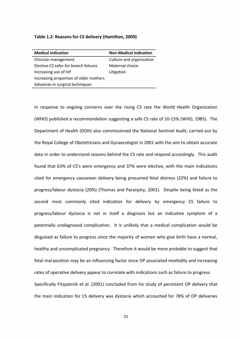
medical and non-medical reasons have been highlighted (Table 1.2).
21
Table 1.2: Reasons for CS delivery (Hamilton, 2009)
Medical indication Non-Medical indication
Clinician management
Elective CS safer for breech fetuses
Increasing use of IVF
Increasing proportion of older mothers
Advances in surgical techniques
Culture and organisation
Maternal choice
Litigation
In response to ongoing concerns over the rising CS rate the World Health Organization
(WHO) published a recommendation suggesting a safe CS rate of 10-15% (WHO, 1985). The
Department of Health (DOH) also commissioned the National Sentinel Audit, carried out by
the Royal College of Obstetricians and Gynaecologist in 2001 with the aim to obtain accurate
data in order to understand reasons behind the CS rate and respond accordingly. This audit
found that 63% of CS’s were emergency and 37% were elective, with the main indications
cited for emergency caesarean delivery being presumed fetal distress (22%) and failure to
progress/labour dystocia (20%) (Thomas and Paranjoty, 2001). Despite being listed as the
second most commonly cited indication for delivery by emergency CS failure to
progress/labour dystocia is not in itself a diagnosis but an indicative symptom of a
potentially undiagnosed complication. It is unlikely that a medical complication would be
disguised as failure to progress since the majority of women who give birth have a normal,
healthy and uncomplicated pregnancy. Therefore it would be more probable to suggest that
fetal mal-position may be an influencing factor since OP associated morbidity and increasing
rates of operative delivery appear to correlate with indications such as failure to progress.
Specifically Fitzpatrick et al. (2001) concluded from his study of persistent OP delivery that
the main indication for CS delivery was dystocia which accounted for 78% of OP deliveries
22
compared to 40% of OA deliveries. Ponkey et al. (2003) reported findings that OP fetuses
had significantly longer first and second stages, again correlating with the main indication for
caesarean delivery. Pearl et al. (1993) stated that the most frequently reported indication
for operative delivery was fetal distress and prolonged second stage. Indeed, all studies that
have investigated the effect of OP delivery on maternal morbidity have concluded that the
rate of operative delivery, both CS and instrumental delivery, have been significantly greater
in the OP group (Ponkey et al., 2003, Sizer and Nirmal, 2000, Pearl et al., 1993, Gardberg et
al., 1998).
The increasing rates of operative delivery and the increasing morbidity for both the mother
and neonate at a time when health care and general health is thought to be at a peak has
urged professionals to seek to improve or curtail the rising rates of intervention. Maternity
associated organisations are as a consequence seeking methods to restrict the rising rates of
both intervention and operative delivery. Obstetricians are exploring ‘active’ management
approaches to determine if early intervention in the form of amniotomy and/or oxytocin
augmentation may reduce the rates of operative delivery (Smyth et al., 2009). However,
such interventions are not without additional risk. The use of early amniotomy is associated
with fetal heart rate abnormalities whilst oxytocin is associated with hyper stimulation and
consequently fetal distress (Fraser et al., 2006). In an attempt to prevent low risk
pregnancies from undergoing unnecessary intervention there is a move towards midwife-led
care and birth units. In particular a steady trend within the Midwifery field has been
observed which encourages alternative management to curtail the problem of dystocia.
23
Labour dystocia is a significant cause of operative delivery in emergency cases and the main
indicator for the transfer of care and management from midwife-led to that of abnormal
labour and consequently consultant-led care (Thomas and Paranjoty, 2001). Thus, it is this
problem that most are attempting to solve as the consequences remain extensive. A theory,
devised by Jean Sutton, a New Zealand based midwife claims to potentially overcome the
morbidity associated with mal-position by implementing Sutton’s theory of Optimal Fetal
Positioning (OFP).
1.5. Jean Sutton’s ‘Optimal Fetal Positioning’
The theory of ‘Optimal Fetal Positioning’ (OFP) was devised by Jean Sutton, a New Zealand
based midwife, now in her late 70s. Sutton’s experience of childbirth started early as an aid
at a local maternity hospital, following which she went on to train as a midwife working in a
variety of hospital settings and childbirth education for over 30 years (Sutton, 2007). She
established a background in maternity care, farming and engineering which she claims were
all key factors that influenced and helped her understand and establish her theory of OFP.
OFP is the term used by Sutton to describe the ‘best possible position’ for the fetus to adopt
prior to labour onset (Sutton and Scott, 1996). This in turn, Sutton claims, increases the
likelihood of SVD compared to all other positions that the fetus may adopt at the onset of
labour.
Initially, Sutton claimed that the OA position was linked to improved outcomes relative to
the OP fetus (Sutton, 2001, Sutton and Scott, 1994). She then went on to claim that it was
specifically the LOA position that was the optimal fetal position and linked to improved
24
outcomes and that the ROA position was linked to least favoured outcomes (Sutton, 2001,
Sutton and Scott, 1996).
Sutton claimed that the LOA position is optimal as the gravid uterus at term gestation in the
nulliparous women adopts a specific shape within the maternal abdomen. The distinct
shape of the uterus encourages anterior rotation of an LOA positioned fetus during labour
and therefore increases the likelihood of SVD (Sutton, 2001, Sutton and Scott, 1994). Sutton
claims that the uterus in a first pregnancy lies with the fundus tilted to the maternal right
and leaning anteriorly against the abdomen referred to as the ‘right obliquity’ of the term
uterus (Sutton and Scott, 1996, Sutton, 1996, Sutton, 2001). The tilting of the uterus in this
manner allows the LOA fetus to lie against the anteriorly inclining convexity of the uterus.
Sutton claims that this encourages anterior rotation of the LOA fetus during labour thus
reducing the possibility of mal-rotation to a posterior position (Sutton and Scott, 1996).
Comparatively, the ROA and ROL positions are thought to be predisposed to mal-rotation.
When the ROA position is assumed the fetus lies against the concavity of the uterus which is
posteriorly inclined, thus lying towards the lumber region of the maternal spine (Sutton
2001). The lean towards the lumber region encourages mal-rotation of the fetus to a
posterior position thus decreasing the likelihood of SVD. This is the fundament of Sutton’s
arguments and is used to explain why LOA position is the optimal fetal position.
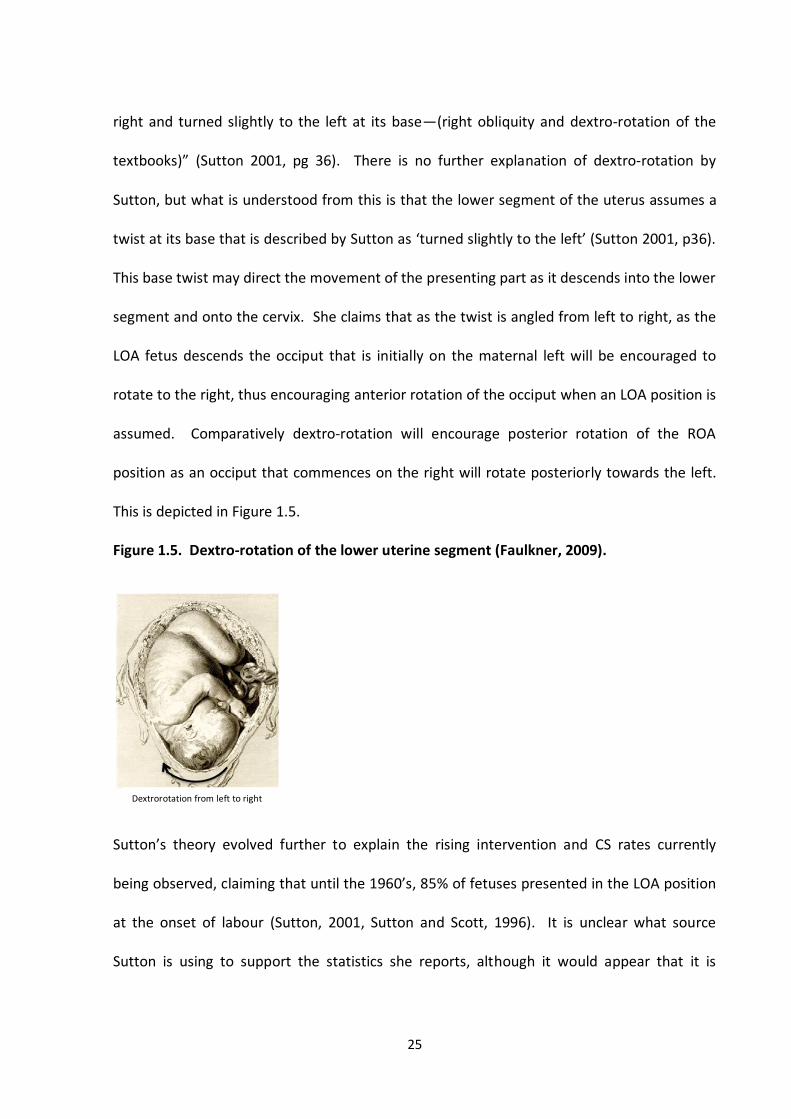
The second characteristic of the uterus that Sutton claims supports the OFP theory is
referred to as ‘dextro-rotation’ of the lower uterine segment, however this aspect of
Sutton’s theory is not explained well in any of her literature. In relation to this particular
aspect Sutton writes; “The uterus will have assumed its final position tilted to the maternal
25
right and turned slightly to the left at its base—(right obliquity and dextro-rotation of the
textbooks)” (Sutton 2001, pg 36). There is no further explanation of dextro-rotation by
Sutton, but what is understood from this is that the lower segment of the uterus assumes a
twist at its base that is described by Sutton as ‘turned slightly to the left’ (Sutton 2001, p36).
This base twist may direct the movement of the presenting part as it descends into the lower
segment and onto the cervix. She claims that as the twist is angled from left to right, as the
LOA fetus descends the occiput that is initially on the maternal left will be encouraged to
rotate to the right, thus encouraging anterior rotation of the occiput when an LOA position is
assumed. Comparatively dextro-rotation will encourage posterior rotation of the ROA
position as an occiput that commences on the right will rotate posteriorly towards the left.
This is depicted in Figure 1.5.
Figure 1.5. Dextro-rotation of the lower uterine segment (Faulkner, 2009).
Sutton’s theory evolved further to explain the rising intervention and CS rates currently
being observed, claiming that until the 1960’s, 85% of fetuses presented in the LOA position
at the onset of labour (Sutton, 2001, Sutton and Scott, 1996). It is unclear what source
Sutton is using to support the statistics she reports, although it would appear that it is
Dextrorotation from left to right
26
anecdotal evidence. Suttons understanding is that mal-position; particularly the OP position
has increased ‘drastically’ in the last few decades and is the primary cause of current
maternity statistics (Sutton, 2000, Sutton, 2001). The increase in intervention and operative
delivery caused by the increase in fetal mal-position is attributed to the change in modern
life style (Sutton and Scott, 1994). She describes Western lifestyle changes since 1960 as
‘quite unlike any other’ (Sutton 2001). The changing role of women from the household
setting into the wider world where they assumed paid work has meant a decrease in
physical activity. The use of the car, the introduction of comfortable sofas, the availability
of labour saving devices such as washing machines and daily use of computers she considers
have all encouraged a physical posture change in women (Sutton, 2007, Sutton, 2001).
Women are now less likely to adopt forward leaning postures that encourage fetuses to
adopt an OA position in-utero but instead adopt postures that are posterior inclining (Sutton
2001; Sutton 2000). It is these lifestyle changes that Sutton believes has led to the current
figures of intervention and operative delivery caused primarily by posterior positioned
fetuses (Sutton 2001).
Sutton’s background in engineering and her theories of the birth process and consideration
of the main problem led her to devise a programme of maternal posturing and movement.
Such, she believed could encourage fetuses into the LOA position and lessen the increasing
rates of intervention and operative delivery. The programme was devised based on the
following 5 main points which aimed to encourage an LOA fetal position (Sutton, 2001,
Sutton, 2007).
27
1. Understanding that from 34-36 weeks the fetus needs to establish its position for
birth (i.e LOA). After this the fetus is too large to manoeuvre readily with the aid of
maternal posturing.
2. From 34 weeks the mother should ensure that when possible her knees are lower
than her pelvis and avoid activities such as using the car on a daily basis, going on
long journeys and sitting on sofas that encourage slouching with raised knees above
the level of the pelvis. The rationale for this is to allow available space at the pelvic
brim so that the fetal head could engage and descend.
3. When resting women should try to lie on their left side, to encourage an LOA fetal
position and engagement.
4. Women should aim to adopt forward leaning postures particularly when the fetus is
active (Sutton, 2001, Sutton, 2007).
5. Discourage semi-recumbent positioning, deep squatting and late maternity leave for
working mothers.
1.6. Evidence Underpinning OFP Theory
Jean Sutton promotes OFP as a theory that is supported by physiology using physiological
and biological principles as the evidence to underpin OFP. Specifically she claims biological
plausibility using anatomical evidence to define the ‘plausible’ relationship between
biological factors of fetal position and uterine anatomy with the causal event of spontaneous
vaginal delivery (Section 1.5). Sutton only provides an explanation supported by the laws of
physics, i.e, that law of gravity states that all object are drawn to the earth and that
28
movement is dependent on mass and availability of space (Gamow, 2003). That is the
descent of the fetus is encouraged by uterine activity and the principles of gravity, yet
direction of fetal movement is influenced by uterine anatomy that may dictate directional
movement within the pelvic cavity.
As Sutton discusses the physiology underpinning OFP, it is these principles that form the
scientific method to support her explanation as to why the LOA position is the optimum fetal
position. It is such that underpins the evidence Sutton provides to support her theory and to
those who refute her theory of whom there are many both published and unpublished, she
claims that biological plausibility with anatomical evidence is suffice (Donna, 2008, Holman,
2001, Nolan, 1997, Coates, 2002). Sutton considers that it is the job of those who disagree
to provide evidence to the contrary.
The author contacted and met with Sutton to obtain further evidence on the theories of
OFP. Sutton explained in detail the anatomical and physiological relationships which
underpinned her theory (outlined in section 1.5). The author requested additional evidence
from Sutton who provided practice audits. Sutton had worked in several hospitals in New
Zealand including the management of a single birth unit, and produced audits showing
delivery outcomes for women delivering on her birth unit compared to a similar unit during
the same period. Sutton claimed it was specifically routine practice for all parents planning
on booking at her birth unit to attend education classes that taught the theory of OFP. She
outlined this as the distinctive difference between associated units and her unit, yet the
rates of CS, instrumental delivery and SVD rates were significantly different. Sutton claimed
the SVD rate at her unit was 85%, and the remaining 15% accounted for operative abdominal
29
and assisted vaginal deliveries. In Sutton’s book she writes “85% of those who grew to full
term got the exit message right that is, LOA.... About 5% are breech....10% chose Vertex ROP
of these half would sort themselves out..... and only 5% would need help.” (Sutton 2001, pg
12). Sutton informed me that the statistics she used in her book were those of the audit
findings. The audits however were unpublished and as a consequence were not available
during the period of thesis write-up.
Suttons theory as with all scientific theory forms the basis of many preliminary concepts, it is
however the ability to test it that provides evidence to support or negate the theory in
question.
1.7. OFP Theory and Acceptance
Jean Sutton has promoted her theory of OFP over a decade. Awareness and acceptance of
Sutton’s theory amongst a range of maternity-related organisations would appear to be
extensive when an everyday search engine (Google) is used to search the theory of OFP and
web associated affiliations. The word ‘Optimum Fetal Positioning’ resulted in 71 pages of
associated links with 815,000 results. All 71 pages of results were viewed by the author of
which, up until the final page, various direct links to the OFP theory were found. What was
also interesting was the large number of active/natural birth and associated organisations
promoting and implementing Sutton’s theory, below is a list of few:

30
NCT Spinning Babies
Homebirth UK Birth Angels
Women’s Health Physiotherapy Better Birth Partners
Mountain View Midwives Maternity Reflexology
Optimal Fetal Positioning/ Facebook Normal Birth at Barnsley
3shiresmidwife.co.uk Birthing Ball Specialist
Pregnancy Yoga Mumsnet
Newcastle Hospitals Acupuncture for Pregnancy
Reflexology for Pregnancy Baby world
The list, which by no means covers the extensive array of organisations now promoting OFP,
indicates the acceptance of the theory not only by maternity organisations but also by allied
health professional.
The teaching of the OFP programme is implemented from 34 weeks and commences at the
education stage where parents are taught the theory underpinning OFP. A very popular
parent education system in England is led by an organisation known as the National
Childbirth Trust (NCT). The NCT refers to itself as the UK’s leading charity for parents,
established since 1956, it has 100,000 volunteers that provide parent education to over
65,000 parents to be. The NCT website has an entire section called ‘Best baby position for
birth’ which includes the leaflet provided by Sutton that promotes OFP (NCT, 2010). The
labour and delivery session of the NCT parent education programme is partly dedicated to
OFP and thus taught as Sutton recommends to parents at 34 weeks.
In addition Sutton herself has written two books, published articles in well-known midwifery
journals, has numerous websites dedicated to OFP and has presented in over a 100 study
31
days and conferences in the UK and across the world. Her theory is also found in midwifery
textbooks verifying the increasing acceptance of the OFP theory (Johnson and Taylor, 2010,
Yates, 2010, Walsh and Downe, 2010, Davis, 2004, Donna, 2008). It is therefore not
imprecise to speculate that due to the diverse group of professionals facilitating Sutton’s
theories there is acceptance of OFP as a way forward in promoting SVD. However, in an age
where all practice should be evidence based, it is critical that the links between fetal position
and birth outcomes are established before the practice of promoting a particular fetal
position as optimal becomes routine. In order to explore the association of fetal position
further the APOLLO Study (Analysis of fetal Position at the Onset of Labour and Labour
Outcomes) was designed and conducted at the Birmingham Women’s NHS Foundation Trust
(BWHCT).
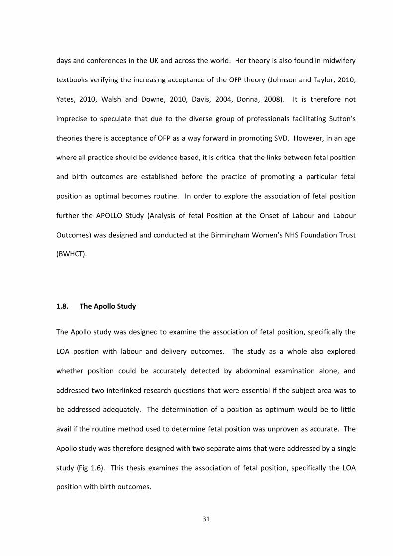
1.8. The Apollo Study
The Apollo study was designed to examine the association of fetal position, specifically the
LOA position with labour and delivery outcomes. The study as a whole also explored
whether position could be accurately detected by abdominal examination alone, and
addressed two interlinked research questions that were essential if the subject area was to
be addressed adequately. The determination of a position as optimum would be to little
avail if the routine method used to determine fetal position was unproven as accurate. The
Apollo study was therefore designed with two separate aims that were addressed by a single
study (Fig 1.6). This thesis examines the association of fetal position, specifically the LOA
position with birth outcomes.
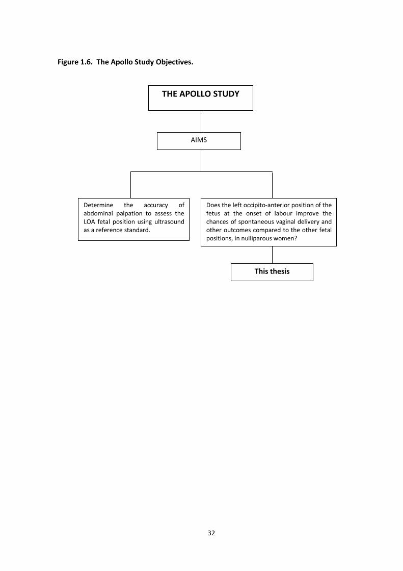
32
Figure 1.6. The Apollo Study Objectives.
THE APOLLO STUDY
AIMS
Determine the accuracy of abdominal palpation to assess the LOA fetal position using ultrasound as a reference standard.
Does the left occipito-anterior position of the fetus at the onset of labour improve the chances of spontaneous vaginal delivery and other outcomes compared to the other fetal positions, in nulliparous women?
This thesis
33
1.9. Summary
Contemporary midwifery and obstetric practice subscribes to the belief that fetal position at
the onset and throughout labour exerts considerable influence on the actual process of
labour and its outcomes for both the mother and the fetus. Despite the obvious importance
of fetal position there is surprisingly an absence of data within the body of midwifery and
obstetric knowledge, specifically in association with mechanism of labour.
The disparity surrounding fetal position is further exacerbated when the CS rate is
investigated. Intriguingly the 2001 National Sentinel Caesarean Section Audit report
concluded that labour dystocia, or more specifically failure to progress, was the second most
commonly cited indication for caesarean delivery (Thomas and Paranjoty, 2001). As failure
to progress is not in itself a descriptive diagnosis but rather a symptom, it raises some
questions as to the actual diagnosis, it is however probable that fetal position maybe an
influencing factor. Fetal position is integral to labour mechanism and is an area that not only
requires further attention but also further research.
The importance of fetal position in the mechanistic aspect of the birth process is becoming
further recognised and discussed particularly in relation to the right or left side positioning.
Commonly the theory of Sutton (2001) that encourage an LOA position to potentially
overcome the morbidity associated with mal-position is being applied (Faber, 1998). Since
Sutton’s work and much of what has been discussed cannot be supported by research
evidence, further study supported by a systematic review were undertaken to determine if
fetal position at the onset of labour influenced mode of delivery.
34
CHAPTER TWO
SYSTEMATIC REVIEW ON THE EFFECT OF FETAL POSITION AT THE ONSET OF LABOUR AND ITS INFLUENCE ON MODE OF DELIVERY
2.1. Introduction
The importance of fetal position and its influence on the process of labour and delivery
especially the active stages is evident, what remains unclear is the influence of fetal position
as that defined at the onset of labour and the subsequent influence on the process of labour
and delivery. Faber (1998) describes such aptly, suggesting that issues associated with fetal
position at labour onset are difficult to establish since no data exists on the subject area.
Furthermore the additional controversy as to whether fetal mal-position is the result of mal-
rotation from an initial anterior position or whether it is the persistence of a posterior
position has meant that understanding fetal position at labour onset has become
paramount. Particularly since labour dystocia, the second most common indication for
caesarean delivery is thought to in fact be related to fetal mal-position (Goer, 1994).
Simpkin (2010) describes the management of fetal mal-position as complex, risk-laden and
expensive, urging the importance of further research into the area, this systematic literature
review will aim to address the following question.
35
2.2. The Review Question
In pregnant women, with a singleton fetus at term gestation, does fetal position at the onset
of labour influence mode of delivery?
2.3. Inclusion Criteria
Population: Term, singleton pregnancies, in early labour/point of labour onset with a
cephalic presenting fetus.
Intervention/exposure: Fetal position of occiput and/or spine confirmed by
ultrasound.
Outcome: Mode of delivery.
2.4. Search Strategy
Studies were reviewed that recruited pregnant women with a singleton cephalic fetus and
that were scanned at the onset of labour to determine fetal position and where position was
subsequently analysed against mode of delivery. The review was performed systematically
of the published literature with no restrictions on year of publication, type of study design
and no language restriction was placed.
Search identification included both manual and electronic search strategies. Electronic
searches involved the electronic databases and predetermined search terms (Mesh
headings). The initial selection criteria was broad to ensure that as many studies as possible
were assessed as to their relevance, this assessment was based on the abstract and titles
presented in the electronic catalogue. Articles that were clearly unsuitable to the review
36
were excluded at this stage. The remaining articles were ordered and read before a decision
to include or exclude was made.
Manual searches were conducted, journals that appear relevant were hand searched and
follow-up searches were conducted on citations found in other studies that may be relevant.
2.5. Expert Contact
As Jean Sutton is the expert within the field of interest she was contacted in order to obtain
any additional information. Sutton was also asked to provide information on ongoing
research that she may be aware of or participating in. All references provided by Sutton
were retrieved.
2.6. Electronic Search Strategy
A review was undertaken of the bibliographic databases using the search strategy outlined,
this included inputting the relevant search terms for the population, exposure and the
outcome. The search included review of the following databases:
MEDLINE
EMBASE
CINAHL
MIDIRS
CENTRAL
As the databases were searched, an individual list was maintained detailing the names of
the databases searched, the keywords used and the results. Titles and abstracts of studies
to be considered for retrieval were printed along with details of where the reference was

37
located. The printed abstracts were then reviewed by the lead researcher and co-
researcher independently with consideration of the inclusion criteria and filed once an
inclusion/exclusion decision was made by both. Once the inclusion/exclusion decision was
made, the two researchers discussed the decision they made and determine agreement.
2.7. Study Selection/Quality Assessment
The selection process was undertaken as a group, alongside a senior university lecturer and
two research colleagues. All studies that were deemed to be suitable were reviewed by the
entire group independently. The senior lecturer of the group provided the quality
assessment tool that was used by the group to critically appraise each study. The quality
assessment tool used was the Newcastle-Ottawa scale (NOS).
The Newcastle-Ottawa scale is used to assess non-randomised studies including case-
control, cohort and observational studies. The scale uses a star allocation method to
determine quality. The scale allocates a maximum of nine stars for quality of selection,
comparability, exposure and outcome.
Following independent review of each study, joint discussion would take place and where
reviewer’s conclusions over the validity differed; a further joint review would take place in
order to achieve agreement.
The results of the search are outlined in Figure 2.1 (see appendix 1 for performed searches).
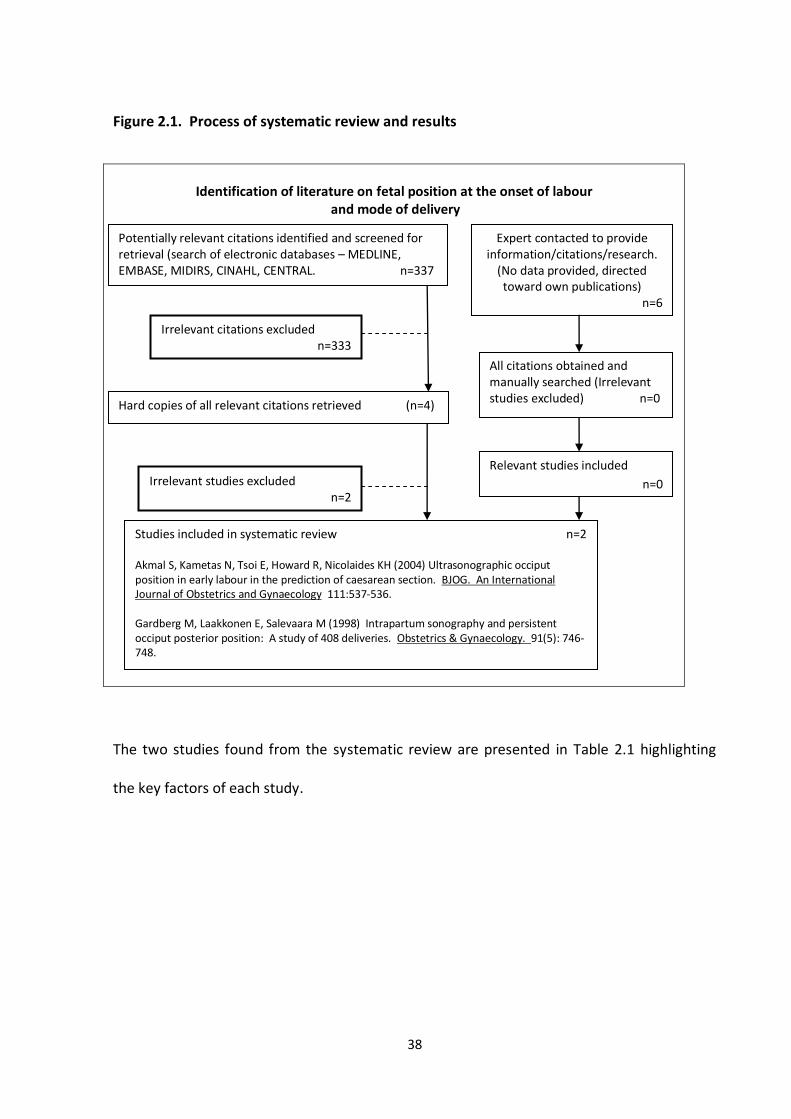
38
Figure 2.1. Process of systematic review and results
Identification of literature on fetal position at the onset of labour
and mode of delivery
The two studies found from the systematic review are presented in Table 2.1 highlighting
the key factors of each study.
Potentially relevant citations identified and screened for retrieval (search of electronic databases – MEDLINE, EMBASE, MIDIRS, CINAHL, CENTRAL. n=337
Irrelevant citations excluded n=333
Hard copies of all relevant citations retrieved (n=4)
Irrelevant studies excluded n=2
Studies included in systematic review n=2
Akmal S, Kametas N, Tsoi E, Howard R, Nicolaides KH (2004) Ultrasonographic occiput position in early labour in the prediction of caesarean section. BJOG. An International Journal of Obstetrics and Gynaecology 111:537-536. Gardberg M, Laakkonen E, Salevaara M (1998) Intrapartum sonography and persistent occiput posterior position: A study of 408 deliveries. Obstetrics & Gynaecology. 91(5): 746-748.
Expert contacted to provide information/citations/research.
(No data provided, directed toward own publications)
n=6
All citations obtained and manually searched (Irrelevant studies excluded) n=0
Relevant studies included
n=0

39
Table 2.1. Synopsis of studies located through systematic review
Author Study design Population/ Sample size
Stage Fetal position defined
Technique used
Outcome NOS Score
Akmal et al. 2004
Prospective study
Singleton pregnancies, cephalic presentation. 601 women.
Cervical dilation 3-5 cm, defined as early labour
Ultrasound by trained sonographers
Prediction of caesarean section
6/9
Gardberg et al. 2004
Prospective Study
Singleton pregnancies ≥ 37 weeks. Vertex presentation. 408 women.
Onset of labour
Ultrasound
Labour duration, mode of delivery, Apgar scores, Fetal rotation.
7/9
2.8. Results
Akmal et al. (2004b) undertook a prospective study with the aim of using ultrasound imagery
to determine fetal position in early labour in order to predict the likelihood of CS. The study
involved 601 women recruited during February 2001 to April 2002 who were between 37-42
weeks pregnant with a singleton cephalic pregnancy. Data on position and outcome were
obtained for all the women included in the study. Akmal et al. (2004b) used ultrasound to
determine fetal occiput position that was defined as either anterior, posterior or lateral with
no further categorisation. Fetal position was determined in early labour; this period was
defined as cervical dilatation ranging from 3-5 cm. The specific aim of the study was to
determine if the occiput posterior position was an independent factor in the prediction of CS
delivery.
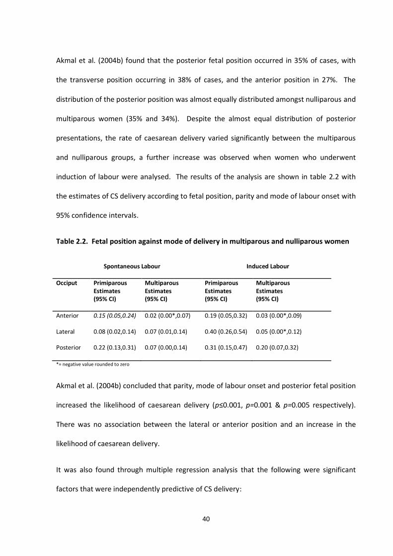
40
Akmal et al. (2004b) found that the posterior fetal position occurred in 35% of cases, with
the transverse position occurring in 38% of cases, and the anterior position in 27%. The
distribution of the posterior position was almost equally distributed amongst nulliparous and
multiparous women (35% and 34%). Despite the almost equal distribution of posterior
presentations, the rate of caesarean delivery varied significantly between the multiparous
and nulliparous groups, a further increase was observed when women who underwent
induction of labour were analysed. The results of the analysis are shown in table 2.2 with
the estimates of CS delivery according to fetal position, parity and mode of labour onset with
95% confidence intervals.
Table 2.2. Fetal position against mode of delivery in multiparous and nulliparous women
Spontaneous Labour Induced Labour
Occiput Primiparous Estimates (95% CI)
Multiparous Estimates (95% CI)
Primiparous Estimates (95% CI)
Multiparous Estimates (95% CI)
Anterior 0.15 (0.05,0.24) 0.02 (0.00*,0.07) 0.19 (0.05,0.32) 0.03 (0.00*,0.09)
Lateral
Posterior
0.08 (0.02,0.14)
0.22 (0.13,0.31)
0.07 (0.01,0.14)
0.07 (0.00,0.14)
0.40 (0.26,0.54)
0.31 (0.15,0.47)
0.05 (0.00*,0.12)
0.20 (0.07,0.32)
*= negative value rounded to zero
Akmal et al. (2004b) concluded that parity, mode of labour onset and posterior fetal position
increased the likelihood of caesarean delivery (p≤0.001, p=0.001 & p=0.005 respectively).
There was no association between the lateral or anterior position and an increase in the
likelihood of caesarean delivery.
It was also found through multiple regression analysis that the following were significant
factors that were independently predictive of CS delivery:
41
Maternal age
Afro-Caribbean origin
Height
Parity
Labour onset
Gestation
Head descent
Male gender – Fetal
The quality assessment of Akmal et al.’s (2004b) study using the NOS achieved 6/9, with
stars not allocated for representativeness in the selection category as no method of
selection was discussed; instead women admitted to a single department were selected to
take part in the study. A lack of control (i.e blinding) in the comparability category also
meant a star was not allocated and finally a lack of independent assessment of ultrasound
findings in the outcome category meant a further star was not allocated. Overall the study
was conducted well and answered the research question posed.
The second study identified through the systematic review was that conducted by Gardberg
et al. (1998). Gardberg et al. (1998) undertook a prospective study of 408 women with
singleton, cephalic fetuses at term gestation. Each woman underwent an ultrasound on
admission to hospital for induction of labour or for spontaneous onset of labour. Fetal
position for the purpose of the study was categorised as either anterior or posterior. A
posterior position was categorised as the observation on ultrasound of both orbits facing the
symphysis pubis and with the spine seen posteriorly. All other observations on ultrasound
were classed as anterior fetal positions. Interestingly no further sub-division of position was
noted; instead the transverse position of the fetus was also classed as an anterior fetal
position.
42
Fetal position on admission was compared to position at delivery as recorded by the
attending clinician. Duration of the first and second stage of labour, mode of delivery, and
Apgar scores were recorded as outcome variables.
Gardberg et al. (1998) found that of the total study population 61 (15%) of fetuses were
posterior at the onset of labour, and 21 (5.1%) were posterior at delivery. Of the posterior
fetuses that were found to be OP on admission 53 (87.5%) rotated and were in an anterior
position at delivery. This supposed that only 8 cases of posterior fetal position persisted
throughout labour and delivery.
Gardberg et al. (1998) however, found that a total of 21 fetuses were posterior at delivery,
concluding that the additional 13 (62%) were as a result of fetal mal-rotation from an initial
anterior fetal position rather than a persistent OP position from labour onset. Gardberg et
al. (1998) found that the OP position was not associated with labour induction, epidural use,
maternal BMI, duration of first and second stage or mode of delivery.
Gardberg et al.’s (1998) study achieved a NOS score of 7/9 stars using the NOS category.
Similarly to Akmal et al.’s (2004b) study a lack of control (i.e blinding) in the comparability
category meant a star was not allocated and a lack of independent assessment of ultrasound
findings in the outcome category meant a further star was not allocated. Gardberg et al.
(1998) achieved full allocation of stars for the selection category.
43
2.9. Discussion
The question posed by the systematic review was:
In pregnant women, with a singleton fetus at term gestation, does fetal position at the onset
of labour influence mode of delivery?
In the outlined studies both the population and the outcome being addressed were included
within the studies. What was absent from both studies was the use of the standard
definition of labour onset and the inclusion of all fetal positions, suggesting that although
population and exposure were addressed, this was not done comprehensively. What was
apparent from the systematic review was that there is an absence of knowledge in the area
of all fetal positions and related outcomes.
Of the two studies located from the systematic review neither answered the review question
in its entirety. Akmal et al.’s (2004) study remains slightly ambiguous, as in the title it stated
that position would be defined in early labour. Throughout both the abstract and the study
however, Akmal et al. (2004) outlined that it was the early yet ‘active’ first stage of labour
that was the point at which position was determined. This is not however, the common
definition for ‘early labour’. In the vast majority of cases early labour is defined as the latent
or non-active phase of the labour process.
Furthermore, Akmal et al. (2004) defined position into three categories this is despite there
being 8 variations in the distribution of fetal position. This limited Akmal et al.’s (2004) study
as the entirety of the exposure (fetal position) was not considered, but instead only a sub-
section of the overall exposure was explored. This would appear to be the main limitation of
the study. Finally the study did not answer the question of the systematic review, the aim of
44
Akmal et al.’s (2004) study was to identify factors that would predict the likelihood of CS
rather than analyse position with mode of delivery.
The study conducted by Gardberg et al. (1998) also had fundamental flaws which were not
addressed and therefore the findings must be interpreted cautiously. Fetal position was
categorised into OA and OP position only, this is despite the understanding that the majority
of fetuses will commence labour in the transverse position. By including the transverse
position as an anterior position it may be argued that this was misclassification of the OA
position and as a consequence associations that others have reported were not observed.
Furthermore in Gardberg et al.’s (1998) study ultrasound was performed upon admission to
the hospital, this criterion alone was used to represent ‘position at labour onset’. What is
not clear is whether there were any additional criteria that allowed Gardberg et al. (1998) to
draw conclusions relating to position at the ‘onset of labour’. Women may had arrived in
either established labour or early labour, in which case descent and rotation may have
already commenced suggesting that position was misdiagnosed. If women were in early
labour further follow-up was not reported, suggesting that women who are often sent home
in early labour and return days later did not have a repeat scan and consequently there was
no way of detecting if the recorded position was the actual position at the onset of labour.
The fundamental flaws in Gardberg et al.’s (1998) study imply that his conclusions are poorly
supported by the study he undertook. Despite this, Gardberg et al. (1998) has challenged
current understanding by concluding that persistent posterior positions at delivery are a
result of mal-rotation from an initial OA position. This is contrary to common belief that the
OP is a persistence of an initial posterior fetal position (Neri et al., 1995).

45
Despite such findings Gardberg et al.’s (1998) study failed to answer the review question,
mainly due to the restrictive definition of fetal position. Thus the absolute absence of data
on all fetal positions at the onset of labour and influence on mode of delivery would suggest
that such factors require clear definition and exploration. This thesis will attempt to address
the issue pertaining to accurate identification of all fetal positions at the point of labour
onset and the association with birth outcomes.
46
CHAPTER 3
METHODOLOGY
This chapter will discuss the methodologies available for the various elements of the
research. The potential research question will be explored and discussed in relation to the
rationale for the most appropriate methods to be used if it is to be answered
comprehensively.
Following review of the literature and systematic review it was evident that insufficient
knowledge exists within the area of fetal position at labour onset and the associated birth
outcomes. The principle aim of the research was to determine all fetal positions at the point
of labour onset, specifically the LOA positions, in association with birth outcomes.
3.1. Study Design
The nature of the question posed meant that a quantitative approach was required in order
to obtain the necessary data. A quantitative approach allowed measure of the variable of
interest in a manner that could then be analysed using statistical methods to determine
statistical significance. Qualitative research is primarily exploratory in nature with the aim of
generating new theories and ideas and therefore not applicable to answer the research
question of this study.
47
The type of quantitative study design required was observational and the most appropriate
and high quality design given the research question was a cohort study. An experimental
design was not suitable. There has already been a randomised controlled trial to determine
if maternal posturing could manipulate fetal position during the antenatal period on the
assumption that fetal position is predicted of birth outcomes (Kariminia et al., 2004). The
clear absence of evidence that position is predictive of outcome however questions the
rationale for an experimental design at this stage. The background and systematic review
supports the approach to determine the relationship, if any, between fetal positioning and
birth outcomes before an intervention to test the ability to manipulate fetal positioning. The
specific characteristics of a prospective cohort study mean recruitment of a cohort of
participants, in this instance pregnant women with a cephalic presenting fetus can be
identified, assessed in terms of exposures of interest and followed-up to record the outcome
of interest which in this case was birth outcomes.
3.2. Composition of Sample
Maternal parity in research has always posed a concern with regards to controlling for
confounding variables since a nulliparous birth has many known different characteristics and
in many studies statistical methods are used to control for this. This study however is based
on a theory that is applicable to the nulliparous women only. Sutton claims that the distinct
shape of the uterus is exclusive to nulliparous women and it is this distinction that dictates
rotational descent. She claims that the distinct uterine shape encourages anterior rotation
of the LOA fetus and posterior rotation of the ROA fetus (See section 1.5. – OFP theory). She
48
considers that the distinct uterine shape is not maintained in subsequent pregnancies and
thus the theory of OFP is not applicable to multiparous women (2001). It is therefore
necessary that the study participants be nulliparous women.
Additionally the increasing rates of CS are substantially influenced by the rates of repeat CS
from an initial operative delivery (Thomas and Paranjoty, 2001). The ability to encourage
SVD amongst nulliparous women could curtail the concerns associated with repeat operative
delivery and therefore aim to reduce the CS rate.
The remaining study inclusion criteria were in the main also dictated by Sutton’s theory
which applied to women who were term, with no known fetal abnormalities, be alive and
singleton. Sutton’s theory is underpinned by the belief that uterine anatomy will influence
directional descent of the fetus consequently participants must therefore be grown to full
term. Finally the requirement of a cephalic presenting fetus was compulsory if the area of
fetal positioning was to be explored.
3.3. Method of Measure
The research involves accurate identification of fetal position, normal clinical practice being
to determine fetal position at the onset of labour/induction of labour by abdominal
palpation only. Abdominal palpation is described as inexact and subjective with limited
evidence supporting its accuracy (Ridley, 2007).
Only one study was found that examined the validity of abdominal examination to
determine fetal presentation and position (McFarlin et al., 1985). McFarlin et al. (1985)
49
conducted a prospective cohort study of 176 participants who had an abdominal palpation
to determine fetal position and an abdominal ultrasound scan to compare its findings.
McFarlin et al. (1985) did not report the ultrasound findings, or highlight how fetal position
was determined on ultrasound. Instead, women who were in antenatal clinic and scheduled
for an ultrasound scan were approached to take part in the study. If consent was obtained,
abdominal palpation was performed prior to their booked ultrasound. The method by which
the sonographers reported, documented or assessed fetal position was not described. Of
the 176 women who participated, only 131 were cephalic so fetal position was only
determined on a 131 women. McFarlin et al. (1985) claimed that of the 131 participants,
correct assessment of fetal position was identified in 60.3% of cases overall, describing the
results as ‘disappointing’. The study had many limitations including a poor sampling
technique and further still despite the study using ultrasound to confirm fetal position it
claimed that some women did not receive an ultrasound scan if the examining clinician was
very certain of his/her findings. These limitations mean that the results must be interpreted
cautiously but indicate concerns about palpation accuracy.
It was apparent that the use of abdominal palpation to determine fetal position would not
be sufficient. The alternative method that has been used and often referred to as the ‘gold
standard’ method is ultrasound. Ultrasound imagery has been described as a major tool in
medical imaging and specifically the most significant technology introduced to Obstetrics
(Doubilet and Benson, 2003, Sherer et al., 1998). A review by Sherer et al. (1998) of
ultrasonography compared the importance of ultrasound in obstetrics to blood banking, the
discovery of antibiotics and anaesthesiology. Ultrasound is understood to represent the
50
superior method of obtaining internal imagery, which can be used at any point in pregnancy
and maybe performed for maternal or fetal reasons (Sherer et al., 1998).
Ultrasound technology uses sound waves produced at a very high frequency to transmit
images (Doubilet and Benson 2003). The vibrations of the sound waves produce wavelengths
or echoes, which are converted into images on a screen. Hard tissues, such as bone, reflect
bigger echoes and are therefore white in the produced image (Dewbury et al., 2001). Soft
tissues such as muscles are reproduced as grey images, and fluids, such as amniotic fluid
appears black as the sound waves travel through the fluid creating no vibrations.
Ultrasound has been adopted into main stream obstetrics and is used throughout the period
of pregnancy, labour and post partum and used routinely to investigate fetal development,
growth, abnormality and to perform certain medical interventions (Sherer et al., 1998).
The importance of determining fetal position in labour and the belief that the standard
palpation method of identifying position may be inaccurate has led to several ultrasound
studies. The first studies reported were as early as 1985 and 1989 that have successfully
used ultrasound to determine fetal position in labour (Rayburn et al., 1989, McFarlin et al.,
1985). Over the past decade various studies have compared digital vaginal examination with
trans-abdominal, trans-perineal and trans-vaginal sonography.
Sherer et al. (2002b) conducted a prospective study to test the null hypothesis that no
correlation exists between digital vaginal examination and trans-abdominal supra-pubic
ultrasound to determine fetal head position during labour. 102 consecutive patients with
normal singleton, cephalic, term fetuses in established labour of 4cms or more were
included and the ultrasound scan was performed immediately after digital examination.
51
Head position was classified as direct OA, direct OP, left or right occiput transverse (LOT,
ROT), left or right occiput anterior or posterior (LOA, LOP, ROA, ROP). The results illustrated
that even when a 45° allowance of angle discrepancy for fetal position was allowed, an error
rate of 53% was encountered (Sherer et al., 2002). Sherer et al. (2002) thus concluded from
his study that intrapartum ultrasound increased the accuracy of determining fetal head
position during the active first stage of labour.
Sherer et al. (2002a) then repeated the study this time including 112 patients in the second
stage of labour. The null hypothesis tested was that no correlation exists between trans-
vaginal digital examination compared with trans-abdominal supra-pubic ultrasound to assess
fetal position in the second stage of labour. The results revealed an error rate of 39% when
a 45° angle of discrepancy was allowed between digital vaginal examinations compared to
trans-abdominal ultrasound findings. Sherer et al. (2002) noted that although compared
with the first study in the active first stage of labour the error rate was lower in the second
stage of labour, it remained ‘considerably high’, thus concluding that intrapartum
sonography increased the accuracy of determining fetal position in active first and second
stage of labour (Sherer et al., 2002).
Akmal et al. (2003) performed a similar study looking at digital vaginal examination
compared to trans-abdominal ultrasound to determine fetal head position prior to
instrumental delivery. 64 women with singleton pregnancies undergoing instrumental
delivery were recruited to the study. Both digital vaginal examination and trans-abdominal
ultrasound were performed one after the other. The vaginal examination was considered
correct if as in Sherer et al’s (2002) study fetal position was within 45° of the ultrasound
52
findings. It found that digital examination during instrumental delivery failed to identify the
correct fetal head position in 26.6% of cases. As a 45° angle of discrepancy was allowed
Akmal et al. (2003) describes the error rate as a ‘substantial discrepancy’. The significance of
such an error is thought to affect instrument application, being incorrectly applied due to
the inaccurate identification of fetal position. The error rate further increased with non
anterior positions. The rate of identifying an anterior position using digital vaginal
examination was 83% compared to 54% for lateral or posterior positions (Akmal et al., 2003).
The study concluded that trans-abdominal ultrasound should be used routinely prior to
instrumental delivery to determine fetal head position.
Kreiser at al. (2001) performed a similar study including 44 women to determine fetal
occiput position by ultrasound during the second stage of labour. The error rate reported by
Kreiser et al. (2001) was 30%, concluding that ultrasound use to determine fetal position was
‘significantly superior’ compared to traditional methods of determining fetal position.
Chou et al. (2004) investigated fetal position in the second stage of labour of vaginal
compared with ultrasound examination to determine fetal occiput position and fetal spine
position in the second stage of labour. 88 patients participated and were given both trans-
abdominal ultrasound and trans-perineal ultrasound to determine fetal occiput position.
The predicted positions by both methods of ultrasound and digital vaginal examination were
compared against position following spontaneous restitution or position at caesarean
delivery to determine accuracy. As with the previously outlined studies Chou et al. (2004)
showed that ultrasound was more accurate than digital vaginal examination, which had an
53
error rate of 28.4%. Chou et al. (2004) did not compare the accuracy of trans-abdominal
ultrasound with trans-perineal ultrasound in this study.
Zahalka et al. (2005) went further to study the determination of fetal head position using 3
different examination techniques, comparing digital vaginal examination with trans-
abdominal ultrasound and with trans-vaginal ultrasound. Zahalka et al. (2005) aimed to
investigate both accuracy and time requirements of all 3 examinations. Sixty women in the
second stage of labour were recruited, and digital vaginal examination was followed by both
trans-vaginal and trans-abdominal ultrasound. A 60° angle of discrepancy was allowed
between each identified fetal position compared to the position recorded following
restitution. The 60° angle of discrepancy was greater than that endorsed in the previous
studies, of only 45°. The results confirmed previous findings that digital examination
remained the least accurate method of determining fetal head position. The error rate
ranged from 15% when the degree of error was greater than 90° to 23.3% when the degree
of error was between 60-90° (Zahalka et al. 2005). Zahalka et al. (2005) further concluded
that no significant difference in accuracy of determining fetal head position was detected
between trans-abdominal and trans-vaginal ultrasound examination. Time requirements
were however significantly shorter for the trans-vaginal examination compared to digital or
trans-abdominal examination.
Over the past decade numerous studies have consistently shown that trans-abdominal
ultrasound examination is the gold standard method of determining fetal head and fetal
spine position. Further studies have looked into more invasive methods such as trans-
vaginal and trans-perineal ultrasound to determine fetal position to increase accuracy.
54
These have shown that invasive methods may cause more discomfort to patients and do not
increase accuracy when determining fetal position against non invasive methods such as
trans-abdominal ultrasound.
All the studies reviewed concluded that ultrasound to determine fetal head position should
be used routinely in view of the identified error rate with use of routine methods (Akmal et
al., 2003, Chou et al., 2004, Zahalka et al., 2005). Peregrine et al. (2007) states that
specifically trans-abdominal ultrasound to determine fetal position is easy to perform and
highly reproducible. This highlights not only the importance of trans-abdominal ultrasound
in reducing error margins but also its ease of use as a routine assessment tool.
3.4. Point of measure
Sutton’s theory was unique since it not only claimed that a single fetal position was optimum
but it also claimed that it was position at a specific point in the labour process that would
dictate the entirety of the labour and birth process. The specific period was the point of
labour onset, interestingly the period of labour onset in the normal process of labour is the
point prior to the commencement of rotational descent. This signifies its importance if, it is
confirmed that the process of rotational descent is influenced by the position the fetus
adopts at the point of labour onset, a position maybe established as optimum.
The ‘onset of labour’ signifies an important period and thus must be understood and defined
with such measure. The point of labour onset, in view of the argument posed by Sutton is
the period prior to the commencement of active labour yet succeeding the latent first stage

55
of labour. As is understood (see section 1.1- Normal Obstetric Labour), the process of labour
for the fetus involves rotational descent through the maternal pelvic. By determining
position once rotation had ensued (i.e active first stage) would not provide information
pertaining to a favoured or otherwise fetal position. This would mainly be due to the
inability to determine what stage of the rotational process the fetus was at, if position was
to be determined during the active first stage of the labour process. Consequently, Suttons
claim was very specific in that fetal position at the point of labour onset must be LOA in
order to encourage anterior rotation and prevent mal-rotation.
3.5. Definition of labour onset and period of measure
The point of measure for the purpose of this study was labour onset, with the aim of
identifying fetal position immediately preceding the point of active labour. The period
immediately prior to the commencement of active labour is referred to as the latent phase,
labour onset and early labour interchangeably. Labour stages are diagnosed retrospectively
with the commencement of a subsequent stage marking the end of the prior stage. The
commencement of active labour which is commonly defined as cervical dilatation greater
than 4cm with regular uterine contractions would mark the end of early labour/latent phase
and labour onset (Tiran, 2008, NICE, 2007).
The latent phase of labour is therefore the point at which measure is to be undertaken
however it is poorly understood and is often claimed to be hard to define (McDonald, 2010).
There is limited research evidence into the areas of recognition, definition and management
of the latent phase. Evidence exists into prolonged and misdiagnosis of the latent phase that
56
often leads to unnecessary admission, leading to increased levels of intervention including
increased rates of operative delivery (Chelmow et al., 1993, Maghoma and Buchmann,
2001). The initial definition of the latent phase of labour and much of labour stages was
described by Friedman (1978). Friedman (1978) claimed that a latent phase greater than 20
hours in nulliparous women would be classed as prolonged, suggesting that a latent phase
up to 20 would be normal. Since then however, many have argued that the length of the
latent phase is 8-12 hours with the majority of textbooks reporting 8 hours (Chelmow et al.,
1993, Maghoma and Buchmann, 2001, McDonald, 2010, McCormick, 2009, Ladwig et al.,
1998, Coad and Dunstall, 2005). There is overall a clear lack of consensus on how long a
latent phase of labour should last. The definition of the latent phase appears to have
greater consensus, described as commencing when women start to experience painful
contractions associated with minimal cervical changes, this definition is also supported by
the National Institute of Clinical Excellence (NICE) (McCormick, 2009, NICE, 2007, McDonald,
2010). NICE was established to set high-quality standards derived from the best available
evidence. NICE intrapartum guidelines provide a clear and concise definition of latent/early
labour up to the point of active labour onset, therefore defining clearly the period during
which position is to be identified for the purpose of the research, the definition being:
A period of time, not necessarily continuous, when:
- there are painful contractions, and
- there is some cervical change, including cervical effacement and dilatation up to 4cm.
Established active first stage of labour is when:
- there are regular painful contractions, and
- progressive cervical dilatation from 4cm. (NICE, 2007)
57
For the purpose of this study the above definition clearly defines the period during which
position is to be defined. Bohra et al. (2003) claimed that 81% of nulliparous women were
admitted to hospital with a cervical dilation of less than 2cm. This suggests that a large
majority of women are likely to be present during the latent phase of labour and therefore
for the purpose of the study position can be measured at the specified point.
3.6. Follow-up
Labour and delivery is regarded as a ‘process’ that is assessed for progress or otherwise over
a period of time. Since the point of measure or the latent phase is identified as ‘a period of
time, not necessarily continuous’ it is appropriate to assume follow-up would be necessary.
As previously discussed a lack of consensus on how long a latent phase is, remains, however
the latent phase requires definition for the purpose of the study in order that a period of
repeat measure could be established. The majority of clinical textbooks suggest that the
normal length of the latent phase is 8-12 hours (Chelmow et al., 1993, Maghoma and
Buchmann, 2001, McDonald, 2010). In view of this it would appear appropriate that fetal
position is reassessed and determined if active labour is not established within the defined
period of the latent phase of 12 hours. To repeat the measure 8 hourly would require 3
ultrasounds within a 24 hour period. To repeat the scan 3 times was considered to be too
much for women who were in pain, potentially sleep-deprived and suffering with increased
exhaustion. Since there is no conclusive argument differentiating between 8 or 12 hours, for
the purpose of the study it was considered appropriate to use a 12 hour rule of repeat
measure.
58
If the study participant did not enter established labour within 12 hours of the ultrasound
scan, a repeat scan was required and the previous scan would become invalid. All study
participants who were being induced or in early labour were therefore followed up every 12
hours, and once they were in established labour subsequent follow-up was not required.
The 12 hour rule ensured that position was determined within the period of early labour and
close to the commencement of active labour. Without such a rule women who may not
have entered labour for several days would not be followed up and therefore it could not be
determined with certainty that position was recorded at labour onset.
3.7. Pilot Study
Following determination of the study methodology it was deemed appropriate that a pilot
study be undertaken. The pilot study was used to assess feasibility of study implementation,
specifically recruitment, up-take of ultrasound during labour, scan technique, 12 hour
follow-up rule and documentation.
Three full-time midwives, who covered a 24 hour shift rota, were trained to scan for the pilot
study, and covered triage (delivery admissions), labour ward and the birth centre. Two scan
machines were available on the delivery suite for use by both the birth centre and triage.
Documentation and recruitment was arranged and commenced at the same time
throughout the community and hospital settings. During the pilot study, 932 women
consented to take part of which 406 were recruited and scanned. Of the 406 women, 280
scans were obtained that contained all the necessary data.
59
Initial consent to take part in the study using the documentation proved successful, with a
large number of participants consenting each month. The manpower however, associated
with producing the consent packs on a large scale and distributing them each month was
time consuming and took a lot of the research midwives time which could have been spent
scanning. As a result it was decided that from the limited study funds available the
researchers would pay an individual to assemble and distribute the consent packs.
No participant in the pilot study refused ultrasound or asked the scanning midwife to
discontinue at any point. Since the ultrasound took place during labour there were some
concerns as to whether women would find it acceptable in view of the possible discomfort
caused by uterine activity. Instead, the scanning midwives reported women requesting the
ultrasound on admission to the hospital.
The scanning technique used by the midwives was considered to be successful, as in 14
cases was the fetal head too low to measure, highlighting that this was not a major concern
for the study. The midwives reported ease of use of the portable scanning machine, clear
images and the available facility of darkening the delivery rooms meant that the
environment for scanning was adequate.
The principal problem identified was a lack of scanning midwives. The hospital based
midwives reported bleeping the scanning midwives but not receiving a response. Three
midwives were not sufficient to cover the demand of scans required. This was the main
reason why the large number of women recruited were not then scanned and included in
the study, which was too low an inclusion rate from those consented.
60
Furthermore women who did not enter established labour within 12 hours and required
subsequent scans were also missed; 75% of women requiring follow-up scans after 12 hours
did not receive a scan. This was mainly due to the shift pattern covered by each midwife.
The scanning midwives felt responsible for the scans required on their department, none of
the midwives took responsibility for the remaining departments whilst on duty. The
midwives did however have clinical responsibilities and could not leave their departments so
12 hour follow-up scans were often missed. Additionally the antenatal ward which did not
have an allocated scanning midwife was regularly missed, since women would be admitted
to the antenatal ward for induction and in the early stages of labour a sizeable group of
women were missed.
The problem highlighted by the pilot study clearly indicated a need for more scanning
midwives including cover of the antenatal ward. This led to the application for a research
midwife to the Research and Development department at the BWHCT. The department
awarded funds to allow a research midwife to be appointed for 3 days a week. The research
midwife (the author) was based on the antenatal ward, and reduced her clinical hours to
ensure she was available 6 days a week to cover the demands of the study. In addition, 3
further midwives were trained to offer additional scanning services. This meant 6 midwives
in total including the research midwife were available for the implementation of the study.
Documentation in relation to data collection was deemed adequate. The pilot study
demonstrated that implementation of the study using the outlined methodology was
adequate. The additional allocation of scanning midwives and the appointment of the
research midwife meant that concerns relating to a lack of available staff to perform the

61
scans were overcome for the main study in order that the women recruited antenatally and
then lost to follow-up would be reduced.
In view of the methodology discussed, the methods of the research have now been
identified for implementation.
62
CHAPTER 4
METHODS
4.1. Aim of the Study
The aim of this study was to explore the association between fetal position at the onset of
labour and birth outcomes for the mother and the neonate. The specific research question
was:
Does the Left Occipito-Anterior (LOA) position of the fetus at the onset of labour improve
the chances of Spontaneous Vaginal Delivery (SVD) and other outcomes compared to the
other fetal positions, in nulliparous women?
4.1.1 Objectives
The main objectives of the study were:
To ascertain the proportion of all fetal occiput and spine positions at the onset of
labour using the ‘gold standard’ method of ultrasound amongst nulliparous women.
To determine the relationship between fetal occiput position at the onset of labour
and birth outcomes(s) amongst nulliparous women.
To ascertain if the LOA fetal occiput position at the onset of labour is associated with
improved outcome(s) amongst nulliparous women.
63
To determine the relationship between fetal spine position at the onset of labour and
mode of delivery amongst nulliparous women.
4.2. Study Design
The study design was a prospective cohort study that was set up to fulfil the above aim and
objectives. Women were recruited to the study and given an ultrasound scan to determine
fetal position at the onset of labour then followed up until birth to determine outcomes.
4.3. Ethics
The study obtained ethical approval from the South Birmingham Research Ethics Committee.
The ethics committee suggested the incorporation of non-English speaking women who
were initially deemed not eligible. It was thought that since link workers were available to
translate during both clinic appointments and during hospital admissions that this group
could be included in the study. The recommended amendment was made and approval was
obtained. The study was also approved locally by BWHCT Research & Development
committee.

64
4.4. Setting
The study was conducted at BWHCT, one of the largest women’s hospitals in England. The
trust has a midwife-led birth centre and a consultant-led delivery suite and the study
included women who were admitted to both units. Recruitment took place over an 18
month period that commenced in April 2007 until September 2008.
4.5. Participants and Inclusion Criteria
All nulliparous women who were booked at the BWH and met the specified inclusion criteria
during the study period were invited to take part. The inclusion criteria are listed below:
Nulliparous
Live, singleton fetus
No known fetal abnormalities
≥ 37 gestational weeks
Cephalic presentation
Labour spontaneous or induced
In early labour (cervical dilatation ≤4 cm)
4.6. Recruitment
Women were invited to take part in the study from 28 gestational weeks. All women
booked at the hospital were routinely given an appointment to attend a blood test
appointment at 28 weeks; this appointment was usually given for a specific clinic (referred to
as the community clinic). The community clinics are co-ordinated by the community
midwives who were the primary recruiters to the study. Since all women were required to
65
attend community clinic it was thought that this would be an ideal primary venue from
which to recruit.
It was anticipated that some women may fail to be recruited from this setting for various
reasons, such as staffing pressures or shortages that may prevent the midwife from
spending the extra time required to obtain consent to participate in the study. In view of
this, it was thought necessary that alongside this recruitment route, link workers, the
research midwives and hospital based midwives would also be involved in the recruitment
process. Recruitment therefore also took place at parent education classes that were
provided at BWHCT for all nulliparous women. Nulliparous women are invited to attend the
classes from 34-36 gestational weeks. The research midwives would attend the antenatal
classes on a regular basis and recruited women to the study.
Prior to the commencement of the study the research midwives visited each community
team and each hospital setting. A presentation of the study was given followed by an
extensive description of the recruitment process. An information leaflet devised for health
professionals was also distributed (Appendix 2). The information was then re-iterated at a
launch presentation, and information clinics were open for 1 week for midwives and women
to attend and ask questions.
Consent packs were devised and distributed to all community teams, hospital settings and
given to the antenatal teachers. The consent packs included consent forms, information
leaflets for women and recruitment stickers to apply to the maternity hand held notes. The
packs were available on every department and refilled every month (Appendix 3).
The recruitment process involved:
66
1. Verbally explaining the research study to the participant and what it involved.
2. Providing prospective participants with an information leaflet.
3. Allowing participants the opportunity to ask questions.
4. If participants agreed to take part in the study, a triplicate consent form was signed.
The first copy was given to the study participant, the second was filed in the hospital
maternity notes and the third was returned to the researchers.
5. A sticker confirming consent to participate in the study was applied to the hand held
maternity notes, so that upon arrival to the hospital the researcher could be
informed.
Once women had agreed to participate in the study, they were aware that they may become
ineligible later on during their pregnancy if at any stage:
A fetal abnormality was diagnosed.
A non-cephalic fetal presentation was diagnosed.
Labour commenced prior to 37 gestational weeks.
An elective caesarean section was required.
Admitted to hospital in established labour (cervical dilatation >4 cm).
Since such information was not known at the initial recruitment stage, numerous women
were likely to become ineligible for the study later on. The recruitment process ensured that
women were recruited to the study in an ethical manner providing consent voluntarily after
assimilating the relevant information. It was also outlined clearly that women could
withdraw their consent to participate in the study at any point.
67
4.7. Determining Fetal Occiput and Fetal Spine Position by Trans-Abdominal Supra-
Pubic Ultrasound
Once consent was obtained, upon admission to the hospital eligibility and consent was
confirmed and fetal position was then determined. Fetal position was determined by trans-
abdominal ultrasound that used intracranial structures identified in the fetal brain as
markers to identify fetal position. All studies using ultrasound to determine position have
used the same markers. The anatomy of the fetal brain allowed clear identification of the
fetal head position in relation to the female pelvis.
For the purpose of this study the following trans-abdominal supra-pubic ultrasound
technique was used.
With the mother in the supine position the ultrasound transducer was placed in the
transverse plane on the supra-pubic area of the maternal pelvis.
The examination was directed to the fetal cranial vault, in the axial plane.
Fetal position was then determined using the cranial contour, cranial midline, fetal
orbits, nasal bridge and the paired thalami (Appendix 4). It was clear from other
studies that not all intracranial landmarks are visualised every time, yet each scan
would have position defining structures recorded. In cases where structures were
difficult to identify ‘slight lateral angulation’ and ‘lateral rocking’ of the transducer
was applied which assisted in the identification of the structures.
The transducer was then placed on the maternal abdomen in a longitudinal manner
to obtain a sagittal plane view of the fetal spine. The sagittal plane view of the fetal
spine is only obtained when the transducer is directly above the fetal spine therefore
allowing detection of its position in relation to the maternal abdomen.
68
Although several structures were used to determine fetal head position, most authors
agreed that the most consistently depicted intra-cranial fetal head position defining
structure was the paired thalami (Souka et al., 2003, Sherer and Abulafia, 2003, Chou et al.,
2004).
4.8. Fetal Position - Occiput
Fetal occiput position was categorised into the following 8 groups:
Direct Occipito-Anterior - DOA
Left Occipito-Anterior - LOA
Right Occipito-Anterior - ROA
Left Occipito-Lateral - LOL
Direct Occipito-Posterior - DOP
Left Occipito-Posterior - LOP
Right Occipito-Posterior - ROP
Right Occipito-Lateral - ROL
Although almost every midwifery and obstetric textbook highlights the above positions,
what is not clear is exactly where one position ends and where the next position commences
within the 360° pelvic cavity. What is generally highlighted is the mid-point of each fetal
occiput position in relation to the 360° female pelvis. With this information, figure 3.1
illustrates the complete identification of each fetal position for the purpose of this study.
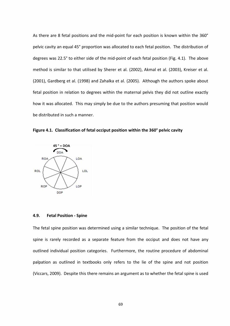
69
As there are 8 fetal positions and the mid-point for each position is known within the 360°
pelvic cavity an equal 45° proportion was allocated to each fetal position. The distribution of
degrees was 22.5° to either side of the mid-point of each fetal position (Fig. 4.1). The above
method is similar to that utilised by Sherer et al. (2002), Akmal et al. (2003), Kreiser et al.
(2001), Gardberg et al. (1998) and Zahalka et al. (2005). Although the authors spoke about
fetal position in relation to degrees within the maternal pelvis they did not outline exactly
how it was allocated. This may simply be due to the authors presuming that position would
be distributed in such a manner.
Figure 4.1. Classification of fetal occiput position within the 360° pelvic cavity
4.9. Fetal Position - Spine
The fetal spine position was determined using a similar technique. The position of the fetal
spine is rarely recorded as a separate feature from the occiput and does not have any
outlined individual position categories. Furthermore, the routine procedure of abdominal
palpation as outlined in textbooks only refers to the lie of the spine and not position
(Viccars, 2009). Despite this there remains an argument as to whether the fetal spine is used
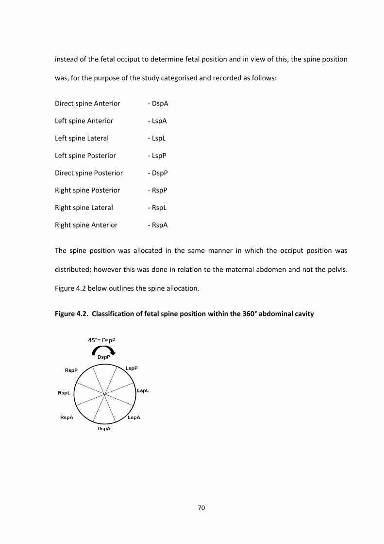
70
instead of the fetal occiput to determine fetal position and in view of this, the spine position
was, for the purpose of the study categorised and recorded as follows:
Direct spine Anterior - DspA
Left spine Anterior - LspA
Left spine Lateral - LspL
Left spine Posterior - LspP
Direct spine Posterior - DspP
Right spine Posterior - RspP
Right spine Lateral - RspL
Right spine Anterior - RspA
The spine position was allocated in the same manner in which the occiput position was
distributed; however this was done in relation to the maternal abdomen and not the pelvis.
Figure 4.2 below outlines the spine allocation.
Figure 4.2. Classification of fetal spine position within the 360° abdominal cavity
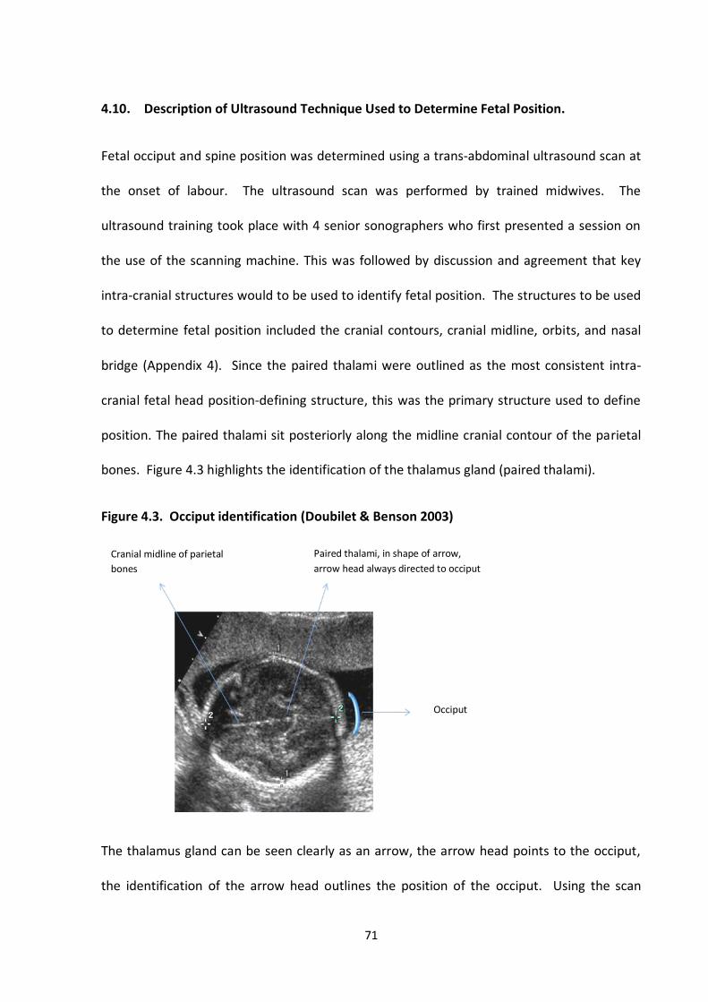
71
4.10. Description of Ultrasound Technique Used to Determine Fetal Position.
Fetal occiput and spine position was determined using a trans-abdominal ultrasound scan at
the onset of labour. The ultrasound scan was performed by trained midwives. The
ultrasound training took place with 4 senior sonographers who first presented a session on
the use of the scanning machine. This was followed by discussion and agreement that key
intra-cranial structures would to be used to identify fetal position. The structures to be used
to determine fetal position included the cranial contours, cranial midline, orbits, and nasal
bridge (Appendix 4). Since the paired thalami were outlined as the most consistent intra-
cranial fetal head position-defining structure, this was the primary structure used to define
position. The paired thalami sit posteriorly along the midline cranial contour of the parietal
bones. Figure 4.3 highlights the identification of the thalamus gland (paired thalami).
Figure 4.3. Occiput identification (Doubilet & Benson 2003)
The thalamus gland can be seen clearly as an arrow, the arrow head points to the occiput,
the identification of the arrow head outlines the position of the occiput. Using the scan
Cranial midline of parietal
bones
Paired thalami, in shape of arrow,
arrow head always directed to occiput
Occiput
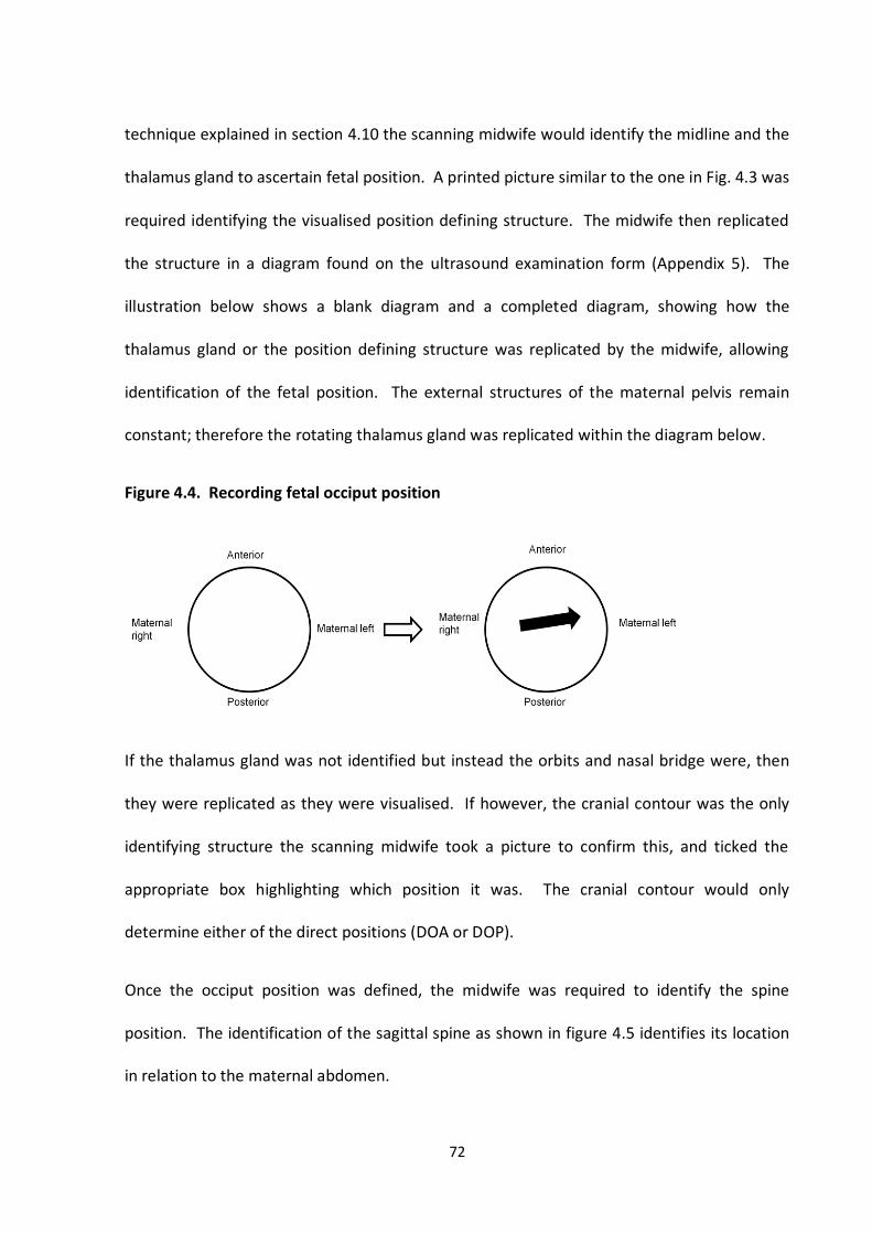
72
technique explained in section 4.10 the scanning midwife would identify the midline and the
thalamus gland to ascertain fetal position. A printed picture similar to the one in Fig. 4.3 was
required identifying the visualised position defining structure. The midwife then replicated
the structure in a diagram found on the ultrasound examination form (Appendix 5). The
illustration below shows a blank diagram and a completed diagram, showing how the
thalamus gland or the position defining structure was replicated by the midwife, allowing
identification of the fetal position. The external structures of the maternal pelvis remain
constant; therefore the rotating thalamus gland was replicated within the diagram below.
Figure 4.4. Recording fetal occiput position
If the thalamus gland was not identified but instead the orbits and nasal bridge were, then
they were replicated as they were visualised. If however, the cranial contour was the only
identifying structure the scanning midwife took a picture to confirm this, and ticked the
appropriate box highlighting which position it was. The cranial contour would only
determine either of the direct positions (DOA or DOP).
Once the occiput position was defined, the midwife was required to identify the spine
position. The identification of the sagittal spine as shown in figure 4.5 identifies its location
in relation to the maternal abdomen.
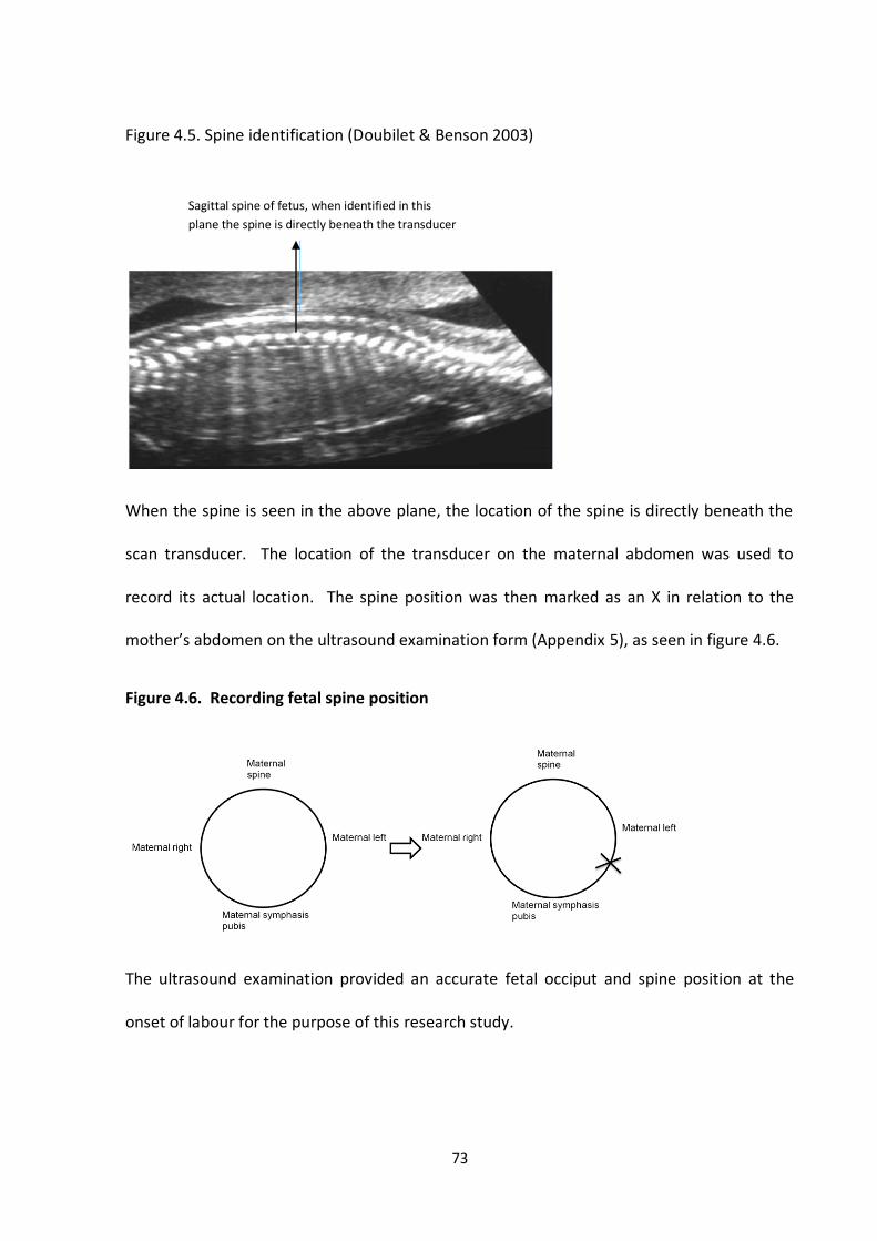
73
Figure 4.5. Spine identification (Doubilet & Benson 2003)
When the spine is seen in the above plane, the location of the spine is directly beneath the
scan transducer. The location of the transducer on the maternal abdomen was used to
record its actual location. The spine position was then marked as an X in relation to the
mother’s abdomen on the ultrasound examination form (Appendix 5), as seen in figure 4.6.
Figure 4.6. Recording fetal spine position
The ultrasound examination provided an accurate fetal occiput and spine position at the
onset of labour for the purpose of this research study.
Sagittal spine of fetus, when identified in this
plane the spine is directly beneath the transducer
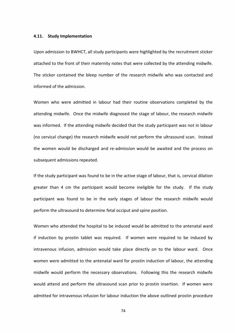
74
4.11. Study Implementation
Upon admission to BWHCT, all study participants were highlighted by the recruitment sticker
attached to the front of their maternity notes that were collected by the attending midwife.
The sticker contained the bleep number of the research midwife who was contacted and
informed of the admission.
Women who were admitted in labour had their routine observations completed by the
attending midwife. Once the midwife diagnosed the stage of labour, the research midwife
was informed. If the attending midwife decided that the study participant was not in labour
(no cervical change) the research midwife would not perform the ultrasound scan. Instead
the women would be discharged and re-admission would be awaited and the process on
subsequent admissions repeated.
If the study participant was found to be in the active stage of labour, that is, cervical dilation
greater than 4 cm the participant would become ineligible for the study. If the study
participant was found to be in the early stages of labour the research midwife would
perform the ultrasound to determine fetal occiput and spine position.
Women who attended the hospital to be induced would be admitted to the antenatal ward
if induction by prostin tablet was required. If women were required to be induced by
intravenous infusion, admission would take place directly on to the labour ward. Once
women were admitted to the antenatal ward for prostin induction of labour, the attending
midwife would perform the necessary observations. Following this the research midwife
would attend and perform the ultrasound scan prior to prostin insertion. If women were
admitted for intravenous infusion for labour induction the above outlined prostin procedure
75
would be followed but ultrasound would take place prior or immediately after
commencement of the intravenous infusion, and before established first stage of labour.
4.12. Obtaining a ‘Valid’ Scan
Once it was confirmed that the study participant was in early labour, the ultrasound scan
could be performed by the research midwife. The attending midwife and the study
participant were blinded to the findings of the ultrasound scan. This ensured that the care
and management of the study participant was not altered or influenced in any way by the
ultrasound findings. In order to obtain a ‘valid’ scan, key observations were mandatory as
follows:
1. The fetal heart was observed and was beating at a normal rate (confirmed and
documented on form B – Appendix 5).
2. The scanning probe was checked for alignment (confirmed and documented on
form B - Appendix 5).
3. Position defining structures were observed within the cranial vault and documented
as seen.
4. The fetal spine was observed and documented as seen.
If a heart rate abnormality was detected at any point, the scan was abandoned and
immediate medical attention sought. If the probe check was not confirmed and
documented the ultrasound scan was not used. The incorrect handling of the probe would
76
alter the direction of the image on the screen therefore fetal position would be
misdiagnosed.
In certain cases the descent of the fetal head into the maternal pelvis meant that the
contents of the cranial vault could not be visualised. Therefore no position defining
structures could be identified. In this instance the scan would be invalid as the test result
would be technically unobtainable.
Although it was not expected, in situations where the research midwife could not define
position of the fetal spine this would also be classed as an ‘invalid’ scan where the test result
was technically unobtainable. When the ultrasound scan was completed the data collection
form was immediately sealed in an envelope and placed in the research file.
A ‘valid’ scan was therefore that which was performed at the onset of labour, with the
observation of both position defining structures present (fetal spine/fetal occiput) and
where the mother was in established first stage of labour within 12 hours of the ultrasound
findings being recorded, only then was a scan classified as a ‘valid’ scan.
Figure 4.7 illustrates the study process following admission to the hospital of a study
participant.
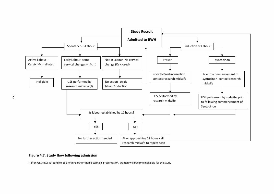
77
(!) If on USS fetus is found to be anything other than a cephalic presentation, women will become ineligible for the study
Figure 4.7. Study flow following admission
Study Recruit
Admitted to BWH
Spontaneous Labour
Active Labour-
Cervix >4cm dilated
Early Labour- some
cervical changes (< 4cm)
Not in Labour- No cervical
change (Os closed)
Ineligible USS performed by
research midwife (!)
No action- await
labour/induction
Induction of Labour
Prostin Syntocinon
Prior to Prostin insertion
contact research midwife
Prior to commencement of
syntocinon contact research
midwife
USS performed by
research midwife USS performed by midwife, prior
to following commencement of
Syntocinon
Is labour established by 12 hours?
YES NO
No further action needed At or approaching 12 hours call
research midwife to repeat scan
78
4.13. Outcome Measures
The outcome measures that were included were determined by two factors: the customary
outcome measures commonly used when investigating factors relating to birth, and those,
that Jean Sutton claimed were improved with the LOA fetal position.
The following definitions of the outcome measures are used at the hospital where the study
was conducted and therefore are also those that were used for the purpose of this study.
4.13.1. Primary outcome
The primary outcome measure was mode of delivery, categorised into 4 main categories as
defined by Tiran (2008):
Spontaneous Vaginal delivery (SVD) - SVD is the complete expulsion of the fetus,
placenta and membranes via the birth canal.
Ventouse delivery (VD) - VD involves the application of an instrument to the fetal
head and use of vacuum or suction to aid delivery of the fetus with maternal
expulsive efforts.
Forceps delivery (FD) - FD involves surgical instruments with two blades applied to
the fetal head to aid rotation and/or delivery. These include Kjellands, Neville Barnes
and Wrigley’s forceps.
Caesarean Section Delivery (CS) - CS involves an operation whereby the fetus is
extracted from the uterus following an incision through the maternal abdomen and
uterine tissue.

79
4.13.2. Secondary outcomes
The secondary outcome measures were:
Augmentation - Augmentation is the acceleration of labour once it has been
diagnosed as not progressing adequately. Inadequate progress is defined by NICE
Intrapartum guidelines as cervical dilatation less then 2cm within a four hour period,
in such a situation labour would be augmented (NICE, 2007). The method used to
augment the labour process is usually performed in order of least severity. Artificial
Rupture of Membranes (ARM) or amniotomy would be performed first. Following
this, intravenous syntocinon infusion would be commenced which is a synthetic
oxytocic drug used to encourage uterine activity. Syntocinon stimulates the uterus
with the aim of increasing strength and duration of uterine contractions. If however,
the membranes have previously ruptured spontaneously, syntocinon would be the
first and only option available to augment labour.
Labour duration - Labour duration was categorised into the active first stage of
labour, the second stage of labour, and duration of membrane rupture. The first
stage of labour was defined by the NICE (2004) intrapartum guidelines as the period
of regular painful contractions, and where there is progressive cervical dilatation
from 4 cm up to 10cm. The second stage of labour is the point at which full cervical
dilatation of 10cm is diagnosed until the delivery of the fetus (Tiran, 2008). Duration
of membrane rupture is defined as the period commencing rupture of the amniotic
sac until the delivery of the fetus (Tiran, 2008).
80
Pain relief - Non-pharmacological forms of pain relief include Transcutaneous
Electrical Neural Stimulation (TENS), inhalation analgesia (entonox) and immersion in
water. Pharmacological forms of pain relief include systemic analgesia such as
Pethidine, which is a sedative and an anti-spasmodic drug. Epidural analgesia
involves an injection of local anaesthetic into the epidural space in the lumbar region
of the spinal cord (Tiran 2008). Other forms of regional and local analgesia are
usually administered prior to intervention or operative delivery in emergency
situations. They include spinal and pudendal block and in certain cases general
anaesthetic.
In many cases women use more than one type of pain relief usually progressing from
non-pharmacological forms to pharmacological forms of pain relief.
Fetal condition at birth - The condition of the neonate was assessed at 1 minute and
5 minutes using the Apgar score. The Apgar score is an acronym for: Activity, Pulse,
Grimace, Appearance and Respiration and is usually assessed by the midwife or
paediatrician at delivery. Each aspect of the Apgar score was assessed with a 0, 1 or
2. A score of 8-10 was considered normal, whilst a score of 4-7 might require some
resuscitative measures, and a neonate with an Apgar score of 3 and below would
require immediate resuscitation.
81
4.14. Data Collection
Data were collected at 3 stages for each study participant. Baseline data was routinely
recorded on maternal parity, age, ethnic group, height, weight and body mass index at the
initial pregnancy booking stage. The data was retrieved for the study following a request to
the informatics department.
The assessment of fetal position required an accessible, accurate and reproducible method
that the ultrasound technique used provided, further highlighting that a consistent data
collection tool was used.
The data collected on fetal position was recorded on an examination sheet (Appendix 5),
verified by an independent clerk who then entered all the data into an excel spreadsheet
ready to merge with the outcome data.
Following delivery of the study participants, outcome data for the primary and secondary
outcome measures which were routinely recorded for all women at the hospital were
obtained from the informatics department. The informatics department was given a list of
the outcome data that was required for each participant. From the list a program was
created and once registration numbers for the study participants were obtained the
program was processed and an excel spreadsheet produced. The spreadsheet incorporated
baseline characteristics and maternal and neonatal outcome data for each study participant.
The obtained data was then merged with the data on fetal position.
82
4.15. Data Verification
The study data were collected from two sources, the scan data collected by the research
midwives and outcome data from the hospital medical system. The scan data were initially
validated by an independent clerk, who inspected all the scans, indentified the accurate fetal
position and entered it into a specifically designed database. A researcher then checked all
1250 entered scans for accuracy. Missing data that was identified was retrieved and
entered. Similarly a researcher inspected all the outcome data comparing dates of scan and
delivery to ensure accuracy. Missing data on delivery outcomes was obtained from
electronic medical records or, maternity notes which were retrieved and thus data was
obtained or clarified. Following this, both the scan data and the requested outcome data
was merged into an excel spreadsheet and transported into STATA, a statistical package
which allowed subsequent analysis.
4.16. Sample Size
There were no data available regarding SVD rates for the LOA position at the onset of labour;
therefore, the sample size of the study was initially computed using the information
available on the proportion of fetuses that present in the OP position at the onset of labour.
The prevalence of the OP position at the onset of labour was anticipated to be 10%
(Gardberg et al., 1998, Gardberg and Tuppurainen, 1994, Fitzpatrick et al., 2001, Sizer and
Nirmal, 2000, Neri et al., 1995, Faber, 1998) and it was assumed that 50% of those
presenting in the OP position at the onset of labour would deliver vaginally (Pearl 1993).
83
To detect an absolute difference of 20% in SVD rates between those that present in the OP
position at the onset of labour and those that do not present in the OP position, with a
significance level of 5% (alpha) and power (1-beta) of 90% at least 125 women with fetuses
in the OP position at the onset of labour were required. As the prevalence of the OP
position was 10% a total of 1250 women were required, that is 125 OP positioned fetuses
and 1125 non-OP positioned fetuses. This calculation actually provided more than 90%
power as the adjustment was conservative. This sample size allowed a smaller absolute
difference, of 15%, between the two groups to be detected with 89% power.
For the employed sample size of 1250, power calculations were performed using SVD rates
among fetuses in the LOA position at the onset of labour as the outcome, and various
combinations of other assumptions. From existing data, an estimate for the local SVD rate
for nulliparous women was 57% (BWH, 2007). For a range of SVD rates (40%, 45%, 50%,
55%, 60%, 65%, 70%, 75% and 80%) for fetuses in the LOA position at the onset of labour,
the power was calculated using 5%, 7.5%, 10%, 12.5% and 15% as the prevalence of the LOA
position at the onset of labour; the results are shown in Table 4.1. The shaded areas
highlight those sections that would allow adequate power to detect effect size.
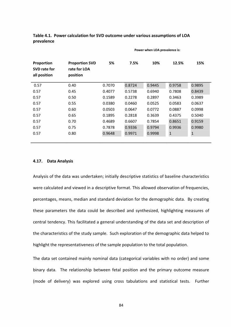
84
Table 4.1. Power calculation for SVD outcome under various assumptions of LOA prevalence
Power when LOA prevalence is:
Proportion
SVD rate for
all position
Proportion SVD
rate for LOA
position
5% 7.5% 10% 12.5% 15%
0.57
0.57
0.57
0.57
0.57
0.57
0.57
0.57
0.57
0.40
0.45
0.50
0.55
0.60
0.65
0.70
0.75
0.80
0.7070
0.4077
0.1589
0.0380
0.0503
0.1895
0.4689
0.7878
0.9648
0.8724
0.5738
0.2278
0.0460
0.0647
0.2818
0.6607
0.9336
0.9971
0.9445
0.6940
0.2897
0.0525
0.0772
0.3639
0.7854
0.9794
0.9998
0.9758
0.7808
0.3463
0.0583
0.0887
0.4375
0.8651
0.9936
1
0.9895
0.8439
0.3989
0.0637
0.0998
0.5040
0.9159
0.9980
1
4.17. Data Analysis
Analysis of the data was undertaken; initially descriptive statistics of baseline characteristics
were calculated and viewed in a descriptive format. This allowed observation of frequencies,
percentages, means, median and standard deviation for the demographic data. By creating
these parameters the data could be described and synthesized, highlighting measures of
central tendency. This facilitated a general understanding of the data set and description of
the characteristics of the study sample. Such exploration of the demographic data helped to
highlight the representativeness of the sample population to the total population.
The data set contained mainly nominal data (categorical variables with no order) and some
binary data. The relationship between fetal position and the primary outcome measure
(mode of delivery) was explored using cross tabulations and statistical tests. Further
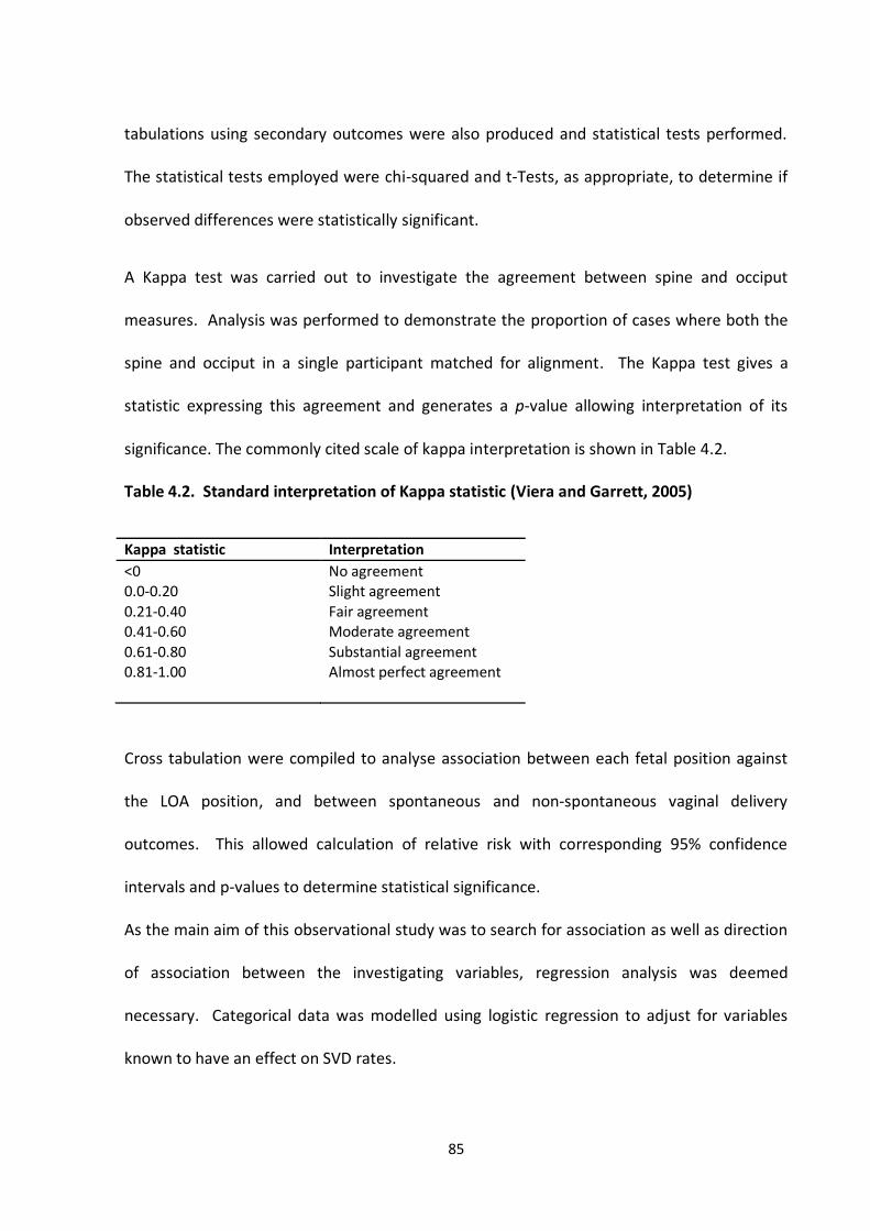
85
tabulations using secondary outcomes were also produced and statistical tests performed.
The statistical tests employed were chi-squared and t-Tests, as appropriate, to determine if
observed differences were statistically significant.
A Kappa test was carried out to investigate the agreement between spine and occiput
measures. Analysis was performed to demonstrate the proportion of cases where both the
spine and occiput in a single participant matched for alignment. The Kappa test gives a
statistic expressing this agreement and generates a p-value allowing interpretation of its
significance. The commonly cited scale of kappa interpretation is shown in Table 4.2.
Table 4.2. Standard interpretation of Kappa statistic (Viera and Garrett, 2005)
Kappa statistic Interpretation
<0 No agreement 0.0-0.20 Slight agreement
0.21-0.40 Fair agreement 0.41-0.60 Moderate agreement
0.61-0.80 Substantial agreement 0.81-1.00 Almost perfect agreement
Cross tabulation were compiled to analyse association between each fetal position against
the LOA position, and between spontaneous and non-spontaneous vaginal delivery
outcomes. This allowed calculation of relative risk with corresponding 95% confidence
intervals and p-values to determine statistical significance.
As the main aim of this observational study was to search for association as well as direction
of association between the investigating variables, regression analysis was deemed
necessary. Categorical data was modelled using logistic regression to adjust for variables
known to have an effect on SVD rates.
86
CHAPTER 5
RESULTS
5.1. Assembly of Study Cohort
The study obtained full scan and outcome data for the required 1250 participants within the
18 month study period of April 2007 to September 2008. During the study period, there
were 2838 births to nulliparous women at the BWH. Of these deliveries, 803 women were
not eligible as they did not meet one or more of the study eligibility criteria.
Of the eligible population of 2035 women prior to labour onset, 80.9% (1647/2035)
consented to take part in the study and were scanned for the study. The remaining 388
women were lost as they either did not consent or consented and were not scanned or
arrived at the hospital in established labour and were therefore ineligible for the study. No
recorded data was available on the proportion of women admitted in established labour at
BWHCT during the study period or at any other period. It was therefore difficult to estimate
what proportion of the 388 women were ineligible as they arrived in established labour and
what proportion were not consented or were not scanned.
Of the 1647 women who consented and were scanned 349 were subsequently lost as shown
in Figure 5.1. In a further 48 cases the test result was technically unobtainable, as the fetal
head was too low to determine fetal occiput position, these women were subsequently
removed from the study. The final number of women for whom a valid scan was available
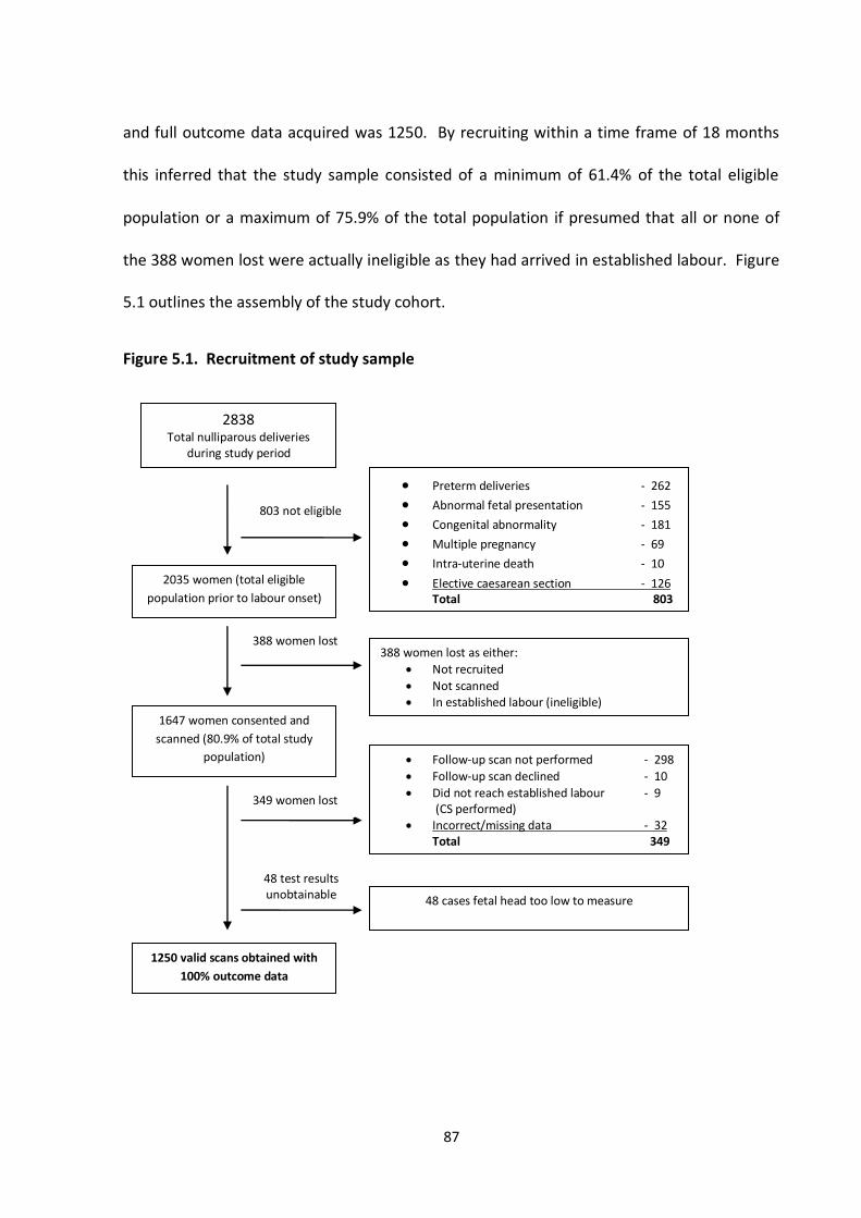
87
and full outcome data acquired was 1250. By recruiting within a time frame of 18 months
this inferred that the study sample consisted of a minimum of 61.4% of the total eligible
population or a maximum of 75.9% of the total population if presumed that all or none of
the 388 women lost were actually ineligible as they had arrived in established labour. Figure
5.1 outlines the assembly of the study cohort.
Figure 5.1. Recruitment of study sample
2838
Total nulliparous deliveries during study period
2035 women (total eligible
population prior to labour onset)
1647 women consented and
scanned (80.9% of total study
population)
1250 valid scans obtained with
100% outcome data
Preterm deliveries - 262 Abnormal fetal presentation - 155 Congenital abnormality - 181 Multiple pregnancy - 69 Intra-uterine death - 10 Elective caesarean section - 126
Total 803
388 women lost as either:
Not recruited
Not scanned In established labour (ineligible)
Follow-up scan not performed - 298
Follow-up scan declined - 10
Did not reach established labour - 9 (CS performed)
Incorrect/missing data - 32 Total 349
48 cases fetal head too low to measure
803 not eligible
388 women lost
349 women lost
48 test results unobtainable
88
5.2. Maternal Characteristics
All women recruited to the study were nulliparous defined as having no live births or
previous pregnancies exceeding the first trimester. In terms of gravida, 95% of the sample
were gravida one or gravida two and had either never been pregnant before or previously
had 1 first trimester miscarriage. The remaining 5.0% of the study recruits had 2 or more
miscarriages with a single participant having seven 1st trimester miscarriages. The mean age
for the study participants was 26 years. The largest group was between the ages of 20-25
and accounted for 31.3% of the total study population.
The largest ethnic group in the study was the White British group that made up almost 56%
of the total sample population. The second largest group of participants was the Pakistani
group at 15%, followed by the Indian ethnic group (6.3%).
The country of origin showed that almost 72.0% of the population were from the United
Kingdom. This was followed by 7.7% of the population which originated from Pakistan and
2.5% from India. The remaining 6.5% consisted of smaller groups of women who originated
from Poland, China, Yemen, Bangladesh and Ireland, 11.4% of the sample however consisted
of the ‘other group’.
The BMI categories showed that the majority of women (50.8%) were within the normal BMI
range (18.5 -29.9). Almost 28% of the sample population had a BMI that was classified as
over-weight. 16.0% of women had a BMI that was classed as obese, with 1.7% of women in
the very obese category. The mean height of the study population was 161cm and the mean
weight was 67kg.
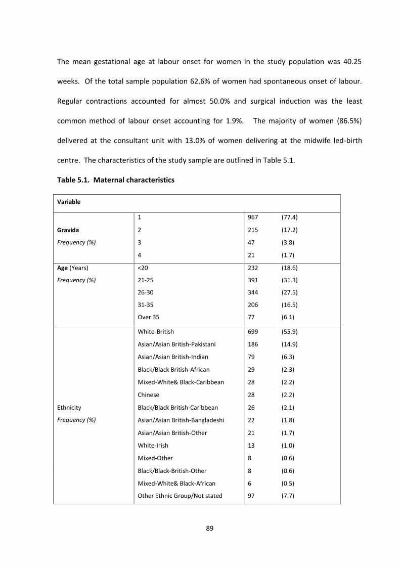
89
The mean gestational age at labour onset for women in the study population was 40.25
weeks. Of the total sample population 62.6% of women had spontaneous onset of labour.
Regular contractions accounted for almost 50.0% and surgical induction was the least
common method of labour onset accounting for 1.9%. The majority of women (86.5%)
delivered at the consultant unit with 13.0% of women delivering at the midwife led-birth
centre. The characteristics of the study sample are outlined in Table 5.1.
Table 5.1. Maternal characteristics
Variable
Gravida
Frequency (%)
1 967 (77.4)
2 215 (17.2)
3 47 (3.8)
4 21 (1.7)
Age (Years)
Frequency (%)
<20
21-25
26-30
31-35
Over 35
232
391
344
206
77
(18.6)
(31.3)
(27.5)
(16.5)
(6.1)
Ethnicity
Frequency (%)
White-British 699 (55.9)
Asian/Asian British-Pakistani 186 (14.9)
Asian/Asian British-Indian 79 (6.3)
Black/Black British-African 29 (2.3)
Mixed-White& Black-Caribbean 28 (2.2)
Chinese 28 (2.2)
Black/Black British-Caribbean 26 (2.1)
Asian/Asian British-Bangladeshi 22 (1.8)
Asian/Asian British-Other 21 (1.7)
White-Irish 13 (1.0)
Mixed-Other 8 (0.6)
Black/Black-British-Other 8 (0.6)
Mixed-White& Black-African 6 (0.5)
Other Ethnic Group/Not stated 97 (7.7)

90
Table 5.1. Maternal characteristics (Continued)
Country of Origin
Frequency (%)
United Kingdom
Pakistan
899 (71.9)
97 (7.8)
India 32 (2.6)
Poland 21 (1.7)
China 18 (1.1)
Yemen 17 (1.4)
Bangladesh 14 (1.1)
S. Ireland
Other
10
142
(0.8)
(11.4)
BMI
Frequency (%)
≤ 18.4
18.5-24.9
25-29.9
30-40
≥40
44
636
349
200
21
(3.2)
(50.8)
(27.9)
(16.0)
(1.7)
Height (cm)
mean (sd)
161.80 (6.98)
Gestation (Weeks)
mean (sd)
40.25 (1.2)
Onset of Labour
Frequency (%)
Regular Contractions 614 (49.1)
Prostin 357 (28.6)
S.R.O.M 164 (13.1)
Surgical Induction & Prostin 33 (2.6)
Surgical Induction & Oxytocic 31 (2.5)
Oxytocic Drugs 25 (2.0)
Surgical Induction 24 (1.9)
ARM 2 (0.2)
Place of Delivery
Frequency (%)
BWH (Consultant Unit) 1,082 (86.5)
BWH (Birth Centre) 168 (13.4)
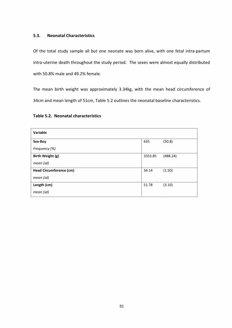
91
5.3. Neonatal Characteristics
Of the total study sample all but one neonate was born alive, with one fetal intra-partum
intra-uterine death throughout the study period. The sexes were almost equally distributed
with 50.8% male and 49.2% female.
The mean birth weight was approximately 3.34kg, with the mean head circumference of
34cm and mean length of 51cm, Table 5.2 outlines the neonatal baseline characteristics.
Table 5.2. Neonatal characteristics
Variable
Sex-Boy
Frequency (%)
635 (50.8)
Birth Weight (g)
mean (sd)
3353.85 (488.24)
Head Circumference (cm)
mean (sd)
34.14 (1.50)
Length (cm)
mean (sd)
51.78 (3.10)
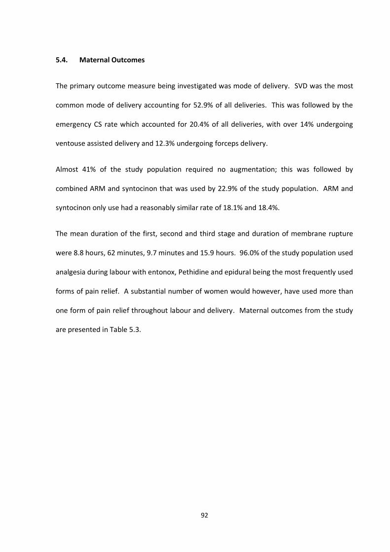
92
5.4. Maternal Outcomes
The primary outcome measure being investigated was mode of delivery. SVD was the most
common mode of delivery accounting for 52.9% of all deliveries. This was followed by the
emergency CS rate which accounted for 20.4% of all deliveries, with over 14% undergoing
ventouse assisted delivery and 12.3% undergoing forceps delivery.
Almost 41% of the study population required no augmentation; this was followed by
combined ARM and syntocinon that was used by 22.9% of the study population. ARM and
syntocinon only use had a reasonably similar rate of 18.1% and 18.4%.
The mean duration of the first, second and third stage and duration of membrane rupture
were 8.8 hours, 62 minutes, 9.7 minutes and 15.9 hours. 96.0% of the study population used
analgesia during labour with entonox, Pethidine and epidural being the most frequently used
forms of pain relief. A substantial number of women would however, have used more than
one form of pain relief throughout labour and delivery. Maternal outcomes from the study
are presented in Table 5.3.
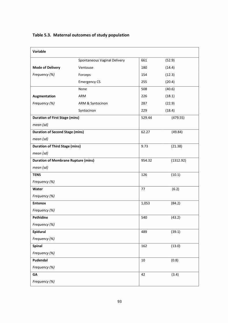
93
Table 5.3. Maternal outcomes of study population
Variable
Mode of Delivery
Frequency (%)
Spontaneous Vaginal Delivery 661 (52.9)
Ventouse 180 (14.4)
Forceps
Emergency CS
154
255
(12.3)
(20.4)
Augmentation
Frequency (%)
None
ARM
508
226
(40.6)
(18.1)
ARM & Syntocinon 287 (22.9)
Syntocinon 229 (18.4)
Duration of First Stage (mins)
mean (sd)
529.44 (479.55)
Duration of Second Stage (mins)
mean (sd)
62.27 (49.84)
Duration of Third Stage (mins)
mean (sd)
9.73 (21.38)
Duration of Membrane Rupture (mins)
mean (sd)
954.32 (1312.92)
TENS
Frequency (%)
126 (10.1)
Water
Frequency (%)
77 (6.2)
Entonox
Frequency (%)
1,053 (84.2)
Pethidine
Frequency (%)
540 (43.2)
Epidural
Frequency (%)
489 (39.1)
Spinal
Frequency (%)
162 (13.0)
Pudendal
Frequency (%)
10 (0.8)
GA
Frequency (%)
42 (3.4)
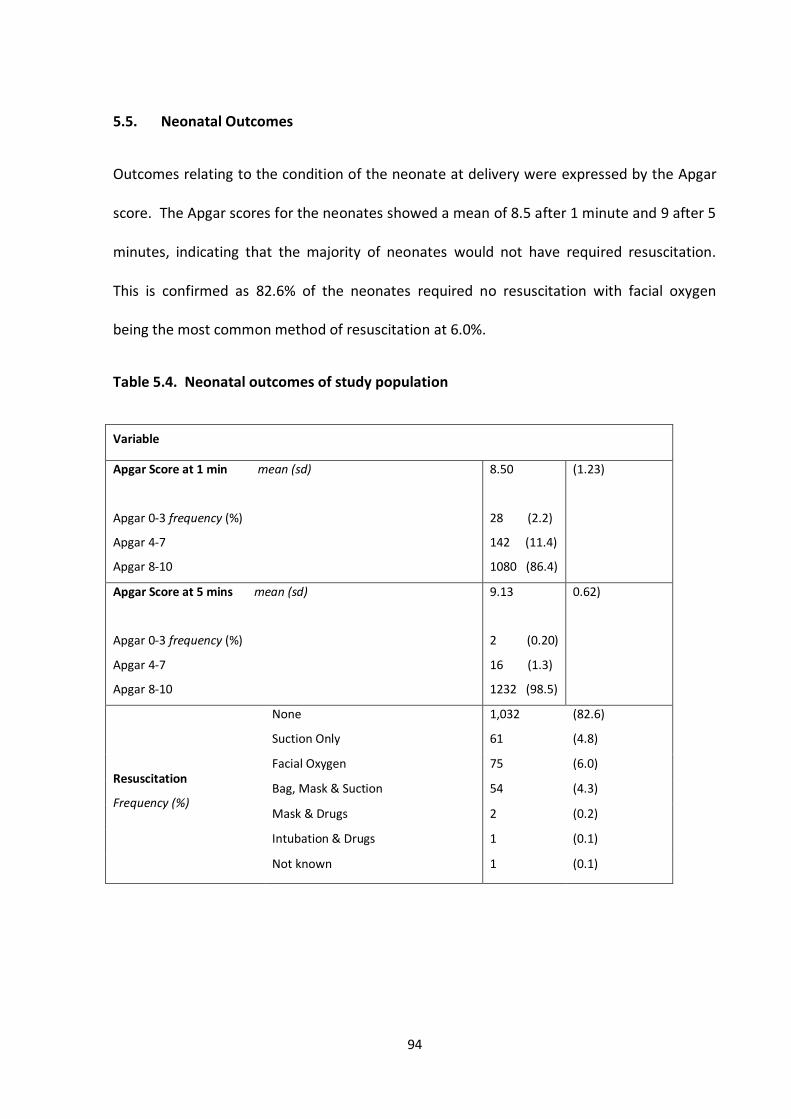
94
5.5. Neonatal Outcomes
Outcomes relating to the condition of the neonate at delivery were expressed by the Apgar
score. The Apgar scores for the neonates showed a mean of 8.5 after 1 minute and 9 after 5
minutes, indicating that the majority of neonates would not have required resuscitation.
This is confirmed as 82.6% of the neonates required no resuscitation with facial oxygen
being the most common method of resuscitation at 6.0%.
Table 5.4. Neonatal outcomes of study population
Variable
Apgar Score at 1 min mean (sd)
Apgar 0-3 frequency (%)
Apgar 4-7
Apgar 8-10
8.50
28 (2.2)
142 (11.4)
1080 (86.4)
(1.23)
Apgar Score at 5 mins mean (sd)
Apgar 0-3 frequency (%)
Apgar 4-7
Apgar 8-10
9.13
2 (0.20)
16 (1.3)
1232 (98.5)
0.62)
Resuscitation
Frequency (%)
None
Suction Only
1,032
61
(82.6)
(4.8)
Facial Oxygen 75 (6.0)
Bag, Mask & Suction 54 (4.3)
Mask & Drugs 2 (0.2)
Intubation & Drugs 1 (0.1)
Not known 1 (0.1)
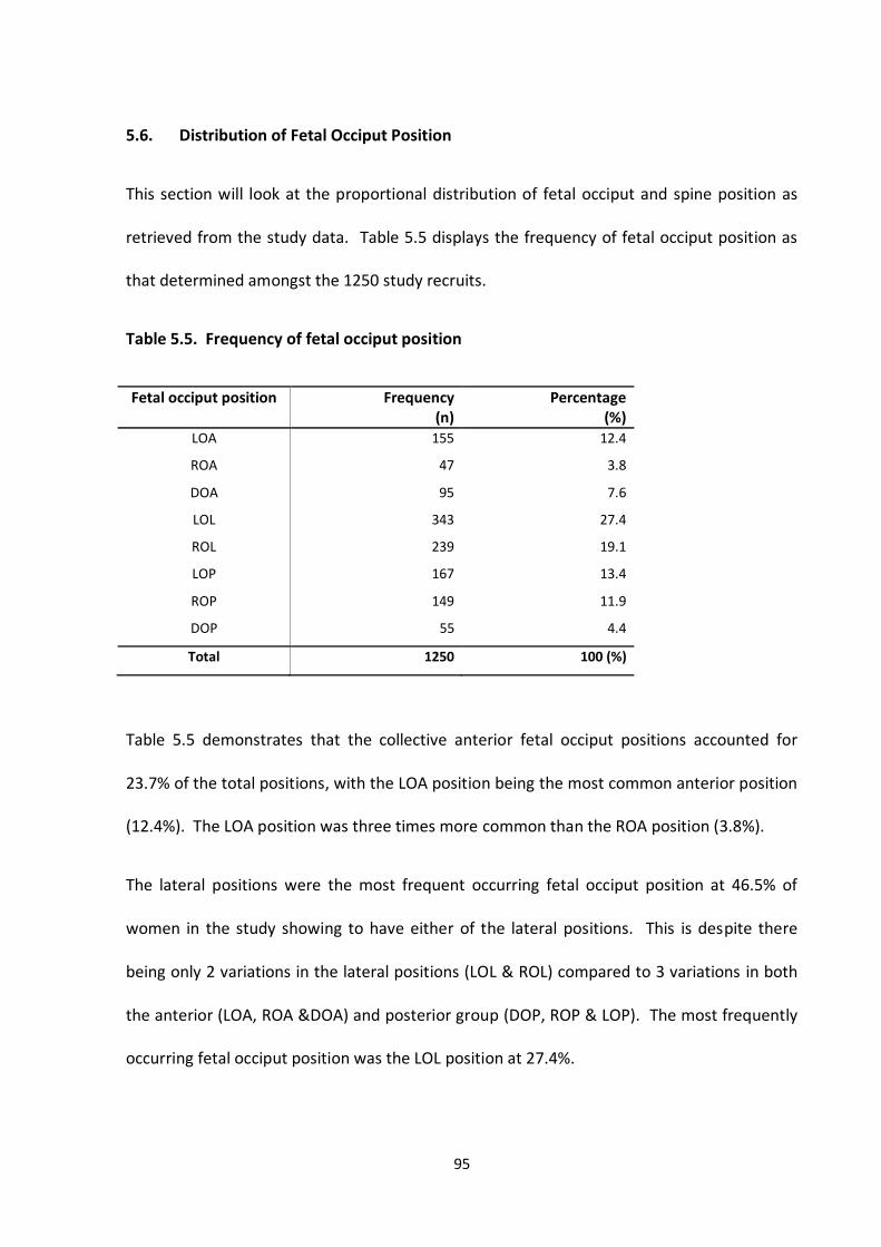
95
5.6. Distribution of Fetal Occiput Position
This section will look at the proportional distribution of fetal occiput and spine position as
retrieved from the study data. Table 5.5 displays the frequency of fetal occiput position as
that determined amongst the 1250 study recruits.
Table 5.5. Frequency of fetal occiput position
Fetal occiput position
Frequency (n)
Percentage (%)
LOA 155 12.4
ROA 47 3.8
DOA 95 7.6
LOL
ROL
LOP
ROP
DOP
343
239
167
149
55
27.4
19.1
13.4
11.9
4.4
Total 1250 100 (%)
Table 5.5 demonstrates that the collective anterior fetal occiput positions accounted for
23.7% of the total positions, with the LOA position being the most common anterior position
(12.4%). The LOA position was three times more common than the ROA position (3.8%).
The lateral positions were the most frequent occurring fetal occiput position at 46.5% of
women in the study showing to have either of the lateral positions. This is despite there
being only 2 variations in the lateral positions (LOL & ROL) compared to 3 variations in both
the anterior (LOA, ROA &DOA) and posterior group (DOP, ROP & LOP). The most frequently
occurring fetal occiput position was the LOL position at 27.4%.
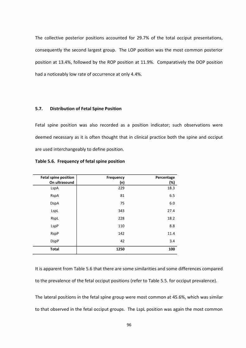
96
The collective posterior positions accounted for 29.7% of the total occiput presentations,
consequently the second largest group. The LOP position was the most common posterior
position at 13.4%, followed by the ROP position at 11.9%. Comparatively the DOP position
had a noticeably low rate of occurrence at only 4.4%.
5.7. Distribution of Fetal Spine Position
Fetal spine position was also recorded as a position indicator; such observations were
deemed necessary as it is often thought that in clinical practice both the spine and occiput
are used interchangeably to define position.
Table 5.6. Frequency of fetal spine position
Fetal spine position On ultrasound
Frequency (n)
Percentage (%)
LspA 229 18.3
RspA 81 6.5
DspA 75 6.0
LspL
RspL
LspP
RspP
DspP
343
228
110
142
42
27.4
18.2
8.8
11.4
3.4
Total 1250 100
It is apparent from Table 5.6 that there are some similarities and some differences compared
to the prevalence of the fetal occiput positions (refer to Table 5.5. for occiput prevalence).
The lateral positions in the fetal spine group were most common at 45.6%, which was similar
to that observed in the fetal occiput groups. The LspL position was again the most common
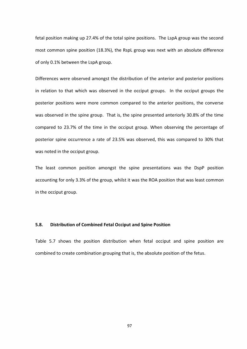
97
fetal position making up 27.4% of the total spine positions. The LspA group was the second
most common spine position (18.3%), the RspL group was next with an absolute difference
of only 0.1% between the LspA group.
Differences were observed amongst the distribution of the anterior and posterior positions
in relation to that which was observed in the occiput groups. In the occiput groups the
posterior positions were more common compared to the anterior positions, the converse
was observed in the spine group. That is, the spine presented anteriorly 30.8% of the time
compared to 23.7% of the time in the occiput group. When observing the percentage of
posterior spine occurrence a rate of 23.5% was observed, this was compared to 30% that
was noted in the occiput group.
The least common position amongst the spine presentations was the DspP position
accounting for only 3.3% of the group, whilst it was the ROA position that was least common
in the occiput group.
5.8. Distribution of Combined Fetal Occiput and Spine Position
Table 5.7 shows the position distribution when fetal occiput and spine position are
combined to create combination grouping that is, the absolute position of the fetus.
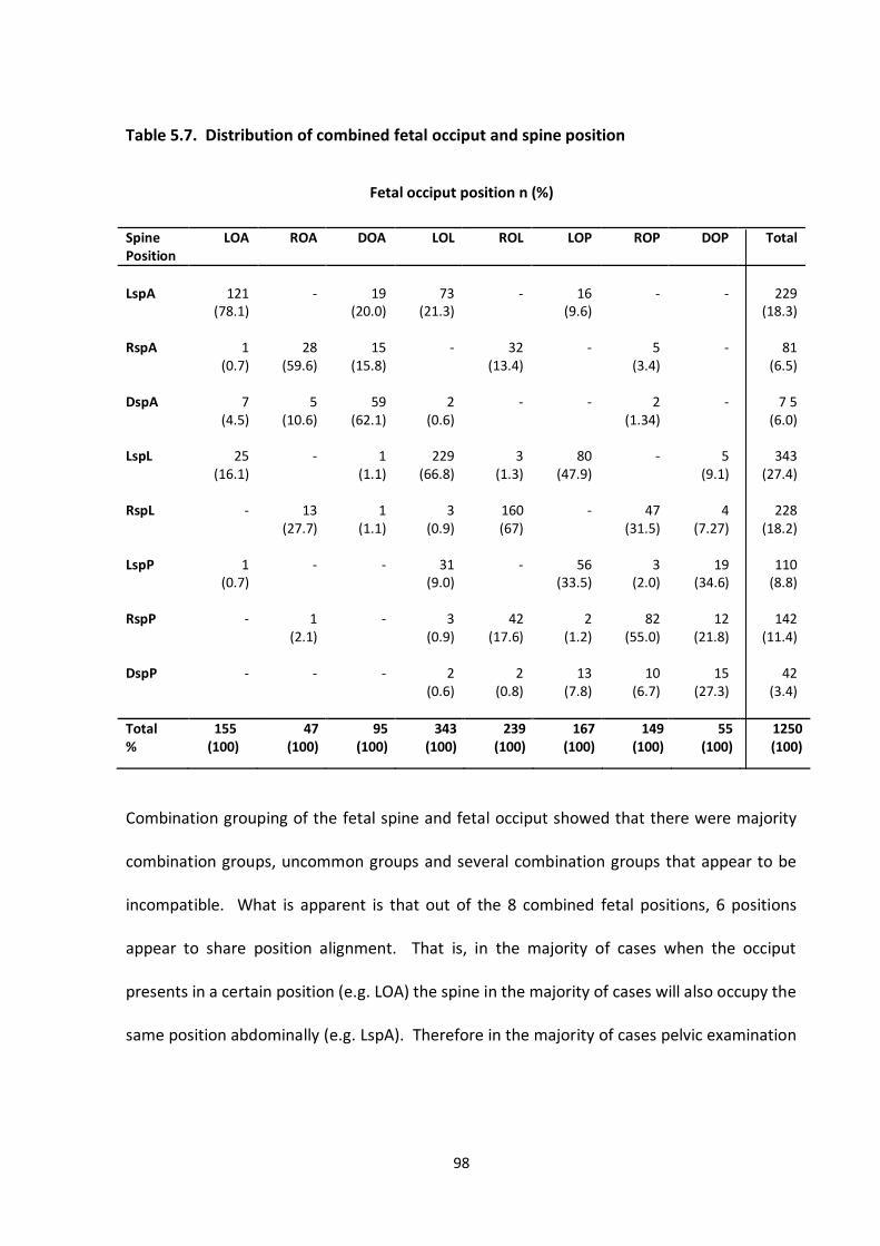
98
Table 5.7. Distribution of combined fetal occiput and spine position
Fetal occiput position n (%)
Spine Position
LOA ROA DOA LOL ROL LOP ROP DOP Total
LspA
121 (78.1)
-
19 (20.0)
73 (21.3)
-
16 (9.6)
-
-
229 (18.3)
RspA
1
(0.7)
28
(59.6)
15
(15.8)
-
32
(13.4)
-
5
(3.4)
-
81
(6.5) DspA
7
(4.5)
5
(10.6)
59
(62.1)
2
(0.6)
-
-
2
(1.34)
-
7 5
(6.0) LspL
25
(16.1)
-
1
(1.1)
229
(66.8)
3
(1.3)
80
(47.9)
-
5
(9.1)
343
(27.4) RspL LspP
-
1 (0.7)
13
(27.7)
-
1
(1.1)
-
3
(0.9)
31 (9.0)
160 (67)
-
-
56 (33.5)
47
(31.5)
3 (2.0)
4
(7.27)
19 (34.6)
228
(18.2)
110 (8.8)
RspP
-
1
(2.1)
-
3
(0.9)
42
(17.6)
2
(1.2)
82
(55.0)
12
(21.8)
142
(11.4)
DspP - - - 2 (0.6)
2 (0.8)
13 (7.8)
10 (6.7)
15 (27.3)
42 (3.4)
Total %
155 (100)
47 (100)
95 (100)
343 (100)
239 (100)
167 (100)
149 55 (100)
55 (100)
1250 (100)
Combination grouping of the fetal spine and fetal occiput showed that there were majority
combination groups, uncommon groups and several combination groups that appear to be
incompatible. What is apparent is that out of the 8 combined fetal positions, 6 positions
appear to share position alignment. That is, in the majority of cases when the occiput
presents in a certain position (e.g. LOA) the spine in the majority of cases will also occupy the
same position abdominally (e.g. LspA). Therefore in the majority of cases pelvic examination
99
to determine occiput presentation should be synonymous to spine position on abdominal
examination. Alignment ranged from 55% in the ROP&RspP to 78% in the LOA&LspA group.
The two positions that did not share occiput and spine alignment were the DOP&DspP
combination, and the LOP&LspP combination. The most frequently occurring combination
for the DOP position was the LspP, and for the LOP position was the LspL spine position.
With the two combination groups that did not share position alignment, in both cases they
were posterior occiput positions that did not share alignment with the corresponding spine
position.
The most frequently occurring combination group was the occiput LOL with spine LspL group
(LOL&LspL). This was followed by the ROL&RspL group, demonstrating that the lateral
positions were the most frequently occurring fetal positions overall. The third most
common combination group was the LOA&LspA. There were several combination groups
that had a frequency of 0, that is, of the 1250 scans performed this combination of spine and
occiput position did not occur.
A weighed Kappa test allowed a Kappa statistic to be reported which indicated the
magnitude of agreement between the two observations (Viera and Garrett, 2005). The
Kappa statistic allowed the magnitude of agreement between fetal occiput and spine
position to be assessed.
To calculate the statistic, agreement was categorised as 1 for total agreement of spine and
occiput position (e.g. LOA + LspA = 1). When the spine position was one either side from
total agreement 0.5 was awarded, the remaining combinations further then the first

100
immediate position either side were awarded 0. Figure 5.2 expresses the allocation used to
calculate the Kappa statistic.
Figure 5.2. Distribution of score used to calculate Kappa Statistic (example DOA and DspA)
Using the above distribution the following Kappa statistic was calculated.
Agreement Expected Agreement Kappa P-Value
77.7% 29.4% 0.68 p<0.001
The Kappa test showed that the expected agreement through chance alone was calculated
to be 29.4%, the actual agreement was 77.7%, with a Kappa statistic of 0.68 and a p-value of
<0.001. Viera and Garrett (2005) Kappa interpretation of a statistic of 0.68 translates to
‘substantial agreement’ between occiput and spine position.
In relation to clinical practice ‘substantial agreement’ would suggest that in the majority of
cases determining spine position would in the greater part express the occiput position.
DOA+DspA=1 (Total agreement)
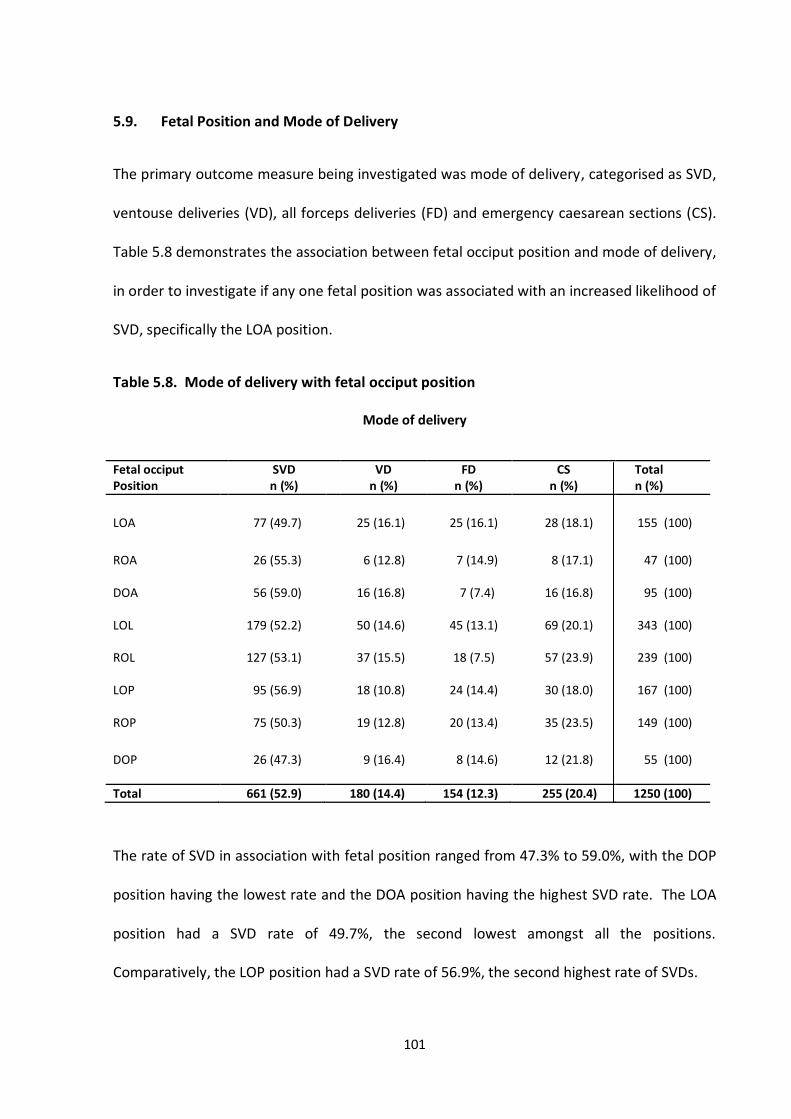
101
5.9. Fetal Position and Mode of Delivery
The primary outcome measure being investigated was mode of delivery, categorised as SVD,
ventouse deliveries (VD), all forceps deliveries (FD) and emergency caesarean sections (CS).
Table 5.8 demonstrates the association between fetal occiput position and mode of delivery,
in order to investigate if any one fetal position was associated with an increased likelihood of
SVD, specifically the LOA position.
Table 5.8. Mode of delivery with fetal occiput position
Mode of delivery
Fetal occiput Position
SVD n (%)
VD n (%)
FD n (%)
CS n (%)
Total n (%)
LOA 77 (49.7) 25 (16.1) 25 (16.1) 28 (18.1) 155 (100)
ROA 26 (55.3) 6 (12.8) 7 (14.9) 8 (17.1) 47 (100)
DOA 56 (59.0) 16 (16.8) 7 (7.4) 16 (16.8) 95 (100)
LOL 179 (52.2) 50 (14.6) 45 (13.1) 69 (20.1) 343 (100)
ROL 127 (53.1) 37 (15.5) 18 (7.5) 57 (23.9)
239 (100)
LOP 95 (56.9) 18 (10.8) 24 (14.4) 30 (18.0) 167 (100)
ROP 75 (50.3) 19 (12.8) 20 (13.4) 35 (23.5) 149 (100)
DOP 26 (47.3) 9 (16.4) 8 (14.6) 12 (21.8)
55 (100)
Total 661 (52.9) 180 (14.4) 154 (12.3) 255 (20.4) 1250 (100)
The rate of SVD in association with fetal position ranged from 47.3% to 59.0%, with the DOP
position having the lowest rate and the DOA position having the highest SVD rate. The LOA
position had a SVD rate of 49.7%, the second lowest amongst all the positions.
Comparatively, the LOP position had a SVD rate of 56.9%, the second highest rate of SVDs.
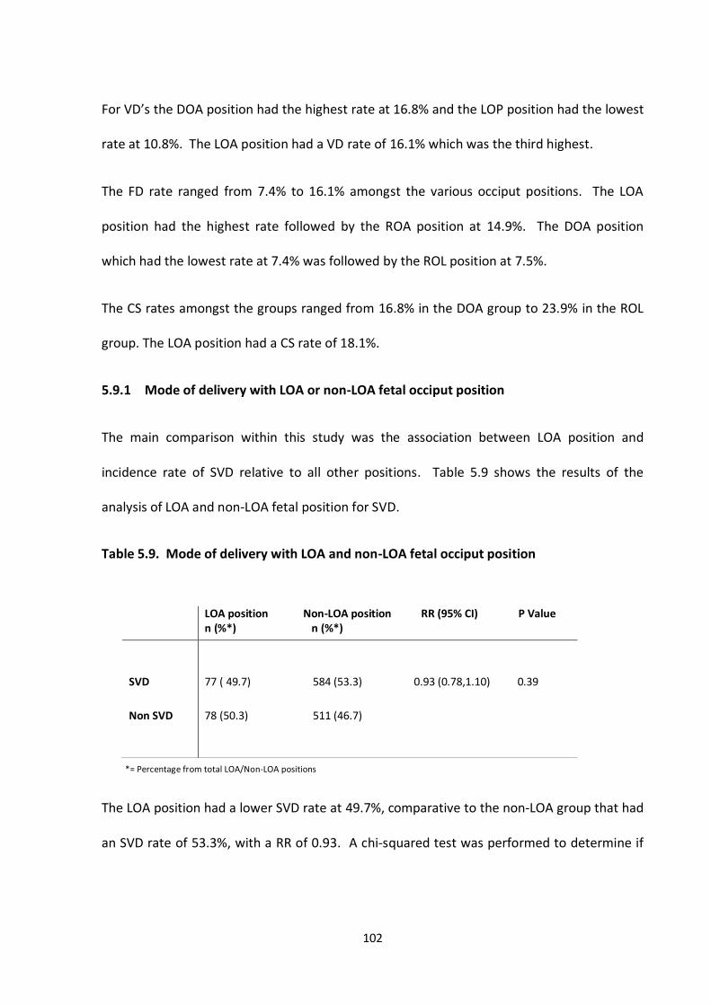
102
For VD’s the DOA position had the highest rate at 16.8% and the LOP position had the lowest
rate at 10.8%. The LOA position had a VD rate of 16.1% which was the third highest.
The FD rate ranged from 7.4% to 16.1% amongst the various occiput positions. The LOA
position had the highest rate followed by the ROA position at 14.9%. The DOA position
which had the lowest rate at 7.4% was followed by the ROL position at 7.5%.
The CS rates amongst the groups ranged from 16.8% in the DOA group to 23.9% in the ROL
group. The LOA position had a CS rate of 18.1%.
5.9.1 Mode of delivery with LOA or non-LOA fetal occiput position
The main comparison within this study was the association between LOA position and
incidence rate of SVD relative to all other positions. Table 5.9 shows the results of the
analysis of LOA and non-LOA fetal position for SVD.
Table 5.9. Mode of delivery with LOA and non-LOA fetal occiput position
*= Percentage from total LOA/Non-LOA positions
The LOA position had a lower SVD rate at 49.7%, comparative to the non-LOA group that had
an SVD rate of 53.3%, with a RR of 0.93. A chi-squared test was performed to determine if
LOA position Non-LOA position RR (95% CI) P Value n (%*) n (%*)
SVD
Non SVD
77 ( 49.7) 584 (53.3) 0.93 (0.78,1.10) 0.39
78 (50.3) 511 (46.7)
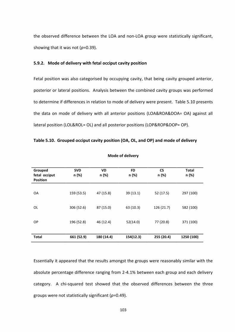
103
the observed difference between the LOA and non-LOA group were statistically significant,
showing that it was not (p=0.39).
5.9.2. Mode of delivery with fetal occiput cavity position
Fetal position was also categorised by occupying cavity, that being cavity grouped anterior,
posterior or lateral positions. Analysis between the combined cavity groups was performed
to determine if differences in relation to mode of delivery were present. Table 5.10 presents
the data on mode of delivery with all anterior positions (LOA&ROA&DOA= OA) against all
lateral position (LOL&ROL= OL) and all posterior positions (LOP&ROP&DOP= OP).
Table 5.10. Grouped occiput cavity position (OA, OL, and OP) and mode of delivery
Mode of delivery
Grouped fetal occiput Position
SVD n (%)
VD n (%)
FD n (%)
CS n (%)
Total n (%)
OA
OL
OP
159 (53.5)
306 (52.6)
196 (52.8)
47 (15.8)
87 (15.0)
46 (12.4)
39 (13.1)
63 (10.3)
52(14.0)
52 (17.5)
126 (21.7)
77 (20.8)
297 (100)
582 (100)
371 (100)
Total 661 (52.9) 180 (14.4) 154(12.3) 255 (20.4) 1250 (100)
Essentially it appeared that the results amongst the groups were reasonably similar with the
absolute percentage difference ranging from 2-4.1% between each group and each delivery
category. A chi-squared test showed that the observed differences between the three
groups were not statistically significant (p=0.49).
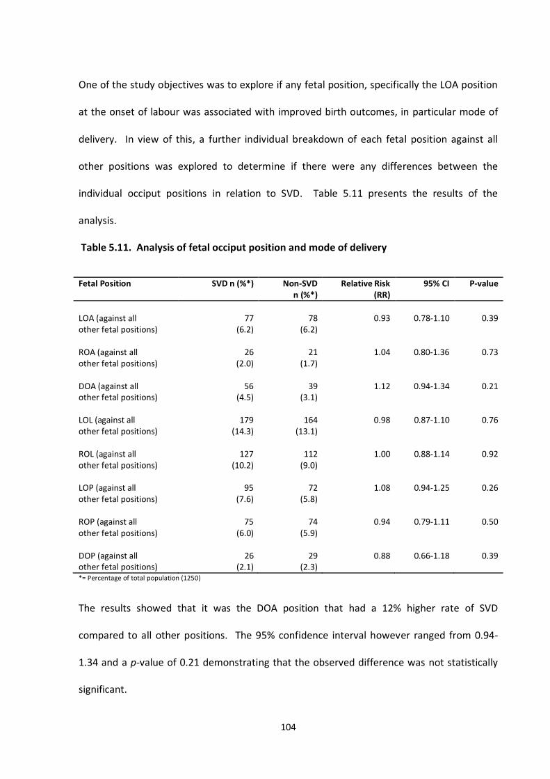
104
One of the study objectives was to explore if any fetal position, specifically the LOA position
at the onset of labour was associated with improved birth outcomes, in particular mode of
delivery. In view of this, a further individual breakdown of each fetal position against all
other positions was explored to determine if there were any differences between the
individual occiput positions in relation to SVD. Table 5.11 presents the results of the
analysis.
Table 5.11. Analysis of fetal occiput position and mode of delivery
Fetal Position SVD n (%*) Non-SVD n (%*)
Relative Risk (RR)
95% CI P-value
LOA (against all other fetal positions) ROA (against all other fetal positions) DOA (against all other fetal positions) LOL (against all other fetal positions) ROL (against all other fetal positions) LOP (against all other fetal positions) ROP (against all other fetal positions) DOP (against all other fetal positions)
77 (6.2)
26 (2.0)
56 (4.5)
179 (14.3)
127 (10.2)
95 (7.6)
75 (6.0)
26 (2.1)
78
(6.2)
21 (1.7)
39 (3.1)
164 (13.1)
112 (9.0)
72 (5.8)
74 (5.9)
29 (2.3)
0.93
1.04
1.12
0.98
1.00
1.08
0.94
0.88
0.78-1.10
0.80-1.36
0.94-1.34
0.87-1.10
0.88-1.14
0.94-1.25
0.79-1.11
0.66-1.18
0.39
0.73
0.21
0.76
0.92
0.26
0.50
0.39
*= Percentage of total population (1250)
The results showed that it was the DOA position that had a 12% higher rate of SVD
compared to all other positions. The 95% confidence interval however ranged from 0.94-
1.34 and a p-value of 0.21 demonstrating that the observed difference was not statistically
significant.
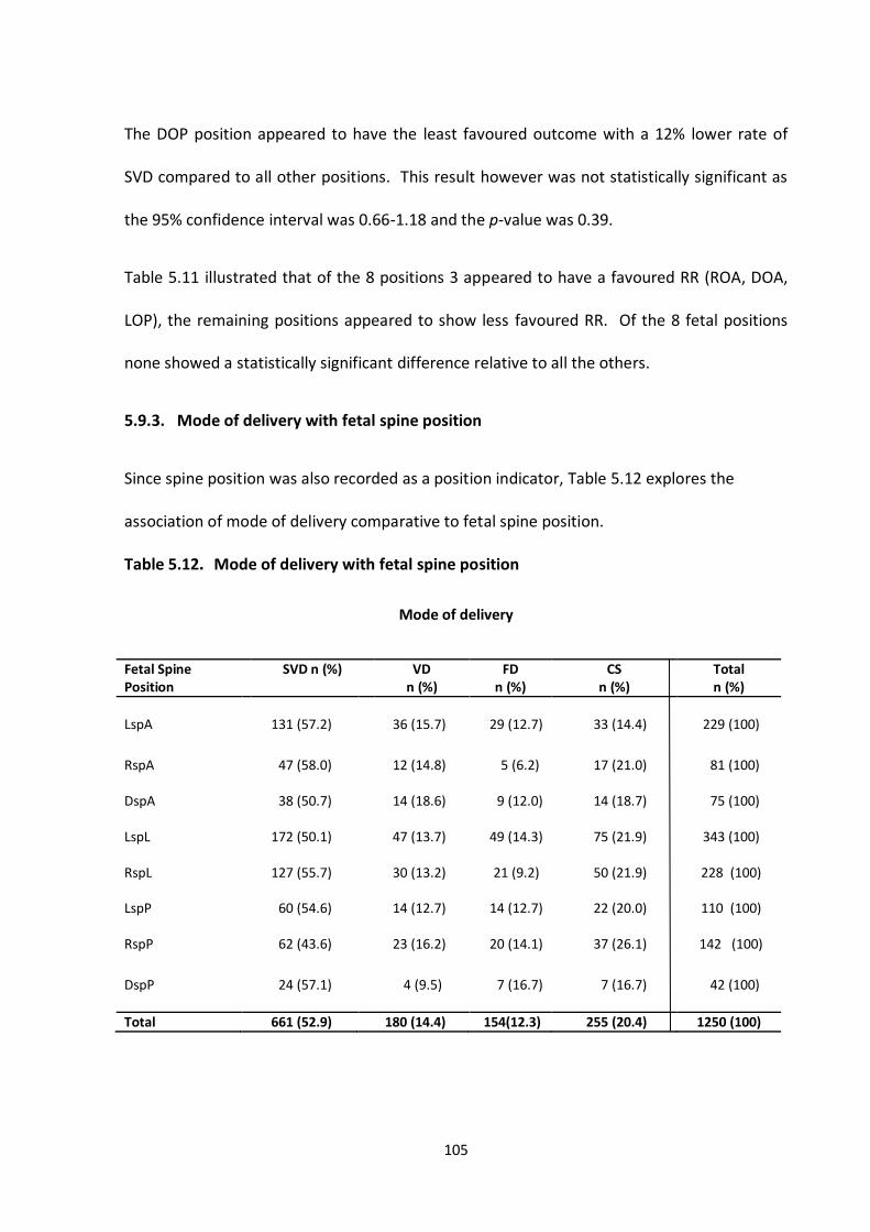
105
The DOP position appeared to have the least favoured outcome with a 12% lower rate of
SVD compared to all other positions. This result however was not statistically significant as
the 95% confidence interval was 0.66-1.18 and the p-value was 0.39.
Table 5.11 illustrated that of the 8 positions 3 appeared to have a favoured RR (ROA, DOA,
LOP), the remaining positions appeared to show less favoured RR. Of the 8 fetal positions
none showed a statistically significant difference relative to all the others.
5.9.3. Mode of delivery with fetal spine position
Since spine position was also recorded as a position indicator, Table 5.12 explores the
association of mode of delivery comparative to fetal spine position.
Table 5.12. Mode of delivery with fetal spine position
Mode of delivery
Fetal Spine Position
SVD n (%) VD n (%)
FD n (%)
CS n (%)
Total n (%)
LspA 131 (57.2) 36 (15.7) 29 (12.7) 33 (14.4) 229 (100)
RspA 47 (58.0) 12 (14.8) 5 (6.2) 17 (21.0) 81 (100)
DspA 38 (50.7) 14 (18.6) 9 (12.0) 14 (18.7) 75 (100)
LspL 172 (50.1) 47 (13.7) 49 (14.3) 75 (21.9) 343 (100)
RspL 127 (55.7) 30 (13.2) 21 (9.2) 50 (21.9)
228 (100)
LspP 60 (54.6) 14 (12.7) 14 (12.7) 22 (20.0) 110 (100)
RspP 62 (43.6) 23 (16.2) 20 (14.1) 37 (26.1) 142 (100)
DspP 24 (57.1) 4 (9.5) 7 (16.7) 7 (16.7)
42 (100)
Total 661 (52.9) 180 (14.4) 154(12.3) 255 (20.4) 1250 (100)
106
In the occiput group the DOA position had the highest rate of SVD’s at 58.9%; however in the
spine group the corresponding DspA position actually had the second lowest rate of SVD’s at
50.7%. It was the RspA group that had the highest rate of SVD’s at 58.0%. This was
followed by the LspA group at 57.2% and surprisingly the DspP had an almost similar rate of
57.1%. Within the spine groups it was in fact the RspP position that actually had the lowest
rate of SVD’s at 43.6%.
The VD rates ranged from 9.5% in the DspP group to 18.6% in the DspA group. This result is
similar to that which was observed with the occiput group. It was also noted that the lowest
rate of VD was amongst the DspP position in the spine group and LOP position in the occiput
group.
The FD rates showed that the RspA position had the lowest rate at 6.2%. The highest rate
was observed amongst the DspP group at 16.7%. Interestingly the following groups that had
the highest rate of FD’s were the LspL (14.3%), LspP (12.7%) and the LspA position (12.7%)
suggesting that foetuses that occupied the left quadrant appeared to have a higher
incidence of FD.
The CS rate ranged from 14.4% to 26.1%, with the lowest CS rate amongst the LspA group
and the highest rate amongst the RspP group.
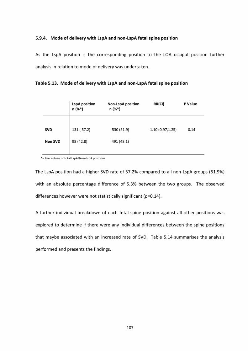
107
5.9.4. Mode of delivery with LspA and non-LspA fetal spine position
As the LspA position is the corresponding position to the LOA occiput position further
analysis in relation to mode of delivery was undertaken.
Table 5.13. Mode of delivery with LspA and non-LspA fetal spine position
*= Percentage of total LspA/Non-LspA positions
The LspA position had a higher SVD rate of 57.2% compared to all non-LspA groups (51.9%)
with an absolute percentage difference of 5.3% between the two groups. The observed
differences however were not statistically significant (p=0.14).
A further individual breakdown of each fetal spine position against all other positions was
explored to determine if there were any individual differences between the spine positions
that maybe associated with an increased rate of SVD. Table 5.14 summarises the analysis
performed and presents the findings.
LspA position Non-LspA position RR(CI) P Value n (%*) n (%*)
SVD
Non SVD
131 ( 57.2) 530 (51.9) 1.10 (0.97,1.25) 0.14
98 (42.8) 491 (48.1)
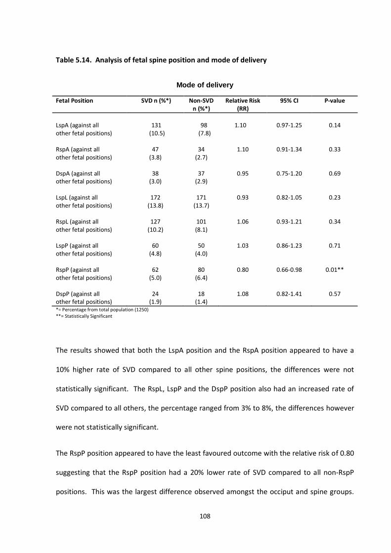
108
Table 5.14. Analysis of fetal spine position and mode of delivery
Mode of delivery
Fetal Position SVD n (%*) Non-SVD n (%*)
Relative Risk (RR)
95% CI P-value
LspA (against all other fetal positions) RspA (against all other fetal positions) DspA (against all other fetal positions) LspL (against all other fetal positions) RspL (against all other fetal positions) LspP (against all other fetal positions) RspP (against all other fetal positions) DspP (against all other fetal positions)
131
(10.5)
47 (3.8)
38
(3.0)
172 (13.8)
127
(10.2)
60 (4.8)
62
(5.0)
24 (1.9)
98
(7.8)
34 (2.7)
37
(2.9)
171 (13.7)
101 (8.1)
50
(4.0)
80 (6.4)
18
(1.4)
1.10
1.10
0.95
0.93
1.06
1.03
0.80
1.08
0.97-1.25
0.91-1.34
0.75-1.20
0.82-1.05
0.93-1.21
0.86-1.23
0.66-0.98
0.82-1.41
0.14
0.33
0.69
0.23
0.34
0.71
0.01**
0.57
*= Percentage from total population (1250) **= Statistically Significant
The results showed that both the LspA position and the RspA position appeared to have a
10% higher rate of SVD compared to all other spine positions, the differences were not
statistically significant. The RspL, LspP and the DspP position also had an increased rate of
SVD compared to all others, the percentage ranged from 3% to 8%, the differences however
were not statistically significant.
The RspP position appeared to have the least favoured outcome with the relative risk of 0.80
suggesting that the RspP position had a 20% lower rate of SVD compared to all non-RspP
positions. This was the largest difference observed amongst the occiput and spine groups.
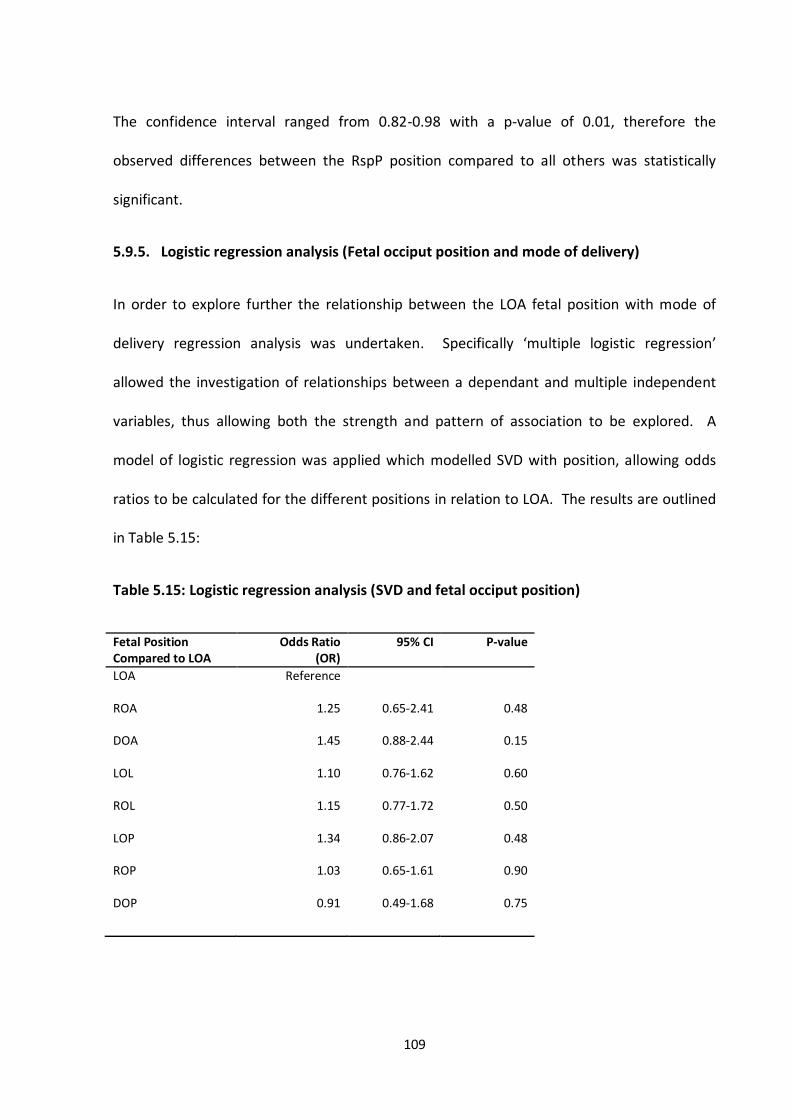
109
The confidence interval ranged from 0.82-0.98 with a p-value of 0.01, therefore the
observed differences between the RspP position compared to all others was statistically
significant.
5.9.5. Logistic regression analysis (Fetal occiput position and mode of delivery)
In order to explore further the relationship between the LOA fetal position with mode of
delivery regression analysis was undertaken. Specifically ‘multiple logistic regression’
allowed the investigation of relationships between a dependant and multiple independent
variables, thus allowing both the strength and pattern of association to be explored. A
model of logistic regression was applied which modelled SVD with position, allowing odds
ratios to be calculated for the different positions in relation to LOA. The results are outlined
in Table 5.15:
Table 5.15: Logistic regression analysis (SVD and fetal occiput position)
Fetal Position Compared to LOA
Odds Ratio (OR)
95% CI P-value
LOA ROA DOA LOL ROL LOP ROP DOP
Reference
1.25
1.45
1.10
1.15
1.34
1.03
0.91
0.65-2.41
0.88-2.44
0.76-1.62
0.77-1.72
0.86-2.07
0.65-1.61
0.49-1.68
0.48
0.15
0.60
0.50
0.48
0.90
0.75
110
The logistic regression model applied showed that comparative to the LOA position,
although differences in SVD rates were observed they were not statistically significant.
Further modelling was undertaken to adjust for variables known to have an effect on SVD
rates. These were maternal age, epidural analgesia, duration of labour and fetal birth
weight, specifically:
Increasing maternal age is associated with an increase in rates of CS delivery (Sharma
et al., 2009, Kirchengast, 2007).
Epidural analgesia is associated with an increase in instrumental delivery rate (Anim-
Somuah et al., 2010, Sharma et al., 2009, Leong et al., 2000).
Duration of labour is associated with an increase in CS delivery (Abu-Heija and Zayer,
1998, El-Hamamy and Arulkumaran, 2005, Gillford et al., 2000).
Neonatal birth weight is associated with an increase in CS delivery (Sharma et al.,
2009, Mocanu et al., 2000).
As expected all four variables within the study data set were analysed and were shown to be
associated with SVD with results that were statistically significant. The analysis verified the
data set showing that the obtained data supports known trends of association and also
enabled the modelling of these variables against the LOA position to determine if these
factors influenced rates of SVD. The results are shown in table 5.16.
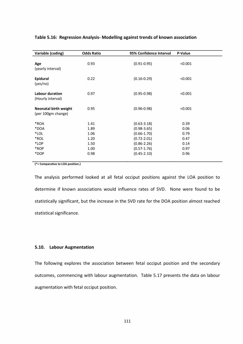
111
Table 5.16: Regression Analysis- Modelling against trends of known association
Variable (coding) Odds Ratio 95% Confidence Interval P-Value
Age (yearly interval)
0.93 (0.91-0.95) <0.001
Epidural (yes/no) Labour duration (Hourly interval) Neonatal birth weight (per 100gm change)
0.22
0.97
0.95
(0.16-0.29)
(0.95-0.98)
(0.96-0.98)
<0.001
<0.001
<0.001
*ROA 1.41 (0.63-3.18) 0.39 *DOA 1.89 (0.98-3.65) 0.06 *LOL 1.06 (0.66-1.70) 0.79 *ROL *LOP *ROP *DOP
1.20 1.50 1.00 0.98
(0.72-2.01) (0.86-2.26) (0.57-1.76) (0.45-2.10)
0.47 0.14 0.97 0.96
(*= Comparative to LOA position.)
The analysis performed looked at all fetal occiput positions against the LOA position to
determine if known associations would influence rates of SVD. None were found to be
statistically significant, but the increase in the SVD rate for the DOA position almost reached
statistical significance.
5.10. Labour Augmentation
The following explores the association between fetal occiput position and the secondary
outcomes, commencing with labour augmentation. Table 5.17 presents the data on labour
augmentation with fetal occiput position.
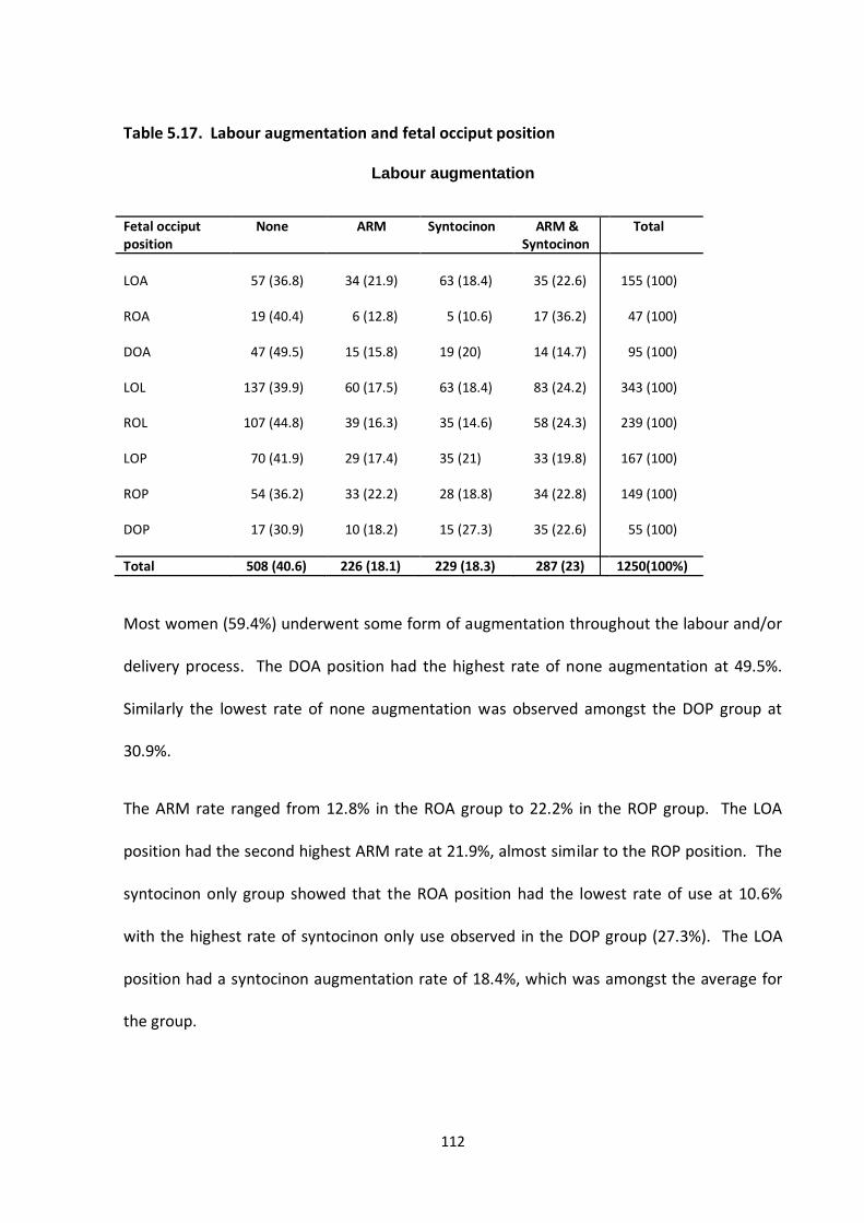
112
Table 5.17. Labour augmentation and fetal occiput position
Labour augmentation
Fetal occiput position
None ARM Syntocinon ARM & Syntocinon
Total
LOA ROA DOA LOL ROL LOP ROP DOP
57 (36.8)
19 (40.4)
47 (49.5)
137 (39.9)
107 (44.8)
70 (41.9)
54 (36.2)
17 (30.9)
34 (21.9)
6 (12.8)
15 (15.8)
60 (17.5)
39 (16.3)
29 (17.4)
33 (22.2)
10 (18.2)
63 (18.4)
5 (10.6)
19 (20)
63 (18.4)
35 (14.6)
35 (21)
28 (18.8)
15 (27.3)
35 (22.6)
17 (36.2)
14 (14.7)
83 (24.2)
58 (24.3)
33 (19.8)
34 (22.8)
35 (22.6)
155 (100)
47 (100)
95 (100)
343 (100)
239 (100)
167 (100)
149 (100)
55 (100)
Total 508 (40.6) 226 (18.1) 229 (18.3) 287 (23) 1250(100%)
Most women (59.4%) underwent some form of augmentation throughout the labour and/or
delivery process. The DOA position had the highest rate of none augmentation at 49.5%.
Similarly the lowest rate of none augmentation was observed amongst the DOP group at
30.9%.
The ARM rate ranged from 12.8% in the ROA group to 22.2% in the ROP group. The LOA
position had the second highest ARM rate at 21.9%, almost similar to the ROP position. The
syntocinon only group showed that the ROA position had the lowest rate of use at 10.6%
with the highest rate of syntocinon only use observed in the DOP group (27.3%). The LOA
position had a syntocinon augmentation rate of 18.4%, which was amongst the average for
the group.

113
The combined ARM and syntocinon augmentation rates appeared to have a visibly large
variation, ranging from 14.7% to 36.1%. The lowest rate was observed in the DOA group,
and the highest rate was observed in the ROA group. Interestingly the highest rate of
combined augmentation was not observed in a posterior or lateral group but instead in an
anterior group. The 36.1% combined augmentation rate for the ROA position did not reflect
the general percentage rates. The remaining positions that had the higher rates of
combined augmentation appear to remain within the 20% range of which all were below
25%, whilst the ROA position had an absolute percentage difference of 11.1%. The LOA
position had an average combined augmentation rate of 22.5%.
5.10.1. Labour augmentation with LOA and non-LOA fetal occiput position.
Table 5.18 presents the data on LOA and non-LOA fetal positions with labour augmentation.
Table 5.18. Labour augmentation with LOA and non-LOA fetal occiput position
Augmentation method
Fetal position None ARM Syntocinon ARM & Syntocinon
Total
LOA position All non-LOA positions
57 (36.8) 451 (41.2)
34 (21.9) 192 (17.5)
29 (18.7) 200 (18.3)
35 (22.6) 252 (23.0)
155 (100) 1095 (100)
Total 508 (40.6) 226 (18.1) 229 (18.3) 287 (23) 1250 (100)
A chi-squared test was performed to see if the observed differences were statistically
significant, a p-value of 0.54 was obtained showing that they were not.
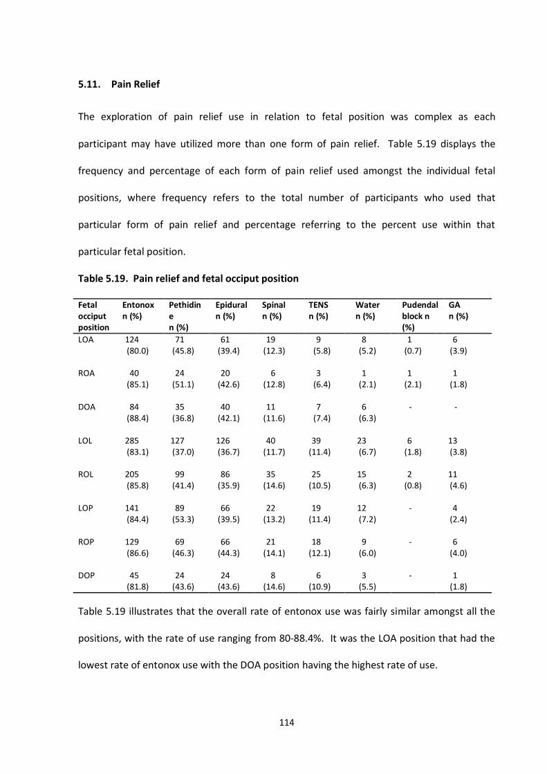
114
5.11. Pain Relief
The exploration of pain relief use in relation to fetal position was complex as each
participant may have utilized more than one form of pain relief. Table 5.19 displays the
frequency and percentage of each form of pain relief used amongst the individual fetal
positions, where frequency refers to the total number of participants who used that
particular form of pain relief and percentage referring to the percent use within that
particular fetal position.
Table 5.19. Pain relief and fetal occiput position Fetal occiput position
Entonox n (%)
Pethidine n (%)
Epidural n (%)
Spinal n (%)
TENS n (%)
Water n (%)
Pudendal block n (%)
GA n (%)
LOA 124 (80.0)
71 (45.8)
61 (39.4)
19 (12.3)
9 (5.8)
8 (5.2)
1 (0.7)
6 (3.9)
ROA 40
(85.1) 24
(51.1) 20
(42.6) 6
(12.8) 3
(6.4) 1
(2.1) 1
(2.1) 1
(1.8)
DOA 84 (88.4)
35 (36.8)
40 (42.1)
11 (11.6)
7 (7.4)
6 (6.3)
- -
LOL 285 (83.1)
127 (37.0)
126 (36.7)
40 (11.7)
39 (11.4)
23 (6.7)
6 (1.8)
13 (3.8)
ROL 205
(85.8) 99
(41.4) 86
(35.9) 35
(14.6) 25
(10.5) 15 (6.3)
2 (0.8)
11 (4.6)
LOP 141
(84.4) 89
(53.3) 66
(39.5) 22
(13.2) 19
(11.4) 12 (7.2)
- 4 (2.4)
ROP 129
(86.6) 69
(46.3) 66
(44.3) 21
(14.1) 18
(12.1) 9
(6.0) - 6
(4.0)
1 (1.8)
DOP
45
(81.8)
24
(43.6)
24
(43.6)
8
(14.6)
6
(10.9)
3
(5.5)
-
Table 5.19 illustrates that the overall rate of entonox use was fairly similar amongst all the
positions, with the rate of use ranging from 80-88.4%. It was the LOA position that had the
lowest rate of entonox use with the DOA position having the highest rate of use.
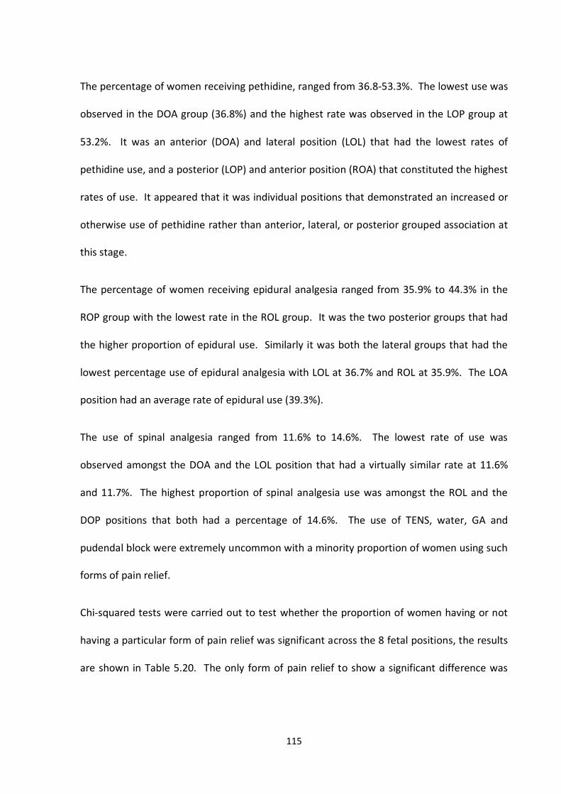
115
The percentage of women receiving pethidine, ranged from 36.8-53.3%. The lowest use was
observed in the DOA group (36.8%) and the highest rate was observed in the LOP group at
53.2%. It was an anterior (DOA) and lateral position (LOL) that had the lowest rates of
pethidine use, and a posterior (LOP) and anterior position (ROA) that constituted the highest
rates of use. It appeared that it was individual positions that demonstrated an increased or
otherwise use of pethidine rather than anterior, lateral, or posterior grouped association at
this stage.
The percentage of women receiving epidural analgesia ranged from 35.9% to 44.3% in the
ROP group with the lowest rate in the ROL group. It was the two posterior groups that had
the higher proportion of epidural use. Similarly it was both the lateral groups that had the
lowest percentage use of epidural analgesia with LOL at 36.7% and ROL at 35.9%. The LOA
position had an average rate of epidural use (39.3%).
The use of spinal analgesia ranged from 11.6% to 14.6%. The lowest rate of use was
observed amongst the DOA and the LOL position that had a virtually similar rate at 11.6%
and 11.7%. The highest proportion of spinal analgesia use was amongst the ROL and the
DOP positions that both had a percentage of 14.6%. The use of TENS, water, GA and
pudendal block were extremely uncommon with a minority proportion of women using such
forms of pain relief.
Chi-squared tests were carried out to test whether the proportion of women having or not
having a particular form of pain relief was significant across the 8 fetal positions, the results
are shown in Table 5.20. The only form of pain relief to show a significant difference was
116
pethidine use amongst women having and not having pethidine across the 8 fetal positions
(p=0.02).
Table 5.20. Chi-squared analysis of individual forms of pain relief against all fetal positions
Pain relief type P-value
Entonox 0.66
Pethidine 0.02*
Epidural 0.71
Spinal 0.97
TENS
Water
Pudendal block
General anesthetic
0.50
0.92
0.27
0.53
*statistically significant
Further analysis of pethidine use was carried out to determine if the observed differences
were related to occupying cavity that is, anterior, lateral or posterior grouped association.
Table 5.21. Grouped cavity fetal occiput position (OA, OL, and OP) and pethidine use
Fetal occiput Position
Pethidine used n (%)
Non-Pethidine use n (%)
Total
OA
OL
OP
130 (43.8)
226 (38.8)
182 (49.1)
167 (56.2)
356 (61.2)
189 (50.9)
297 (100)
582 (100)
371 (100)
Total 538 (43) 712 (57) 1250 (100%)
This analysis showed that despite it being both an individual anterior and an individual
posterior position that had the highest use of pethidine (see Table 5.19), when categorised
into collective anterior, lateral and posterior groups, it was the posterior group that showed

117
increased use of pethidine overall. A chi-squared test suggested that the posterior fetal
positions had a significantly higher use of pethidine compared to the anterior and lateral
positions (p=0.008).
Further analysis was undertaken to determine if specifically the LOA position comparative to
all other positions was associated with use of pethidine (Table 5.22).
Table 5.22. Pethidine with LOA against non-LOA fetal occiput position
Fetal occiput Position
Pethidine used n (%)
Non-Pethidine use n (%)
Total
LOA position
71 (45.8)
84 (54.2)
155 (100%)
All non-LOA position
467 (42.7)
628 (57.4)
1095 (100%)
Total 538 (43) 712 (57) 1250 (100%)
Table 5.22 shows that the LOA position had a 3.2% increased rate of pethidine use compared
to all other fetal positions, but a chi-squared test showed that this was not statistically
significant (p=0.46).
5.12. Labour Duration
The duration of labour was analysed at three period points of the labour and delivery
process which were:
duration of the active first stage of labour.
duration of the second stage of labour.
duration of membrane rupture.
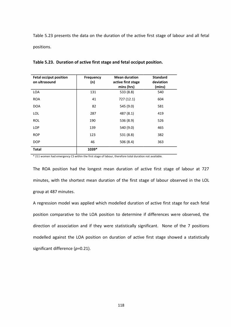
118
Table 5.23 presents the data on the duration of the active first stage of labour and all fetal
positions.
Table 5.23. Duration of active first stage and fetal occiput position.
Fetal occiput position on ultrasound
Frequency (n)
Mean duration active first stage
mins (hrs)
Standard deviation
(mins)
LOA 131 533 (8.8) 540
ROA 41 727 (12.1) 604
DOA 82 545 (9.0) 581
LOL
ROL
LOP
ROP
DOP
287
190
139
123
46
487 (8.1)
536 (8.9)
540 (9.0)
531 (8.8)
506 (8.4)
419
526
465
382
363
Total 1039*
* 211 women had emergency CS within the first stage of labour, therefore total duration not available.
The ROA position had the longest mean duration of active first stage of labour at 727
minutes, with the shortest mean duration of the first stage of labour observed in the LOL
group at 487 minutes.
A regression model was applied which modelled duration of active first stage for each fetal
position comparative to the LOA position to determine if differences were observed, the
direction of association and if they were statistically significant. None of the 7 positions
modelled against the LOA position on duration of active first stage showed a statistically
significant difference (p=0.21).
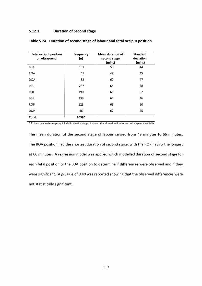
119
5.12.1. Duration of Second stage
Table 5.24. Duration of second stage of labour and fetal occiput position
Fetal occiput position on ultrasound
Frequency (n)
Mean duration of second stage
(mins)
Standard deviation
(mins)
LOA 131 55 44
ROA 41 49 45
DOA 82 62 47
LOL
ROL
LOP
ROP
DOP
287
190
139
123
46
64
61
64
66
62
48
52
46
60
45
Total 1039*
* 211 women had emergency CS within the first stage of labour, therefore duration for second stage not available.
The mean duration of the second stage of labour ranged from 49 minutes to 66 minutes.
The ROA position had the shortest duration of second stage, with the ROP having the longest
at 66 minutes. A regression model was applied which modelled duration of second stage for
each fetal position to the LOA position to determine if differences were observed and if they
were significant. A p-value of 0.40 was reported showing that the observed differences were
not statistically significant.
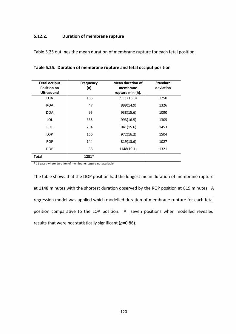
120
5.12.2. Duration of membrane rupture
Table 5.25 outlines the mean duration of membrane rupture for each fetal position.
Table 5.25. Duration of membrane rupture and fetal occiput position
Fetal occiput Position on Ultrasound
Frequency (n)
Mean duration of membrane
rupture min (h).
Standard deviation
LOA 155 953 (15.8) 1250
ROA 47 899(14.9) 1326
DOA 95 938(15.6) 1090
LOL
ROL
LOP
ROP
DOP
335
234
166
144
55
993(16.5)
941(15.6)
972(16.2)
819(13.6)
1148(19.1)
1305
1453
1504
1027
1321
Total 1231*
* 11 cases where duration of membrane rupture not available.
The table shows that the DOP position had the longest mean duration of membrane rupture
at 1148 minutes with the shortest duration observed by the ROP position at 819 minutes. A
regression model was applied which modelled duration of membrane rupture for each fetal
position comparative to the LOA position. All seven positions when modelled revealed
results that were not statistically significant (p=0.86).
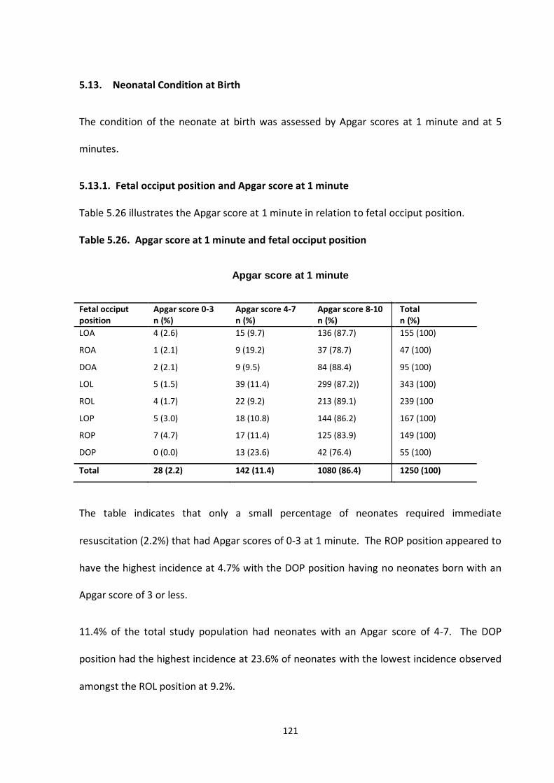
121
5.13. Neonatal Condition at Birth
The condition of the neonate at birth was assessed by Apgar scores at 1 minute and at 5
minutes.
5.13.1. Fetal occiput position and Apgar score at 1 minute
Table 5.26 illustrates the Apgar score at 1 minute in relation to fetal occiput position.
Table 5.26. Apgar score at 1 minute and fetal occiput position
Apgar score at 1 minute
Fetal occiput position
Apgar score 0-3 n (%)
Apgar score 4-7 n (%)
Apgar score 8-10 n (%)
Total n (%)
LOA
ROA
DOA
LOL
ROL
LOP
ROP
DOP
4 (2.6)
1 (2.1)
2 (2.1)
5 (1.5)
4 (1.7)
5 (3.0)
7 (4.7)
0 (0.0)
15 (9.7)
9 (19.2)
9 (9.5)
39 (11.4)
22 (9.2)
18 (10.8)
17 (11.4)
13 (23.6)
136 (87.7)
37 (78.7)
84 (88.4)
299 (87.2))
213 (89.1)
144 (86.2)
125 (83.9)
42 (76.4)
155 (100)
47 (100)
95 (100)
343 (100)
239 (100
167 (100)
149 (100)
55 (100)
Total 28 (2.2) 142 (11.4) 1080 (86.4) 1250 (100)
The table indicates that only a small percentage of neonates required immediate
resuscitation (2.2%) that had Apgar scores of 0-3 at 1 minute. The ROP position appeared to
have the highest incidence at 4.7% with the DOP position having no neonates born with an
Apgar score of 3 or less.
11.4% of the total study population had neonates with an Apgar score of 4-7. The DOP
position had the highest incidence at 23.6% of neonates with the lowest incidence observed
amongst the ROL position at 9.2%.
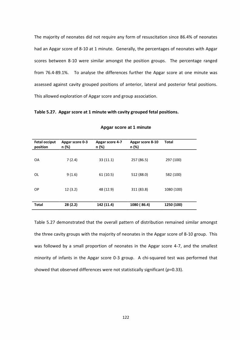
122
The majority of neonates did not require any form of resuscitation since 86.4% of neonates
had an Apgar score of 8-10 at 1 minute. Generally, the percentages of neonates with Apgar
scores between 8-10 were similar amongst the position groups. The percentage ranged
from 76.4-89.1%. To analyse the differences further the Apgar score at one minute was
assessed against cavity grouped positions of anterior, lateral and posterior fetal positions.
This allowed exploration of Apgar score and group association.
Table 5.27. Apgar score at 1 minute with cavity grouped fetal positions.
Apgar score at 1 minute
Fetal occiput position
Apgar score 0-3 n (%)
Apgar score 4-7 n (%)
Apgar score 8-10 n (%)
Total
OA
OL
7 (2.4)
9 (1.6)
33 (11.1)
61 (10.5)
257 (86.5)
512 (88.0)
297 (100)
582 (100)
OP 12 (3.2) 48 (12.9) 311 (83.8) 1080 (100)
Total 28 (2.2) 142 (11.4) 1080 ( 86.4) 1250 (100)
Table 5.27 demonstrated that the overall pattern of distribution remained similar amongst
the three cavity groups with the majority of neonates in the Apgar score of 8-10 group. This
was followed by a small proportion of neonates in the Apgar score 4-7, and the smallest
minority of infants in the Apgar score 0-3 group. A chi-squared test was performed that
showed that observed differences were not statistically significant (p=0.33).
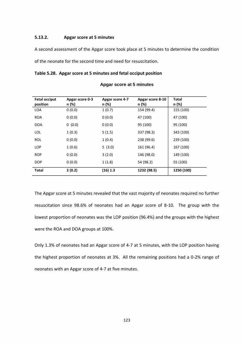
123
5.13.2. Apgar score at 5 minutes
A second assessment of the Apgar score took place at 5 minutes to determine the condition
of the neonate for the second time and need for resuscitation.
Table 5.28. Apgar score at 5 minutes and fetal occiput position
Apgar score at 5 minutes
Fetal occiput position
Apgar score 0-3 n (%)
Apgar score 4-7 n (%)
Apgar score 8-10 n (%)
Total n (%)
LOA
ROA
DOA
LOL
ROL
LOP
ROP
DOP
0 (0.0)
0 (0.0)
0 (0.0)
1 (0.3)
0 (0.0)
1 (0.6)
0 (0.0)
0 (0.0)
1 (0.7)
0 (0.0)
0 (0.0)
5 (1.5)
1 (0.4)
5 (3.0)
3 (2.0)
1 (1.8)
154 (99.4)
47 (100)
95 (100)
337 (98.3)
238 (99.6)
161 (96.4)
146 (98.0)
54 (98.2)
155 (100)
47 (100)
95 (100)
343 (100)
239 (100)
167 (100)
149 (100)
55 (100)
Total 2 (0.2) (16) 1.3 1232 (98.5) 1250 (100)
The Apgar score at 5 minutes revealed that the vast majority of neonates required no further
resuscitation since 98.6% of neonates had an Apgar score of 8-10. The group with the
lowest proportion of neonates was the LOP position (96.4%) and the groups with the highest
were the ROA and DOA groups at 100%.
Only 1.3% of neonates had an Apgar score of 4-7 at 5 minutes, with the LOP position having
the highest proportion of neonates at 3%. All the remaining positions had a 0-2% range of
neonates with an Apgar score of 4-7 at five minutes.
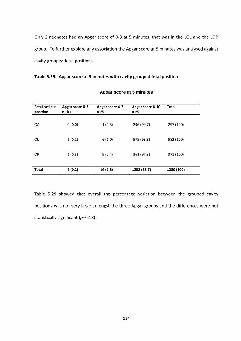
124
Only 2 neonates had an Apgar score of 0-3 at 5 minutes, that was in the LOL and the LOP
group. To further explore any association the Apgar score at 5 minutes was analysed against
cavity grouped fetal positions.
Table 5.29. Apgar score at 5 minutes with cavity grouped fetal position
Apgar score at 5 minutes
Fetal occiput position
Apgar score 0-3 n (%)
Apgar score 4-7 n (%)
Apgar score 8-10 n (%)
Total
OA
OL
0 (0.0)
1 (0.2)
1 (0.3)
6 (1.0)
296 (99.7)
575 (98.8)
297 (100)
582 (100)
OP 1 (0.3) 9 (2.4) 361 (97.3) 371 (100)
Total 2 (0.2) 16 (1.3) 1232 (98.7) 1250 (100)
Table 5.29 showed that overall the percentage variation between the grouped cavity
positions was not very large amongst the three Apgar groups and the differences were not
statistically significant (p=0.13).
125
CHAPTER 6
DISCUSSION
6.1. Main Results
The aim of this prospective study was to establish the association between fetal position at
the onset of labour, and birth outcomes in particular mode of delivery for the mother and
the neonate. Specifically it was the LOA position which based on Sutton’s theory
hypothesised as being linked to improved outcomes that was explored.
The data from the study demonstrated that the LOA position at the onset of labour was not
associated with mode of delivery which was the primary outcome measure. None of the
eight positions identified at the point of labour onset were associated with mode of delivery.
The secondary outcome measures of pain relief, duration of labour, augmentation, and
Apgar scores, did not reveal any association with fetal position, with the exception of pain
relief. Of the eight forms of pain relief available, the proportion of women using pethidine
to those who did not across the 8 fetal positions was statistically significant. Further
categorisation into cavity grouped positions of anterior, lateral and posterior groups showed
that the posterior group was more likely to use pethidine compared to the other groups.
The study also provided data on prevalence of fetal occiput position at the point of labour
onset (Table 5.5 – Occiput Prevalence), such data not having been published before, nor
126
reported in any Midwifery or Obstetric textbook. The study data demonstrated that of the
cavity grouped fetal positions the anterior group at the onset of labour was the least
frequently occurring at 23.7%. Specifically the ROA position was the least occurring position
amongst all 8 fetal positions. The posterior positions occurred amongst 29.7% of the
women, which is two to three times the reported figures of OP presentations at the onset of
labour (Gardberg et al., 1998, Gardberg and Tuppurainen, 1994, Fitzpatrick et al., 2001, Sizer
and Nirmal, 2000, Neri et al., 1995, Faber, 1998). The lateral positions were the most
commonly occurring fetal occiput position with 46.5% of the women in the study shown to
have this position. The most prevalent of the 8 fetal occiput positions was the LOL position.
The prevalence of fetal spine position reflected similar patterns to that observed for the
occiput groups, with the lateral groups most prevalent, specifically the LspL position (Table
5.6 – Spine Prevalence). The only difference observed between the distribution of the
occiput and spine position was that the cavity grouped anterior spine position was the
second most common group with the posterior group being least common, the reversal of
what was observed in the occiput group. The importance of the prevalence of fetal spine
position was that no figures to date have been published on prevalence of spine position,
thus data from the study provided additional new knowledge.. Furthermore, the prevalence
of the combined fetal occiput and spine position, referred to as combination grouping has
also never been matched or reported.
The results of the combination grouping showed that there were common groups;
uncommon groups are several combination groups that were incompatible. The analysis of
data on fetal position revealed that in the majority of cases the occiput and the spine share
127
corresponding identical positions, and that the most common position for the fetus to adopt
at the onset of labour in nulliparous women at term gestation was the LOL&LspL position.
6.2. Strengths and Weaknesses of Study
The rigorous planning and piloting of the study aimed to minimise weaknesses and enhance
scientific strengths. During the planning of the study the use of a prospective design was
deemed mandatory if the associative relationship between fetal position and labour
outcomes was to provide best available evidence. The ability to determine the eligibility
criteria minimised confounding variables that would otherwise generate arguments relating
to validity. The prospective approach allowed controls to be imposed in order to standardise
processes of assessment of fetal position. Once such controls were applied it allowed study
participants to be followed until the outcome of delivery was observed and recorded. The
primary outcome measure of mode of delivery was an objective measure and did not give
rise to issues of subjectivity. Moreover it was routinely collected so not influenced at all by
study purposes. The ability to design a study and impose controls increased the validity of
the study and eliminated potential bias, particularly relating to possible confounding
variables that may otherwise result in competing explanations.
Since the point of measure during which fetal position was to be determined was
paramount, the definition of early labour as outlined by the National Institute of Clinical
Excellence was used and is the one also used in the centre in which the study was
undertaken (See section 3.5 - Definition of Labour Onset and Period of Measure) (NICE,
2007). The use of the definition clearly identified the point commencing and ending early
128
labour, outlining the period during which measure of fetal position was to be undertaken.
To control the point of interest further a rule of 12 hour repeat measure was applied so if
the mother did not enter established labour within 12 hours of the fetal position being
identified the measure would be repeated.
The use of a ‘gold standard’ technique of ultrasound to measure fetal position further
enhanced the validity of this independent variable, ensuring that misclassification caused by
subjective measures ware decreased. The midwives trained to perform the ultrasound were
taught by the same sonographers and underwent an identical assessment including
completion of a training log of 40 individual cases. The successful implementation of the
study also showed that the use of ultrasound to accurately detect fetal position at the onset
of labour was feasible and acceptable to women during early labour.
Internal validity was further enhanced as the scans performed were validated by an
independent clerk. The clerk observed the scan pictures against the diagrams outlined by
the scanning midwife to determine agreement and define position. The diagram that was
used by the scanning midwife was not labelled with the individual positions; instead the
scanning midwife was asked to simply replicate the position defining structure as observed
on ultrasound and not to define position which was done independently by the clerk. The
routine procedures applied ensured constancy of condition which intern highlighted the
validity achieved. Random checks were also performed where two trained scanning
midwives would freeze the position defining structure on the ultrasound machine and
replicate the structure independently. This was then sealed and sent to the independent
clerk for agreement. Further contingencies included blinding of the scan findings to the
129
attending clinician and the woman being scanned so that such information would not
influence the care received or behaviour and expectations of the woman/clinician.
To maximise representativeness and generalisability, all areas that women would attend
throughout their pregnancy were involved with the recruitment process. The primary
recruiters were the community midwives who recruited from a specific clinic that all women
were required to attend. This not only ensured a high recruitment rate but also that the
sample population was similar to that at the recruiting hospital. The extensive recruitment
process meant that no one particular group of women were targeted but instead the entire
population had the opportunity to take part in the study.
Similarities were therefore observed with ethnic diversity and mode of delivery between the
BWH eligible population and that represented in the sample population (Refer to tables 5.1
& 5.3). The largest ethnic group in both the sample population and the total population was
the White-British group at 56.0% and 58.0%. The next largest was the Pakistani group which
constituted 18.0% of the sample and 15% of the total population and the third largest was
the Indian group, forming 6.3% of the sample and 6.5% of the total population. The
remaining smaller groups also showed similarities.
Mode of delivery rates also showed consistency between sample and total hospital
population, with the SVD rate being 53% in the sample and 57% at the BWH. The small
discrepancy is likely to be due to the study inclusion criteria. The documented delivery
figures available for nulliparous women at BWHCT did not distinguish between gestation,
multiple pregnancy, fetal abnormality, breech vaginal deliveries and other characteristics
that were excluded from the study. It is therefore possible that this small discrepancy may
130
account for women whom would otherwise due to fetal abnormality, multiple pregnancy
and breech presentation be excluded from the study.
Ventouse deliveries accounted for 14% of the deliveries in the sample compared to 12% at
BWHCT. Forceps deliveries, inclusive of all types, accounted for 12% of the total deliveries in
the sample compared to 10% reported at BWH. The CS rate was 20% in the sample, the
same as the rate at the BWH. Therefore the observed mode of delivery rates appear to
show good representativeness in this sample.
The calculation of sample size was extremely important if the pre-determined effect size was
to be detected reliably. The limited data on SVD rates for the LOA position meant that
figures on OP position were used instead. The sample size allowed an absolute difference of
15% in SVD rates to be detected between groups with a power of 89%. This required a
sample size of 1250 women which was achieved within a period of 18 months thus ensuring
an adequate recruitment rate of the total population.
Of the 1250 women recruited and scanned to the study 100% of the outcome data was
obtained for every study participant including both the primary and secondary outcome
measures required to undertake complete analysis.
Logistic regression analysis was undertaken to explore both the strength and pattern of
association, between SVD rates and fetal occiput position, and to adjust for variables that
were known to be associated with SVD. The variables were maternal age, epidural
analgesia, labour duration and neonatal birth weight. Despite the multiple logistic
regression finding demonstrating no significant associations for fetal position, all four above
131
variables confirmed known trends of association, therefore increasing confidence in the
study data and thus study findings.
The main limitation of the study, which was evident retrospectively, was the inability to
obtain data on fetal position during the second stage of labour and at delivery. The main
aim of the study was to determine fetal position at the onset of labour and examine the
association with birth outcomes. Since the study revealed that fetal position at the onset of
labour was not associated with mode of delivery, the availability of data on position during
second stage may have provided further explanatory information. If data on position during
second stage was obtained analysis into mal-rotation may have been determined, since
there remains a debate as to whether the persistent OP, which is associated with increased
morbidity, is as a result of mal-rotation or the persistence of an initial OP position. Data on
position at second stage may have provided the answer to this question, and clarified the
overall issue.
The hospital computer system was reviewed to determine whether routine data on fetal
position at delivery was available, but fetal position at delivery was entered as:
Spontaneous anterior
Spontaneous other
Ventouse
Forceps
Breech
Emergency CS
Elective
The above categorisation did not provide adequate information to undertake the analysis.
Another limitation was that it was not possible to identify from the hospital medical systems
women who arrived at the hospital in active labour therefore not eligible, thus an exact
132
calculation of recruitment rate was not possible. Of the 2035 women who were eligible,
1647 were recruited and scanned with a total of 388 potential women lost. The recruitment
rate was therefore recorded as a range of a minimum of 61.4% to a maximum of 75.9% if
presumed that either all or none of the 388 women lost were actually ineligible (Refer to
figure 5.1 – Recruitment of study sample).
6.3. Interpretation of Findings
The main initiative for this study was to contribute scientific knowledge to the increasing
move towards promoting a single fetal position (LOA) as optimum. Such a move was led by
a midwife, Jean Sutton who used anecdotal evidence from a life time of experience to
support her claims. At a time where all practice should be supported by scientific evidence,
the continuation of such practice with the absence of scientific evidence was undesirable.
The fetal position described as optimum was the LOA position, that Sutton (2001) claimed
would facilitate anterior rotation preventing mal-rotation and consequently resulting in a:
shorter labour duration
less need for pain relief
less intervention
increased rate of SVD
Findings from this study revealed no association between the LOA position or the LspA
position and mode of delivery. Instead the LOA position showed to have a slightly lower rate
of SVD compared to all non-LOA positions although this was not statistically significant.
Furthermore, the LOA position was not associated with either a reduction in need for pain
133
relief, labour duration or use of intervention. The only association was between the use of
pethidine analgesia and cavity grouped posterior fetal positions. Individual position analysis
of pethidine use did not reveal any significant results, suggesting that the association was
connected to grouped posterior cavity positions only.
The relationship between increasing use of pain relief and the posterior fetal position in
active labour is well documented (Simpkin, 2010, Fitzpatrick et al., 2001, Gardberg et al.,
1998, Ponkey et al., 2003). One argument as to why the link between OP at labour onset
and pethidine use was also evident may be related to the physiological principles of OP
engagement. The increased diameter entering the pelvic brim in early labour may only be
achieved with increased uterine activity consequently requiring further pain relief.
Additionally it may be linked to the perception of OP labours being longer and associated
with morbidity, thus influencing women and professionals to the increase use, if position is
thought to be OP. Other forms of pain relief did not reveal a similar association, which may
be because further pain relief is only available in active labour.
Sutton’s theory that the LOA fetal position is the optimum position for the fetus to adopt at
the onset of labour was not supported by the findings of this study thus confirming that
antenatal practices to encourage an LOA fetus should not be promoted.
In addition, the study did not confirm known associations of fetal position, mainly OP mal-
position with increased operative delivery, oxytocin augmentation, labour duration and low
Apgar scores (Ponkey et al., 2003, Fitzpatrick et al., 2001, Cheng et al., 2006). The most likely
reason for this is that the studies were undertaken with position measured during the active
first or second stage of labour, therefore investigating the persistent occiput posterior

134
position comparative to grouped anterior and/or lateral positions, where as the present
study measured position at labour onset prior to the commencement of active labour. The
main comparison was undertaken against ‘all’ individual fetal positions at ‘labour onset’ and
not solely grouped positions. The dissimilar results are therefore not a consequence of
contradictory findings but are new findings that have not previously been reported.
The lack of association between fetal position at labour onset and delivery outcomes
suggests that no cause/effect relationship exists between the variables. To speculate on the
reason for such, is that the onset of labour mainly involves fetal manoeuvres of engagement,
descent and flexion which are manoeuvres which are less likely to deviate. The manoeuvres
required during active labour however are that of rotation, extension and external rotation
which due to the increased nature of movement required are more likely to deviate.
The specific delivery modes of CS, ventouse and forceps deliveries did not show any
association with any of the fetal occiput positions. In the analysis of spine position the RspL
position showed a significant association suggesting a 20% lower rate of SVD compared to all
other position groups (p=0.02). Nonetheless, the identification of a single result as
significant following a large number of non-significant results is not considered of huge
importance mainly because it is probably a consequence of a chance finding rather than a
true significant result (Bland and Altman, 1995). The rates of SVD amongst the overall cavity
grouped fetal positions of anterior, lateral and posterior positions were very similar.
Some of the findings reported by this study are supported by Gardberg et al.’s (1998)
findings, who reported no association between fetal position and mode of delivery, epidural
use, and duration of first or second stage of labour. Gardberg et al.’s (1998) study however
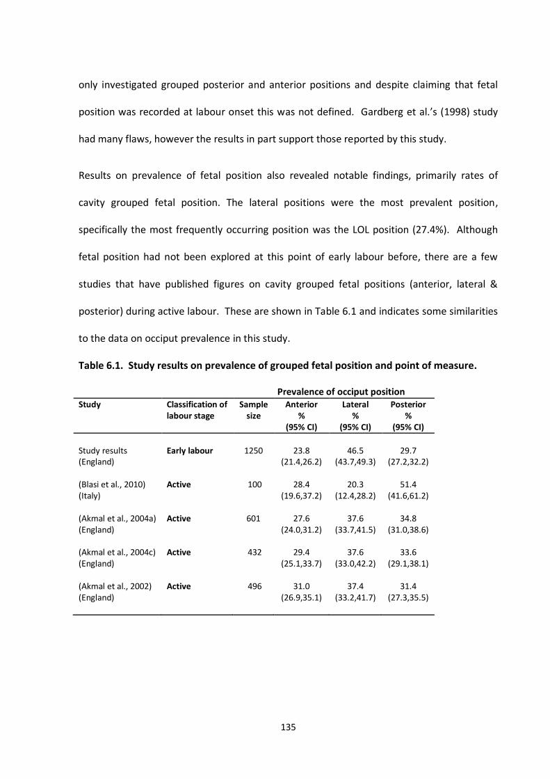
135
only investigated grouped posterior and anterior positions and despite claiming that fetal
position was recorded at labour onset this was not defined. Gardberg et al.’s (1998) study
had many flaws, however the results in part support those reported by this study.
Results on prevalence of fetal position also revealed notable findings, primarily rates of
cavity grouped fetal position. The lateral positions were the most prevalent position,
specifically the most frequently occurring position was the LOL position (27.4%). Although
fetal position had not been explored at this point of early labour before, there are a few
studies that have published figures on cavity grouped fetal positions (anterior, lateral &
posterior) during active labour. These are shown in Table 6.1 and indicates some similarities
to the data on occiput prevalence in this study.
Table 6.1. Study results on prevalence of grouped fetal position and point of measure.
Prevalence of occiput position Study Classification of
labour stage Sample
size Anterior
% (95% CI)
Lateral %
(95% CI)
Posterior %
(95% CI)
Study results (England)
Early labour
1250
23.8
(21.4,26.2)
46.5
(43.7,49.3)
29.7
(27.2,32.2) (Blasi et al., 2010) (Italy)
Active 100 28.4 (19.6,37.2)
20.3 (12.4,28.2)
51.4 (41.6,61.2)
(Akmal et al., 2004a) (England)
Active 601 27.6 (24.0,31.2)
37.6 (33.7,41.5)
34.8 (31.0,38.6)
(Akmal et al., 2004c) (England)
Active
432
29.4
(25.1,33.7)
37.6
(33.0,42.2)
33.6
(29.1,38.1)
(Akmal et al., 2002) (England)
Active 496 31.0 (26.9,35.1)
37.4 (33.2,41.7)
31.4 (27.3,35.5)
136
Despite all these studies having a different point at which position was diagnosed, excluding
that of Blasi et al.’s (2010) which yielded unexpected figures that remain unexplained, the
remaining 3 studies report similar data on prevalence to this study. In all the studies the
lateral group was the most prevalent which was expected in accordance with labour
mechanism, with the fetus commencing labour in the transverse plane ready to descend into
the maternal pelvis (Caruthers, 2000, Cunningham et al., 2008, McCormick, 2008).
The posterior group was the next most prevalent, and the anterior positions were least
prevalent, a pattern of distribution synonymous across all three studies (Akmal et al., 2004a,
Akmal et al., 2004c, Akmal et al., 2002). The small variations amongst the individual rates of
lateral, posterior and anterior prevalence may be due to the stage at which position was
identified, and reflect fetal position prior and post rotational descent.
Studies have reported figures on the prevalence of the posterior fetal position to be 10-15%
in ‘early labour’, such claims are not supported by this study (Sizer and Nirmal, 2000,
Gardberg et al., 1998, Gardberg and Tuppurainen, 1994, Fitzpatrick et al., 2001, Neri et al.,
1995). The discrepancy in the figures on posterior fetal position is difficult to explain since
determining whether posterior positions are increasing as some claim, or, if it is as a
consequence of studies using ultrasound to report accurate figures is difficult to distinguish
(Sutton, 2007).
What remains intriguing about the findings on grouped positions and labour process, is that
review of textbooks suggests that occiput engagement takes place in the transverse/lateral
position (Cunningham et al., 2008, Dietze, 2001, McCormick, 2008, Selman and Johnston,
2010). No textbook describes the mechanism of labour when a position other than a lateral
137
position is identified at the onset of labour, that is, despite this study identifying that over
half of the fetal positions engaged in a position other than the transverse position (53.5% -
Table 5.5 Frequency of fetal occiput position). The majority of textbooks outline that
complete fetal engagement in the posterior position prior to the onset of labour is unusual.
The understanding is that the majority of posterior fetuses will remain unengaged until
uterine contractions and membrane rupture urge engagement and descent (Coates, 2002,
Coates, 2008). This study revealed that 29.7% of fetuses were posterior and entered
established labour within 12 hours thus suggesting some degree of engagement at the pelvic
brim was achieved, yet mechanism of labour pertaining to the posterior position remains
unreported.
Furthermore, it is thought that the direct positions (DOA & DOP) are only adopted during the
final labour stage prior to the expulsive phase (Dietze, 2001, Selman and Johnston, 2010,
McCormick, 2008). Yet again mechanism with a fetus in the direct position is not reported in
any literature, yet this study identified that 12% of fetuses were in either of the direct
positions. Such findings are atypical since literature that reports pelvic measures state that
the anterior-posterior plane of the pelvic brim is 11cm, where as a fetus engaging in the
direct position must descent with its widest diameter reported to be 11.5cm, suggesting that
such is not possible (McCormick, 2008, Cunningham et al., 2008). In this study however, not
only were 12% of fetuses observed in a direct position but the DOA position also had the
highest SVD rate at 59.0%, although contrary to that, the DOP position had the lowest rate
of SVD at 47.3%. This discrepancy is however understandable as the DOA fetus is able to
achieve complete flexion whilst the DOP fetus must descent in a deflexed manner as the
maternal spine prevents flexion (refer section 1.2- Fetal Mal-position).
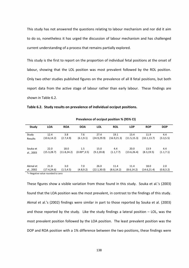
138
This study has not answered the questions relating to labour mechanism and nor did it aim
to do so, nonetheless it has urged the discussion of labour mechanism and has challenged
current understanding of a process that remains partially explored.
This study is the first to report on the proportion of individual fetal positions at the onset of
labour, showing that the LOL position was most prevalent followed by the ROL position.
Only two other studies published figures on the prevalence of all 8 fetal positions, but both
report data from the active stage of labour rather than early labour. These findings are
shown in Table 6.2.
Table 6.2. Study results on prevalence of individual occiput positions.
Prevalence of occiput position % (95% CI)
Study LOA ROA DOA LOL ROL LOP ROP DOP
Study
Results
12.4 (10.6,14.2)
3.8 (2.7,4.9)
7.6 (6.1,9.1)
27.4 (24.9,29.9)
19.1 (16.9,21.3)
13.4 (11.5,15.3)
11.9 (10.1,13.7)
4.4 (3.3,5.5)
Souka et
al., 2003
22.0 (15.3,28.7)
18.0 (11.8,24.2)
1.5 (0.00*,3.5)
15.0 (9.2,20.8)
4.4 (1.1,7.7)
20.0 (13.6,26.4)
13.9 (8.3,19.5)
4.4 (1.1,7.1)
Akmal et al., 2002
21.0 (17.4,24.6)
3.0 (1.5,4.5)
7.0 (4.8,9.2)
26.0 (22.1,30.0)
11.4 (8.6,14.2)
11.4 (8.6,14.2)
18.0 (14.6,21.4)
2.0 (0.8,3.2)
*= Negative value rounded to zero
These figures show a visible variation from those found in this study. Souka et al.’s (2003)
found that the LOA position was the most prevalent, in contrast to the findings of this study.
Akmal et al.’s (2002) findings were similar in part to those reported by Souka et al. (2003)
and those reported by the study. Like the study findings a lateral position – LOL, was the
most prevalent position followed by the LOA position. The least prevalent position was the
DOP and ROA position with a 1% difference between the two positions, these findings were
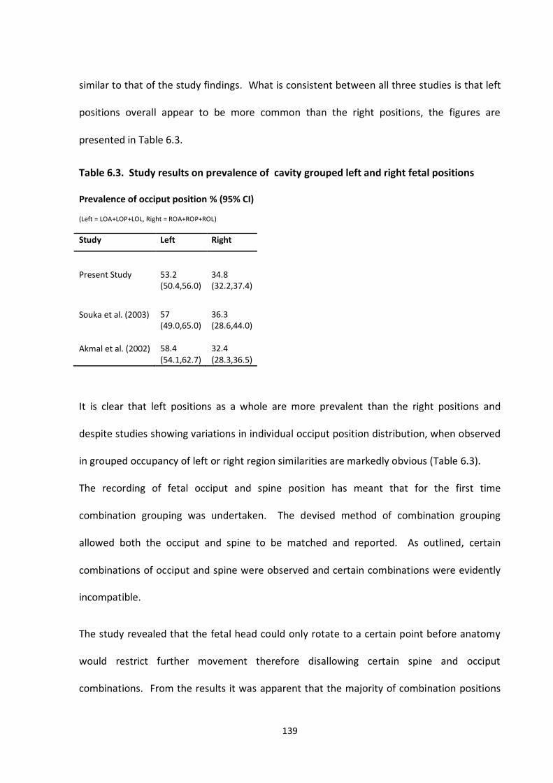
139
similar to that of the study findings. What is consistent between all three studies is that left
positions overall appear to be more common than the right positions, the figures are
presented in Table 6.3.
Table 6.3. Study results on prevalence of cavity grouped left and right fetal positions
Prevalence of occiput position % (95% CI)
(Left = LOA+LOP+LOL, Right = ROA+ROP+ROL)
Study Left Right
Present Study 53.2 (50.4,56.0)
34.8 (32.2,37.4)
Souka et al. (2003)
Akmal et al. (2002)
57 (49.0,65.0) 58.4 (54.1,62.7)
36.3 (28.6,44.0) 32.4 (28.3,36.5)
It is clear that left positions as a whole are more prevalent than the right positions and
despite studies showing variations in individual occiput position distribution, when observed
in grouped occupancy of left or right region similarities are markedly obvious (Table 6.3).
The recording of fetal occiput and spine position has meant that for the first time
combination grouping was undertaken. The devised method of combination grouping
allowed both the occiput and spine to be matched and reported. As outlined, certain
combinations of occiput and spine were observed and certain combinations were evidently
incompatible.
The study revealed that the fetal head could only rotate to a certain point before anatomy
would restrict further movement therefore disallowing certain spine and occiput
combinations. From the results it was apparent that the majority of combination positions
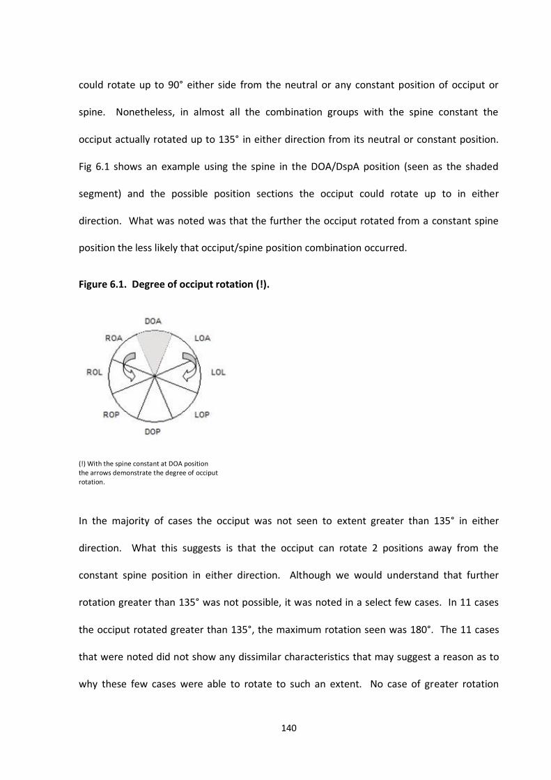
140
could rotate up to 90° either side from the neutral or any constant position of occiput or
spine. Nonetheless, in almost all the combination groups with the spine constant the
occiput actually rotated up to 135° in either direction from its neutral or constant position.
Fig 6.1 shows an example using the spine in the DOA/DspA position (seen as the shaded
segment) and the possible position sections the occiput could rotate up to in either
direction. What was noted was that the further the occiput rotated from a constant spine
position the less likely that occiput/spine position combination occurred.
Figure 6.1. Degree of occiput rotation (!).
(!) With the spine constant at DOA position the arrows demonstrate the degree of occiput rotation.
In the majority of cases the occiput was not seen to extent greater than 135° in either
direction. What this suggests is that the occiput can rotate 2 positions away from the
constant spine position in either direction. Although we would understand that further
rotation greater than 135° was not possible, it was noted in a select few cases. In 11 cases
the occiput rotated greater than 135°, the maximum rotation seen was 180°. The 11 cases
that were noted did not show any dissimilar characteristics that may suggest a reason as to
why these few cases were able to rotate to such an extent. No case of greater rotation
141
extending 180° was noted; therefore it is assumed that anatomically greater rotation is not
physically possible. The data also revealed that in the majority of cases the fetal spine
position corresponds with the fetal occiput position. A Kappa test gave a statistic of 0.68
suggesting ‘substantial agreement’ with occiput and spine combination.
Such data may influence clinical practice which utilises the fetal occiput to determine fetal
position. The advantage of position alignment could ease the discomfort and risk of
infection associated with deep pelvic and vaginal examination used to assess fetal position.
If position could be determined by abdominal palpation alone by using spine position and
assuming alignment of the occiput, this would reduce the need to perform deep pelvic
palpation or/and vaginal examination. The study has shown that the identification of the
fetal spine would at least demonstrate that the occiput would be either aligned or two
positions either side. This however requires extensive exploration as a method of accurately
identifying fetal position particularly in light of Webb’s (2009) findings.
This APOLLO study as a whole addressed two inter-linked questions, firstly the association of
fetal position and birth outcomes and secondly the accuracy of abdominal palpation to
assess the LOA position. Webb (2009) undertook analysis of the second aim, pertaining to
abdominal palpation, where she used the findings of position on ultrasound as a reference
standard and compared this against the findings of abdominal palpation by midwife
clinicians. Webb (2009) reported sensitivity and specificity to detect the LOA position on
abdominal palpation. She found that of the 61 LOA positions identified on ultrasound, only
21 were correctly identified demonstrating 34% (95% CI 24,47) sensitivity and 78% (95% CI
67,75) specificity. The conclusion overall was that abdominal palpation as a means to
142
correctly identify the LOA fetal occiput position was poor. Although Webb (2009) aimed
primarily to address the accurate assessment of the LOA position, she also reported that in
total only 16% of all fetal positions were identified accurately by abdominal palpation. Such
findings are concerning, particularly in relation to the validity of a routine assessment tool,
and would have created greater concerns if a position was identified as optimum.
The findings of both aspects of the overall APOLLO study have confirmed that scientific
evidence is essential before any practice is adopted as routine into mainstream Midwifery or
Obstetrics.
6.4. Implication for Practice
The concerns associated with increasing rates of intervention namely operative delivery has
encouraged management options to be explored as a way of addressing such issues. The
theory of OFP was one such approach, however the evidence from this study has
demonstrated that such an approach is not associated with beneficial maternal or neonatal
outcomes and consequently should not be promoted.
The availability of information is vital if women are to make an informed choice, and
therefore information pertaining to alternative approaches should be made available by
maternity associated organisation. It is however the promotion of a theory such as OFP as a
means of reducing intervention and operative delivery that is not supported this study that
must be conveyed accordingly.
143
The apparent implications for practice are evident, however the findings of the research
extend further to associated areas of every day practice. Principally the area of early labour
and latent phase, where examination to determine fetal position is recommended in both
midwifery textbooks and by NICE in the published intrapartum guidelines (Coad and
Dunstall, 2005, McCormick, 2009, NICE, 2007). It is questionable as to why the additional
discomfort associated with clinical procedures to determine fetal position are required at all
during the point of labour onset. Additionally the results to the other part of the overall
study provide an additional argument to the practice and ethical implications of a procedure
that has a low rate of accuracy and the findings of which are shown not to be associated
with outcome (Webb, 2009). Such practices however require further discussion and debate
alongside current evidence to determine the best way forward.
In view of the inaccuracy in determining fetal position subsequent studies would require use
of ultrasound to ensure fetal position is determined accurately.
6.5. Generalisability
BWHCT is the largest provider of maternity services in Birmingham and a tertiary referral
centre consequently serving a diverse population. Birmingham is also the second largest city
in the UK, and is known for its ethnic diversity. The main reason for conducting the study at
BWHCT was to achieve the diverse mix within the sample population. As outlined in section
5.2 the sample population was representative of the overall population in terms of mode of
delivery and ethnic diversity. The recruitment rate of 61.4%-75.9% supports the claims of
sample representativeness and thus the ability to generalise the findings to the target
144
population. The sample population was also representative of the Birmingham population
and the ethnic mix observed nationally as outlined by the National Consensus, with the
largest group being white, followed by the Pakistani, Indian and then the Black
African/Caribbean groups (DOH, 2001). This is the same pattern of majority groups
observed at BWHCT and in the sample for the study. Although the proportions amongst
each group vary this was expected as national statistics on ethnic diversity report collective
figures on all ages, gender and area of residence. The generalisability therefore of the study
findings would be suited to at least the childbearing population of the Birmingham district.
The study was designed so that it could be reproduced in the majority of clinical settings
where access to ultrasound and trained sonographers were available. The point at which
ultrasound is to be performed is routinely assessed by midwives upon admission to most
labour wards, so no additional services would be required. The technique used to
determine fetal position on ultrasound is well published and understood as the ‘gold
standard’ method of determining fetal position (Akmal et al., 2003, Chou et al., 2004,
Zahalka et al., 2005).
Finally the sample representativeness, the power of 89% obtained from the sample size, the
recruitment rate of 61.4%-75.9% and the detail applied to the implementation of the study
increased the generalisability of the study findings.
It is argued that at a time when all practice should be research based, promoting a single
position as optimum must be confirmed as beneficial before such practices become routine.
The study has shown that the LOA fetal position is not associated with improved outcomes
and therefore such findings should be generalised to the wider population.
145
6.6. Recommendations for Further Research
The study has answered the question initially posed; however it has also highlighted a dearth
of knowledge with regards to mechanism of labour, and bought to question the validity of
basic beliefs of the labour and delivery process. Consequently this has established that
further exploration is paramount. The areas that require further research include:
Mechanism of labour: In all the textbooks that were reviewed the labour process is
described with the fetus engaging in the transverse position. This study has shown
that not all fetuses engage in this position but instead over half engage in the
anterior, posterior or the direct positions. The mechanism involved when a fetus
commences labour in a position other than the lateral position has never been
documented, suggesting that mechanism remains undetermined. Further research is
required into labour mechanism when fetuses adopt positions other then the lateral
position for engagement.
Fetal position (active labour): This study explored the association of ‘all’ fetal
positions at the onset of labour, yet no study to date has reviewed ‘all’ fetal positions
in active labour and determined the association with birth outcomes. The
investigation of all positions in active labour may identify yet a position that is
optimum.
Fetal position (second stage): Since the process of labour involves rotational decent,
the identification of all fetal positions at labour onset and follow-up during second
stage would determine if fetal position at labour onset influenced the direction of

146
rotation. This is particularly important since mal-position is associated with
increased morbidity for the mother and the newborn.
Abdominal palpation: The use of the occiput to determine fetal position was found
to be poor, this study has shown that the fetal spine corresponds with the fetal
occiput in the majority of cases. More research is required into new or adapted
techniques and training that may enhance the clinical skills of palpation.
147
CONCLUSION
The position of the fetus during and throughout labour exerts considerable influence on the
process of active labour and delivery, with mal-position known to contribute towards
maternal and neonatal morbidity. The morbidity associated with mal-position and the belief
that this is contributing towards the increasing rates of operative delivery has led to the
implementation of theories such as OFP. OFP advocates the promotion of a single position
that being the LOA position as optimum in achieving spontaneous vaginal delivery
comparative to all other fetal positions. The lack of scientific evidence resulted in much
criticism of a theory that was otherwise attempting to salvage the normal birth process.
This prospective study aimed to establish the association between fetal position at the onset
of labour and birth outcomes for the mother and her newborn. In particular the optimum
characteristics of the LOA position in achieving SVD were explored in order that Sutton’s
theory could be supported or refuted. It demonstrated that the LOA position was not
associated with increased rates of SVD or other improved outcomes, therefore negating
Sutton’s claim of an optimal fetal position. None of the fetal positions were associated with
mode of delivery or improved birth outcomes.
The unease associated with the increasing rates of intervention and operative delivery is of
concern to all those involved with maternity care. Despite such concerns unsupported
theories should not be applied as a way in solving the complicated issues of childbirth.

148
Instead, ideas must be tested through rigorous scientific measure before any theory is
applied to routine practice as a way forward.

149
Appendix 1
Search Result of Systematic Review

150
Appendix 1 – Results of Systematic Review

151
Appendix 1 – Results of Systematic Review
152
Appendix 2
Information Leaflet for Midwives
THE APOLLO STUDY
Analysis of Fetal Position at the Onset of Labour and Labour Outcomes
1. Introduction to the research project
The APOLLO Study is a midwifery study examining the relationship between specific fetal
cephalic positions at the onset of labour and labour and birth outcomes in nulliparous
women. This study is a preliminary project needing to be done in order to establish an
evidence base to underpin theories promoting maternal posturing to encourage the fetus to
lie in a supposed optimal position.
2. What is the purpose of the study?
We believe that the exact position a baby is lying in when it lies head down at the beginning
of labour influences how labour progresses and how the baby is born. We hope this study
will provide the information needed to determine if this is true.
We are inviting as many women expecting a straightforward birth with their first baby as we
can to join the study. We are aiming for 1250 women in total to be involved in the study and
anticipate that it will take 12-18 months to complete the study.
3. What will happen to the women who take part in the study?
There is nothing special that they have to do. Their agreement to participate in the study will
give permission for a research midwife to perform an additional abdominal palpation to
determine fetal lie, presentation and position when they attend hospital in early labour or if
labour is being induced. The research midwife will also confirm her abdominal palpation by
performing an ultrasound scan.
Page 1
153
Appendix 2 – Information Leaflet for Midwives
4. What will I have to do?
We need you to recruit as many nulliparous women as you can to the study from 28
gestational weeks. Eligible women are nulliparous women with a singleton pregnancy and
where the baby is not expected to have an abnormality.
We need you to explain the study to eligible women and gain their informed consent to join
the study. This will involve you getting the consent form signed, placing it in the notes and
marking the front of the green hand-held notes with a sticker so that it is visible.
You will need to give the women the information leaflet explaining what the study involves
and who to contact if they have more questions, and also give them a copy of the signed
consent form
5. What are the risks?
Abdominal examination is not an invasive procedure and ultrasound is safe. The APOLLO
Study involves an additional abdominal examination to determine specific fetal cephalic
position with confirming ultrasound carried out by the midwife researcher at the beginning of
labour.
6. What are the benefits?
Eventually we hope to collect enough information about enough women and babies to see if
there is any particular position that benefits labour progress and how the baby is born. If this
is shown, then we hope to be able to inform women how to promote this position for their
own babies.
7. What happens at the end of the study?
The information we obtain from the abdominal examination, ultrasound and labour and birth
details will be studied carefully. We hope to see if any particular position the baby’s head is
in at the beginning of labour is related to a better labour and birth outcome. We will keep you
up to date with the progress of the study and your contribution to the recruitment. We will
present our findings to you at the completion of the study and advise you of any other
relevant information related to the study. We aim to share the findings from the study with
women and midwives by performing presentations and publishing the work in midwifery
journals.
Page 2
154
Appendix 2 – Information Leaflet for Midwives
8. What if something goes wrong?
It is extremely unlikely that having an additional abdominal examination and ultrasound scan
will cause anything to go wrong. However if there is a problem or if the woman is not
satisfied with her care, being involved in a research study does not affect her right to
complain. The patient advice and liaison service (PALS) at the Birmingham Women’s
Hospital can inform the woman of how to make a complaint.
9. Will the information of those taking part in the study be kept confidential?
All information that is collected during the course of the research will be kept strictly
confidential. Any information that leaves the hospital will have the patients’ name and
address removed so that individuals cannot be recognized from it.
10. What will happen to the results of the research study?
The information we obtain from the abdominal examination, ultrasound and labour and birth
details will be studied carefully. We hope to see if any particular position the baby’s head is
in at the beginning of labour is related to a better labour and birth outcome. We aim to share
the findings from the study with both yourselves and the women by performing presentations
and publishing the work in midwifery journals.
11. Who is organizing and funding the research?
Birmingham Women’s Healthcare NHS Trust funds the research. It will be organized by a
team of four midwives and has the support of both the Medical Director and the Director of
Nursing & Midwifery at Birmingham Women’s Healthcare NHS Trust, the Professor of
Midwifery, School of Women’s Health Studies, University of Central England, and the Royal
College of Midwives.
12. Who has reviewed the study?
South Birmingham Research Ethics Committee, Birmingham Women’s Hospital Research &
Development Committee, and the Royal College of Midwives have reviewed the study.
If you want to know more about the study or there are any issues you would like to clarify
please contact: Sara Webb on Delivery Suite, 0121 472 1377 ext 2665
Bernadette Earley at the Birth Centre, 0121 472 1377 ext 6093
155
Appendix 3
Consent Pack
A consent pack included a 4 page information leaflet for women and a consent form.
Information leaflet for women.
156
Appendix 3 – Consent Pack (Information Leaflet for Women)
THE APOLLO STUDY
Analysis of Fetal Position at the Onset of Labour and Labour Outcomes
Information Leaflet
1. Invitation to the research project
You are being invited to take part in a research study. Before you decide it is important
for you to understand why the research is being done and what it will involve. Please take
time to read the following information carefully and discuss it with others if you wish. Ask
us if there is anything that is not clear or if you would like more information. Take time to
decide whether or not you wish to take part.
Thank you for reading this.
2. What is the purpose of the study? We believe that the exact position a baby is lying in when it lies head down at the
beginning of labour influences how labour progresses and how the baby is born. We
hope this study will provide the information needed to determine if this is true.
3. Why have I been chosen?
You have been invited to take part in the APOLLO Study because you are expecting your
first baby and everything is expected to be straightforward with the birth. We are inviting
as many women expecting a straightforward birth with their first baby as we can to join the
study. We are aiming for 1250 women in total to be involved in the study and anticipate
that it will take 12-18 months to complete the study.
4. Do I have to take part?
It is up to you to decide whether or not to take part. If you do decide to take part you will
be given this information sheet to keep and be asked to sign a consent form. You will
also be given a copy of the consent form to keep. If you decide to take part you are free
to withdraw at any time and without giving a reason. A decision to withdraw at any time,
or a decision not to take part, will not affect the standard of care you receive.
Page 2

157
Appendix 3 – Consent Packs (Information Leaflet for Women)
5. What will happen to me if I take part? A sticker will be attached to the front of your notes to indicate that you have agreed to take
part.
When you come to the hospital to have your baby you will be visited soon after admission
by a research midwife. She will check first that you still wish to take part in the study.
With your consent, the research midwife will feel your abdomen to find out how your baby
is lying and the position of your baby’s head, and also perform an ultrasound scan to
confirm your baby’s position. You will not be given the results of this scan, however this
will not affect your care in any way.
Your agreement will also give permission to the researchers to read your notes and collect
relevant information about your labour and birth after your baby is born.
6. What do I have to do?
There is nothing special that you have to do. It is standard practice that when you come
to hospital to have a baby your abdomen is felt by the midwife caring for you to find out
how your baby is lying and the position of your baby’s head. If you agree to join the study
your abdomen will be felt once more by a research midwife who will also perform an
ultrasound scan just to confirm your baby’s position.
7. What are the risks?
Abdominal examination is not an invasive procedure and ultrasound is safe.
8. What are the benefits? We will be more certain about how your baby is lying and the position of your baby’s head.
Eventually we hope to collect enough information about enough women and babies to see
if there is any particular position that benefits labour progress and how the baby is born. If
this is shown, then we hope to be able to inform women how to promote this position for
their own babies.
9. What happens at the end of the research study? If you would like to know the results of the study we will be pleased to advise you of them
when the study is complete.
Page 3

158
Appendix 3 – Consent Packs (Information Leaflet for Women)
10. What if something goes wrong? It is extremely unlikely that having an additional abdominal examination and ultrasound
scan when you come to hospital expecting to have your baby will cause anything to go
wrong. However if there is a problem or you are not satisfied with your care, being
involved in a research study does not affect your right to complain. The patient advice
and liaison service (PALS) at the Birmingham Women’s Hospital can inform you of how to
make a complaint.
11. Will my taking part in this study be kept confidential?
All information that is collected about you during the course of the research will be kept
strictly confidential. Any information about you that leaves the hospital will have your
name and address removed so that you cannot be recognized from it.
12. What will happen to the results of the research study? The information we obtain from the abdominal examination, ultrasound and labour and
birth details will be studied carefully. We hope to see if any particular position the baby’s
head is in at the beginning of labour is related to a better labour and birth outcome. We
aim to share the findings from the study with women and midwives by performing
presentations and publishing the work in midwifery journals.
13. Who is organizing and funding the research? The research is funded by Birmingham Women’s Healthcare NHS Trust. It will be
organized by a team of four midwives and has the support of both the Medical Director
and the Director of Nursing & Midwifery at Birmingham Women’s Healthcare NHS Trust,
the Professor of Midwifery ( School of Women’s Health Studies) at the University of
Central England, and the Royal College of Midwives.
14. Who has reviewed the study?
South Birmingham Research Ethics Committee, Birmingham Women’s Hospital Research & Development Committee, and the Royal College of Midwives have reviewed the study.
15. Contact for further information If you want to know more about the study or there are any issues you would like to clarify
please contact:
Sara Webb on Delivery Suite, 0121 472 1377 ext 2665
Bernadette Earley at the Birth Centre, 0121 472 1377 ext 6099
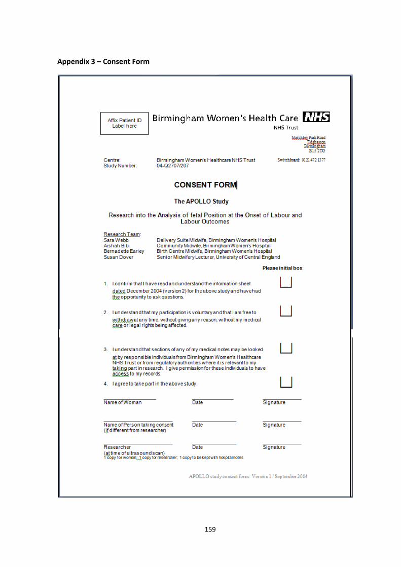
159
Appendix 3 – Consent Form
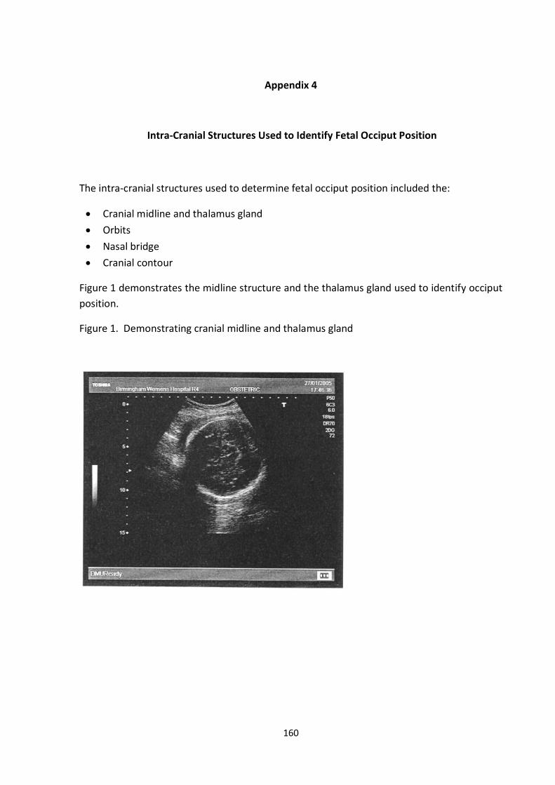
160
Appendix 4
Intra-Cranial Structures Used to Identify Fetal Occiput Position
The intra-cranial structures used to determine fetal occiput position included the:
Cranial midline and thalamus gland
Orbits
Nasal bridge
Cranial contour
Figure 1 demonstrates the midline structure and the thalamus gland used to identify occiput
position.
Figure 1. Demonstrating cranial midline and thalamus gland
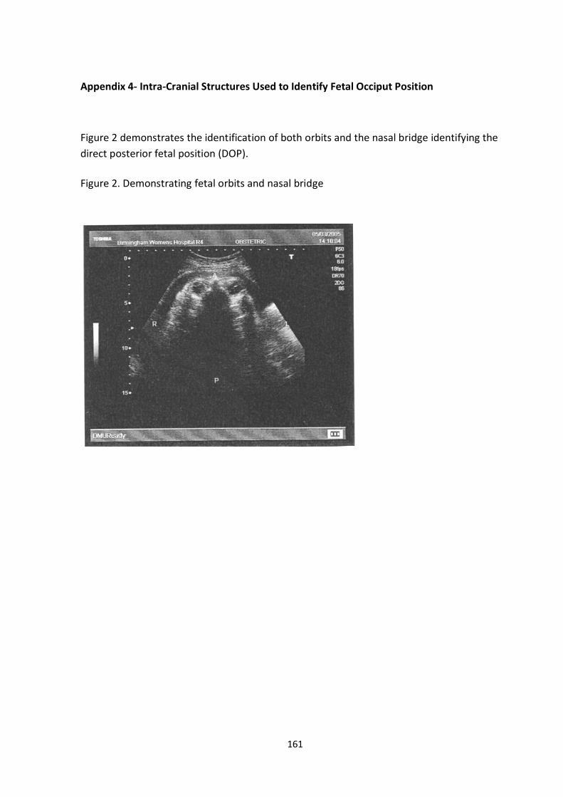
161
Appendix 4- Intra-Cranial Structures Used to Identify Fetal Occiput Position
Figure 2 demonstrates the identification of both orbits and the nasal bridge identifying the
direct posterior fetal position (DOP).
Figure 2. Demonstrating fetal orbits and nasal bridge
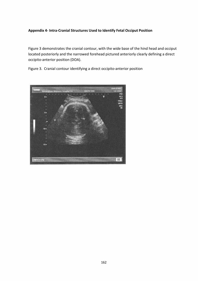
162
Appendix 4- Intra-Cranial Structures Used to Identify Fetal Occiput Position
Figure 3 demonstrates the cranial contour, with the wide base of the hind head and occiput
located posteriorly and the narrowed forehead pictured anteriorly clearly defining a direct
occipito-anterior position (DOA).
Figure 3. Cranial contour identifying a direct occipito-anterior position
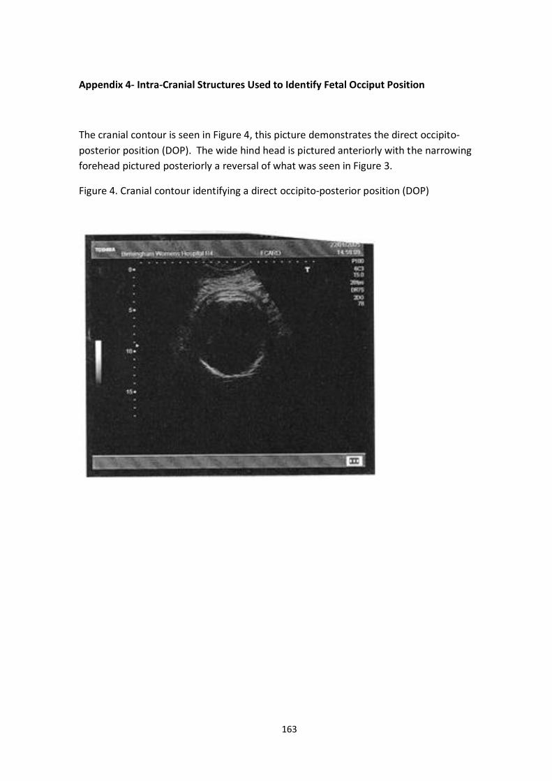
163
Appendix 4- Intra-Cranial Structures Used to Identify Fetal Occiput Position
The cranial contour is seen in Figure 4, this picture demonstrates the direct occipito-
posterior position (DOP). The wide hind head is pictured anteriorly with the narrowing
forehead pictured posteriorly a reversal of what was seen in Figure 3.
Figure 4. Cranial contour identifying a direct occipito-posterior position (DOP)

164
Appendix 5
Form B – Ultrasound Examination Form
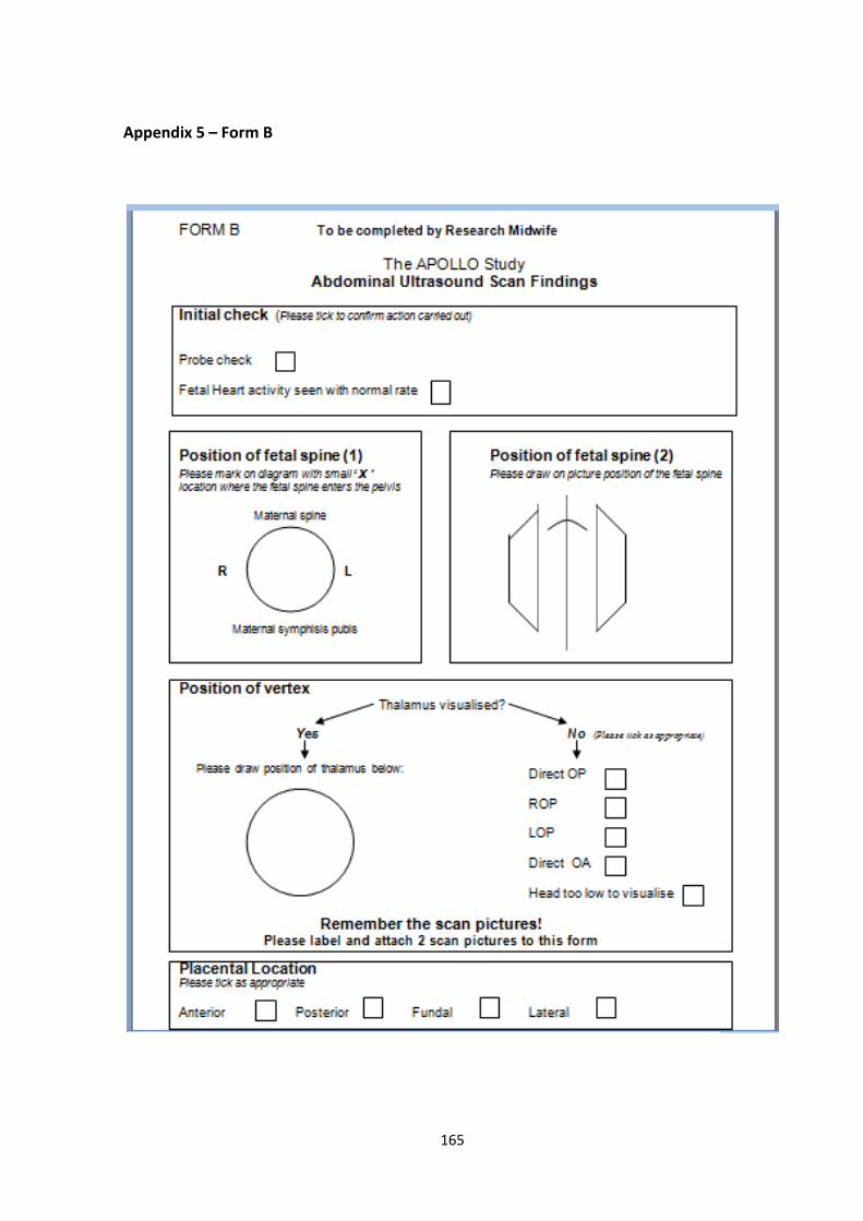
165
Appendix 5 – Form B

166
Appendix 6
Author’s contribution to the research study
The author is referred to as ‘the researcher’ throughout this thesis.
I was responsible for the idea and design of the research study. I had a main role in
obtaining ethical approval, documenting the research protocol and formulating the
methodology.
I was responsible for the overall management of the study, and performed over 90% of
ultrasound scans carried out.
Finally, once the study was complete I validated the data and undertook statistical analysis.
167
REFERENCES
ABU-HEIJA, A. & ZAYER, F. (1998) Primary and repeat caesarean sections:Comparison of indicators. Journal of Obstetrics & Gynaecology, 18, 432-434. AKMAL, S., KAMETA, N., TSOI, E., HOWARD, R. & NICOLAIDES, K. H. (2004a) Ultrasonographic occiput position in early labour in the prediction of caesarean section. BJOG: An International Journal of Obstetrics and Gynaecology, 111, 532-536. AKMAL, S., KAMETAS, N., TSOI, E., HARGREAVES, C. & NICOLAIDES, K. H. (2003) Comparison of transvaginal digital examination with intrapartum sonography to determine fetal head position before instrumental delivery. Ultrasound in Obstetrics and Gynaecology, 21, 437- 440. AKMAL, S., KAMETAS, N., TSOI, E., HOWARD, R. & NICOLAIDES, K. H. (2004b) Ultrasonographic occiput position in early labour in the prediction of caesarean section. BJOG: an International Journal of Obstetrics and Gynaecology, 111, 532-536. AKMAL, S. & PATERSON-BROWN, S. (2009) Malpositions and malpresentations of the foetal head. Obstetrics, Gynaecology and Reproductive Medicine, 19, 240-246. AKMAL, S., TSOI, E., HOWARD, R., OSEI, E. & NICOLAIDES, K. H. (2004c) Investigation of occiput posterior delivery by intrapartum sonography. Ultrasound in Obstetrics and Gynaecology, 24, 425-428. AKMAL, S., TSOI, E., KAMETA, N., HOWARD, R. & NICOLAIDES, K. H. (2002) Intrapartum sonography to determine fetal head position. The Journal of Maternal-Fetal and Neonatal Medicine, 12, 172-177. ANIM-SOMUAH, M., SMYTH, R. & HOWELL, C. (2010) Epidural versus non-epidural or no analgesia in labour The Cochrane Database of Systematic Reviews. CD000331. BENAVIDES, L., WU, J., HUNDLEY, A. F., IVESTER, T. S. & VISCO, A. G. (2005) The impact of occiput posterior fetal head position on the risk of anal sphincter injury in forceps-assisted vaginal deliveries. American Journal of Obstetrics and Gynaecology, 192, 1702-1706. BIANCUZZO, M. (1993) How to recognise and rotate an occiput posterior fetus. American Journal of Nursing, 93, 38-41. BLAND, M. & ALTMAN, D. (1995) Multiple significance tests: the Bonferroni method. BMJ, 310, 170.
168
BLASI, I., D'AMICO, R., FENU, V., VOLPE, A., FUCHS, I., HENRICH, W. & MAZZA, V. (2010) Sonographic assessment of fetal spine and head position during the first and second stages of labor for the diagnosis of persistent occiput posterior position: a pilot study. Ultrasound in Obstetrics and Gynaecology, 35, 210-215. BOHRA, U., DONNELLY, J., GEARY, M. & MACQUILLAN, K. (2003) Active management of labour revisited: The first 1000 primiparous labour in 2000. Journal of Obstetrics & Gynaecology, 23, 118-120. BWH (2007) Obstetrics Annual Report, April 2006-March 2007. Birmingham, BWH. CARUTHERS, B. (2000) Structural and mechanical factors association with the birth process. The Surgical Technologist, Oct 1, 19-27. CHAMBERLAIN, G. & STEER, P. (1999) ABC of labour care. Unusual presentations and positions and multiple pregnancy. BMJ, 318, 1192-1194. CHELMOW, D., KILPATRICK, S. & RK., L. (1993) Maternal and neonatal outcomes after prolonged latent phase. Obstetrics and Gynaecology, 201, 486-491. CHENG, Y. W., SHAFFER, B. L. & CAUGHEY, A. B. (2006) The association between persistent occiput posterior position and neonatal outcomes. Obstetrics and Gynaecology, 107, 837- 844. CHOU, M. R., KREISER, D., TASLIMI, M., DRUZIN, M. & EL-SAYED, Y. (2004) Vaginal versus ultrasound examination of the fetal occiput position during the second stage of labor. American Journal of Obstetrics and Gynaecology, 191, 521-524. COAD, J. & DUNSTALL, M. (2005) Anatomy and Physiology for Midwives, London, Churchill Livingstone. COATES, T. (2002) Malpositions and malpresentations of the occiput: current research and practice tips. MIDIRS Midwifery Digest, 12, 152-154. COATES, T. (2008) Malpositions of the occiput and malpresentations. IN FRASER, D. & COOPER, M. (Eds.) Myles Textbook for Midwives. London, Churchill Livingstone. CUNNINGHAM, F. G., LEVENO, M. D., BLOOM, S., HAUTH, J. C., GILSTRAP, L. & WENSTROM, K. (2008) Normal Labour & Delivery. Williams Obstetrics, London, McGraw-Hill. DAVIS, E. (2004) Heart & Hands. A Midwifery Guide to Pregnancy and Birth, London, Celestial Arts. DEWBURY, K., HYLTON, M., COSGROVE, D. & FARRANT, P. (2001) Ultrasound in Obstetrics and Gynaecology, London, Churchill Livingstone.
169
DIETZE, M. (2001) A re-evaluation of the mechanism of labour for contemporary midwifery practice. Midwifery Matters, 3-8. DOH (2000) Why Mother's Die 1997-1999. The Confidential Enquiries into Maternal Deaths in the UK. London, HMSO. DOH (2001) The UK population: by ethnic group, April 2001. London, Office of National Statistics. Retrieved 15-04-2010 DOH (2002) NHS Maternity Statistics England 1998-99 to 2000-01. London, The Health and Social Care Information Centre. DOH (2005) NHS Maternity Statistics - England 2004-05. London, The Health and Social Care Information Centre. DOH (2010) NHS Maternity Statistics, England - 2009-10. London, The Information Centre for Health and Social Care. DONNA, S. (2008) Birth Countdown to Optimal, London, Fresh Heart Publishing. DOUBILET, P. M. & BENSON, C. B. (2003) Atlas of Ultrasound in Obstetrics and Gynaecology, Philadelphia, Williams & Wilkins. DOWNE, S. (2008) The transition and the second stage of labour: physiology and the role of the midwife. IN FRASER, D. & COOPER, M. (Eds.) Myles Textbook for Midwives. 15 th ed. London, Churchill Livingstone. EL-HAMAMY, E. & ARULKUMARAN, S. (2005) Poor progress in labour. Current Obstetrics & Gynaecology, 15, 1-8. FABER, P. (1998) The persistent OP: Can maternal position rotate the baby? The Practising Midwife, 1, 16-19. FAULKNER, A. (2009) Doula Momma [online] available at www.googleimages.com [Accessed 01-04-2010]. FITZPATRICK, M., MCQUILLAN, K. & O'HERLIHY, C. (2001) Influence of persistent occiput posterior position on delivery outcomes. The American College of Obstetricians and Gynaecologists, 98, 1027-1031. FRASER, W., TURCOT, L., KRAUSS, I. & BRISSON-CAROL, G. (2006) Amniotomy for shortening spontaneous labour. The Cochrane Database of Systematic Reviews. CD000015. FRIEDMAN, E. (1978) Labor. Clinical evaluation and management., New York, Appleton- Century-Croft.
170
GAMOW, G. (2003) Gravity, Dover, Dover Publications Inc. GARDBERG, M., LAAKKONEN, E. & SALEVAARA, M. (1998) Intrapartum sonography and persistent occiput posterior position: A study of 408 deliveries. Obstetrics and Gynaecology, 91, 746-749. GARDBERG, M. & TUPPURAINEN, M. (1994) Anterior placental location predisposes for occiput posterior presentation near term. Acta Obstetrics and Gynaecology Scandinavia, 73, 151-152. GILLFORD, D. S., MORTON, S. C., FISKE, M., KEESEY, J., KEELER, E. & KAHN, K. L. (2000) Lack of progress as a reason for caesarean. Obstetrics and Gynaecology, 95, 589-595. GOER, H. (1994) The Occiput Posterior Baby: It's more than a pain in the back. Childbirth Instructor Magazine, Summer, 36-40. HAMILTON, A. (2009) Assisted Births. IN FRASER, D. & COOPER, M. (Eds.) Myles Textbook For Midwives. 15 ed. Edinburgh, Churchill Livingstone. HOLMAN, L. (2001) Optimal Fetal Positioning: Making a great turn around. Midwifery Today, Sping, 41. JOHANSON, R. & MENON, V. (2009) Vacuum extraction versus forceps for assisted vaginal delivery. The Cochrane Database of Systematic Reviews. CD000224. JOHNSON, R. & TAYLOR, W. (2010) Skills for Midwifery Practice, Edinburgh, Churchill Livingstone. KARIMINIA, A., CHAMBERLAIN, M. E., KEOGH, J. & SHEA, A. (2004) Randomised controlled trial of effect of hands and knees posturing on incidence of occiput position at birth. BMJ, 1- 5. KIRCHENGAST, S. (2007) Maternal age and pregnancy outcome- an anthropological approach. Anthroplogischer Anzeiger, 65, 181-191. KREISER, D., SCHIFF, E., KAYAM, Z., AVARAHAM, A. & ACHIRON, R. (2001) Determination of fetal occiput position by ultrasound during the second stage of labor. The Journal of Maternal-Fetal Medicine, 10, 283-286. LADWIG, P. W., LONDON, M. L. & OLDS, S. B. (1998) Maternal-Newborn Nursing Care, California, Addison-Wesley. LEONG, E., SIVANESARATNAM, V., OH, L. & CHAN, Y. (2000) Epidural analgesia in primigravidae in spontaneous labour at term: A prospective study. Journal of Obstetrics & Gynaecology Research, 26, 271-275.
171
MAGHOMA, J. & BUCHMANN, E. (2001) Maternal and fetal risks associated with prolonged latent phase of labour Journal of Obstetrics & Gynaecology, 22, 16-19. MATHAI, M., SANGHVI, H. & GUIDOTTOI, R. J. (2000) Managing Complications in Pregnancy and Childbirth (MCPC) - A Guide for Midwives and Doctors. United Kingdom, WHO. MCCORMICK, C. (2008) The first stage of labour: physiology and early care. IN FRASER, D. M. & COOPER, M. A. (Eds.) Myles Textbook for Midwives. 15th ed. London, Churchill Livingstone. MCCORMICK, C. (2009) The first stage of labour: physiology and early care IN FRASER, D. & COOPER, M. (Eds.) Myles Textbook For Midwives. Fifteenth Edition ed. Edinburgh, Churchill Livingstone. MCDONALD, G. (2010) Diagnosing the latent phase of labour: use of the partogram. British Journal of Midwifery, 18, 9. MCFARLIN, B., ENGSTROM, J., SAMPSON, M. B. & CATTLEDGE, F. (1985) Concurrent Validity of Leopold's Maneuvers in Determining Fetal Presentation and Position. Journal of Nurse- Midwifery, 30, 280-284. MOCANU, E. V., GREENE, R. A., BYRNE, B. M. & TURNER, M. J. (2000) Obstetrics and neonatal outcome for babies weighing more then 4.5kg: An analysis by parity. European Journal of Obstetrics, Gynaecology & Reproductive Biology, 92, 229-233. NCT (2010) NCT The UK's largest charity for parents. Available at: http://www.nct.org.uk. (Accessed: 19 April 2010) NERI, A., KAPLAN, B., RABINERSON, D. & OVADIA, J. (1995) The management of the persistent occiput-posterior position. Clinical Expertise in Obstetrics and Gynaecology, 22, 126-131. NICE (2004) Caesarean Section. London, National Institute for Clinical Excellence (NICE). Clinical Guideline 13. NICE (2007) Intrapartum Care. Care of the healthy women and their babies during childbirth. London, National Institute of Clinical Excellence. Clinical Guideline 55. NOLAN, M. (1997) Jean Sutton and Optimal fetal positioning. Modern Midwife, 7, 15-17. PEARL, M. L., ROBERTS, J. M., LAROS, R. R. & HURD, W. (1993) Vaginal delivery from the persistent occiput posterior position. Influence on maternal and neonatal morbidity. The Journal of Reproductive Medicine, 38, 955-961.
172
PEREGRINE, E., O'BRIEN, P. & JAUNIAUX, E. (2007) Impact on delivery outcomes of ultrasonographic fetal head position prior to induction of labor. The American College of Obstetricians and Gynaecologists, 109, 618-625. PONKEY, S., COHEN, A., HEFFNER, L. & LIEBERMAN, E. (2003) Persistent fetal occiput posterior position: Obstetric outcomes. Obstetrics and Gynaecology, 5, 915-920. RAYBURN, W., SIEMERS, K., LEGINO, L., NABITY, M., ANDERSON, J. & PATIL, K. (1989) Dystocia in labor: Determining fetal position by clinical and ultrasonic techniques. American Journal of Perinatology, 6, 316-319. RIDLEY, R. (2007) Diagnosis and intervention for occiput posterior malposition Journal of Obstetrics, Gynaecology & Neonatal Nursing (JOGNN), 36, 135-143. SAVAGE, W. (2007) The rising Caesarean section rate: a loss of obstetric skill? Journal of Obstetrics & Gynaecology, 27, 339-346. SELMAN, T. & JOHNSTON, T. (2010) Mechanism and management of normal labour. Obstetrics, Gynaecology and Reproductive Medicine, 20, 201-206. SENECAL, J., XIONG, X. & FRASER, W. (2005) Effect of fetal position on second-stage duration and labour outcome. The American College of Obstetricians and Gynaecologists, 105, 763- 772. SHARMA, V., COLLERAN, G., DINEEN, B., HESSION, M. B., AVALOS, G. & MORRISON, J. J. (2009) Factors influencing delivery mode for nulliparous women with singleton pregnancy and cephalic presentation during a 17-year period. European Journal of Obstetrics, Gynaecology & Reproductive Biology, 147, 173-177. SHERER, D. & ABULAFIA, O. (2003) Intrapartum assessment of fetal head engagement: comparison between transvaginal digital and transabdominal ultrasound determinations. Ultrasound in Obstetrics and Gynaecology, 21, 430-436. SHERER, D., MIODOVNIK, M., BRADLEY, K. & LANGER, O. (2002a) Intrapartum fetal head position II: comparison between transvaginal digital examination and transabdominal ultrasound assessment during the second stage of labor. Ultrasound in Obstetrics and Gynaecology, 19, 264-268. SHERER, D. M., ABULAFIA, O. & ANYAEGBUNAM, A. M. (1998) Intra- and early postpartum ultrasonography: A review. Obstetrics and Gynaecology Survey, 53, 107-116. SHERER, D. M., MIODOVNIK, M., BRADLEY, K. & LANGER, O. (2002b) Intrapartum fetal head position 1: comparison between transvaginal digital examination and transabdominal ultrasound assessment during the active stage of labor. Ultrasound in Obstetrics and Gynaecology, 19, 258-263.
173
SIMPKIN, P. (2010) The fetal occiput posterior position: State of the science and a new perspective. Birth, 37, 61-71. SIZER, A. & NIRMAL, D. (2000) Occipitoposterior position: Associated factors and obstetric outcome in Nulliparas. Obstetrics and Gynaecology, 96, 749-752. SMYTH, R., ALLDRED, S. K. & MARKHAM, C. (2009) Amniotomy for shortening spontaneous labour. Cochrane Database of Systematic Reviews CD006167. SOUKA, A., HARITOS, K., BASAYIANNIS, N., NOIKOKYRI, N. & ANTSAKLIS, A. (2003) Intrapartum ultrasound for the examination of the fetal head position in normal and obstructed labor. The Journal of Maternal-Fetal and Neonatal Medicine, 13, 59-63. SUTTON, J. (1996) Birth: Medical emergency or engineering miracle? A midwifery approach to keeping birth normal. MIDIRS Midwifery Digest, 6, 170-173. SUTTON, J. (2000) Occipito-posterior positioning. Practising Midwife, 6, 20-22. SUTTON, J. (2001) Let birth be born again. Rediscovering and Reclaiming our Midwifery Heritage, Middlesex, Birth Concepts UK. SUTTON, J. (2007) Get your baby lined up! Optimum foetal positioning [online] available at www.homebirthreferencesite.com [Accessed 12-06-2010]. SUTTON, J. & SCOTT, P. (1994) Optimal fetal positioning. A midwifery approach to increasing the number of normal births. MIDIRS Midwifery Digest, 4, 283-286. SUTTON, J. & SCOTT, P. (1996) Understanding and teaching optimal foetal positioning, New Zealand, Birth Concepts. SWEET, B. (1997) The Female Pelvis. IN SWEET, B. & TIRAN, D. (Eds.) Mayes' Midwifery: A Textbook for Midwives. 12 th ed. London, Bailliere Tindall. THOMAS, J. & PARANJOTY, S. (2001) National Sentinel Caesarean Section Audit Report London, Royal College of Obstetricians and Gynaecologists. TIRAN, D. (2008) Bailliere's Midwives Dictionary. IN SARAH, J. (Ed.) 11 ed. London, Bailllliere Tindall. VICCARS, A. (2009) Antenatal care. IN FRASER, D. & COOPER, M. (Eds.) Myles Textbook for Midwives. 15 th ed. Edinburgh, Churchill Livingstone. VIERA, A. & GARRETT, J. (2005) Understanding Interobserver Agreement: The Kappa Statistic. Family Medicine, 37, 360-363.
174
WALSH, D. & DOWNE, S. (2010) Essential midwifery practice intrapartum care, England, Wiley-Blackwell. WEBB, S. S. (2009) Abdominal Palpation to Determine Fetal Position at the Onset of Labour: An Accuracy Study. Department of Obstetrics and Gynaecology. Birmingham, The University of Birmingham. WHO (1985) Appropriate technology for birth. Lancet, 2, 436-437. WU, J., WILLIAMS, K., HUNDLEY, A., CONNOLLY, A. & VISCO, A. (2005) Occiput posterior fetal head position increases the risk of anal sphincter injury in vacuum-assisted deliveries. American Journal of Obstetrics and Gynaecology, 193, 525-529. YATES, S. (2010) Pregnancy and Childbirth. A holistic approach to massage and body work, London, Churchill Livingstone. ZAHALKA, N., SADAN, O., MALINGER, G., LIBERATI, M., BOAZ, M., GLEZERMAN, M. & ROTMENSCH, S. (2005) Comparison of transvaginal sonography with digital examination and transabdominal sonography for the determination of fetal head position in the second stage of labor. American Journal of Obstetrics and Gynaecology, 193, 381-386.