The association between clinical symptoms of lumbar spinal ...
11
Fukushima J. Med. Sci., Vol. 67, No. 3, 2021 [Original article] The association between clinical symptoms of lumbar spinal stenosis and MRI axial imaging findings Yuki Fushimi, Koji Otani, Ryoji Tominaga, Masataka Nakamura, Miho Sekiguchi and Shin - ichi Konno Department of Orthopaedic Surgery, Fukushima Medical University School of Medicine, Fukushima, Ja- pan (Received August 24, 2021, accepted October 27, 2021) Abstract Purpose : In diagnosing lumbar spinal stenosis (LSS), Magnetic Resonance Imaging (MRI) is ap- propriate to confirm the presence of anatomical stenosis of the spinal canal or compression of the nerve roots. However, it is known that morphological LSS is often present in asymptomatic sub- jects. There is still controversy about the relationship between anatomical LSS and symptomatic LSS. The aim of this study was to assess the association between qualitative imaging findings on MRI of the lumbar spine and symptomatic LSS. Patients and methods : This was a cross - sectional study of 239 volunteers from an epidemiologi- cal survey that included 1,862 participants in total. MRI of the lumbar spine was evaluated in four categories : morphological grading of central stenosis and lateral recess stenosis, presence of the sedimentation sign, and severity of facet joint effusion. The relationship between these morpho- logical evaluations and typical LSS symptoms as assessed by the self - administered, self - reported history questionnaire for lumbar spinal stenosis (LSS - SSHQ) was investigated by multiple logistic regression analysis. Results : The odds ratio of the most severe central stenosis to no stenosis was 15.5 (95%CI : 1.4 - 164.9). Only the most severe central stenosis was associated with typical LSS symptoms, but not all cases with typical LSS symptoms were due to severe central stenosis. Conclusion : Extreme severe central stenosis was strongly related to typical LSS symptoms. However, although subjects with severe central stenosis showed symptoms suggestive of LSS, these subjects did not always show typical LSS symptoms. Key words : cross - sectional study, central stenosis, lateral recess stenosis, sedimentation sign, facet joint effusion Introduction Lumbar spinal stenosis (LSS) is characterized by narrowing of the spinal canal including the central spinal canal, in the area under the facet joints (sub- articular, lateral recess, or foraminal stenosis), or far laterally (extraforaminal stenosis) 1 - 3) . The anatomi- cal condition of LSS causes variable clinical symp- toms, such as gluteal and/or lower extremity pain, numbness, and/or neurogenic intermittent claudica- tion. These symptoms are thought to be caused by the diminished space available for the neural and vascular elements. However, the pathogenesis of LSS is not fully clarified, and there is no definition with clear criteria for the imaging findings and clini- cal symptoms 1,4) . Magnetic resonance imaging (MRI) is appropri- ate to confirm the anatomical condition of the spinal Corresponding author : Koji Otani E - mail : [email protected] ©2021 The Fukushima Society of Medical Science. This article is licensed under a Creative Commons [Attribu- tion - NonCommercial - ShareAlike 4.0 International] license. https://creativecommons.org/licenses/by - nc - sa/4.0/ 150
-
Upload
khangminh22 -
Category
Documents
-
view
0 -
download
0
Transcript of The association between clinical symptoms of lumbar spinal ...
150 Y. Fushimi et al.Fukushima J. Med. Sci.,Vol. 67, No. 3, 2021
[Original article]
The association between clinical symptoms of lumbar spinal stenosis and MRI axial imaging findings
Yuki Fushimi, Koji Otani, Ryoji Tominaga, Masataka Nakamura, Miho Sekiguchi and Shin-ichi Konno
Department of Orthopaedic Surgery, Fukushima Medical University School of Medicine, Fukushima, Ja-pan
(Received August 24, 2021, accepted October 27, 2021)
Abstract Purpose : In diagnosing lumbar spinal stenosis (LSS), Magnetic Resonance Imaging (MRI) is ap-propriate to confirm the presence of anatomical stenosis of the spinal canal or compression of the nerve roots. However, it is known that morphological LSS is often present in asymptomatic sub-jects. There is still controversy about the relationship between anatomical LSS and symptomatic LSS. The aim of this study was to assess the association between qualitative imaging findings on MRI of the lumbar spine and symptomatic LSS. Patients and methods : This was a cross-sectional study of 239 volunteers from an epidemiologi-cal survey that included 1,862 participants in total. MRI of the lumbar spine was evaluated in four categories : morphological grading of central stenosis and lateral recess stenosis, presence of the sedimentation sign, and severity of facet joint effusion. The relationship between these morpho-logical evaluations and typical LSS symptoms as assessed by the self-administered, self-reported history questionnaire for lumbar spinal stenosis (LSS-SSHQ) was investigated by multiple logistic regression analysis. Results : The odds ratio of the most severe central stenosis to no stenosis was 15.5 (95%CI : 1.4-
164.9). Only the most severe central stenosis was associated with typical LSS symptoms, but not all cases with typical LSS symptoms were due to severe central stenosis. Conclusion : Extreme severe central stenosis was strongly related to typical LSS symptoms. However, although subjects with severe central stenosis showed symptoms suggestive of LSS, these subjects did not always show typical LSS symptoms.
Key words : cross-sectional study, central stenosis, lateral recess stenosis, sedimentation sign,
facet joint effusion
Introduction
Lumbar spinal stenosis (LSS) is characterized by narrowing of the spinal canal including the central spinal canal, in the area under the facet joints (sub-articular, lateral recess, or foraminal stenosis), or far laterally (extraforaminal stenosis)1-3). The anatomi-cal condition of LSS causes variable clinical symp-toms, such as gluteal and/or lower extremity pain,
numbness, and/or neurogenic intermittent claudica-tion. These symptoms are thought to be caused by the diminished space available for the neural and vascular elements. However, the pathogenesis of LSS is not fully clarified, and there is no definition with clear criteria for the imaging findings and clini-cal symptoms1,4).
Magnetic resonance imaging (MRI) is appropri-ate to confirm the anatomical condition of the spinal
Corresponding author : Koji Otani E-mail : [email protected]©2021 The Fukushima Society of Medical Science. This article is licensed under a Creative Commons [Attribu-tion-NonCommercial-ShareAlike 4.0 International] license. https://creativecommons.org/licenses/by-nc-sa/4.0/
150

151The association between LSS symptoms and MRI
canal or compression of the nerve roots. However, it is known that radiographic LSS is found in asymp-tomatic patients5). According to the North Ameri-can Spine Society (NASS) Guideline of LSS, there is insufficient evidence for a correlation between clini-cal symptoms or function with the anatomic narrow-ing of the spinal canal on imaging4). One of the rea-sons for this might be variations in assessment methods due to the lack of clear criteria for the as-sessment of imaging findings.
In recent studies, the most commonly used meth-od for evaluating anatomical spinal stenosis on imaging was dural sac cross-sectional area (DCSA)4,6-10). However, the correlation between DCSA and symp-toms is still controversial. In addition, the mea-surement of DCSA is not easy in routine clinical settings ; therefore, a well-defined and simple mor-phological classification for evaluating the severity of anatomical spinal stenosis would be useful. Moreover, DCSA is insufficient to evaluate nerve root impingement due to lateral recess stenosis (LRS), since DCSA does not include the lateral re-cess. LRS is most commonly caused by degenera-tive changes of the spine, such as facet joint osteoar-t h r i t i s , l i g a m e n t u m f l a v u m h y p e r t ro p h y, intervertebral disc degeneration, and endplate spurs2,11,12). Therefore, central stenosis and LRS should be evaluated separately for anatomical spinal stenosis. Furthermore, there are other findings as-sociated with anatomical spinal stenosis, such as the sedimentation sign and facet joint effusion (FJE), which are considered to be related to symptomatic LSS13-17). However, evidence for a correlation be-tween clinical symptoms and anatomic stenosis on imaging is still insufficient. Since most previous studies were hospital-based and their control groups
had diseases other than LSS, the results are unlikely to be generalizable. Therefore, there is a need for population-based studies with higher external valid-ity. The main objective of this study was to evalu-ate the association between qualitative assessments of MRI axial images of the lumbar spine and symp-tomatic LSS in a community cohort.
Materials and methods
This study was approved by the ethics commit-tee of Fukushima Medical University School of Med-icine (Approval No. 295). All participants gave their written informed consent to participate in the study.
Participants
This cross-sectional study was based on an epi-demiological survey from 2004, for which 1,862 peo-ple (697 males and 1,165 females) enrolled when a routine public health survey was conducted by their local governments in Tadami Town, Tateiwa Village, and Ina Village of Fukushima Prefecture. Their ages ranged from 19 to 93 years18). Of the 1,862 survey participants, 459 agreed to undergo lumbar spine MRI for additional assessment. Participants with no cerebral infarction or bleeding history who could walk independently were included. The ex-clusion criteria were if they were unable to walk in-dependently, fill out questionnaires due to visual im-pairment, or had ever undergone brain or spinal surgery. Those without sufficient imaging findings in all classifications or those with missing question-naire data were excluded, leaving 239 who were ana-lysed in the present study (Figure 1).
Fig. 1. Flow chart of the analysis for evaluation of MRI.

152 Y. Fushimi et al.
Assessment of MRI
Axial T2-weighted images were obtained at each level of the vertebral bodies and intervertebral discs from L1 to S1. The details of the MRI imag-ing conditions are shown in Table 1.
The classifications of central stenosis, LRS, the sedimentation sign, and FJE were evaluated using MRI (Table 2)2,13,19,20). Central stenosis, nerve root compression by classification of LRS, and FJE were evaluated in the axial images of each intervertebral disc level from L1-2 to L5-S1. For all findings, the highest grade was used as representative for analy-sis. To evaluate the intra/inter-observer reliabili-ties of each classification, two orthopaedic surgeons (YF & MN) did independent evaluations two times every two weeks. According to the sample size calculation estimating ρ = 0.8 with 0.4 of 95% confi-dence interval rated by two examiners, at least 20 subjects would be needed. Therefore, 30 subjects were randomly selected for determination using kappa analysis21). The intra-observer reliabilities were determined to be substantial for the assess-ment of central stenosis, the sedimentation sign, and
FJE (0.68, 0.71, and 0.70, respectively) and moder-ate for the assessment of LRS (0.54). The inter-observer reliabilities were determined to be sub-stantial for the assessment of central stenosis, the sedimentation sign, and FJE (0.65, 0.68, and 0.63, respectively) and fair for the assessment of LRS (0.31). Therefore, the intra-observer and inter-ob-server reliabilities were considered acceptable. Fi-nally, one orthopaedic surgeon (YF) examined the images without any participants’ information, includ-
Table 1. MRI specifications and utilization
Manufacturer Philips Toshiba
Product name Gyroscan EXCELART/P2
Intera Power Pianissimo
Tesla 1.0 T 1.5 T
Slice thickness (mm) 5 5
Slice gap (mm) 0.5 1
TE (ms) 120 108
TR (ms) 4,500 4,000
No. of participants 170 69
Abbreviations : MRI, magnetic resonance imaging ; TE, echo time ; TR, repetition time.
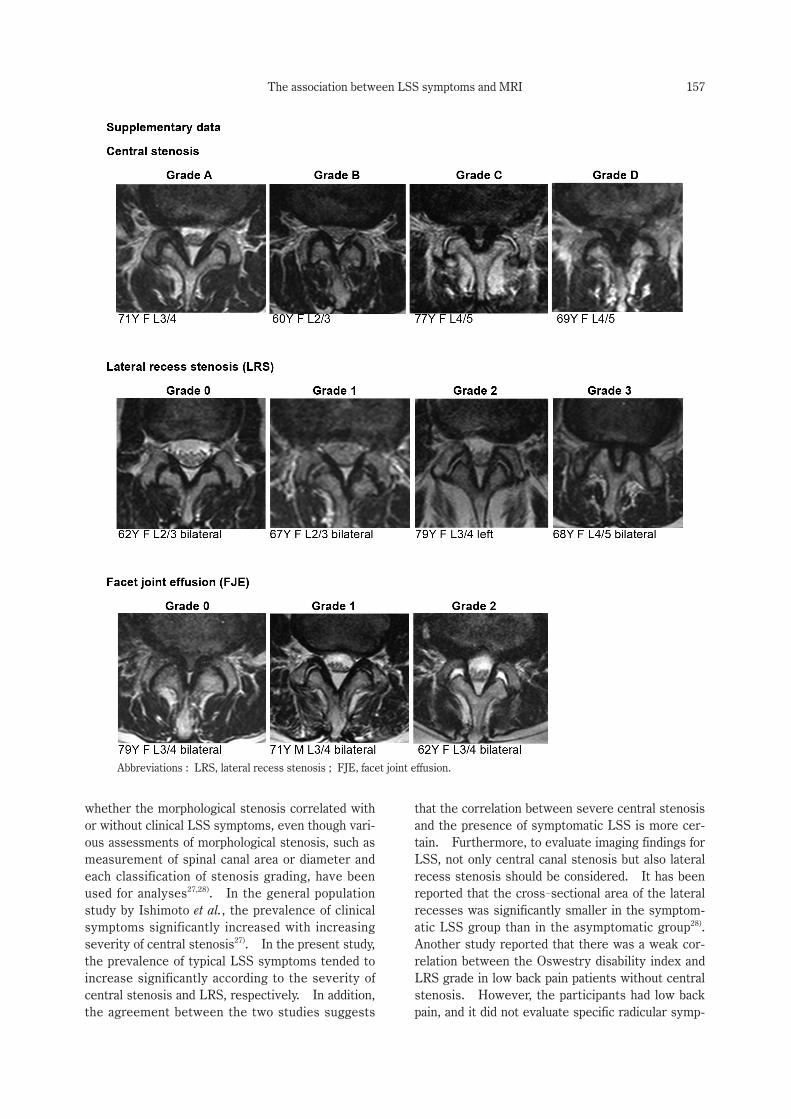
Table 2. Classifications of MRI findings
Central stenosis Schizas C et al.19)
Grade A There is clearly CSF visible inside the dural sac, but its distribution is inhomogeneous.
Grade B The rootlets occupy the whole of the dural sac, but they can still be individualized. Some CSF is still present giv-ing a grainy appearance to the sac.
Grade C No rootlets can be recognized, the dural sac demonstrating a homogeneous gray signal with no CSF signal visi-ble. There is epidural fat present posteriorly.
Grade D In addition to no rootlets being recognizable, there is no epidural fat posteriorly.
Lateral recess stenosis (LRS) Bartynski WS et al.2)
Grade 0 Normal
Grade 1 Reduced size of the corner of the lateral canal or recess ; trefoil shape to the lateral recess, either congenital or acquired ; early acute angular narrowing of the corner of the canal and thecal sac ; nerve root is visualized and not widened, flattened, or altered.
Grade 2 Reduced size of the corner of the lateral canal or lateral recess, trefoil shape and narrowing of the lateral recess, angular pinch-like shape and narrowing of the lateral canal and thecal sac, nerve root judged compressed in the small trefoil recess or angled pinch but recess judged not totally obliterated, nerve root may be deviated medially.
Grade 3 Severe facet hypertrophy and disc/end plate changes, no CSF or space identified in the lateral recess or corner of the canal, severe angular pinch of the lateral corner of the canal, root may or may not be clearly visible, root may be seen coursing through the compressed lateral recess, root may be seen as medially displaced.
Sedimentation sign Barz T et al.13)
Positive The absence of nerve root sedimentation.
Facet joint effusion (FJE) Chaput C et al.20)
Grade 0 No effusion
Grade 1 Measurable effusion < 1.5 mm
Grade 2 Large effusion > 1.5 mm
Abbreviations : CSF, cerebrospinal fluid.

153The association between LSS symptoms and MRI
ing their symptoms. The highest severity of each classification in individual subjects was used as the representative value in the analysis.
Assessment of clinical symptoms
The presence or absence of typical LSS symp-toms was determined using the LSS-SSHQ to stan-dardize self-reporting of symptoms. The LSS-
SSHQ was developed for the identification of LSS based solely on self-reported patient information. This questionnaire is a just a screening tool for LSS, therefore not all symptomatic LSS cases could be detected. In order to identify symptomatic LSS by a self-reported questionnaire, a cut-off value is set even though false positive and negative cases were limitations for the diagnosis of LSS. “Symptomatic LSS as assessed by LSS-SSHQ” will be referred to as “typical LSS symptoms” in this study. LSS-
SSHQ consists of 10 question items and has a sensi-tivity of 84%, specificity of 78%, positive likelihood ratio of 1.89, and negative likelihood ratio of 0.21 ac-cording to the validation study22). Evidence grading has established that the SSHQ can be useful to as-sist with providing clinical evidence of lumbar spinal stenosis as level II diagnostic evidence by the De-generative Lumbar Spinal Stenosis Work Group of the North American Spine Society’s Evidence-
Based Clinical Guideline Development Committee4).
Statistical analysis
Baseline characteristics are described using ap-propriate summary statistics. Univariate analysis of the correlation between each MRI finding and typical LSS symptoms was performed using the chi-squared test and Cochran-Armitage’s propensity test. Then, all MRI findings were tested for multi-collinearity. After variables were removed if they showed a correlation of over r = 0.70 with any other variables in the model23), on multivariate analysis, adjusted odds ratios (ORs) were estimated using a logistic regression model with adjustments for age and sex. A two-sided p < 0.05 was considered sig-nificant. Statistical analyses were performed using IBM SPSS Statistics for Windows, ver. 26 (IBM Corp., Armonk, NY, USA) and R ver. 3.5.3 (The R Foundation, Vienna, Austria).
Results
Demographic data
The 239 participants consisted of 86 men and 153 women. Their mean age was 65.3 years, and most participants were aged over 70 years. The demographic data are shown in Table 3.
Central stenosis of grade B or higher was ob-served in 46% of the participants. LRS was ob-served in 96.7%, with Grade 2 the most common, at 41.0%. The sedimentation sign was positive in 33.5% of the participants. With regard to FJE, only 2.1% of participants were assessed as having no FJE, and 97.9% had FJE. There were no significant differences in MRI findings of all classification be-tween the sexes (Table 3). The distribution of grades in three classifications are shown in Table 4 and the almost all of the severe MRI findings were observed at L3/4 or L4/5. Furthermore, grades of MRI findings progressed with age in all classifica-tions (Figure 2).
Table 3. Demographic characteristics of participants
Total Male Female
n=239 n=86 n=153
Age (y) (mean ±SD) 65.3±11.0 65.4±11.3 65.3±10.9
BMI (kg/m2) 23.4±3.1 23.3±3.0 23.4±3.1
Distribution of age (y) (n [%])
<50 18 (7.5) 7 (8.1) 11 (7.2)
50-59 46 (19.2) 17 (19.8) 29 (20.0)
60-69 78 (32.6) 26 (30.2) 52 (34.0)
≥70 97 (40.6) 36 (41.9) 61 (39.9)
Classifications of MRI (n [%])
Central stenosis
Grade A 129 (54.0) 44 (51.2) 85 (55.6)
Grade B 73 (30.5) 28 (32.6) 45 (29.4)
Grade C 30 (12.6) 11 (12.8) 19 (12.4)
Grade D 7 (2.9) 3 (3.5) 4 (2.6)
LRS
Grade 0 8 (3.3) 3 (3.5) 5 (3.3)
Grade 1 67 (28.0) 25 (29.1) 42 (27.5)
Grade 2 98 (41.0) 30 (34.9) 68 (44.4)
Grade 3 66 (27.6) 28 (32.6) 38 (24.8)
Sedimentation sign
Positive 80 (33.5) 28 (32.6) 52 (34.0)
Negative 159 (66.5) 58 (67.4) 101 (66.0)
FJE
Grade 0 5 (2.1) 1 (1.2) 4 (2.6)
Grade 1 171 (71.5) 59 (68.6) 112 (73.2)
Grade 2 63 (26.4) 26 (30.2) 37 (24.2)
Abbreviations : LSS, lumbar spinal stenosis ; LRS, lateral recess stenosis ; FJE, facet joint effusion.

154 Y. Fushimi et al.
Table 4. Distribution of MRI findings
Classifications of MRI (n [%])L1/2 L2/3 L3/4 L4/5 L5/S1
Central stenosis
Grade A212 200 171 158 233
(88.7) (83.7) (71.5) (66.1) (97.5)
Grade B26 34 51 55 2
(10.9) (14.2) (21.3) (23.0) (0.8)
Grade C1 5 16 20 3
(0.4) (2.1) (6.7) (8.4) (1.3)
Grade D0 0 1 6 1
(0.0) (0.0) (0.4) (2.5) (0.4)
Left RightL1/2 L2/3 L3/4 L4/5 L5/S1 L1/2 L2/3 L3/4 L4/5 L5/S1
LRS
Grade 0180 113 56 23 107 176 113 58 25 113
(75.3) (47.3) (23.4) (9.6) (44.8) (73.6) (47.3) (24.3) (10.5) (47.3)
Grade 149 89 111 110 91 42 82 105 104 89
(20.5) (37.2) (46.4) (46.0) (38.1) (17.6) (34.3) (43.9) (43.5) (7.2)
Grade 210 32 58 60 32 21 35 56 70 29
(4.2) (13.4) (24.3) (25.1) (13.4) (8.8) (14.6) (23.4) (29.3) (12.1)
Grade 30 5 14 46 9 0 9 20 40 8
(0.0) (2.1) (5.9) (19.2) (3.8) (0.0) (3.8) (9.4) (16.7) (3.3)FJE
Grade 0130 101 89 96 108 142 107 92 105 109
(54.4) (42.3) (37.2) (40.2) (45.2) (59.4) (44.8) (38.5) (43.9) (45.6)
Grade 1105 120 139 129 125 92 111 129 123 124
(43.9) (50.2) (58.2) (54.0) (52.3) (38.5) (46.4) (54.0) (51.5) (51.9)
Grade 21 18 11 14 6 5 21 18 11 6
(1.7) (7.5) (4.6) (5.9) (2.5) (2.1) (8.8) (7.5) (4.6) (2.5)
Abbreviations : LRS, lateral recess stenosis ; FJE.
Fig. 2. The distribution of grades in each classification by age. (A) The rate of severe central stenosis increases with age. (B) The rate of severe LRS increases with age. (C)
The sedimentation sign increases with age. (D) The rate of Grade 2 FJE increases with age.
155The association between LSS symptoms and MRI
Association of grading on MRI findings and LSS symptoms in each classification
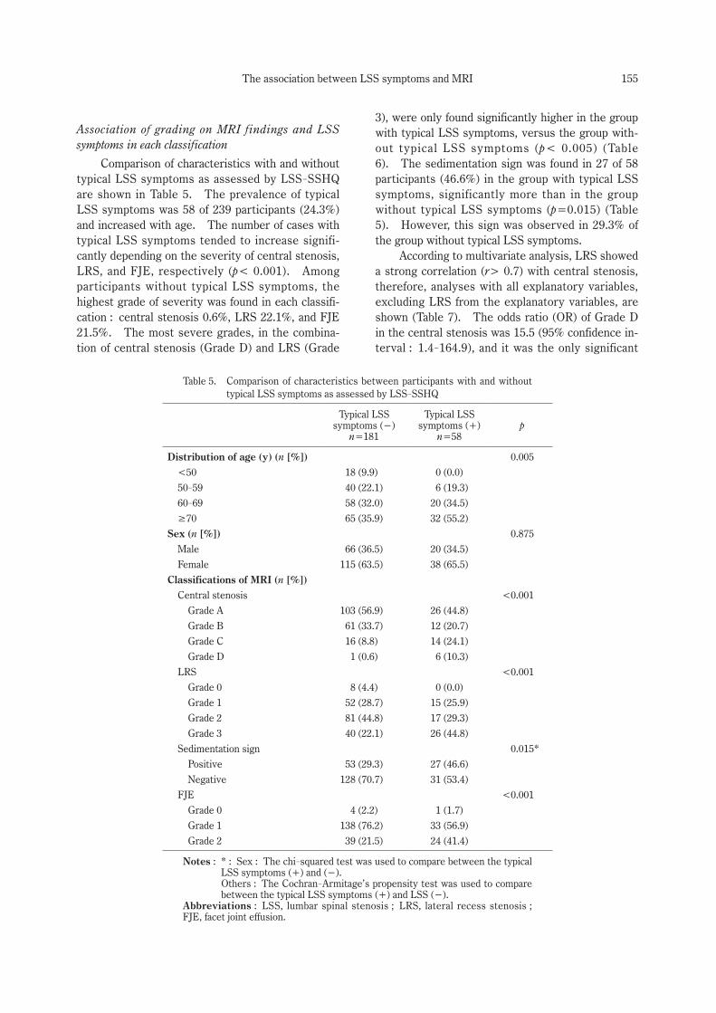
Comparison of characteristics with and without typical LSS symptoms as assessed by LSS-SSHQ are shown in Table 5. The prevalence of typical LSS symptoms was 58 of 239 participants (24.3%) and increased with age. The number of cases with typical LSS symptoms tended to increase signifi-cantly depending on the severity of central stenosis, LRS, and FJE, respectively (p< 0.001). Among participants without typical LSS symptoms, the highest grade of severity was found in each classifi-cation : central stenosis 0.6%, LRS 22.1%, and FJE 21.5%. The most severe grades, in the combina-tion of central stenosis (Grade D) and LRS (Grade
3), were only found significantly higher in the group with typical LSS symptoms, versus the group with-out typical LSS symptoms (p< 0.005) (Table 6). The sedimentation sign was found in 27 of 58 participants (46.6%) in the group with typical LSS symptoms, significantly more than in the group without typical LSS symptoms (p=0.015) (Table 5). However, this sign was observed in 29.3% of the group without typical LSS symptoms.
According to multivariate analysis, LRS showed a strong correlation (r> 0.7) with central stenosis, therefore, analyses with all explanatory variables, excluding LRS from the explanatory variables, are shown (Table 7). The odds ratio (OR) of Grade D in the central stenosis was 15.5 (95% confidence in-terval : 1.4-164.9), and it was the only significant
Table 5. Comparison of characteristics between participants with and without typical LSS symptoms as assessed by LSS-SSHQ
Typical LSS symptoms (−)
n=181
Typical LSS symptoms (+)
n=58p
Distribution of age (y) (n [%]) 0.005
<50 18 (9.9) 0 (0.0)
50-59 40 (22.1) 6 (19.3)
60-69 58 (32.0) 20 (34.5)
≥70 65 (35.9) 32 (55.2)
Sex (n [%]) 0.875
Male 66 (36.5) 20 (34.5)
Female 115 (63.5) 38 (65.5)
Classifications of MRI (n [%])
Central stenosis <0.001
Grade A 103 (56.9) 26 (44.8)
Grade B 61 (33.7) 12 (20.7)
Grade C 16 (8.8) 14 (24.1)
Grade D 1 (0.6) 6 (10.3)
LRS <0.001
Grade 0 8 (4.4) 0 (0.0)
Grade 1 52 (28.7) 15 (25.9)
Grade 2 81 (44.8) 17 (29.3)
Grade 3 40 (22.1) 26 (44.8)
Sedimentation sign 0.015*
Positive 53 (29.3) 27 (46.6)
Negative 128 (70.7) 31 (53.4)
FJE <0.001
Grade 0 4 (2.2) 1 (1.7)
Grade 1 138 (76.2) 33 (56.9)
Grade 2 39 (21.5) 24 (41.4)
Notes : * : Sex : The chi-squared test was used to compare between the typical LSS symptoms (+) and (−).
Others : The Cochran-Armitage’s propensity test was used to compare between the typical LSS symptoms (+) and LSS (−).
Abbreviations : LSS, lumbar spinal stenosis ; LRS, lateral recess stenosis ; FJE, facet joint effusion.
156 Y. Fushimi et al.
explanatory variable (p=0.023).
Discussion
The present study evaluated correlations be-tween the presence or absence of typical LSS symp-toms and four classifications of lumbar MRI : mor-phological grading of central stenosis and of LRS, presence of the sedimentation sign, and severity of
FJE. It was found that only the most severe grade (D) of central stenosis was strongly associated with the presence of typical LSS symptoms.
Recent focus has been on the association be-tween anatomical central stenosis on imaging and clinical symptoms. There are previous studies linking morphological stenosis with the LSS symp-toms in hospital-based surveys7-10,24-26). According to these studies, it has remained controversial
Table 6. Distribution for combination of LRS and central stenosis with and without typi-cal LSS symptoms as assessed by LSS-SSHQ
Total (Typical LSS symptoms [−]/Typical LSS symptoms [+]) (n=239)
LRS
Grade 0 Grade 1 Grade 2 Grade 3
Central stenosis
Grade A 8 (8/0) 62 (50/12) 55 (42/13) 4 (3/1)
Grade B 0 (0/0) 4 (1/3) 41 (38/3) 28 (22/6)
Grade C 0 (0/0) 1 (1/0) 2 (1/1) 27 (14/13)
Grade D 0 (0/0) 0 (0/0) 0 (0/0) 7 (1/6)*
Notes : *p<0.05 : The chi-squared test was used to compare between the typical LSS symptoms (+) and (−).
Abbreviations : LSS, lumbar spinal stenosis ; LRS, lateral recess stenosis.
Table 7. Associations of MRI findings with the presence or absence of typical LSS symptoms as assessed by LSS-SSHQ in multivariant regression analysis
Model 1 Model 2
OR 95% CI p value OR 95% CI p value
Sex (Female) 1.262 0.629-2.530 0.513 1.211 0.609-2.405 0.585
Age 1.048 1.010-1.088 0.013 1.049 1.012-1.088 0.009
Classifications of MRI findings
Central stenosis
Grade A Ref. Ref.
Grade B 0.561 0.207-1.523 0.257 0.561 0.234-1.346 0.195
Grade C 2.000 0.469-8.530 0.349 2.518 0.793-7.997 0.117
Grade D 11.885 0.919-153.74 0.058 15.453 1.448-164.94 0.023
LRS
Grade 0 Ref.
Grade 1 254965047 0.000 0.999 − − −
Grade 2 147471147 0.000 0.999 − − −
Grade 3 240462533 0.000 0.999 − − −
Sedimentation sign (Positive) 0.872 0.341-2.232 0.776 0.846 0.334-2.141 0.723
FJE
Grade 0 Ref. Ref.
Grade 1 1.062 0.095-11.833 0.961 1.033 0.093-11.454 0.979
Grade 2 2.444 0.211-28.264 0.474 2.314 0.201-26.58 0.501
R2 0.235 0.209
Notes : Model 1 : The results of the analysis with all MRI findings. Model 2 : The results of the analysis adjusted LRS.Abbreviations : LSS, lumbar spinal stenosis ; LRS, lateral recess stenosis ; OR, odds ratio ; CI, confidence interval ; FJE, facet joint effusion.
157The association between LSS symptoms and MRI
whether the morphological stenosis correlated with or without clinical LSS symptoms, even though vari-ous assessments of morphological stenosis, such as measurement of spinal canal area or diameter and each classification of stenosis grading, have been used for analyses27,28). In the general population study by Ishimoto et al., the prevalence of clinical symptoms significantly increased with increasing severity of central stenosis27). In the present study, the prevalence of typical LSS symptoms tended to increase significantly according to the severity of central stenosis and LRS, respectively. In addition, the agreement between the two studies suggests
that the correlation between severe central stenosis and the presence of symptomatic LSS is more cer-tain. Furthermore, to evaluate imaging findings for LSS, not only central canal stenosis but also lateral recess stenosis should be considered. It has been reported that the cross-sectional area of the lateral recesses was significantly smaller in the symptom-atic LSS group than in the asymptomatic group28). Another study reported that there was a weak cor-relation between the Oswestry disability index and LRS grade in low back pain patients without central stenosis. However, the participants had low back pain, and it did not evaluate specific radicular symp-
Abbreviations : LRS, lateral recess stenosis ; FJE, facet joint effusion.

158 Y. Fushimi et al.
toms11). In the present study, the prevalence of typical LSS symptoms increased significantly ac-cording to the severity of LRS compared with the group without typical LSS symptoms. It is known that degenerative spinal stenosis is primarily caused by age-related degeneration, such as protruding discs, osteophytes, hypertrophy of facet joints, and accompanying thickening of the ligamentum fla-vum1,12). Therefore, it is likely that advanced cen-tral stenosis is often associated with LRS. In the present study, the combination of higher severity grades of central stenosis (Grade C, D) and LRS (Grade 2, 3) was found in approximately 15%, and half of them were in the symptomatic LSS group. Since LRS showed a strong correlation (r> 0.7) with central stenosis in the present study, it was consid-ered inappropriate to include both central stenosis and LRS in the logistic regression analysis due to their multicollinearity ; therefore, the correlation between LRS and central stenosis was not evaluat-ed. The present study showed that only the most severe central stenosis was strongly related to the presence of typical LSS symptoms. Conversely, this and another study27) found that not all of the higher severity grades of MRI imaging cases pre-sented with typical LSS symptoms. In the present study, the LSS-SSHQ score of one case with Grade D central stenosis, determined not to have typical LSS symptoms, was 2 points in questions 1-4 and 1 point in questions 5-10. This case showed some symptoms suggestive of LSS, but did not meet the defining criteria of the LSS-SSHQ. Moreover, the severity of stenosis on MRI was not associated with preoperative disability and pain, or clinical outcome at 1 year26). Furthermore, in the natural history of symptomatic LSS, it was shown that more than half of symptomatic LSS subjects improved their symp-toms, whereas 10% of asymptomatic LSS subjects developed clinically diagnosed symptomatic LSS at 1 year follow up6). According to the abovementioned studies, morphological spinal stenosis might not be equal to the presence of typical LSS symptoms.
The sedimentation sign was first reported as a finding with high sensitivity and specificity for symp-tomatic LSS13). The presence of a positive sedimen-tation sign was greater depending on the severity of the morphological grade29). However, validation of the sedimentation sign is insufficient in patients with mild to moderate anatomical LSS29,30). Al-though the prevalence of the positive sedimentation sign was significantly higher in the group with typi-cal LSS symptoms (46.4%) than in the group without typical symptoms (29.3%), logistic regression analy-
sis showed that the sedimentation sign was not a significant explanatory variable in the present study. These results suggest that the sedimenta-tion sign itself was not associated with the presence or absence of typical LSS symptoms, just associated with the presence of anatomical central steno-sis. This may be due to the fact that the present study involved a general population, and various de-grees of anatomical LSS were included ; therefore, these results might indicate a more generalized as-sociation between imaging findings and symptoms.
It has recently been reported that FJE may be associated with symptomatic LSS. In degenerative lumbar spinal disorders, high levels of inflammatory cytokines in facet joint tissue have been found to be released into the spinal canal, which is suspected to be the cause of pain11,16,17). In addition, increased facet fluid on MRI has been reported to be highly predictive of the dynamic reduction of DCSA detect-ed on axial-loaded MRI in the clinical assessment of LSS14,15). In the present study, the severity of FJE was not associated with the presence or absence of typical LSS symptoms. However, in the present analysis, the highest severity was used as a repre-sentative value of the respective findings ; the level and right/left sidedness of occurrence points were not taken into account. These factors might be useful to improve the accuracy of detecting symp-tomatic LSS.
The presence and severity of each MRI finding were often related to each other, suggesting a corre-lation between findings. Correlations strong enough to suggest multicollinearity were found only between central stenosis and LRS, but correlations between independent variables may have influenced the results of multivariate analysis. It was also necessary to evaluate whether combining each find-ing would result in a correlation with the presence or absence of symptoms ; however, statistical analy-sis was difficult because the correlation between each finding resulted in a large bias in the distribu-tion of the number of cases. A larger sample size may reduce the influence on multivariate analysis and allow us to analyze the association between the combination of findings and the presence of symp-toms.
A strength of this study is that four different kinds of MRI evaluation items were performed in all participants. In addition, the distributions of each MRI item with and without typical LSS symptoms were evaluated and associations between them were analyzed using a logistic regression model. There-fore, various morphological findings were compared
159The association between LSS symptoms and MRI
for the possibility of pathogenesis in symptomatic LSS. A second strength is that the data were ob-tained from a large community-dwelling population on which various analyses have been performed, in-cluding the present study. Therefore, in contrast to a hospital-based survey, these results reflect a real-world setting and relevance for pathogenesis of LSS.
This study has several limitations. First, all participants in this study were volunteers, so it is possible that those participants who had any symp-toms or more severe LSS symptoms might self-se-lect for receiving MRI. However, each item from the MRI findings was distributed across all grades, and participants without symptoms also agreed to undergo MRI. Therefore, compared to a hospital-based study, the benefit of this study was that all grades of morphological stenosis, including mild ste-nosis and no stenosis, could be evaluated. Second, the most severe grade was taken as representative of each finding, therefore, detailed analyses for rela-tionships between the responsible anatomical steno-sis level inferred from the site of the symptoms or multiple level lesions were not considered. Be-cause there is no established method of evaluation that takes into account multiple level lesions, and because it requires detailed grouping combined with the severity of the disease, a larger number of cases is considered necessary for the analysis. Third, since the research location was in a rural and moun-tainous area, one may not be completely able to ex-trapolate the findings to a more typical Japanese (ur-ban) population. Finally, this was a cross-sectional study, therefore no causal relationships between morphological and symptomatic LSS could be estab-lished.
Conclusion
The most severe central stenosis was found to be strongly related to typical LSS symptoms. How-ever, although subjects with severe central stenosis showed symptoms suggestive of LSS, these subjects did not always show typical LSS symptoms. More-over, mild central and lateral recess stenosis may or may not present with typical LSS symptoms. Fur-ther studies are needed to clarify the mechanism of onset and induction of LSS symptoms and its rela-tionship to anatomical and radiological stenosis.
Acknowledgments
The authors wish to thank Dr. Akira Onda, Dr. Kazuya Yamauchi, Dr. Yoshiaki Takeyachi, Dr. Ichiro
Takahashi, Dr. Hisayoshi Tachihara, and Dr. Bunji Takayama for participating in the data collec-tion. The authors would also like to thank 5 public health nurses (Ms. Nobuko Fujita, Ms. Nakako Hoshi, Ms. Misako Hoshi, Ms. Naoko Imada, and Ms. Seiko Kanno) for their support in carrying out this study.
Conflict of interest disclosure
The authors declare no conflicts of interest as-sociated with this manuscript.
References
1. Siebert E, Prüss H, Klingebiel R, et al. Lumbar spinal stenosis : syndrome, diagnostics and treat-ment. Nat Rev Neurol, 5 : 392-403, 2009.
2. Bartynski WS, Lin L. Lumbar root compression in the lateral recess : MR imaging, conventional myelography, and CT myelography comparison with surgical confirmation. AJNR Am J Neurora-diol, 24 : 348-360, 2003.
3. Kitamura M, Eguchi Y, Inoue G, et al. A case of symptomatic extra-foraminal lumbosacral stenosis (“far-out syndrome”) diagnosed by diffusion tensor imaging. Spine (Phila Pa 1976), 37 : E854-E857, 2012.
4. North American Spine Society Clinical Guidelines. III. Definition and Natural History of Degenerative Lumbar Spinal Stenosis [homepage on the Internet]. North American Spine Society ; 2011. Available from : https://www.spine.org/Portals/0/Assets/Downloads/ResearchClinicalCare/Guidelines/LumbarStenosis.pdf Accessed May 1, 2020.
5. Boden SD, Davis DO, Dina TS, et al. Abnormal magnetic-resonance scans of the lumbar spine in asymptomatic subjects. A prospective investiga-tion. J Bone Joint Surg Am, 72 : 403-408, 1990.
6. Otani K, Kikuchi SI, Yabuki S, et al. Prospective one-year follow-up of lumbar spinal stenosis in a regional community. J Pain Res, 11 : 455-464, 2018.
7. Ogikubo O, Forsberg L, Hansson T. The relation-ship between the cross-sectional area of the cauda equina and the preoperative symptoms in central lumbar spinal stenosis. Spine (Phila Pa 1976), 32 : 1423-1428 ; discussion 1429, 2007.
8. Kuittinen P, Sipola P, Saari T, et al. Visually as-sessed severity of lumbar spinal canal stenosis is paradoxically associated with leg pain and objective walking ability. BMC Musculoskelet Disord, 15 : 348, 2014.
9. Kim YU, Kong YG, Lee J, et al. Clinical symp-toms of lumbar spinal stenosis associated with

160 Y. Fushimi et al.
morphological parameters on magnetic resonance images. Eur Spine J, 24 : 2236-2243, 2015.
10. Andrasinova T, Adamova B, Buskova J, et al. Is there a Correlation Between Degree of Radiologic Lumbar Spinal Stenosis and its Clinical Manifesta-tion? Clin Spine Surg, 31 : E403-E408, 2018.
11. Splettstößer A, Khan MF, Zimmermann B, et al. Correlation of lumbar lateral recess stenosis in magnetic resonance imaging and clinical symp-toms. World J Radiol, 9 : 223-229, 2017.
12. Kushchayev SV, Glushko T, Jarraya M, et al. ABCs of the degenerative spine. Insights Imaging, 9 : 253-274, 2018.
13. Barz T, Melloh M, Staub LP, et al. Nerve root sedimentation sign : evaluation of a new radiologi-cal sign in lumbar spinal stenosis. Spine (Phila Pa 1976), 35 : 892-897, 2010.
14. Kim YK, Lee JW, Kim HJ, et al. Diagnostic ad-vancement of axial loaded lumbar spine MRI in pa-tients with clinically suspected central spinal canal stenosis. Spine (Phila Pa 1976), 38 : E1342-
E1347, 2013. 15. Kanno H, Ozawa H, Koizumi Y, et al. Increased
Facet Fluid Predicts Dynamic Changes in the Dural Sac Size on Axial-Loaded MRI in Patients with Lumbar Spinal Canal Stenosis. AJNR Am J Neu-roradiol, 37 : 730-735, 2016.
16. Igarashi A, Kikuchi S, Konno S. Correlation be-tween inflammatory cytokines released from the lumbar facet joint tissue and symptoms in degen-erative lumbar spinal disorders. J Orthop Sci, 12 : 154-160, 2007.
17. Vardeh D, Mannion RJ, Woolf CJ. Toward a Mech-anism-Based Approach to Pain Diagnosis. J Pain, 17 : T50-T69, 2016.
18. Otani K, Kikuchi S, Yabuki S, et al. Lumbar spinal stenosis has a negative impact on quality of life compared with other comorbidities : an epidemio-logical cross-sectional study of 1862 community-
dwelling individuals. ScientificWorldJournal, 2013 : 590652, 2013.
19. Schizas C, Theumann N, Burn A, et al. Qualita-tive grading of severity of lumbar spinal stenosis based on the morphology of the dural sac on mag-netic resonance images. Spine (Phila Pa 1976), 35 : 1919-1924, 2010.
20. Chaput C, Padon D, Rush J, et al. The significance of increased fluid signal on magnetic resonance im-aging in lumbar facets in relationship to degenera-
tive spondylolisthesis. Spine (Phila Pa 1976), 32 : 1883-1887, 2007.
21. Landis JR, Koch GG. The measurement of ob-server agreement for categorical data. Biomet-rics, 33 : 159-174, 1977.
22. Konno S, Kikuchi S, Tanaka Y, et al. A diagnostic support tool for lumbar spinal stenosis : a self-ad-ministered, self-reported history questionnaire. BMC Musculoskelet Disord, 8 : 102, 2007.
23. Vatcheva KP, Lee M, McCormick JB, et al. Multi-collinearity in Regression Analyses Conducted in Epidemiologic Studies. Epidemiology (Sunny-vale), 6 : 227, 2016.
24. Kapural L, Mekhail N, Bena J, et al. Value of the magnetic resonance imaging in patients with pain-ful lumbar spinal stenosis (LSS) undergoing lumbar epidural steroid injections. Clin J Pain, 23 : 571-
575, 2007. 25. Sigmundsson FG, Kang XP, Jönsson B, et al. Cor-
relation between disability and MRI findings in lumbar spinal stenosis : a prospective study of 109 patients operated on by decompression. Acta Or-thop, 82 : 204-210, 2011.
26. Weber C, Giannadakis C, Rao V, et al. Is There an Association Between Radiological Severity of Lum-bar Spinal Stenosis and Disability, Pain, or Surgical Outcome? : A Multicenter Observational Study. Spine (Phila Pa 1976), 41 : E78-E83, 2016.
27. Ishimoto Y, Yoshimura N, Muraki S, et al. Associ-ations between radiographic lumbar spinal stenosis and clinical symptoms in the general popula-tion : the Wakayama Spine Study. Osteoarthritis Cartilage, 21 : 783-738, 2013.
28. Majidi H, Shafizad M, Niksolat F, et al. Relation-ship Between Magnetic Resonance Imaging Find-ings and Clinical Symptoms in Patients with Sus-pected Lumbar Spinal Canal Stenosis : a Case-
control Study. Acta Inform Med, 27 : 229-233, 2019.
29. Laudato PA, Kulik G, Schizas C. Relationship be-tween sedimentation sign and morphological grade in symptomatic lumbar spinal stenosis. Eur Spine J, 24 : 2264-2268, 2015.
30. Zhang L, Chen R, Xie P, et al. Diagnostic value of the nerve root sedimentation sign, a radiological sign using magnetic resonance imaging, for detect-ing lumbar spinal stenosis : a meta-analysis. Skeletal Radiol, 44 : 519-527, 2015.