The application of polythiol molecules for protein immobilisation on sensor surfaces
7
Biosensors and Bioelectronics 25 (2010) 1049–1055 Contents lists available at ScienceDirect Biosensors and Bioelectronics journal homepage: www.elsevier.com/locate/bios The application of polythiol molecules for protein immobilisation on sensor surfaces Dimitris Kyprianou a , Antonio R. Guerreiro a,∗ , Martin Nirschl b,c , Iva Chianella a , Sreenath Subrahmanyam a , Anthony P.F. Turner a , Sergey Piletsky a a Cranfield Health, Cranfield University, College Road, Cranfield, Bedfordshire, MK34 0AL, UK b Siemens AG Munich, Corporate Technology, Otto-Hahn-Ring 6, 81739 Munich, Germany c Laboratory of Biosensors and Bioelectronics, Institute for Biomedical Engineering, ETH Zurich, Switzerland article info Article history: Received 22 July 2009 Received in revised form 14 September 2009 Accepted 18 September 2009 Available online 12 October 2009 Keywords: Biosensor SPR Thiol monolayer Detection Salmonella PSA abstract The immobilisation of bio-receptors on transducer surfaces is a key step in the development of biosensors. The immobilisation needs to be fast, cheap and most importantly should not affect the biorecognition activity of the immobilised receptor. The development of a protocol for biomolecule immobilisation onto a surface plasmon resonance (SPR) sensor surface using inexpensive polythiol compounds is pre- sented here. The method used here is based on the reaction between primary amines and thioacetal groups, formed upon reaction of o-phthaldialdehyde (OPA) and thiol compounds. The self-assembled thiol monolayers were characterised using contact angle and XPS. The possibility to immobilise proteins on monolayers was assessed by employing BSA as a model protein. For the polythiol layers exhibiting the best performance, a general protocol was optimised suitable for the immobilisation of enzymes and antibodies such as anti-prostate specific antigen (anti-PSA) and anti Salmonella typhimurium. The kinetic data was obtained for PSA binding to anti-PSA and for S. typhimurium cells with a detection limit of 5 × 10 6 cells mL −1 with minimal non-specific binding of other biomolecules. These findings make this technique a very promising alternative for amine coupling compared to peptide bond formation. Addi- tionally, it offers opportunity for immobilising proteins (even those with low isoelectric point) on neutral polythiol layers without any activation step. © 2009 Elsevier B.V. All rights reserved. 1. Introduction Immunoassay technology is currently growing rapidly due to market demands for low cost, easy to use and sensitive biosen- sors (Vikholm, 2005; Sadana, 2006). Surface plasmon resonance (SPR), quartz crystal microbalance (QCM), cantilever and electro- chemical detectors are the most widespread platforms used with immunosensors. The main advantages of these when compared with immunoassays as ELISA, are the label free detection and the opportunity for measuring biochemical interactions in real time. This way kinetic and affinity constants can easily be obtained (Haga et al., 2008; Katsamba et al., 2006; Regnault et al., 1998). The ligands (biomolecules) are usually attached on sensor sur- faces by physical adsorption (Predki, 2004), covalent attachment (Kusnezow and Hoheisel, 2003; O’Shannessy et al., 1992) or lig- and capture, which mainly refers to the strong interaction between biotilynated ligands and immobilised steptavidin or avidin (Craft et al., 1998; Panayotou et al., 1998). Covalent attachment is ∗ Corresponding author. Tel.: +44 1234758326; fax: +44 1525863533. E-mail address: a.guerreiro@cranfield.ac.uk (A.R. Guerreiro). used because it provides a strong and stable binding of the lig- and/receptor to the sensor surface. This allows easy regeneration of sensors using conditions which can remove the analyte from the surface, but not the attached ligand itself. Covalent immobil- isation includes amino coupling (Lofas et al., 1995; Piletska et al., 2001), aldehyde coupling (Abraham et al., 1995) and thiol coupling methods (Johnson et al., 1995). The covalent attachment can also occur on gold surfaces modified with polymers such as carboxydex- tran matrix (Lofas et al., 1995) and thioacetal matrix (Kyprianou et al., 2009) or self-assembled monolayers (Nuzzo and Allara, 1983). The selection of the immobilisation procedure is a critical point for the development of a successful sensor. This is because the immo- bilisation may cause denaturation of ligand/receptor or alter the structure of binding sites (Butler, 2000) with consequent loss of bioreactivity. The direct attachment of the receptor on the sen- sor surface is however unadvisable since it can cause irreversible denaturation of the bound proteins (Su et al., 1998). The application of SAMs or polymers has advantages and disadvantages and selec- tion of one over the other depends on the application. For example, flat surfaces with self-assembled monolayers (SAMs) are benefi- cial compared to polymeric layers (carboxydextran) both when the analytes of interest are large molecules such as cells and viruses 0956-5663/$ – see front matter © 2009 Elsevier B.V. All rights reserved. doi:10.1016/j.bios.2009.09.030
-
Upload
independent -
Category
Documents
-
view
0 -
download
0
Transcript of The application of polythiol molecules for protein immobilisation on sensor surfaces
Ts
DAa
b
c
a
ARR1AA
KBSTDSP
1
ms(ciwoTe
f(abe
0d
Biosensors and Bioelectronics 25 (2010) 1049–1055
Contents lists available at ScienceDirect
Biosensors and Bioelectronics
journa l homepage: www.e lsev ier .com/ locate /b ios
he application of polythiol molecules for protein immobilisation on sensorurfaces
imitris Kyprianoua, Antonio R. Guerreiroa,∗, Martin Nirschlb,c, Iva Chianellaa, Sreenath Subrahmanyama,nthony P.F. Turnera, Sergey Piletskya
Cranfield Health, Cranfield University, College Road, Cranfield, Bedfordshire, MK34 0AL, UKSiemens AG Munich, Corporate Technology, Otto-Hahn-Ring 6, 81739 Munich, GermanyLaboratory of Biosensors and Bioelectronics, Institute for Biomedical Engineering, ETH Zurich, Switzerland
r t i c l e i n f o
rticle history:eceived 22 July 2009eceived in revised form4 September 2009ccepted 18 September 2009vailable online 12 October 2009
eywords:iosensor
a b s t r a c t
The immobilisation of bio-receptors on transducer surfaces is a key step in the development of biosensors.The immobilisation needs to be fast, cheap and most importantly should not affect the biorecognitionactivity of the immobilised receptor. The development of a protocol for biomolecule immobilisationonto a surface plasmon resonance (SPR) sensor surface using inexpensive polythiol compounds is pre-sented here. The method used here is based on the reaction between primary amines and thioacetalgroups, formed upon reaction of o-phthaldialdehyde (OPA) and thiol compounds. The self-assembledthiol monolayers were characterised using contact angle and XPS. The possibility to immobilise proteinson monolayers was assessed by employing BSA as a model protein. For the polythiol layers exhibiting
PRhiol monolayeretectionalmonellaSA
the best performance, a general protocol was optimised suitable for the immobilisation of enzymes andantibodies such as anti-prostate specific antigen (anti-PSA) and anti Salmonella typhimurium. The kineticdata was obtained for PSA binding to anti-PSA and for S. typhimurium cells with a detection limit of5 × 106 cells mL−1 with minimal non-specific binding of other biomolecules. These findings make thistechnique a very promising alternative for amine coupling compared to peptide bond formation. Addi-tionally, it offers opportunity for immobilising proteins (even those with low isoelectric point) on neutral
ny a
polythiol layers without a. Introduction
Immunoassay technology is currently growing rapidly due toarket demands for low cost, easy to use and sensitive biosen-
ors (Vikholm, 2005; Sadana, 2006). Surface plasmon resonanceSPR), quartz crystal microbalance (QCM), cantilever and electro-hemical detectors are the most widespread platforms used withmmunosensors. The main advantages of these when compared
ith immunoassays as ELISA, are the label free detection and thepportunity for measuring biochemical interactions in real time.his way kinetic and affinity constants can easily be obtained (Hagat al., 2008; Katsamba et al., 2006; Regnault et al., 1998).
The ligands (biomolecules) are usually attached on sensor sur-aces by physical adsorption (Predki, 2004), covalent attachment
Kusnezow and Hoheisel, 2003; O’Shannessy et al., 1992) or lig-nd capture, which mainly refers to the strong interaction betweeniotilynated ligands and immobilised steptavidin or avidin (Craftt al., 1998; Panayotou et al., 1998). Covalent attachment is∗ Corresponding author. Tel.: +44 1234758326; fax: +44 1525863533.E-mail address: [email protected] (A.R. Guerreiro).
956-5663/$ – see front matter © 2009 Elsevier B.V. All rights reserved.oi:10.1016/j.bios.2009.09.030
ctivation step.© 2009 Elsevier B.V. All rights reserved.
used because it provides a strong and stable binding of the lig-and/receptor to the sensor surface. This allows easy regenerationof sensors using conditions which can remove the analyte fromthe surface, but not the attached ligand itself. Covalent immobil-isation includes amino coupling (Lofas et al., 1995; Piletska et al.,2001), aldehyde coupling (Abraham et al., 1995) and thiol couplingmethods (Johnson et al., 1995). The covalent attachment can alsooccur on gold surfaces modified with polymers such as carboxydex-tran matrix (Lofas et al., 1995) and thioacetal matrix (Kyprianou etal., 2009) or self-assembled monolayers (Nuzzo and Allara, 1983).The selection of the immobilisation procedure is a critical point forthe development of a successful sensor. This is because the immo-bilisation may cause denaturation of ligand/receptor or alter thestructure of binding sites (Butler, 2000) with consequent loss ofbioreactivity. The direct attachment of the receptor on the sen-sor surface is however unadvisable since it can cause irreversibledenaturation of the bound proteins (Su et al., 1998). The application
of SAMs or polymers has advantages and disadvantages and selec-tion of one over the other depends on the application. For example,flat surfaces with self-assembled monolayers (SAMs) are benefi-cial compared to polymeric layers (carboxydextran) both when theanalytes of interest are large molecules such as cells and viruses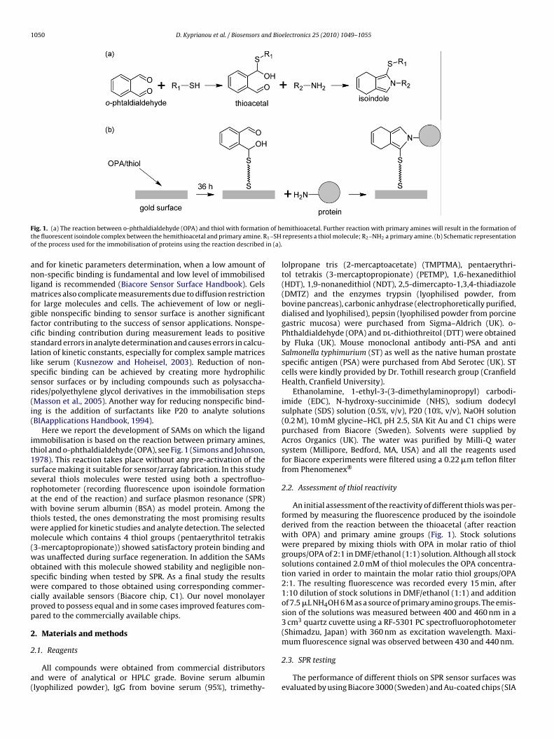
1050 D. Kyprianou et al. / Biosensors and Bioelectronics 25 (2010) 1049–1055
F n of ht 1–SHo in (a).
anlmfgfcsllssr(i(
it1ssrawtwm(woswcpp
2
2
a(
ig. 1. (a) The reaction between o-phthaldialdehyde (OPA) and thiol with formatiohe fluorescent isoindole complex between the hemithioacetal and primary amine. Rf the process used for the immobilisation of proteins using the reaction described
nd for kinetic parameters determination, when a low amount ofon-specific binding is fundamental and low level of immobilised
igand is recommended (Biacore Sensor Surface Handbook). Gelsatrices also complicate measurements due to diffusion restriction
or large molecules and cells. The achievement of low or negli-ible nonspecific binding to sensor surface is another significantactor contributing to the success of sensor applications. Nonspe-ific binding contribution during measurement leads to positivetandard errors in analyte determination and causes errors in calcu-ation of kinetic constants, especially for complex sample matricesike serum (Kusnezow and Hoheisel, 2003). Reduction of non-pecific binding can be achieved by creating more hydrophilicensor surfaces or by including compounds such as polysaccha-ides/polyethylene glycol derivatives in the immobilisation stepsMasson et al., 2005). Another way for reducing nonspecific bind-ng is the addition of surfactants like P20 to analyte solutionsBIAapplications Handbook, 1994).
Here we report the development of SAMs on which the ligandmmobilisation is based on the reaction between primary amines,hiol and o-phthaldialdehyde (OPA), see Fig. 1 (Simons and Johnson,978). This reaction takes place without any pre-activation of theurface making it suitable for sensor/array fabrication. In this studyeveral thiols molecules were tested using both a spectrofluo-ophotometer (recording fluorescence upon isoindole formationt the end of the reaction) and surface plasmon resonance (SPR)ith bovine serum albumin (BSA) as model protein. Among the
hiols tested, the ones demonstrating the most promising resultsere applied for kinetic studies and analyte detection. The selectedolecule which contains 4 thiol groups (pentaerythritol tetrakis
3-mercaptopropionate)) showed satisfactory protein binding andas unaffected during surface regeneration. In addition the SAMs
btained with this molecule showed stability and negligible non-pecific binding when tested by SPR. As a final study the resultsere compared to those obtained using corresponding commer-
ially available sensors (Biacore chip, C1). Our novel monolayerroved to possess equal and in some cases improved features com-ared to the commercially available chips.
. Materials and methods
.1. Reagents
All compounds were obtained from commercial distributorsnd were of analytical or HPLC grade. Bovine serum albuminlyophilized powder), IgG from bovine serum (95%), trimethy-
emithioacetal. Further reaction with primary amines will result in the formation ofrepresents a thiol molecule; R2–NH2 a primary amine. (b) Schematic representation
lolpropane tris (2-mercaptoacetate) (TMPTMA), pentaerythri-tol tetrakis (3-mercaptopropionate) (PETMP), 1,6-hexanedithiol(HDT), 1,9-nonanedithiol (NDT), 2,5-dimercapto-1,3,4-thiadiazole(DMTZ) and the enzymes trypsin (lyophilised powder, frombovine pancreas), carbonic anhydrase (electrophoretically purified,dialised and lyophilised), pepsin (lyophilised powder from porcinegastric mucosa) were purchased from Sigma–Aldrich (UK). o-Phthaldialdehyde (OPA) and dl-dithiothreitol (DTT) were obtainedby Fluka (UK). Mouse monoclonal antibody anti-PSA and antiSalmonella typhimurium (ST) as well as the native human prostatespecific antigen (PSA) were purchased from Abd Serotec (UK). STcells were kindly provided by Dr. Tothill research group (CranfieldHealth, Cranfield University).
Ethanolamine, 1-ethyl-3-(3-dimethylaminopropyl) carbodi-imide (EDC), N-hydroxy-succinimide (NHS), sodium dodecylsulphate (SDS) solution (0.5%, v/v), P20 (10%, v/v), NaOH solution(0.2 M), 10 mM glycine–HCl, pH 2.5, SIA Kit Au and C1 chips werepurchased from Biacore (Sweden). Solvents were supplied byAcros Organics (UK). The water was purified by Milli-Q watersystem (Millipore, Bedford, MA, USA) and all the reagents usedfor Biacore experiments were filtered using a 0.22 �m teflon filterfrom Phenomenex®
2.2. Assessment of thiol reactivity
An initial assessment of the reactivity of different thiols was per-formed by measuring the fluorescence produced by the isoindolederived from the reaction between the thioacetal (after reactionwith OPA) and primary amine groups (Fig. 1). Stock solutionswere prepared by mixing thiols with OPA in molar ratio of thiolgroups/OPA of 2:1 in DMF/ethanol (1:1) solution. Although all stocksolutions contained 2.0 mM of thiol molecules the OPA concentra-tion varied in order to maintain the molar ratio thiol groups/OPA2:1. The resulting fluorescence was recorded every 15 min, after1:10 dilution of stock solutions in DMF/ethanol (1:1) and additionof 7.5 �L NH4OH 6 M as a source of primary amino groups. The emis-sion of the solutions was measured between 400 and 460 nm in a3 cm3 quartz cuvette using a RF-5301 PC spectrofluorophotometer(Shimadzu, Japan) with 360 nm as excitation wavelength. Maxi-mum fluorescence signal was observed between 430 and 440 nm.
2.3. SPR testing
The performance of different thiols on SPR sensor surfaces wasevaluated by using Biacore 3000 (Sweden) and Au-coated chips (SIA
d Bio
Kp
2
tcpwtgstfADa
tbfl(tabn
S(wtibAsc
2
ew
cta5wTtt
2
ctouScwtTrTwt
D. Kyprianou et al. / Biosensors an
it Au) purchased from Biacore (Sweden). All the experiments wereerformed at 25 ◦C.
.3.1. Treatment of gold chips–gold surface modificationGold sensor chips, SIA Au (Biacore, Sweden) were used to assess
he ability of polythiol/OPA monolayer to bind biomolecules. Auhips were cleaned for 3 min by oxygen plasma at 40 W using alasma chamber (Emitech, UK). SAMs (except for DTT and DMTZ)ere created on the gold surface by immersing the chips in 10 mL
hiol/OPA solution in DMF/ethanol (1:1) with 2:1 molar ratio thiolroups/OPA for 36 h. Triethylamine TEA (50 �L) was added to theolution in order to facilitate thioacetal formation. The concen-ration was 0.1 M for di-thiol, 0.066 M for tri-thiol and 0.05 Mor tetrathiol. OPA concentration was kept in all cases at 0.1 M.fter immobilisation the gold surface was rinsed thoroughly withMF/ethanol (1:1, HPLC grade), dried with nitrogen and the chipsssembled on the holder.
For the water soluble dithiol molecules, DTT and DMTZ forma-ion of SAMs was performed and recorded on-line using Biacorey injecting 200 �L on a cleaned gold chip (2 injections × 100 �L,ow rate 5 �L min−1) of DTT/OPA (0.066 M/0.033 M) or DMTZ/OPA0.02 M/0.01 M) prepared in 50 mM Na2B4O7, pH 9.0. All the solu-ions were purged with argon for 5 min and kept under inerttmosphere in order to avoid oxidation. This experiment could note performed with other thiols (insoluble in water) as Biacore isot compatible with organic solvents.
For the PETMP/OPA (the thiol with the best performance) theAM formation was studied using film bulk acoustic resonatorsFBAR) with gold electrodes. With a cell mounted on the FBARhich is open on the top it is possible to pipette amounts of solu-
ions directly to the sensor gold surface. The technique is describedn detail in Nirschl et al. (in press). To monitor the adsorption, theaseline was recorded with 10 �L of DMF/ethanol (1:1) in the cell.fter a stable baseline was reached, 90 �L of PETMP/OPA monomerolution was added at a concentration of 0.1 M. The cell was thenlosed with a lid to avoid evaporation of the solution.
.3.2. SAM coated sensor surface characterisationThe static water contact angle was determined with a CCD cam-
ra Supplied by Spectra Source Equipment model MCD400S (USA)ith the software provided.
X-ray photoelectron spectroscopy (XPS) measurements werearried out on a VG ESCA lab-Mark-2 X-ray Photoelectron Spec-rometer (East Grinstead, UK). The X-ray gun was operated at 14 kVnd 20 mA. Survey and high-resolution spectra were collected at0 and 100 eV respectively, with Mg K� 1253.6 eV radiation. Scansere obtained in the C1s, N1s, O1s, and S2p regions of the spectrum.
he decomposition of the XPS peaks into different components andhe quantitative interpretation was performed after subtraction ofhe background using the Shirley method.
.3.3. Protein immobilisation on coated gold surfaceThe biomolecules used for evaluation of performance of SAM
oated surfaces were BSA (bovine serum albumin), the enzymesrypsin, carbonic anhydrase, pepsin and the antibodies mouse mon-clonal anti-PSA and anti-ST. Non-immunoactive mouse IgG wassed as control on reference channel for experiments with PSA andT cells detection. BSA was used for the initial assessment of theapacity of SAM surfaces to immobilise protein. Biacore C1 chipsere used for comparison. C1 was initially cleaned with 2 min injec-
ion (20 �L, flow rate 10 �L min−1) of NaOH 1 mM containing 0.03%
riton X-100 the chip was then activated by injecting 70 �L (flowate 10 �L min−1) of 0.2 M EDC/0.05 M NHS (Fagerstam et al., 1992).ypically, BSA and enzymes immobilisation on SAM coated SIA Auas carried out by injecting 75 �L of 100 �g mL−1 of protein solu-ion in 0.01 M phosphate buffered saline (PBS), pH 7.4 with a flow
electronics 25 (2010) 1049–1055 1051
rate 15 �L min−1. For the study of pH effect on protein immobil-isation to thiol SAM, the proteins were diluted in buffer (0.05 Macetate buffer pH 4.5 and pH 5.0, 0.1 M PBS pH 7.4 and Na-boratebuffer 0.05 M pH 9.0). For immobilisation of biomolecules on C10.05 M acetate buffer, pH 5.0 was used instead of PBS.
The stability of the immobilised biomolecules on SAM modifiedsurfaces was tested by injection of 10 �L of regeneration solution:0.1%, sodium dodecyl sulphate (SDS) at a flow rate of 30 �L min−1.
For antibodies immobilisation (anti-ST anti-PSA and mouseIgG) 75 �L of antibodies (50 �g mL−1) diluted in PBS pH 7.4 wereinjected with flow rate of 15 �L min−1. Running buffer was alsoPBS, pH 7.4. For kinetic studies after antibodies immobilisation thebuffer was switched from PBS to PBS containing 0.005% surfactant(P20) in order to eliminate nonspecific binding and improve fit-ting to the Langmuir 1:1 binding model. After covalent coupling ofthe antibodies, remaining thioacetal groups were deactivated with25 �L 1 M ethanolamine hydrochloride at pH 8.5 and 2–4 injec-tions BSA 30–50 �L (100 �g mL−1). The evaluation of immobilisedantibodies was performed by injecting the antigens PSA and STcells into chip with corresponding antibodies. The antigens werediluted in PBS containing 0.005% of surfactant P20 and injected, for3–5 min with a flow rate of 20 �L min−1 and 5 �L min−1 for PSA andcells correspondingly. PSA was injected at concentrations rangingfrom 3.3 to 832.5 nM. The dissociation time for assessing the dis-sociation constant Kd was 120–180 s. Kinetic data was obtained byBiaevaluation software provided by Biacore. In all experiments areference channel with immobilised mouse IgG was used in orderto assess the binding specificity. In case of anti-PSA/PSA the sur-face was regenerated with a pulse of 5–30 �L of HCl/glycine 10 mM(pH 2.5) at a flow rate of 30 �L min−1. For the surface with immo-bilised anti-ST/ST cells, regeneration was performed by injection of10–90 �L 1 mM NaOH, 30 �L min−1.
3. Results and discussion
3.1. Reactivity of thiols
The ability of the selected molecules (DTT, PETMP, TMPTMA,DMTZ, HDT, and NDT) to form a fluorescent isoindole after reactionwith OPA and NH4OH was initially assessed for the selection ofthe most promising thiol molecules. Under the experimental con-ditions the maximum fluorescent was obtained at 3 h after NH4OHaddition. In higher molecule/OPA concentrations the maximumsignal was reached 5–10 min after addition of NH4OH. Nearly nofluorescence was observed for the molecule/OPA solutions in theabsence of primary amines. The stability of the thioacetal groups(thiol/OPA solutions) was also studied by recording the fluores-cence signal during 6 weeks at regular intervals after addition ofNH4OH. The fluorescent intensities, recorded 3 h after addition ofNH4OH for each molecule/OPA solution, are illustrated in Table 1.The experiments were performed in triplicate. Table 1 shows thatthe molecules exhibiting the highest fluorescence and thereforethe strongest ability to form the isoindole and bind primary aminewere DTT and PETMP. This can be explained by the presence ofelectron withdrawing groups (–OH for DTT and ester groups forPETMP) in the structures (See Fig. S1, Supporting Information)which increase the thiols acidity. As a result the thiol group is depro-tonated more easily and the formation of thioacetal is facilitated.As expected, DTT showed limited stability because of its tendencyto oxidise and decompose as the recorded fluorescence decreased
considerably in 6 weeks. On the contrary, PETMP after 6 weeksin solution with OPA exhibited only a slight decrease in fluores-cence. The fluorescence derived from the NDT/OPA and HDT/OPAreaction with NH4OH was lower than the one obtained with theDTT and PETMP due to the lack of electron withdrawing groups.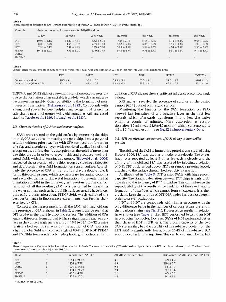
1052 D. Kyprianou et al. / Biosensors and Bioelectronics 25 (2010) 1049–1055
Table 1The fluorescence emission at 430–440 nm after reaction of thiol/OPA solutions with NH4OH in DMF/ethanol 1:1.
Molecule Maximum recorded fluorescence after NH4OH addition
1st day 1st week 2nd week 3rd week 4th week 5th week 6th week
DTT 10.93 ± 3.1% 10.47 ± 4.3% 8.52 ± 6.3% 7.55 ± 2.1% 5.45 ± 4.4% 3.18 ± 6.3% 0.65 ± 9.2%HDT 8.09 ± 2.7% 8.61 ± 3.3% 7.53 ± 4.2% 6.62 ± 5.3% 6.04 ± 5.2% 5.16 ± 3.4% 4.98 ± 3.3%NDT 7.83 ± 5.1% 7.99 ± 4.2% 6.75 ± 2.9% 6.89 ± 3.1% 5.82 ± 3.5% 4.08 ± 2.8% 3.56 ± 5.9%PETMP 10.11 ± 3.6% 9.93 ± 1.7% 9.40 ± 5.4% 9.40 ± 4.7% 9.58 ± 3.7% 9.31 ± 1.1% 9.14 ± 1.7%DMTZ – – – – – – –TMPTMA – – – – – –
Table 2Contact angle measurements of surface with polythiol molecules with and without OPA. The measurements were repeated three times.
HD
5562
Tddflass
3
isogoesaifaottuba
tDlfraa
TBs
DTT DMTZ
Contact angle thiol 16.3 ± 0.1 33.1 ± 0.6Contact angle (thiol + OPA) 32.1 ± 1.5 63.4 ± 0.6
MPTMA and DMTZ did not show significant fluorescence possiblyue to the formation of an unstable isoindole, which can undergoecomposition quickly. Other possibility is the formation of non-uorescent derivatives (Nakamura et al., 1982). Compounds withlong alkyl spacer between sulphur and oxygen and branching
ide-chains near thiol groups will yield isoindoles with increasedtability (Jacobs et al., 1986; Stobaugh et al., 1983).
.2. Characterisation of SAM coated sensor surfaces
SAMs were created on the gold surface by immersing the chipsn thiol/OPA solutions. Immersing the gold chips into a polythiololution without prior reaction with OPA can result in formationf a flat and disordered layer with restricted availability of thiolroups on the surface due to adsorption (on the gold) of more thanne thiol group. In order to prevent this and produced ‘well ori-nted’ SAMs with thiol terminating groups, Niklewski et al. (2004)uggested the protection of one thiol group by creating a thioesternd deprotection after SAM formation on sensor surface. Accord-ngly the presence of OPA in the solution plays a double role. Itorms thioacetal groups, which are necessary for amino couplingnd secondly, thanks to thioacetal formation, it prevents the flatrientation of SAM in the same way as thioesters do. The charac-erisation of all the resulting SAMs was performed by measuringhe water contact angle as hydrophilic surfaces usually have lowernspecific protein adsorption. PETMP SAM, which exhibited theest performance in fluorescence experiments, was further char-cterised by XPS.
Contact angle measurement for all the SAMs with and withouthe presence of OPA is shown in Table 2, where it can be seen thatTT produces the most hydrophilic surface. The addition of OPA
eads to thioacetal formation, which has a significant impact on sur-ace as the contact angle increases from 16.3 to 32.1. DMTZ createselatively hydrophilic surfaces, but the addition of OPA results inhydrophobic SAM with contact angle of 63.4◦. HDT, NDT, PETMPnd TMPTMA form a relatively hydrophobic gold surface and the
able 3iacore responses to BSA immobilised on different molecule SAMs. The standard deviationhows material removed after injection SDS 0.1%.
Thiol na Immobilised BSA (RU) (
DTT 6 1613 ± 21.4% 6HDT 3 1203 ± 7.7% 4DMTZ 3 1095 ± 14.1% 9NDT 3 1194 ± 24.2% 2PETMP 6 1487 ± 4.7% 2TMPTMA 4 1327 ± 10.0% 3
a Number of chips used.
T NDT PETMP TMPTMA
.0 ± 3.1 63.3 ± 0.1 51.6 ± 1.2 48.6 ± 1.3
.3 ± 0.1 63.3 ± 0.1 62.6 ± 0.7 53.1 ± 1.9
addition of OPA did not show significant influence on contact anglevalues.
XPS analysis revealed the presence of sulphur on the coatedsample (6.2%) but not on the gold surface.
Monitoring the kinetics of the SAM formation on FBARshowed fast formation of a dissipative layer in the first fewseconds which afterwards transforms into a less dissipativewithin a couple of minutes. Mass adsorption at satura-tion after 15 min was 31.6 ± 4.5 ng cm−2 which corresponds to4.3 × 1013 molecules cm−2, see Fig. S2 in Supplementary Data.
3.3. SPR experiments: assessment of SAM ability to immobiliseprotein
The ability of the SAM to immobilise proteins was studied usingBiacore 3000. BSA was used as a model biomolecule. The exper-iment was repeated at least 3 times for each molecule and theaffinity of immobilised BSA was assessed by injecting a solutionof 0.1% SDS as described above. SDS can remove proteins looselyattached to the surface through hydrophobic interactions.
As illustrated in Table 3, DTT creates SAMs with high proteincapacity. The standard deviation between DTT chips is high, prob-ably due to the tendency of DTT to oxidise. This can influence thereproducibility of the results, since oxidation of thiols will lead toformation of disulfides which cannot form thioacetals. It is thencrucial to keep the solution of DTT/OPA under inert atmosphere inorder to prevent oxidation.
NDT and HDT are compounds with similar structure with theonly difference being in the number of carbons atoms present intheir carbon chains (see Fig. S1). Fluorescence results in solutionhave shown (see Table 1) that HDT performed better than NDT
in producing isoindoles. However SAMs of NDT performed betterthan those of HDT in SPR tests. The protein capacity of the twoSAMs is similar, but the stability of immobilised protein on theHDT SAM is significantly lower, since 26.4% of immobilised BSAwas removed after SDS injection. This can be explained by the fact(STD) within the chip and between different chips is also reported. The last column
%) STD within each chip % Removed BSA after injection SDS 0.1%
.1 4.9 ± 0.4
.9 26.4 ± 3.7
.4 6.5 ± 1.3
.9 9.7 ± 1.6
.2 4.3 ± 2.2
.4 9.1 ± 5.5
d Bioelectronics 25 (2010) 1049–1055 1053
tmaHi
tclerTbsssape(
mtDasP
3c
3i
dbatrisleatcBs
S0bweasbi
sboEdwc
Table 4The calculated kinetic constants for anti-PSA/PSA interaction on C1 and on PETMP-OPA SAM.
4 −3 2
D. Kyprianou et al. / Biosensors an
hat a longer alkane chain (as on NDT) provides opportunity forore van der Waals interactions between neighboring molecules,
llowing the formation of an ordered thiol layer (Bain et al., 1989;olmes-Farley et al., 1988). HDT most likely produces a SAM, which
s not as ordered.TMPTMA and PETMP are molecules containing three and four
hiol groups respectively. PETMP SAMs showed higher surfaceapacity for protein immobilisation with 1487 RU and TMPTMAayer 1327 RU. Additionally, as illustrated in Table 3 PETMP layerxhibited higher stability as only 4.3% of the immobilised BSA wasemoved after washing with SDS, whereas 9.1% was removed fromMPTMA layer. The high stability of this molecule was also proveny the low STD (4.7%) for protein immobilisation calculated usingix different chips over a 3 months period. On the contrary TMPTMAeemed to be affected by stability problems especially after expo-ure to atmospheric conditions. Despite the fact that TMPTMAnd DMTZ did not show fluorescence in solution the observedrotein immobilisation can be due to other reactions of thioac-tals with amino groups, which results in non-fluorescent productsNakamura et al., 1982).
It can be concluded that SPR experiments are generally in agree-ent with fluorescence studies (Section 3.1). Both have shown that
he molecules with highest performance for amine coupling areTT and PETMP. The fluorescence experiments have shown PETMPs one of the most stable molecule and DTT as one of the leasttable. Due to its high reproducibility, stability and good affinityETMP was selected for further studies.
.4. Application of PETMP/OPA SAM and comparison with Biacorehip C1
.4.1. Kinetic analysis of PSA/anti-PSA monoclonal antibodynteraction
Flat sensor surface modifications are useful especially for theetermination of kinetic constants and evaluation of affinity ofinding reactions. Application of flat surfaces with low volumend restricted surface capacity is important due to the fact that inhis condition mass transport limitation has a minor impact on theesulting sensogram (Önell and Andersson, 2005). In order to min-mise the limitation of mass transport when a sensor chip with highurface capacity is applied for kinetic studies the immobilisationevel of the ligand should be kept low (500–2000 RU) (Katsambat al., 2006). Functionalisation providing low volume surfaces canlso result in less nonspecific binding (due to limited charge attrac-ions and hydrophobic interactions), which, if it is not eliminated,an have a prominent effect on the calculation of kinetic constants.iacore also recommends low capacity sensor surfaces for kinetictudies (Biaevaluation Handbook).
The effect of the pH on protein immobilisation on PETMP/OPAAM was studied in the range 4.5–9.0 (0.05 M acetate buffer pH 4.5,.05 M acetate buffer pH 5.0, 0.1 M PBS pH 7.4 and 0.05 M borateuffer pH 9.0). The highest immobilisation was achieved when BSAas immobilised in PBS buffer pH 7.4 and borate buffer pH 9.0 as
xpected since the fluorescent isoindole formation is inhibited incidic conditions. Consequently the immobilisation at pH 7.4 waselected for further studies on PETMP/OPA SAM since it resem-les physiological conditions while still allowing for good protein
mmobilisation.Comparative studies between PETMP/OPA SAMs and Biacore C1
ensor chips were performed with monoclonal anti-PSA. Immo-ilisation of antibody on PETMP-OPA and C1 produced a signal
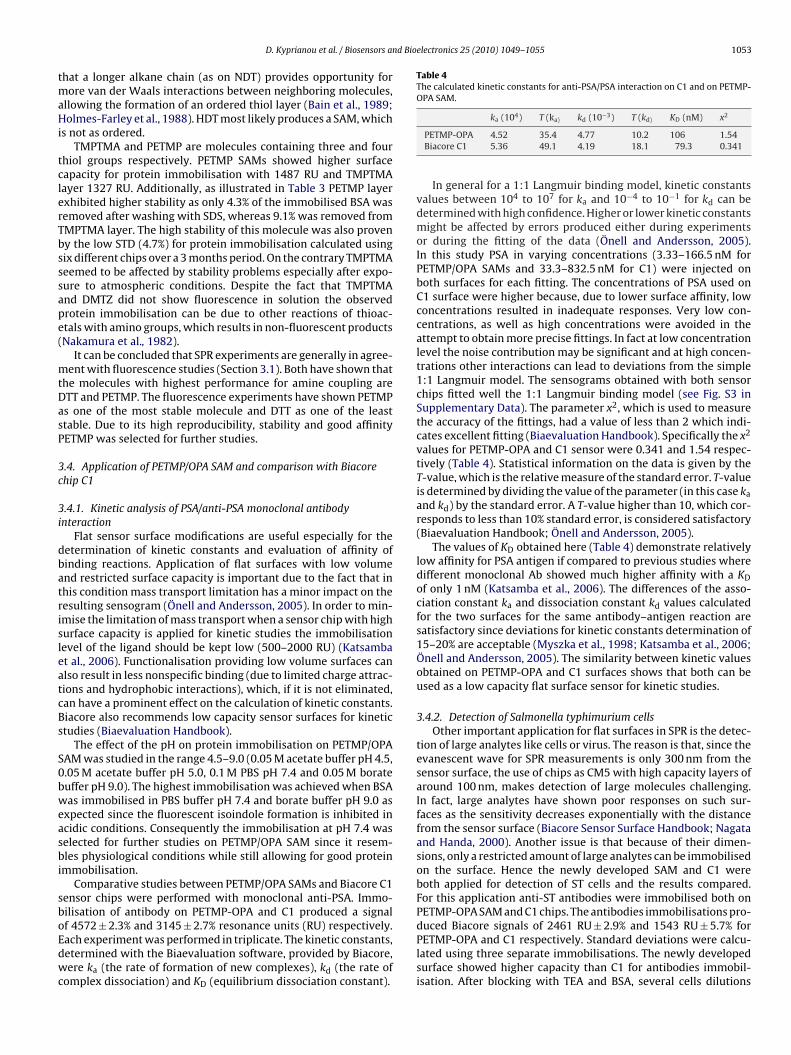
f 4572 ± 2.3% and 3145 ± 2.7% resonance units (RU) respectively.ach experiment was performed in triplicate. The kinetic constants,etermined with the Biaevaluation software, provided by Biacore,ere ka (the rate of formation of new complexes), kd (the rate ofomplex dissociation) and KD (equilibrium dissociation constant).
ka (10 ) T (ka) kd (10 ) T (kd) KD (nM) x
PETMP-OPA 4.52 35.4 4.77 10.2 106 1.54Biacore C1 5.36 49.1 4.19 18.1 79.3 0.341
In general for a 1:1 Langmuir binding model, kinetic constantsvalues between 104 to 107 for ka and 10−4 to 10−1 for kd can bedetermined with high confidence. Higher or lower kinetic constantsmight be affected by errors produced either during experimentsor during the fitting of the data (Önell and Andersson, 2005).In this study PSA in varying concentrations (3.33–166.5 nM forPETMP/OPA SAMs and 33.3–832.5 nM for C1) were injected onboth surfaces for each fitting. The concentrations of PSA used onC1 surface were higher because, due to lower surface affinity, lowconcentrations resulted in inadequate responses. Very low con-centrations, as well as high concentrations were avoided in theattempt to obtain more precise fittings. In fact at low concentrationlevel the noise contribution may be significant and at high concen-trations other interactions can lead to deviations from the simple1:1 Langmuir model. The sensograms obtained with both sensorchips fitted well the 1:1 Langmuir binding model (see Fig. S3 inSupplementary Data). The parameter x2, which is used to measurethe accuracy of the fittings, had a value of less than 2 which indi-cates excellent fitting (Biaevaluation Handbook). Specifically the x2
values for PETMP-OPA and C1 sensor were 0.341 and 1.54 respec-tively (Table 4). Statistical information on the data is given by theT-value, which is the relative measure of the standard error. T-valueis determined by dividing the value of the parameter (in this case ka
and kd) by the standard error. A T-value higher than 10, which cor-responds to less than 10% standard error, is considered satisfactory(Biaevaluation Handbook; Önell and Andersson, 2005).
The values of KD obtained here (Table 4) demonstrate relativelylow affinity for PSA antigen if compared to previous studies wheredifferent monoclonal Ab showed much higher affinity with a KDof only 1 nM (Katsamba et al., 2006). The differences of the asso-ciation constant ka and dissociation constant kd values calculatedfor the two surfaces for the same antibody–antigen reaction aresatisfactory since deviations for kinetic constants determination of15–20% are acceptable (Myszka et al., 1998; Katsamba et al., 2006;Önell and Andersson, 2005). The similarity between kinetic valuesobtained on PETMP-OPA and C1 surfaces shows that both can beused as a low capacity flat surface sensor for kinetic studies.
3.4.2. Detection of Salmonella typhimurium cellsOther important application for flat surfaces in SPR is the detec-
tion of large analytes like cells or virus. The reason is that, since theevanescent wave for SPR measurements is only 300 nm from thesensor surface, the use of chips as CM5 with high capacity layers ofaround 100 nm, makes detection of large molecules challenging.In fact, large analytes have shown poor responses on such sur-faces as the sensitivity decreases exponentially with the distancefrom the sensor surface (Biacore Sensor Surface Handbook; Nagataand Handa, 2000). Another issue is that because of their dimen-sions, only a restricted amount of large analytes can be immobilisedon the surface. Hence the newly developed SAM and C1 wereboth applied for detection of ST cells and the results compared.For this application anti-ST antibodies were immobilised both onPETMP-OPA SAM and C1 chips. The antibodies immobilisations pro-
duced Biacore signals of 2461 RU ± 2.9% and 1543 RU ± 5.7% forPETMP-OPA and C1 respectively. Standard deviations were calcu-lated using three separate immobilisations. The newly developedsurface showed higher capacity than C1 for antibodies immobil-isation. After blocking with TEA and BSA, several cells dilutions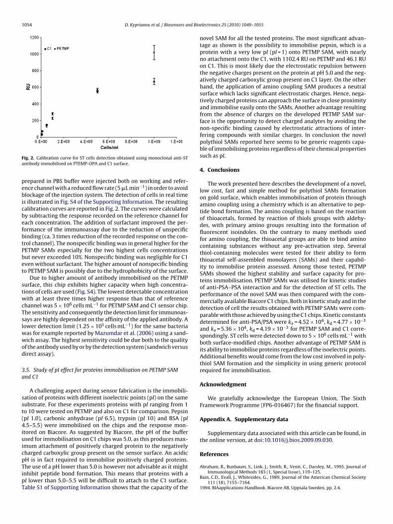
1054 D. Kyprianou et al. / Biosensors and Bio
Fa
pebicbefbtPbet
stwcTslwwod
3a
sst(4iuicpTipT
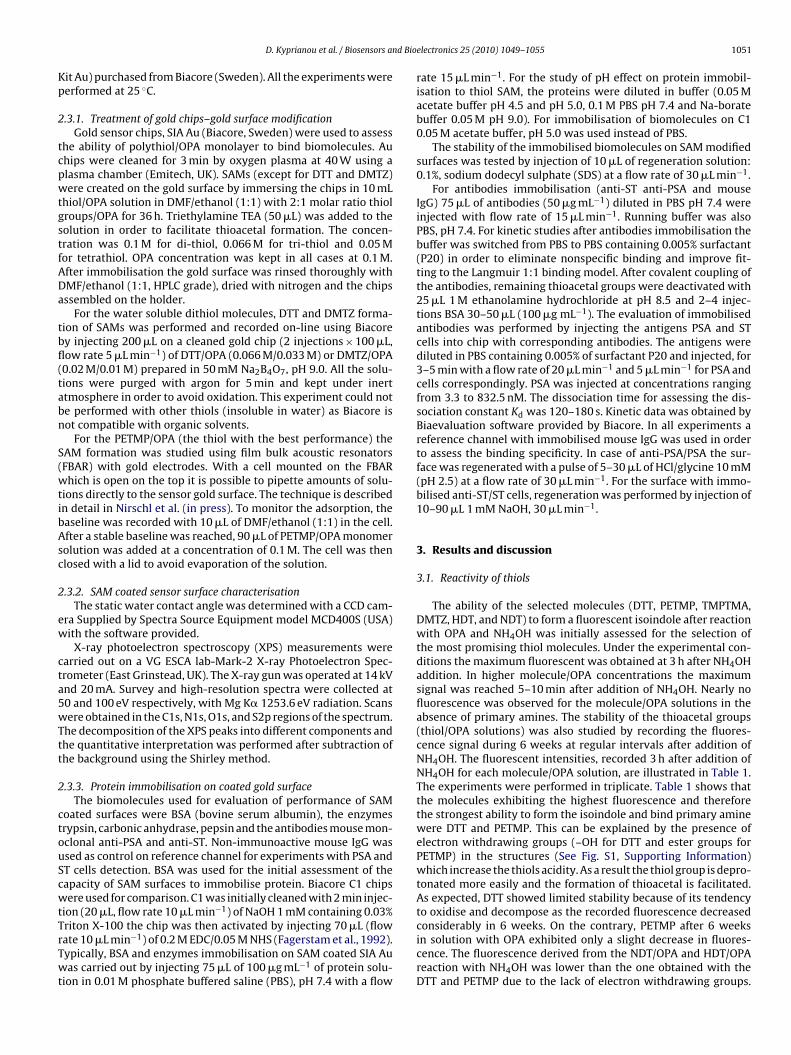
ig. 2. Calibration curve for ST cells detection obtained using monoclonal anti-STntibody immobilised on PTEMP-OPA and C1 surface.
repared in PBS buffer were injected both on working and refer-nce channel with a reduced flow rate (5 �L min−1) in order to avoidlockage of the injection system. The detection of cells in real time
s illustrated in Fig. S4 of the Supporting Information. The resultingalibration curves are reported in Fig. 2. The curves were calculatedy subtracting the response recorded on the reference channel forach concentration. The addition of surfactant improved the per-ormance of the immunoassay due to the reduction of unspecificinding (ca. 3 times reduction of the recorded response on the con-rol channel). The nonspecific binding was in general higher for theETMP SAMs especially for the two highest cells concentrationsut never exceeded 10%. Nonspecific binding was negligible for C1ven without surfactant. The higher amount of nonspecific bindingo PETMP SAM is possibly due to the hydrophobicity of the surface.
Due to higher amount of antibody immobilised on the PETMPurface, this chip exhibits higher capacity when high concentra-ions of cells are used (Fig. S4). The lowest detectable concentrationith at least three times higher response than that of reference
hannel was 5 × 10⁶ cells mL−1 for PETMP SAM and C1 sensor chip.he sensitivity and consequently the detection limit for immunoas-ays are highly dependent on the affinity of the applied antibody. Aower detection limit (1.25 × 10⁵ cells mL−1) for the same bacteria
as for example reported by Mazumdar et al. (2006) using a sand-ich assay. The highest sensitivity could be due both to the quality
f the antibody used by or by the detection system (sandwich versusirect assay).
.5. Study of pI effect for proteins immobilisation on PETMP SAMnd C1
A challenging aspect during sensor fabrication is the immobili-ation of proteins with different isoelectric points (pI) on the sameubstrate. For these experiments proteins with pI ranging from 1o 10 were tested on PETMP and also on C1 for comparison. PepsinpI 1.0), carbonic anhydrase (pI 6.5), trypsin (pI 10) and BSA (pI.5–5.5) were immobilised on the chips and the response mon-
tored on Biacore. As suggested by Biacore, the pH of the buffersed for immobilisation on C1 chips was 5.0, as this produces max-
mum attachment of positively charged protein to the negativelyharged carboxylic group present on the sensor surface. An acidicH is in fact required to immobilise positively charged proteins.
he use of a pH lower than 5.0 is however not advisable as it mightnhibit peptide bond formation. This means that proteins with aI lower than 5.0–5.5 will be difficult to attach to the C1 surface.able S1 of Supporting Information shows that the capacity of theelectronics 25 (2010) 1049–1055
novel SAM for all the tested proteins. The most significant advan-tage as shown is the possibility to immobilise pepsin, which is aprotein with a very low pI (pI = 1) onto PETMP SAM, with nearlyno attachment onto the C1, with 1102.4 RU on PETMP and 46.1 RUon C1. This is most likely due the electrostatic repulsion betweenthe negative charges present on the protein at pH 5.0 and the neg-atively charged carboxylic group present on C1 layer. On the otherhand, the application of amino coupling SAM produces a neutralsurface which lacks significant electrostatic charges. Hence, nega-tively charged proteins can approach the surface in close proximityand immobilise easily onto the SAMs. Another advantage resultingfrom the absence of charges on the developed PETMP SAM sur-face is the opportunity to detect charged analytes by avoiding thenon-specific binding caused by electrostatic attractions of inter-fering compounds with similar charges. In conclusion the novelpolythiol SAMs reported here seems to be generic reagents capa-ble of immobilising proteins regardless of their chemical propertiessuch as pI.
4. Conclusions
The work presented here describes the development of a novel,low cost, fast and simple method for polythiol SAMs formationon gold surface, which enables immobilisation of protein throughamino coupling using a chemistry which is an alternative to pep-tide bond formation. The amino coupling is based on the reactionof thioacetals, formed by reaction of thiols groups with aldehy-des, with primary amino groups resulting into the formation offluorescent isoindoles. On the contrary to many methods usedfor amino coupling, the thioacetal groups are able to bind aminocontaining substances without any pre-activation step. Severalthiol-containing molecules were tested for their ability to formthioacetal self-assembled monolayers (SAMs) and their capabil-ity to immobilise protein assessed. Among those tested, PETMPSAMs showed the highest stability and surface capacity for pro-teins immobilisation. PETMP SAMs was utilised for kinetic studiesof anti-PSA–PSA interaction and for the detection of ST cells. Theperformance of the novel SAM was then compared with the com-mercially available Biacore C1 chips. Both in kinetic study and in thedetection of cell the results obtained with PETMP SAMs were com-parable with those achieved by using the C1 chips. Kinetic constantsdetermined for anti-PSA/PSA were ka = 4.52 × 10⁴, kd = 4.77 × 10⁻³
and ka = 5.36 × 10⁴, kd = 4.19 × 10⁻³ for PETMP SAM and C1 corre-spondingly. ST cells were detected down to 5 × 10⁶ cells mL−1 withboth surface-modified chips. Another advantage of PETMP SAM isits ability to immobilise proteins regardless of the isoelectric points.Additional benefits would come from the low cost involved in poly-thiol SAM formation and the simplicity in using generic protocolrequired for immobilisation.
Acknowledgment
We gratefully acknowledge the European Union, The SixthFramework Programme (FP6-016467) for the financial support.
Appendix A. Supplementary data
Supplementary data associated with this article can be found, inthe online version, at doi:10.1016/j.bios.2009.09.030.
References
Abraham, R., Buxbaum, S., Link, J., Smith, R., Venti, C., Darsley, M., 1995. Journal ofImmunological Methods 183 (1, Special Issue), 119–125.
Bain, C.D., Evall, J., Whitesides, G., 1989. Journal of the American Chemical Society111 (18), 7155–7164.
1994. BIAapplications Handbook. Biacore AB, Uppsala Sweden, pp. 2.4.

d Bio
22
BC
F
H
HJ
J
KK
K
L
M
M
M
D. Kyprianou et al. / Biosensors an
003. Biacore Sensor Surface Handbook. Biacore AB, Uppsala Sweden, pp. 3, 13–14.004. Biaevaluation Handbook. Biacore AB, Uppsala Sweden, pp. 4.8, 4.14–4.16,
7.13–7.14.utler, E.J., 2000. Methods 22 (1), 4–23.raft, C.M., Xu, J., Slepak, V.Z., Zhan-Poe, X., Zhu, X., Brown, B., Lolley, R.N., 1998.
Biochemistry 37 (45), 15758–15772.agerstam, L.G., Frostell-Karlsson, A., Karlsson, R., Persson, B., Ronnberg, I., 1992.
Journal of Chromatography 597 (1–2), 397–410.aga, Y., Hakomori, S., Hatanaka, K., 2008. Carbohydrate Research 343 (18),
3034–3038.olmes-Farley, S.R., Bain, C.D., Whitesides, G.M., 1988. Langmuir 4 (4), 921–937.
acobs, W.A., Leburg, M.W., Madaj, E.J., 1986. Analytical Biochemistry 156 (2),334–440.
ohnson, B., Lofas, S., Lindquist, G., Edstrom, A., Muller, R., Hannson, A., 1995. Journalof Molecular Recognition 8 (1–2), 125–131.
atsamba, P.S., et al., 2006. Analytical Biochemistry 352 (2), 208–221.usnezow, W., Hoheisel, J.D., 2003. Journal of Molecular Recognition 16 (4),
165–176.yprianou, D., Guerreiro, A.R., Chianella, I., Piletska, E.V., Fowler, S., Karim, K., Whit-
combe, M.J., Turner, A., Piletsky, S., 2009. Biosensors and Bioelectronics 24 (5),1356–1371.
ofas, S., Johnson, B., Edstrom, A., Hansson, G., Linquist, G., Muller, R., Stigh, L., 1995.Biosensors and Bioelectronics 10 (10), 813–822.
azumdar, S.D., Hartmann, M., Kämpfer, P., Keusgen, M., 2006. Biosensors and Bio-electronics 22 (9–10), 2040–2046.
asson, J.F., Battaglia, M., Davidson, J., Kim, Y., Prakash, A.M.C., Beaudoin, S., Booksh,K.S., 2005. Talanta 67 (5), 918–925.
yszka, D.G., He, X., Dembo, M., Morton, T.A., Goldstein, B., 1998. Biophysics Journal75 (2), 583–594.
electronics 25 (2010) 1049–1055 1055
Nakamura, H., Matsumoto, A., Tarmura, Z., 1982. Analytical Letters 15 (17),1393–1410.
Nagata, K., Handa, H., 2000. Real-Time Analysis of Biomolecular Interactions.Springer-Verlag, Tokyo (Japan).
Niklewski, W., Azzam, T., Strunskus, R., Fischer, C., 2004. Langmuir 20 (20),8620–8624.
Nirschl, M., Blüher, A., Erler, C., Katzschner, B., Vikholm-Lundin, I., Auer, S., Vörös,J., Pompe, W., Schreiter, M., Mertig, M., in press DOI:10.1016/j.sna.2009.02.021.Sensors and Actuators A.
Nuzzo, R., Allara, D., 1983. Journal of the American Chemical Society 105 (13),4481–4483.
Önell, A., Andersson, K., 2005. Journal of Molecular Recognition 18 (4), 307–317.O’Shannessy, D.J., Brigham-Burke, M., Peck, K., 1992. Analytical Biochemistry 205
(1), 132–136.Panayotou, G., Brown, T., Barlow, T., Pearl, L.H., Savva, R., 1998. Journal of Biological
Chemistry 273 (1), 45–50.Piletska, E.V., Piletsky, S., Subrahmanyam, S., Karim, K., Turner, A.P.F., 2001. Polymer
42 (8), 3603–3608.Predki, F.P., 2004. Current Opinion in Chemical Biology 8 (1), 8–13.Regnault, V., Arvieux, J., Vallar, L., Lecompte, T., 1998. Journal of Immunological
Methods 211 (1–2), 191–197.Sadana, A., 2006. Market Size and Economics for Biosensors. Binding and Disso-
ciation Kinetics for Different Biosensor Applications Using Fractals. Elsevier
Science, Amsterdam, pp. 319–342.Simons, S., Johnson, D., 1978. Journal of Organic Chemistry 43 (14), 2286–2891.Stobaugh, F., Repta, A.J., Sternson, L.A., Garren, K.W., 1983. Analytical Biochemistry
135 (2), 495–504.Su, T.J., Lu, J.R., Thomas, R.K., Cui, Z.F., Penfold, J., 1998. Langmuir 14 (2), 438–445.Vikholm, I., 2005. Sensors and Actuators B 106 (1), 311–316.