The application of in situ mid-FTIR fibre-optic reflectance spectroscopy and GC–MS analysis to...
7
Spectrochimica Acta Part A 74 (2009) 1182–1188 Contents lists available at ScienceDirect Spectrochimica Acta Part A: Molecular and Biomolecular Spectroscopy journal homepage: www.elsevier.com/locate/saa The application of in situ mid-FTIR fibre-optic reflectance spectroscopy and GC–MS analysis to monitor and evaluate painting cleaning Kenza Kahrim a , Alessia Daveri a , Paola Rocchi b , Grazia de Cesare c , Laura Cartechini a,b , Costanza Miliani a,b,∗ , B.G. Brunetti a , A. Sgamellotti a,b a SMAArt, Dipartimento di Chimica, Via Elce di Sotto 8, 06123 Perugia, Italy b CNR-ISTM, Dipartimento di Chimica, Via Elce di Sotto 8, 06123 Perugia, Italy c Istituto Centrale per il Restauro e la Conservazione del Patrimonio Archivistico e Librario, MiBAC, Laboratorio dipinti e materiali contemporanei, Piazza S. Francesco di Paola 9, 00184 Rome, Italy article info Article history: Received 31 January 2009 Accepted 20 August 2009 Keywords: Non-invasive Fibre-optic reflectance spectroscopy Triammonium citrate Oxalate Painting cleaning abstract The development of non-invasive methodologies and portable instrumentation for in situ studies has been subject to great research and development in recent years in the field of conservation science. Despite such interest, very few reported studies employ these versatile techniques in the monitoring of cleaning treatments. This paper describes the application of mid-FTIR fibre-optic reflectance spectroscopy to monitor and evaluate the cleaning treatment of an oil painting using the chelating agent, triammonium citrate, a task undertaken in close collaboration with the painting conservator. Results obtained on site verify the removal of calcium oxalate and an organic component from the surface of the painting, later identified as a terpenic varnish. The subsequent, in laboratory FTIR and GC–MS analysis of the cotton swabs employed during the cleaning treatment acts as an additional non-invasive manner to support the results obtained in situ by mid-FTIR spectroscopy and to better understand the mechanism of the chosen cleaning agent. © 2009 Elsevier B.V. All rights reserved. 1. Introduction 1.1. Background and research aim A range of approaches exists to aid the conservator in the clean- ing of painted works of art. These may be loosely classified as mechanical cleaning, solvent cleaning, laser cleaning, etc., and often are applied in combination to deal most effectively with the chal- lenges that may be encountered in restoring a painting to a greater aesthetic coherence and a more sound state of conservation. The practice of solvent cleaning dates to almost 300 years, fol- lowed closely by proposed classification schemes of solvents to aid the practitioner in their application. Today, one convenient and commonly accepted scheme [1] may account for hundreds of solu- tions upon the basis of solvent action (physical, physico-chemical, aqueous solutions containing additives, etc.). It is of no surprise that such a wide range of substances, often employed on the basis of empirical observation rather than an understanding of the mechanisms at hand, has resulted in inter- ventions of varying success and, as such, subject to debate. The ∗ Corresponding author at: CNR-ISTM, Dipartimento di Chimica, Via Elce di Sotto 8, 06123 Perugia, Italy. Tel.: +39 075 585 5639; fax: +39 075 585 5606. E-mail address: [email protected] (C. Miliani). advent of science in the field of conservation has led to a greater understanding of the agents employed for cleaning, possible expla- nations for conservation interventions gone awry as well as newly proposed agents that undergo systematic testing before their appli- cation to veritable works of art. Although this has been of invaluable help for making informed decisions regarding the choice of cleaning agent, the complex system of (cleaning agent, unwanted material to be removed and original artist materials to be respected) is unique for every undertaken project and hardly reproducible in the labo- ratory. As a consequence, every cleaning procedure may yield an unpredictable result, thus necessitating in situ, on-line monitor- ing of the painting treatment for an accurate understanding of the processes taking place. To date, a number of published studies [2–9] report the mon- itoring of cleaning procedures in situ making use of a range of techniques including SEM, GC–MS, Py-GC–MS, DTMS, FORS and some less frequently encountered techniques such as holographic interferometry, time-resolved shadowgraphy and plasma spec- troscopy employed to study the effects and the interaction process created during laser cleaning [8,9]. The singular studies are unques- tionably of great interest but, taken as a body of work, present a number of features that may be of concern to both scientists and conservators. The first concern regards the use of destructive tests. Many of the publications rely upon the use of techniques that neces- sitate sampling which, even in the case of micro-sampling, may 1386-1425/$ – see front matter © 2009 Elsevier B.V. All rights reserved. doi:10.1016/j.saa.2009.08.051
-
Upload
independent -
Category
Documents
-
view
0 -
download
0
Transcript of The application of in situ mid-FTIR fibre-optic reflectance spectroscopy and GC–MS analysis to...
TG
KCa
b
c
P
a
ARA
KNFTOP
1
1
imala
ltcta
euv
8
1d
Spectrochimica Acta Part A 74 (2009) 1182–1188
Contents lists available at ScienceDirect
Spectrochimica Acta Part A: Molecular andBiomolecular Spectroscopy
journa l homepage: www.e lsev ier .com/ locate /saa
he application of in situ mid-FTIR fibre-optic reflectance spectroscopy andC–MS analysis to monitor and evaluate painting cleaning
enza Kahrima, Alessia Daveria, Paola Rocchib, Grazia de Cesarec, Laura Cartechinia,b,ostanza Miliania,b,∗, B.G. Brunetti a, A. Sgamellotti a,b
SMAArt, Dipartimento di Chimica, Via Elce di Sotto 8, 06123 Perugia, ItalyCNR-ISTM, Dipartimento di Chimica, Via Elce di Sotto 8, 06123 Perugia, ItalyIstituto Centrale per il Restauro e la Conservazione del Patrimonio Archivistico e Librario, MiBAC, Laboratorio dipinti e materiali contemporanei,iazza S. Francesco di Paola 9, 00184 Rome, Italy
r t i c l e i n f o
rticle history:eceived 31 January 2009ccepted 20 August 2009
eywords:
a b s t r a c t
The development of non-invasive methodologies and portable instrumentation for in situ studies hasbeen subject to great research and development in recent years in the field of conservation science.Despite such interest, very few reported studies employ these versatile techniques in the monitoring ofcleaning treatments.
on-invasiveibre-optic reflectance spectroscopyriammonium citratexalateainting cleaning
This paper describes the application of mid-FTIR fibre-optic reflectance spectroscopy to monitor andevaluate the cleaning treatment of an oil painting using the chelating agent, triammonium citrate, atask undertaken in close collaboration with the painting conservator. Results obtained on site verify theremoval of calcium oxalate and an organic component from the surface of the painting, later identified asa terpenic varnish. The subsequent, in laboratory FTIR and GC–MS analysis of the cotton swabs employedduring the cleaning treatment acts as an additional non-invasive manner to support the results obtained
oscop
in situ by mid-FTIR spectr. Introduction
.1. Background and research aim
A range of approaches exists to aid the conservator in the clean-ng of painted works of art. These may be loosely classified as
echanical cleaning, solvent cleaning, laser cleaning, etc., and oftenre applied in combination to deal most effectively with the chal-enges that may be encountered in restoring a painting to a greateresthetic coherence and a more sound state of conservation.
The practice of solvent cleaning dates to almost 300 years, fol-owed closely by proposed classification schemes of solvents to aidhe practitioner in their application. Today, one convenient andommonly accepted scheme [1] may account for hundreds of solu-ions upon the basis of solvent action (physical, physico-chemical,queous solutions containing additives, etc.).
It is of no surprise that such a wide range of substances, oftenmployed on the basis of empirical observation rather than annderstanding of the mechanisms at hand, has resulted in inter-entions of varying success and, as such, subject to debate. The
∗ Corresponding author at: CNR-ISTM, Dipartimento di Chimica, Via Elce di Sotto, 06123 Perugia, Italy. Tel.: +39 075 585 5639; fax: +39 075 585 5606.
E-mail address: [email protected] (C. Miliani).
386-1425/$ – see front matter © 2009 Elsevier B.V. All rights reserved.oi:10.1016/j.saa.2009.08.051
y and to better understand the mechanism of the chosen cleaning agent.© 2009 Elsevier B.V. All rights reserved.
advent of science in the field of conservation has led to a greaterunderstanding of the agents employed for cleaning, possible expla-nations for conservation interventions gone awry as well as newlyproposed agents that undergo systematic testing before their appli-cation to veritable works of art. Although this has been of invaluablehelp for making informed decisions regarding the choice of cleaningagent, the complex system of (cleaning agent, unwanted material tobe removed and original artist materials to be respected) is uniquefor every undertaken project and hardly reproducible in the labo-ratory. As a consequence, every cleaning procedure may yield anunpredictable result, thus necessitating in situ, on-line monitor-ing of the painting treatment for an accurate understanding of theprocesses taking place.
To date, a number of published studies [2–9] report the mon-itoring of cleaning procedures in situ making use of a range oftechniques including SEM, GC–MS, Py-GC–MS, DTMS, FORS andsome less frequently encountered techniques such as holographicinterferometry, time-resolved shadowgraphy and plasma spec-troscopy employed to study the effects and the interaction processcreated during laser cleaning [8,9]. The singular studies are unques-
tionably of great interest but, taken as a body of work, present anumber of features that may be of concern to both scientists andconservators. The first concern regards the use of destructive tests.Many of the publications rely upon the use of techniques that neces-sitate sampling which, even in the case of micro-sampling, may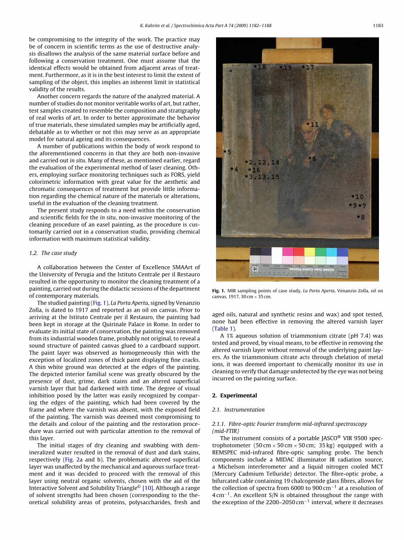
ca Acta Part A 74 (2009) 1182–1188 1183
bbsfimsv
ntoodm
tatecctu
acti
1
trpo
ZabefsTeATpviifotdt
irlmlIoo
K. Kahrim et al. / Spectrochimi
e compromising to the integrity of the work. The practice maye of concern in scientific terms as the use of destructive analy-is disallows the analysis of the same material surface before andollowing a conservation treatment. One must assume that thedentical effects would be obtained from adjacent areas of treat-
ent. Furthermore, as it is in the best interest to limit the extent ofampling of the object, this implies an inherent limit in statisticalalidity of the results.
Another concern regards the nature of the analyzed material. Aumber of studies do not monitor veritable works of art, but rather,est samples created to resemble the composition and stratigraphyf real works of art. In order to better approximate the behaviorf true materials, these simulated samples may be artificially aged,ebatable as to whether or not this may serve as an appropriateodel for natural ageing and its consequences.A number of publications within the body of work respond to
he aforementioned concerns in that they are both non-invasivend carried out in situ. Many of these, as mentioned earlier, regardhe evaluation of the experimental method of laser cleaning. Oth-rs, employing surface monitoring techniques such as FORS, yieldolorimetric information with great value for the aesthetic andhromatic consequences of treatment but provide little informa-ion regarding the chemical nature of the materials or alterations,seful in the evaluation of the cleaning treatment.
The present study responds to a need within the conservationnd scientific fields for the in situ, non-invasive monitoring of theleaning procedure of an easel painting, as the procedure is cus-omarily carried out in a conservation studio, providing chemicalnformation with maximum statistical validity.
.2. The case study
A collaboration between the Center of Excellence SMAArt ofhe University of Perugia and the Istituto Centrale per il Restauroesulted in the opportunity to monitor the cleaning treatment of aainting, carried out during the didactic sessions of the departmentf contemporary materials.
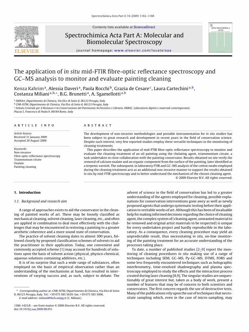
The studied painting (Fig. 1), La Porta Aperta, signed by Venanzioolla, is dated to 1917 and reported as an oil on canvas. Prior torriving at the Istituto Centrale per il Restauro, the painting hadeen kept in storage at the Quirinale Palace in Rome. In order tovaluate its initial state of conservation, the painting was removedrom its industrial wooden frame, probably not original, to reveal aound structure of painted canvas glued to a cardboard support.he paint layer was observed as homogeneously thin with thexception of localized zones of thick paint displaying fine cracks.
thin white ground was detected at the edges of the painting.he depicted interior familial scene was greatly obscured by theresence of dust, grime, dark stains and an altered superficialarnish layer that had darkened with time. The degree of visualnhibition posed by the latter was easily recognized by compar-ng the edges of the painting, which had been covered by therame and where the varnish was absent, with the exposed fieldf the painting. The varnish was deemed most compromising tohe details and colour of the painting and the restoration proce-ure was carried out with particular attention to the removal ofhis layer.
The initial stages of dry cleaning and swabbing with dem-neralized water resulted in the removal of dust and dark stains,espectively (Fig. 2a and b). The problematic altered superficialayer was unaffected by the mechanical and aqueous surface treat-
ent and it was decided to proceed with the removal of thisayer using neutral organic solvents, chosen with the aid of thenteractive Solvent and Solubility Triangle© [10]. Although a rangef solvent strengths had been chosen (corresponding to the the-retical solubility areas of proteins, polysaccharides, fresh and
Fig. 1. MIR sampling points of case study, La Porta Aperta, Venanzio Zolla, oil oncanvas, 1917, 30 cm × 35 cm.
aged oils, natural and synthetic resins and wax) and spot tested,none had been effective in removing the altered varnish layer(Table 1).
A 1% aqueous solution of triammonium citrate (pH 7.4) wastested and proved, by visual means, to be effective in removing thealtered varnish layer without removal of the underlying paint lay-ers. As the triammonium citrate acts through chelation of metalions, it was deemed important to chemically monitor its use incleaning to verify that damage undetected by the eye was not beingincurred on the painting surface.
2. Experimental
2.1. Instrumentation
2.1.1. Fibre-optic Fourier transform mid-infrared spectroscopy(mid-FTIR)
The instrument consists of a portable JASCO® VIR 9500 spec-trophotometer (50 cm × 50 cm × 50 cm; 35 kg) equipped with aREMSPEC mid-infrared fibre-optic sampling probe. The benchcomponents include a MIDAC illuminator IR radiation source,a Michelson interferometer and a liquid nitrogen cooled MCT(Mercury Cadmium Telluride) detector. The fibre-optic probe, a
bifurcated cable containing 19 chalcogenide glass fibres, allows forthe collection of spectra from 6000 to 900 cm−1 at a resolution of4 cm−1. An excellent S/N is obtained throughout the range withthe exception of the 2200–2050 cm−1 interval, where it decreases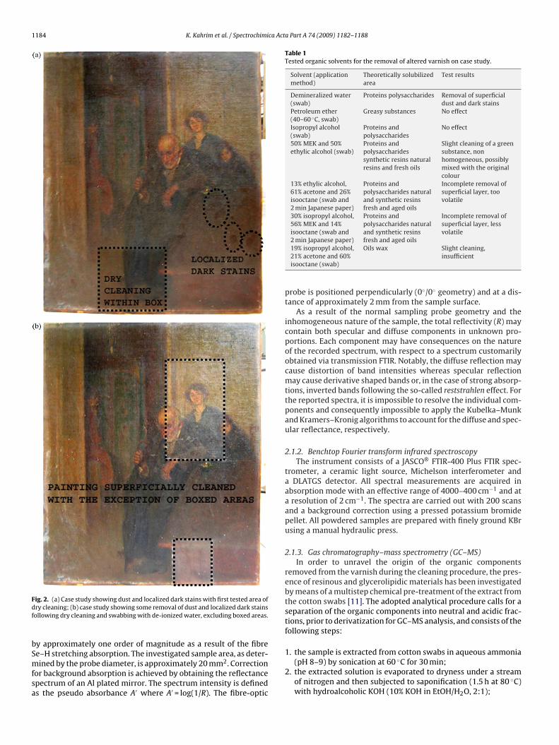
1184 K. Kahrim et al. / Spectrochimica Acta Part A 74 (2009) 1182–1188
Fdf
bSmfsa
Table 1Tested organic solvents for the removal of altered varnish on case study.
Solvent (applicationmethod)
Theoretically solubilizedarea
Test results
Demineralized water(swab)
Proteins polysaccharides Removal of superficialdust and dark stains
Petroleum ether(40–60 ◦C, swab)
Greasy substances No effect
Isopropyl alcohol(swab)
Proteins andpolysaccharides
No effect
50% MEK and 50%ethylic alcohol (swab)
Proteins andpolysaccharidessynthetic resins naturalresins and fresh oils
Slight cleaning of a greensubstance, nonhomogeneous, possiblymixed with the originalcolour
13% ethylic alcohol,61% acetone and 26%isooctane (swab and2 min Japanese paper)
Proteins andpolysaccharides naturaland synthetic resinsfresh and aged oils
Incomplete removal ofsuperficial layer, toovolatile
30% isopropyl alcohol,56% MEK and 14%isooctane (swab and2 min Japanese paper)
Proteins andpolysaccharides naturaland synthetic resinsfresh and aged oils
Incomplete removal ofsuperficial layer, lessvolatile
ig. 2. (a) Case study showing dust and localized dark stains with first tested area ofry cleaning; (b) case study showing some removal of dust and localized dark stainsollowing dry cleaning and swabbing with de-ionized water, excluding boxed areas.
y approximately one order of magnitude as a result of the fibree–H stretching absorption. The investigated sample area, as deter-
ined by the probe diameter, is approximately 20 mm2. Correctionor background absorption is achieved by obtaining the reflectancepectrum of an Al plated mirror. The spectrum intensity is defineds the pseudo absorbance A′ where A′ = log(1/R). The fibre-optic
19% isopropyl alcohol,21% acetone and 60%isooctane (swab)
Oils wax Slight cleaning,insufficient
probe is positioned perpendicularly (0◦/0◦ geometry) and at a dis-tance of approximately 2 mm from the sample surface.
As a result of the normal sampling probe geometry and theinhomogeneous nature of the sample, the total reflectivity (R) maycontain both specular and diffuse components in unknown pro-portions. Each component may have consequences on the natureof the recorded spectrum, with respect to a spectrum customarilyobtained via transmission FTIR. Notably, the diffuse reflection maycause distortion of band intensities whereas specular reflectionmay cause derivative shaped bands or, in the case of strong absorp-tions, inverted bands following the so-called reststrahlen effect. Forthe reported spectra, it is impossible to resolve the individual com-ponents and consequently impossible to apply the Kubelka–Munkand Kramers–Kronig algorithms to account for the diffuse and spec-ular reflectance, respectively.
2.1.2. Benchtop Fourier transform infrared spectroscopyThe instrument consists of a JASCO® FTIR-400 Plus FTIR spec-
trometer, a ceramic light source, Michelson interferometer anda DLATGS detector. All spectral measurements are acquired inabsorption mode with an effective range of 4000–400 cm−1 and ata resolution of 2 cm−1. The spectra are carried out with 200 scansand a background correction using a pressed potassium bromidepellet. All powdered samples are prepared with finely ground KBrusing a manual hydraulic press.
2.1.3. Gas chromatography–mass spectrometry (GC–MS)In order to unravel the origin of the organic components
removed from the varnish during the cleaning procedure, the pres-ence of resinous and glycerolipidic materials has been investigatedby means of a multistep chemical pre-treatment of the extract fromthe cotton swabs [11]. The adopted analytical procedure calls for aseparation of the organic components into neutral and acidic frac-tions, prior to derivatization for GC–MS analysis, and consists of thefollowing steps:
1. the sample is extracted from cotton swabs in aqueous ammonia
(pH 8–9) by sonication at 60 ◦C for 30 min;2. the extracted solution is evaporated to dryness under a streamof nitrogen and then subjected to saponification (1.5 h at 80 ◦C)with hydroalcoholic KOH (10% KOH in EtOH/H2O, 2:1);
ca Acta Part A 74 (2009) 1182–1188 1185
3
4
5
6
mt
(atr21w
wofiTtwwhra
2
p
(
(
(
(
(
a
3
3
m
K. Kahrim et al. / Spectrochimi
. the unsaponifiable fraction is extracted with n-hexane threetimes (neutral fraction) and the residual solution is kept for step5;
. an aliquot of this extract is evaporated to dryness under a streamof nitrogen and subjected to derivatization for GC–MS analysiswith N,O-bis-trimethylsilyltrifluoroacetamide (BSTFA) at 60 ◦Cfor 30 min. Following the addition of isooctane as solvent, 1 �lof the final solution is analyzed by GC–MS with hexadecane asinternal standard;
. the residual solution of the n-hexane extraction is acidified andextracted with diethyl ether three times;
. an aliquot of the acidic fraction is then subjected to the samederivatization procedure as described for the neutral fraction.
The derivatization step converts the acidic and alcoholicoieties to the corresponding trimethylsilyl ester and ether deriva-
ives.Samples were analyzed by using a 6890N gas chromatography
Agilent Technologies) with split/splitless injector, interfaced tomodel 5973 quadropole mass spectrometer (MS). Injector and
ransfer lines were kept at a temperature of 230 ◦C and 280 ◦C,espectively. While the MS ion source temperature was kept at30 ◦C, the mass quadropole was maintained at a temperature of50 ◦C. The MS operated in the EI mode at 70 eV. The carrier gasas helium at a constant flow of 1 ml/min.
The GC employs a fused silica capillary column, model HP-5MS,ith a 5% diphenyl, 95% dimethyl-polysiloxane stationary phase,
f 30 m length with internal diameter of 0.25 mm and 0.25 �mlm thickness (J&W Scientific, Agilent Technologies, Palo Alto, CA).he temperature program of the oven adopted for the analysis ofhe neutral and acidic fractions has an initial temperature of 80 ◦C,hich is held for 2 min and then ramped at 10 ◦C/min to 200 ◦C,here it is held for 3 min, ramped at 10 ◦C/min to 280 ◦C, where it iseld for 3 min, ramped at 20 ◦C/min to 300 ◦C and held for 20 min,esulting in a total of approximately 49 min of chromatographicnalysis.
.2. Methodology
The in situ on-line monitoring of the cleaning treatment waserformed on the basis of the following steps:
1) a mid-FTIR reflectance spectrum of the chosen area of the paint-ing surface was acquired;
2) the localized area of the painting was gently treated with a 1%triammonium citrate infused cotton swab, by repeated rollingon the surface;
3) a mid-FTIR reflectance spectrum was subsequently acquired onthe treated area;
4) the localized area was gently rinsed with a de-ionized waterinfused cotton swab, by repeated rolling on the surface (thesecotton swabs were conserved for subsequent laboratory anal-ysis);
5) the final mid-FTIR reflectance spectrum was acquired on thetreated and rinsed painting area.
The complete procedure was repeated four times in varyingreas of the painting surface.
. Results and discussion
.1. In situ on-line monitoring
In order to gain a better understanding of the altered varnishaterial in question, an initial range of mid-FTIR reflectance spectra
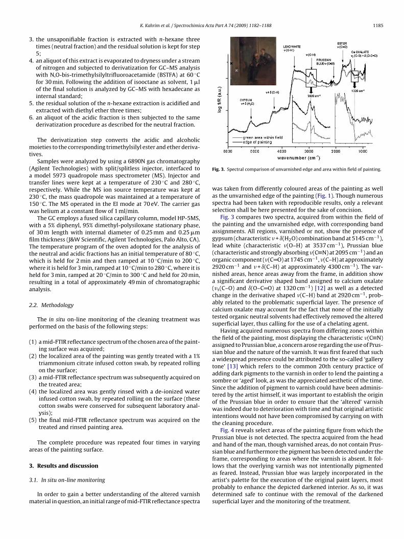
Fig. 3. Spectral comparison of unvarnished edge and area within field of painting.
was taken from differently coloured areas of the painting as wellas the unvarnished edge of the painting (Fig. 1). Though numerousspectra had been taken with reproducible results, only a relevantselection shall be here presented for the sake of concision.
Fig. 3 compares two spectra, acquired from within the field ofthe painting and the unvarnished edge, with corresponding bandassignments. All regions, varnished or not, show the presence ofgypsum (characteristic � + ı(H2O) combination band at 5145 cm−1),lead white (characteristic �(O–H) at 3537 cm−1), Prussian blue(characteristic and strongly absorbing �(C N) at 2095 cm−1) and anorganic component (�(C O) at 1745 cm−1, �(C–H) at approximately2920 cm−1 and � + ı(C–H) at approximately 4300 cm−1). The var-nished areas, hence areas away from the frame, in addition showa significant derivative shaped band assigned to calcium oxalate(�s(C–O) and ı(O–C O) at 1320 cm−1) [12] as well as a detectedchange in the derivative shaped �(C–H) band at 2920 cm−1, prob-ably related to the problematic superficial layer. The presence ofcalcium oxalate may account for the fact that none of the initiallytested organic neutral solvents had effectively removed the alteredsuperficial layer, thus calling for the use of a chelating agent.
Having acquired numerous spectra from differing zones withinthe field of the painting, most displaying the characteristic �(C N)assigned to Prussian blue, a concern arose regarding the use of Prus-sian blue and the nature of the varnish. It was first feared that sucha widespread presence could be attributed to the so-called ‘gallerytone’ [13] which refers to the common 20th century practice ofadding dark pigments to the varnish in order to lend the painting asombre or ‘aged’ look, as was the appreciated aesthetic of the time.Since the addition of pigment to varnish could have been adminis-tered by the artist himself, it was important to establish the originof the Prussian blue in order to ensure that the ‘altered’ varnishwas indeed due to deterioration with time and that original artisticintentions would not have been compromised by carrying on withthe cleaning procedure.
Fig. 4 reveals select areas of the painting figure from which thePrussian blue is not detected. The spectra acquired from the headand hand of the man, though varnished areas, do not contain Prus-sian blue and furthermore the pigment has been detected under theframe, corresponding to areas where the varnish is absent. It fol-lows that the overlying varnish was not intentionally pigmentedas feared. Instead, Prussian blue was largely incorporated in the
artist’s palette for the execution of the original paint layers, mostprobably to enhance the depicted darkened interior. As so, it wasdetermined safe to continue with the removal of the darkenedsuperficial layer and the monitoring of the treatment.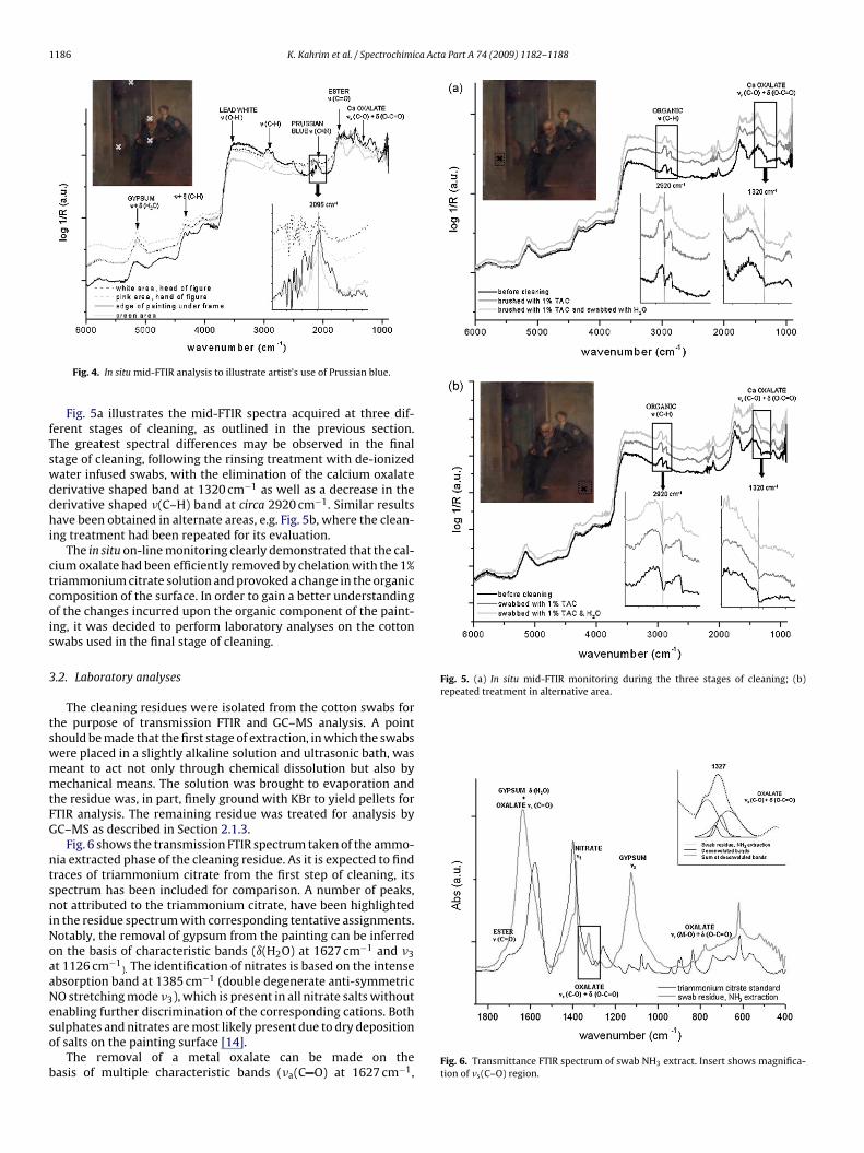
1186 K. Kahrim et al. / Spectrochimica Acta Part A 74 (2009) 1182–1188
fTswddhi
ctcois
3
tswmmtFG
ntsniNoaaNeso
b
Fig. 5. (a) In situ mid-FTIR monitoring during the three stages of cleaning; (b)repeated treatment in alternative area.
Fig. 4. In situ mid-FTIR analysis to illustrate artist’s use of Prussian blue.
Fig. 5a illustrates the mid-FTIR spectra acquired at three dif-erent stages of cleaning, as outlined in the previous section.he greatest spectral differences may be observed in the finaltage of cleaning, following the rinsing treatment with de-ionizedater infused swabs, with the elimination of the calcium oxalateerivative shaped band at 1320 cm−1 as well as a decrease in theerivative shaped �(C–H) band at circa 2920 cm−1. Similar resultsave been obtained in alternate areas, e.g. Fig. 5b, where the clean-
ng treatment had been repeated for its evaluation.The in situ on-line monitoring clearly demonstrated that the cal-
ium oxalate had been efficiently removed by chelation with the 1%riammonium citrate solution and provoked a change in the organicomposition of the surface. In order to gain a better understandingf the changes incurred upon the organic component of the paint-ng, it was decided to perform laboratory analyses on the cottonwabs used in the final stage of cleaning.
.2. Laboratory analyses
The cleaning residues were isolated from the cotton swabs forhe purpose of transmission FTIR and GC–MS analysis. A pointhould be made that the first stage of extraction, in which the swabsere placed in a slightly alkaline solution and ultrasonic bath, waseant to act not only through chemical dissolution but also byechanical means. The solution was brought to evaporation and
he residue was, in part, finely ground with KBr to yield pellets forTIR analysis. The remaining residue was treated for analysis byC–MS as described in Section 2.1.3.
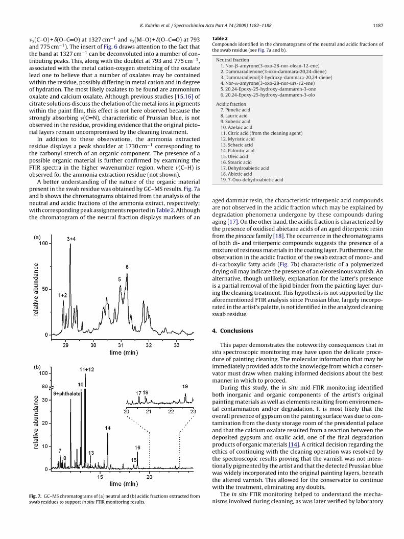
Fig. 6 shows the transmission FTIR spectrum taken of the ammo-ia extracted phase of the cleaning residue. As it is expected to findraces of triammonium citrate from the first step of cleaning, itspectrum has been included for comparison. A number of peaks,ot attributed to the triammonium citrate, have been highlighted
n the residue spectrum with corresponding tentative assignments.otably, the removal of gypsum from the painting can be inferredn the basis of characteristic bands (ı(H2O) at 1627 cm−1 and �3t 1126 cm−1
). The identification of nitrates is based on the intensebsorption band at 1385 cm−1 (double degenerate anti-symmetricO stretching mode �3), which is present in all nitrate salts withoutnabling further discrimination of the corresponding cations. Both
ulphates and nitrates are most likely present due to dry depositionf salts on the painting surface [14].The removal of a metal oxalate can be made on theasis of multiple characteristic bands (�a(C O) at 1627 cm−1,
Fig. 6. Transmittance FTIR spectrum of swab NH3 extract. Insert shows magnifica-tion of �s(C–O) region.
ca Acta Part A 74 (2009) 1182–1188 1187
�attalwoocwsor
rtpFo
panwt
Fs
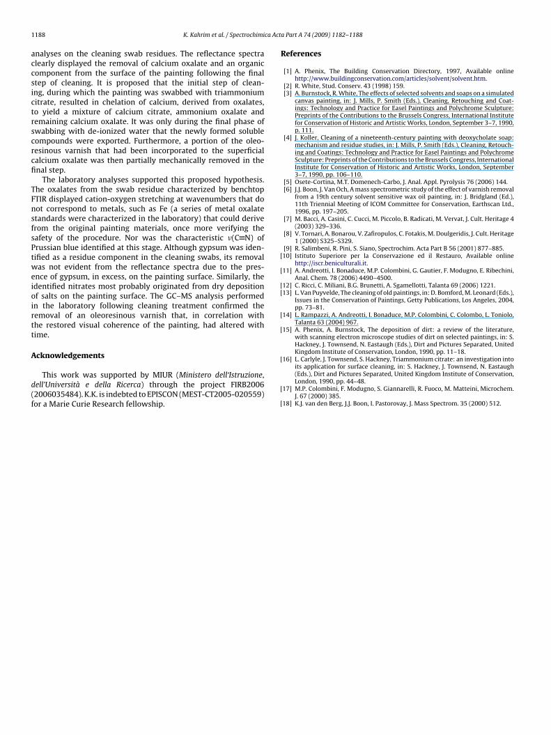
Table 2Compounds identified in the chromatograms of the neutral and acidic fractions ofthe swab residue (see Fig. 7a and b).
Neutral fraction1. Nor-�-amyrone(3-oxo-28-nor-olean-12-ene)2. Dammaradienone(3-oxo-dammara-20,24-diene)3. Dammaradienol(3-hydroxy-dammara-20,24-diene)4. Nor-�-amyrone(3-oxo-28-nor-urs-12-ene)5. 20,24-Epoxy-25-hydroxy-dammaren-3-one6. 20,24-Epoxy-25-hydroxy-dammaren-3-olo
Acidic fraction7. Pimelic acid8. Lauric acid9. Suberic acid10. Azelaic acid11. Citric acid (from the cleaning agent)12. Myristic acid13. Sebacic acid14. Palmitic acid15. Oleic acid16. Stearic acid
K. Kahrim et al. / Spectrochimi
s(C–O) + ı(O–C O) at 1327 cm−1 and �s(M–O) + ı(O–C O) at 793nd 775 cm−1). The insert of Fig. 6 draws attention to the fact thathe band at 1327 cm−1 can be deconvoluted into a number of con-ributing peaks. This, along with the doublet at 793 and 775 cm−1,ssociated with the metal cation-oxygen stretching of the oxalateead one to believe that a number of oxalates may be contained
ithin the residue, possibly differing in metal cation and in degreef hydration. The most likely oxalates to be found are ammoniumxalate and calcium oxalate. Although previous studies [15,16] ofitrate solutions discuss the chelation of the metal ions in pigmentsithin the paint film, this effect is not here observed because the
trongly absorbing �(C N), characteristic of Prussian blue, is notbserved in the residue, providing evidence that the original picto-ial layers remain uncompromised by the cleaning treatment.
In addition to these observations, the ammonia extractedesidue displays a peak shoulder at 1730 cm−1 corresponding tohe carbonyl stretch of an organic component. The presence of aossible organic material is further confirmed by examining theTIR spectra in the higher wavenumber region, where �(C–H) isbserved for the ammonia extraction residue (not shown).
A better understanding of the nature of the organic materialresent in the swab residue was obtained by GC–MS results. Fig. 7a
nd b shows the chromatograms obtained from the analysis of theeutral and acidic fractions of the ammonia extract, respectively;ith corresponding peak assignments reported in Table 2. Althoughhe chromatogram of the neutral fraction displays markers of an
ig. 7. GC–MS chromatograms of (a) neutral and (b) acidic fractions extracted fromwab residues to support in situ FTIR monitoring results.
17. Dehydroabietic acid18. Abietic acid19. 7-Oxo-dehydroabietic acid
aged dammar resin, the characteristic triterpenic acid compoundsare not observed in the acidic fraction which may be explained bydegradation phenomena undergone by these compounds duringaging [17]. On the other hand, the acidic fraction is characterized bythe presence of oxidised abietane acids of an aged diterpenic resinfrom the pinacae family [18]. The occurrence in the chromatogramsof both di- and triterpenic compounds suggests the presence of amixture of resinous materials in the coating layer. Furthermore, theobservation in the acidic fraction of the swab extract of mono- anddi-carboxylic fatty acids (Fig. 7b) characteristic of a polymerizeddrying oil may indicate the presence of an oleoresinous varnish. Analternative, though unlikely, explanation for the latter’s presenceis a partial removal of the lipid binder from the painting layer dur-ing the cleaning treatment. This hypothesis is not supported by theaforementioned FTIR analysis since Prussian blue, largely incorpo-rated in the artist’s palette, is not identified in the analyzed cleaningswab residue.
4. Conclusions
This paper demonstrates the noteworthy consequences that insitu spectroscopic monitoring may have upon the delicate proce-dure of painting cleaning. The molecular information that may beimmediately provided adds to the knowledge from which a conser-vator must draw when making informed decisions about the bestmanner in which to proceed.
During this study, the in situ mid-FTIR monitoring identifiedboth inorganic and organic components of the artist’s originalpainting materials as well as elements resulting from environmen-tal contamination and/or degradation. It is most likely that theoverall presence of gypsum on the painting surface was due to con-tamination from the dusty storage room of the presidential palaceand that the calcium oxalate resulted from a reaction between thedeposited gypsum and oxalic acid, one of the final degradationproducts of organic materials [14]. A critical decision regarding theethics of continuing with the cleaning operation was resolved bythe spectroscopic results proving that the varnish was not inten-tionally pigmented by the artist and that the detected Prussian blue
was widely incorporated into the original painting layers, beneaththe altered varnish. This allowed for the conservator to continuewith the treatment, eliminating any doubts.The in situ FTIR monitoring helped to understand the mecha-nisms involved during cleaning, as was later verified by laboratory
1 ca Act
accsictrscrcfi
TFnsfsPtweioirtt
A
d(f
[
[
[[
[
[
[
188 K. Kahrim et al. / Spectrochimi
nalyses on the cleaning swab residues. The reflectance spectralearly displayed the removal of calcium oxalate and an organicomponent from the surface of the painting following the finaltep of cleaning. It is proposed that the initial step of clean-ng, during which the painting was swabbed with triammoniumitrate, resulted in chelation of calcium, derived from oxalates,o yield a mixture of calcium citrate, ammonium oxalate andemaining calcium oxalate. It was only during the final phase ofwabbing with de-ionized water that the newly formed solubleompounds were exported. Furthermore, a portion of the oleo-esinous varnish that had been incorporated to the superficialalcium oxalate was then partially mechanically removed in thenal step.
The laboratory analyses supported this proposed hypothesis.he oxalates from the swab residue characterized by benchtopTIR displayed cation-oxygen stretching at wavenumbers that doot correspond to metals, such as Fe (a series of metal oxalatetandards were characterized in the laboratory) that could deriverom the original painting materials, once more verifying theafety of the procedure. Nor was the characteristic �(C N) ofrussian blue identified at this stage. Although gypsum was iden-ified as a residue component in the cleaning swabs, its removalas not evident from the reflectance spectra due to the pres-
nce of gypsum, in excess, on the painting surface. Similarly, thedentified nitrates most probably originated from dry depositionf salts on the painting surface. The GC–MS analysis performedn the laboratory following cleaning treatment confirmed theemoval of an oleoresinous varnish that, in correlation withhe restored visual coherence of the painting, had altered withime.
cknowledgements
This work was supported by MIUR (Ministero dell‘Istruzione,ell’Università e della Ricerca) through the project FIRB20062006035484). K.K. is indebted to EPISCON (MEST-CT2005-020559)or a Marie Curie Research fellowship.
[
[
a Part A 74 (2009) 1182–1188
References
[1] A. Phenix, The Building Conservation Directory, 1997, Available onlinehttp://www.buildingconservation.com/articles/solvent/solvent.htm.
[2] R. White, Stud. Conserv. 43 (1998) 159.[3] A. Burnstock, R. White, The effects of selected solvents and soaps on a simulated
canvas painting, in: J. Mills, P. Smith (Eds.), Cleaning, Retouching and Coat-ings: Technology and Practice for Easel Paintings and Polychrome Sculpture:Preprints of the Contributions to the Brussels Congress, International Institutefor Conservation of Historic and Artistic Works, London, September 3–7, 1990,p. 111.
[4] J. Koller, Cleaning of a nineteenth-century painting with deoxycholate soap:mechanism and residue studies, in: J. Mills, P. Smith (Eds.), Cleaning, Retouch-ing and Coatings: Technology and Practice for Easel Paintings and PolychromeSculpture: Preprints of the Contributions to the Brussels Congress, InternationalInstitute for Conservation of Historic and Artistic Works, London, September3–7, 1990, pp. 106–110.
[5] Osete-Cortina, M.T. Domenech-Carbo, J. Anal. Appl. Pyrolysis 76 (2006) 144.[6] J.J. Boon, J. Van Och, A mass spectrometric study of the effect of varnish removal
from a 19th century solvent sensitive wax oil painting, in: J. Bridgland (Ed.),11th Triennial Meeting of ICOM Committee for Conservation, Earthscan Ltd.,1996, pp. 197–205.
[7] M. Bacci, A. Casini, C. Cucci, M. Piccolo, B. Radicati, M. Vervat, J. Cult. Heritage 4(2003) 329–336.
[8] V. Tornari, A. Bonarou, V. Zafiropulos, C. Fotakis, M. Doulgeridis, J. Cult. Heritage1 (2000) S325–S329.
[9] R. Salimbeni, R. Pini, S. Siano, Spectrochim. Acta Part B 56 (2001) 877–885.10] Istituto Superiore per la Conservazione ed il Restauro, Available online
http://iscr.beniculturali.it.11] A. Andreotti, I. Bonaduce, M.P. Colombini, G. Gautier, F. Modugno, E. Ribechini,
Anal. Chem. 78 (2006) 4490–4500.12] C. Ricci, C. Miliani, B.G. Brunetti, A. Sgamellotti, Talanta 69 (2006) 1221.13] L. Van Puyvelde, The cleaning of old paintings, in: D. Bomford, M. Leonard (Eds.),
Issues in the Conservation of Paintings, Getty Publications, Los Angeles, 2004,pp. 73–81.
14] L. Rampazzi, A. Andreotti, I. Bonaduce, M.P. Colombini, C. Colombo, L. Toniolo,Talanta 63 (2004) 967.
15] A. Phenix, A. Burnstock, The deposition of dirt: a review of the literature,with scanning electron microscope studies of dirt on selected paintings, in: S.Hackney, J. Townsend, N. Eastaugh (Eds.), Dirt and Pictures Separated, UnitedKingdom Institute of Conservation, London, 1990, pp. 11–18.
16] L. Carlyle, J. Townsend, S. Hackney, Triammonium citrate: an investigation into
its application for surface cleaning, in: S. Hackney, J. Townsend, N. Eastaugh(Eds.), Dirt and Pictures Separated, United Kingdom Institute of Conservation,London, 1990, pp. 44–48.17] M.P. Colombini, F. Modugno, S. Giannarelli, R. Fuoco, M. Matteini, Microchem.J. 67 (2000) 385.
18] K.J. van den Berg, J.J. Boon, I. Pastorovay, J. Mass Spectrom. 35 (2000) 512.