The amnestic syndrome of hippocampal type in Alzheimer's disease: an MRI study
10
Journal of Alzheimer’s Disease 22 (2010) 285–294 285 DOI 10.3233/JAD-2010-091150 IOS Press The Amnestic Syndrome of Hippocampal type in Alzheimer’s Disease: An MRI Study Marie Sarazin a,b,1,* , Val´ erie Chauvir´ e a,c,1 , Emilie Gerardin d , Olivier Colliot d , Serge Kinkingn´ ehun a,g , Leonardo Cruz de Souza a,b , Laurence Hugonot-Diener a , Line Garnero d , St´ ephane Leh´ ericy a,e,f , Marie Chupin d,1 and Bruno Dubois a,b a CRICM UPMC Inserm UMR 975 Piti´ e-Salpˆ etri` ere Hospital, Paris, France b Research and Resource Memory Centre, Piti´ e-Salpˆ etri` ere Hospital, Paris, France c Research and Resource Memory Centre, University Hospital, rue Larrey, France d Cognitive Neuroscience and Brain Imaging Laboratory, CNRS UPR 640-LENA, UPMC Univ Paris, France e Neuroradiology, Piti´ e-Salpˆ etri` ere Hospital, Paris, France f Center for NeuroImaging Research – CENIR, Piti´ e-Salpˆ etri` ere Hospital, University Pierre and Marie Curie, Paris, France g e(ye)BRAIN, 3 bis rue Maurice Grandcoing, France Handling Associate Editor: Eric Salmon Accepted 14 June 2010 Abstract. The Free and Cued Selective Reminding Test (FCSRT) is a verbal episodic memory test used to identify patients with mild Alzheimer’s disease (AD). The present study investigates the relationships between performance on FCSRT and grey matter atrophy assessed with structural MRI in patients with AD. Three complementary MRI-based analyses (VBM analysis, ROI-based analysis, and three-dimensional hippocampal surface-based shape analysis) were performed in 35 patients with AD to analyze correlations between regional atrophy and their scores for episodic memory using the FCSRT. With VBM analysis, the total score on the FCSRT was correlated with left medial temporal lobe atrophy including the left hippocampus but also the thalami. In addition, using ROI-based analysis, the total recall score on the FCSRT was correlated with the left hippocampal volume. With three-dimensional hippocampal surface-based shape analysis, both free recall and total recall scores were correlated with regions corresponding approximately to the CA1 field. No correlation was found with short term memory scores using any of these methods of analysis. In AD, the FCSRT may be considered as a useful clinical marker of memory disorders due to medial temporal damage, specially the CA1 field of the hippocampus. Keywords: Alzheimer’s disease, assessment of cognitive disorders/dementia, cognitive neuropsychology in dementia, memory, MRI 1 These authors contributed equally. * Correspondence to: Dr. Marie Sarazin, F´ ed´ eration des maladies du Syst` eme Nerveux, Research and Resource Memory Centre, Pavil- lon Paul Castaigne, Hˆ opital de la Salpˆ etri` ere, 47 Bd de l’Hˆ opital, 75013 Paris, France. Tel.: +33 1 42 16 19 86; Fax: +33 1 42 16 27 39; E-mail: [email protected]. INTRODUCTION Increasing diagnostic accuracy of patients with mild AD will be crucial in a near future with the development of drugs aimed at slowing Alzheimer’s disease (AD) progression. New criteria for early diagnosis were re- cently proposed that include a core criterion of signifi- cant episodic memory impairment together with one or more supportive criteria from structural and molecular ISSN 1387-2877/10/$27.50 2010 – IOS Press and the authors. All rights reserved
-
Upload
independent -
Category
Documents
-
view
0 -
download
0
Transcript of The amnestic syndrome of hippocampal type in Alzheimer's disease: an MRI study
Journal of Alzheimer’s Disease 22 (2010) 285–294 285DOI 10.3233/JAD-2010-091150IOS Press
The Amnestic Syndrome of Hippocampaltype in Alzheimer’s Disease: An MRI Study
Marie Sarazina,b,1,∗, Valerie Chauvirea,c,1, Emilie Gerardind, Olivier Colliotd, Serge Kinkingnehuna,g,Leonardo Cruz de Souzaa,b, Laurence Hugonot-Dienera, Line Garnerod, Stephane Lehericya,e,f,Marie Chupind,1 and Bruno Duboisa,b
aCRICM UPMC Inserm UMR 975 Pitie-Salpetriere Hospital, Paris, FrancebResearch and Resource Memory Centre, Pitie-Salpetriere Hospital, Paris, FrancecResearch and Resource Memory Centre, University Hospital,rue Larrey, FrancedCognitive Neuroscience and Brain Imaging Laboratory, CNRSUPR 640-LENA, UPMC Univ Paris, FranceeNeuroradiology, Pitie-Salpetriere Hospital, Paris, FrancefCenter for NeuroImaging Research – CENIR, Pitie-Salpetriere Hospital, University Pierre and Marie Curie,Paris, Francege(ye)BRAIN, 3 bis rue Maurice Grandcoing, France
Handling Associate Editor: Eric Salmon
Accepted 14 June 2010
Abstract. The Free and Cued Selective Reminding Test (FCSRT) is a verbal episodic memory test used to identify patients withmild Alzheimer’s disease (AD). The present study investigates the relationships between performance on FCSRT and greymatteratrophy assessed with structural MRI in patients with AD. Three complementary MRI-based analyses (VBM analysis, ROI-basedanalysis, and three-dimensional hippocampal surface-based shape analysis) were performed in 35 patients with AD to analyzecorrelations between regional atrophy and their scores forepisodic memory using the FCSRT. With VBM analysis, the totalscore on the FCSRT was correlated with left medial temporal lobe atrophy including the left hippocampus but also the thalami.In addition, using ROI-based analysis, the total recall score on the FCSRT was correlated with the left hippocampal volume.With three-dimensional hippocampal surface-based shape analysis, both free recall and total recall scores were correlated withregions corresponding approximately to the CA1 field. No correlation was found with short term memory scores using any ofthese methods of analysis. In AD, the FCSRT may be consideredas a useful clinical marker of memory disorders due to medialtemporal damage, specially the CA1 field of the hippocampus.
Keywords: Alzheimer’s disease, assessment of cognitive disorders/dementia, cognitive neuropsychology in dementia, memory,MRI
1These authors contributed equally.∗Correspondence to: Dr. Marie Sarazin, Federation des maladies
du Systeme Nerveux, Research and Resource Memory Centre, Pavil-lon Paul Castaigne, Hopital de la Salpetriere, 47 Bd de l’Hopital,75013 Paris, France. Tel.: +33 1 42 16 19 86; Fax: +33 1 42 16 2739; E-mail: [email protected].
INTRODUCTION
Increasing diagnostic accuracy of patients with mildAD will be crucial in a near future with the developmentof drugs aimed at slowing Alzheimer’s disease (AD)progression. New criteria for early diagnosis were re-cently proposed that include a core criterion of signifi-cant episodic memory impairment together with one ormore supportive criteria from structural and molecular
ISSN 1387-2877/10/$27.50 2010 – IOS Press and the authors. All rights reserved

286 M. Sarazin et al. / The Amnestic Syndrome of Hippocampal typein Alzheimer’s Disease: An MRI Study
neuroimaging and cerebrospinal fluid (CSF) biomark-ers [1]. The use of memory test remains a first step forthe diagnosis and it is essential to choose tests that candetect the specific features of memory disorders due toearly hippocampal damage.
Previous studies on the relationships between hip-pocampal volume and episodic memory performancein AD found very inconstant correlations [2–7]. Theseapparent discrepancies may be explained by differencesin the episodic memory tests that are chosen in the stud-ies, including the control of encoding and the existenceof cueing or recognition procedures. Only few stud-ies showed correlations between verbal memory andthe left entorhinal cortex (when verbal memory wasassessed with either the Rey or the CERAD learninglists) [8,9] or the hippocampus (when verbal memorywas assessed with the Free and Cued Selective Remind-ing Test (FCSRT) [2] or with an word recall test [10]),whereas in another study, a low memory performancewas correlated with the atrophy of the temporal polarcortex and frontal areas (when verbal memory was as-sessed with the California Verbal Learning Test) [11].
In a previous longitudinal study with the follow-upof 251 patients with mild cognitive impairment (MCI),we showed that both free and total recall scores as-sessed by the FCSRT [12] were able to detect the spe-cific memory deficit of AD, even at a prodromal, pre-dementia stage, with a high sensitivity and specifici-ty [13]. The efficacy of the test is probably due tothe use of semantic categories which permit to con-trol for an effective encoding of the verbal items andto facilitate the retrieval of stored information. Con-sequently, the FCRST can isolate storage deficits dueto involvement of medial temporal structures that char-acterizes the early neuropathologic changes of AD [2,14,15] (so-called “amnestic syndrome of the medialtemporal type”) from other memory disorders due toattention disorders or retrieval impairment. Memoryscores of the FCSRT are therefore expected to be main-ly correlated with the hippocampal volume assessedwith structural MRI in patients with AD.
The aim of this study was to analyze in mild AD cor-relations between episodic memory performance, as-sessed by the FCSRT, and regional grey matter volumeusing three MRI anatomical imaging complementarymethods; 1) with voxel-based morphometry (VBM)analysis, because this method has the advantage of con-sidering grey matter atrophy in the whole brain withoutregional preferences; 2) with region of interest (ROI)-based anatomical analysis using an automatic methodfor the measurement of hippocampal volume; 3) with
a three-dimensional hippocampal surface-based shapeanalysis for the study of specific anatomical subregions.Correlation analysis results were compared with thoseobtained from short term memory scores.
METHODS
Subjects
Thirty-five patients with AD were recruited fromthe Memory Clinic of the Salpetriere Hospital. To beincluded in this study, patients had to: 1) fulfill theNational Institute of Neurological and CommunicationDisorders and Stroke/Alzheimer Disease and RelatedDisorders Association (NINCDS-ADRDA) criteria forprobable AD [16]; 2) have a score on the Clinical De-mentia Rating Scale (CDR)> 0.5 [17]; 3) present noclinical or neuroimaging evidence of focal lesions; 4)present no cortical or subcortical vascular lesions onMRI; 5) have no medical conditions that would inter-fere with cognitive performance; and 6) have no majordepression . All subjects were living in the local com-munity and all were right-handed. Each patient signedan informed consent after the nature of the procedureshad been fully explained. The study was approved bythe Ethics Committee of the Salpetriere Hospital. Allpatients underwent the same clinical and neuroimagingprocedures.
Procedures
Neuropsychological assessmentAt baseline, all patients were tested using a stan-
dardized neuropsychological battery, including the Mi-ni Mental State Examination (MMSE) for global cog-nition [18], FCRST for verbal episodic memory [19],and the digit span test (forward and backward condi-tions) for verbal short-term memory. The FCSRT wasselected because it is based on a semantic relationshipbetween the presented items and their categories (e.g.,what is the name of the fruit?), which allowed us tocontrol for an initial and effective registration of thelist of words, and secondarily to facilitate the retrievalfrom stored information. The FCSRT was adminis-tered according to the procedure described by Groberand Buschke [19,20] modified with verbal items. The16 items to be learned were presented as sets of fouritems each on successive cards. Items were represent-ed in each quadrant by a word (e.g., grapes) that cor-responds with a unique category cue (e.g., fruit). The

M. Sarazin et al. / The Amnestic Syndrome of Hippocampal typein Alzheimer’s Disease: An MRI Study 287
subject was asked to name aloud and point to each itemafter its cue was orally presented. In order to control forencoding, after all four items were correctly identified,the card was removed and immediate cued recall of thefour items was tested by presenting the cues again inorder to control for encoding. Once immediate recallfor a set of four items was completed, the next set offour items was presented. This first phase of the testallows for controlling encoding and provides a scorecalled immediate recall. The memory phase was thenperformed using three successive recall trials, each pre-ceded by 20 s during which the subjects were asked tocount backward to obtain recall from long-term mem-ory. Each recall trial consisted of two parts. First, eachsubject had up to 2 min to freely recall as many items aspossible. Next, an orally presented semantic category(e.g., “what was the name of the fruit?”) was providedfor those items that were not spontaneously retrieved.This provided a free recall score (out of 48) and a totalrecall score, which was the sum of free and cued recall(out of 48).
MRI methodsMRI data acquisition
MRI acquisition was performed on a 1.5 Tesla sys-tem (GE Medical Systems, Milwaukee, WI, USA).Anatomical high resolution scans were acquired us-ing a three-dimensional acquisition (TR/TE/flip angle:23 ms/5 ms/35◦, 256× 256 matrix; voxel size, 1× 1×
1.5 mm3) using a standard head coil. To prevent invol-untary head movements inside the magnet, the subject’shead was taped on the forehead.
Data processing for VBMWe used Voxel Based Morphometry (VBM) in order
to evaluate the correlation between the scores obtainedby subjects on testing and the grey matter density ontheir anatomical MRI scans. Data processing and anal-ysis was performed using statistical parametric map-ping software (SPM2, Welcome Laboratory of the De-partment of Cognitive Neurology, Institute of Neurol-ogy, London, UK, http://www.fil.ion.ucl.ac.uk/spm/)with MATLAB version 7.0.1 (The Mathworks, Inc.,MA, USA). To analyze brain volumes, we used an op-timized VBM procedure [21–23]. The image data setswere analyzed using the following automated imageprocessing steps prior to statistical analysis. The MRIscans of AD patients were used for creating customtemplates. Each T1 – weighted MR volume was spa-tially normalized with an affine registration to the T1
Montreal Neurological Institute (MNI) template. Sub-
sequently, the normalized scans were averaged, andthe resulting mean image was smoothed with an 8-mmfull width at half maximum (FWHM) Gaussian Ker-nel. A study-specific GM template was created ac-cording to the following steps: spatial normalizationof all structural images to the customized whole braintemplate, segmentation into gray matter, and brain ex-traction (for removing non-brain voxels from the graymatter images). The extracted GM images were thensmoothed with a 10-mm FWHM isotropic Gaussiankernel. The resulting smoothed, normalized regionscontained the average amount of gray matter within aregion surrounding a voxel. Following the creation of astudy-specific gray matter template, the optimized pro-tocol was subsequently applied to the original data [22].Briefly, the protocol included 1) segmentation of GM,white matter (WM), and CSF images and removal ofnon-brain voxels in the native space; 2) estimation ofthe nonlinear normalization parameters using the GMtemplate previously built; 3) application of these pa-rameters to the original whole-brain MR image, whichwas then resliced to a voxel size of 1× 1 × 1 mm3;and 4) segmentation of the normalized image into GM,WM, and CSF, using the customized prior probabilitymap. The GM probability maps were modulated bythe Jacobian determinant and then smoothed with a 12-mm FWHM isotropic Gaussian kernel. The resultingsmoothed, normalized regions contained the averageamount of GM within a region surrounding a voxel.Total intracranial volume (TIV) was defined by the sumof GM, white matter and CSF extracted in the processfor each subject.
Automatic hippocampal volumetryThe segmentation of the hippocampus was per-
formed using an automatic method [24–26]. Thismethod has been validated by comparison with manualtracing in young healthy participants and patients withAD and has been proven to be reliable, fast and accu-rate [27]. It has also been applied successfully to auto-matically discriminate between patients with AD andelderly controls [26,27]. This approach extracts bothhippocampus and amygdala based on simultaneous re-gion deformation, with competition between these twostructures. The method includes prior knowledge ofthe locations of the amygdala and hippocampus, de-rived from a probabilistic atlas and from the relativepositions of these structures with respect to anatomicallandmarks that are automatically identified during thedeformationprocess. Segmentations were evaluated bya trained operator who was blind to all clinical data.

288 M. Sarazin et al. / The Amnestic Syndrome of Hippocampal typein Alzheimer’s Disease: An MRI Study
Three-dimensional hippocampal surface-based shapeanalysis
To investigate the specific anatomical contributionwithin the hippocampus, we performed a statistical 3Dsurface-based shape analysis relying on the sphericalharmonics (SPHARM) approach [28,29]. The anal-ysis was performed using the SPHARM-PDM soft-ware developed at the University of North Carolina andthe National Alliance for Medical Imaging Comput-ing (http://www.namic.org/Wiki/index.php/Algorithm:UNC: Shape Analysis). SPHARM is a mathemati-cal framework to parameterize surfaces with sphericaltopology, which can be seen as a 3D analog of Fouri-er series expansion. In brief, the SPHARM approachrelies on the following steps, which were performed inour analysis. First, hippocampal segmentations wereconverted to surface meshes, and a spherical parame-terization was computed, creating a one-to-one map-ping between each point on the surface and each pointon a sphere. The surface was expanded into a seriesof spherical harmonics, which were then truncated at agiven degree (we chose a degree ofk = 20, which re-sults in an acceptable degree of smoothing). The coeffi-cients of the series expansion were normalized in orderto make them invariant to rotation,translation and scale.The SPHARM parameterization was transformed in-to a triangulated surface (called the SPHARM-PDM),based on a uniform subdivision of the spherical param-eterization. Each hippocampus was described by a setof n = 4002 landmarks. Finally, the SPHARM-PDMwas spatially aligned using rigid Procustes alignment,giving a one-to-one mapping between surface pointsfor each pair of hippocampi.
Statistical analysis
VBM analysisData analysis was performed using SPM software.
We performed one linear regression per neuropsycho-logical test for all the subjects. In order to test for thepossible influence of disease severity, we conducteda multiple regression analysis including age, total in-tracranial volume (TIV) and MMSE as nuisance vari-ables. All the analyses were performed using a statisti-cal threshold ofp 6 0.005 for the voxels and a clustercorrection ofp 6 0.05.
Hippocampal volume analysisAll statistical analyses were performed using R soft-
ware for Windows version 2.0.1 (R Development CoreTeam 2004). Hippocampal volume measurements were
normalized with respect to TIV. The relations betweenhippocampal volume measures and neuropsychologi-cal tests (immediate, free and total recall of FCSRT,short term memory scores) were examined using mul-tiple linear regression analysis, controlling for age andMMSE. Bonferroni correction was applied.
Three-dimensional hippocampal surface-based shapeanalysis
All hippocampal surfaces were averaged in order tocompute a mean hippocampal template. The signeddistance between each individual hippocampal surfaceand this template was computed at each vertex. Wesearched for correlations between neuropsychologi-cal tests and local hippocampal atrophy by comput-ing Pearson’s correlation coefficient between a givenneuropsychological measure and the signed distance ateach vertex, using the intracranial volume, age, andMMSE as covariates. In the 3D statistical maps, re-gions with p < 0.05 were considered as statisticallysignificant.
RESULTS
Characteristics of patients
Age was 72.8± 5.6 (mean± SD) years, with agender ratio (M/F) of 22/13. Education level was 12±
3 years. MMSE score was 22.8± 3.4 (range 13–29).The CDR score was 0.5 forn = 1 patient, CDR= 1for 31 patients and CDR= 2 for 3 patients.
Immediate recall score on the FCSRT was 11.2±
2.7 (maximum= 16, norms for age and education=14 to 16); free recall score was 7.6± 5.1 (maximum=48, norms= 24 to 29); total (free+ cued) recall scorewas 23.6± 10 (maximum= 48; norms= 45 to 48);number of intrusions was 12.6± 8.9 (norms= 1 to 2).For details of norms of the FCSRT in elderly adults,see [20,30].
Digit span test forwards score was 5.1± 0.9; digitspan test backward was 3.7± 0.6.
Correlations between episodic memory scores andMRI
VBM methodScores of FCSRT total recall were significantly cor-
related with a large cluster of 17,679 voxels includedthe left medial temporal lobe and the thalami. Moreprecisely, this area included on the left: hippocampus,

M. Sarazin et al. / The Amnestic Syndrome of Hippocampal typein Alzheimer’s Disease: An MRI Study 289
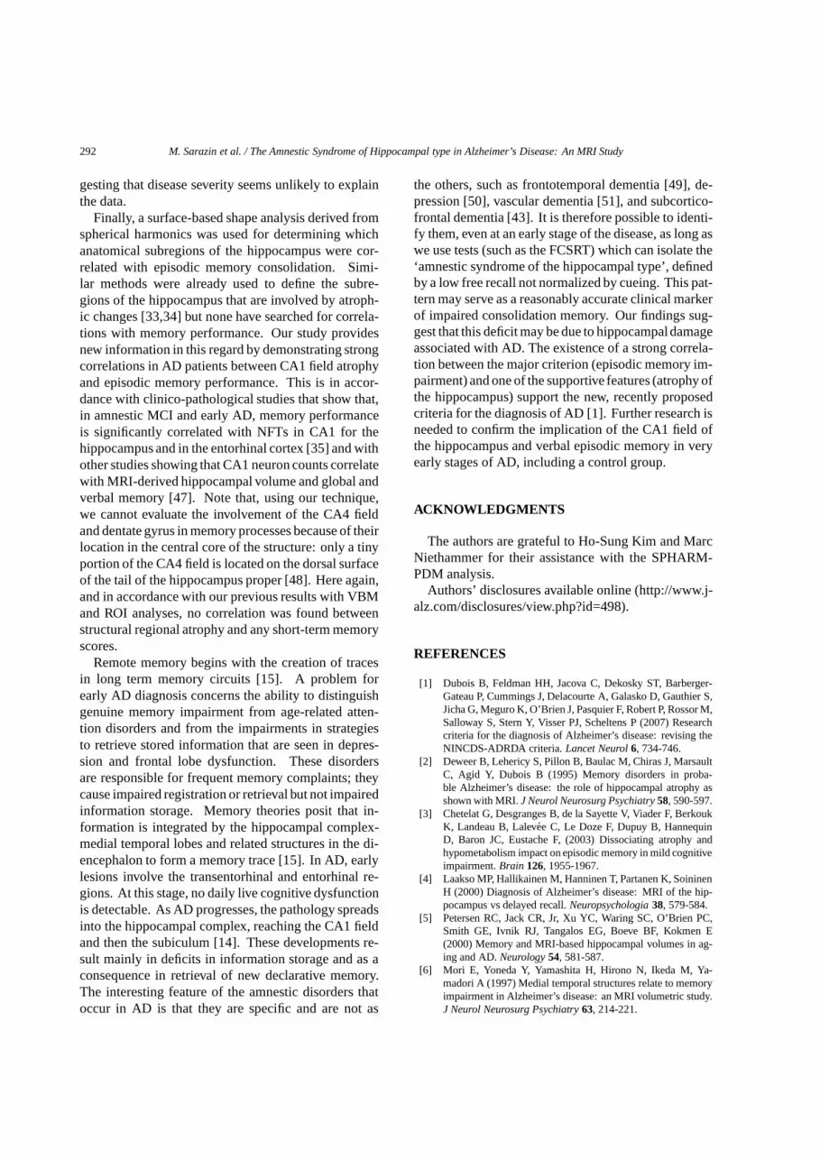
Fig. 1. Areas of significant correlation between total recall of theFCSRT∗ and gray matter concentration in AD patients. Statisti-cal maps of correlation were thresholded atp 6 0.005 using anadjusted cutoff ofp 6 0.05 for cluster extension. The cluster isdisplayed using glass brain representation (neurologicalconvention)and anatomical images (radiological convention) from the average ofnormalized T1-weighted images AD patients. The regression modelincluded as covariate parameters total intracranial volume (TIV), ageand MMSE.∗Free and Cued Selective Reminding Test.
parahippocampal gyrus, entorhinal cortex, amygdalaand thalamus and on the right only the thalamus.
Including the MMSE score as a covariate of no in-terest, the cluster of significant correlation was small-er (10,013 voxels) included only on the left side: hip-pocampus, parahippocampal gyrus, entorhinal cortex,amygdala, and thalamus (see Fig. 1).
There was a trend (p = 0.082) toward a correlationbetween scores of FCSRT free recall and a cluster of7151 voxels located on the left hippocampus and leftparahippocampus.
Automatic hippocampal volumetryThe results of the correlation analysis between nor-
malized hippocampal volumes and neuropsychologicalscores are displayed in Table 1. Using Bonferroni cor-rection, the total recall of the FCSRT was significantlycorrelated with the left hippocampal volume.
Three-dimensional hippocampal surface-based shapeanalysis
Three-dimensional hippocampal maps showed sig-nificant positive correlations between scores in bothfree and total recall on the FSCRT and atrophy in re-gions approximately corresponding to the CA1 field ofthe left hippocampus, and to a smaller extent with theright hippocampus (Figs 2a and 2b). A figure with atemplate of hippocampal subregions can be found inApostolova et al. [31].
No significant correlation was found with the imme-diate recall of the FCSRT score.
Correlations between short term memory scores andMRI
No significant correlation was found between thedigit forward span and backward scores and the volumeof brain anatomical structures with any of the threeMRI methods.
To confirm the absence of correlations was not dueto the small amplitude of these scores, we determineda short term memory composite score which was thesum of the digit forward span and backward scores. Nocorrelation was found between this score and the threeMRI methods.
DISCUSSION
The study shows significant correlations between thetotal recall score of the FCSRT and left medial tem-poral lobe atrophy assessed by VBM analysis, and theleft hippocampal volume assessed by automatic vol-umetry method. In addition, both free and total recallscores were correlated with local hippocampal atrophyassessed by the three dimensional surface based shapeanalysis. Interestingly, these correlations within thehippocampus were specially localized in the CA1 field,a region known to be involved in memory storage [32]and to be early affected by AD neurobiology [33–36].The results suggest that both encoding and storage pro-cesses are dependent from the hippocampal structures.In agreement with previous evidence, this study high-

290 M. Sarazin et al. / The Amnestic Syndrome of Hippocampal typein Alzheimer’s Disease: An MRI Study
Fig. 2. (a): Areas of correlation between free recall on the FCSRT* and hippocampal surface. (b): Areas of correlation between total recall onthe FCSRT* and hippocampal surface. (Colors are visible in the electronic version of this article at http://dx.doi.org/10.3233/JAD-2010-091150)

M. Sarazin et al. / The Amnestic Syndrome of Hippocampal typein Alzheimer’s Disease: An MRI Study 291
Table 1Correlations between normalized hippocampal volumes and neuropsychological scores. Normalized hippocampalvolumes were corrected for the effects of age and MMSE; Bonferroni correction was applied
Neuropsychological tests Left hippocampus Right hippocampusr p value r p value
Long term memory FCSRT: Free recall 0.35 NS 0.37 NSFCSRT: Total (free+ cued) recall 0.43 0.01 0.39 NS
Short term memory FCSRT: Immediate recall −0.01 NS −0.02 NSDigit span test forward condition 0.06 NS 0.03 NS
“r” corresponds to the adjusted coefficient of correlation.NS: not significative.
lights the role of the hippocampus in long term episodicmemory [31,43].
Overall, the main results of the study are the consis-tent correlations between episodic memory scores as-sessed by the FCSRT, especially the total recall, and theatrophy of hippocampus in patients with AD, while nocorrelation was found with short term memory scores.
Previous studies with VBM analysis showed incon-sistent results in this matter [8–11,36–42] and correla-tions were found with right and left hippocampus [3],left entorhinal cortex [8], parts of temporal polar cor-tex, orbital cortex and inferior frontal gyri [11] or ento-and perirhinal cortex [9,10]. Such discrepancies mayresult from the choice of the memory tests used in thesestudies. The neuropsychological paradigm of the FC-SRT seems particularly well suited in this respect be-cause it can distinguish the three different componentsof episodic memory:registration(by controlling thatall the items have been truly registered),storage(byproviding the semantic cues for facilitating the accessto stored information), andretrieval (by the sponta-neous recall of items after delay). Total recall, whichis the sum of the spontaneous and the cued recalls,is an interesting parameter to consider because it re-flects the amount of information that is stored by thepatient during the delay, the most important marker oflong term episodic memory. In agreement with pre-vious evidence, this study highlights the role of thehippocampus in long term episodic memory in linewith the hippocampal/subcortical-frontal dissociationaccording to which: i) the hippocampus is more in-volved in encoding and storage process and ii) thesubscortical-frontal axes are more involved in retrieval(33,44). It is noteworthy that the neuropsychologicalbattery of the present study did not include a non-cuedmemory test because of the risk of interferences be-tween different verbal stimuli.
In our study, the total recall of verbal items was cor-related with atrophy of the left hippocampus, in accor-dance with the expected role of the hippocampus in the
consolidation and retention of new verbal informationover delays [2,3,5,44–46].
In addition, VBM analysis showed that patients’ totalrecall score on the FCSRT was also associated with thevolume of other brain structures known to be involvedin consolidation of new verbal information, such as theleft parahippocampal gyrus, entorhinal cortex, and tha-lamus. All these structures are part of the polysynapticpathway of the neurobiological theory of memory: theparahippocampalcortex projects onto the hippocampusvia the entorhinal cortex; within the hippocampus, aset of intrinsic connections proceeds from the dentategyrus to the CA3 field and hence to the CA1 field. Themajor efferent hippocampal connections arise from thepyramidal cells of the CA1 field and project direct-ly or via the subiculum to the cerebral cortex. Hip-pocampal efferent fibers reach the anterior thalamic nu-clei through the fimbria-fornix system, the mammil-lary bodies, and the mammillo-thalamic tract; subse-quently, the main cortical projections from the anteriorthalamus are directed toward the posterior cingulate,retrospenial, and anterior cingulate cortices [32]. Thethresholds on VBM analysis might not be significantwith a FWE correction, but with a statistical thresholdof p 6 0.005 for the voxels and a cluster correction ofp 6 0.05, due to the limited number of patients in ourstudy. Therefore, these data have to be considered asexploratory.
VBM analysis was the first step of the study becauseit relies on noa priori. It demonstrates strong rela-tionships between episodic memory performance andhippocampal volume in patients with AD that were fur-ther reinforced with the results of the ROI-based anal-ysis using the fully automatic method for hippocampussegmentation. When compared to manual segmenta-tion, this new method has the advantage of reducingthe subjective component of the evaluation [24,26,27].Results from ROI analysis confirmed that the total re-call score of the FCSRT is correlated with the severityof hippocampal atrophy. Including MMSE scores as acovariate in the analysis did not modify the results, sug-
292 M. Sarazin et al. / The Amnestic Syndrome of Hippocampal typein Alzheimer’s Disease: An MRI Study
gesting that disease severity seems unlikely to explainthe data.
Finally, a surface-based shape analysis derived fromspherical harmonics was used for determining whichanatomical subregions of the hippocampus were cor-related with episodic memory consolidation. Simi-lar methods were already used to define the subre-gions of the hippocampus that are involved by atroph-ic changes [33,34] but none have searched for correla-tions with memory performance. Our study providesnew information in this regard by demonstrating strongcorrelations in AD patients between CA1 field atrophyand episodic memory performance. This is in accor-dance with clinico-pathological studies that show that,in amnestic MCI and early AD, memory performanceis significantly correlated with NFTs in CA1 for thehippocampus and in the entorhinal cortex [35] and withother studies showing that CA1 neuron counts correlatewith MRI-derived hippocampal volume and global andverbal memory [47]. Note that, using our technique,we cannot evaluate the involvement of the CA4 fieldand dentate gyrus in memory processes because of theirlocation in the central core of the structure: only a tinyportion of the CA4 field is located on the dorsal surfaceof the tail of the hippocampus proper [48]. Here again,and in accordance with our previous results with VBMand ROI analyses, no correlation was found betweenstructural regional atrophy and any short-term memoryscores.
Remote memory begins with the creation of tracesin long term memory circuits [15]. A problem forearly AD diagnosis concerns the ability to distinguishgenuine memory impairment from age-related atten-tion disorders and from the impairments in strategiesto retrieve stored information that are seen in depres-sion and frontal lobe dysfunction. These disordersare responsible for frequent memory complaints; theycause impaired registration or retrieval but not impairedinformation storage. Memory theories posit that in-formation is integrated by the hippocampal complex-medial temporal lobes and related structures in the di-encephalon to form a memory trace [15]. In AD, earlylesions involve the transentorhinal and entorhinal re-gions. At this stage, no daily live cognitive dysfunctionis detectable. As AD progresses, the pathology spreadsinto the hippocampal complex, reaching the CA1 fieldand then the subiculum [14]. These developments re-sult mainly in deficits in information storage and as aconsequence in retrieval of new declarative memory.The interesting feature of the amnestic disorders thatoccur in AD is that they are specific and are not as
the others, such as frontotemporal dementia [49], de-pression [50], vascular dementia [51], and subcortico-frontal dementia [43]. It is therefore possible to identi-fy them, even at an early stage of the disease, as long aswe use tests (such as the FCSRT) which can isolate the‘amnestic syndrome of the hippocampal type’, definedby a low free recall not normalized by cueing. This pat-tern may serve as a reasonably accurate clinical markerof impaired consolidation memory. Our findings sug-gest that this deficit may be due to hippocampaldamageassociated with AD. The existence of a strong correla-tion between the major criterion (episodic memory im-pairment) and one of the supportive features (atrophy ofthe hippocampus) support the new, recently proposedcriteria for the diagnosis of AD [1]. Further research isneeded to confirm the implication of the CA1 field ofthe hippocampus and verbal episodic memory in veryearly stages of AD, including a control group.
ACKNOWLEDGMENTS
The authors are grateful to Ho-Sung Kim and MarcNiethammer for their assistance with the SPHARM-PDM analysis.
Authors’ disclosures available online (http://www.j-alz.com/disclosures/view.php?id=498).
REFERENCES
[1] Dubois B, Feldman HH, Jacova C, Dekosky ST, Barberger-Gateau P, Cummings J, Delacourte A, Galasko D, Gauthier S,Jicha G, Meguro K, O’Brien J, Pasquier F, Robert P, Rossor M,Salloway S, Stern Y, Visser PJ, Scheltens P (2007) Researchcriteria for the diagnosis of Alzheimer’s disease: revising theNINCDS-ADRDA criteria.Lancet Neurol6, 734-746.
[2] Deweer B, Lehericy S, Pillon B, Baulac M, Chiras J, MarsaultC, Agid Y, Dubois B (1995) Memory disorders in proba-ble Alzheimer’s disease: the role of hippocampal atrophy asshown with MRI.J Neurol Neurosurg Psychiatry58, 590-597.
[3] Chetelat G, Desgranges B, de la Sayette V, Viader F, BerkoukK, Landeau B, Lalevee C, Le Doze F, Dupuy B, HannequinD, Baron JC, Eustache F, (2003) Dissociating atrophy andhypometabolism impact on episodic memory in mild cognitiveimpairment.Brain 126, 1955-1967.
[4] Laakso MP, Hallikainen M, Hanninen T, Partanen K, SoininenH (2000) Diagnosis of Alzheimer’s disease: MRI of the hip-pocampus vs delayed recall.Neuropsychologia38, 579-584.
[5] Petersen RC, Jack CR, Jr, Xu YC, Waring SC, O’Brien PC,Smith GE, Ivnik RJ, Tangalos EG, Boeve BF, Kokmen E(2000) Memory and MRI-based hippocampal volumes in ag-ing and AD.Neurology54, 581-587.
[6] Mori E, Yoneda Y, Yamashita H, Hirono N, Ikeda M, Ya-madori A (1997) Medial temporal structures relate to memoryimpairment in Alzheimer’s disease: an MRI volumetric study.J Neurol Neurosurg Psychiatry63, 214-221.

M. Sarazin et al. / The Amnestic Syndrome of Hippocampal typein Alzheimer’s Disease: An MRI Study 293
[7] Basso M, Yang J, Warren L, MacAvoy MG, Varma P, BronenRA, van Dyck CH (2006) Volumetry of amygdala and hip-pocampus and memory performance in Alzheimer’s disease.Psychiatry Res146, 251-261.
[8] Di Paola M, Macaluso E, Carlesimo GA, Tomaiuolo F, Wors-ley KJ, Fadda L, Caltagirone C (2007) Episodic memory im-pairment in patients with Alzheimer’s disease is correlatedwith entorhinal cortex atrophy A voxel-based morphometrystudy.J Neurol254, 774-781.
[9] Schmidt-Wilcke T, Poljansky S, Hierlmeier S, Hausner J,Ibach B (2009) Memory performance correlates with graymatter density in the ento-/perirhinal cortex and posterior hip-pocampus in patients with mild cognitive impairment andhealthy controls – a voxel based morphometry study.Neuroim-age47, 1914-1920.
[10] Leube DT, Weis S, Freymann K, Erb M, Jessen F, HeunR, Grodd W, Kircher TT (2008) Neural correlates of verbalepisodic memory in patients with MCI and Alzheimer’s dis-ease – a VBM study.Int J Geriatr Psychiatry23, 1114-1118.
[11] Duarte A, Hayasaka S, Du A, Schuff N, Jahng GH, Kramer J,Miller B, Weiner M (2006) Volumetric correlates of memoryand executive function in normal elderly, mild cognitive im-pairment and Alzheimer’s disease.Neurosci Lett406, 60-65.
[12] Grober E, Buschke H (1987) Genuine memory deficits in de-mentia.Dev Neuropsychol3, 13-36.
[13] Sarazin M, Berr C, De Rotrou J, Fabrigoule C, Pasquier F,Legrain S, Michel B, Puel M, Volteau M, Touchon J, Verny M,Dubois B (2007) Amnestic syndrome of the medial temporaltype identifies prodromal AD: a longitudinal study.Neurology69, 1859-1867.
[14] Delacourte A, David JP, Sergeant N, Buee L, Wattez A, Ver-mersch P, Ghozali F, Fallet-Bianco C, Pasquier F, Lebert F,Petit H, Di Menza C (1999) The biochemical pathway of neu-rofibrillary degeneration in aging and Alzheimer’s disease.Neurology52, 1158-1165.
[15] Moscovitch M, Nadel L, Winocur G, Gilboa A, RosenbaumRS (2006) The cognitive neuroscience of remote episodic,semantic and spatial memory.Curr Opin Neurobiol16, 179-190.
[16] McKhann G, Drachman D, Folstein M, Katzman R, Price D,Stadlan EM (1984) Clinical diagnosis of Alzheimer’s disease:report of the NINCDS-ADRDA Work Group under the aus-pices of Department of Health and Human Services Task Forceon Alzheimer’s Disease.Neurology34, 939-944.
[17] Morris JC (1993) The Clinical Dementia Rating (CDR): cur-rent version and scoring rules.Neurology43, 2412-2414.
[18] Folstein MF, Folstein SE, McHugh PR (1975) Mini-mentalstate A practical method for grading the cognitive state ofpatients for the clinician.J Psychiatr Res12, 189-198.
[19] Grober E, Buschke H, Crystal H, Bang S, Dresner R (1988)Screening for dementia by memory testing.Neurology38,900-903.
[20] Van der Linden M, Coyette F, Poitrenaud J, et les membresduGREMEM L’epreuve de rappel libre/rappel indice a 16 items(RL/RI – 16) In: Van der Linden M, Adam S, Agniel A etles membres du GREMEM, editors (2004) L’evaluation destroubles de la memoire Presentation de quatre tests de memoireepisodique (avec leuretalonnage) Marseille, France: Solal,25-47.
[21] Ashburner J, Friston KJ (2000) Voxel-based morphometry –the methods.Neuroimage11, 805-821.
[22] Good CD, Johnsrude IS, Ashburner J, Henson RN, Friston KJ,Frackowiak RS (2001) A voxel-based morphometric study of
ageing in 465 normal adult human brains.Neuroimage14,21-36.
[23] Kinkingnehun S, Sarazin M, Lehericy S, Guichart-GomezE,Hergueta T, Dubois B (2008) VBM anticipates the rate ofprogression of Alzheimer disease: a 3-year longitudinal study.Neurology70, 2201-2211
[24] Chupin M, Mukuna-Bantumbakulu AR, Hasboun D, BardinetE, Baillet S, Kinkingnehun S, Lemieux L, Dubois B, GarneroL (2007) Anatomically constrained region deformation for theautomated segmentation of the hippocampus and the amyg-dala: Method and validation on controls and patients withAlzheimer’s disease.Neuroimage34, 996-1019
[25] Chupin M, Hammers A, Bardinet E, Colliot O, Liu RSN, Dun-can JS, Garnero L, Lemieux L (2009) Automatic segmenta-tion of the hippocampus and the amygdala driven by hybridconstraints: Method and validation.Neuroimage46, 749-761.
[26] Colliot O, Chetelat G, Chupin M, Desgranges B, Magnin B,Benali H, Dubois B, Garnero L, Eustache F, Lehericy S (2008)Discrimination between Alzheimer disease, mild cognitiveim-pairment, and normal aging by using automated segmentationof the hippocampus.Radiology248, 194-201.
[27] Chupin M, Chetelat G, Lemieux L, Dubois B, Garnero L,Benali H, Eustache F, Lehericy S, Desgranges B, Colliot O(2008) Fully automatic hippocampus segmentation discrim-inates between early Alzheimer’s disease and normal aging.ISBI2008, 97-100.
[28] Styner M, Lieberman JA, Pantazis D, Gerig G (2004)Boundary and medial shape analysis of the hippocampus inschizophrenia.Med Image Anal8, 197-203.
[29] Styner M, Oguz I, Xu S, Brechbuehler C, Pantazis D, LevittJ, Shenton M, Gerig G (2006) Framework for the statisticalshape analysis of brain structures using SPHARM-PDM MIC-CAI Open Source Workshop. Insight Journal DSpace link:http://hdl.handle.net/1926/215.
[30] Amieva H, Carcaillon L, Rouze L’Alzit-Schuermans P, MilletX, Dartigues JF, Fabrigoule C (2007) [Cued and uncued mem-ory tests: norms in elderly adults from the 3 Cities epidemio-logical study].Rev Neurol163, 205-221.
[31] Apostolova LG, Dinov ID, Dutton RA, Hayashi KM, TogaAW, Cummings JL, Thompson PM (2006) 3D comparison ofhippocampal atrophy in amnestic mild cognitive impairmentand Alzheimer’s disease.Brain, 129, 2867-2873.
[32] Moscovitch M, Rosenbaum RS, Gilboa A, Addis DR, West-macott R, Grady C, McAndrews MP, Levine B, Black S,Winocur G, Nadel L, (2005) Functional neuroanatomy of re-mote episodic, semantic and spatial memory: a unified ac-count based on multiple trace theory.J Anat207, 35-66.
[33] Frisoni GB, Sabattoli F, Lee AD, Dutton RA, Toga AW,Thompson PM (2006)In vivo neuropathology of the hip-pocampal formation in AD: a radial mapping MR-based study.Neuroimage, 32, 104-110.
[34] Apostolova LG, Dutton RA, Dinov ID, Hayashi KM, TogaAW, Cummings JL, Thompson PM (2006) Conversion of mildcognitive impairment to Alzheimer disease predicted by hip-pocampal atrophy maps.Arch Neurol63, 693-699.
[35] Markesbery WR, Schmitt FA, Kryscio RJ, Davis DG, SmithCD, Wekstein DR (2006) Neuropathologic substrate of mildcognitive impairment.Arch Neurol63, 38-46.
[36] Baron JC, Chetelat G, Desgranges B, Perchey G, Landeau B,de la Sayette V, Eustache F (2001)In vivomapping of gray mat-ter loss with voxel-based morphometry in mild Alzheimer’sdisease.Neuroimage14, 298-309.
[37] Frisoni GB, Testa C, Zorzan A, Sabattoli F, BeltramelloA,Soininen H, Laakso MP (2002) Detection of grey matter loss

294 M. Sarazin et al. / The Amnestic Syndrome of Hippocampal typein Alzheimer’s Disease: An MRI Study
in mild Alzheimer’s disease with voxel based morphometry.JNeurol Neurosurg Psychiatry73, 657-664.
[38] Boxer AL, Kramer JH, Du AT, Schuff N, Weiner MW, MillerBL, Rosen HJ (2003) Focal right inferotemporal atrophy in ADwith disproportionate visual constructive impairment.Neurol-ogy61, 1485-1491.
[39] Grossman M, McMillan C, Moore P, Ding L, Glosser G, WorkM, Gee J (2004) What’s in a name: voxel-based morphometricanalyses of MRI and naming difficulty in Alzheimer’s disease,frontotemporal dementia and corticobasal degeneration.Brain127, 628-649.
[40] Tyler LK, Marslen-Wilson W, Stamatakis EA (2005) Dissoci-ating neuro-cognitive component processes: voxel-based cor-relational methodology.Neuropsychologia43, 771-778.
[41] Baxter LC, Sparks DL, Johnson SC, Lenoski B, Lopez JE,Connor DJ, Sabbagh MN (2006) Relationship of cognitivemeasures and gray and white matter in Alzheimer’s disease.JAlzheimers Dis9, 253-260.
[42] Venneri A, McGeown WJ, Hietanen HM, Guerrini C, EllisAW, Shanks MF (2008) The anatomical bases of semantic re-trieval deficits in early Alzheimer’s disease.Neuropsychologia46, 497-510.
[43] Pillon B, Blin J, Vidailhet M, Deweer B, Sirigu A, DuboisB,Agid Y (1995) The neuropsychological pattern of corticobasaldegeneration: comparison with progressive supranuclear pal-sy and Alzheimer’s disease.Neurology45, 1477-1483.
[44] Kramer JH, Schuff N, Reed BR, Mungas D, Du AT, RosenHJ, Jagust WJ, Miller BL, Weiner MW, Chui HC (2004) Hip-
pocampal volume and retention in Alzheimer’s disease.J IntNeuropsychol Soc10, 639-643.
[45] Pillon B, Bazin B, Deweer B, Ehrle N, Baulac M, DuboisB (1999) Specificity of memory deficits after right or lefttemporal lobectomy.Cortex35, 561-571.
[46] de Toledo-Morrell L, Dickerson B, Sullivan MP, Spanovic C,Wilson R, Bennett DA (2000) Hemispheric differences in hip-pocampal volume predict verbal and spatial memory perfor-mance in patients with Alzheimer’s disease.Hippocampus10,136-142.
[47] Zarow C, Vinters HV, Ellis WG, Weiner MW, Mungas D,White L, Chui HC (2005) Correlates of hippocampal neuronnumber in Alzheimer’s disease and ischemic vascular demen-tia. Ann Neurol57, 896-903.
[48] Duvernoy HM (1998) The Human Hippocampus FunctionalAnatomy, Vascularization and Serial Sections with MRI, 3rded, Springer, Berlin.
[49] Lavenu I, Pasquier F, Lebert F, Pruvo JP, Petit H (1998) Ex-plicit memory in frontotemporal dementia: the role of medialtemporal atrophy.Dement Geriatr Cogn Disord9, 99-102.
[50] Fossati P, Coyette F, Ergis AM, Allilaire JF (2002) Influenceof age and executive functioning on verbal memory in patientswith depression.J Affect Disord68, 261-271.
[51] Traykov L, Baudic S, Raoux N, Latour F, Rieu D, SmaggheA, Rigaud AS (2005) Patterns of memory impairment andperseverative behavior discriminate early Alzheimer’s diseasefrom subcortical vascular dementia.J Neurol Sci229-230,75-79.


![A distinct [18F]MPPF PET profile in amnestic mild cognitive impairment compared to mild Alzheimer's disease](https://static.fdokumen.com/doc/165x107/63361f3bb5f91cb18a0bb07c/a-distinct-18fmppf-pet-profile-in-amnestic-mild-cognitive-impairment-compared.jpg)