Tensile Forces Stimulate Vascular Remodeling and Epidermal Cell Proliferation in Living Skin
7
ORIGINAL ARTICLES Tensile Forces Stimulate Vascular Remodeling and Epidermal Cell Proliferation in Living Skin Giorgio Pietramaggiori, MD,*† Perry Liu, MD,* Saja S. Scherer, MD,* Arja Kaipainen, MD, PhD,‡ Michael J. Prsa,§ Horacio Mayer, MD,* Jennifer Newalder, M Arch,* Michael Alperovich, BS,* Steven J. Mentzer, MD,¶ Moritz A. Konerding, MD, Sui Huang, MD, PhD,‡ Donald E. Ingber, MD, PhD,‡ and Dennis P. Orgill, MD, PhD* Objectives: To quantify tissue remodeling induced by static and cyclical application of tensional forces in a living perfused tissue. Background: Cells are able to respond to mechanical cues from the environment and can switch between proliferation and quiescence. However, the effects of different regimens of tension on living, perfused skin have not been characterized. Methods: The ears of living rats were mechanically loaded by applying tensile forces (0.5 Newtons) either statically or cyclically and then analyzing tissue responses using in vivo microscopy, immunohistochemistry, and corrosion casting. Results: Quantitative immunohistochemistry showed that in the static group (4-day continuous tension) there was up to 4-fold increase in cellular proliferation in the epidermis after 4 days and a 2.8-fold increase in the vascularity in the dermis that peaked after 2 days. Comparable effects could be achieved in just 8 hours using a cyclic loading protocol. We also modeled the resultant stress pro- duced in the ear using a linear finite element model and demon- strated a correlation between the level of applied stress and both epidermal cell proliferation and blood vessel density. Conclusions: Mechanical forces stimulate cell proliferation and vascular remodeling in living skin. As cell growth and vascular supply are critical to wound healing and tissue expansion, devices applying controlled mechanical loads to tissues may be a powerful therapy to treat tissue defects. (Ann Surg 2007;246: 896 –902) R econstructive surgeons treat an increasing number of skin tissue defects due to the aging population and the epidemics of diabetes and obesity. 1 Previous attempts to promote wound closure by using growth factors and cytokines to stimulate cell proliferation, motility, and angiogenesis have been disappoint- ing 2,3 possibly because the physical environment of the wound has largely been ignored. Mechanical tension, for example, plays a critical role in control of cell growth and function, and is important for tissue remodeling in many tissues, including skin. 4,5 In particular, cell proliferation requires an extracellular matrix (ECM) that physically resists cell traction forces and increases isometric tension within the cells adherent to it that is required for movement 6 and growth. 7 Numerous in vitro studies have established that soluble growth factors cannot stimulate proliferation when cells are adherent to ECM substrates that cannot resist cytoskeletal tension. 8,9 In the absence of an ECM that can resist cell traction forces, endothelial cells retract, and become round and apoptotic. 10 Thus, soluble growth factors must act in concert with ECM and mechanical stimuli to pro- mote cell growth and motility. Clinical studies with tissue expanders has illustrated the importance of mechanics for tissue development and wound healing. 11,12 For example, recent findings show that closure of chronic nonhealing wounds in humans can be accomplished using a device that exerts mechanical stresses on the healing tissue. 13 This device that is now used clinically applies a vacuum through an open pore foam that is adherent to the wound bed and covered with an occlusive dressing. 14 We recently proposed that the positive clinical results seen in this form of microdeforma- tional wound therapy are caused in part through the application of mechanical forces at the cellular level, 14,15 which we refer to as micromechanical forces. Therefore, application of a moderate external force to intact perfused tissues would be predicted to promote growth of both blood vessels and parenchymal cells within that tissue. We test this hypothesis by analyzing the effects of the application of different mechanical force regimens to the ears of living rats. MATERIALS AND METHODS Rat Ear Stretch Model Wistar rats (Charles River Laboratory, Wilmington, MA) were cared for in an Association for Assessment and From the *Tissue Engineering and Wound Healing Laboratory, Division of Plastic Surgery, Department of Surgery, Brigham and Women’s Hospi- tal, Harvard Medical School, Boston, Massachusetts; †Plastic Surgery Department, University of Padova, Italy; ‡Department of Pathology and Surgery Children’s Hospital, Harvard Medical School, Boston, Massa- chusetts; §Wentworth Institute of Technology, Boston, Massachusetts; ¶Division Thoracic Surgery, Department of Surgery, Brigham and Wom- en’s Hospital, Harvard Medical School, Boston, Massachusetts; and Institut Fur Anatomie, Universitatsklinkum Mainz, Germany. Supported by CIMIT and by the Plastic Surgery Educational Foundation (National Endowment for Plastic Surgery). Reprints: Dennis P. Orgill, MD, PhD, Division of Plastic Surgery, 75 Francis Street, Brigham and Women’s Hospital, Boston, MA 02115. E-mail: [email protected]. Copyright © 2007 by Lippincott Williams & Wilkins ISSN: 0003-4932/07/24605-0896 DOI: 10.1097/SLA.0b013e3180caa47f Annals of Surgery • Volume 246, Number 5, November 2007 896
-
Upload
mainankids -
Category
Documents
-
view
2 -
download
0
Transcript of Tensile Forces Stimulate Vascular Remodeling and Epidermal Cell Proliferation in Living Skin
ORIGINAL ARTICLES
Tensile Forces Stimulate Vascular Remodelingand Epidermal Cell Proliferation in Living Skin
Giorgio Pietramaggiori, MD,*† Perry Liu, MD,* Saja S. Scherer, MD,* Arja Kaipainen, MD, PhD,‡Michael J. Prsa,§ Horacio Mayer, MD,* Jennifer Newalder, M Arch,* Michael Alperovich, BS,*
Steven J. Mentzer, MD,¶ Moritz A. Konerding, MD,� Sui Huang, MD, PhD,‡Donald E. Ingber, MD, PhD,‡ and Dennis P. Orgill, MD, PhD*
Objectives: To quantify tissue remodeling induced by static andcyclical application of tensional forces in a living perfused tissue.Background: Cells are able to respond to mechanical cues from theenvironment and can switch between proliferation and quiescence.However, the effects of different regimens of tension on living,perfused skin have not been characterized.Methods: The ears of living rats were mechanically loaded byapplying tensile forces (0.5 Newtons) either statically or cyclicallyand then analyzing tissue responses using in vivo microscopy,immunohistochemistry, and corrosion casting.Results: Quantitative immunohistochemistry showed that in thestatic group (4-day continuous tension) there was up to 4-foldincrease in cellular proliferation in the epidermis after 4 days and a2.8-fold increase in the vascularity in the dermis that peaked after 2days. Comparable effects could be achieved in just 8 hours using acyclic loading protocol. We also modeled the resultant stress pro-duced in the ear using a linear finite element model and demon-strated a correlation between the level of applied stress and bothepidermal cell proliferation and blood vessel density.Conclusions: Mechanical forces stimulate cell proliferation andvascular remodeling in living skin. As cell growth and vascularsupply are critical to wound healing and tissue expansion, devicesapplying controlled mechanical loads to tissues may be a powerfultherapy to treat tissue defects.
(Ann Surg 2007;246: 896–902)
Reconstructive surgeons treat an increasing number of skintissue defects due to the aging population and the epidemics
of diabetes and obesity.1 Previous attempts to promote woundclosure by using growth factors and cytokines to stimulate cellproliferation, motility, and angiogenesis have been disappoint-ing2,3 possibly because the physical environment of the woundhas largely been ignored. Mechanical tension, for example, playsa critical role in control of cell growth and function, and isimportant for tissue remodeling in many tissues, includingskin.4,5 In particular, cell proliferation requires an extracellularmatrix (ECM) that physically resists cell traction forces andincreases isometric tension within the cells adherent to it that isrequired for movement6 and growth.7 Numerous in vitro studieshave established that soluble growth factors cannot stimulateproliferation when cells are adherent to ECM substrates thatcannot resist cytoskeletal tension.8,9 In the absence of an ECMthat can resist cell traction forces, endothelial cells retract, andbecome round and apoptotic.10 Thus, soluble growth factorsmust act in concert with ECM and mechanical stimuli to pro-mote cell growth and motility.
Clinical studies with tissue expanders has illustrated theimportance of mechanics for tissue development and woundhealing.11,12 For example, recent findings show that closure ofchronic nonhealing wounds in humans can be accomplishedusing a device that exerts mechanical stresses on the healingtissue.13 This device that is now used clinically applies a vacuumthrough an open pore foam that is adherent to the wound bed andcovered with an occlusive dressing.14 We recently proposed thatthe positive clinical results seen in this form of microdeforma-tional wound therapy are caused in part through the applicationof mechanical forces at the cellular level,14,15 which we refer toas micromechanical forces. Therefore, application of a moderateexternal force to intact perfused tissues would be predicted topromote growth of both blood vessels and parenchymal cellswithin that tissue. We test this hypothesis by analyzing theeffects of the application of different mechanical force regimensto the ears of living rats.
MATERIALS AND METHODS
Rat Ear Stretch ModelWistar rats (Charles River Laboratory, Wilmington,
MA) were cared for in an Association for Assessment and
From the *Tissue Engineering and Wound Healing Laboratory, Division ofPlastic Surgery, Department of Surgery, Brigham and Women’s Hospi-tal, Harvard Medical School, Boston, Massachusetts; †Plastic SurgeryDepartment, University of Padova, Italy; ‡Department of Pathology andSurgery Children’s Hospital, Harvard Medical School, Boston, Massa-chusetts; §Wentworth Institute of Technology, Boston, Massachusetts;¶Division Thoracic Surgery, Department of Surgery, Brigham and Wom-en’s Hospital, Harvard Medical School, Boston, Massachusetts; and�Institut Fur Anatomie, Universitatsklinkum Mainz, Germany.
Supported by CIMIT and by the Plastic Surgery Educational Foundation(National Endowment for Plastic Surgery).
Reprints: Dennis P. Orgill, MD, PhD, Division of Plastic Surgery, 75 FrancisStreet, Brigham and Women’s Hospital, Boston, MA 02115. E-mail:[email protected].
Copyright © 2007 by Lippincott Williams & WilkinsISSN: 0003-4932/07/24605-0896DOI: 10.1097/SLA.0b013e3180caa47f
Annals of Surgery • Volume 246, Number 5, November 2007896

Accreditation of Laboratory Animal Care certified facilityunder an approved experimental protocol. Ears were shaved24 to 48 hours before the start of each experiment andattached to custom-built ring-shaped devices through 3 latexadhesive pads using cyanoacrylate glue (Dermabond, Ethi-con, Sommerville, NJ) and rubber bands. The rubber bandswere calibrated for tension versus distance by stretchingusing a digital force gauge (DFI 10: Chantillon, San Leandro,CA). Continuous tension, 0.50 Newtons (N) (�50g force)was applied for a period of 2 days, and 4 days continuously,or 8 hours cyclically (2 hours on and 1 hour off). The ears ofthe tension-free animals received sham devices, with rubberbands free of tension. Each animal received a stretch deviceon 1 side and a sham device contralaterally.
Finite Element Analysis of Rat Ear StretchThe rat ear subjected to the stretch device was modeled
using FEMLAB 3.1 (Comsol, Paris, France) software. Theexperiment was modeled as a 3-dimensional, structural me-chanics, solid (stress and strain) representation with staticanalysis. The rat ear was modeled as a linear, homogenous,elastoplastic material, and a number of specific mechanicalproperties was used. A Young’s modulus of elasticity (E) of
2 � 107N
m2,16 which is the approximate modulus of elas-
ticity for human skin, was used, and a Poisson’s ratio (mea-sure of the tendency of a stretched material to get thinner) (�)of 0.49 was assigned.15
The system was designed with the average dimensionsof a rat ear (2.0 cm � 1.6 cm � 0.1 cm). The 3 adhesive pads(used to apply tension) were then added, and a compositeobject was created. Boundary conditions were implementedat A (left lateral pad), B (top pad), C (right lateral pad), andD (base of the ear). In accordance with the experimentoutline, boundary A was given a force of �0.5 N in the xdirection, boundary B was given a force of �0.5 N in the ydirection, and boundary C was given a force of �0.5 N in thex direction. Boundary D is where the rat ear is attached to thebody and therefore was constrained in the x, y, and z direc-tions. The model was then meshed, refined, and solved.Results were expressed in kPa (1.0 kPa � 1000 N/m2).
Intravital MicroscopyIntravital microscopy pictures of the rat ear vessels
were taken daily to monitor changes in vessel size andmorphology. The main dorsal blood vessel along the y axis ofthe ear was identified macroscopically. Areas of interest weremarked using India ink before stimulating the ears. Serialimages of the vasculature were taken of these predesignatedareas. Blood vessels in ears stretched for 4 days continuouslyand 8 hours cyclically were compared with the sham coun-terparts. Three groups of animals were included in the study:2- and 4-day continuous stretch and 8-hour cyclical stretch,15 animals per group were studied. During stimulation, ratears were viewed under a light microscope at 10� magnifi-cation at a fixed distance below the microscope lens. The earswere transilluminated from below using a light source ofconstant intensity. Digital images of the ear vasculature
were taken from 3 different areas using a Kodak DC120Digital Camera, (Kodak, Rochester, NY). Vessel diameterswere measured digitally using Adobe Photoshop (Adobe,San Jose, CA).
ImmunohistochemistryEar samples (n � 3), collected 72 hours after force
application, were bisected along the longitudinal axes andfixed in 10% neutral buffered formalin. Paraffin-embeddedsections were rehydrated and antigen retrieval for proliferat-ing cell nuclear antigen (PCNA) analysis was accomplishedby microwaving in 10 mM sodium citrate (pH 6.0) for 10minutes. Sections for endothelial cell marker platelet endo-thelial cell adhesion molecule 1 (PECAM-1) were treatedwith 40 �g/mL proteinase K (Roche Diagnostics Corp.,Mannheim, Germany) for 25 minutes at 37°C. PECAM-1(Pharmingen, San Jose, CA) and PCNA (Dako Corp., Carpin-teria, CA) primary antibodies were incubated at 4°C over-night. PECAM-1 signal was intensified using tyramide am-plification system (Perkin—Elmer, Boston, MA). PCNAstaining was performed using Vectastain ABC Kit (VectorLaboratories, Burlingame, CA).
Quantification of the Endothelial Surface andCell Proliferation
Digital color images of the samples stained for PE-CAM-1 were preprocessed before quantification to ensureuniform contrast of PECAM-1 positive areas relative to thebackground as previously described.17 Briefly, images wereconverted in pure black and white representations, whereblack is the endothelial surface and white the background.Between 6 and 12 microscopic fields at 40� magnificationwere used to quantify endothelial surface extension % (blacksignal/white background) in stretched and sham ears.
Ear sections stained for PCNA were analyzed for cellproliferation in a manner similar to the method of vesseldensity quantification. High-power digital images of PCNAstained ears sections were used to measure the number ofPCNA-positive cells relative to the total number of nuclei.The degree of proliferation was quantified over the entire earsection using 4 to 6 fields at 40� magnification and expressedas a ratio of proliferating nuclei (PCNA-positive) to totalnuclei. Keratinocytes, fibroblasts, and endothelial cell prolif-eration were separately quantified.
Mercox Corrosion CastingSeventy-two hours after 2 or 4 days of continuous
stretch or 8 hours of cyclical stretch, animals (n � 3–6animals per group) were euthanized for corrosion casting.
Mercox Embedding Resin Kits were obtained from SPISupplies (West Chester, PA). Intravascular access, using anolive-tipped metal cannula, was established via the left ven-tricle while the rat was under deep anesthesia. After thedescending aorta was suture ligated, a vent was created in theright atrium. The entire vasculature was subsequently irri-gated with normal saline until clear of blood. After the bloodhad been washed out, the vessels were perfusion fixed byflushing with 15 mL of a 2.5% glutaraldehyde solution.Subsequently, 15 to 20 mL of a Mercox/methyl-methacrylate/
Annals of Surgery • Volume 246, Number 5, November 2007 Tensile Forces Stimulate Tissue Growth
© 2007 Lippincott Williams & Wilkins 897
catalyst mixture was injected through the cannula to com-pletely fill the ear vasculature and allowed to set. Ears werethen carefully harvested and frozen in distilled water untiltime of processing. The tissue encasing the Mercox resin wascorroded by boiling the ears in water (38°C), followed bypotassium hydroxide. The resulting vessel cast was thensputter coated using gold dust and viewed at 10 kV using ascanning electron microscope.
StatisticsAll statistics and graphs were calculated and plotted using
Sigma Plot Software (Systat Software; Point Richmond, CA). Ineach animal the ratio stretch/sham was calculated. A pairedStudent t test was used to determine statistical significance. AP value of 0.05 was considered statistically significant.
RESULTS
Tension Application System and PredictedResponses
For studying the effects of tension on perfused systems,we developed a new in vivo rat ear model. The ear, being thinand semitransparent, allowed for the quantification of theresponse of the blood vessels to forces using in vivo micros-copy over time, without harvesting the tissues.
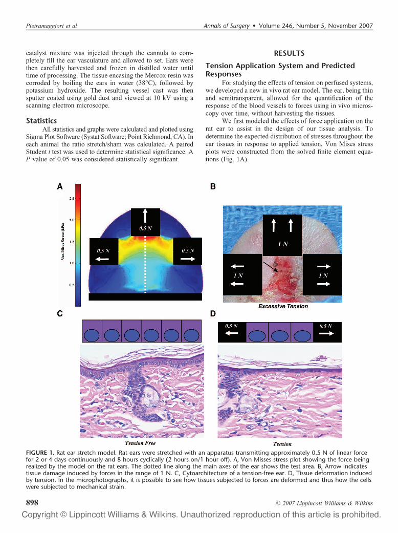
We first modeled the effects of force application on therat ear to assist in the design of our tissue analysis. Todetermine the expected distribution of stresses throughout theear tissues in response to applied tension, Von Mises stressplots were constructed from the solved finite element equa-tions (Fig. 1A).
FIGURE 1. Rat ear stretch model. Rat ears were stretched with an apparatus transmitting approximately 0.5 N of linear forcefor 2 or 4 days continuously and 8 hours cyclically (2 hours on/1 hour off). A, Von Misses stress plot showing the force beingrealized by the model on the rat ears. The dotted line along the main axes of the ear shows the test area. B, Arrow indicatestissue damage induced by forces in the range of 1 N. C, Cytoarchitecture of a tension-free ear. D, Tissue deformation inducedby tension. In the microphotographs, it is possible to see how tissues subjected to forces are deformed and thus how the cellswere subjected to mechanical strain.
Pietramaggiori et al Annals of Surgery • Volume 246, Number 5, November 2007
© 2007 Lippincott Williams & Wilkins898
Although skin is generally considered to be a nonlinear,viscoelastic material18 results from the Von Mises stress plotalong the y axis appeared linear until approximately 2 kPa offorce (Fig. 1A); thus, the ears were modeled as linear mate-rials. The Von Mises stress curve can be broken into 2 mainlinear ranges of approximately 0 to 1(low level) and 1 to 2kPa (midlevel) of force, both extending approximately 2 mminto the tissue (Fig. 1A). A third range of 2 to 3 kPa (highlevel), extending approximately 1 mm closer to the apicaladhesive pad, was also identified. Preliminary studies usingthe tension application device showed that forces in the rangeof 1 N (�100g) induced tissue injury within 4 hours ofapplication, characterized by edema, oozing, and bleedingthrough the damaged epithelium, de-epithelialization (Fig.1B), and the appearance of extravasated cells in the intersti-tial space. When this type of injury is induced, the inflam-matory response masks the effects of micromechanicalforces. Thus, we chose to use a lower tensile load of 0.5 N,which elicited vascular changes without inflammatory re-sponse as confirmed with histologic sections. Forces appliedto the ears induced cell displacement, particularly in theepidermal layer, and straightening and realignment of colla-gen fibers in the dermal layer (Fig. 1C, D).
Tension Application Increases Blood VesselDiameter
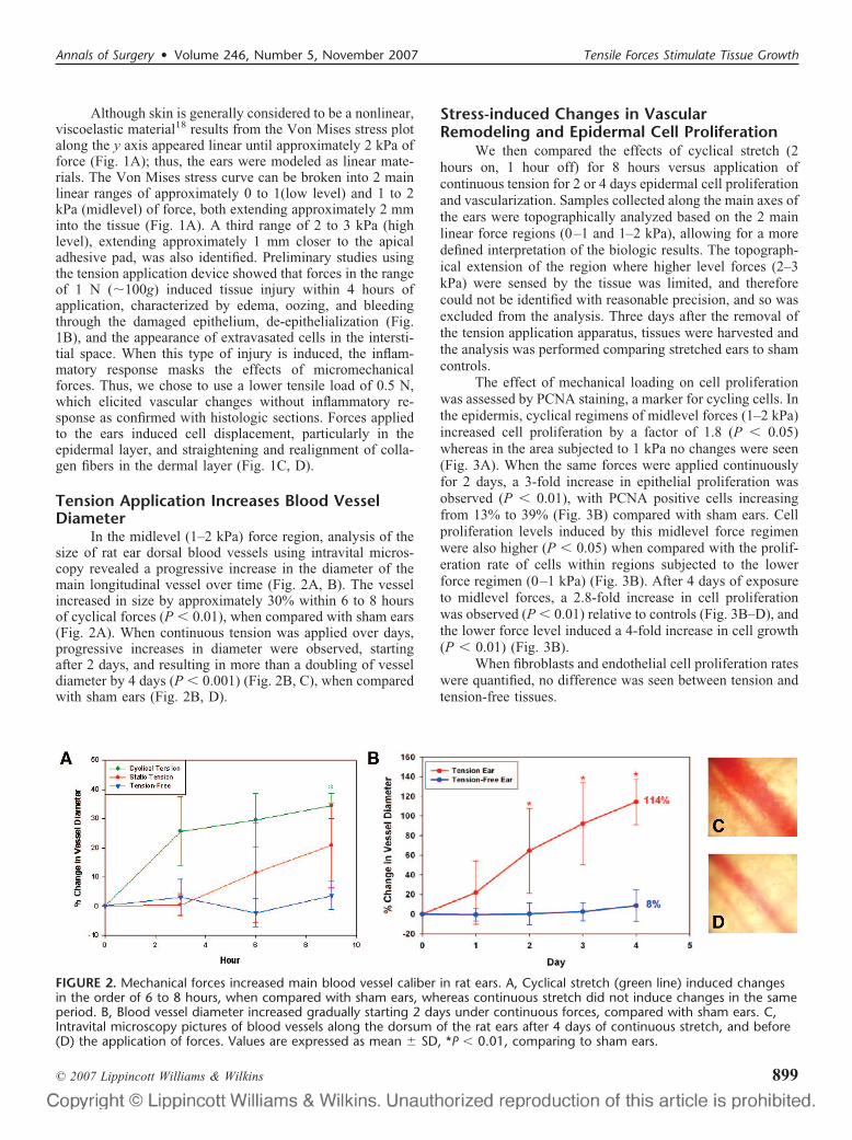
In the midlevel (1–2 kPa) force region, analysis of thesize of rat ear dorsal blood vessels using intravital micros-copy revealed a progressive increase in the diameter of themain longitudinal vessel over time (Fig. 2A, B). The vesselincreased in size by approximately 30% within 6 to 8 hoursof cyclical forces (P � 0.01), when compared with sham ears(Fig. 2A). When continuous tension was applied over days,progressive increases in diameter were observed, startingafter 2 days, and resulting in more than a doubling of vesseldiameter by 4 days (P � 0.001) (Fig. 2B, C), when comparedwith sham ears (Fig. 2B, D).
Stress-induced Changes in VascularRemodeling and Epidermal Cell Proliferation
We then compared the effects of cyclical stretch (2hours on, 1 hour off) for 8 hours versus application ofcontinuous tension for 2 or 4 days epidermal cell proliferationand vascularization. Samples collected along the main axes ofthe ears were topographically analyzed based on the 2 mainlinear force regions (0–1 and 1–2 kPa), allowing for a moredefined interpretation of the biologic results. The topograph-ical extension of the region where higher level forces (2–3kPa) were sensed by the tissue was limited, and thereforecould not be identified with reasonable precision, and so wasexcluded from the analysis. Three days after the removal ofthe tension application apparatus, tissues were harvested andthe analysis was performed comparing stretched ears to shamcontrols.
The effect of mechanical loading on cell proliferationwas assessed by PCNA staining, a marker for cycling cells. Inthe epidermis, cyclical regimens of midlevel forces (1–2 kPa)increased cell proliferation by a factor of 1.8 (P � 0.05)whereas in the area subjected to 1 kPa no changes were seen(Fig. 3A). When the same forces were applied continuouslyfor 2 days, a 3-fold increase in epithelial proliferation wasobserved (P � 0.01), with PCNA positive cells increasingfrom 13% to 39% (Fig. 3B) compared with sham ears. Cellproliferation levels induced by this midlevel force regimenwere also higher (P � 0.05) when compared with the prolif-eration rate of cells within regions subjected to the lowerforce regimen (0–1 kPa) (Fig. 3B). After 4 days of exposureto midlevel forces, a 2.8-fold increase in cell proliferationwas observed (P � 0.01) relative to controls (Fig. 3B–D), andthe lower force level induced a 4-fold increase in cell growth(P � 0.01) (Fig. 3B).
When fibroblasts and endothelial cell proliferation rateswere quantified, no difference was seen between tension andtension-free tissues.
FIGURE 2. Mechanical forces increased main blood vessel caliber in rat ears. A, Cyclical stretch (green line) induced changesin the order of 6 to 8 hours, when compared with sham ears, whereas continuous stretch did not induce changes in the sameperiod. B, Blood vessel diameter increased gradually starting 2 days under continuous forces, compared with sham ears. C,Intravital microscopy pictures of blood vessels along the dorsum of the rat ears after 4 days of continuous stretch, and before(D) the application of forces. Values are expressed as mean � SD, *P � 0.01, comparing to sham ears.
Annals of Surgery • Volume 246, Number 5, November 2007 Tensile Forces Stimulate Tissue Growth
© 2007 Lippincott Williams & Wilkins 899

The response of the vasculature to stretch was evalu-ated by quantifying the area of endothelial cells withinhistologic sections after staining for the vascular endothelialcell marker PECAM-1, a molecule that is also involved inmechanotransduction.19 Cyclical regimens produced a 2.8-and 1.5-fold increase (P � 0.05) in vascular area within 8hours, compared with the sham ears in the areas subjected tomid- and low-level forces, respectively (Fig. 3E, G, H).Comparing the 2 areas subjected to forces, total vascularendothelial area present in regions exposed to midlevel forceswas significantly higher that that observed in regions sub-jected to low-level forces (P � 0.01) (Fig. 3E).
Continuous exposure of ears to the midlevel forceregimen for 2 days induced a 2.8-fold increase (P � 0.05) invascular area (Fig. 3F). No changes were seen when low-level forces were applied, and no vascular changes wereobserved in tissues after 4 days of exposure to continuoustension (Fig. 3F). From the trendline analysis of the results,2.25 days (ˆ mark, Fig. 3B, F) were predicted to be the pointin which both the vascular surface and cell proliferation peak.
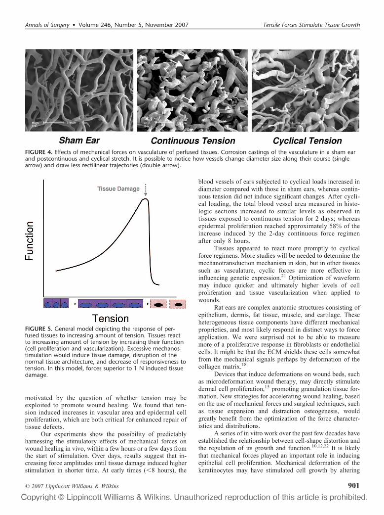
We also made casts of the microcirculation to visualizethe morphologic changes in the vasculature subjected toforces. Overall, the microcirculatory network subjected tomidlevel forces displayed microvessels with a less homoge-neous caliber and higher curvature (Fig. 4). Interestingly,
microvessels subjected to forces showed the highly orientedtridimensional direction, as if forces redirected their distribu-tion in the dermal space along the main force vector direction.Heterogeneity in caliber of the vessels and increased curva-ture are early signs of vascular remodeling. These peculiarchanges are characteristic of the casts collected after theapplication of continuous force, and cyclical stretch regimenswere also able to induce similar initial signs of vascular remod-eling (Fig. 4).
After observing the characteristics of the physical data andtrendline curves, an empirical graph general model was gener-ated to predict cell response to the application of forces usingincreased vascular remodeling and proliferation as markers oftissue function (Fig. 5). As shown, tissue growth is induced withthe application of increasing tension. The curve depicting thetissue response to increasing amounts of force has a maximumslope illustrating the point at which maximal safe stimulation isachieved. After the maximal stimulation, tissue damage occurs,the tissue loses its physical properties, homeostasis is lost, andcells lose their ability to respond to mechanical forces andundergo apoptosis, as previously shown in vitro.20
DISCUSSIONWe characterized the response of perfused mamma-
lian ear tissues to different regimens of mechanical forces
FIGURE 3. Changes in cell proliferation and vascularity after stretch. A, 8 hours of cyclical, 1 to 2 kPa tension increased cellproliferation 1.8-fold over baseline. B, Continuous forces in the range of 1 to 2 kPa increased cell proliferation starting from 2days. Similar increase was induced by 0 to 1 kPa of continuous tension in 4 days. C, D, Microphotographs depicting cellularproliferation as assessed by PCNA staining, which stains proliferating cells. Sham ears show lower levels of PCNA staining inthe epithelium when compared with ears stretched for 4 days. E, Cyclical forces, over the 8-hour period increased vascularsurface by a factor of 2.8 (1–2 kPa) and 1.5 (0–1 kPa). F, Continuous 1 to 2 kPa of forces increased vascular surface at the2-day time-point. G, H, Staining for vasculature using PECAM-1 shows lower endothelial surface in sham ears when comparedwith cyclically stretched ears. Results are expressed as a ratio stretched/sham, with the value of 1 as the baseline level in thesham. **P � 0.01, *P � 0.05, �� � 1.5 � 0.5 kPa � 1 kPa, ˆ � timepoint corresponding to maximal predicted tissue re-sponse. Values are expressed as mean � standard error.
Pietramaggiori et al Annals of Surgery • Volume 246, Number 5, November 2007
© 2007 Lippincott Williams & Wilkins900
motivated by the question of whether tension may beexploited to promote wound healing. We found that ten-sion induced increases in vascular area and epidermal cellproliferation, which are both critical for enhanced repair oftissue defects.
Our experiments show the possibility of predictablyharnessing the stimulatory effects of mechanical forces onwound healing in vivo, within a few hours or a few days fromthe start of stimulation. Over days, results suggest that in-creasing force amplitudes until tissue damage induced higherstimulation in shorter time. At early times (�8 hours), the
blood vessels of ears subjected to cyclical loads increased indiameter compared with those in sham ears, whereas contin-uous tension did not induce significant changes. After cycli-cal loading, the total blood vessel area measured in histo-logic sections increased to similar levels as observed intissues exposed to continuous tension for 2 days; whereasepidermal proliferation reached approximately 58% of theincrease induced by the 2-day continuous force regimenafter only 8 hours.
Tissues appeared to react more promptly to cyclicalforce regimens. More studies will be needed to determine themechanotransduction mechanism in skin, but in other tissuessuch as vasculature, cyclic forces are more effective ininfluencing genetic expression.21 Optimization of waveformmay induce quicker and ultimately higher levels of cellproliferation and tissue vascularization when applied towounds.
Rat ears are complex anatomic structures consisting ofepithelium, dermis, fat tissue, muscle, and cartilage. Theseheterogeneous tissue components have different mechanicalproprieties, and most likely respond in distinct ways to forceapplication. We were surprised not to be able to measuremore of a proliferative response in fibroblasts or endothelialcells. It might be that the ECM shields these cells somewhatfrom the mechanical signals perhaps by deformation of thecollagen matrix.18
Devices that induce deformations on wound beds, suchas microdeformation wound therapy, may directly stimulatedermal cell proliferation,15 promoting granulation tissue for-mation. New strategies for accelerating wound healing, basedon the use of mechanical forces and surgical techniques, suchas tissue expansion and distraction osteogenesis, wouldgreatly benefit from the optimization of the force character-istics and distributions.
A series of in vitro work over the past few decades haveestablished the relationship between cell-shape distortion andthe regulation of its growth and function.10,12,22 It is likelythat mechanical forces played an important role in inducingepithelial cell proliferation. Mechanical deformation of thekeratinocytes may have stimulated cell growth by altering
FIGURE 4. Effects of mechanical forces on vasculature of perfused tissues. Corrosion castings of the vasculature in a sham earand postcontinuous and cyclical stretch. It is possible to notice how vessels change diameter size along their course (singlearrow) and draw less rectilinear trajectories (double arrow).
FIGURE 5. General model depicting the response of per-fused tissues to increasing amount of tension. Tissues reactto increasing amount of tension by increasing their function(cell proliferation and vascularization). Excessive mechanos-timulation would induce tissue damage, disruption of thenormal tissue architecture, and decrease of responsiveness totension. In this model, forces superior to 1 N induced tissuedamage.
Annals of Surgery • Volume 246, Number 5, November 2007 Tensile Forces Stimulate Tissue Growth
© 2007 Lippincott Williams & Wilkins 901

biochemical signaling responses within the cell, as demon-strated in many studies with cultured cells.10
Increased vascular area noticed after the application oftension can also be a consequence of endothelial cell migra-tion, cell signaling from the epithelium and vascular remod-eling, rather than endothelial proliferation. Additional studiesare required to characterize the changes in blood flow and theresponse of endothelial cells and the cross-talk betweenepithelial and dermal cells to mechanical stretch to enhancevascular stimulation in nonhealing wounds. After the removalof the forces, the mechano-induced proliferative and remod-eling response would likely result in tissue gain, important tocover tissue defects, as shown in tissue expansion.
CONCLUSIONSThese results show that tension on perfused tissues
determines an amplitude and waveform dependent cellularresponse in both the epidermis and in the blood vessel ofthe dermal tissue. We hope that a better understanding of theresponse of living tissues to mechanical forces will allow thedevelopment of novel surgical techniques.
ACKNOWLEDGMENTSThe authors thank Lauren Bayer and Jasmine Mathews
for critical review of the manuscript, Emy Chen for excellenttechnical assistance, and Kristin Johnson for photography.
REFERENCES1. Diabetes epidemic out of control. www.idf.org/home/index.cfm. Ac-
cessed June 29, 2007.2. Steed DL. Clinical evaluation of recombinant human platelet-derived
growth factor for the treatment of lower extremity ulcers. Plast ReconstrSurg. 2006;117(7 suppl):143S–149S; discussion 150S–151S.
3. Wieman TJ; Becaplermin Gel Studies Group. Clinical efficacy of be-caplermin (rhPDGF-BB) gel. Am J Surg. 1998;176(2A suppl):74S–79S.
4. Ingber DE. Cellular mechanotransduction: putting all the pieces togetheragain. FASEB J. 2006;20:811–827.
5. Ryan TJ. Exchange and the mechanical properties of the skin: oncotic
and hydrostatic forces control by blood supply and lymphatic drainage.Wound Repair Regen. 1995;3:258–264.
6. Parker KK, Brock AL, Brangwynne C, et al. Directional control oflamellipodia extension by constraining cell shape and orienting celltractional forces. FASEB J. 2002;16:1195–1204.
7. Huang S, Chen CS, Ingber DE. Control of cyclin D1, p27(Kip1), and cellcycle progression in human capillary endothelial cells by cell shape andcytoskeletal tension. Mol Biol Cell. 1998;9:3179–3193.
8. Chen CS, Mrksich M, Huang S, et al. Geometric control of cell life anddeath. Science. 1997;276:1425–1428.
9. Ingber DE, Folkman J. Mechanochemical switching between growth anddifferentiation during fibroblast growth factor-stimulated angiogenesisin vitro: role of extracellular matrix. J Cell Biol. 1989;109:317–330.
10. Huang S, Ingber DE. The structural and mechanical complexity ofcell-growth control. Nat Cell Biol. 1999;1:E131–E138.
11. Manders EK, Schenden MJ, Furrey JA, et al. Soft-tissue expansion:concepts and complications. Plast Reconstr Surg. 1984;74:493–507.
12. Olenius M, Dalsgaard CJ, Wickman M. Mitotic activity in expandedhuman skin. Plast Reconstr Surg. 1993;91:213–216.
13. Argenta LC, Morykwas MJ, Marks MW, et al. Vacuum-assisted closure:state of clinic art. Plast Reconstr Surg. 2006;117(7 suppl):127S–142S.
14. Greene AK, Puder M, Roy R, et al. Microdeformational wound therapy:effects on angiogenesis and matrix metalloproteinases in chronicwounds of 3 debilitated patients. Ann Plast Surg. 2006;56:418–422.
15. Saxena V, Hwang CW, Huang S, et al. Vacuum-assisted closure:microdeformations of wounds and cell proliferation. Plast ReconstrSurg. 2004;114:1086–1096; discussion 1097–1098.
16. Sanders R. Torsional elasticity of human skin in vivo. Pflugers Arch.1973;342:255–260.
17. Pietramaggiori G, Kaipainen A, Czeczuga JM, et al. Freeze-dried plate-let-rich plasma shows beneficial healing properties in chronic wounds.Wound Repair Regen. 2006;14:573–580.
18. Wilhelmi BJ, Blackwell SJ, Mancoll JS, et al. Creep vs. stretch: a reviewof the viscoelastic properties of skin. Ann Plast Surg. 1998;41:215–219.
19. Tzima E, Irani-Tehrani M, Kiosses WB, et al. A mechanosensorycomplex that mediates the endothelial cell response to fluid shear stress.Nature. 2005;437:426–431.
20. Ingber D. How cells (might) sense microgravity. FASEB J. 1999;13(suppl):S3–S15.
21. Blackman BR, Garcia-Cardena G, Gimbrone MA Jr. A new in vitromodel to evaluate differential responses of endothelial cells to simulatedarterial shear stress waveforms. J Biomech Eng. 2002;124:397–407.
22. Ingber DE. Cellular tensegrity: defining new rules of biological designthat govern the cytoskeleton. J Cell Sci. 1993;104(Pt 3):613–627.
Pietramaggiori et al Annals of Surgery • Volume 246, Number 5, November 2007
© 2007 Lippincott Williams & Wilkins902