Tenascin-W Is a Novel Marker for Activated Tumor Stroma in Low-grade Human Breast Cancer and...
12
Tenascin-W Is a Novel Marker for Activated Tumor Stroma in Low-grade Human Breast Cancer and Influences Cell Behavior Martin Degen, 1 Florence Brellier, 1 Renate Kain, 4 Christian Ruiz, 2 Luigi Terracciano, 2 Gertraud Orend, 3 and Ruth Chiquet-Ehrismann 1 1 Friedrich Miescher Institute for Biomedical Research, Novartis Research Foundation; 2 Institute of Pathology and 3 Institute of Biochemistry and Genetics, Center for Biomedicine, University of Basel, Basel, Switzerland; and 4 Department of Clinical Pathology, University of Vienna, Vienna, Austria Abstract This is the first report about human tenascin-W, the fourth and final member of the extracellular matrix protein family of tenascins. Sixty-three human breast tumor extracts were analyzed by Western blotting for the presence of tenascin-W and compared with tenascin-C, an established marker of tumor stroma. Interestingly, we found tenascin-W expression in the majority of the tumor tissues, but no detectable expression in the normal mammary parenchyma. Eighty- one percent of the breast tumor samples were tenascin-W positive and 86% showed expression of tenascin-C. However, tenascin-W and tenascin-C amounts varied greatly between tumors and some contained either tenascin-W or tenascin-C exclusively, indicating independent mechanisms regulating their expression. Although there was no difference between high- or low-grade tumors with respect to the presence of tenascin-C, tenascin-W was more prominent in low-grade tumors. For 42 of the breast cancer tissues, a frozen tumor microarray was available to confirm the Western blot data by immunohistochemistry. Similar to tenascin-C, tenascin-W was detected in the tumor stroma. Fibroblasts adhered to tenascin-W in a B 1 integrin–dependent manner and spread with a distinctive morphology under conditions where they remained round on tenascin-C. CHOB2 cells expressing A v B 1 or A 4 B 1 integrins were able to spread on tenascin-W. Furthermore, addition of tenascin-W to the culture medium increased migration of breast cancer cells toward a fibronec- tin substratum in vitro . These data imply that tenascin-W expression in the activated tumor stroma facilitates tumori- genesis by supporting the migratory behavior of breast cancer cells. [Cancer Res 2007;67(19):9169–79] Introduction Epithelial tissues depend on normal stromal cells and the basement membrane for maintenance of tissue homeostasis, cell adhesion to extracellular matrix (ECM) through integrins, and generation and preservation of epithelial cell polarity (1, 2). These interactions continue to be important in epithelial pathologies. During carcinogenesis, the stromal cells get activated by an initial trigger that usually comes from the cancer cell, leading to stromal changes and the formation of a tumor-permissive microenviron- ment (1). More and more evidence is accumulating that this aberrant tumor stroma influences cancer development and has an effect on the malignancy of a tumor ( for reviews, see refs. 3–6). Tumor-associated cells of the stromal compartment express proproliferative paracrine signals for epithelial cells (7), stimulate angiogenesis (8, 9), and can even show loss of tumor-suppressor genes (10). Therefore, understanding the mechanisms of the complex crosstalk between the cancerous epithelial cells and the tumor stroma might lead to novel approaches for cancer therapies that target the functions of the activated stromal cells ( for reviews, see refs. 3, 11–13). Because of the newly recognized importance of the tumor stroma in cancer development, it is necessary to fully characterize this tissue compartment. A prominent ECM protein specifically present in tumor stroma is tenascin-C (ref. 14; for reviews, see refs. 15–17). Interestingly, tenascin-C was shown to be expressed around angiogenic vessels in many tumors (18–20) as well as to promote angiogenesis in cell culture studies (21). Furthermore, tenascin-C addition to a fibronectin substratum stimulated cancer cell growth in in vitro studies (22, 23). Therefore, tenascin-C is one of the potential candidate molecules mediating the protumorigenic effects of tumor stroma ( for review, see ref. 15). Recently, we found in the stroma of mouse mammary tumors the induction of a second member of the tenascin family of ECM proteins, tenascin-W (24). Because the human orthologue has never been analyzed, we cloned the human tenascin-W cDNA, expressed the protein, and raised antibodies against it. We determined the presence of tenascin-W in a large number of breast cancers where it was more prevalent in low-grade tumors. In vitro , tenascin-W did not interfere with cancer cell adhesion to fibronectin, but promoted migration of breast cancer cells toward fibronectin. Furthermore, fibroblasts were able to adhere to a tenascin-W substratum. Our data suggest tenascin-W as a marker for transformation of the normal physiologic stroma to an activated stroma in breast cancer, and that tenascin-W can influence cancer cell behavior. Materials and Methods Cloning, expression, and purification of full-length human tenascin- W and tenascin-C. cDNA was synthesized from total RNA extracted from the human osteosarcoma cell line KRIB with TRIzol (Invitrogen). PCR was done using the KRIB cDNA as template and primers for nested PCR were designed that amplified three separate but overlapping parts of human tenascin-W that we call part A, part B, and part C. The first reaction was done with the primer sets 5¶-CAGGCATCCTGGAGGGTCTGCTCCC-3¶/5¶- GTGAGGGCATTGGTGTCAGCTTTC-3¶ (for part A), 5¶-GCGCTA- CACTTCTGCTGATG-3¶/5¶-CTGTGGAGAGGGTGGTGG-3¶ ( for part B), and Requests for reprints: Ruth Chiquet-Ehrismann, Friedrich Miescher Institute for Biomedical Research, Novartis Research Foundation, Maulbeerstrasse 66, CH-4058 Basel, Switzerland. Phone: 41-61-697-2494; Fax: 41-61-697-3976; E-mail: chiquet@ fmi.ch. I2007 American Association for Cancer Research. doi:10.1158/0008-5472.CAN-07-0666 www.aacrjournals.org 9169 Cancer Res 2007; 67: (19). October 1, 2007 Research Article Research. on February 7, 2016. © 2007 American Association for Cancer cancerres.aacrjournals.org Downloaded from
Transcript of Tenascin-W Is a Novel Marker for Activated Tumor Stroma in Low-grade Human Breast Cancer and...

Tenascin-W Is a Novel Marker for Activated Tumor Stroma
in Low-grade Human Breast Cancer and Influences
Cell Behavior
Martin Degen,1Florence Brellier,
1Renate Kain,
4Christian Ruiz,
2Luigi Terracciano,
2
Gertraud Orend,3and Ruth Chiquet-Ehrismann
1
1Friedrich Miescher Institute for Biomedical Research, Novartis Research Foundation; 2Institute of Pathology and 3Institute of Biochemistryand Genetics, Center for Biomedicine, University of Basel, Basel, Switzerland; and 4Department of Clinical Pathology,University of Vienna, Vienna, Austria
Abstract
This is the first report about human tenascin-W, the fourthand final member of the extracellular matrix protein family oftenascins. Sixty-three human breast tumor extracts wereanalyzed by Western blotting for the presence of tenascin-Wand compared with tenascin-C, an established marker oftumor stroma. Interestingly, we found tenascin-W expressionin the majority of the tumor tissues, but no detectableexpression in the normal mammary parenchyma. Eighty-one percent of the breast tumor samples were tenascin-Wpositive and 86% showed expression of tenascin-C. However,tenascin-W and tenascin-C amounts varied greatly betweentumors and some contained either tenascin-W or tenascin-Cexclusively, indicating independent mechanisms regulatingtheir expression. Although there was no difference betweenhigh- or low-grade tumors with respect to the presence oftenascin-C, tenascin-W was more prominent in low-gradetumors. For 42 of the breast cancer tissues, a frozen tumormicroarray was available to confirm the Western blot databy immunohistochemistry. Similar to tenascin-C, tenascin-Wwas detected in the tumor stroma. Fibroblasts adhered totenascin-W in a B1 integrin–dependent manner and spreadwith a distinctive morphology under conditions where theyremained round on tenascin-C. CHOB2 cells expressing AvB1
or A4B1 integrins were able to spread on tenascin-W.Furthermore, addition of tenascin-W to the culture mediumincreased migration of breast cancer cells toward a fibronec-tin substratum in vitro . These data imply that tenascin-Wexpression in the activated tumor stroma facilitates tumori-genesis by supporting the migratory behavior of breast cancercells. [Cancer Res 2007;67(19):9169–79]
Introduction
Epithelial tissues depend on normal stromal cells and thebasement membrane for maintenance of tissue homeostasis, celladhesion to extracellular matrix (ECM) through integrins, andgeneration and preservation of epithelial cell polarity (1, 2). Theseinteractions continue to be important in epithelial pathologies.During carcinogenesis, the stromal cells get activated by an initialtrigger that usually comes from the cancer cell, leading to stromal
changes and the formation of a tumor-permissive microenviron-ment (1). More and more evidence is accumulating that thisaberrant tumor stroma influences cancer development and has aneffect on the malignancy of a tumor ( for reviews, see refs. 3–6).Tumor-associated cells of the stromal compartment expressproproliferative paracrine signals for epithelial cells (7), stimulateangiogenesis (8, 9), and can even show loss of tumor-suppressorgenes (10). Therefore, understanding the mechanisms of thecomplex crosstalk between the cancerous epithelial cells and thetumor stroma might lead to novel approaches for cancer therapiesthat target the functions of the activated stromal cells ( for reviews,see refs. 3, 11–13).Because of the newly recognized importance of the tumor
stroma in cancer development, it is necessary to fully characterizethis tissue compartment. A prominent ECM protein specificallypresent in tumor stroma is tenascin-C (ref. 14; for reviews, seerefs. 15–17). Interestingly, tenascin-C was shown to be expressedaround angiogenic vessels in many tumors (18–20) as well as topromote angiogenesis in cell culture studies (21). Furthermore,tenascin-C addition to a fibronectin substratum stimulated cancercell growth in in vitro studies (22, 23). Therefore, tenascin-C is oneof the potential candidate molecules mediating the protumorigeniceffects of tumor stroma ( for review, see ref. 15).Recently, we found in the stroma of mouse mammary tumors the
induction of a second member of the tenascin family of ECMproteins, tenascin-W (24). Because the human orthologue has neverbeen analyzed, we cloned the human tenascin-W cDNA, expressedthe protein, and raised antibodies against it. We determined thepresence of tenascin-W in a large number of breast cancers whereit was more prevalent in low-grade tumors. In vitro , tenascin-W didnot interfere with cancer cell adhesion to fibronectin, but promotedmigration of breast cancer cells toward fibronectin. Furthermore,fibroblasts were able to adhere to a tenascin-Wsubstratum. Our datasuggest tenascin-W as a marker for transformation of the normalphysiologic stroma to an activated stroma in breast cancer, and thattenascin-W can influence cancer cell behavior.
Materials and Methods
Cloning, expression, and purification of full-length human tenascin-W and tenascin-C. cDNA was synthesized from total RNA extracted from
the human osteosarcoma cell line KRIB with TRIzol (Invitrogen). PCR was
done using the KRIB cDNA as template and primers for nested PCR weredesigned that amplified three separate but overlapping parts of human
tenascin-W that we call part A, part B, and part C. The first reaction was
done with the primer sets 5¶-CAGGCATCCTGGAGGGTCTGCTCCC-3¶/5¶-GTGAGGGCATTGGTGTCAGCTTTC-3¶ ( for part A), 5¶-GCGCTA-CACTTCTGCTGATG-3¶/5¶-CTGTGGAGAGGGTGGTGG-3¶ ( for part B), and
Requests for reprints: Ruth Chiquet-Ehrismann, Friedrich Miescher Institute forBiomedical Research, Novartis Research Foundation, Maulbeerstrasse 66, CH-4058Basel, Switzerland. Phone: 41-61-697-2494; Fax: 41-61-697-3976; E-mail: [email protected].
I2007 American Association for Cancer Research.doi:10.1158/0008-5472.CAN-07-0666
www.aacrjournals.org 9169 Cancer Res 2007; 67: (19). October 1, 2007
Research Article
Research. on February 7, 2016. © 2007 American Association for Cancercancerres.aacrjournals.org Downloaded from

5¶-CGCAGTCTGGTGGCATATTG-3¶/5¶-CATGATTTGTTCTGCGGGC-3¶ ( forpart C). Primer sets for the second reactions included XhoI and BamHI
restriction sites, respectively. Primer pair 5¶-GACTCGAGCTTTCCAAGGAT-GAGTCTCCAGG-3¶/5¶-GAGGATCCCCTGGTTGCCCCTTTCAGCCC-3¶ wasused for part A, primer pair 5¶-GACTCGAGTGCACAAGGATGAGAGCAG-3¶/5¶-GAGGATCCACCCTTAAAGGCAACAAGGG-3¶ was used for part B, andprimer pair 5¶-GACTCGAGCGGCTACATTCTGACTTACC-3¶/5¶-GAG-GATCCTCAGTGATGGTGATGGTGATGGAACGTTCGCAGCCTTCCTC-3¶was used for part C. The reverse primer for the second PCR of part Ccontained the sequence encoding a 6�His-tag followed by a stop codon.
The first two fragments (A and B) were ligated using an internal AccI
restriction site present in the overlapping region, and the third fragment
was added using an internal NarI restriction site located in the overlap ofpart B and C. This resulted in a full-length human tenascin-W cDNA of 3,885
bp (corresponds to the sequence 16–3,900 of Genbank accession no.
NM_022093). This full-length cDNA was cloned into the expression vectorpCEP/Pu (provided by J. Engel, Biozentrum, Basel, Switzerland) and
transfected into EBNA293 cells [American Type Culture Collection (ATCC)]
using FuGENE6 (Roche). Transfected cells were selected with puromycin,
and serum-free medium containing the secreted recombinant humantenascin-W was collected. After ammonium sulfate precipitation (0–50%)
and dialysis against PBS, the protein was passed over a gelatin-agarose
column to remove fibronectin followed by affinity purification on a Ni-NTA
matrix (Qiagen) in the presence of 0.5 mol/L urea. After extensive washingof the column, tenascin-W was eluted with 250 mmol/L imidazole (pH 6.9)
and finally dialyzed against PBS. The protein appeared as a single band on a
Coomassie-stained acrylamide gel, and fibronectin could not be detected inthe purified sample by immunoblotting or by ELISA.
A full-length human tenascin-C cDNA called HxBL was kindly obtained
from H.P. Erickson (Department of Cell Biology, Duke University, Durham,
NC). It was modified by the addition of a 6�His-tag at the COOH terminusand subcloned into pCEP/Pu for transfection into EBNA293 cells as
described above for human tenascin-W (25). The same purification
procedure as described above for tenascin-W was used for the isolation
of human tenascin-C.Anti–tenascin-W antibody production. To raise polyclonal antisera in
rabbits, a recombinant fragment of human tenascin-W was cloned,
bacterially expressed, and purified. To clone the recombinant fragment,
specific primers were designed to amplify the sequence encoding the last
two fibronectin type III domains (Fig. 1A) with the Expand High Fidelity
PCR System (Roche). The cDNA of the full-length human tenascin-W
(described above) was used as template and the PCR was done with the
primer set 5 ¶-GAGGATCCGAAATTGACGGCCCCAAAAACC-3 ¶/5 ¶-ATAAGCTTATGTGGAGAGGGTGGTGGA-3¶. The forward primer included
a BamHI restriction site and the reverse primer a stop codon immediately
followed by a HindIII restriction site to enable the directional cloning into
the bacterial expression vector pQE30 (Qiagen), supplying a COOH-terminal
His tag for the purification of the recombinant fragment. The recombinant
fragment corresponding to fibronectin type III domains 3F/4 (Fig. 1A) was
expressed and purified by affinity chromatography to a Ni-NTA matrix
(Qiagen) following the supplier’s instructions. Purification was done under
native conditions and elution by 250 mmol/L imidazole (pH 6.9). The
bacterially expressed fragment of tenascin-W was then used to raise
polyclonal antisera in rabbits using standard immunization procedures.
To raise monoclonal antibodies (mAb) in mice, a recombinant fragment
of human tenascin-W was cloned containing the last three fibronectin typeIII domains (Fig. 1A), bacterially expressed, and purified as described above.
To clone the recombinant fragment, cDNA was synthesized from total RNA
that was extracted from Saos-2 cells (ATCC) by TRIzol reagent (Invitrogen).
Primers, amplifying the last three FN type III domains, were used for nestedPCR reactions with the Expand High Fidelity System (Roche) using the Saos-
2 cDNA as template. The first reaction was done with the primer set 5¶-GGGAAGGAGCAGAGTAGCACTG-3¶/5¶-CCGCCTCTGGAAGACAATCC-3¶,the second reaction with the primers 5¶-AGGGATCCGACATTGACAGCCCC-CAAAACC-3¶/5¶-CTAAGCTTTCATGTGGAGAGGGTGGTGGATAC-3¶. Theforward primer for the second PCR included a BamHI restriction site and
the reverse primer a stop codon immediately followed by a HindIII
restriction site to enable the directional cloning into the bacterialexpression vector pQE30 (Qiagen), supplying a COOH-terminal His tag for
the purification of the recombinant fragment. mAbs were obtained by
immunizing female BALB/c mice with 34.6 Ag of the purified recombinanttenascin-W fragment emulsified with STIMUNE (ID-Lelystad, Institute forAnimal Sciences and Health). For boosting, mice were injected twice with a
4-week interval with 25 Ag tenascin-W fragment. Splenic lymphocytes were
fused with the myeloma cell line P3X63Ag8.653 (ATTC) and cultured
according to standard protocols. The hybridoma supernatants wereanalyzed by ELISA and Western blot analysis of a tenascin-W fragment
expressed in HEK293 cells using a construct containing the sequence of the
last three FN III domains of tenascin-W fused to an NH2-terminal fragment
of chicken tenascin-C containing its secretion signal and the epitope for thewell-characterized monoclonal anti-chicken tenascin-C antibody mAb60.
IgGs from two mAb hybridoma clones were purified from conditioned
medium by protein G Sepharose (Amersham) and clone 56O was used inthis study.
Human tissue extracts and Western blot analysis. The following study
was done in accordance with the guidelines of the ethical committee of the
University of Basel. Fresh human tissue was frozen on dry ice immediately
after surgery. For the processing of the tissue, it was thawed on ice, minced,
and homogenized in lysis buffer [100 mmol/L phosphate buffer (pH 8.0),
300 mmol/L NaCl, 8 mol/L urea, 1% Triton X-100, 10 mmol/L h-mercaptoe-thanol, 50 mmol/L guanidinium hydrochloride, and complete protease
inhibitor cocktail (Roche)]. Insoluble material was pelleted, and reducing
SDS-PAGE sample buffer was added to the supernatant and boiled for 5 min
at 95jC. After electrophoresis on 6% polyacrylamide gels, proteins were
electrotransferred onto polyvinyldifluoride membranes (Millipore) using a
semidry blotting apparatus (Millipore). After the transfer, membranes were
stained with Amido Black to control equal protein loading. After blocking for
1 h at room temperature in TBS containing 0.05% Tween and 5% skim milk
powder, membranes were incubated overnight with either the polyclonal
tenascin-W antiserum (1:750), the mAb 56O raised against tenascin-W
(1:1,000), the mAb B28-13 raised against tenascin-C (1:100; ref. 26), or the
monoclonal antivinculin antibody (1:2,000; Sigma) followed by an incubation
for 1 h with anti-rabbit IgG or anti-mouse IgG coupled to horseradish
peroxidase (1:10,000), respectively. Blots were developed using SuperSignal
(Pierce) and exposed to Kodak BioMax MR Films.
For Western blot quantification, the software Gene Tools from SynGene
was used. Briefly, the quantity of 25 ng was assigned to the specific band
obtained from 25 ng of the purified protein loaded on the same gel. Bydividing the densitometric values of the bands of the tissue extracts by the
value obtained with 25 ng of the purified protein loaded and developed on
the same gel, a quantity for each band could be calculated and furthernormalized to vinculin.
Frozen tissue microarrays and immunohistochemistry. A frozen
tissue microarray (TMA) was constructed from frozen tissue samples of
40 breast carcinomas and 2 fibroadenomas. Pathologic features of thesesamples are summarized in Table 1 (patients 1–42). Histologic grading of
the breast carcinomas was done according to the Bloom, Richardson, Elston
grading system. A second TMA was built from 10 frozen tissue samples of
normal breast tissue. Both TMAs were constructed in frozen Tissue-Tekoptimum cutting temperature compound (Miles Laboratories) as described
previously (27). We optimized a commercial microarray device (Beecher
Instruments) by using a 0.6-mm drill for recipient whole making instead ofthe conventional hollow needle.
All immunostainings were done using the Discovery XT automated
stainer (Ventana Medical Systems), with 3,3¶-diaminobenzidine Map
detection kit (Ventana). Frozen TMA slides were dried 1 h at roomtemperature, fixed for 10 min at 4jC in acetone, and then introduced into theautomate. No pretreatment was required for any staining. Slides were first
blocked twice for 12 min with the AB Block reagent (Ventana). Afterward,
they were incubated for 1 h at 37jC with mouse monoclonal anti–tenascin-C(B28-13; 1:2,500), rabbit polyclonal anti–tenascin-W (1:40), and mouse
monoclonal anti–tenascin-W (clone 56O; 1:800). Slides were then treated for
32 min at 37jC with a biotinylated universal secondary antibody (Ventana)and counterstained with hematoxylin and bluing reagent (Ventana).
Cancer Research
Cancer Res 2007; 67: (19). October 1, 2007 9170 www.aacrjournals.org
Research. on February 7, 2016. © 2007 American Association for Cancercancerres.aacrjournals.org Downloaded from

Cell culture. EBNA293 (ATCC), T47D (ATCC), MCF-7 (ATCC), MDA-MB-435 (ATCC), and Detroit 551 cells (ATCC) were grown in DMEM
supplemented with 10% FCS, 100 units/mL penicillin, and 100 Ag/mLstreptomycin and cultured under standard conditions. CHOB2 (28) and
sublines CHOB2a27 (ref. 29; subclone 2C8), CHOB2a4h1 (30), and CHOB2v7(31), expressing a5a1, a4h1, and avh1, respectively, were grown in MEMasupplemented with 10% FCS, 100 units/mL penicillin, and 100 Ag/mLstreptomycin and cultured under standard conditions.
Cell adhesion assays and cell morphology. Cell adhesion assays weredone with the human mammary carcinoma cell lines T47D, MCF-7, MDA-
MB-435; the integrin-deficient CHOB2 cells; CHOB2 cells expressing single
integrins; and with human dermal fibroblasts Detroit 551. Sixty-well
microtiter plates (Nunc) were coated for 1 h at room temperature with 5 ALper well PBS containing 0.01% Tween and 10 Ag/mL of the indicated ECMprotein and blocked for 30 min with PBS containing 1% bovine serum
albumin (BSA). Cells for the adhesion assays were detached using trypsin-EDTA, resuspended in serum-free medium, and counted. Cells (1,500 per
well) were plated on the different substrata for the indicated time at 37jC.Adherent cells were then fixed by the addition of 4% formaldehyde and
stained with 0.1% crystal violet. Pictures of the entire wells were taken andcells were counted in triplicate wells in at least three independent
experiments. Mixed substrata were prepared as follows: After coating the
wells with a first ECM protein, they were washed with PBS and coated for
another hour with the second ECM protein. The order of coating for themixed substrata was not important as tested by ELISA.
To analyze cell morphology, 3.5-cm dishes containing four separate wells
(Greiner) were coated as described above with the different ECM proteins.After 30 min of blocking in PBS/1% BSA, 103 Detroit 551 fibroblasts,
resuspended in serum-free medium, were plated on the individual wells and
incubated for 90 min at 37jC. To block h1 integrin function, fibroblasts wereplated in the presence of a function-blocking anti-integrin h1 antibody(Chemicon, clone P5D2) at a concentration of 10 Ag/mL. Fibroblasts werefixed by 4% paraformaldehyde in PBS for 30 min and permeabilized with
PBS/0.1% Triton X-100 for 5 min followed by incubation with a monoclonal
antivinculin antibody for 1 h at room temperature. Cells were washed thricewith PBS and incubated with a fluorescein-coupled anti-mouse IgG (Cappel,
1:1,000) and rhodamine-labeled phalloidin (Sigma; 1:500) for 1 h. After
incubation with the secondary antibody, cells were washed thrice with PBS,once with H2O, mounted using Mowiol (Calbiochem), examined, and
photographed using an Axiophot microscope (Carl Zeiss MicroImaging)
connected to a DFC480 camera (Leica).
Transwell migration assays. The transwell migration assay has beendescribed previously (24). Briefly, the lower side of the membrane (Costar;
porosity: 8 Am) was coated with 10 Ag/mL ECM proteins for 2 h at 37jC.Serum-starved cells were trypsinized and resuspended in serum-free DMEM
and a total of 105 cells were added to the upper chamber of each well coatedon the lower side with 10 Ag/mL ECM proteins. DMEM containing 10 Ag/mLpurified tenascin-W or tenascin-C was added to the bottom chamber and
cells were allowed to migrate across the filter for 24 h at 37jC. Cellsremaining on the upper side of the membrane were removed with a cottonswab and the cells that had migrated to the underside of the filter were fixed
with 3.7% formaldehyde in PBS. Fixed cells were then stained with 0.1%
crystal violet solution. Migration was quantified by counting cells per eightrandomly selected fields of view using �10 magnifications in triplicates inat least three independent experiments.
Results
Characterization of human tenascin-W. The gene encodinghuman tenascin-W is located on chromosome 1 in a tail-to-tailconfiguration next to the tenascin-R gene (32). The derived cDNAand amino acid sequences can be found under accession no.NM_022093. Because no data existed about the human tenascin-Wprotein, we decided to clone parts of tenascin-W for antibodyproduction as well as the full-length protein for cell biologicalstudies (described in Materials and Methods). The primary
sequence of human tenascin-W encodes a protein of 1,294 aminoacids. The structural motifs and their arrangement are shown inFig. 1A . Tenascin-W contains an NH2-terminal oligomerizationdomain, including heptad repeats followed by 3.5 epidermal growth
Figure 1. Tenascin-W protein and specific antibodies. A, schematicrepresentation of human tenascin-W (TNW ) and the recombinant fragmentsthat served as antigens to raise polyclonal and mAbs. The following symbolshave been used to identify the structural domains: heptad repeats (wavy line ),EGF-like repeats (diamonds ), FNIII domains (boxes ), FNIII domains generatedby duplication (dark boxes ), fibrinogen globe (circle ). B, SDS-PAGE analysis ofthe purified full-length human tenascin-W compared with purified fibronectin (FN )and tenascin-C (TNC ) reveals a monomeric tenascin-W subunit of 160 kDaunder reducing (left ), and a hexameric structure under nonreducing (right ),conditions. bME, h-mercaptoethanol. C, immunoblots showing the specificityof the antibodies used in this study. The anti–tenascin-W antibodies onlyrecognize tenascin-W but not tenascin-C and vice versa, and their activities canbe blocked by preincubation with the recombinant fragment or with purifiedfull-length protein shown in A .
Tenascin-W in Human Breast Cancer
www.aacrjournals.org 9171 Cancer Res 2007; 67: (19). October 1, 2007
Research. on February 7, 2016. © 2007 American Association for Cancercancerres.aacrjournals.org Downloaded from

factor (EGF)–like repeats, 9 fibronectin type III (FNIII) domains,and a COOH-terminal fibrinogen globe. Human tenascin-W showshigh sequence conservation with mouse tenascin-W. There are,however, some noticeable differences. First, the mouse tenascin-Wgene contains an RGD sequence located in the second FNIII domain,whereas the human tenascin-W does not. Interestingly, it is theopposite in the case of tenascin-C where the human protein containsan RGD sequence and the mouse orthologue does not. Second, thetwo tenascin-W orthologues differ in the number of FNIII domains(32). To analyze the structure and function of tenascin-W, we raisedantibodies against bacterially expressed tenascin-W fragmentsconsisting of FNIII domains (Fig. 1A). Full-length tenascin-W waspurified from transfected EBNA293 cells. SDS-PAGE analysisrevealed a molecular weight of 160 kDa per subunit under reducingconditions, which corresponds to the calculated size deduced fromthe cDNA sequence. Tenascin-W monomers are smaller than themonomeric forms of human tenascin-C and fibronectin (Fig. 1B).Under nonreducing conditions, tenascin-W migrates as a hexamerslightly faster than hexameric tenascin-C (Fig. 1B). To provespecificity of the tenascin-W antibodies, we did immunoblots ofpurified full-length tenascin-W and tenascin-C (Fig. 1C). The poly-clonal as well as the monoclonal anti–tenascin-W antibodiesspecifically react with tenascin-W and show no cross-reactivity withtenascin-C. Furthermore, the immunoreactivity of both antibodiescan be abolished by preincubation with either the bacteriallyexpressed tenascin-W fragment or the full-length human tenascin-Wprotein (Fig. 1C).
Tenascin-W is present in extracts of human breast tumorsbut not in the corresponding normal tissue. To test whethertenascin-C shares its prominent expression in neoplasms withtenascin-W, we analyzed extracts from cancer tissues of 63 breasttumor patients (carcinomas as well as benign tumors; see Table 1for details) compared with normal breast tissue. As shown by theimmunoblots in Fig. 2, the majority of breast tumor tissue samplescontained detectable levels of tenascin-C (54 of 63; 86%). Also, 81%of breast cancer samples (51 of 63) were positive for tenascin-W.For tenascin-C, we detected different isoforms due to alternativesplicing as previously reported (33). In the case of tenascin-W, weonly observed a single major band for all extracts. Thus, we do nothave evidence for the existence of prominent alternatively splicedtenascin-W isoforms in breast cancer (Fig. 2). However, in somecases, the detected tenascin-W band migrated slightly differently
Table 1. Clinicopathologic features and tenascin levels(Cont’d)
Patient pT pN Grade Type TNW TNC
59 n.a. n.a. n.a. n.a. 3.8 32.2
60 pT2 pN1 G2 l.c./d.c. 17.2 65.861 pT1 pN1 G1 d.c. n.d. n.d.
62 a.d. 26.0 38.0
63 pT2 pN0 G3 d.c. 18.8 56.7
Abbreviations: pT, tumor size; pN, palpable nodes; n.a., not available;
n.d., not detectable; d.c., ductal carcinoma; lc., lobular carcinoma; m.c.,
medullary carcinoma; a.d., fibroadenoma; TNW, TNC, protein levels
deduced by immunoblotting (Fig. 2).*These tumors show stromal and epithelial tenascin-C staining.
Table 1. Clinicopathologic features and tenascin levels
Patient pT pN Grade Type TNW TNC
1 pT3 pN1 G2 d.c. n.d. 28.6
2 pT2 pNx G2 d.c. 86.4 37.5
3 pT2 pN0 G2 d.c. 74.8 42.4
4 n.a. n.a. G2 d.c. n.d. 13.5
5 pT2 pN0 G3 d.c. 16.9 40.8
6 pT2 pN1 G2 d.c. 44.8 22.2
7 pT4 pN1 G3 d.c. n.d. 3.1
8 pT2 pN0 G3 d.c. 106.0 16.7
9 pT2 pN1 G3 d.c. 12.4 2.9*
10 pT2 pN1 G2 d.c. 39.2 8.2
11 pT2 pN0 G3 d.c. 32.8 75.5*
12 pT2 pN1 G3 d.c. 75.4 27.9
13 pT2 pN1 G2 d.c. 172.0 41.4
14 pT4 pN1 G3 d.c. 2.2 1.7
15 pT3 pN2 G3 d.c. 1.8 10.2
16 pT2 pN1 G2 d.c. 43.3 40.8
17 pT2 pN1 G2 l.c. 95.1 67.2
18 pT2 pN1 G2 d.c. 22.5 16.3
19 pT2 pN0 G2 l.c. 56.4 45.2
20 pT4 pN2 G2 l.c. 66.8 56.8
21 pT2 pN1 G1 d.c. 178.0 3.4
22 pT2 pN1 G2 d.c. 27.4 16.7
23 pT2 pN0 G2 d.c. 15.2 15.8
24 pT3 pN1 G3 d.c. n.d. 6.0
25 pT2 pN1 G2 d.c. 1.8 27.3
26 pT2 pN0 G3 d.c. n.d 5.8*
27 pT4 pN1 G3 d.c. n.d. 48.7*
28 pT3 pN2 G2 l.c. 1.3 3.4
29 pT4 pN1 G2 d.c. 21.4 1.6
30 PT2 pN1 G3 d.c. 42.3 45.6
31 pT4 pN1 G3 l.c. 37.5 37.5
32 pT2 pN1 G2 d.c. n.d. n.d.
33 a.d. 162.0 41.1
34 pT2 pN1 G2 m.c. 25.2 n.d.
35 a.d. 86.9 41.3
36 pT2 pN0 G2 d.c. 22.3 12.3
37 pT3 pN1 G2 l.c. 88.6 5.9
38 pT2 pN1 G2 d.c. 64.8 31.2
39 pT2 pN1 G2 d.c. 7.2 44.1
40 pT4 pN1 G3 d.c. n.d. n.d.
41 pT2 pN1 G2 d.c. 3.6 n.d.
42 pT2 pN0 G2 d.c. 129.0 5.7
43 n.a. n.a n.a. n.a. 28.6 3.9
44 n.a. n.a. n.a. n.a. 52.2 1.0
45 n.a. n.a. n.a. n.a. 54.1 10.2
46 n.a. n.a. n.a. n.a. n.d. 86.8
47 n.a. n.a. n.a. n.a. 49.9 16.8
48 pT1 pN0 G3 d.c. 20.4 32.2
49 a.d. 93.4 73.3
50 a.d. 2.8 n.d.
51 pT2 pN1 G2 d.c. 48.8 70.5
52 pT2 n.a. n.a. l.c. n.d. n.d.
53 pT1 pN0 G2 d.c. 66.9 62.8
54 n.a. pN0 n.a. n.a. 18.6 n.d.
55 pT1 pN0 G2 d.c. 86.4 26.5
56 pT4 pN0 G2 d.c. 62.2 18.1
57 pT2 pN1 n.a. m.c. 66.8 58.9
58 pT2 pN1 G3 d.c. n.d. n.d.
Cancer Research
Cancer Res 2007; 67: (19). October 1, 2007 9172 www.aacrjournals.org
Research. on February 7, 2016. © 2007 American Association for Cancercancerres.aacrjournals.org Downloaded from

between patients, possibly representing differences in posttransla-tional modifications and in rare cases a very faint second lowerband was detectable. We assume that the lower bands representdegradation products of tenascin-W, but we cannot exclude thatalternative splicing might occur at very low levels. For patients 54to 63, extracts from corresponding normal breast tissue were alsoavailable. In these normal tissues, neither tenascin-C nor tenascin-W could be detected, although most of the tumor extracts of thesame patients revealed expression of tenascin proteins (Fig. 2). Insummary, the majority of human breast tumors express bothtenascins, but their relative amount varies largely between patients.In addition, some cancers express either tenascin-W or tenascin-Calone. This observation suggests that expression and/or depositionand degradation of tenascin-C and tenascin-W can be regulateddifferentially.To confirm these results by immunohistochemistry and to
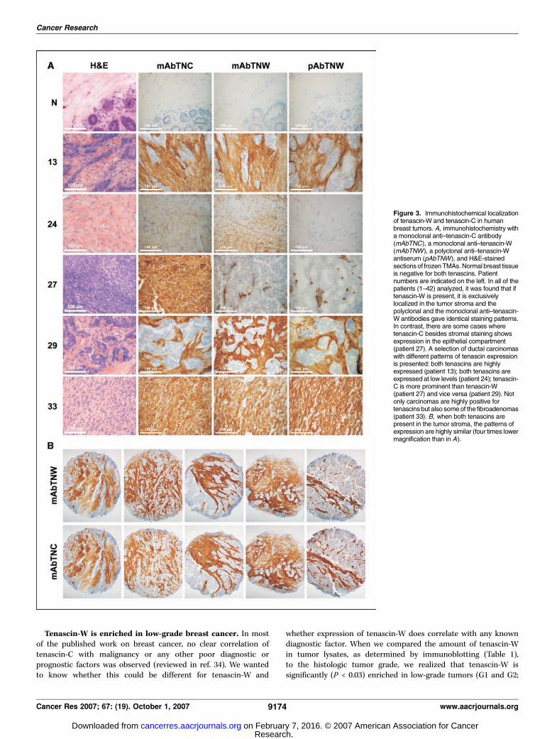
localize the proteins within the tissues, we made use of frozenTMAs. The breast cancer TMA contained spots from the tumorsof patients 1 to 42 (see Table 1; Fig. 2) and a second TMAcontained spots from 10 normal mammary tissues. In 36 of the 42cases, the staining revealed a very strong expression of tenascin-Cin the tumor stroma surrounding the transformed epithelial cellsbut no staining in the normal tissues. In four cases, tenascin-Cstaining was also present within the epithelial compartment of
the tumors (Table 1). These tumors all belong to high-grade G3breast cancers. In contrast, tenascin-W was exclusively detectedin the tumor stroma in 34 of the 42 patients and normal tissuewas negative. Examples of the different staining patterns areshown in Fig. 3. Both monoclonal and polyclonal anti–tenascin-Wantibodies gave the same staining patterns (Fig. 3A) and in mostcases stromal staining for tenascin-C overlapped with tenascin-Wstaining (Fig. 3B). In patient 24, only very faint stromal stainingis seen for either tenascin antibody consistent with the corres-ponding Western blots that were weakly positive for tenascin-Cand negative for tenascin-W. In a rare case of ductal carcinoma(patient 27), tenascin-C staining was observed throughout thetumor, including the transformed epithelial cells, whereastenascin-W is only expressed in the stromal compartment. Also,benign tumors were rich in tenascin-W and tenascin-C. Patient 33gives an example of a fibroadenoma with strong staining by allantibodies throughout the tumor mass. The relative amounts oftenascin-W and tenascin-C in these benign tumors variedgreatly (cf. Table 1) with mean tenascin-W levels of 74.2 F 62(Fig. 4A).In summary, the immunohistochemical staining of the TMAs
confirmed our results from the immunoblotting experiments andshowed that tenascin-W is a specific marker for breast tumorstroma of benign as well as malignant lesions.
Figure 2. Tenascin-W is expressed in human mammary tumors but not in the corresponding normal tissue. Tumor extracts from 63 breast tumor patients weretested by Western blot for the presence of tenascin-W and tenascin-C. Analysis revealed high expression of tenascin-W and tenascin-C in the majority of thetested extracts. For patients 54 to 63, the corresponding normal breast tissue could also be analyzed and did not show any tenascin expression. To quantify theimmunoblots (see Materials and Methods), 25 ng of the purified proteins were loaded on each gel (see patient 63) and vinculin detection was used to normalizefor protein loading. N, normal tissue; T, tumor tissue.
Tenascin-W in Human Breast Cancer
www.aacrjournals.org 9173 Cancer Res 2007; 67: (19). October 1, 2007
Research. on February 7, 2016. © 2007 American Association for Cancercancerres.aacrjournals.org Downloaded from
Tenascin-W is enriched in low-grade breast cancer. In mostof the published work on breast cancer, no clear correlation oftenascin-C with malignancy or any other poor diagnostic orprognostic factors was observed (reviewed in ref. 34). We wantedto know whether this could be different for tenascin-W and
whether expression of tenascin-W does correlate with any knowndiagnostic factor. When we compared the amount of tenascin-Win tumor lysates, as determined by immunoblotting (Table 1),to the histologic tumor grade, we realized that tenascin-W issignificantly (P < 0.03) enriched in low-grade tumors (G1 and G2;
Figure 3. Immunohistochemical localizationof tenascin-W and tenascin-C in humanbreast tumors. A, immunohistochemistry witha monoclonal anti–tenascin-C antibody(mAbTNC), a monoclonal anti–tenascin-W(mAbTNW), a polyclonal anti–tenascin-Wantiserum (pAbTNW), and H&E-stainedsections of frozenTMAs.Normal breast tissueis negative for both tenascins. Patientnumbers are indicated on the left. In all of thepatients (1–42) analyzed, it was found that iftenascin-W is present, it is exclusivelylocalized in the tumor stroma and thepolyclonal and the monoclonal anti–tenascin-W antibodies gave identical staining patterns.In contrast, there are some cases wheretenascin-C besides stromal staining showsexpression in the epithelial compartment(patient 27). A selection of ductal carcinomaswith different patterns of tenascin expressionis presented: both tenascins are highlyexpressed (patient 13); both tenascins areexpressed at low levels (patient 24); tenascin-C is more prominent than tenascin-W(patient 27) and vice versa (patient 29). Notonly carcinomas are highly positive fortenascinsbut also someof the fibroadenomas(patient 33). B, when both tenascins arepresent in the tumor stroma, the patterns ofexpression are highly similar (four times lowermagnification than in A).
Cancer Research
Cancer Res 2007; 67: (19). October 1, 2007 9174 www.aacrjournals.org
Research. on February 7, 2016. © 2007 American Association for Cancercancerres.aacrjournals.org Downloaded from

49.0 F 47) compared with high-grade tumors (G3; 21.6 F 30). Themean relative amount of tenascin-W in G1/G2 tumors is >2-foldhigher than in G3 tumors (Fig. 4A). In contrast, there is nosignificant difference between high-grade (24.2 F 23) and low-grade tumors (25.9 F 22) with respect to tenascin-C expression(Fig. 4B). This correlation between higher tenascin-W expressionand low-grade breast tumors could be confirmed in anindependent RNA profiling study of different breast cancerpatients done by van’t Veer et al. (35), accessible in theONCOMINE database (36). The heat maps presented in Fig. 4Cshow enrichment in tenascin-W transcripts in G1/G2 tumorscompared with G3 tumors, whereas there is no obvious tendencyvisible in the same samples for the tenascin-C transcripts. Inanother study by Farmer et al. (37), transcript profiling was doneof basal versus luminal breast carcinomas. Interestingly, tenascin-W is highly elevated in luminal compared with basal carcinomas,indicating that it may correlate with the estrogen receptor (ER)status as well (Fig. 4D).
Tenascin-W promotes fibroblast adhesion and stimulatescancer cell migration toward fibronectin. Because tenascin-W ispresent in the stroma surrounding tumors, we investigated its effectson stromal cells such as fibroblasts as well as on the neighboringcancer cells. Tenascin-C is an antiadhesive protein with adhesion-modulating effects leading to the expression of growth-promotingproteins (22, 23). To elucidate whether tenascin-W might act in asimilar way, we first did cell adhesion assays using different breastcancer cell lines and normal human fibroblasts (Fig. 5A). The breastcancer cells T47D neither spread nor attached to a tenascin-Wsubstratum, whereas they adhered to fibronectin or type I collagen.The same was the case for two other cell lines tested (MCF-7 andMDA-MB-435; not shown). Furthermore, we could confirm theadhesion-modulating effect by tenascin-C on breast cancer celladhesion to fibronectin. In this respect, tenascin-W differed fromtenascin-C because it did not affect tumor cell adhesion when it wasoffered as a mixed substratum with fibronectin or type I collagen. Incontrast to tumor cells, fibroblasts did attach to a tenascin-W
Figure 4. Tenascin-W is enriched in low-grade tumors. A, histograms showing the quantification of the immunoblot analysis of breast cancer extracts indicate astatistically significant higher mean tenascin-W expression in low-grade tumors (G1/G2; 49 F 47) compared with its expression in high-grade tumors (G3; 21.6 F 30).The mean tenascin-W amount in fibroadenomas (Ad ; 74.2F 62) is similar to the mean value in low-grade tumors. Quantification was done as described in Materials andMethods. Below detection has been set to 0.1. B, in contrast to tenascin-W, there is no correlation between tenascin-C expression and tumor grade. Bars, meanexpression value of the corresponding tumor grade. C, heat maps of tenascin-W and tenascin-C of an RNA profiling study of 117 breast cancer patients (35, 36).Enrichment of tenascin-W transcripts in low-grade tumors is confirmed in this study (P = 4.8E10�5), whereas tenascin-C transcripts do not show any correlationwith tumor grade. D, heat maps of tenascin-W and tenascin-C of an independent RNA profiling study of 43 breast cancer patient reveals that tenascin-W isalmost exclusively found to be elevated in luminal breast cancers, whereas some of the basal breast cancers show high levels of tenascin-C (36, 37).
Tenascin-W in Human Breast Cancer
www.aacrjournals.org 9175 Cancer Res 2007; 67: (19). October 1, 2007
Research. on February 7, 2016. © 2007 American Association for Cancercancerres.aacrjournals.org Downloaded from

substratum and partially spread (Fig. 5A). This property is unique fortenascin-W, because on tenascin-C fibroblasts remained round. Whena mixed substratum of the two tenascin proteins was used, theantiadhesive effect of tenascin-C was slightly counteracted bytenascin-W. However, there is a distinct morphology of the spreadfibroblasts on tenascin-W as revealed by phalloidin staining whencompared with fibroblasts plated on fibronectin, type I collagen, or
tenascin-C (Fig. 5B). Fibroblasts plated on fibronectin and type Icollagen form actin stress fibers and a lot of focal contacts visualizedby staining for vinculin (Fig. 5B, arrowhead). The same cells plated ontenascin-W appeared much more compact and irregularly shaped.They fail to form long actin cables, but instead produce many longactin-rich protrusions (Fig. 5B, arrows), which are also rich in vinculin(Fig. 5B, arrows). In contrast, the few fibroblasts that attached to
Figure 5. Tenascin-W promotes fibroblast adhesion and stimulates cancer cell migration. A, T47D breast cancer cells and Detroit 551 fibroblasts were plated for 2 hon the indicated single or mixed substrata before fixation, staining, and counting of adherent cells. Breast cancer cells (T47D) did not adhere to tenascin-W andtenascin-W does not have adhesion-modulating effects when mixed with fibronectin or collagen type I. In contrast, the adhesion-modulating effect of tenascin-Cmixed with fibronectin was confirmed. Compared with T47D cells, Detroit 551 fibroblasts can attach to and partially spread on tenascin-W. However, an adhesion-modulating effect of tenascin-W was not observed when mixed with other ECM proteins. Again, addition of tenascin-C statistically significantly (P < 0.002) inhibits cellspreading on fibronectin. B, phalloidin and vinculin staining of Detroit 551 fibroblasts plated on tenascin-W or fibronectin. On fibronectin, fibroblasts formed longand thick actin cables and many focal contacts (arrowhead ). On tenascin-W, actin fibers were absent and cells exhibited irregular shapes and formed long actin-rich protrusions (arrow) that were also positive for vinculin (arrow ). Adhesion to tenascin-W is integrin mediated because addition of a function-blocking anti-integrin h1antibody (h1 blocking) inhibited cell attachment and spreading. C, integrin-deficient CHOB2 cells and single integrin-expressing CHOB2 cells were plated for 20 hon the indicated ECM proteins before they were fixed, stained, and photographed. Integrin avh1- and a4h1-expressing cells were able to adhere and spread ontenascin-W, whereas a5h1-expressing cells preferentially attached and spread on fibronectin. None of the cells adhered to tenascin-C or BSA. D, transwell migrationassay shows that addition of soluble tenascin-W or tenascin-C to the lower chamber leads to a statistically significant (P < 0.001) stimulation of T47D breastcancer cells across a fibronectin-coated filter.
Cancer Research
Cancer Res 2007; 67: (19). October 1, 2007 9176 www.aacrjournals.org
Research. on February 7, 2016. © 2007 American Association for Cancercancerres.aacrjournals.org Downloaded from

tenascin-C did not spread and remained with a round morphology(not shown). Adhesion to tenascin-Wwas integrin mediated, becauseaddition of anti-h1 integrin antibodies inhibited adhesion to tenascin-W (Fig. 5B). To find out which h1 integrin(s) is able to mediateadhesion to tenascin-W, wemade use of CHOB2 cells expressing singleintegrin a chains (29–31) and compared their morphology uponadhesion to tenascin-W, tenascin-C, or fibronectin (Fig. 5C). Integrin-deficient CHOB2 cells did not adhere to any substratum. Integrina5h1–expressing cells adhered and spread preferentially on fibronec-tin, whereas avh1- and a4h1-expressing cells adhered to fibronectinand tenascin-W substrata with similar efficiency. Therefore, avh1 anda4h1 can serve as receptors for cell adhesion to tenascin-W. None ofthe CHOB2 integrin-expressing cells tested adhered to tenascin-C– orBSA-coated plates (Fig. 5C).Finally, we investigated the effect of tenascin-W on cell migration
of breast cancer cells using transfilter migration chambers with theunderside of the filters coated with fibronectin. Addition of solubletenascin-W to the lower chamber stimulated T47D cell migrationacross the filters toward the fibronectin substratum (Fig. 5D).Migration toward fibronectin was enhanced 2-fold by the presenceof tenascin-W in the culture medium, whereas cells did not migratetoward BSA. A similar migration-stimulatory effect was observedby the addition of soluble tenascin-C to the lower chamber of thetranswell system (Fig. 5D). Therefore, the presence of tenascin-C aswell as tenascin-W in tumor stroma may stimulate migration ofcancer and cancer-associated cells.
Discussion
Here, we present the first report about human tenascin-W, thefinal member of the human tenascin family. We cloned the humantenascin-W full-length cDNA, expressed it in mammalian cellculture, and purified the protein. Human tenascin-W is built upfrom the same domain types as the other three known tenascins,tenascin-C, tenascin-R, and tenascin-X, namely heptad repeats,3.5 EGF-like repeats, 9 FN III domains, and a COOH-terminalfibrinogen globe (see review in ref. 32). Although there are a lot ofalternatively spliced variants in the other tenascins (38–41), whichshow different expression pattern and functions (42–44), there isno evidence for the existence of splice variants in tenascin-W,because we detected a single protein on immunoblots of tumorextracts.Tenascin-W was originally identified in zebrafish, where it is
coexpressed with tenascin-C in somites and by neural crest cells(45). More recently, murine (46) and chicken (47) tenascin-W havebeen described. In both species, tenascin-W is expressed insmooth muscle cells and bone. Chicken tenascin-W modulatedadhesion and spreading of calvarial cells in vitro (47). In certainmurine mammary tumors, tenascin-W was up-regulated and itspresence appeared to correlate with the metastatic potential of thetumors (24).In this report, we investigated the expression of tenascin-W in
human breast cancer and found that 81% of the tumors testedexpressed tenascin-W and 86% were positive for tenascin-C.However, the amount of tenascin-C and tenascin-W differedbetween samples, indicating independent mechanisms that regulatetheir expression. In our previous study on mouse tenascin-W inprimary mouse embryo fibroblasts, we found that BMP-2 was apotent inducer of tenascin-W but not tenascin-C expression, andtransforming growth factor-h showed the opposite effect andinduced tenascin-C expression much more than that of tenascin-W(24). On the other hand, tumor necrosis factor-a strongly induced
both proteins. It is possible that the relative amounts of thesecytokines could account at least partially for the differentialexpression of the two tenascins in the tumor stroma of breastcancer.We did not detect tenascin-W in normal human mammary tissue
but could correlate tenascin-W expression levels with tumor grade.There is a statistically significant higher mean expression oftenascin-W in low-grade tumors (G1/G2) compared with high-grade tumors (G3). In contrast, in the present study, tenascin-Ccould not be correlated with tumor grade in mammary cancer. Thedifferentially expressed tenascin-C isoforms could not be correlatedwith tumor grade either, although there is one report showing thatsome specific tenascin-C isoforms are only expressed in invasivebreast carcinomas (48). However, in the transcript profiling studyby Farmer et al. (37) extracted from ONCOMINE (36), only cases ofbasal cancers showed very high levels of tenascin-C, whereastenascin-W was almost exclusively found to be elevated in luminalcancers. This indicates that tenascin-W might be elevated in ER-positive cancers, because ER-positive cancer cells tend to beenriched in luminal cancers. In contrast, high tenascin-Cexpression in basal cancers suggests a correlation with ER-negativecells enriched in basal cancers known to have a worse prognosis(49). In the literature, contradictory studies have been published onthe value of tenascin-C as a prognostic marker in breast cancer,indicating that the correlations may not be very strong and maydepend on the sampling (reviewed in ref. 34). In the case oftenascin-W, the data seem to be more consistent. In support of ourfindings, tenascin-W enrichment in low-grade breast cancers wasindependently confirmed in a different patient cohort by RNAprofiling studies of breast cancer patients (35). These data areavailable from the ONCOMINE database. A good correlationbetween tenascin-W but not tenascin-C transcript levels andtumor grade could be found in this data set (cf. Fig. 4). Tenascin-Cis, however, a useful prognostic marker for other types of tumorsthan breast cancer such as gliomas and lung cancer and in thesecases seems to play a role in tumorigenesis ( for review, see ref. 34).In many tumors, tenascin-C expression correlates with invasionand angiogenesis, whereas tenascin-C–deficient mice exhibitimpaired angiogenesis (50, 51).In the majority of breast cancers analyzed by immunohisto-
chemistry, there was an almost perfect overlap between tenascin-Cand tenascin-W expression in the tumor stroma, suggesting thatboth tenascins may serve similar but not identical functions. Incontrast to tenascin-C, tenascin-W is an adhesive substratum forfibroblasts. They attached to tenascin-W and partially spread withan irregular cell shape that differed from fibroblasts on fibronectinor collagen type I where cells formed actin stress fibers and focaladhesions. The adhesion to tenascin-W was dependent on h1integrins and CHOB2 cells expressing either avh1 or a4h1 adheredand spread on tenascin-W to a similar extent as to their knownECM ligand fibronectin. Thus, tenascin-W is a novel ligand forthese two integrins.Although both tenascins do not support breast cancer cell
adhesion when they are used as a single substratum, they differ intheir action when used as mixed substrata with fibronectin.Although tenascin-C inhibited cancer cell spreading on fibronectin,tenascin-W did not interfere with cell binding to fibronectin or typeI collagen. Interestingly, both tenascins were able to induce cancercell migration toward fibronectin. Because in vivo , the stromalECM surrounding cancer cells contains a mixture of both tenascinstogether with many other ECM proteins, they may be part of an
Tenascin-W in Human Breast Cancer
www.aacrjournals.org 9177 Cancer Res 2007; 67: (19). October 1, 2007
Research. on February 7, 2016. © 2007 American Association for Cancercancerres.aacrjournals.org Downloaded from
activated tumor stroma promoting cancer cell migration andinvasion. Because benign tumors can also exhibit high levels ofstromal tenascin-W and tenascin-C, the presence of these twoproteins is not sufficient to induce invasion. In fibroadenomas, theintact basement membrane separating the epithelial cells from thestromal compartment may protect them from the promigratoryeffect of the tenascins. The situation is different in carcinomaswhere additional factors lead to the breakdown of basementmembranes and expose the epithelial cancer cells to the stromalenvironment.For a long time, cancer research was mainly focused on the
cancer cells alone. An amazing wealth of information aboutoncogenes and tumor suppressors was obtained, which led to theimprovement of our understanding of the molecular eventsoccurring in cancer cells and the proteins and signaling pathwaysaffected represent promising new therapeutic targets. However,in recent years, it became clear that a cancer cell requires apermissive environment for progression and that carcinogenesis is
accompanied by several changes in the stroma, which finally leadsto an aberrant microenvironment that facilitates tumor growth andinvasion (reviewed in refs. 4, 5). Because tenascin-W is expressed inlower-grade breast cancers and enhances cell migration, it mightbe an early marker of activated tumor stroma and thus a goodantitumor target.
Acknowledgments
Received 2/20/2007; revised 7/10/2007; accepted 7/18/2007.Grant support: Swiss National Science Foundation SNF 3100A0-114103/1 and
Oncosuisse OCS-01419-08-2003 (G. Orend).The costs of publication of this article were defrayed in part by the payment of page
charges. This article must therefore be hereby marked advertisement in accordancewith 18 U.S.C. Section 1734 solely to indicate this fact.
We thank Peter Hans Schraml for supplying us with breast cancer lysates, SusanneSchenk for the generation of mAbs, Sandrine Bichet for help with immunohisto-chemistry, Erika Fluri for generating the subclone 2C8 of CHOB2a27 cells, and ErkkiRuoslahti (Burnham Institute, La Jolla, CA) and Jean Schwarzbauer (Department ofMelecular Biology, Princeton University, Princeton, NJ) for the gifts of CHOB2v7 andCHOa4b1 cells.
References1. Bissell MJ, Radisky D. Putting tumours in context. NatRev Cancer 2001;1:46–54.
2. Kalluri R. Basement membranes: structure, assemblyand role in tumour angiogenesis. Nat Rev Cancer 2003;3:422–33.
3. Mueller MM, Fusenig NE. Friends or foes—bipolareffects of the tumour stroma in cancer. Nat Rev Cancer2004;4:839–49.
4. Beacham DA, Cukierman E. Stromagenesis: thechanging face of fibroblastic microenvironmentsduring tumor progression. Semin Cancer Biol 2005;15:329–41.
5. Bhowmick NA, Moses HL. Tumor-stroma interactions.Curr Opin Genet Dev 2005;15:97–101.
6. Kalluri R, Zeisberg M. Fibroblasts in cancer. Nat RevCancer 2006;6:392–401.
7. Joesting MS, Perrin S, Elenbaas B, et al. Identificationof SFRP1 as a candidate mediator of stromal-to-epithelial signaling in prostate cancer. Cancer Res2005;65:10423–30.
8. Yang F, Tuxhorn JA, Ressler SJ, McAlhany SJ, Dang TD,Rowley DR. Stromal expression of connective tissuegrowth factor promotes angiogenesis and prostatecancer tumorigenesis. Cancer Res 2005;65:8887–95.
9. Cruz-Munoz W, Kim I, Khokha R. TIMP-3 deficiency inthe host, but not in the tumor, enhances tumor growthand angiogenesis. Oncogene 2005;26:650–5.
10. Hill R, Song Y, Cardiff RD, Van Dyke T. Selectiveevolutionof stromalmesenchymewith p53 loss in responseto epithelial tumorigenesis. Cell 2005;123:1001–11.
11. Kammertoens T, Schuler T, Blankenstein T. Immu-notherapy: target the stroma to hit the tumor. TrendsMol Med 2005;11:225–31.
12. Lee J, Fassnacht M, Nair S, Boczkowski D, Gilboa E.Tumor immunotherapy targeting fibroblast activationprotein, a product expressed in tumor-associatedfibroblasts. Cancer Res 2005;65:11156–63.
13. Liotta LA, Kohn EC. The microenvironment of thetumour-host interface. Nature 2001;411:375–9.
14. Chiquet-Ehrismann R, Mackie EJ, Pearson CA,Sakakura T. Tenascin: an extracellular matrix proteininvolved in tissue interactions during fetal developmentand oncogenesis. Cell 1986;47:131–9.
15. Orend G. Potential oncogenic action of tenascin-C intumorigenesis. Int J Biochem Cell Biol 2005;37:1066–83.
16. Chiquet-Ehrismann R, Chiquet M. Tenascins: regula-tion and putative functions during pathological stress.J Pathol 2003;200:488–99.
17. Jones FS, Jones PL. The tenascin family of ECMglycoproteins: structure, function, and regulation during
embryonic development and tissue remodeling. DevDyn 2000;218:235–59.
18. Zagzag D, Friedlander DR, Dosik J, et al. Tenascin-Cexpression by angiogenic vessels in human astrocyto-mas and by human brain endothelial cells in vitro.Cancer Res 1996;56:182–9.
19. Schnyder B, Semadeni RO, Fischer RW, et al. Distribu-tion pattern of tenascin-C in normal and neoplasticmesenchymal tissues. Int J Cancer 1997;72:217–24.
20. Miller DW, Vosseler S, Mirancea N, et al. Rapid vesselregression, protease inhibition, and stromal normaliza-tion upon short-term vascular endothelial growth factorreceptor 2 inhibition in skin carcinoma heterotrans-plants. Am J Pathol 2005;167:1389–403.
21. Zagzag D, Capo V. Angiogenesis in the centralnervous system: a role for vascular endothelial growthfactor/vascular permeability factor and tenascin-C.Common molecular effectors in cerebral neoplasticand non-neoplastic ‘‘angiogenic diseases.’’ Histol Histo-pathol 2002;17:301–21.
22. Huang W, Chiquet-Ehrismann R, Moyano JV, Garcia-Pardo A, Orend G. Interference of tenascin-C withsyndecan-4 binding to fibronectin blocks cell adhesionand stimulates tumor cell proliferation. Cancer Res2001;61:8586–94.
23. Ruiz C, Huang W, Hegi ME, et al. Growth promotingsignaling by tenascin-C [corrected]. Cancer Res 2004;64:7377–85.
24. Scherberich A, Tucker RP, Degen M, Brown-Luedi M,Andres AC, Chiquet-Ehrismann R. Tenascin-W is foundin malignant mammary tumors, promotes a8 integrin-dependent motility and requires p38MAPK activity forBMP-2 and TNF-a induced expression in vitro . Onco-gene 2005;24:1525–32.
25. Lange K, Kammerer M, Hegi M, et al. Endothelinreceptor type B counteracts tenascin-C-induced endothe-lin receptor type A-dependent focal adhesion and actinstress fiber disorganization. Cancer Res 2007;67:6163–73.
26. Schenk S, Muser J, Vollmer G, Chiquet-Ehrismann R.Tenascin-C in serum: a questionable tumor marker. IntJ Cancer 1995;61:443–9.
27. Schoenberg Fejzo M, Slamon DJ. Frozen tumor tissuemicroarray technology for analysis of tumor RNA, DNA,proteins. Am J Pathol 2001;159:1645–50.
28. Schreiner CL, Bauer JS, Danilov YN, Hussein S,Sczekan MM, Juliano RL. Isolation and characterizationof Chinese hamster ovary cell variants deficient in theexpression of fibronectin receptor. J Cell Biol 1989;109:3157–67.
29. Giancotti FG, Ruoslahti E. Elevated levels of the a5h1fibronectin receptor suppress the transformed pheno-type of Chinese hamster ovary cells. Cell 1990;60:849–59.
30. Irie A, Kamata T, Takada Y. Multiple loop structurescritical for ligand binding of the integrin a4 subunit inthe upper face of the h-propeller mode 1. Proc NatlAcad Sci U S A 1997;94:7198–203.
31. Zhang Z, Morla AO, Vuori K, Bauer JS, Juliano RL,Ruoslahti E. The avh1 integrin functions as a fibronectinreceptor but does not support fibronectin matrixassembly and cell migration on fibronectin. J Cell Biol1993;122:235–42.
32. Tucker R, Drabikowski K, Hess J, Ferralli J, Chiquet-Ehrismann R, Adams J. Phylogenetic analysis of thetenascin gene family: evidence of origin early in thechordate lineage. BMC evolutionary biology 2006;6:60.
33. Borsi L, Carnemolla B, Nicolo G, Spina B, Tanara G,Zardi L. Expression of different tenascin isoforms innormal, hyperplastic and neoplastic human breasttissues. Int J Cancer 1992;52:688–92.
34. Orend G, Chiquet-Ehrismann R. Tenascin-C inducedsignaling in cancer. Cancer Lett 2006;244:143–63.
35. van’t Veer L, Dau H, van de Vijver M, et al. Geneexpression profiling predicts clinical outcome of breastcancer. Nature 2002;415:484–5.
36. Rhodes DR, Yu J, Shanker K, et al. ONCOMINE: acancer microarray database and integrated data-miningplatform. Neoplasia 2004;6:1–6.
37. Farmer P, Bonnefoi H, Becette V, et al. Identificationof molecular apocrine breast tumours by microarrayanalysis. Oncogene 2005;24:4660–71.
38. Chiquet-Ehrismann R. Tenascins, a growing familyof extracellular matrix proteins. Experientia 1995;51:853–62.
39. Norenberg U, Wille H, Wolff JM, Frank R, Rathjen FG.The chicken neural extracellular matrix moleculerestrictin: similarity with EGF-, fibronectin type III-,and fibrinogen-like motifs. Neuron 1992;8:849–63.
40. Ikuta T, Sogawa N, Ariga H, Ikemura T, Matsumoto K.Structural analysis of mouse tenascin-X: evolutionaryaspects of reduplication of FNIII repeats in the tenascingene family. Gene 1998;217:1–13.
41. von Holst A, Egbers U, Prochiantz A, Faissner A.Neural stem/progenitor cells express 20 tenascin Cisoforms that are differentially regulated by pax6. J BiolChem 2007;282:9172–81.
42. Meiners S, Nur-e-Kamal MS, Mercado ML. Identifi-cation of a neurite outgrowth-promoting motif withinthe alternatively spliced region of human tenascin-C.J Neurosci 2001;21:7215–25.
43. Meiners S, Geller HM. Long and short splice variantsof human tenascin differentially regulate neurite out-growth. Mol Cell Neurosci 1997;10:100–16.
44. Fischer D, Tucker RP, Chiquet-Ehrismann R, AdamsJC. Cell-adhesive responses to tenascin-C splice variants
Cancer Research
Cancer Res 2007; 67: (19). October 1, 2007 9178 www.aacrjournals.org
Research. on February 7, 2016. © 2007 American Association for Cancercancerres.aacrjournals.org Downloaded from

involve formation of fascin microspikes. Mol Biol Cell1997;8:2055–75.
45. Weber P, Montag D, Schachner M, Bernhardt RR.Zebrafish tenascin-W, a new member of the tenascinfamily. J Neurobiol 1998;35:1–16.
46. Scherberich A, Tucker RP, Samandari E, Brown-LuediM, Martin D, Chiquet-Ehrismann R. Murine tenascin-W:a novel mammalian tenascin expressed in kidney and atsites of bone and smooth muscle development. J Cell Sci2004;117:571–81.
47. Meloty-Kapella CV, Degen M, Chiquet-Ehrismann R,Tucker RP. Avian tenascin-W: expression in smoothmuscle and bone, and effects on calvarial cell spreadingand adhesion in vitro . Dev Dyn 2006;235:1532–42.
48. Adams M, Jones JL, Walker RA, Pringle JH, Bell SC.Changes in tenascin-C isoform expression in invasiveand preinvasive breast disease. Cancer Res 2002;62:3289–97.
49. Sorlie T, Perou CM, Tibshirani R, et al. Geneexpression patterns of breast carcinomas distinguish
tumor subclasses with clinical implications. Proc NatlAcad Sci U S A 2001;98:10869–74.
50. Tanaka K, Hiraiwa N, Hashimoto H, Yamazaki Y,Kusakabe M. Tenascin-C regulates angiogenesis intumor through the regulation of vascular endothe-lial growth factor expression. Int J Cancer 2004;108:31–40.
51. Ballard VL, Sharma A, Duignan I, et al. Vasculartenascin-C regulates cardiac endothelial phenotype andneovascularization. Faseb J 2006;20:717–9.
Tenascin-W in Human Breast Cancer
www.aacrjournals.org 9179 Cancer Res 2007; 67: (19). October 1, 2007
Research. on February 7, 2016. © 2007 American Association for Cancercancerres.aacrjournals.org Downloaded from

2007;67:9169-9179. Cancer Res Martin Degen, Florence Brellier, Renate Kain, et al. BehaviorLow-grade Human Breast Cancer and Influences Cell Tenascin-W Is a Novel Marker for Activated Tumor Stroma in
Updated version
http://cancerres.aacrjournals.org/content/67/19/9169
Access the most recent version of this article at:
Cited articles
http://cancerres.aacrjournals.org/content/67/19/9169.full.html#ref-list-1
This article cites 51 articles, 17 of which you can access for free at:
Citing articles
http://cancerres.aacrjournals.org/content/67/19/9169.full.html#related-urls
This article has been cited by 3 HighWire-hosted articles. Access the articles at:
E-mail alerts related to this article or journal.Sign up to receive free email-alerts
Subscriptions
Reprints and
To order reprints of this article or to subscribe to the journal, contact the AACR Publications
Permissions
To request permission to re-use all or part of this article, contact the AACR Publications
Research. on February 7, 2016. © 2007 American Association for Cancercancerres.aacrjournals.org Downloaded from