Temporal requirement of the alternative-splicing factor Sfrs1 for the survival of retinal neurons
11
3923 RESEARCH ARTICLE INTRODUCTION As the genomes of different organisms are sequenced and annotated it is becoming apparent that the complexity of an organism does not depend on the total number of protein-coding genes. The current estimate for the number of protein-coding genes in the mouse (~23,049) (http://www.ensembl.org/Mus_musculus/index.html) is similar to that in Arabidopsis thaliana (Seki et al., 2002). Thus, it has been proposed that the complexity of higher metazoans must arise via the regulation of these genes at the transcriptional and post- transcriptional level. Alternative splicing (AS) is the mechanism by which exons of a single gene can be spliced in various combinations to encode a diverse set of proteins. Indeed, 74% of all human genes are known to be alternatively spliced and different tissues exhibit varying degrees of AS (Cheng et al., 2005; Johnson et al., 2003; Kampa et al., 2004). Moreover, when one considers that a single gene can produce multiple isoforms, the number of proteins encoded by the genome will most likely be much higher than the current estimates. In humans, the mRNAs that are expressed in the central nervous system (CNS) are subjected to the highest degree of AS when compared with other mature tissues (McCullough et al., 2005). However, the role of AS in CNS development is relatively unexplored. Given the complexity of AS, combined with that of CNS development, it is important that a system employed to investigate this question should be accessible, well characterized during development and relatively amenable to functional perturbation. The vertebrate retina is part of the CNS, yet is a relatively simple tissue with six neuronal cell classes (rod photoreceptors, cone photoreceptors, horizontal cells, bipolar cells, amacrine cells and ganglion cells) and one glial type (Müller glia) organized in a stereotypic manner. The birth order of each cell type is conserved, such that ganglion cells, cone photoreceptors and horizontal cells are among the first-born cell types, followed by amacrine cells, rod photoreceptors, bipolar cells and Müller glia (Rapaport et al., 2004; Sidman, 1961; Young, 1985a). The production of each postmitotic cell type begins in the central retina and expands from the center to the periphery (Rapaport et al., 2004; Young, 1985a; Young, 1985b). Moreover, retinal development has been the focus of many studies, leading to a better understanding of mechanisms that govern cell fate determination and differentiation (Cepko, 1996; Livesey and Cepko, 2001). Thus, the current investigation focuses on understanding the role of an alternative-splicing factor (ASF) called splicing factor arginine/serine-rich 1 (Sfrs1) in retinal development. Sfrs1 belongs to a highly conserved arginine/serine-rich (SR) protein family of RNA processing factors found throughout metazoans and in plants (Zahler, 1999; Zahler et al., 1992). The role of Sfrs1 in splicing has been well documented, but only recently has its role in any developmental context been investigated (Xu et al., 2005). In C. elegans, ablation of the Sfrs1 homolog resulted in late embryonic lethality, suggesting that its function is non-redundant in at least one critical stage of development (Kawano et al., 2000; Longman et al., 2001). In mice, the loss of Sfrs1 also resulted in embryonic lethality (Xu et al., 2005). A conditional knockout (cKO) mouse in which the Sfrs1 gene was flanked with loxP sites (Sfrs1 fl/fl ) has enabled functional studies of Sfrs1. Xu et al. have shown that loss of Sfrs1 does not cause aberrant proliferation and/or cell death of cardiac progenitor cells and that embryonic heart development proceeds normally (Xu et al., 2005). However, during postnatal remodeling of the heart, aberrant splicing of specific target genes such as cardiac troponin T (cTnT; Tnnt2 – Mouse Genome Informatics), the Z-line protein cypher (Ldb3) and Ca 2+ /calmodulin- dependent kinase II δ (CaMKIIδ; Camk2d), resulted in physiological defects in the heart (Xu et al., 2005). In this report, we show that Sfrs1 is expressed in the developing mouse retina and is itself regulated by AS. We have identified a new isoform that is expressed during late embryonic development and continues to be expressed during postnatal retinal development. This Temporal requirement of the alternative-splicing factor Sfrs1 for the survival of retinal neurons Rahul N. Kanadia 1,2 , Victoria E. Clark 1 , Claudio Punzo 1 , Jeffrey M. Trimarchi 1 and Constance L. Cepko 1,2,3, * Alternative splicing is the primary mechanism by which a limited number of protein-coding genes can generate proteome diversity. We have investigated the role of the alternative-splicing factor Sfrs1, an arginine/serine-rich (SR) protein family member, during mouse retinal development. Loss of Sfrs1 function during embryonic retinal development had a profound effect, leading to a small retina at birth. In addition, the retina underwent further degeneration in the postnatal period. Loss of Sfrs1 function resulted in the death of retinal neurons that were born during early to mid-embryonic development. Ganglion cells, cone photoreceptors, horizontal cells and amacrine cells were produced and initiated differentiation. However, these neurons subsequently underwent cell death through apoptosis. By contrast, Sfrs1 was not required for the survival of the neurons generated later, including later- born amacrine cells, rod photoreceptors, bipolar cells and Müller glia. Our results highlight the requirement of Sfrs1-mediated alternative splicing for the survival of retinal neurons, with sensitivity defined by the window of time in which the neuron was generated. KEY WORDS: Alternative splicing, Sfrs1, Survival of retinal neurons, Temporal Development 135, 3923-3933 (2008) doi:10.1242/dev.024620 1 Department of Genetics, Harvard Medical School, Boston, MA 02115, USA. 2 Howard Hughes Medical Institute, Chevy Chase, MD 20815, USA. 3 Department of Ophthalmology, Harvard Medical School, Boston, MA 02115, USA. *Author for correspondence (e-mail: [email protected]) Accepted 2 October 2008 DEVELOPMENT
Transcript of Temporal requirement of the alternative-splicing factor Sfrs1 for the survival of retinal neurons
3923RESEARCH ARTICLE
INTRODUCTIONAs the genomes of different organisms are sequenced and annotatedit is becoming apparent that the complexity of an organism does notdepend on the total number of protein-coding genes. The currentestimate for the number of protein-coding genes in the mouse(~23,049) (http://www.ensembl.org/Mus_musculus/index.html) issimilar to that in Arabidopsis thaliana (Seki et al., 2002). Thus, ithas been proposed that the complexity of higher metazoans mustarise via the regulation of these genes at the transcriptional and post-transcriptional level. Alternative splicing (AS) is the mechanism bywhich exons of a single gene can be spliced in various combinationsto encode a diverse set of proteins. Indeed, 74% of all human genesare known to be alternatively spliced and different tissues exhibitvarying degrees of AS (Cheng et al., 2005; Johnson et al., 2003;Kampa et al., 2004). Moreover, when one considers that a singlegene can produce multiple isoforms, the number of proteins encodedby the genome will most likely be much higher than the currentestimates. In humans, the mRNAs that are expressed in the centralnervous system (CNS) are subjected to the highest degree of ASwhen compared with other mature tissues (McCullough et al., 2005).However, the role of AS in CNS development is relativelyunexplored. Given the complexity of AS, combined with that ofCNS development, it is important that a system employed toinvestigate this question should be accessible, well characterizedduring development and relatively amenable to functionalperturbation.
The vertebrate retina is part of the CNS, yet is a relatively simpletissue with six neuronal cell classes (rod photoreceptors, conephotoreceptors, horizontal cells, bipolar cells, amacrine cells and
ganglion cells) and one glial type (Müller glia) organized in astereotypic manner. The birth order of each cell type is conserved,such that ganglion cells, cone photoreceptors and horizontal cells areamong the first-born cell types, followed by amacrine cells, rodphotoreceptors, bipolar cells and Müller glia (Rapaport et al., 2004;Sidman, 1961; Young, 1985a). The production of each postmitoticcell type begins in the central retina and expands from the center tothe periphery (Rapaport et al., 2004; Young, 1985a; Young, 1985b).Moreover, retinal development has been the focus of many studies,leading to a better understanding of mechanisms that govern cell fatedetermination and differentiation (Cepko, 1996; Livesey and Cepko,2001). Thus, the current investigation focuses on understanding therole of an alternative-splicing factor (ASF) called splicing factorarginine/serine-rich 1 (Sfrs1) in retinal development.
Sfrs1 belongs to a highly conserved arginine/serine-rich (SR)protein family of RNA processing factors found throughoutmetazoans and in plants (Zahler, 1999; Zahler et al., 1992). The roleof Sfrs1 in splicing has been well documented, but only recently hasits role in any developmental context been investigated (Xu et al.,2005). In C. elegans, ablation of the Sfrs1 homolog resulted in lateembryonic lethality, suggesting that its function is non-redundant inat least one critical stage of development (Kawano et al., 2000;Longman et al., 2001). In mice, the loss of Sfrs1 also resulted inembryonic lethality (Xu et al., 2005). A conditional knockout (cKO)mouse in which the Sfrs1 gene was flanked with loxP sites (Sfrs1fl/fl)has enabled functional studies of Sfrs1. Xu et al. have shown thatloss of Sfrs1 does not cause aberrant proliferation and/or cell deathof cardiac progenitor cells and that embryonic heart developmentproceeds normally (Xu et al., 2005). However, during postnatalremodeling of the heart, aberrant splicing of specific target genessuch as cardiac troponin T (cTnT; Tnnt2 – Mouse GenomeInformatics), the Z-line protein cypher (Ldb3) and Ca2+/calmodulin-dependent kinase II δ (CaMKIIδ; Camk2d), resulted in physiologicaldefects in the heart (Xu et al., 2005).
In this report, we show that Sfrs1 is expressed in the developingmouse retina and is itself regulated by AS. We have identified a newisoform that is expressed during late embryonic development andcontinues to be expressed during postnatal retinal development. This
Temporal requirement of the alternative-splicing factor Sfrs1for the survival of retinal neuronsRahul N. Kanadia1,2, Victoria E. Clark1, Claudio Punzo1, Jeffrey M. Trimarchi1 and Constance L. Cepko1,2,3,*
Alternative splicing is the primary mechanism by which a limited number of protein-coding genes can generate proteome diversity.We have investigated the role of the alternative-splicing factor Sfrs1, an arginine/serine-rich (SR) protein family member, duringmouse retinal development. Loss of Sfrs1 function during embryonic retinal development had a profound effect, leading to a smallretina at birth. In addition, the retina underwent further degeneration in the postnatal period. Loss of Sfrs1 function resulted in thedeath of retinal neurons that were born during early to mid-embryonic development. Ganglion cells, cone photoreceptors,horizontal cells and amacrine cells were produced and initiated differentiation. However, these neurons subsequently underwentcell death through apoptosis. By contrast, Sfrs1 was not required for the survival of the neurons generated later, including later-born amacrine cells, rod photoreceptors, bipolar cells and Müller glia. Our results highlight the requirement of Sfrs1-mediatedalternative splicing for the survival of retinal neurons, with sensitivity defined by the window of time in which the neuron wasgenerated.
KEY WORDS: Alternative splicing, Sfrs1, Survival of retinal neurons, Temporal
Development 135, 3923-3933 (2008) doi:10.1242/dev.024620
1Department of Genetics, Harvard Medical School, Boston, MA 02115, USA.2Howard Hughes Medical Institute, Chevy Chase, MD 20815, USA. 3Department ofOphthalmology, Harvard Medical School, Boston, MA 02115, USA.
*Author for correspondence (e-mail: [email protected])
Accepted 2 October 2008 DEVELO
PMENT
3924
novel isoform lacks the SR domain that is crucial for the nuclearlocalization of the Sfrs1 protein (Kataoka et al., 1999; Lai et al., 2000;Lai et al., 2001). Furthermore, the present investigation employs theSfrs1fl/fl mice created by Xu et al. to determine the role of Sfrs1 in thedeveloping mouse retina. To specifically ablate Sfrs1 function duringretinal development, the Sfrs1fl/fl mice were crossed to mice thatexpress Cre recombinase under the regulation of the ceh-10homeodomain-containing homolog (Chx10; Vsx2), a gene that isexpressed in retinal progenitor cells (Rowan and Cepko, 2004). Theloss of Sfrs1 function resulted in a small eye at birth. We found thatloss of Sfrs1 function did not have a significant effect on proliferation.However, neurons generated during early embryonic developmentunderwent apoptosis, whereas those generated after birth did not.Consequently, neurons generated in the embryo, such as ganglioncells, cone photoreceptors, horizontal cells and amacrine cells, weresignificantly reduced in the Sfrs1-cKO retina. By contrast, rodphotoreceptors, bipolar cells, late-born amacrine cells and Müller gliasurvived in the Sfrs1-cKO retina. The subset of susceptible neuronswas defined primarily by the time of their birth, a possible reflectionof the temporal heterogeneity in gene expression that has been definedfor retinal progenitor cells (Trimarchi et al., 2008).
MATERIALS AND METHODSAnimal proceduresTo generate Sfrs1-cKO retinae, Sfrs1fl/fl mice were crossed to Chx10::Cremice. Mice were genotyped for Sfrs1 by PCR (Xu et al., 2005). The presenceof the Cre allele was also confirmed by PCR (Rivera-Feliciano and Tabin,2006). When mice were subjected to surgical procedures, all IACUCprotocols to minimize pain during and after surgery were observed.
RT-PCRRetinae from different developmental time points were harvested and totalRNA prepared in Trizol following the manufacturer’s protocol (Invitrogen).For cDNA synthesis, 5 μg of total RNA from retinae harvested at varioustime points was used (Kanadia et al., 2006). PCR to detect Sfrs1 isoformswas performed with a forward (5′-ATGTCGGGAGGTGGTGTGATCC-3′)and a reverse (5′-CCAATCATCTTATGTACGAGAGCGAGATC-3′)primer for 30 cycles (95°C for 35 seconds; 58°C for 25 seconds; 68°C for 2minutes). PCR to detect Tnnt2 isoforms was performed as describedpreviously (Kanadia et al., 2003).
In situ hybridization on sections and dissociated cellsIn situ hybridization (ISH) on 16 μm cryosections and dissociated cells wasperformed as described previously (Trimarchi et al., 2007). All the probes usedin this report are as published previously (Trimarchi et al., 2008; Trimarchi etal., 2007). For Sfrs1 we employed a 3�UTR probe corresponding to bp 1620-2639 in the clone NM_173374 in the NCBI database.
Immunofluorescence For immunofluorescence (IF), 16 μm cryosections were first hydrated inphosphate-buffered saline (PBS, pH 7.4), followed by theimmunohistochemistry protocol of Kim et al. (Kim et al., 2008). Thedilutions of the primary antibodies were: chicken anti-GFP (1:2000)(Abcam); mouse anti-Pax6 (1:300) (Covance); rabbit anti-Chx10 (1:300)(Cepko laboratory); mouse anti-rhodopsin (4D2; 1:300) (Molday andMacKenzie, 1983); mouse anti-glutamine synthetase (1:300) (Chemicon);rabbit anti-red/green opsin (1:300) (Chemicon); and mouse anti-Ki67(1:250) (BD Pharmingen).
ImmunoblotThe nuclear/cytoplasmic extraction protocol from Pierce was employed onretinae (n=10) from different stages. Upon fractionation, 30 μg of proteinwas resolved on a 4-20% Tris-glycine gradient gel (Invitrogen), followed bytransfer of the proteins to a positively charged nylon membrane (Invitrogen),which was then subjected to immunoblot analysis as described previously(Kanadia et al., 2006). The primary antibodies used were mouse anti-Sfrs1(1:1000) (Lifespan) and mouse anti-Cugbp1 (1:500) (Abcam).
Electron microscopyP0 pups were harvested in 0.1 M sodium cacodylate buffer (pH 7.4),followed by fixation in 2% paraformaldehyde (PFA) and 2.5%glutaraldehyde in 0.1 M sodium cacodylate buffer. Retinae were thenprocessed by the Harvard Medical School Electron Microscopy CoreFacility.
P0 electroporationP0 pups were electroporated with pCAG-Cre and pCAG-LoxP-Stop-LoxP-GFP plasmids or pCAG-GFP plasmid alone (Matsuda and Cepko, 2004).
Viral infectionsRetroviral vectors were generated by cloning Cre after the IRES in the pQC-H2B-GFP-IRES-MCS vector (Punzo and Cepko, 2008). All viruses wereprepared as described previously (Cepko, 1989). E10 viral infections byultrasound-assisted delivery in timed-pregnant Sfrs1fl/fl and/or Sfrs1wt/wt
females were performed as described previously (Punzo and Cepko, 2008).P0 viral infections were performed as described previously (Matsuda andCepko, 2004).
BrdU pulse labelingA pregnant female at E16 was weighed and then injected with 3 μg ofBrdU/g body weight. At P7, retinae were harvested and processed for BrdUstaining by antigen retrieval as described by the manufacturer (Vector Labs).Next, the slides were fixed in 4% PFA for 20 minutes followed by two 5-minute washes with PBS. Slides were treated with 2M HCl for 30 minutesat room temperature, followed by a brief wash with 0.1 M boric acid (pH8.5) and IF was performed as described previously (Kim et al., 2008).
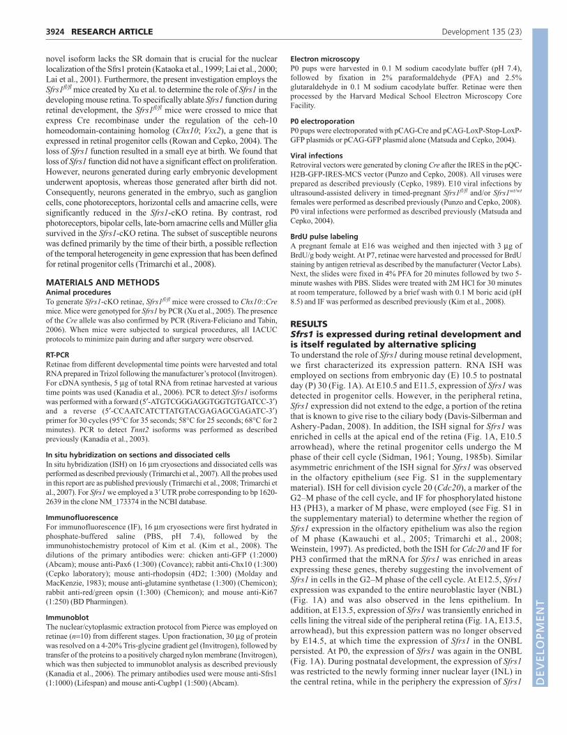
RESULTSSfrs1 is expressed during retinal development andis itself regulated by alternative splicingTo understand the role of Sfrs1 during mouse retinal development,we first characterized its expression pattern. RNA ISH wasemployed on sections from embryonic day (E) 10.5 to postnatalday (P) 30 (Fig. 1A). At E10.5 and E11.5, expression of Sfrs1 wasdetected in progenitor cells. However, in the peripheral retina,Sfrs1 expression did not extend to the edge, a portion of the retinathat is known to give rise to the ciliary body (Davis-Silberman andAshery-Padan, 2008). In addition, the ISH signal for Sfrs1 wasenriched in cells at the apical end of the retina (Fig. 1A, E10.5arrowhead), where the retinal progenitor cells undergo the Mphase of their cell cycle (Sidman, 1961; Young, 1985b). Similarasymmetric enrichment of the ISH signal for Sfrs1 was observedin the olfactory epithelium (see Fig. S1 in the supplementarymaterial). ISH for cell division cycle 20 (Cdc20), a marker of theG2–M phase of the cell cycle, and IF for phosphorylated histoneH3 (PH3), a marker of M phase, were employed (see Fig. S1 inthe supplementary material) to determine whether the region ofSfrs1 expression in the olfactory epithelium was also the regionof M phase (Kawauchi et al., 2005; Trimarchi et al., 2008;Weinstein, 1997). As predicted, both the ISH for Cdc20 and IF forPH3 confirmed that the mRNA for Sfrs1 was enriched in areasexpressing these genes, thereby suggesting the involvement ofSfrs1 in cells in the G2–M phase of the cell cycle. At E12.5, Sfrs1expression was expanded to the entire neuroblastic layer (NBL)(Fig. 1A) and was also observed in the lens epithelium. Inaddition, at E13.5, expression of Sfrs1 was transiently enriched incells lining the vitreal side of the peripheral retina (Fig. 1A, E13.5,arrowhead), but this expression pattern was no longer observedby E14.5, at which time the expression of Sfrs1 in the ONBLpersisted. At P0, the expression of Sfrs1 was again in the ONBL(Fig. 1A). During postnatal development, the expression of Sfrs1was restricted to the newly forming inner nuclear layer (INL) inthe central retina, while in the periphery the expression of Sfrs1
RESEARCH ARTICLE Development 135 (23)
DEVELO
PMENT
3925RESEARCH ARTICLESfrs1 is required for retinal neurons
Fig. 1. Sfrs1 expression during mouse retinal development. (A) In situ hybridization detecting Sfrs1 RNA during retinal development. Theschematic to the left indicates the location (box) of each row of images in the developing retina. Scale bars: 50 μm. (B) Schematic representation ofthe Sfrs1 gene, showing exons (blue boxes, 1-5), introns (green lines) and the primers (red arrows) used for RT-PCR analysis. (C) Two RT-PCR productsfrom the Sfrs1 coding sequence. The bottom panel shows Rps17, which was used as a control. The top band for Sfrs1 retains intron 3 (Sfrs1a),whereas the lower band is the canonical isoform with all four exons (Sfrs1b). (D) RT-PCR products of Sfrs1 coding sequence from heart cDNA fromdifferent developmental stages are shown along with PCR products for Tnnt2 as assessed by amplifying exon 2 to exon 6. (E) Immunoblot analysis(nuclear and cytoplasmic retinal extracts) to detect production of the new isoform of Sfrs1 at E14.5, P2 and P10. Fractionation and equal proteinloading were assayed by probing for Cugbp1. (F) Localization of the two isoforms of Sfrs1 as achieved by fusing Sfrs1a and Sfrs1b with GFP followedby transfection of NIH3T3 cells. The cells are counterstained with phalloidin (red) and DAPI (blue).RPE, retinal pigmented epithelium; GCL, ganglioncell layer; NBL, neuroblastic layer; INBL, inner neuroblastic layer; ONBL, outer neuroblastic layer. D
EVELO
PMENT
3926
was observed in progenitor cells. At P6, Sfrs1 expression becamerestricted to the lower half of the INL, where mostly amacrine anddisplaced ganglion cells are found, and to the ganglion cell layer(GCL), where ganglion and displaced amacrine cells are found(Fig. 1A). This pattern was also observed at P10 and P30 (Fig.1A).
The expression of Sfrs1 was also investigated by RT-PCRanalysis. Recent reports have indicated that the protein level ofmost of the SR-protein family members is regulated by AS(Lareau et al., 2007; Ni et al., 2007). Thus, we investigatedwhether AS might modulate the protein levels of Sfrs1 duringretinal development. To determine the AS status of Sfrs1, whichhas 5 exons (Fig. 1B), a forward primer in exon 1 (start codon)and reverse primer in exon 4 (stop codon) (Fig. 1B, red arrows)were utilized. Indeed, Sfrs1 was found to be regulated via AS ina temporal manner, such that only one isoform (Sfrs1b) wasobserved in the embryo, whereas an additional isoform at a highermolecular weight (Sfrs1a) was also expressed postnatally (Fig.1C). The temporal regulation of Sfrs1 via AS led us to investigatewhether a similar form of regulation was also employed duringthe development of other tissues. Because the role of Sfrs1 inheart development has been investigated (Xu et al., 2005), thistissue was examined. RT-PCR analysis with the same primers
showed that during embryonic heart development, both isoformsof Sfrs1 were present. By contrast, the predominant isoform at P2was the Sfrs1b isoform, which was also the only one expressed inthe adult (Fig. 1D).
Sequence analysis revealed that Sfrs1b was the canonical isoform,whereas Sfrs1a retained intron 3. Consequently, in the Sfrs1aisoform there was a frame shift in the coding sequence resulting inthe truncation of the RS domain. This change is of functionalsignificance because Sfrs1 shuttles between the cytoplasm and thenucleus and the phosphorylation of the RS domain is required for itsnuclear localization (Caceres et al., 1998; Ma et al., 2008). Thus,during retinal development, the AS of Sfrs1 produces an isoform thatwould most likely fail to translocate into the nucleus. In agreementwith the RT-PCR analysis, immunoblot analysis showed twoimmunoreactive bands in the cytoplasmic fraction from thepostnatal, but not the embryonic, retina (Fig. 1E). In addition, thelower molecular weight isoform (Sfrs1a) of Sfrs1 was significantlymore abundant in the cytoplasmic than in the nuclear fraction (Fig.1E). Since the Sfrs1a isoform was predicted to be cytoplasmic, thelow-level detection of Sfrs1a in the nuclear fraction might be due tocross-contamination between the two fractions. The inability of theSfrs1a isoform to shuttle into the nucleus was further investigatedby fusing the coding sequence of Sfrs1a and Sfrs1b to GFP, followed
RESEARCH ARTICLE Development 135 (23)
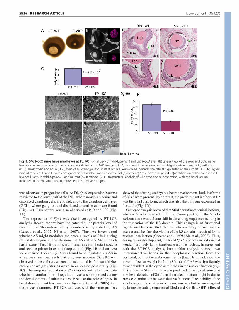
Fig. 2. Sfrs1-cKO mice have small eyes at P0. (A) Frontal view of wild-type (WT) and Sfrs1-cKO eyes. (B) Lateral view of the eyes and optic nerve.Insets show cross-sections of the optic nerves stained with DAPI (magenta). (C) Total weight comparison of wild-type (n=4) and mutant (n=4) eyes.(D,E) Hematoxylin and Eosin (H&E) stain of P0 wild-type and mutant retinae. Arrowhead indicates the retinal pigmented epithelium (RPE). (F,G) Highermagnification of D and E, with each ganglion cell nucleus marked with a dot (arrowhead) Scale bars: 100 μm. (H) Quantification of the ganglion celllayer cellularity in wild-type (n=3) and mutant (n=3) retinae. (I-L) Ultrastructural analysis of wild-type and mutant retina, with the basal laminaindicated in the mutant retina (J, arrowhead). Scale bars: 10 μm.
DEVELO
PMENT
by transfection of each plasmid into NIH3T3 cells. As predicted, thecanonical isoform, Sfrs1b, was predominantly located in thenucleus, whereas Sfrs1a was not observed in the nucleus (Fig. 1F).In summary, Sfrs1 is expressed throughout retinal development andis regulated by AS in a temporal manner.
Sfrs1-cKO mice have small eyes at birthThe role of Sfrs1 during retinal development was investigated bygenetic ablation of Sfrs1 function in retinal progenitor cells bycrossing the Sfrs1fl/fl mouse to a Chx10::Cre transgenic mouse(Rowan and Cepko, 2004). Consequently, the majority of thedeveloping retina lacked Sfrs1 function. The Sfrs1-cKO retinaeunderwent aberrant embryonic development, which was inferredfrom the observation that Sfrs1-cKO mice were born with small eyes(Fig. 2A). Sfrs1-cKO eyes also had a thin optic nerve and the totaleye weight was half that of the wild type (Fig. 2B,C). Hematoxylinand Eosin (H&E) staining of the retinal sections revealed asignificant decrease in the size of the retina, but not in the retinalpigmented epithelium (RPE) or the lens (Fig. 2D,E). The retina wasdetached from the RPE (Fig. 2E, arrowhead) while abutting the lensand exhibited a marked decrease in the cellularity of the GCL, alongwith a perturbed inner limiting membrane (ILM) (Fig. 2G,arrowhead). Quantification of the number of nuclei in the GCL (Fig.2F,G, arrowhead) confirmed the decrease in GCL cellularity (Fig.2H), which in turn was reflected in the thin optic nerve (Fig. 2B,insets). Ultrastructural analysis showed that cells in the mutant retinawere highly disorganized, with vacuolar inclusions, unlike the wild-
type retina, which had elongated nuclei with a defined polarity (Fig.2I-L). The absence of the ILM in the H&E-stained sections wasconfirmed by ultrastructural analysis (Fig. 2L, arrowhead). Themutant retina exhibited a dysmorphic fibrous layer instead of thewell-organized ILM seen in the wild-type control retina (Fig. 2K).In summary, loss of Sfrs1 function had a profound effect on theembryonic development of the retina and, possibly, also on that ofthe ciliary body, which together might have contributed to themicrophthalmia observed at birth.
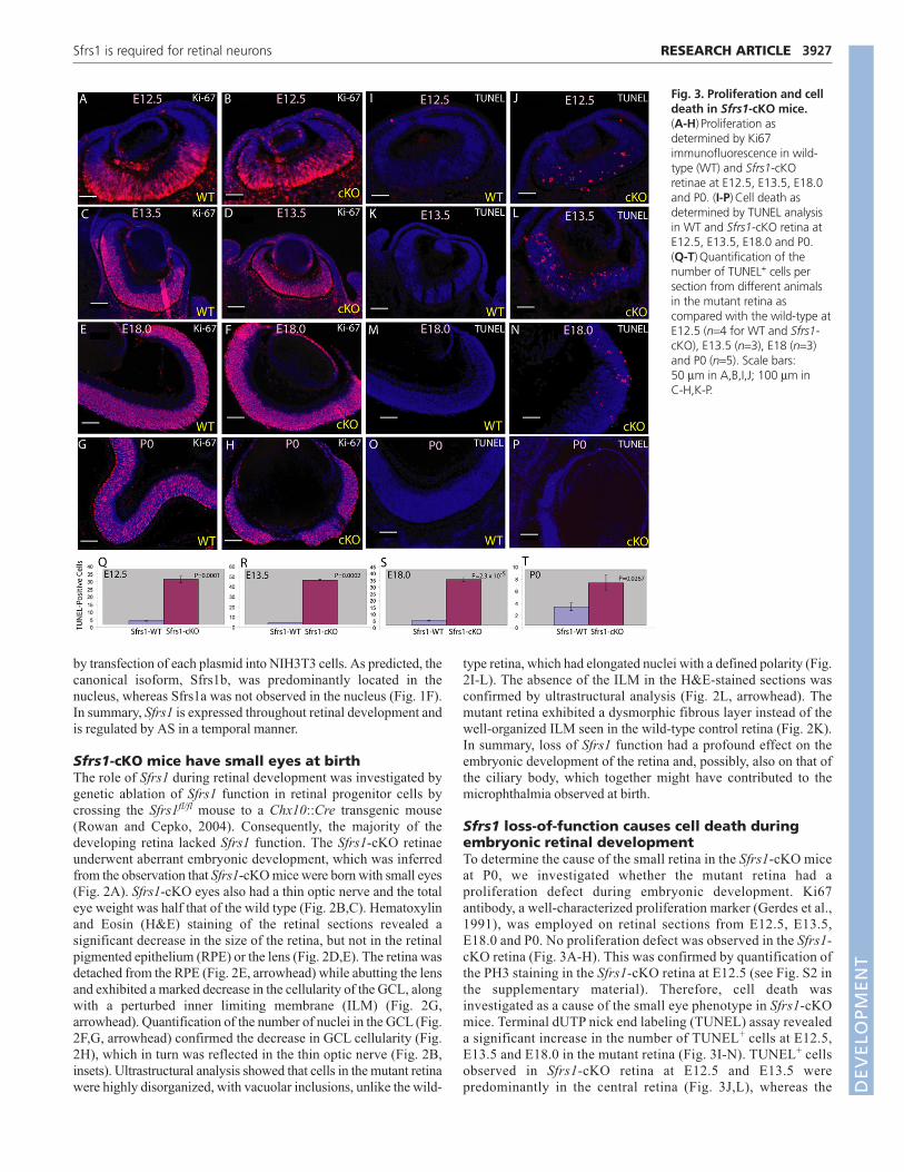
Sfrs1 loss-of-function causes cell death duringembryonic retinal developmentTo determine the cause of the small retina in the Sfrs1-cKO miceat P0, we investigated whether the mutant retina had aproliferation defect during embryonic development. Ki67antibody, a well-characterized proliferation marker (Gerdes et al.,1991), was employed on retinal sections from E12.5, E13.5,E18.0 and P0. No proliferation defect was observed in the Sfrs1-cKO retina (Fig. 3A-H). This was confirmed by quantification ofthe PH3 staining in the Sfrs1-cKO retina at E12.5 (see Fig. S2 inthe supplementary material). Therefore, cell death wasinvestigated as a cause of the small eye phenotype in Sfrs1-cKOmice. Terminal dUTP nick end labeling (TUNEL) assay revealeda significant increase in the number of TUNEL+ cells at E12.5,E13.5 and E18.0 in the mutant retina (Fig. 3I-N). TUNEL+ cellsobserved in Sfrs1-cKO retina at E12.5 and E13.5 werepredominantly in the central retina (Fig. 3J,L), whereas the
3927RESEARCH ARTICLESfrs1 is required for retinal neurons
Fig. 3. Proliferation and celldeath in Sfrs1-cKO mice.(A-H) Proliferation asdetermined by Ki67immunofluorescence in wild-type (WT) and Sfrs1-cKOretinae at E12.5, E13.5, E18.0and P0. (I-P) Cell death asdetermined by TUNEL analysisin WT and Sfrs1-cKO retina atE12.5, E13.5, E18.0 and P0.(Q-T) Quantification of thenumber of TUNEL+ cells persection from different animalsin the mutant retina ascompared with the wild-type atE12.5 (n=4 for WT and Sfrs1-cKO), E13.5 (n=3), E18 (n=3)and P0 (n=5). Scale bars:50 μm in A,B,I,J; 100 μm inC-H,K-P.
DEVELO
PMENT

3928
majority of the TUNEL+ cells at E18.0 were in the periphery(Fig. 3N). At P0, few TUNEL+ cells were observed in theperiphery (Fig. 3P). Furthermore, quantification of the TUNEL+
cells in the mutant retina at each stage revealed that the cell deathpeaked at ~E13.5, followed by a decrease in the number ofTUNEL+ cells at E18, with few TUNEL+ cells at P0 (Fig. 3Q-T).In summary, loss of Sfrs1 function during embryonicdevelopment caused cell death and did not cause an observablereduction in proliferation; however, a subtle reduction inproliferation could not be ruled out.
Sfrs1 loss-of-function does not result in the deathof retinal progenitor cellsThere were three different scenarios that could explain the smalleye phenotype. The dying cells might be progenitor cells, theymight be postmitotic cells, or they might comprise both types ofcells. Since proliferation was not reduced, it was unlikely thatprogenitor cells died. Nonetheless, this was investigated furtherbecause Sfrs1 is expressed in progenitor cells during embryonicdevelopment. For this, Sfrs1 function was ablated in retinalprogenitor cells utilizing a retrovirus expressing Cre along withnuclear GFP (histone H2B-GFP fusion). Because retroviruses canonly infect mitotic cells, the retinal progenitor cells would loseSfrs1. Moreover, the viral genome inserts into the cell genome andthereby marks all its progeny. This retrovirus was delivered toE10.5 Sfrs1fl/fl embryos with the aid of an ultrasound-guidedinjection device, followed by detection of GFPimmunofluorescence at P14 (Fig. 4). A virus encoding membrane-GFP (mGFP), without Cre, was also injected as a control for theinfection. An additional control was to repeat the sameexperiment in Sfrs1wt/wt mice. Analysis at P14 showed severalclones with nuclear GFP in the retinae of infected Sfrs1fl/fl mice(Fig. 4C), indicating the survival of the initially infectedprogenitor cells. However, there was a difference in the cell-typecomposition of the clones in the Sfrs1fl/fl and Sfrs1wt/wt mice.Based on their position, the majority of GFP+ cells in the Sfrs1fl/fl
mice were either rod photoreceptors, bipolar cells or Müller glia(Fig. 4C). By contrast, the cell types observed in the Sfrs1wt/wt
mice, as determined by the position of the nuclei, included rodphotoreceptors, horizontal cells, bipolar cells, Müller glia,amacrine cells and ganglion cells (Fig. 4B). The Cre-negativemGFP+ clones were similar in composition to those of the wildtype (data not shown). In conclusion, the loss of Sfrs1 did notcause retinal progenitor cell death, at least not in all infectedprogenitor cells.
This conclusion was further bolstered by the genetic ablationstrategy that employed the Chx10::Cre line along with a reporterline, RC::PFWE, in the Sfrs1fl/fl background. The RC::PFWEstrain reports Cre activity by expressing nuclear β-galactosidase(nlacZ) (Farago et al., 2006). Thus, every progenitor cell thatexpresses Cre should be positive for nlacZ and negative for Sfrs1function. The results showed that at least some retinal progenitorcells did not die, as there were nlacZ+ neurons in the P7 mutantretina (Fig. 4E). These neurons, as judged from their position,were mostly photoreceptors, bipolars and Müller glia (Fig. 4E).By contrast, the wild-type littermate retina showed many cellsexpressing nlacZ in the ONL (outer nuclear layer) (rodphotoreceptors, cone receptors), INL (bipolar cells, horizontalcells, amacrine cells and Müller Glia) and in the GCL (ganglionand amacrine cells) (Fig. 4D). In both the wild-type and themutant retinae there were cells that were negative for nlacZ,which was likely to result from the mosaic expression of
Chx10::Cre (Rowan and Cepko, 2004). In summary, loss of Sfrs1function did not lead to the death of all progenitor cells, althoughsome progenitor cell death could not be ruled out.
Postmitotic cells are generated in the Sfrs1-cKOretina during early embryonic developmentTo examine whether the postmitotic cells were dying in the Sfrs1-cKOretinae, we first determined whether postmitotic cells were produced.For this, RNA ISH with probes that label differentiated neurons wasemployed on E13.5 retinal sections. First, loss of the Sfrs1 transcriptin the Sfrs1-cKO retina was confirmed (Fig. 5B). Although themajority of the retina lacked Sfrs1, a few cells (Fig. 5B, arrowhead)were positive for Sfrs1, most likely reflecting the Chx10::Cremosaicism (Rowan and Cepko, 2004). ISH analysis was performedon serial sections with probes that mark postmitotic cells, includingneurofilament-like light chain 68 (Nf68; Nefl – Mouse Genome
RESEARCH ARTICLE Development 135 (23)
Fig. 4. Retinal progenitor cells survive the loss of Sfrs1.(A) Snapshot of a live ultrasound image of an E10.5 mouse embryoimaged through the uterine wall. Inset shows the newly formed opticvesicles and the diencephalon is traced in white. The right-hand panelshows a glass needle that was introduced into the ventricle where thevirus was delivered. (B) P14 retinal sections from Sfrs1wt/wt embryos thatwere injected at E10.5 with a virus expressing Cre and nuclear-GFP.Stained for GFP. The arrowhead shows the position of the amacrinecells. (C) P14 retinal sections from Sfrs1fl/fl embryos injected at E10.5 withthe same virus. The arrowhead highlights the absence of the amacrinecells. (D,E) P7 retinal sections from Sfrs1wt/wt and Sfrs1fl/fl mice that werecrossed to the RC::PFWE line, which reports the Chx10::Cre-mediatedexcision event by activating nlacZ expression. GCL, ganglion cell layer;INL, inner nuclear layer; ONL, outer neuroblastic later. Scale bars: 50 μm.
DEVELO
PMENT
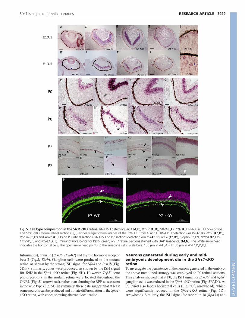
Informatics), brain 3b (Brn3b; Pou4f2) and thyroid hormone receptorbeta 2 (Trβ2; Thrb). Ganglion cells were produced in the mutantretina, as shown by the strong ISH signal for Nf68 and Brn3b (Fig.5D,F). Similarly, cones were produced, as shown by the ISH signalfor Trβ2 in the Sfrs1-cKO retina (Fig. 5H). However, Trβ2+ conephotoreceptors in the mutant retina were located throughout theONBL (Fig. 5J, arrowhead), rather than abutting the RPE as was seenin the wild type (Fig. 5I). In summary, these data suggest that at leastsome neurons can be produced and initiate differentiation in the Sfrs1-cKO retina, with cones showing aberrant localization.
Neurons generated during early and mid-embryonic development die in the Sfrs1-cKOretinaTo investigate the persistence of the neurons generated in the embryo,the above-mentioned strategy was employed on P0 retinal sections.This analysis showed that at P0, the ISH signal for Brn3b+ and Nf68+
ganglion cells was reduced in the Sfrs1-cKO retina (Fig. 5B�,D�). AtP0, Nf68 also labels horizontal cells (Fig. 5C�, arrowhead), whichwere significantly reduced in the Sfrs1-cKO retina (Fig. 5D�,arrowhead). Similarly, the ISH signal for rabphilin 3a (Rph3a) and
3929RESEARCH ARTICLESfrs1 is required for retinal neurons
Fig. 5. Cell type composition in the Sfrs1-cKO retina. RNA ISH detecting Sfrs1 (A,B), Brn3b (C,D), Nf68 (E,F), Trβ2 (G,H) RNA in E13.5 wild-typeand Sfrs1-cKO mouse retinal sections. (I,J) Higher magnification images of the Trβ2 ISH from G and H. RNA ISH detecting Brn3b (A�,B�), Nf68 (C�,D�),Rph3a (E�,F�) and Ap2b (G�,H�) on P0 retinal sections. RNA ISH on P7 sections detecting Brn3b (A�,B�), Nf68 (C�,D�), S opsin (E�,F�), Ndrg4 (G�,H�),Otx2 (I�,J�) and Nr2e3 (K,L). Immunofluorescence for Pax6 (green) on P7 retinal sections stained with DAPI (magenta) (M,N). The white arrowheadindicates the horizontal cells, the open arrowhead points to the amacrine cells. Scale bars: 100 μm in A-H,A�-H�; 50 μm in A�-H�,I�,J�,K,L.
DEVELO
PMENT
3930
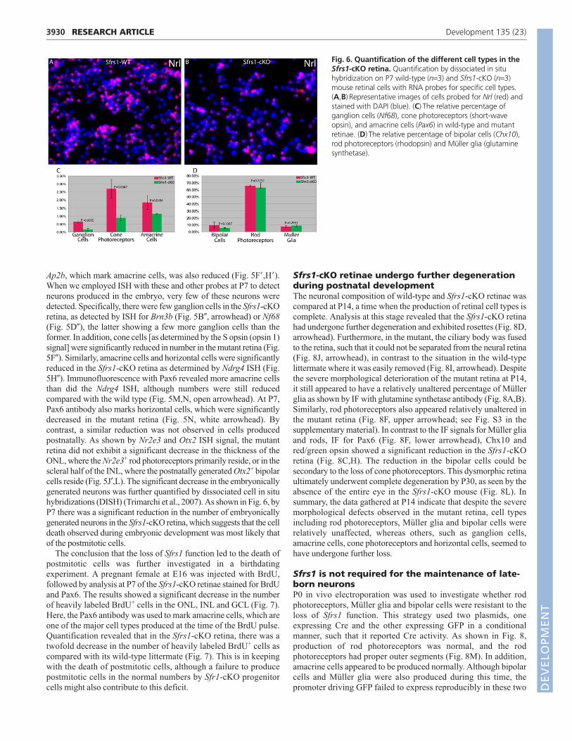
Ap2b, which mark amacrine cells, was also reduced (Fig. 5F�,H�).When we employed ISH with these and other probes at P7 to detectneurons produced in the embryo, very few of these neurons weredetected. Specifically, there were few ganglion cells in the Sfrs1-cKOretina, as detected by ISH for Brn3b (Fig. 5B�, arrowhead) or Nf68(Fig. 5D�), the latter showing a few more ganglion cells than theformer. In addition, cone cells [as determined by the S opsin (opsin 1)signal] were significantly reduced in number in the mutant retina (Fig.5F�). Similarly, amacrine cells and horizontal cells were significantlyreduced in the Sfrs1-cKO retina as determined by Ndrg4 ISH (Fig.5H�). Immunofluorescence with Pax6 revealed more amacrine cellsthan did the Ndrg4 ISH, although numbers were still reducedcompared with the wild type (Fig. 5M,N, open arrowhead). At P7,Pax6 antibody also marks horizontal cells, which were significantlydecreased in the mutant retina (Fig. 5N, white arrowhead). Bycontrast, a similar reduction was not observed in cells producedpostnatally. As shown by Nr2e3 and Otx2 ISH signal, the mutantretina did not exhibit a significant decrease in the thickness of theONL, where the Nr2e3+ rod photoreceptors primarily reside, or in thescleral half of the INL, where the postnatally generated Otx2+ bipolarcells reside (Fig. 5J′,L). The significant decrease in the embryonicallygenerated neurons was further quantified by dissociated cell in situhybridizations (DISH) (Trimarchi et al., 2007). As shown in Fig. 6, byP7 there was a significant reduction in the number of embryonicallygenerated neurons in the Sfrs1-cKO retina, which suggests that the celldeath observed during embryonic development was most likely thatof the postmitotic cells.
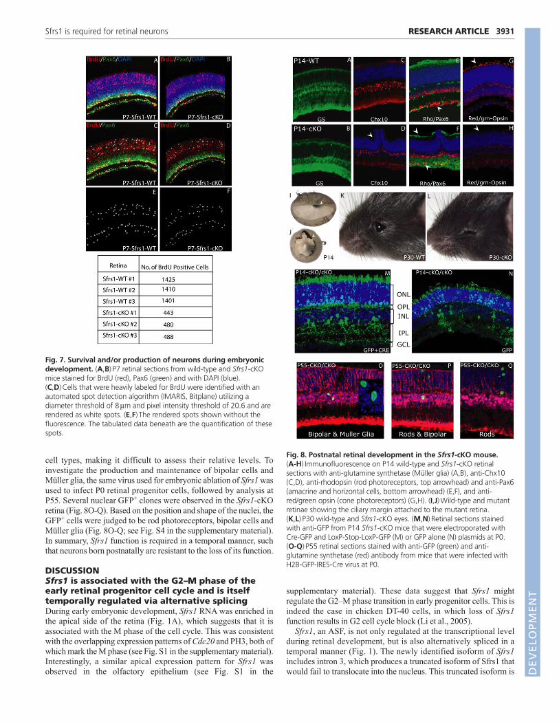
The conclusion that the loss of Sfrs1 function led to the death ofpostmitotic cells was further investigated in a birthdatingexperiment. A pregnant female at E16 was injected with BrdU,followed by analysis at P7 of the Sfrs1-cKO retinae stained for BrdUand Pax6. The results showed a significant decrease in the numberof heavily labeled BrdU+ cells in the ONL, INL and GCL (Fig. 7).Here, the Pax6 antibody was used to mark amacrine cells, which areone of the major cell types produced at the time of the BrdU pulse.Quantification revealed that in the Sfrs1-cKO retina, there was atwofold decrease in the number of heavily labeled BrdU+ cells ascompared with its wild-type littermate (Fig. 7). This is in keepingwith the death of postmitotic cells, although a failure to producepostmitotic cells in the normal numbers by Sfr1-cKO progenitorcells might also contribute to this deficit.
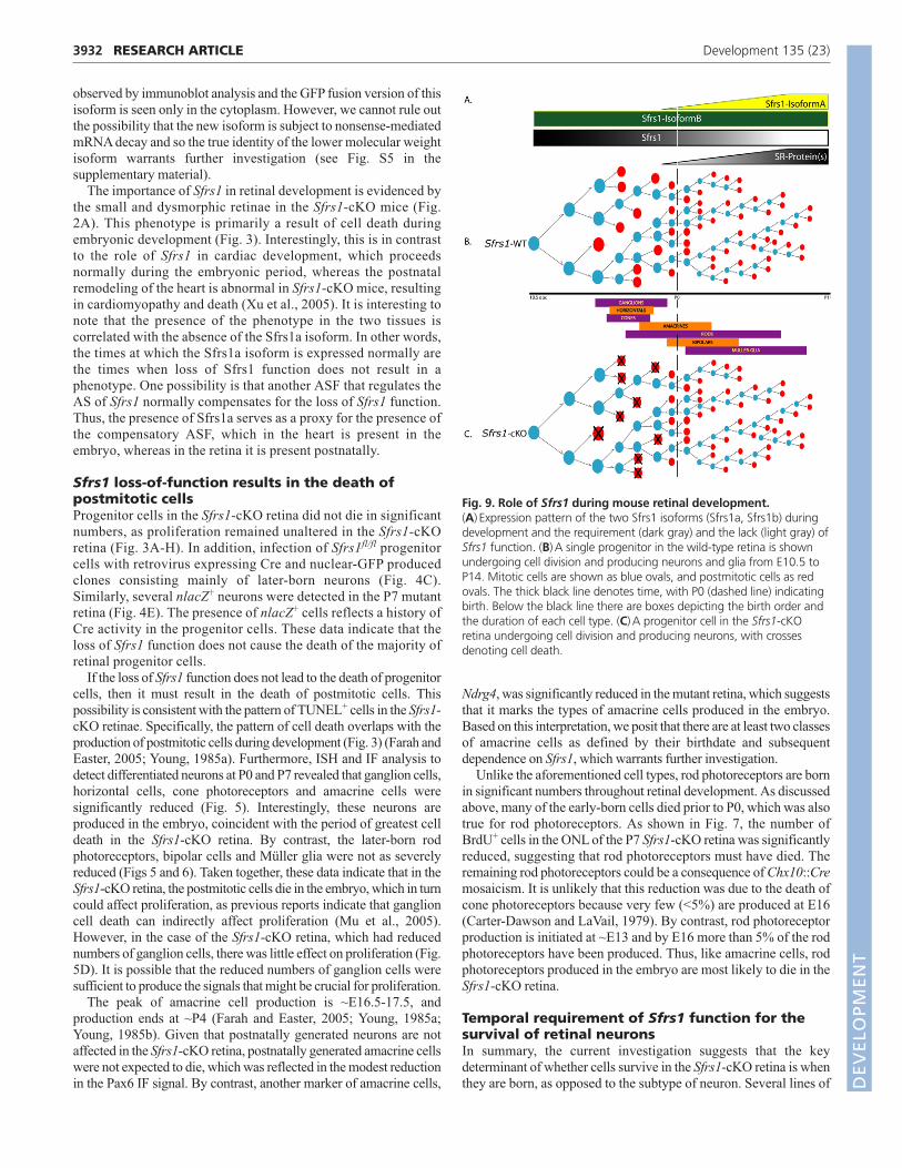
Sfrs1-cKO retinae undergo further degenerationduring postnatal developmentThe neuronal composition of wild-type and Sfrs1-cKO retinae wascompared at P14, a time when the production of retinal cell types iscomplete. Analysis at this stage revealed that the Sfrs1-cKO retinahad undergone further degeneration and exhibited rosettes (Fig. 8D,arrowhead). Furthermore, in the mutant, the ciliary body was fusedto the retina, such that it could not be separated from the neural retina(Fig. 8J, arrowhead), in contrast to the situation in the wild-typelittermate where it was easily removed (Fig. 8I, arrowhead). Despitethe severe morphological deterioration of the mutant retina at P14,it still appeared to have a relatively unaltered percentage of Müllerglia as shown by IF with glutamine synthetase antibody (Fig. 8A,B).Similarly, rod photoreceptors also appeared relatively unaltered inthe mutant retina (Fig. 8F, upper arrowhead; see Fig. S3 in thesupplementary material). In contrast to the IF signals for Müller gliaand rods, IF for Pax6 (Fig. 8F, lower arrowhead), Chx10 andred/green opsin showed a significant reduction in the Sfrs1-cKOretina (Fig. 8C,H). The reduction in the bipolar cells could besecondary to the loss of cone photoreceptors. This dysmorphic retinaultimately underwent complete degeneration by P30, as seen by theabsence of the entire eye in the Sfrs1-cKO mouse (Fig. 8L). Insummary, the data gathered at P14 indicate that despite the severemorphological defects observed in the mutant retina, cell typesincluding rod photoreceptors, Müller glia and bipolar cells wererelatively unaffected, whereas others, such as ganglion cells,amacrine cells, cone photoreceptors and horizontal cells, seemed tohave undergone further loss.
Sfrs1 is not required for the maintenance of late-born neuronsP0 in vivo electroporation was used to investigate whether rodphotoreceptors, Müller glia and bipolar cells were resistant to theloss of Sfrs1 function. This strategy used two plasmids, oneexpressing Cre and the other expressing GFP in a conditionalmanner, such that it reported Cre activity. As shown in Fig. 8,production of rod photoreceptors was normal, and the rodphotoreceptors had proper outer segments (Fig. 8M). In addition,amacrine cells appeared to be produced normally. Although bipolarcells and Müller glia were also produced during this time, thepromoter driving GFP failed to express reproducibly in these two
RESEARCH ARTICLE Development 135 (23)
Fig. 6. Quantification of the different cell types in theSfrs1-cKO retina. Quantification by dissociated in situhybridization on P7 wild-type (n=3) and Sfrs1-cKO (n=3)mouse retinal cells with RNA probes for specific cell types.(A,B) Representative images of cells probed for Nrl (red) andstained with DAPI (blue). (C) The relative percentage ofganglion cells (Nf68), cone photoreceptors (short-waveopsin), and amacrine cells (Pax6) in wild-type and mutantretinae. (D) The relative percentage of bipolar cells (Chx10),rod photoreceptors (rhodopsin) and Müller glia (glutaminesynthetase).
DEVELO
PMENT
cell types, making it difficult to assess their relative levels. Toinvestigate the production and maintenance of bipolar cells andMüller glia, the same virus used for embryonic ablation of Sfrs1 wasused to infect P0 retinal progenitor cells, followed by analysis atP55. Several nuclear GFP+ clones were observed in the Sfrs1-cKOretina (Fig. 8O-Q). Based on the position and shape of the nuclei, theGFP+ cells were judged to be rod photoreceptors, bipolar cells andMüller glia (Fig. 8O-Q; see Fig. S4 in the supplementary material).In summary, Sfrs1 function is required in a temporal manner, suchthat neurons born postnatally are resistant to the loss of its function.
DISCUSSIONSfrs1 is associated with the G2–M phase of theearly retinal progenitor cell cycle and is itselftemporally regulated via alternative splicingDuring early embryonic development, Sfrs1 RNA was enriched inthe apical side of the retina (Fig. 1A), which suggests that it isassociated with the M phase of the cell cycle. This was consistentwith the overlapping expression patterns of Cdc20 and PH3, both ofwhich mark the M phase (see Fig. S1 in the supplementary material).Interestingly, a similar apical expression pattern for Sfrs1 wasobserved in the olfactory epithelium (see Fig. S1 in the
supplementary material). These data suggest that Sfrs1 mightregulate the G2–M phase transition in early progenitor cells. This isindeed the case in chicken DT-40 cells, in which loss of Sfrs1function results in G2 cell cycle block (Li et al., 2005).
Sfrs1, an ASF, is not only regulated at the transcriptional levelduring retinal development, but is also alternatively spliced in atemporal manner (Fig. 1). The newly identified isoform of Sfrs1includes intron 3, which produces a truncated isoform of Sfrs1 thatwould fail to translocate into the nucleus. This truncated isoform is
3931RESEARCH ARTICLESfrs1 is required for retinal neurons
Fig. 7. Survival and/or production of neurons during embryonicdevelopment. (A,B) P7 retinal sections from wild-type and Sfrs1-cKOmice stained for BrdU (red), Pax6 (green) and with DAPI (blue).(C,D) Cells that were heavily labeled for BrdU were identified with anautomated spot detection algorithm (IMARIS, Bitplane) utilizing adiameter threshold of 8μm and pixel intensity threshold of 20.6 and arerendered as white spots. (E,F) The rendered spots shown without thefluorescence. The tabulated data beneath are the quantification of thesespots.
Fig. 8. Postnatal retinal development in the Sfrs1-cKO mouse. (A-H) Immunofluorescence on P14 wild-type and Sfrs1-cKO retinalsections with anti-glutamine synthetase (Müller glia) (A,B), anti-Chx10(C,D), anti-rhodopsin (rod photoreceptors, top arrowhead) and anti-Pax6(amacrine and horizontal cells, bottom arrowhead) (E,F), and anti-red/green opsin (cone photoreceptors) (G,H). (I,J) Wild-type and mutantretinae showing the ciliary margin attached to the mutant retina.(K,L) P30 wild-type and Sfrs1-cKO eyes. (M,N) Retinal sections stainedwith anti-GFP from P14 Sfrs1-cKO mice that were electroporated withCre-GFP and LoxP-Stop-LoxP-GFP (M) or GFP alone (N) plasmids at P0.(O-Q) P55 retinal sections stained with anti-GFP (green) and anti-glutamine synthetase (red) antibody from mice that were infected withH2B-GFP-IRES-Cre virus at P0.
DEVELO
PMENT
3932
observed by immunoblot analysis and the GFP fusion version of thisisoform is seen only in the cytoplasm. However, we cannot rule outthe possibility that the new isoform is subject to nonsense-mediatedmRNA decay and so the true identity of the lower molecular weightisoform warrants further investigation (see Fig. S5 in thesupplementary material).
The importance of Sfrs1 in retinal development is evidenced bythe small and dysmorphic retinae in the Sfrs1-cKO mice (Fig.2A). This phenotype is primarily a result of cell death duringembryonic development (Fig. 3). Interestingly, this is in contrastto the role of Sfrs1 in cardiac development, which proceedsnormally during the embryonic period, whereas the postnatalremodeling of the heart is abnormal in Sfrs1-cKO mice, resultingin cardiomyopathy and death (Xu et al., 2005). It is interesting tonote that the presence of the phenotype in the two tissues iscorrelated with the absence of the Sfrs1a isoform. In other words,the times at which the Sfrs1a isoform is expressed normally arethe times when loss of Sfrs1 function does not result in aphenotype. One possibility is that another ASF that regulates theAS of Sfrs1 normally compensates for the loss of Sfrs1 function.Thus, the presence of Sfrs1a serves as a proxy for the presence ofthe compensatory ASF, which in the heart is present in theembryo, whereas in the retina it is present postnatally.
Sfrs1 loss-of-function results in the death ofpostmitotic cellsProgenitor cells in the Sfrs1-cKO retina did not die in significantnumbers, as proliferation remained unaltered in the Sfrs1-cKOretina (Fig. 3A-H). In addition, infection of Sfrs1fl/fl progenitorcells with retrovirus expressing Cre and nuclear-GFP producedclones consisting mainly of later-born neurons (Fig. 4C).Similarly, several nlacZ+ neurons were detected in the P7 mutantretina (Fig. 4E). The presence of nlacZ+ cells reflects a history ofCre activity in the progenitor cells. These data indicate that theloss of Sfrs1 function does not cause the death of the majority ofretinal progenitor cells.
If the loss of Sfrs1 function does not lead to the death of progenitorcells, then it must result in the death of postmitotic cells. Thispossibility is consistent with the pattern of TUNEL+ cells in the Sfrs1-cKO retinae. Specifically, the pattern of cell death overlaps with theproduction of postmitotic cells during development (Fig. 3) (Farah andEaster, 2005; Young, 1985a). Furthermore, ISH and IF analysis todetect differentiated neurons at P0 and P7 revealed that ganglion cells,horizontal cells, cone photoreceptors and amacrine cells weresignificantly reduced (Fig. 5). Interestingly, these neurons areproduced in the embryo, coincident with the period of greatest celldeath in the Sfrs1-cKO retina. By contrast, the later-born rodphotoreceptors, bipolar cells and Müller glia were not as severelyreduced (Figs 5 and 6). Taken together, these data indicate that in theSfrs1-cKO retina, the postmitotic cells die in the embryo, which in turncould affect proliferation, as previous reports indicate that ganglioncell death can indirectly affect proliferation (Mu et al., 2005).However, in the case of the Sfrs1-cKO retina, which had reducednumbers of ganglion cells, there was little effect on proliferation (Fig.5D). It is possible that the reduced numbers of ganglion cells weresufficient to produce the signals that might be crucial for proliferation.
The peak of amacrine cell production is ~E16.5-17.5, andproduction ends at ~P4 (Farah and Easter, 2005; Young, 1985a;Young, 1985b). Given that postnatally generated neurons are notaffected in the Sfrs1-cKO retina, postnatally generated amacrine cellswere not expected to die, which was reflected in the modest reductionin the Pax6 IF signal. By contrast, another marker of amacrine cells,
Ndrg4, was significantly reduced in the mutant retina, which suggeststhat it marks the types of amacrine cells produced in the embryo.Based on this interpretation, we posit that there are at least two classesof amacrine cells as defined by their birthdate and subsequentdependence on Sfrs1, which warrants further investigation.
Unlike the aforementioned cell types, rod photoreceptors are bornin significant numbers throughout retinal development. As discussedabove, many of the early-born cells died prior to P0, which was alsotrue for rod photoreceptors. As shown in Fig. 7, the number ofBrdU+ cells in the ONL of the P7 Sfrs1-cKO retina was significantlyreduced, suggesting that rod photoreceptors must have died. Theremaining rod photoreceptors could be a consequence of Chx10::Cremosaicism. It is unlikely that this reduction was due to the death ofcone photoreceptors because very few (<5%) are produced at E16(Carter-Dawson and LaVail, 1979). By contrast, rod photoreceptorproduction is initiated at ~E13 and by E16 more than 5% of the rodphotoreceptors have been produced. Thus, like amacrine cells, rodphotoreceptors produced in the embryo are most likely to die in theSfrs1-cKO retina.
Temporal requirement of Sfrs1 function for thesurvival of retinal neuronsIn summary, the current investigation suggests that the keydeterminant of whether cells survive in the Sfrs1-cKO retina is whenthey are born, as opposed to the subtype of neuron. Several lines of
RESEARCH ARTICLE Development 135 (23)
Fig. 9. Role of Sfrs1 during mouse retinal development.(A) Expression pattern of the two Sfrs1 isoforms (Sfrs1a, Sfrs1b) duringdevelopment and the requirement (dark gray) and the lack (light gray) ofSfrs1 function. (B) A single progenitor in the wild-type retina is shownundergoing cell division and producing neurons and glia from E10.5 toP14. Mitotic cells are shown as blue ovals, and postmitotic cells as redovals. The thick black line denotes time, with P0 (dashed line) indicatingbirth. Below the black line there are boxes depicting the birth order andthe duration of each cell type. (C) A progenitor cell in the Sfrs1-cKOretina undergoing cell division and producing neurons, with crossesdenoting cell death.
DEVELO
PMENT
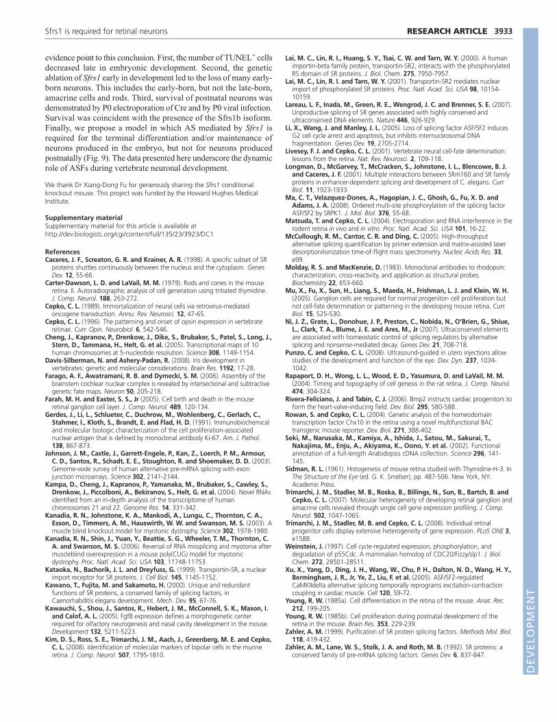
evidence point to this conclusion. First, the number of TUNEL+ cellsdecreased late in embryonic development. Second, the geneticablation of Sfrs1 early in development led to the loss of many early-born neurons. This includes the early-born, but not the late-born,amacrine cells and rods. Third, survival of postnatal neurons wasdemonstrated by P0 electroporation of Cre and by P0 viral infection.Survival was coincident with the presence of the Sfrs1b isoform.Finally, we propose a model in which AS mediated by Sfrs1 isrequired for the terminal differentiation and/or maintenance ofneurons produced in the embryo, but not for neurons producedpostnatally (Fig. 9). The data presented here underscore the dynamicrole of ASFs during vertebrate neuronal development.
We thank Dr Xiang-Dong Fu for generously sharing the Sfrs1 conditionalknockout mouse. This project was funded by the Howard Hughes MedicalInstitute.
Supplementary materialSupplementary material for this article is available athttp://dev.biologists.org/cgi/content/full/135/23/3923/DC1
ReferencesCaceres, J. F., Screaton, G. R. and Krainer, A. R. (1998). A specific subset of SR
proteins shuttles continuously between the nucleus and the cytoplasm. GenesDev. 12, 55-66.
Carter-Dawson, L. D. and LaVail, M. M. (1979). Rods and cones in the mouseretina. II. Autoradiographic analysis of cell generation using tritiated thymidine.J. Comp. Neurol. 188, 263-272.
Cepko, C. L. (1989). Immortalization of neural cells via retrovirus-mediatedoncogene transduction. Annu. Rev. Neurosci. 12, 47-65.
Cepko, C. L. (1996). The patterning and onset of opsin expression in vertebrateretinae. Curr. Opin. Neurobiol. 6, 542-546.
Cheng, J., Kapranov, P., Drenkow, J., Dike, S., Brubaker, S., Patel, S., Long, J.,Stern, D., Tammana, H., Helt, G. et al. (2005). Transcriptional maps of 10human chromosomes at 5-nucleotide resolution. Science 308, 1149-1154.
Davis-Silberman, N. and Ashery-Padan, R. (2008). Iris development invertebrates: genetic and molecular considerations. Brain Res. 1192, 17-28.
Farago, A. F., Awatramani, R. B. and Dymecki, S. M. (2006). Assembly of thebrainstem cochlear nuclear complex is revealed by intersectional and subtractivegenetic fate maps. Neuron 50, 205-218.
Farah, M. H. and Easter, S. S., Jr (2005). Cell birth and death in the mouseretinal ganglion cell layer. J. Comp. Neurol. 489, 120-134.
Gerdes, J., Li, L., Schlueter, C., Duchrow, M., Wohlenberg, C., Gerlach, C.,Stahmer, I., Kloth, S., Brandt, E. and Flad, H. D. (1991). Immunobiochemicaland molecular biologic characterization of the cell proliferation-associatednuclear antigen that is defined by monoclonal antibody Ki-67. Am. J. Pathol.138, 867-873.
Johnson, J. M., Castle, J., Garrett-Engele, P., Kan, Z., Loerch, P. M., Armour,C. D., Santos, R., Schadt, E. E., Stoughton, R. and Shoemaker, D. D. (2003).Genome-wide survey of human alternative pre-mRNA splicing with exonjunction microarrays. Science 302, 2141-2144.
Kampa, D., Cheng, J., Kapranov, P., Yamanaka, M., Brubaker, S., Cawley, S.,Drenkow, J., Piccolboni, A., Bekiranov, S., Helt, G. et al. (2004). Novel RNAsidentified from an in-depth analysis of the transcriptome of humanchromosomes 21 and 22. Genome Res. 14, 331-342.
Kanadia, R. N., Johnstone, K. A., Mankodi, A., Lungu, C., Thornton, C. A.,Esson, D., Timmers, A. M., Hauswirth, W. W. and Swanson, M. S. (2003). Amuscle blind knockout model for myotonic dystrophy. Science 302, 1978-1980.
Kanadia, R. N., Shin, J., Yuan, Y., Beattie, S. G., Wheeler, T. M., Thornton, C.A. and Swanson, M. S. (2006). Reversal of RNA missplicing and myotonia aftermuscleblind overexpression in a mouse poly(CUG) model for myotonicdystrophy. Proc. Natl. Acad. Sci. USA 103, 11748-11753.
Kataoka, N., Bachorik, J. L. and Dreyfuss, G. (1999). Transportin-SR, a nuclearimport receptor for SR proteins. J. Cell Biol. 145, 1145-1152.
Kawano, T., Fujita, M. and Sakamoto, H. (2000). Unique and redundantfunctions of SR proteins, a conserved family of splicing factors, inCaenorhabditis elegans development. Mech. Dev. 95, 67-76.
Kawauchi, S., Shou, J., Santos, R., Hebert, J. M., McConnell, S. K., Mason, I.and Calof, A. L. (2005). Fgf8 expression defines a morphogenetic centerrequired for olfactory neurogenesis and nasal cavity development in the mouse.Development 132, 5211-5223.
Kim, D. S., Ross, S. E., Trimarchi, J. M., Aach, J., Greenberg, M. E. and Cepko,C. L. (2008). Identification of molecular markers of bipolar cells in the murineretina. J. Comp. Neurol. 507, 1795-1810.
Lai, M. C., Lin, R. I., Huang, S. Y., Tsai, C. W. and Tarn, W. Y. (2000). A humanimportin-beta family protein, transportin-SR2, interacts with the phosphorylatedRS domain of SR proteins. J. Biol. Chem. 275, 7950-7957.
Lai, M. C., Lin, R. I. and Tarn, W. Y. (2001). Transportin-SR2 mediates nuclearimport of phosphorylated SR proteins. Proc. Natl. Acad. Sci. USA 98, 10154-10159.
Lareau, L. F., Inada, M., Green, R. E., Wengrod, J. C. and Brenner, S. E. (2007).Unproductive splicing of SR genes associated with highly conserved andultraconserved DNA elements. Nature 446, 926-929.
Li, X., Wang, J. and Manley, J. L. (2005). Loss of splicing factor ASF/SF2 inducesG2 cell cycle arrest and apoptosis, but inhibits internucleosomal DNAfragmentation. Genes Dev. 19, 2705-2714.
Livesey, F. J. and Cepko, C. L. (2001). Vertebrate neural cell-fate determination:lessons from the retina. Nat. Rev. Neurosci. 2, 109-118.
Longman, D., McGarvey, T., McCracken, S., Johnstone, I. L., Blencowe, B. J.and Caceres, J. F. (2001). Multiple interactions between SRm160 and SR familyproteins in enhancer-dependent splicing and development of C. elegans. Curr.Biol. 11, 1923-1933.
Ma, C. T., Velazquez-Dones, A., Hagopian, J. C., Ghosh, G., Fu, X. D. andAdams, J. A. (2008). Ordered multi-site phosphorylation of the splicing factorASF/SF2 by SRPK1. J. Mol. Biol. 376, 55-68.
Matsuda, T. and Cepko, C. L. (2004). Electroporation and RNA interference in therodent retina in vivo and in vitro. Proc. Natl. Acad. Sci. USA 101, 16-22.
McCullough, R. M., Cantor, C. R. and Ding, C. (2005). High-throughputalternative splicing quantification by primer extension and matrix-assisted laserdesorption/ionization time-of-flight mass spectrometry. Nucleic Acids Res. 33,e99.
Molday, R. S. and MacKenzie, D. (1983). Monoclonal antibodies to rhodopsin:characterization, cross-reactivity, and application as structural probes.Biochemistry 22, 653-660.
Mu, X., Fu, X., Sun, H., Liang, S., Maeda, H., Frishman, L. J. and Klein, W. H.(2005). Ganglion cells are required for normal progenitor- cell proliferation butnot cell-fate determination or patterning in the developing mouse retina. Curr.Biol. 15, 525-530.
Ni, J. Z., Grate, L., Donohue, J. P., Preston, C., Nobida, N., O’Brien, G., Shiue,L., Clark, T. A., Blume, J. E. and Ares, M., Jr (2007). Ultraconserved elementsare associated with homeostatic control of splicing regulators by alternativesplicing and nonsense-mediated decay. Genes Dev. 21, 708-718.
Punzo, C. and Cepko, C. L. (2008). Ultrasound-guided in utero injections allowstudies of the development and function of the eye. Dev. Dyn. 237, 1034-1042.
Rapaport, D. H., Wong, L. L., Wood, E. D., Yasumura, D. and LaVail, M. M.(2004). Timing and topography of cell genesis in the rat retina. J. Comp. Neurol.474, 304-324.
Rivera-Feliciano, J. and Tabin, C. J. (2006). Bmp2 instructs cardiac progenitors toform the heart-valve-inducing field. Dev. Biol. 295, 580-588.
Rowan, S. and Cepko, C. L. (2004). Genetic analysis of the homeodomaintranscription factor Chx10 in the retina using a novel multifunctional BACtransgenic mouse reporter. Dev. Biol. 271, 388-402.
Seki, M., Narusaka, M., Kamiya, A., Ishida, J., Satou, M., Sakurai, T.,Nakajima, M., Enju, A., Akiyama, K., Oono, Y. et al. (2002). Functionalannotation of a full-length Arabidopsis cDNA collection. Science 296, 141-145.
Sidman, R. L. (1961). Histogenesis of mouse retina studied with Thymidine-H-3. InThe Structure of the Eye (ed. G. K. Smelser), pp. 487-506. New York, NY:Academic Press.
Trimarchi, J. M., Stadler, M. B., Roska, B., Billings, N., Sun, B., Bartch, B. andCepko, C. L. (2007). Molecular heterogeneity of developing retinal ganglion andamacrine cells revealed through single cell gene expression profiling. J. Comp.Neurol. 502, 1047-1065.
Trimarchi, J. M., Stadler, M. B. and Cepko, C. L. (2008). Individual retinalprogenitor cells display extensive heterogeneity of gene expression. PLoS ONE 3,e1588.
Weinstein, J. (1997). Cell cycle-regulated expression, phosphorylation, anddegradation of p55Cdc. A mammalian homolog of CDC20/Fizzy/slp1. J. Biol.Chem. 272, 28501-28511.
Xu, X., Yang, D., Ding, J. H., Wang, W., Chu, P. H., Dalton, N. D., Wang, H. Y.,Bermingham, J. R., Jr, Ye, Z., Liu, F. et al. (2005). ASF/SF2-regulatedCaMKIIdelta alternative splicing temporally reprograms excitation-contractioncoupling in cardiac muscle. Cell 120, 59-72.
Young, R. W. (1985a). Cell differentiation in the retina of the mouse. Anat. Rec.212, 199-205.
Young, R. W. (1985b). Cell proliferation during postnatal development of theretina in the mouse. Brain Res. 353, 229-239.
Zahler, A. M. (1999). Purification of SR protein splicing factors. Methods Mol. Biol.118, 419-432.
Zahler, A. M., Lane, W. S., Stolk, J. A. and Roth, M. B. (1992). SR proteins: aconserved family of pre-mRNA splicing factors. Genes Dev. 6, 837-847.
3933RESEARCH ARTICLESfrs1 is required for retinal neurons
DEVELO
PMENT