
Temporal dynamics of perisylvian activation during language processing in children and adults
17
Temporal dynamics of perisylvian activation during language processing in children and adults Jens Brauer, Jane Neumann, and Angela D. Friederici Max Planck Institute for Human Cognitive and Brain Sciences, Leipzig, Germany Abstract The perisylvian region of the human cortex is known to play a major role in language processing. Especially the superior temporal cortex (STC) and the inferior frontal cortex (IFC) have been investigated with respect to their particular involvement in language comprehension. In the present research, the timing of recruitment of these language-related brain areas in both hemispheres was examined as a function of age using functional imaging data of 6-year-old children and adults with a special focus on blood oxygenation level dependent (BOLD) response time courses. The results show that children’s activation time courses differ from that of adults. First, children show an overall later peak of BOLD responses. Second, children’s IFC responds much later than their STC, while in adults the difference between both regions is less pronounced. Within the STC, both groups show similar regionally U-shaped activation patterns with fastest peaks in voxels at the STC’s mid-portion around Heschl’s gyrus and longer latencies in anterior and posterior directions, suggesting a coarsely similar information flow in adults and children in the temporal region. Finally, children in contrast to adults, display a temporal primacy of right over left hemispheric activation. The observed overall latency differences between children and adults are in line with the assumption of ongoing maturation in perisylvian brain regions and the connections between them. A functional perspective on BOLD timing argues for a developmental change from higher processing costs in children compared to adults due to slower and less automatic language processes, in particular those located in the IFC. The observed hemispheric differences are discussed in the context of developmental models assuming a high reliance on right-hemisphere-based suprasegmental information processing during language comprehension in childhood. Introduction By means of brain imaging methods, in particular functional magnetic resonance imaging (fMRI), we have progressively learned about the involvement of the perisylvian region of the human cortex in language processing and respective contributions of frontal, temporal, and parietal brain areas in different linguistic aspects such as syntax, semantics, and phonology (Friederici, 2002; Hickok and Poeppel, 2007). For auditory language perception, specialized left and right hemispheric involvement has been reported, with the left hemispheric (LH) perisylvian cortex supporting the processing of semantic and syntactic information (Friederici, 2002), and with the perisylvian cortex of the right hemisphere (RH) being responsible for processing prosodic information (Meyer et al., 2002; Zatorre et al., 2002). These experimental Address for Correspondence: Jens Brauer, Max Planck Institute for Human Cognitive and Brain Sciences, Stephanstr. 1a, 04103 Leipzig, Germany, Phone: ++49 (0)341/99 40 238, Fax: ++49 (0)341/99 40 260, E-mail: [email protected]. Publisher's Disclaimer: This is a PDF file of an unedited manuscript that has been accepted for publication. As a service to our customers we are providing this early version of the manuscript. The manuscript will undergo copyediting, typesetting, and review of the resulting proof before it is published in its final citable form. Please note that during the production process errors may be discovered which could affect the content, and all legal disclaimers that apply to the journal pertain. NIH Public Access Author Manuscript Neuroimage. Author manuscript; available in PMC 2009 July 15. Published in final edited form as: Neuroimage. 2008 July 15; 41(4): 1484–1492. NIH-PA Author Manuscript NIH-PA Author Manuscript NIH-PA Author Manuscript
Transcript of Temporal dynamics of perisylvian activation during language processing in children and adults
Temporal dynamics of perisylvian activation during languageprocessing in children and adults
Jens Brauer, Jane Neumann, and Angela D. FriedericiMax Planck Institute for Human Cognitive and Brain Sciences, Leipzig, Germany
AbstractThe perisylvian region of the human cortex is known to play a major role in language processing.Especially the superior temporal cortex (STC) and the inferior frontal cortex (IFC) have beeninvestigated with respect to their particular involvement in language comprehension. In the presentresearch, the timing of recruitment of these language-related brain areas in both hemispheres wasexamined as a function of age using functional imaging data of 6-year-old children and adults witha special focus on blood oxygenation level dependent (BOLD) response time courses. The resultsshow that children’s activation time courses differ from that of adults. First, children show an overalllater peak of BOLD responses. Second, children’s IFC responds much later than their STC, while inadults the difference between both regions is less pronounced. Within the STC, both groups showsimilar regionally U-shaped activation patterns with fastest peaks in voxels at the STC’s mid-portionaround Heschl’s gyrus and longer latencies in anterior and posterior directions, suggesting a coarselysimilar information flow in adults and children in the temporal region. Finally, children in contrastto adults, display a temporal primacy of right over left hemispheric activation. The observed overalllatency differences between children and adults are in line with the assumption of ongoing maturationin perisylvian brain regions and the connections between them. A functional perspective on BOLDtiming argues for a developmental change from higher processing costs in children compared toadults due to slower and less automatic language processes, in particular those located in the IFC.The observed hemispheric differences are discussed in the context of developmental modelsassuming a high reliance on right-hemisphere-based suprasegmental information processing duringlanguage comprehension in childhood.
IntroductionBy means of brain imaging methods, in particular functional magnetic resonance imaging(fMRI), we have progressively learned about the involvement of the perisylvian region of thehuman cortex in language processing and respective contributions of frontal, temporal, andparietal brain areas in different linguistic aspects such as syntax, semantics, and phonology(Friederici, 2002; Hickok and Poeppel, 2007). For auditory language perception, specializedleft and right hemispheric involvement has been reported, with the left hemispheric (LH)perisylvian cortex supporting the processing of semantic and syntactic information (Friederici,2002), and with the perisylvian cortex of the right hemisphere (RH) being responsible forprocessing prosodic information (Meyer et al., 2002; Zatorre et al., 2002). These experimental
Address for Correspondence: Jens Brauer, Max Planck Institute for Human Cognitive and Brain Sciences, Stephanstr. 1a, 04103 Leipzig,Germany, Phone: ++49 (0)341/99 40 238, Fax: ++49 (0)341/99 40 260, E-mail: [email protected]'s Disclaimer: This is a PDF file of an unedited manuscript that has been accepted for publication. As a service to our customerswe are providing this early version of the manuscript. The manuscript will undergo copyediting, typesetting, and review of the resultingproof before it is published in its final citable form. Please note that during the production process errors may be discovered which couldaffect the content, and all legal disclaimers that apply to the journal pertain.
NIH Public AccessAuthor ManuscriptNeuroimage. Author manuscript; available in PMC 2009 July 15.
Published in final edited form as:Neuroimage. 2008 July 15; 41(4): 1484–1492.
NIH
-PA Author Manuscript
NIH
-PA Author Manuscript
NIH
-PA Author Manuscript
data suggest a model for adult language comprehension that assumes segmental informationto be processed predominantly in LH and suprasegmental information to be processed primarilyin RH (Friederici and Alter, 2004). A right hemispheric specialization similar to adults wasreported already in young children, e.g. for the processing of prosodic information (Homae etal., 2006; Wartenburger et al., 2007). An increase in language lateralization to the lefthemisphere, however, was observed with increasing age (Holland et al., 2001; Szaflarski etal., 2006).
A recent developmental fMRI study on syntactic and semantic processing during languagecomprehension found higher involvement of the perisylvian language areas in 6-year-oldchildren compared to adults (Brauer and Friederici, 2007). Crucial areas of activation wereprimarily Broca’s area, its right-hemispheric homologue and the deep frontal operculum (FO)bilaterally in the inferior frontal cortex (IFC) as well as the superior temporal cortex (STC)bilaterally. In adults, IFC and STC were also involved, but IFC activation was limited to theleft hemispheric FO, whereas activation of the more lateral part of IFC (Broca’s area) remainedbelow threshold. Interestingly, adults showed differentiation between semantic and syntacticprocesses in the FO and along the STC, whereas children only showed such a functionaldifferentiation in Broca’s area.
The present research extended on this initial work on children’s language comprehension byfocusing on the time course of activation in language-related brain regions. Specifically, herewe investigated temporal dynamics of the BOLD signal within brain areas involved in languagecomprehension. The time course of brain activation has been usually examined by means ofevent-related brain potentials (ERPs) or event-related fields (ERFs). While temporalinformation in ERP and ERF data is of high precision, spatial information is unfortunatelyrather imprecise. Conversely, very high spatial resolution about brain areas involved incognitive processing can be gained from fMRI. As the underlying physiology also containsinformation about temporal dynamics of brain activation (Friston et al., 1995), thehemodynamic timing of functionally identified brain areas can also be obtained from fMRIdata in addition to the spatial information.
Several methods have been developed to extract and investigate temporal information fromhemodynamic brain responses. For example, spectral analysis using measures of coherenceand phase of the BOLD signal have been administered to human brain data (Dehaene-Lambertzet al., 2006a; Müller et al., 2003; Sun et al., 2005). With these approaches, sequences of fMRIbrain activation were separated at temporal resolutions down to about 100 milliseconds(Sigman et al., 2007). Also time-to-peak and other parameters have been extracted from theBOLD time course to describe its temporal behaviour (Bellgowan et al., 2003; Neumann et al.,2003; Thierry et al., 1999). Both measures of hemodynamic latency i.e., spectral phase shiftand time-to-peak of the BOLD time course, were confirmed to highly correlate with each other,in particular for short stimulation times up to several seconds (Müller et al., 2005). Otherapproaches have employed independent component analysis (Duann et al., 2002) or nonlinearregression analysis (Kruggel and von Cramon, 2001) to model the BOLD response underspecific assumptions.
It was shown that the time course of the BOLD response varies between different brain areas(Anemueller et al., 2006; Duann et al., 2002; Thierry et al., 1999). Moreover, the latency ofthe BOLD response can even be selectively affected in specific brain regions by cognitivedemands such as verbal working memory load (Thierry et al., 2003), by stimulus repetition(Dehaene-Lambertz et al., 2006a), or lexical decision (Henson et al., 2002). While temporalactivation (Wernicke’s area) appears earlier than inferior frontal activation (Broca’s area) inlanguage comprehension, language production, in contrast, is characterized by an opposite
Brauer et al. Page 2
Neuroimage. Author manuscript; available in PMC 2009 July 15.
NIH
-PA Author Manuscript
NIH
-PA Author Manuscript
NIH
-PA Author Manuscript
pattern of peak activation with temporal primacy for Broca’s over Wernicke’s area (Heim andFriederici, 2003).
The present research on a comparison of children’s activation time courses to the adult timecourse pattern will provide a developmental perspective on temporal hemodynamics. In orderto gain a more fine-grained insight into the temporal dynamics’ development of language-related brain recruitment in IFC and STC, we analyzed the time course of the BOLD responseduring sentence processing in these regions. Time-to-peak information was extracted from thehemodynamic response. The time-to-peak measure has been confirmed a very robust parameteralong the BOLD time course (Neumann et al., 2003).
Materials and MethodsParticipants and Material
The present study is a reanalysis of previously published data (Brauer and Friederici, 2007),this time investigating hemodynamic activation time courses. Data of 13 adults (7 female meanage 25.9 years, SD = 2.7) and 12 children (8 girls, mean age 6.2 years, SD = 0.7) were available.Parents of the children and the adult participants themselves gave written, informed consent.Children gave verbal assent for attendance. All children had normal intelligence and languageskills and no known neurological or psychiatric disease or disorder or medical treatmentaffecting the central nervous system. None of the adult participants had any history ofneurological or psychiatric disorder. All participants were right-handed German nativespeakers (Oldfield, 1971). The study was approved by the Research Ethics Committee of theUniversity of Leipzig (Germany).
Stimulus material consisted of short sentences in active voice with age appropriate vocabulary.Items were spoken by a trained female native speaker in a well-pronounced, child-directedmanner. All sentences were recorded and digitized at 44.1 kHz, 16 bit mono. They had anaverage length of about 2 seconds. For the adult sample, the session contained 200 trials plus25 null events, in which the BOLD response was allowed to return to baseline state (Burocket al., 1998). For children, the session contained 120 trials plus 15 null events. Otherwise theprocedure was the same as that used for adults. Trials were presented every 8 seconds in asingle session. While listening to stimuli and during the entire measurement, participants couldsee an aquarium screensaver with fishes swimming calmly across the scene. Onset of everystimulus presentation relative to the beginning of the first scan was randomly jittered between0, 500, 1,000, and 1,500 ms to get measurements at numerous time points along the BOLDsignal curve, thus providing a higher resolution of the BOLD response (Miezin et al., 2000).
Scanning ParametersFor functional measurements, a gradient-echo EPI sequence was used (TE 30 ms, flip angle90°, TR 2 s, bandwidth 100 kHz, matrix 64 × 64 voxels, FOV 192 mm, in-plane resolution 3× 3 mm,) at 3 T (Siemens TRIO, Germany) for 20 slices (slice thickness 4 mm, 1 mm gap),covering a range of z = −40 mm to z = 60 mm from the AC-PC line. T1-weighted modifieddriven equilibrium Fourier transform (MDEFT) images (Ugurbil et al., 1993), matrix 256 ×256, TR 1.3 s, TE 7.4 ms, with a non slice-selective inversion pulse followed by a singleexcitation of each slice (Norris, 2000) were used for registration. For anatomical data, a T1-weighted 3D magnetization-prepared rapid gradient echo (MP-RAGE) sequence was obtainedwith magnetization preparation consisting of a non-selective inversion pulse (TI 650 ms, TR1.3 s, snapshot FLASH 10 ms, TE 3.97 ms, angle 10 degrees, bandwidth 67 kHz, matrix 256× 240, slab thickness 192 mm, sagittal orientation, spatial resolution 1 × 1 × 1.5 mm. To avoidaliasing, oversampling was performed in read direction (head-foot).
Brauer et al. Page 3
Neuroimage. Author manuscript; available in PMC 2009 July 15.
NIH
-PA Author Manuscript
NIH
-PA Author Manuscript
NIH
-PA Author Manuscript
Data AnalysisFor increased signal-to-noise ratio, no distinction between syntactic and semantic informationprocessing was made. Rather, language stimulation in general was contrasted against restingbaseline (null events) since our main interest was the temporal dynamics of the BOLD responsein general language comprehension. Data processing was conducted with the LIPSIA softwarepackage (Lohmann et al., 2001) and included motion correction using three translational andthree rotational parameters, slice time correction (cubic-spline-interpolation), highpassfiltering (1/60 Hz), and spatial smoothing (4.24 mm full width at half maximum, FWHM).Motion correction was allowed up to 3 mm (one voxel). Three datasets of children were cutafter 376, 460, and 532 of 540 repetitions for too much movement. Rotational and translationalparameters of rigid linear registration were transformed to standard size by linear scaling(Talairach and Tournoux, 1988), followed by a nonlinear normalization (Thirion, 1998).
Statistical evaluation of functional activation was based on a general linear regression withpre-whitening (Worsley et al., 2002). Specifically, autocorrelation parameters were estimatedfrom the least squares residuals using the Yule-Walker equations. These parameters weresubsequently used to whiten both data and design matrix. Finally, the linear model was re-estimated using least squares on the whitened data to produce estimates of effects and theirstandard errors. The design matrix was generated with a synthetic hemodynamic responsefunction (Friston et al., 1998; Josephs et al., 1997) and its first and second derivatives.Movement correction parameters and stimulus duration were included as regressors. For eachparticipant, one contrast image was generated to represent the main effect of sentencepresentation vs. baseline. Individual functional datasets were aligned with the stereotacticgroup reference space.
Statistical evaluation of BOLD time course was based on the following procedure. Individualfixed-effects z-maps at z > 2.33 (p < .01, uncorrected) were generated and used to maskindividual preprocessed raw data. This was to guarantee that only reliably activated voxelswould enter subsequent analysis where BOLD response information was obtained voxel-wisely from trial-averaged time courses for each subject by aligning the onsets for individualtrials and averaging across these trials at a sampling rate of 5 Hz. Time points falling betweenmeasured data points due to jittering and the lower sampling rate for measuring were linearlyinterpolated from weighted values of their neighbors. Trial averages obtained for null-eventswere subtracted from critical trial averages. Subsequently, maximum amplitude (in percentsignal change, peak maximum minus preceding minimum) and the corresponding time-to-peakmeasures were determined for every time course within a time range from 3 to 12 seconds asdescribed by Neumann et al. (2003). This methodological approach of parameter extractionfrom trial averaged time courses relies on the assumption of stationarity and reproducibility ofthe hemodynamic response over trials. In cases where these assumption might not hold (e.g.,habituation paradigms), there are alternative methods like model-based approaches fordiscriminating adaptation phenomenae (e.g., Marrelec et al., 2003).
Finally, time course parameters were averaged for both groups separately and entered in groupmaps. Further analyses investigated the perisylvian language region more closely. The regionwas subdivided into subclusters of activation to examine their contributions in more detail. TheIFC was subdivided into Broca’s area and FO, the STC was subdivided along the y-axis inthree subparts: anterior (ant STC), mid-portion (mid STC), and posterior STC (post STC). Inorder to acquire ROI time-to-peak values, voxel-wisely extracted time course information wasaveraged across subjects for each group and ROI, and obtained values were entered in repeated-measures GLM for statistical comparison.
Bonferroni correction for multiple comparisons of post-hoc analyses and Greenhouse-Geissercorrection of degrees of freedom were applied as required (Greenhouse and Geisser, 1959).
Brauer et al. Page 4
Neuroimage. Author manuscript; available in PMC 2009 July 15.
NIH
-PA Author Manuscript
NIH
-PA Author Manuscript
NIH
-PA Author Manuscript
ResultsFMRI data on sentence comprehension in adults and children were analyzed with a focus ontime courses of IFC and STC contributions to language processing in both groups. BOLDresponse parameters extracted voxel-wisely from individual preprocessed EPI maps wereobtained. Only active voxels within IFC and STC were entered in subsequent analyses (seeFigure 1).
We first tested contributions of the IFC in adults at the lower threshold. This preceding analysisshould confirm a sufficient involvement of this area in adults, since strong activation withinthe IFC was found for both groups in the FO, but only for children in Broca’s area at a higherthreshold in a previous analysis (Brauer and Friederici, 2007). Analysis revealed Broca’s areaactivation in adults with a maximum z-value of 2.80 (children: 3.88). To get further evidencefor a lower but significant involvement of Broca’s area in adults, data were investigated forpercent signal change (PSC) within the IFC. PSC values (incl. SD) for adults were 0.78 (0.23)in Broca’s area and 0.84 (0.24) in Broca’s homologue, 0.50 (0.10) in left FO and 0.53 (0.11)in right FO, for children 1.10 (0.47) in Broca and 0.97 (0.48) in Broca’s homologue, 0.70 (0.22)in left FO and 0.74 (0.33) in right FO. Data were investigated by a 2 × 2 × 2 repeated-measuresGLM with between-subject factor Group (children, adults) and within-subject factors Area(Broca, FO) and Hemisphere (left, right). A significantly lower PSC of Broca’s area in adultswould result in a Group × Area interaction. However, we found significant effects for Area, F(1,23) = 38.12, p < .001, and Group, F(1,23) = 4.63, p < .05, but no effect for hemisphere, F(1,23) < 1, and no significant interaction: Group × Area: F(1,23) < 1, Group × Hemisphere: F(1,23) = 1.54, p = .23, Area × Hemisphere: F(1,23) = 1.40, p = .25, Group × Area × Hemisphere:F(1,23) = 2.65, p = .12. Thus, children show higher PSC values than adults, and Broca’s areahigher PSC values than the FO. The main effect for Area without a Group × Area interactionindicates the same tendency for IFC recruitment in children and adults.
In order to investigate temporal dynamics of language-related brain activation in the perisylvianregion, we investigated BOLD response time-to-peak latencies more closely in inferior frontalcortex and superior temporal cortex. Time-to-peak information was obtained and statisticallycompared between groups and hemispheres for Broca’s area, FO, ant STC, mid STC, and postSTC. Active voxels within these areas were entered in a 5 (Area) × 2 (Hemisphere) × 2 (Group)repeated-measures GLM. Analysis revealed a significant main effect for Group with time-to-peak mean values (incl. SD) of 5.8 s (0.4) for adults and 6.7 s (0.6) for children. There wasalso a significant main effect for Hemisphere, LH: 6.4 s (0.9), RH: 6.1 s (0.6), and a main effectfor Area with fastest responses in the STC [mid STC: 5.7 s (0.6), ant STC: 6.2 s (0.8), postSTC: 6.3 s (1.0)] and slower responses in the IFC [Broca: 6.6 s (0.9), FO: 6.6 s (1.0)].Interactions were found for Group × Area and for Group × Hemisphere, and also the Group ×Area × Hemisphere interaction yielded significance (see Figure 2 and Table 1). Consequently,post-hoc analyses were run for each level of factor Area with the following results. In the IFC,a Group main effect was found for Broca’s area with BOLD latencies of 6.0 s (0.5) for adultsand 7.2 s (0.8) for children. Also for the FO area, analysis yielded a significant Group effect[adults: 6.0 s (0.6), children: 7.2 s (0.9)], and, moreover, a Group × Hemisphere interaction,based on a significant hemispheric difference in children [LH: 7.8 s (1.3), RH: 6.6 s (0.7)], F(1,11) = 8.37, p < .05, while there was no such hemispheric effect in adults [LH: 5.8 s (0.6),RH: 6.1 s (0.8), F(1,12) = 2.77, p = .37]. In the STC, for mid STC and post STC, a significantGroup effect was observed: mid STC 5.4 s (0.4) for adults and 6.0 s (0.6) for children, postSTC 5.7 s (0.4) for adults and 6.8 s (1.2) for children, while no significant effect was observedfor ant STC (see Table 2). A post-hoc analysis of the overall Group × Hemisphere interactionfrom the initial 5 × 2 × 2 analysis revealed this effect to rely on hemispheric differences inchildren [LH: 7.0 s (0.8), RH: 6.4 s (0.6), F(1,11) = 8.27, p < .05], not in adults [LH: 5.8 s(0.5), RH: 5.8 s (0.4), F(1,12) < 1].
Brauer et al. Page 5
Neuroimage. Author manuscript; available in PMC 2009 July 15.
NIH
-PA Author Manuscript
NIH
-PA Author Manuscript
NIH
-PA Author Manuscript
Results indicated a robust main effect of Group in the initial 5 × 2 × 2 GLM, confirmed by thesame effect in nearly all areas in the post-hoc analysis. That indicates overall longer BOLDlatencies in children than in adults. The Area effect in the initial analysis is based on anequivalent tendency in both groups with shortest latencies in the mid STC and longest latenciesin the IFC. While in post-hoc analyses in the FO area of the IFC a significant Group ×Hemisphere interaction was found, no such interaction was observed in STC areas. The overallmain effect of Hemisphere was confirmed in the children’s FO, and for both groups marginally(p < .10) in the STC (ant and post).
In a separate region analysis, the timing of inferior frontal and superior temporal contributionswas contrasted. For that purpose, a 2 × 2 × 2 repeated-measures GLM was conducted withbetween-subject factor Group (children, adults) and within-subject factors Area (IFC, STC)and Hemisphere (left, right). Statistical comparison yielded significant main effects for all threefactors as well as significant Group × Area, Group × Hemisphere, and Group × Area ×Hemisphere interactions (see Table 3A). Interactions were further investigated by a follow-upanalysis comparing each level of factor Group separately. Results revealed an Area main effectfor adults with IFC: 6.0 s (0.5), STC: 5.7 s (0.4). For children, too, an Area main effect wasobserved, IFC: 7.2 s (0.8), STC: 6.4 s (0.6), and also the Hemisphere main effect in childrenwas confirmed, LH: 7.0 s (0.8), RH: 6.4 s (0.6) (see Table 3B). Theses results reveal thedifferences between children and adults with respect to the timing of inferior frontal andsuperior temporal contributions, as pointed out by the Group × Area interaction. Although bothgroups demonstrate later IFC than STC activation, this effect size was only moderate in adults(ηp
2 = .41), while the same effect was large in children (ηp2 = .65).
DiscussionThis study examined BOLD response properties in perisylvian language areas in 6-year-oldchildren and adults. In both groups, similar brain regions in inferior frontal and superiortemporal cortices were activated bilaterally. Analysis of BOLD amplitudes in Broca’s area andFO revealed reliable involvement of Broca’s area in adults albeit observable only at a lowerthreshold than in a previous analysis (Brauer and Friederici, 2007).
For BOLD response latencies a systematic progression was observed along the STC with fastestBOLD responses in the mid-portion around Heschl’s gyrus and longer latencies in anterior andposterior directions, suggesting initial processing of sensory information in primary auditorycortex and later involvement of anterior and posterior STC and the IFC. This pattern was ingeneral equivalent for both groups and represents a finding that is in line with earlier studiesin infants (Dehaene-Lambertz et al., 2006b) and adults (Dehaene-Lambertz et al., 2006a;Thierry et al., 1999). A direct comparison between age groups revealed that children showedoverall longer BOLD latencies than adults. We excluded a possible confound of experimentlength on the group differences by an additional analysis. Since the experimental session forchildren was shorter than for adults (540 vs. 900 scanning repetitions), the effect for BOLDlatencies could have been based on the longer experiment in adults. We simulated a shorterexperiment in adults by truncating the adult datasets after 540 repetitions. Accordingly, weobtained a short and a long version of the experiment for adult participants. A repeated-measures GLM for these two versions yielded no effect of factor Length [F(1,12) = 1.20, p = .29] and no interaction of Length with any other factor: Length × Area [F(4,48) < 1], Length ×Hemisphere [F(1,12) < 1], Length × Area × Hemisphere [F(4,48) < 1]. Hence, length ofexperiment cannot explain the observed group differences.
Besides the overall longer BOLD latencies in children compared to adults, time-to-peaklatencies were observed to be longer in IFC than in STC for both groups. However, in childrenthis effect was much more pronounced, as pointed out by the significant interaction in the
Brauer et al. Page 6
Neuroimage. Author manuscript; available in PMC 2009 July 15.
NIH
-PA Author Manuscript
NIH
-PA Author Manuscript
NIH
-PA Author Manuscript
region analysis. But children did not only show a stronger temporal-frontal regional effect,they, moreover, demonstrated a significant effect across hemispheres with slower left and fasterright-hemispheric BOLD responses, a finding which was absent in adults. Particularly thesegroup by area and group by hemisphere effects require some more profound discussion.
In two previous studies, BOLD timing properties in perisylvian areas during sentencecomprehension were reported for adults and for 3-month-old infants, respectively. Thesestudies suggested similar patterns in the temporal organizations of superior temporal andinferior frontal cortices when considering the infants’ data (Dehaene-Lambertz et al., 2006b)and those referred for adults (Dehaene-Lambertz et al., 2006a). They did not report aquantitative comparison between infants and adults, but their conclusion of a qualitativelysimilar temporal organization in the developing and the mature brain seems to confirm theresults of the present study. However, in absence of a direct comparison between infants andadults, an evaluation of temporal dynamics between the two groups in these areas remainsdifficult. The present data of 6-year-old children and adults rather argue for a developmentaleffect of these contributions to sentence comprehension with later BOLD responses in inferiorfrontal areas and a hemispheric effect with earlier right than left hemispheric hemodynamicresponses in the developing language system.
Differences between children and adults in the present study might reflect processingdifferences between age groups in a way that higher cognitive processing demand may causedelayed BOLD responses. The present study cannot yield such conclusions by itself. However,studies in adults have shown that the hemodynamic timing of brain responses can be influencedby experimental manipulation (Dehaene-Lambertz et al., 2006a; Heim and Friederici, 2003;Henson et al., 2002; Thierry et al., 2003). For instance, the latency of BOLD peaks for languageprocessing has been shown to be delayed by additional verbal working memory requirements(Thierry et al., 2003). They demonstrated that evoked hemodynamic responses in inferiorprefrontal cortex including Broca’s area depended on experimental manipulation with varyingdemands for verbal working memory. The finding that in this study no such sensitivity ofBOLD time course was observed in the superior temporal gyrus, argues for region-specificeffects based on varying cognitive demands. Accordingly, our data suggest higher cognitiveprocessing demands for the processing of sentences in the developing brain as opposed to theadult brain. More automatic and thereby faster language processing in the mature as comparedto the developing brain might account for the differences in BOLD time courses in IFC betweenchildren and adults.
The interpretation of quantitatively different processes in language processing in adults andchildren is supported by results from electrophysiological studies. ERP brain responses relatedto sentence comprehension processes have been reported to be delayed in children comparedto adults (Hahne et al., 2004; Holcomb et al., 1992; Oberecker et al., 2005). In the semanticdomain, this delay has been interpreted to reflect increasing demands on contextual integrationprocesses (Holcomb et al., 1992), and in the syntactic domain component delay has beeninterpreted to reflect slower processes. The absence of a particular ERP component indexingautomatic syntactic processes has been argued to indicate that the automaticity of syntacticprocesses only develops slowly during childhood (Hahne et al., 2004; Oberecker and Friederici,2006; Oberecker et al., 2005).
We observed equal overall hemodynamic time-to-peak values in adults for both hemispheres,but smaller values for RH in children, based particularly on the right FO. The right-hemisphericFO has been shown to be sensitive to suprasegmental, prosodic information in functionalimaging studies in adults (Dehaene-Lambertz et al., 2006a; Friederici and Alter, 2004; Meyeret al., 2004). A right hemispheric involvement for prosodic processes was also reported for 4-year-old children (Wartenburger et al., 2007) and for infants (Homae et al., 2006), both by
Brauer et al. Page 7
Neuroimage. Author manuscript; available in PMC 2009 July 15.
NIH
-PA Author Manuscript
NIH
-PA Author Manuscript
NIH
-PA Author Manuscript
means of near-infrared spectroscopy (NIRS). Moreover, ERP data have demonstrated that inadults prosodic information influences syntactic parsing very fast, that is in a very early phaseduring speech comprehension (Eckstein and Friederici, 2006) and that the brain’s sensitivityto prosodic features is present not only in adults (Pannekamp et al., 2005), but also in infants(Pannekamp et al., 2006).
Psycholinguistic studies in adults (Marslen-Wilson et al., 1992; Warren et al., 1995) haveprovided evidence for an interaction of prosodic and syntactic processes during auditorylanguage comprehension (Frazier et al., 2006), and psycholinguistic models of languageacquisition state a strong reliance on prosodic information during early language processing(Weissenborn and Höhle, 2001). The shorter right than left BOLD latencies for children in ourstudy seem to match these electrophysiological data and, moreover, are consistent with thepsycholinguistic models. The present data indicate a temporal hemodynamic primacy of theright hemisphere in the developing brain, particularly the right FO, possibly reflecting theintense use of prosodic information during language processing.
However, a direct comparison of hemodynamic and electrophysiological event-relatedresponses should be interpreted with caution. Although the BOLD contrast mechanism isconsidered to reflect neural responses to a stimulus (Logothetis et al., 2001), the neurovascularinterrelation of delay in neural activity and temporal properties of hemodynamic processes isin need for further clarification. It is still an open question whether observed BOLD responselatency differences reflect a hemodynamic or neuronal origin or a synthesis of both.Hemodynamic response timing may reflect the timing of neuronal activity, but the inverseproblem regarding inferences from hemodynamic responses to underlying neural activity onlystarts to be addressed (Buckner, 2003).
On the basis of the present study, it is up to now not possible to exactly evaluate to what extentobserved differences in hemodynamic timing between adults and children are grounded ondifferences in local vasculature and on differences in functional recruitment of involved brainareas. A factor to be considered for an interpretation of the present findings might also be thepotential influence of cerebral blood flow (CBF). The BOLD signal reflects changes in CBFrelative to changes in cerebral metabolic rate of oxygen (CMRO2) (Buxton et al., 2004). Byimplementing a vascular model of the hemodynamic response, Vazquez et al. (2006) havesuggested that changes in baseline CBF might influence latency and amplitude parameters ofthe BOLD signal. Concerning development, global cortical CBF was shown to increase duringearly childhood, peaking at about age 5 to 6, and then to decline, reaching an adult level in lateadolescence (Chiron et al., 1992; Takahashi et al., 1999). Moreover, developmental changesin regional CBF were argued to be related to cognitive development and higher order functionssuch as language (Chiron et al., 1992; Devous et al., 2006). Thus, a potential influence of CBFdifferences between adults and children is conceivable to contribute to the present findings.However, there are still too many open questions in our current understanding of CBFdevelopment and its relation to evoked BOLD responses to agree upon robust conclusions atpresent.
In addition to functional and physiological aspects of the observed hemodynamic differencesbetween adults and children, structural aspects of brain maturation must be considered. Ourobservation of overall longer BOLD latencies in children agrees with the assumption ofongoing maturational changes within language relevant brain areas and the structuralconnections between them. Regarding the developmental courses of white matter myelinationin language-related temporal and frontal brain regions, Pujol et al. (2006) described temporaland frontal regions to coincide during rather early stages of maturation. The frontal cortex,however, is among the last brain regions to fully mature (Sowell et al., 1999). Structuralmaturation of white matter tracts in those fronto-temporal pathways which support language
Brauer et al. Page 8
Neuroimage. Author manuscript; available in PMC 2009 July 15.
NIH
-PA Author Manuscript
NIH
-PA Author Manuscript
NIH
-PA Author Manuscript
functions is even reported to continue until late childhood and adolescence (Paus et al.,1999). Relations between the maturation of brain structure and cognitive functions have beenreported for gray and white matter. Nagy et al. (2004), for example, have shown that cognitivefunctions are related to maturation of white matter for children older than 8 years of age.Changes in gray matter maturation are known to continue until adulthood (Toga et al., 2006)and are correlated with changes in cognitive abilities such as vocabulary (Sowell et al.,2004). The relationship between maturation of brain structure and development of cognitivefunction so far, however, is correlative only and has to be investigated more thoroughly beforespecific causal inferences from synchronous brain maturation and progress in cognitivefunctions can be drawn (Aslin and Schlaggar, 2006). Nonetheless, maturation of gray and whitematter must be considered as one aspect in the explanation for the apparent changes of BOLDtime courses during development, even though the precise impact of brain maturation on thepresent results remains an open issue.
Taken together, a combination of neurophysiological and structural factors might account fordifferences in the temporal dynamics of brain responses between children and adults as it wasobserved in the present study. Moreover, a functional account can help to better understandthe present findings. A possible scenario regarding functional and structural contributions tothe development of language comprehension might be that the overall time course differencesof hemodynamic responses between adults and children exist mainly due to ongoingmaturational changes in children, whereas specific age differences between particular brainareas might be mainly based on differences in functional processing with structural propertiescontributing less. A case of almost purely functional influences might be the hemispheric agedifferences which most likely results from different processing strategies in children and adultswith children relying more on right hemispheric prosodic processes than adults. In general,this might suggest that as long as children’s brains do not possess mature structural means,they need to compensate that disadvantage by strategy and/or effort. Progressing with furtherbrain development (through maturation and experience), more effective informationtransmission and processing become possible.
ConclusionThe present study demonstrated distinct temporal dynamics of the BOLD response in theperisylvian language cortex for 6-year-old children and adults during language comprehension.Children’s BOLD responses showed overall longer latencies when compared to adults.Moreover, a temporal primacy of right over left hemispheric activation was found, especiallyfor the children’s FO. While in adults, inferior frontal activation showed peak latencies laterthan but close to superior temporal activation, children’s IFC activation peaked much later thanSTC activation. These latency differences between children and adults in the functional BOLDresponse during language comprehension are in line with our current understanding ofmaturational changes in language-related brain areas and the structural connections betweenthem. The data also support the view that developmental changes evolve from higherprocessing costs in the developing brain to faster and more automatic language processing inthe mature brain.
Acknowledgements
JN’s work is supported by the NIH (Grant R01MH74457).
ReferencesAnemueller J, Duann JR, Sejnowski TJ, Makeig S. Spatio-temporal dynamics in fMRI recordings
revealed with complex independent component analysis. Neurocomputing 2006;69:1502–1512.
Brauer et al. Page 9
Neuroimage. Author manuscript; available in PMC 2009 July 15.
NIH
-PA Author Manuscript
NIH
-PA Author Manuscript
NIH
-PA Author Manuscript
Aslin RN, Schlaggar BL. Is myelination the precipitating neural event for language development in infantsand toddlers? Neurology 2006;66:304–305. [PubMed: 16476926]
Bellgowan PSF, Saad ZS, Bandettini PA. Understanding neural system dynamics through taskmodulation and measurement of functional MRI amplitude, latency, and width. Proceedings of theNational Academy of Sciences of the United States of America 2003;100:1415–1419. [PubMed:12552093]
Brauer J, Friederici AD. Functional Neural Networks of Semantic and Syntactic Processes in theDeveloping Brain. Journal of Cognitive Neuroscience 2007;19:1609–1623. [PubMed: 18271736]
Buckner RL. The hemodynamic inverse problem: Making inferences about neural activity from measuredMRI signals. Proceedings of the National Academy of Sciences of the United States of America2003;100:2177–2179. [PubMed: 12606715]
Burock MA, Buckner RL, Woldorff MG, Rosen BR, Dale AM. Randomized event-related experimentaldesigns allow for extremely rapid presentation rates using functional MRI. Neuroreport 1998;9:3735–3739. [PubMed: 9858388]
Buxton RB, Uludag K, Dubowitz DJ, Liu TT. Modeling the hemodynamic response to brain activation.NeuroImage 2004;23:S220–S233. [PubMed: 15501093]
Chiron C, Raynaud C, Maziere B, Zilbovicius M, Laflamme L, Masure MC, Dulac O, Bourguignon M,Syrota A. Changes in Regional Cerebral Blood Flow During Brain Maturation in Children andAdolescents. J Nucl Med 1992;33:696–703. [PubMed: 1569478]
Dehaene-Lambertz G, Dehaene S, Anton JL, Campagne A, Ciuciu P, Dehaene GP, Denghien I, JobertA, LeBihan D, Sigman M, Pallier C, Poline JB. Functional segregation of cortical language areas bysentence repetition. Human Brain Mapping 2006a;27:360–371. [PubMed: 16565949]
Dehaene-Lambertz G, Hertz-Pannier L, Dubois J, Meriaux S, Roche A, Sigman M, Dehaene S. Functionalorganization of perisylvian activation during presentation of sentences in preverbal infants.Proceedings of the National Academy of Sciences of the United States of America 2006b;103:14240–14245. [PubMed: 16968771]
Devous MD Sr, Altuna D, Furl N, Cooper W, Gabbert G, Ngai WT, Chiu S, Scott JM III, Harris TS,Payne JK, Tobey EA. Maturation of Speech and Language Functional Neuroanatomy in PediatricNormal Controls. J Speech Lang Hear Res 2006;49:856–866. [PubMed: 16908880]
Duann JR, Jung TP, Kuo WJ, Yeh TC, Makeig S, Hsieh JC, Sejnowski TJ. Single-trial variability inevent-related BOLD signals. NeuroImage 2002;15:823–835. [PubMed: 11906223]
Eckstein K, Friederici AD. It’s early: Event-related potential evidence for initial interaction of syntaxand prosody in speech comprehension. Journal of Cognitive Neuroscience 2006;18:1696–1711.[PubMed: 17014374]
Frazier L, Carlson K, Clifton C. Prosodic phrasing is central to language comprehension. Trends inCognitive Sciences 2006;10:244–249. [PubMed: 16651019]
Friederici AD. Towards a neural basis of auditory sentence processing. Trends in Cognitive Sciences2002;6:78–84. [PubMed: 15866191]
Friederici AD, Alter K. Lateralization of auditory language functions: A dynamic dual pathway model.Brain and Language 2004;89:267–276. [PubMed: 15068909]
Friston KJ, Fletcher P, Josephs O, Holmes A, Rugg MD, Turner R. Event-related fMRI: characterizingdifferential responses. NeuroImage 1998;7:30–40. [PubMed: 9500830]
Friston KJ, Frith CD, Turner R, Frackowiak RSJ. Characterizing Evoked Hemodynamics with fMRI.NeuroImage 1995;2:157–165. [PubMed: 9343598]
Greenhouse SW, Geisser S. On methods in the analysis of profile data. Psychometrika 1959;24:95–112.Hahne A, Eckstein K, Friederici AD. Brain signatures of syntactic and semantic processes during
children’s language development. Journal of Cognitive Neuroscience 2004;16:1302–1318.[PubMed: 15453981]
Heim SCA, Friederici AD. Phonological processing in language production: time course of brain activity.Neuroreport 2003;14:2031–2033. [PubMed: 14600492]
Henson RNA, Price CJ, Rugg MD, Turner R, Friston KJ. Detecting latency differences in event-relatedBOLD responses: Application to words versus nonwords and initial versus repeated facepresentations. NeuroImage 2002;15:83–97. [PubMed: 11771976]
Brauer et al. Page 10
Neuroimage. Author manuscript; available in PMC 2009 July 15.
NIH
-PA Author Manuscript
NIH
-PA Author Manuscript
NIH
-PA Author Manuscript
Hickok G, Poeppel D. Opinion - The cortical organization of speech processing. Nature ReviewsNeuroscience 2007;8:393–402.
Holcomb PJ, Coffey SA, Neville HJ. Visual and auditory sentence processing: A developmental analysisusing event-related brain potentials. Developmental Neuropsychology 1992;8:203–241.
Holland SK, Plante E, Weber Byars A, Strawsburg RH, Schmithorst VJ, Ball JWS. Normal fMRI BrainActivation Patterns in Children Performing a Verb Generation Task. NeuroImage 2001;14:837–843.[PubMed: 11554802]
Homae F, Watanabe H, Nakano T, Asakawa K, Taga G. The right hemisphere of sleeping infant perceivessentential prosody. Neuroscience Research 2006;54:276–280. [PubMed: 16427714]
Josephs O, Turner R, Friston K. Event-related fMRI. Human Brain Mapping 1997;5:243–248.Kruggel F, von Cramon DY. Nonlinear regression analysis of the hemodynamic response in functional
MRI. Pattern Recognition Letters 2001;22:1247–1252.Logothetis NK, Pauls J, Augath M, Trinath T, Oeltermann A. Neurophysiological investigation of the
basis of the fMRI signal. Nature 2001;412:150–157. [PubMed: 11449264]Lohmann G, Müller K, Bosch V, Mentzel H, Hessler S, Chen L, Zysset S, von Cramon DY. LIPSIA - a
new software system for the evaluation of functional magnetic resonance images of the human brain.Computerized Medical Imaging and Graphics 2001;25:449–457. [PubMed: 11679206]
Marrelec G, Benali H, Ciuciu P, Pelegrini-Issac M, Poline JB. Robust Bayesian estimation of theHemodynamic Response Function in event-related BOLD fMRI using basic physiologicalinformation. Human Brain Mapping 2003;19:1–17. [PubMed: 12731100]
Marslen-Wilson WD, Tyler LK, Warren P, Grenier P, Lee CS. Prosodic effects in minimal attachment.Quarterly Journal of Experimental Psychology 1992;45:73–87.
Meyer M, Alter K, Friederici AD, Lohmann G, von Cramon DY. FMRI reveals brain regions mediatingslow prosodic modulations in spoken sentences. Human Brain Mapping 2002;17:73–88. [PubMed:12353242]
Meyer M, Steinhauer K, Alter K, Friederici AD, von Cramon DY. Brain activity varies with modulationof dynamic pitch variance in sentence melody. Brain & Language 2004;89:277–289. [PubMed:15068910]
Miezin FM, Maccotta L, Ollinger JM, Petersen SE, Buckner RL. Characterizing the hemodynamicresponse: effects of presentation rate, sampling procedure, and the possibility of ordering brainactivity based on relative timing. NeuroImage 2000;11:735–759. [PubMed: 10860799]
Müller K, Mildner T, Lohmann G, von Cramon DY. Investigating the stimulus-dependent temporaldynamics of the BOLD signal using spectral methods. Journal of Magnetic Resonance Imaging2003;17:375–382. [PubMed: 12594729]
Müller K, Neumann J, Lohmann G, Mildner T, von Cramon DY. The correlation between bloodoxygenation level-dependent signal strength and latency. Journal of Magnetic Resonance Imaging2005;21:489–494. [PubMed: 15779024]
Nagy Z, Westerberg H, Klingberg T. Maturation of White Matter is Associated with the Developmentof Cognitive Functions during Childhood. J Cogn Neurosci 2004;16:1227–1233. [PubMed:15453975]
Neumann J, Lohmann G, Zysset S, von Cramon DY. Within-subject variability of BOLD responsedynamics. NeuroImage 2003;19:784–796. [PubMed: 12880807]
Norris DG. Reduced power multislice MDEFT imaging. JMRI-Journal of Magnetic Resonance Imaging2000;11:445–451.
Oberecker R, Friederici AD. Syntactic event-related potential components in 24-month-olds’ sentencecomprehension. Neuroreport 2006;17:1017–1021. [PubMed: 16791095]
Oberecker R, Friedrich M, Friederici AD. Neural correlates of syntactic processing in two-year-olds.Journal of Cognitive Neuroscience 2005;17:1667–1678. [PubMed: 16269104]
Oldfield RC. The assessment and analysis of handedness: the Edinburgh inventory. Neuropsychologia1971;9:97–113. [PubMed: 5146491]
Pannekamp A, Toepel U, Alter K, Hahne A, Friederici AD. Prosody-driven sentence processing: Anevent-related brain potential study. Journal of Cognitive Neuroscience 2005;17:407–421. [PubMed:15814001]
Brauer et al. Page 11
Neuroimage. Author manuscript; available in PMC 2009 July 15.
NIH
-PA Author Manuscript
NIH
-PA Author Manuscript
NIH
-PA Author Manuscript
Pannekamp A, Weber C, Friederici AD. Prosodic processing at the sentence level in infants. Neuroreport2006;17:675–678. [PubMed: 16603934]
Paus T, Zijdenbos A, Worsley K, Collins DL, Blumenthal J, Giedd JN, Rapoport JL, Evans AC. Structuralmaturation of neural pathways in children and adolescents: In vivo study. Science 1999;283:1908–1911. [PubMed: 10082463]
Pujol J, Soriano-Mas C, Ortiz H, Sebastian-Galles N, Losilla JM, Deus J. Myelination of language-relatedareas in the developing brain. Neurology 2006;66:339–343. [PubMed: 16476931]
Sigman M, Jobert A, LeBihan D, Dehaene S. Parsing a sequence of brain activations at psychologicaltimes using fMRI. NeuroImage 2007;35:655–668. [PubMed: 17275341]
Sowell ER, Thompson PM, Holmes CJ, Jernigan TL, Toga AW. In vivo evidence for post-adolescentbrain maturation in frontal and striatal regions. Nature Neuroscience 1999;2:859–861.
Sowell ER, Thompson PM, Leonard CM, Welcome SE, Kan E, Toga AW. Longitudinal mapping ofcortical thickness and brain growth in normal children. Journal of Neuroscience 2004;24:8223–8231.[PubMed: 15385605]
Sun FT, Miller LM, D’Esposito M. Measuring temporal dynamics of functional networks using phasespectrum of fMRI data. NeuroImage 2005;28:227–237. [PubMed: 16019230]
Szaflarski JP, Holland SK, Schmithorst VJ, Byars AW. fMRI study of language lateralization in childrenand adults. Human Brain Mapping 2006;27:202–212. [PubMed: 16035047]
Takahashi T, Shirane R, Sato S, Yoshimoto T. Developmental changes of cerebral blood flow and oxygenmetabolism in children. American Journal of Neuroradiology 1999;20:917–922. [PubMed:10369366]
Talairach, J.; Tournoux, P. Co-planar stereotactic atlas of the human brain. Thieme; New York: 1988.Thierry G, Boulanouar K, Kherif F, Ranjeva JP, Demonet JF. Temporal sorting of neural components
underlying phonological processing. Neuroreport 1999;10:2599–2603. [PubMed: 10574376]Thierry G, Ibarrola D, Demonet JF, Cardebat D. Demand on verbal working memory delays
haemodynamic response in the inferior prefrontal cortex. Human Brain Mapping 2003;19:37–46.[PubMed: 12731102]
Thirion JP. Image matching as a diffusion process: an analogy with Maxwell’s demons. Medical ImageAnalysis 1998;2:243–260. [PubMed: 9873902]
Toga AW, Thompson PM, Sowell ER. Mapping brain maturation. Trends in Neurosciences 2006;29:148–159. [PubMed: 16472876]
Ugurbil K, Garwood M, Ellermann J, Hendrich K, Hinke R, Hu XP, Kim SG, Menon R, Merkle H, OgawaS, Salmi R. Imaging at high magnetic fields -initial experiences at 4-t. Magnetic Resonance Quarterly1993;9:259–277. [PubMed: 8274375]
Vazquez AL, Cohen ER, Gulani V, Hernandez-Garcia L, Zheng Y, Lee GR, Kim SG, Grotberg JB, NollDC. Vascular dynamics and BOLD fMRI: CBF level effects and analysis considerations.NeuroImage 2006;32:1642–1655. [PubMed: 16860574]
Warren P, Grabe E, Nolan F. Prosody, phonology, and parsing in closure ambiguities. Language andCognitive Processes 1995;10:457–486.
Wartenburger I, Steinbrink J, Telkemeyer S, Friedrich M, Friederici AD, Obrig H. The processing ofprosody: Evidence of interhemispheric specialization at the age of four. NeuroImage 2007;34:416–425. [PubMed: 17056277]
Weissenborn, J.; Höhle, B. Approaches to Bootstrapping: Phonological, Lexical, Syntactic, andNeurophysiological Aspects of Early Language Acquisition. Benjamins; Amsterdam: 2001.
Worsley KJ, Liao CH, Aston J, Petre V, Duncan GH, Morales F, Evans AC. A General Statistical Analysisfor fMRI Data. NeuroImage 2002;15:1–15. [PubMed: 11771969]
Zatorre RJ, Belin P, Penhune VB. Structure and function of auditory cortex: music and speech. Trendsin Cognitive Sciences 2002;6:37–46. [PubMed: 11849614]
Brauer et al. Page 12
Neuroimage. Author manuscript; available in PMC 2009 July 15.
NIH
-PA Author Manuscript
NIH
-PA Author Manuscript
NIH
-PA Author Manuscript
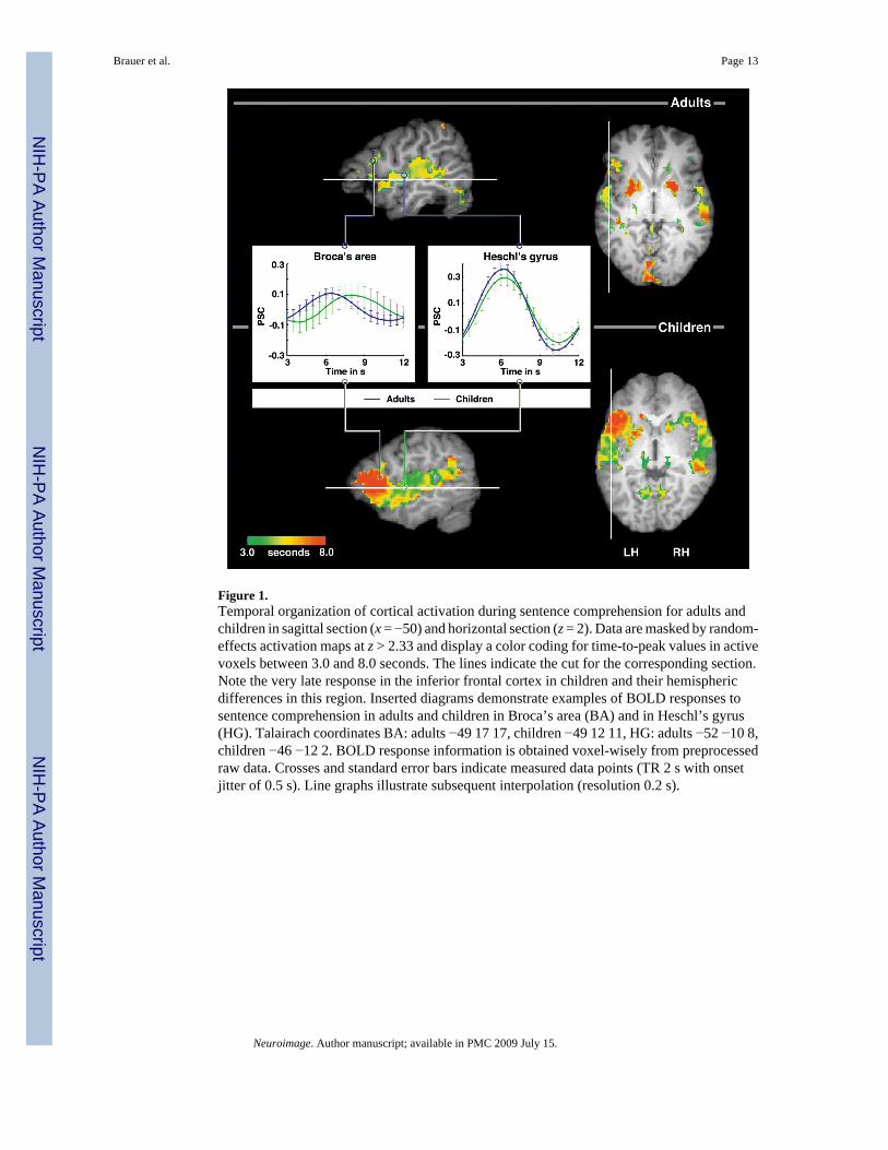
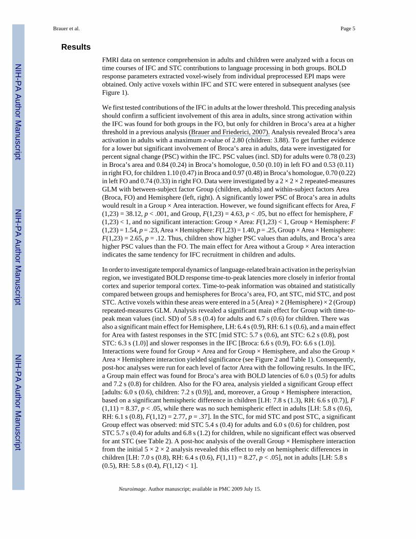
Figure 1.Temporal organization of cortical activation during sentence comprehension for adults andchildren in sagittal section (x = −50) and horizontal section (z = 2). Data are masked by random-effects activation maps at z > 2.33 and display a color coding for time-to-peak values in activevoxels between 3.0 and 8.0 seconds. The lines indicate the cut for the corresponding section.Note the very late response in the inferior frontal cortex in children and their hemisphericdifferences in this region. Inserted diagrams demonstrate examples of BOLD responses tosentence comprehension in adults and children in Broca’s area (BA) and in Heschl’s gyrus(HG). Talairach coordinates BA: adults −49 17 17, children −49 12 11, HG: adults −52 −10 8,children −46 −12 2. BOLD response information is obtained voxel-wisely from preprocessedraw data. Crosses and standard error bars indicate measured data points (TR 2 s with onsetjitter of 0.5 s). Line graphs illustrate subsequent interpolation (resolution 0.2 s).
Brauer et al. Page 13
Neuroimage. Author manuscript; available in PMC 2009 July 15.
NIH
-PA Author Manuscript
NIH
-PA Author Manuscript
NIH
-PA Author Manuscript
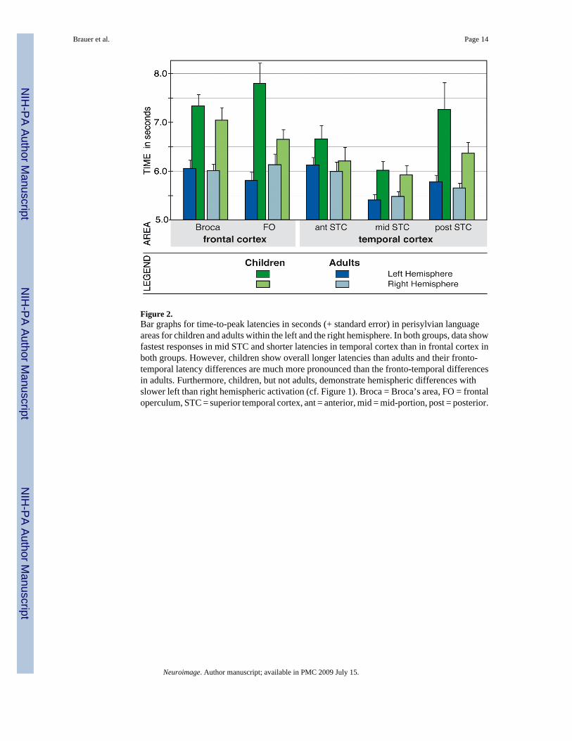
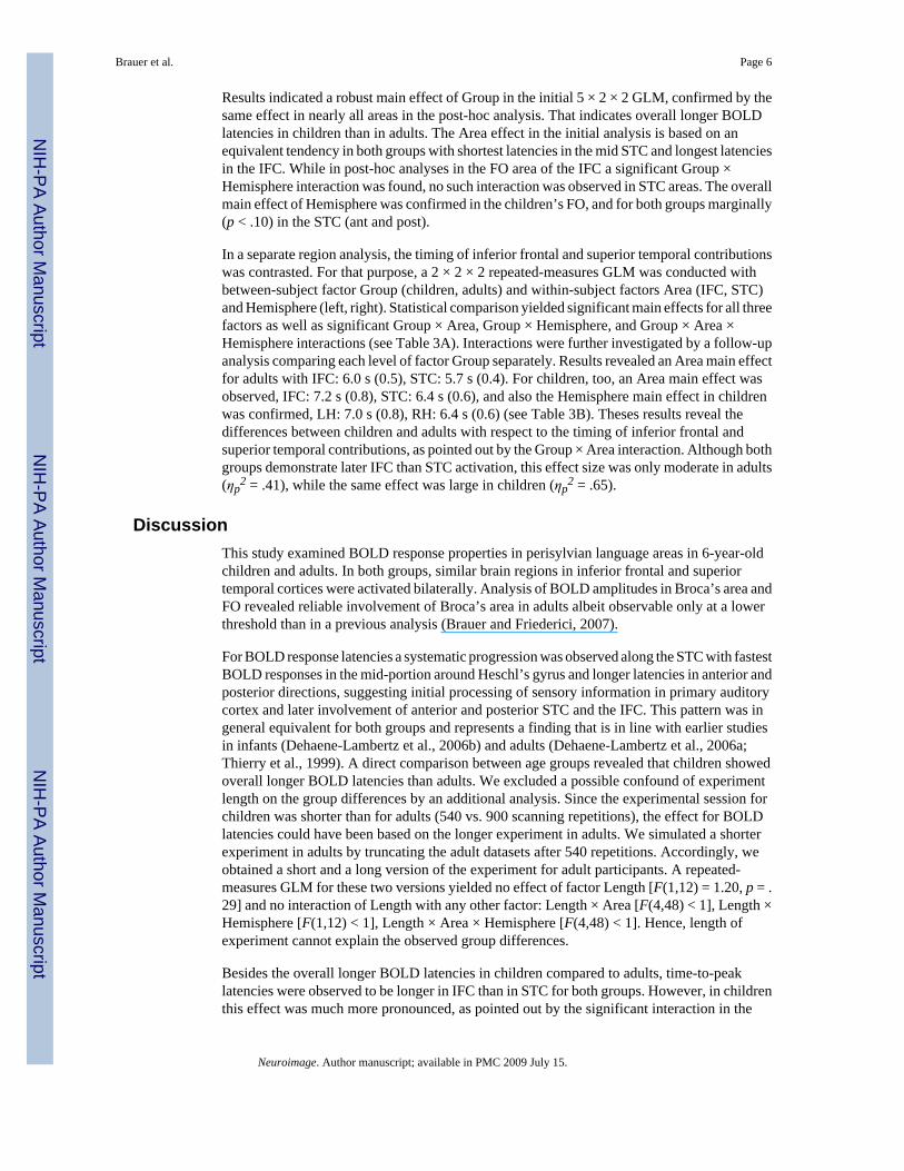
Figure 2.Bar graphs for time-to-peak latencies in seconds (+ standard error) in perisylvian languageareas for children and adults within the left and the right hemisphere. In both groups, data showfastest responses in mid STC and shorter latencies in temporal cortex than in frontal cortex inboth groups. However, children show overall longer latencies than adults and their fronto-temporal latency differences are much more pronounced than the fronto-temporal differencesin adults. Furthermore, children, but not adults, demonstrate hemispheric differences withslower left than right hemispheric activation (cf. Figure 1). Broca = Broca’s area, FO = frontaloperculum, STC = superior temporal cortex, ant = anterior, mid = mid-portion, post = posterior.
Brauer et al. Page 14
Neuroimage. Author manuscript; available in PMC 2009 July 15.
NIH
-PA Author Manuscript
NIH
-PA Author Manuscript
NIH
-PA Author Manuscript
NIH
-PA Author Manuscript
NIH
-PA Author Manuscript
NIH
-PA Author Manuscript
Brauer et al. Page 15
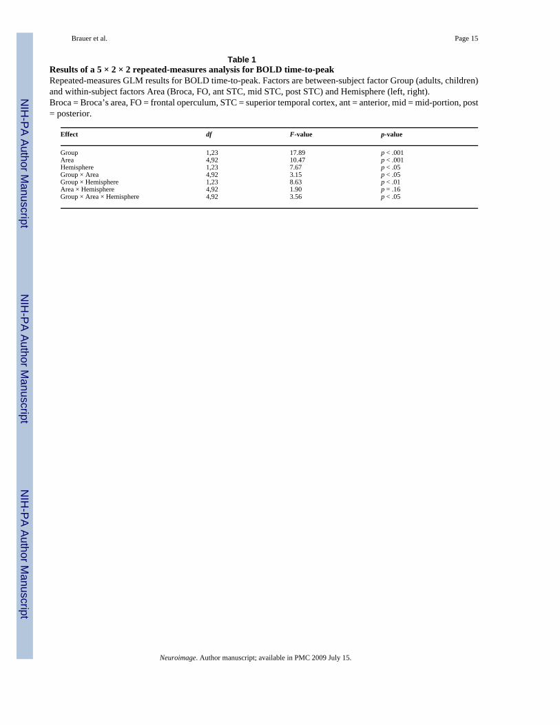
Table 1Results of a 5 × 2 × 2 repeated-measures analysis for BOLD time-to-peakRepeated-measures GLM results for BOLD time-to-peak. Factors are between-subject factor Group (adults, children)and within-subject factors Area (Broca, FO, ant STC, mid STC, post STC) and Hemisphere (left, right).Broca = Broca’s area, FO = frontal operculum, STC = superior temporal cortex, ant = anterior, mid = mid-portion, post= posterior.
Effect df F-value p-value
Group 1,23 17.89 p < .001Area 4,92 10.47 p < .001Hemisphere 1,23 7.67 p < .05Group × Area 4,92 3.15 p < .05Group × Hemisphere 1,23 8.63 p < .01Area × Hemisphere 4,92 1.90 p = .16Group × Area × Hemisphere 4,92 3.56 p < .05
Neuroimage. Author manuscript; available in PMC 2009 July 15.
NIH
-PA Author Manuscript
NIH
-PA Author Manuscript
NIH
-PA Author Manuscript
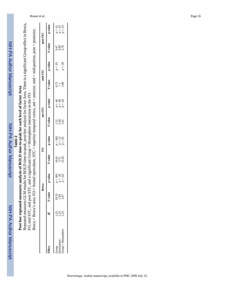
Brauer et al. Page 16Ta
ble
2Po
st-h
oc r
epea
ted-
mea
sure
s ana
lysi
s of B
OL
D ti
me-
to-p
eak
for
each
leve
l of f
acto
r A
rea
Rep
eate
d-m
easu
res G
LM re
sults
for B
OLD
tim
e-to
-pea
k, p
ost-h
oc a
naly
sis f
or fa
ctor
Are
a. T
here
is a
sign
ifica
nt G
roup
eff
ect i
n B
roca
,FO
, mid
STC
, and
pos
t STC
, and
a si
gnifi
cant
Gro
up ×
Hem
isph
ere
inte
ract
ion
in th
e FO
.B
roca
= B
roca
’s a
rea,
FO
= fr
onta
l ope
rcul
um, S
TC =
supe
rior t
empo
ral c
orte
x, a
nt =
ant
erio
r, m
id =
mid
-por
tion,
pos
t = p
oste
rior.
Bro
caFO
ant S
TC
mid
ST
Cpo
st S
TC
Effe
ctdf
F-va
lue
p-va
lue
F-va
lue
p-va
lue
F-va
lue
p-va
lue
F-va
lue
p-va
lue
F-va
lue
p-va
lue
Gro
up1,
2319
.55
p <
.001
16.8
1p
< .0
011.
51p
= .4
66.
75p
< .0
59.
47p
< .0
1H
emis
pher
e1,
231.
94p
= .3
63.
72p
= .1
35.
48p
= .0
6<
14.
90p
= .0
7G
roup
× H
emis
pher
e1,
231.
07p
= .6
211
.65
p <
.01
1.61
p =
.43
1.80
p =
.39
2.70
p =
.23
Neuroimage. Author manuscript; available in PMC 2009 July 15.
NIH
-PA Author Manuscript
NIH
-PA Author Manuscript
NIH
-PA Author Manuscript
Brauer et al. Page 17
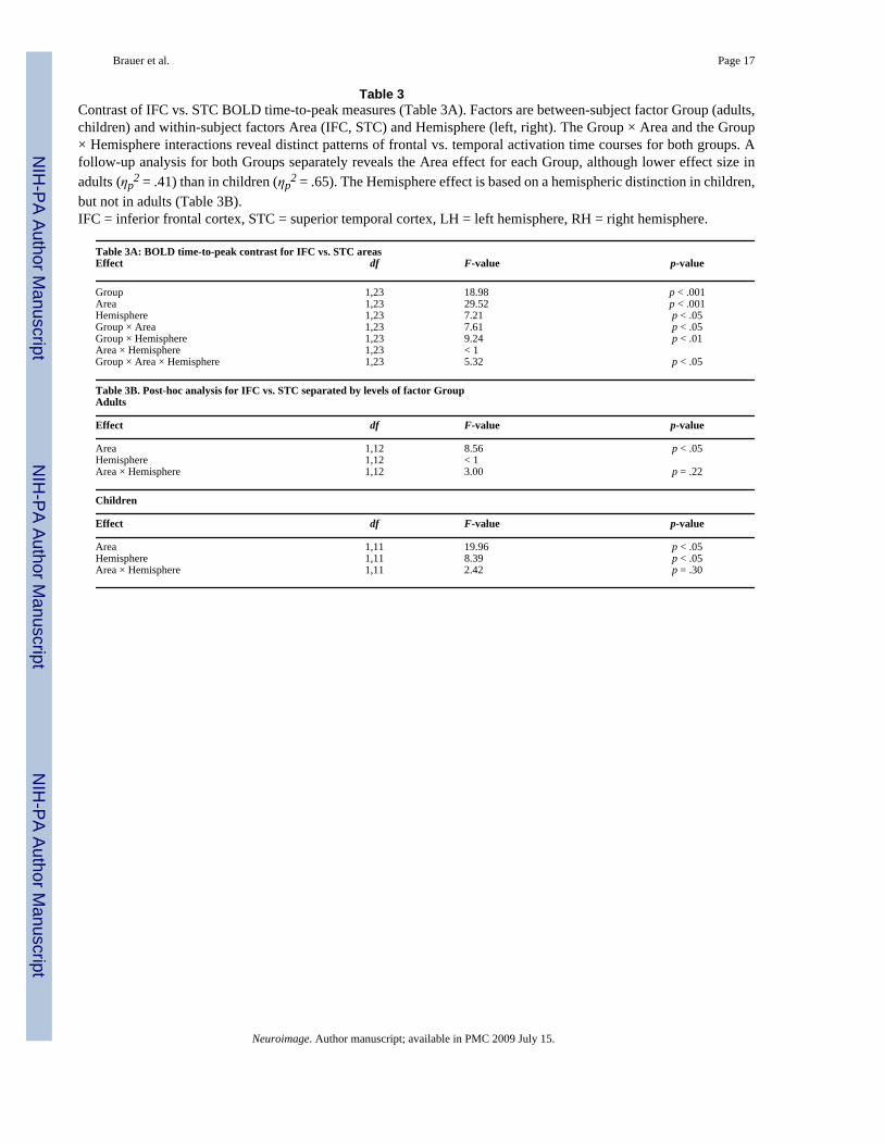
Table 3Contrast of IFC vs. STC BOLD time-to-peak measures (Table 3A). Factors are between-subject factor Group (adults,children) and within-subject factors Area (IFC, STC) and Hemisphere (left, right). The Group × Area and the Group× Hemisphere interactions reveal distinct patterns of frontal vs. temporal activation time courses for both groups. Afollow-up analysis for both Groups separately reveals the Area effect for each Group, although lower effect size inadults (ηp
2 = .41) than in children (ηp2 = .65). The Hemisphere effect is based on a hemispheric distinction in children,
but not in adults (Table 3B).IFC = inferior frontal cortex, STC = superior temporal cortex, LH = left hemisphere, RH = right hemisphere.
Table 3A: BOLD time-to-peak contrast for IFC vs. STC areasEffect df F-value p-value
Group 1,23 18.98 p < .001Area 1,23 29.52 p < .001Hemisphere 1,23 7.21 p < .05Group × Area 1,23 7.61 p < .05Group × Hemisphere 1,23 9.24 p < .01Area × Hemisphere 1,23 < 1Group × Area × Hemisphere 1,23 5.32 p < .05
Table 3B. Post-hoc analysis for IFC vs. STC separated by levels of factor GroupAdults
Effect df F-value p-value
Area 1,12 8.56 p < .05Hemisphere 1,12 < 1Area × Hemisphere 1,12 3.00 p = .22
Children
Effect df F-value p-value
Area 1,11 19.96 p < .05Hemisphere 1,11 8.39 p < .05Area × Hemisphere 1,11 2.42 p = .30
Neuroimage. Author manuscript; available in PMC 2009 July 15.