TAF6δ Controls Apoptosis and Gene Expression in the Absence of p53
13
TAF6d Controls Apoptosis and Gene Expression in the Absence of p53 Emmanuelle Wilhelm 1 , Franc ¸ois-Xavier Pellay 2 , Arndt Benecke 2 , Brendan Bell 1 * 1 RNA Group, De ´ partement de microbiologie et d’infectiologie, Faculte ´ de me ´decine et sciences de la sante ´, Universite ´ de Sherbrooke, Sherbrooke, Que ´bec, Canada, 2 Institut des Hautes E ´ tudes Scientifiques and Institut de Recherche Interdisciplinaire – CNRS USR3078 - Universite ´ de Lille, Bures sur Yvette, France Abstract Background: Life and death decisions of metazoan cells hinge on the balance between the expression of pro- versus anti- apoptotic gene products. The general RNA polymerase II transcription factor, TFIID, plays a central role in the regulation of gene expression through its core promoter recognition and co-activator functions. The core TFIID subunit TAF6 acts in vitro as an essential co-activator of transcription for the p53 tumor suppressor protein. We previously identified a splice variant of TAF6, termed TAF6d that can be induced during apoptosis. Methodology/Principal Findings: To elucidate the impact of TAF6d on cell death and gene expression, we have employed modified antisense oligonucleotides to enforce expression of endogenous TAF6d. The induction of endogenous TAF6d triggered apoptosis in tumor cell lines, including cells devoid of p53. Microarray experiments revealed that TAF6d activates gene expression independently of cellular p53 status. Conclusions: Our data define TAF6d as a pivotal node in a signaling pathway that controls gene expression programs and apoptosis in the absence of p53. Citation: Wilhelm E, Pellay F-X, Benecke A, Bell B (2008) TAF6d Controls Apoptosis and Gene Expression in the Absence of p53. PLoS ONE 3(7): e2721. doi:10.1371/journal.pone.0002721 Editor: Axel Imhof, University of Munich and Center of Integrated Protein Science, Germany Received April 15, 2008; Accepted June 18, 2008; Published July 16, 2008 Copyright: ß 2008 Wilhelm et al. This is an open-access article distributed under the terms of the Creative Commons Attribution License, which permits unrestricted use, distribution, and reproduction in any medium, provided the original author and source are credited. Funding: F.X.P. is recipient of a doctoral fellowship from the Nausicaa Combat Sa Leuce ´mie association. A.B.’s group received funds from the European Hematology Association - Jose ´ Carreras Foundation, and the French Ministry of Research through the ‘‘Complexite ´ du Vivant - Action STICS-Sante ´ ’’ program. Work in B.B.’s laboratory was funded through a Discovery grant from Natural Sciences and Engineering Research Council of Canada. B.B. holds the Canada Research Chair in Genomic Regulation. Competing Interests: The authors have declared that no competing interests exist. * E-mail: [email protected] Introduction Tightly controlled programmed cell death is essential for the development and tissue homeostasis of all animals. Metazoan cells possess an evolutionary conserved network of proteins (e.g. caspases, Bcl-2 family members and death receptors) that execute appropriate life or death decisions in response to extrinsic and intrinsic cellular cues [1]. Deregulated apoptosis underlies numerous human disease states including neurodegenerative disorders and cancer [2]. The identification and molecular characterization of the critical control points in apoptotic pathways is therefore essential to reveal novel therapeutic avenues to treat a broad array of human pathologies. Gene expression plays a key role in the response of cells to death-inducing stimuli. A growing body of evidence indicates that the levels of numerous death-related genes can be induced during apoptosis [3]. The integration of cellular signals from diverse apoptotic pathways requires the finely balanced expression of pro- versus anti-apoptotic proteins. Gene expression patterns of pro- and anti-apoptotic genes, established by the levels of transcription [4] as well as alternative splicing [5], can dictate the life-or-death decisions of cells. The most intensely studied protein known to control apoptosis by altering gene expression is the p53 tumor suppressor. Current paradigms link the p53 tumor suppression function to its capacity to induce apoptosis in response to genotoxic stress [6]. The pro-apoptotic activity of p53 depends largely on its function as a transcriptional activator that binds directly to the promoters of pro-apoptotic genes including FDXR [7,8], PUMA [9], Noxa [10], Bax [11] and p53AIP1 [12]. TFIID is a multi-protein complex that plays a pivotal role in the transcription of protein-coding genes in eukaryotes. TFIID is composed of the TATA-binding protein (TBP) and up to 14 evolutionarily conserved TBP-associated factors (TAFs) [13,14]. TFIID can play a rate-limiting role in the regulation of transcription through the recognition of the core promoter elements such as the TATA-box, the initiator, and downstream promoter element (DPE) [15]. The TFIID complex also engages in direct contacts with DNA- binding transcriptional factors to co-activate gene expression [16]. The architecture and integrity of TFIID complexes depends on a network of TAF-TAF interactions that are predominantly mediated by dimerization of TAFs via their interlocking histone fold motifs [17]. Histone-fold pairs within TFIID include TAF6-TAF9 [18], TAF4-TAF12 [19], TAF11-13 [20], TAF8-TAF10 [21] and TAF3- TAF10 [22]. TAF4 and TAF5 together with the histone fold containing TAF12, TAF9 and TAF6 are defined as core TAFs, since their depletion from Drosophila cells results in overall destabilization of TFIID complexes [23]. The core TFIID subunit TAF6 (hTAF II 70/80) has been shown to be broadly required for RNA polymerase II (Pol II) transcription in yeast when total poly(A) + mRNA levels were PLoS ONE | www.plosone.org 1 July 2008 | Volume 3 | Issue 7 | e2721
Transcript of TAF6δ Controls Apoptosis and Gene Expression in the Absence of p53
TAF6d Controls Apoptosis and Gene Expression in theAbsence of p53Emmanuelle Wilhelm1, Francois-Xavier Pellay2, Arndt Benecke2, Brendan Bell1*
1 RNA Group, Departement de microbiologie et d’infectiologie, Faculte de medecine et sciences de la sante, Universite de Sherbrooke, Sherbrooke, Quebec, Canada,
2 Institut des Hautes Etudes Scientifiques and Institut de Recherche Interdisciplinaire – CNRS USR3078 - Universite de Lille, Bures sur Yvette, France
Abstract
Background: Life and death decisions of metazoan cells hinge on the balance between the expression of pro- versus anti-apoptotic gene products. The general RNA polymerase II transcription factor, TFIID, plays a central role in the regulation ofgene expression through its core promoter recognition and co-activator functions. The core TFIID subunit TAF6 acts in vitroas an essential co-activator of transcription for the p53 tumor suppressor protein. We previously identified a splice variant ofTAF6, termed TAF6d that can be induced during apoptosis.
Methodology/Principal Findings: To elucidate the impact of TAF6d on cell death and gene expression, we have employedmodified antisense oligonucleotides to enforce expression of endogenous TAF6d. The induction of endogenous TAF6dtriggered apoptosis in tumor cell lines, including cells devoid of p53. Microarray experiments revealed that TAF6d activatesgene expression independently of cellular p53 status.
Conclusions: Our data define TAF6d as a pivotal node in a signaling pathway that controls gene expression programs andapoptosis in the absence of p53.
Citation: Wilhelm E, Pellay F-X, Benecke A, Bell B (2008) TAF6d Controls Apoptosis and Gene Expression in the Absence of p53. PLoS ONE 3(7): e2721.doi:10.1371/journal.pone.0002721
Editor: Axel Imhof, University of Munich and Center of Integrated Protein Science, Germany
Received April 15, 2008; Accepted June 18, 2008; Published July 16, 2008
Copyright: � 2008 Wilhelm et al. This is an open-access article distributed under the terms of the Creative Commons Attribution License, which permitsunrestricted use, distribution, and reproduction in any medium, provided the original author and source are credited.
Funding: F.X.P. is recipient of a doctoral fellowship from the Nausicaa Combat Sa Leucemie association. A.B.’s group received funds from the EuropeanHematology Association - Jose Carreras Foundation, and the French Ministry of Research through the ‘‘Complexite du Vivant - Action STICS-Sante’’ program. Workin B.B.’s laboratory was funded through a Discovery grant from Natural Sciences and Engineering Research Council of Canada. B.B. holds the Canada ResearchChair in Genomic Regulation.
Competing Interests: The authors have declared that no competing interests exist.
* E-mail: [email protected]
Introduction
Tightly controlled programmed cell death is essential for the
development and tissue homeostasis of all animals. Metazoan cells
possess an evolutionary conserved network of proteins (e.g.
caspases, Bcl-2 family members and death receptors) that execute
appropriate life or death decisions in response to extrinsic and
intrinsic cellular cues [1]. Deregulated apoptosis underlies
numerous human disease states including neurodegenerative
disorders and cancer [2]. The identification and molecular
characterization of the critical control points in apoptotic
pathways is therefore essential to reveal novel therapeutic avenues
to treat a broad array of human pathologies.
Gene expression plays a key role in the response of cells to
death-inducing stimuli. A growing body of evidence indicates that
the levels of numerous death-related genes can be induced during
apoptosis [3]. The integration of cellular signals from diverse
apoptotic pathways requires the finely balanced expression of pro-
versus anti-apoptotic proteins. Gene expression patterns of pro-
and anti-apoptotic genes, established by the levels of transcription
[4] as well as alternative splicing [5], can dictate the life-or-death
decisions of cells. The most intensely studied protein known to
control apoptosis by altering gene expression is the p53 tumor
suppressor. Current paradigms link the p53 tumor suppression
function to its capacity to induce apoptosis in response to
genotoxic stress [6]. The pro-apoptotic activity of p53 depends
largely on its function as a transcriptional activator that binds
directly to the promoters of pro-apoptotic genes including FDXR
[7,8], PUMA [9], Noxa [10], Bax [11] and p53AIP1 [12].
TFIID is a multi-protein complex that plays a pivotal role in the
transcription of protein-coding genes in eukaryotes. TFIID is
composed of the TATA-binding protein (TBP) and up to 14
evolutionarily conserved TBP-associated factors (TAFs) [13,14].
TFIID can play a rate-limiting role in the regulation of transcription
through the recognition of the core promoter elements such as the
TATA-box, the initiator, and downstream promoter element (DPE)
[15]. The TFIID complex also engages in direct contacts with DNA-
binding transcriptional factors to co-activate gene expression [16].
The architecture and integrity of TFIID complexes depends on a
network of TAF-TAF interactions that are predominantly mediated
by dimerization of TAFs via their interlocking histone fold motifs
[17]. Histone-fold pairs within TFIID include TAF6-TAF9 [18],
TAF4-TAF12 [19], TAF11-13 [20], TAF8-TAF10 [21] and TAF3-
TAF10 [22]. TAF4 and TAF5 together with the histone fold
containing TAF12, TAF9 and TAF6 are defined as core TAFs, since
their depletion from Drosophila cells results in overall destabilization of
TFIID complexes [23].
The core TFIID subunit TAF6 (hTAFII70/80) has been shown
to be broadly required for RNA polymerase II (Pol II)
transcription in yeast when total poly(A)+ mRNA levels were
PLoS ONE | www.plosone.org 1 July 2008 | Volume 3 | Issue 7 | e2721
monitored [24]. A more recent microarray analysis estimated that
approximately 18% of the yeast Pol II transcriptome depends on
TAF6 [25]. TAF6 has been shown to be essential for viability in
yeast [24,26], plants [27], insects [28] and fish [29]. The
requirement for TAF6 in all model organisms studied, together
with the fact that human cells have a single TAF6 gene [30,31],
strongly implies that TAF6 is essential for human cell viability.
TAF6 and TAF6-TAF9 dimers can bind to the downstream
promoter element (DPE) [32,33]. In addition to its core promoter
recognition function, TAF6 can also interact with transcriptional
activators. For example, in vitro experiments have shown that both
TAF6 [34], and its dimerization partner TAF9 [35], interact
directly with p53 and are required for the activation of
transcription by p53. The available evidence implies that the
TAF6-p53 interaction is required for the activation of at least
some, and potentially all, p53 target genes in vivo [36,37,38].
We have identified and characterized a splice variant termed
TAF6d that lacks 10 amino acids in the centre of its histone fold
domain [39]. TAF6d is unable to interact with TAF9, but retains
interactions with other TFIID subunits. TAF6d expression is
induced in promyelocytic HL-60 cells undergoing retinoic-acid
dependent apoptosis. TAF6d overexpression induces apoptosis in
HeLa cells, evoking the possibility that TFIID function could be
coupled to certain apoptotic pathways via TAF6d. Importantly,
however, it is not currently known if changes in the expression of
endogenous TAF6d can influence tumor cell death. Furthermore,
despite the physical and functional interactions between the pivotal
tumor suppressor p53 and TAF6 [34], nothing is currently known
about whether p53 is required for TAF6d-mediated apoptosis. Here,
we have used splice-site switching modified antisense RNA
technology to demonstrate that endogenous TAF6d controls
apoptosis and that p53 is not required for TAF6d-dependent
apoptosis or TAF6d-dependent gene expression.
Materials and Methods
Cell cultureHeLa cells were grown in DMEM containing 2.5% CS and
2.5% FCS. Saos-2 and H1299 cell lines were cultured in DMEM
with 10% FCS. A549 cells were grown in Ham’s F12 medium with
10% FCS. HCT-116 cells were grown in McCoy’s media
supplemented with 10% FCS. When indicated, cells were treated
with the proteasome inhibitor MG-132 (Calbiochem) at 0.5 mM
and/or pan-caspase inhibitor Z-VAD-FMK (Biomol) at 100 mM.
TransfectionsOligonucleotides were transfected with lipofectamine 2000
(Invitrogen) as a delivery agent (1.6 ml/ml) according to the
manufacturer’s recommendations. 29-O-methyl-oligoribonucleoside
phosphorothioate antisense 20-mers were from Sigma-Proligo.
‘‘TAF6 AS1’’ 59-CGAUCUCUUUGAUGCGGUAG-39 targets
the central 20 nucleotides of the alternative exon 2 of TAF6,
‘‘TAF6 AS2’’ 59-GCCGGGUCACCUGUGCGAUC-39 the con-
stitutive alpha 59 splice site. ‘‘Control AS’’ 59-AUGGCCUCGAC-
GUGCGCGCU-39 is a scrambled oligo used as a negative control.
‘‘Bcl-x AS’’ 59-ACCCAGCCGCCGUUCUCC-39 targets the 59-
splice site of Bcl-xL [40]. Plasmids were transfected using 1 ml
DMRIE-C (Invitrogen) as a delivery agent in a 24 well plate
according to the manufacturer’s recommendations. All transfections
were performed in OptiMEM medium (Invitrogen).
RT-PCRTotal RNA was isolated from cells using Trizol (Invitrogen)
according to the manufacturer’s recommendations. 1 mg of total
RNA was reverse transcribed using AMV-RT (Roche). 1/10 of the
total cDNA was used per PCR reaction : 95uC, 3 min; 25 cycles of
94uC for 1 min, 58uC for 45 sec, 68uC for 50 sec; final extension
at 68uC for 5 min with the following oligonucleotide pairs. For
Taf6; forward 59-ATGGGCATCGCCCAGATTCAGG-39 and
reverse 59-AAGGCGTAGTCAATGTCACTGG-39. For Bcl-x;
forward 59-TCATTTCCGACTGAAGAGTGA-39 and reverse
59-ATGGCAGCAGTAAAGCAAGCG-39
Apoptosis assaysDetection of caspase cleaved cytokeratin-18 by flow cytometry
was performed using Cytodeath reagent (Roche) according to the
manufacturer’s recommendations. Flow cytometric analysis of sub-
G1 DNA content was performed as described [39].
PlasmidsTo construct pASTAF6, the genomic region of TAF6
containing exon2 to exon4 was amplified by PCR from HEK
293 genomic DNA with primers 59-GGAGAAGAGGGACTC-
CAGAATGGCTG-39 (forward) and 59-TCCCCCAACCTTT-
GAGGCAGACG-39 (reverse). The resulting product was digested
with HindIII and SmaI and inserted into the same sites of the
plasmid pXJ42hTAFII80a [39].
AntibodiesMonoclonal antibodies directed against TAF6d (37TA-1 &
37TA-2), TAF6a (25TA) [39], TBP (3G3) [41], and TAF5 (2D2)
[42] have been described. Monoclonal antibodies against TAF6
and PARP-1 were purchased from BD Transduction Laboratories
and Biomol, respectively.
ImmunocytochemistryCells were fixed in 4% PFA, permeabilized with PBS-0.1%
Triton X-100 (PBS-Tx) and incubated for 30 min in blocking
buffer (PBS-Tx containing 1% bovine serum albumin (BSA) and
0.5% fish gelatine (Sigma-Aldrich)). Cells were then sequentially
incubated one hour at room temperature, followed by washes,
with each of the following antibodies diluted in blocking buffer;
anti-TAF6 mAb (1/400), Oregon Green goat anti-mouse IgG
secondary antibody (Molecular Probes), anti-TAF6d mAb (37TA-
1: 1/1000), Alexa Fluor 546 goat anti-mouse IgG1 secondary
antibody (Molecular Probes). Cells were then treated with Hoechst
33342 (2 mg/ml) and visualized by fluorescence microscopy.
Microarray Analysis of Gene ExpressionTranscriptome Acquisition. Total RNA was analyzed
using ABI Human Whole Genome Survey Arrays v1.0 arrays
(Prod. No.: 4359030), containing 31,700 60-mer oligonucleotide
probes representing a set of 27,868 individual annotated human
genes. Chemiluminescence detection technology is used to detect
as little as a femtomole of expressed mRNA. One single round of
linear amplification was performed from total RNA according to
the Applied Biosystems RT-IVT (Applied Biosystems, ProdNo:
4339628) protocol using 2 mg of total RNA. cDNA synthesis, in
vitro transcription and labeling, fragmentation, hybridization,
staining, and scanning were performed as directed by the
supplier (Applied Biosystems, ProdNo: 4346875).
Transcriptome Data Analysis. Applied Biosystems
Expression Array System Software v1.1.1. (ProdNo: 4364137)
has been used to acquire the chemiluminescence and fluorescence
images and primary data analysis. We renormalized the resulting
data according to the logarithmic signal median once more after
having removed control probes and those probes for which the
TAF6d Controls Death Sans p53
PLoS ONE | www.plosone.org 2 July 2008 | Volume 3 | Issue 7 | e2721

Applied Biosystems Software has set flags equal to or greater than
212, indicating compromised measurements (as recommended by
Applied Biosystems). Log2 subtractions were determined using
averages over the weighted individual signal values. The weights are
anti-proportional to the corresponding coefficient of variation. For
these inter-assay comparisons the NeONORM method was used for
normalization using sensitivity parameter k = 0.20 [43]. P-values
were determined using a standard ANOVA method. Multiple
probes for a single gene, cross-reactivity of a single probe to several
genes, as well as the resolution of probe-ID annotations were done
according to the standards defined previously [44]. Gene lists
corresponding to statistically significant changes in expression
(P,0.05) are available as supplementary data files: genes changing
in response TAF6d in HCT-116 p53 +/+ cells (Supplementary Data
File S1), genes changing in response to TAF6d in HCT-116 p53 2/
2 (Supplementary Data File S2), genes differentially expressed in the
HCT-116 p53 +/+ cells versus HCT-116 p53 2/2 cells both in
cells treated with control oligonucleotide and TAF6d-inducing
oligonucleotides (Supplementary Data File S3), and genes
differentially regulated by TAF6d in both HCT-116 p53 +/+ cells
and HCT-116 p53 2/2 cells (Supplementary Data File S4).
The microarray data for the experiments described here were
deposited in the Gene Expression Omnibus database (http://www.
ncbi.nlm.nih.gov/geo/) under accession number: GSE10795.
The supplementary data files can also be accessed from the
Benecke group webpage: http://seg.ihes.fr/.
Real time PCRReal Time PCR was performed on cDNA prepared as for
microarray experiments (above) using ABI TaqManH assays. The
genes and their respective assay numbers were ATF3 (Hs
00231069_m1), ACRC (Hs 00369516_m1), FNBP4 (Hs
00392543_m1) HES1 (Hs 00172878_m1), and HOM-TES-103
(Hs 00209961_m1). Real-time PCR was performed on 10 ng of
cDNA with 1.25 ml of 206 TaqManH probes and 12.5 ml 26TaqManH Universal Master Mix (ABI) in a final 25 ml reaction.
Real-time PCR relative quantification assay was running for 2 min
at 50uC, 10 min at 95uC, followed by 40 cycles of 15 sec at 95uCand 1 min at 60uC on an ABI 7500 system. Relative quantity of
target genes was calculated using the comparative CT (DDCT)
method using FNBP4 as the internal control.
Results
Selective induction of endogenous TAF6d mRNAexpression by splice-switching oligonucleotides
To date four splice variants of TAF6 have been identified and
termed a, b, c, and d [31,39,45]. Here we focus on the TAF6d splice
variant due to its potentially important role in programmed cell
death [39]. The total number of distinct TAF6 mRNA species
produced by alternative splicing has not yet been established. For
clarity, we therefore refer here collectively to all TAF6 splice variants
lacking the 30 nucleotide exon IIa as TAF6d and to TAF6a as all
species of mRNA containing exon IIa (Fig. 1). The TAF6 genomic
locus shows that the major TAF6a isoform is produced by the
selection of an intron proximal 59 splice site (SS) (Fig. 1A). In
contrast, the TAF6d isoform is produced by an alternative splicing
event at the intron distal 59 SS (labelled d in Fig. 1A).
To dissect the biological role of endogenous TAF6d, we
exploited splice-switching oligonucleotides (SSOs) [40,46] to
experimentally manipulate endogenous TAF6 alternative splicing.
The HeLa cell system represents a natural cellular context to study
TAF6d function because the TAF6d variant was originally cloned
from a HeLa cell cDNA library [39]. We transfected HeLa cells
with synthetic 29-O-methyl-modified oligoribonucleoside phos-
phorothioate that hybridizes to the central 20 nucleotides of
alternative exon IIa (Fig. 1A). The ratio of the alternative TAF6dmRNA level with respect to TAF6a mRNA level was analyzed by
RT-PCR of RNA samples from transfected HeLa cells. The
transfection of the antisense oligonucleotide Taf6 AS1 resulted in a
marked increase in the level of the TAF6d mRNA and a
concurrent decrease in the level of the major TAF6a mRNA
(Fig. 1B, lane 3). In contrast, transfection of an oligonucleotide of
scrambled sequence had no effect on the TAF6d/TAF6a mRNA
ratio (Fig. 1B, lane 2). To further demonstrate the specificity of the
SSOs, we transfected antisense RNA oligonucleotides shown to
enforce the expression of the Bcl-xS splice variant [40]. The
TAF6d/TAF6a+d mRNA ratio was increased ,3-fold by
treatment with the Taf6 AS1 oligonucleotide, but unchanged by
the Bcl-x AS oligonucleotide (Fig. 1C). Conversely the ratio of Bcl-
xS/Bcl-xL+xS mRNA was increased ,10-fold by Bcl-x AS but
unaffected by Taf6 AS1 (Fig. 1C). Control RT-PCR reactions
showed that ratio of TAF6d with respect to total TAF6 mRNA is
increased by Taf6 SSO but none of the ratios of any other known
TAF6 alternative splice variants was affected (data not shown).
The SSOs used therefore impact specifically on TAF6d alternative
splicing without influencing overall expression patterns of TAF6
mRNA. These results demonstrate that TAF6-directed SSOs are
an efficient and selective method to enforce the expression of the
endogenous TAF6d mRNA in HeLa cells.
To further characterize the cellular response to TAF6 splice site
switching antisense oligonucleotides we performed a time course
analysis. The level of TAF6d mRNA is detectably increased after
4 hours and increases until 24 hours (Supplementary Fig. S1A).
These results are consistent with a previous study targeting the Bcl-x
gene [40], and establish a kinetic framework to follow the early
outcomes of TAF6d mRNA expression in transfected cells. We next
investigated the concentration dependence for the Taf6 response to
treatment with the AS1 oligonucleotide. The induction of TAF6dmRNA was observed with transfection of as little as 50 nM Taf6
AS1 and sharply increased until treatment with 200 nM Taf6 AS1,
after which a plateau was reached (Supplementary Fig. S1B). Based
on these results, we have employed 200 nM oligonucleotide
concentrations herein, unless otherwise stated, for robust and specific
induction of endogenous TAF6d mRNA in living cells.
Splice-switching oligonucleotides increase endogenousTAF6d protein levels
We next investigated the influence of the splice site switching
oligonucleotides on the levels of TAF6d and TAF6a proteins. TAF6
was detected by immunocytochemistry using monoclonal antibodies
that recognize an epitope present in all of the known isoforms of
TAF6. HeLa cells treated with negative control oligonucleotides
showed strong TAF6 staining throughout the entire nucleoplasm
(Fig. 2A). The nuclear total TAF6 immunofluorescent signal is
diminished in cells treated with SSOs that increase TAF6d mRNA
production (Fig. 2A), presumably due to decreased expression of
TAF6a (see also below). TAF6d was detected by immunofluores-
cence with monoclonal antibodies that specifically recognize the
delta TAF6 isoform [39]. HeLa cells transfected with negative
control antisense oligonucleotides exhibited undetectable cellular
staining with anti-TAF6d monoclonal antibodies (Fig. 2A). In
contrast, transfection of HeLa cells with oligonucleotides that induce
TAF6d mRNA expression resulted in punctate nuclear staining
(Fig. 2A). We further quantified the influence of antisense treatment
by scoring the number of cells displaying clear nuclear TAF6dimmunofluorescent signals. We found that treatment with the Taf6
AS1 oligonucleotide resulted in nearly ,10 fold more cells with
TAF6d Controls Death Sans p53
PLoS ONE | www.plosone.org 3 July 2008 | Volume 3 | Issue 7 | e2721
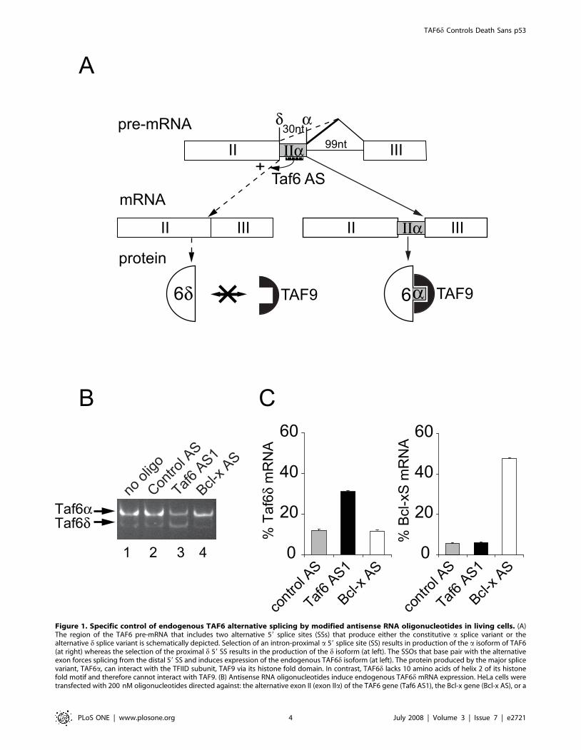
Figure 1. Specific control of endogenous TAF6 alternative splicing by modified antisense RNA oligonucleotides in living cells. (A)The region of the TAF6 pre-mRNA that includes two alternative 59 splice sites (SSs) that produce either the constitutive a splice variant or thealternative d splice variant is schematically depicted. Selection of an intron-proximal a 59 splice site (SS) results in production of the a isoform of TAF6(at right) whereas the selection of the proximal d 59 SS results in the production of the d isoform (at left). The SSOs that base pair with the alternativeexon forces splicing from the distal 59 SS and induces expression of the endogenous TAF6d isoform (at left). The protein produced by the major splicevariant, TAF6a, can interact with the TFIID subunit, TAF9 via its histone fold domain. In contrast, TAF6d lacks 10 amino acids of helix 2 of its histonefold motif and therefore cannot interact with TAF9. (B) Antisense RNA oligonucleotides induce endogenous TAF6d mRNA expression. HeLa cells weretransfected with 200 nM oligonucleotides directed against: the alternative exon II (exon IIa) of the TAF6 gene (Taf6 AS1), the Bcl-x gene (Bcl-x AS), or a
TAF6d Controls Death Sans p53
PLoS ONE | www.plosone.org 4 July 2008 | Volume 3 | Issue 7 | e2721

TAF6d staining compared to control treated cells (Fig. 2B). As a
further control of specificity, oligonucleotide Bcl-x AS was
transfected and caused no increase in nuclear TAF6d immunoflu-
orescent staining (Fig. 2B). We conclude that TAF6d protein in
discrete nuclear loci is significantly increased by SSO targeting of the
TAF6 pre-mRNA.
scrambled control oligonucleotide (Control AS). 24 hours post-transfection total RNA was isolated and subjected to RT-PCR with primers that amplifyboth the TAF6a and the alternative TAF6d mRNAs. (C) Specificity of TAF6 splice site switching oligonucleotides. HeLa cells were transfected withantisense RNA oligonucleotides as in A. RT-PCR was perfomed with primers sets that amplify the both the a and d TAF6 splice variants, or both theBcl-xS and Bcl-xL splice variants. PCR products were separated by microfluidity and analyzed using a 2100 Agilent bioanalyzer. The ratio of TAF6dmRNA over total TAF6 mRNA and the ratio of Bcl-Xs mRNA over total Bcl-X mRNA are expressed on the y-axis. The values from cells treated withscrambled control (grey bars), Taf6 AS1 (black bars), or Bcl-X AS (white bars) are shown. Error bars represent the standard deviation of threeindependent transfections.doi:10.1371/journal.pone.0002721.g001
Figure 2. Specific induction of TAF6d protein expression by modified antisense RNA oligonucleotides. (A) HeLa cells were transfectedwith splice site-switching oligonucleotides directed against exon IIa of the TAF6 gene and treated with MG-132 5 hours later. 21 hours posttransfection cells were fixed and stained with the indicated antibodies for immunocytochemistry (B) Quantification of endogenous TAF6d expressingcells transfected with splice site-switching antisense oligonucleotides. Results are expressed as the percentage of cells displaying a clear TAF6dpunctate staining on a total of at least 500 cells. (C) Translation of exogenous TAF6d is induced by modified antisense RNA oligonucleotides.Schematic representation of plasmid pASTAF6 containing sequences derived from the TAF6 cDNA (white) or from genomic DNA (grey). HeLa cellswere first transfected with pASTAF6 and 19 hours later with splice site switching oligonucleotides and treated with MG132 and ZVAD-FMK 6 hoursafter this second transfection. 38 hours post-transfection protein extracts from cells were analyzed by immunoblotting with monoclonal antibodiesdirected against TFIID subunits.doi:10.1371/journal.pone.0002721.g002
TAF6d Controls Death Sans p53
PLoS ONE | www.plosone.org 5 July 2008 | Volume 3 | Issue 7 | e2721
Immunofluorescence experiments measure the levels of detect-
able antigen in fixed cells. Since the levels of total cellular protein
can potentially differ from the antigenically recognizable levels of
protein we employed immunoblotting under denaturing condi-
tions to directly examine the effect of TAF6d-inducing oligonu-
cleotides on the translation of the TAF6d mRNA in treated cells.
Endogenous TAF6d is undetectable by Western blotting of
extracts from HeLa cell due to its rapid turnover by the
proteasome (data not shown) and by caspase-dependent cleavage
in apoptotic cells [39]. We therefore developed a TAF6 minigene
plasmid that is responsive to SSO (pASTAF6, Fig. 2C). Immu-
noblots on total protein extracts from HeLa cells transfected with
the spliceable minigene construct and later treated with TAF6d-
inducing oligonucleotides resulted in a marked increase in TAF6dprotein levels, with a corresponding reduction in TAF6a (Fig. 2C).
The levels of two other TFIID subunits, TAF5 and TBP, remained
relatively constant. These data demonstrate a selective induction
of TAF6d translation and concomitant reduction in TAF6a levels
by TAF6d-inducing SSOs.
Endogenous TAF6d expression causes apoptosis in HeLacells
We next investigated the physiological consequences of SSO-
induced endogenous TAF6d expression in HeLa cells. The
transfection of HeLa cells with a scrambled antisense oligonucle-
otide resulted in no obvious morphological changes or changes in
cell number when visualized by light microscopy (Fig. 3A, left
image). In stark contrast, TAF6d-inducing SSO resulted in an
obvious loss of adherent cells and produced significant numbers of
cells that exhibit the classical features of apoptosis, including
membrane blebbing (Fig. 3A, right image). To obtain further
evidence that TAF6d induction causes apoptosis we measured
cleavage of the well-known caspase substrate PARP-1, since
activation of the caspase protease cascade is a defining biochemical
feature of apoptosis. Immunoblotting revealed readily detectable
cleavage of PARP-1 in cells when TAF6d was induced (Fig. 3B,
lane 2). As an additional control for the specificity of the Taf6 AS1
oligonucleotide, we used a Bcl-xS-inducing SSO. Consistent with a
previous report [47], the induction of Bcl-xS expression has little
effect on apoptosis in HeLa cells (Fig. 3B, lane 3). To further
substantiate and quantify TAF6d-induced apoptosis we employed
flow cytometry to measure the levels of caspase-cleaved cytoker-
atin-18 (KRT18c), another established marker of apoptosis [48].
Treatment of HeLa cells with the Taf6 AS1 oligonucleotide
resulted in a 3.5 fold increase in KRT18c positive cells (Fig. 3C).
As an independent quantification of apoptosis, we employed flow
cytometry to measure the level of Sub-G1 DNA content. This
assay showed that TAF6d induction resulted in a 2.8 fold increase
in apoptosis whereas Bcl-xS induction resulted in a 1.3 fold
increase in apoptosis in HeLa cells (Fig. 3D). Thus, four distinct
assays show that the induction of endogenous TAF6d triggers a
robust apoptotic response in HeLa cells.
TAF6d induces apoptosis in the absence of p53The tumor suppressor p53 interacts physically and functionally
with TAF6a (see Introduction). Mutations in the p53 pathway are
thought to allow human tumor cells to escape apoptotic death and
therefore allow cancer development [6]. It was therefore of
fundamental importance to establish whether TAF6d-induced
apoptosis can occur in the absence of p53. To address whether
TAF6d-dependent apoptosis requires p53 we transfected the Saos-
2 osteosarcoma cell line that is devoid of a functional p53 gene [49]
with oligonucleotide Taf6 AS1. Transfection of Taf6 AS1, but not
Bcl-x AS into Saos-2 cells effectively increased endogenous TAF6d
mRNA levels (Fig. 4A, lane 3). Analysis of the RT-PCR results
showed an approximately ,5 fold induction in the TAF6d/
TAF6a+d mRNA ratio (Fig. 4B). The expression of TAF6dinduced a 3.3 fold increase in apoptosis in Saos-2 as measured by
Sub-G1 DNA content (Fig. 4C). Similar results were obtained in
another cell line (H1299 lung carcinoma) that does not contain
p53 (data not shown). Because HeLa cells have impaired p53
function due to the expression of the Human Papilloma Virus E6
gene product [50], we also compared the efficiency of induction of
apoptosis in the A549 lung carcinoma cells because they express
wild type p53. Taf6 AS1 transfection increased apoptosis by 3.1
fold (Fig. 4D), whereas Bcl-x AS transfection caused no increase in
apoptosis over background levels (Fig. 4D). The fact that Saos-2
are at least as susceptible as A549 cells to TAF6d-induced
programmed cell death was further verified by measuring PARP-1
cleavage by immunoblotting (Fig. 4E versus 4F). To reinforce the
fact that p53 is dispensable for TAF6d-induced apoptosis, we
employed the HCT-116 human colon carcinoma cell line and its
isogenic counterpart HCT-116 p53 2/2 in which the p53 gene
has been deleted by homologous recombination [51]. The
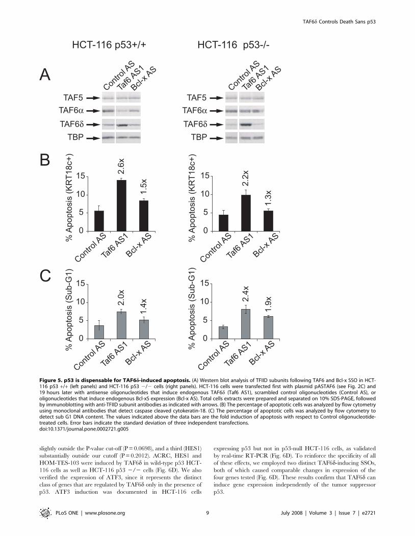
induction of apoptosis by TAF6d in isogenic cells lacking p53 is
equally robust as in wild-type cells, as judged by significant
increases in both caspase-cleavage of cytokeratin-18 (Fig. 5B) and
Sub-G1 DNA content (Fig. 5C). The induction of TAF6d protein
levels by the SSO strategy was efficient in both cells lines (Fig. 5A).
The results demonstrate that p53 is dispensable for TAF6d-
induced cell death. We conclude that TAF6d controls apoptosis
irrespective of cellular p53 status.
TAF6d activates gene expression independently of p53TAF6d can induce apoptosis of several cancer cell lines
independent of their p53 status. We have previously shown that
TAF6d can bind the TFIID subunits TAF1, TAF5, TBP and
TAF12 in vitro, and forms a TFIID-like complex that contains
several TAFs but lacks TAF9 (TFIIDp) in vivo [39]. TAF6 is an
essential gene that plays a broad role in the regulation of
transcription programs (see Introduction). To investigate whether
TAF6d can regulate transcription, with an emphasis on potentially
p53-independent transcription, we employed genome-wide micro-
array technology. The transcriptional effects of TAF6d are
technically difficult to measure because endogenous TAF6d is
not induced in all cells during antisense transfection (Fig. 2B) and
because endogenous TAF6d-expressing cells are lost rapidly from
the culture by apoptosis (Fig. 3). In order to achieve maximal
sensitivity we chose a recently developed microarray technology
based on chemiluminescent detection and longer oligonucleotide
probes (60 nucleotides), that has been shown to provide increased
signal dynamic range and higher sensitivity when compared to
traditional microarray technologies [52,53]. The microarrays used
represent 27,868 annotated human genes (Material and Methods).
The design of the microarray experiments enables detection of
direct TAF6d target genes without excluding potentially informa-
tive rapid secondary changes in mRNA levels. Wild-type HCT-
116 and their p53-null isogenic counterparts (HCT-116 p53 2/2)
were transfected with oligonucleotides Taf6 AS2 and Control AS,
and total RNA was isolated and subjected to microarray analysis
after 24 hours. The scrambled control oligonucleotide was
employed as a reference to exclude any non-specific changes in
gene expression due to the transfection protocol or the
introduction of exogenous oligonucleotide into cells. Biological
triplicates (three independent transfections) were performed for
each condition and statistical analysis and filtering was performed,
as detailed in Materials and Methods, to identify significantly
(P,0.05) regulated mRNAs.
TAF6d Controls Death Sans p53
PLoS ONE | www.plosone.org 6 July 2008 | Volume 3 | Issue 7 | e2721

The induction of endogenous TAF6d in wild-type HCT-116
cells resulted in significant changes in the levels of 321 mRNAs out
of a total of 27,868 independent genes measured by microarray
analysis (Fig. 6A). The induction of endogenous TAF6d in HCT-
116 lacking p53 expression resulted in significant changes in the
levels of 444 mRNAs. In both cells the majority of mRNAs are
increased in response to TAF6d. These data establish that TAF6dacts primarily as a positive regulator of gene expression and rule
out the possibility that TAF6d-induced cell death is a result of a
global reduction in mRNA transcription.
TAF6a physically interacts with p53 [34], yet TAF6d induces
apoptosis in cells lacking p53. We therefore analyzed the
microarray data to determine whether TAF6d can control gene
expression independently of p53. The p53-dependent genes were
identified by filtering for genes that are significantly changed in the
wild-type HCT-116 versus HCT-116 p53 2/2 in both the
presence of the TAF6 SSO or a scrambled control oligonucleotide
(Fig. 6B). As expected, well-established p53 target genes, including
FAS [54], FDXR [8], SESN1 [55] and p21/CDKN1A [56] were
found in the p53-dependent gene set (Supplementary Data File
S3), confirming the sensitivity and accuracy of the microarray
methodology. We focused on the identification of genes regulated
in both wild-type HCT-116 and HCT-116 p53 2/2 because
these mRNAs represent candidates for genes that function to
induce p53-independent apoptosis. The different gene sets
significantly regulated by TAF6d in wild-type and p53 negative
Figure 3. Expression of endogenous TAF6d causes cell death by apoptosis. HeLa cells were transfected with antisense oligonucleotides thatinduce endogenous TAF6d (Taf6 AS1), scrambled control oligonucleotides (Control AS), or oligonucleotides that induce endogenous Bcl-xSexpression (Bcl-x AS). (A) 24 hours post transfection cells were observed by differential interference contrast microscopy. (B) Proteins were extractedfrom transfected cells and subjected to immunoblot analysis with anti-PARP monoclonal antibodies. PARPc indicates caspase cleaved PARP. (C) Thepercentage of apoptotic cells was analyzed by flow cytometry using monoclonal antibodies that detect caspase cleaved cytokeratin-18. (D) Thepercentage of apoptotic cells was analyzed by flow cytometry to detect sub G1 DNA content. The values indicated above the data bars are the foldinduction of apoptosis with respect to Control oligonucleotide-treated cells.doi:10.1371/journal.pone.0002721.g003
TAF6d Controls Death Sans p53
PLoS ONE | www.plosone.org 7 July 2008 | Volume 3 | Issue 7 | e2721

HCT-116 cells, as well as the p53-dependent genes, are shown by
Venn diagrams in Fig. 6C. The absolute numbers of TAF6d-
dependent genes is underestimated when compared to p53-
dependent genes because the two gene sets are derived from very
technically different approaches. p53-dependence is defined here
through the use of an isogenic cell line in which p53 expression is
eliminated completely in 100% of cells by genetic ablation through
homologous recombination. In contrast TAF6d-dependency is
defined by the induction of endogenous TAF6d via transient
transfection with splice switching oligonucleotides, that occurs only
partially (Fig. 1C) and in fraction of the cells (Fig. 2B). Nonetheless,
the analysis revealed 21 TAF6d-dependent, p53-independent
genes (Fig. 6C). To independently validate the TAF6d-dependent
genes we selected 4 genes (and the internal control FNBP4) for
real-time quantitative RT-PCR analysis. One gene (HOM-TES-
103) is within our P-value cut-of (P,0.05), another (ACRC) is
Figure 4. TAF6d induces apoptosis in cancer cell lines lacking p53. Saos-2 human bone osteosarcoma cells, that do not express p53, weretransfected with antisense oligonucleotides that induce endogenous TAF6d (Taf6 AS1), scrambled control oligonucleotides (Control AS), oroligonucleotides that induce endogenous Bcl-xS expression (Bcl-x AS). (A) 24 hours post transfection total RNA was isolated for analysis by RT-PCRwith primers that amplify both the TAF6a and TAF6d mRNAs. (B) RT-PCR products were analyzed on an Agilent Bioanalyzer. Error bars indicate thestandard deviation of three independent transfections. (C) Percentage of apoptotic Saos-2 cells was analyzed by flow cytometry to detect sub G1DNA content. Error bars indicate the standard deviation of three independent transfections. (D) As in C except that A549 human lung carcinoma cells,that express wild-type p53, were transfected. (E) As in C except that proteins were extracted from transfected Saos-2 cells and subjected toimmunoblot analysis with anti-PARP monoclonal antibodies. PARPc indicates caspase cleaved PARP. (F) As in E except with A549 cells.doi:10.1371/journal.pone.0002721.g004
TAF6d Controls Death Sans p53
PLoS ONE | www.plosone.org 8 July 2008 | Volume 3 | Issue 7 | e2721
slightly outside the P-value cut-off (P = 0.0698), and a third (HES1)
substantially outside our cutoff (P = 0.2012). ACRC, HES1 and
HOM-TES-103 were induced by TAF6d in wild-type p53 HCT-
116 cells as well as HCT-116 p53 2/2 cells (Fig. 6D). We also
verified the expression of ATF3, since it represents the distinct
class of genes that are regulated by TAF6d only in the presence of
p53. ATF3 induction was documented in HCT-116 cells
expressing p53 but not in p53-null HCT-116 cells, as validated
by real-time RT-PCR (Fig. 6D). To reinforce the specificity of all
of these effects, we employed two distinct TAF6d-inducing SSOs,
both of which caused comparable changes in expression of the
four genes tested (Fig. 6D). These results confirm that TAF6d can
induce gene expression independently of the tumor suppressor
p53.
Figure 5. p53 is dispensable for TAF6d-induced apoptosis. (A) Western blot analysis of TFIID subunits following TAF6 and Bcl-x SSO in HCT-116 p53 +/+ (left panels) and HCT-116 p53 2/2 cells (right panels). HCT-116 cells were transfected first with plasmid pASTAF6 (see Fig. 2C) and19 hours later with antisense oligonucleotides that induce endogenous TAF6d (Taf6 AS1), scrambled control oligonucleotides (Control AS), oroligonucleotides that induce endogenous Bcl-xS expression (Bcl-x AS). Total cells extracts were prepared and separated on 10% SDS-PAGE, followedby immunoblotting with anti-TFIID subunit antibodies as indicated with arrows. (B) The percentage of apoptotic cells was analyzed by flow cytometryusing monoclonal antibodies that detect caspase cleaved cytokeratin-18. (C) The percentage of apoptotic cells was analyzed by flow cytometry todetect sub G1 DNA content. The values indicated above the data bars are the fold induction of apoptosis with respect to Control oligonucleotide-treated cells. Error bars indicate the standard deviation of three independent transfections.doi:10.1371/journal.pone.0002721.g005
TAF6d Controls Death Sans p53
PLoS ONE | www.plosone.org 9 July 2008 | Volume 3 | Issue 7 | e2721
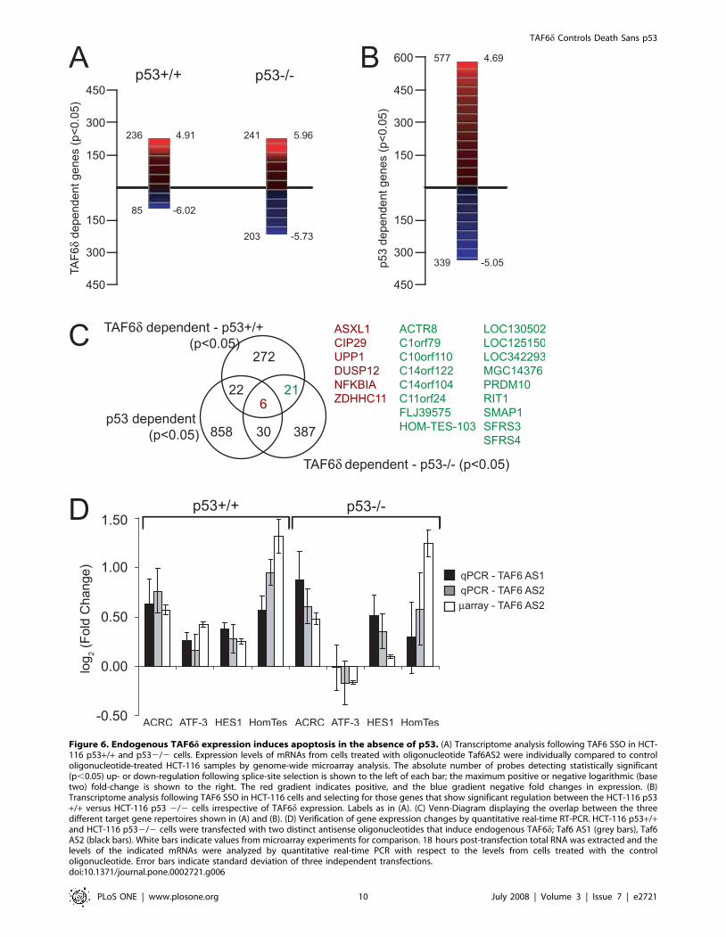
Figure 6. Endogenous TAF6d expression induces apoptosis in the absence of p53. (A) Transcriptome analysis following TAF6 SSO in HCT-116 p53+/+ and p532/2 cells. Expression levels of mRNAs from cells treated with oligonucleotide Taf6AS2 were individually compared to controloligonucleotide-treated HCT-116 samples by genome-wide microarray analysis. The absolute number of probes detecting statistically significant(p,0.05) up- or down-regulation following splice-site selection is shown to the left of each bar; the maximum positive or negative logarithmic (basetwo) fold-change is shown to the right. The red gradient indicates positive, and the blue gradient negative fold changes in expression. (B)Transcriptome analysis following TAF6 SSO in HCT-116 cells and selecting for those genes that show significant regulation between the HCT-116 p53+/+ versus HCT-116 p53 2/2 cells irrespective of TAF6d expression. Labels as in (A). (C) Venn-Diagram displaying the overlap between the threedifferent target gene repertoires shown in (A) and (B). (D) Verification of gene expression changes by quantitative real-time RT-PCR. HCT-116 p53+/+and HCT-116 p532/2 cells were transfected with two distinct antisense oligonucleotides that induce endogenous TAF6d; Taf6 AS1 (grey bars), Taf6AS2 (black bars). White bars indicate values from microarray experiments for comparison. 18 hours post-transfection total RNA was extracted and thelevels of the indicated mRNAs were analyzed by quantitative real-time PCR with respect to the levels from cells treated with the controloligonucleotide. Error bars indicate standard deviation of three independent transfections.doi:10.1371/journal.pone.0002721.g006
TAF6d Controls Death Sans p53
PLoS ONE | www.plosone.org 10 July 2008 | Volume 3 | Issue 7 | e2721
Discussion
Here we have combined splice-switching oligonucleotides
(SSOs) with high-sensitivity genome-wide microarrays to shed
light on the function of endogenous TAF6d. By experimentally
inducing endogenous TAF6d expression we show that TAF6dtriggers robust cell death in several cancer cell types. The
expression of the p53 tumor suppressor is dispensable for
TAF6d-dependent cell death and gene expression in several cell
lines. The data establish the TAF6d pathway as an important
signaling hub that can control apoptosis in the absence of p53.
Our microarray results show that TAF6d expression activates
genes, such as HOM-TES-103, HES1 and ACRC independently
of p53. The 21 genes we identify here that are controlled by
TAF6d independently of p53 represent candidate genes that could
mediate TAF6d-dependent apoptosis. The majority of genes
identified through the unbiased microarray approach are of as yet
unknown biological function; a finding that is not unexpected
given that TAF6d represents a newly discovered signaling
pathway. Therefore, further work will be required to determine
their contributions to the apoptotic program. Given the fact that
TAF6d activates minimally hundreds of genes, it is improbable
that any single gene product could account fully for TAF6d-driven
apoptosis. The microarray data presented here represent, to our
knowledge, the first documentation of changes in gene expression
due to induction of an endogenous TFIID subunit. Based on the
increases in gene expression we have demonstrated here, we
propose a model in which TAF6d drives a pro-apoptotic
transcription program to initiate the apoptotic cascade.
Despite intensive efforts, the molecular mechanisms by which
the TAFs specifically influence gene expression remain obscure.
The advantage of the SSO strategy employed here is that it reveals
for the first time a pro-apoptotic role for endogenous TAF6dlevels. As is the case for all physiological alternative splicing events,
the TAF6d isoform is produced with a concomitant reduction in
the levels of the major alpha isoform. The reduction in TAF6amay contribute to changes in gene expression during the SSO-
enforced as well as the normal physiological switch from alpha to
delta expression. It is, however, important to note that the
overexpression of TAF6d alone is sufficient to induce apoptosis in
cells that express endogenous TAF6a [39]. Therefore, although
decreased TAF6a expression necessarily accompanies increased
TAF6d expression, all the available evidence indicates that the
increase of the minor TAF6d isoform is a critical molecular event
triggering apoptosis.
Mounting evidence suggests that functionally distinct forms of
TFIID exist [57], and that TFIID composition is dynamic [58].
Recently, the dynamic nature of core promoter recognition
complexes in vivo has been underscored by the observation of a
drastic replacement of the cellular pools of canonical TFIID with a
complex containing TRF3/TAF3 during myogenesis [59]. The
number of combinatorial possibilities for more subtle changes
within the TFIID complex itself continues to expand with the
discovery of new TAF isoforms; TAF4 and TAF4b possess distinct
target gene specificities [60], TAF9 and TAF9b can both
incorporate into TFIID but are functionally non-redundant [61],
and TAF1-2 and TAF1-4 are signal-inducible and functionally
distinct splice variants of TAF1 [62,63]. To date, however, the
functional consequences for changes in the cellular levels of
endogenous TFIID subunits have remained unknown. The current
findings show that the induction of a single TFIID subunit within
living cells can orchestrate gene expression programs to alter cell
physiology. TAF6d mRNA levels can be dramatically induced in
HL-60 cells after differentiation by retinoic-acid, demonstrating at
least one physiological situation where TAF6d is induced in living
cells [39]. Recent genetic evidence that TAF12 is required for
ethylene-responsive transcription in plants [64] further argues that
TFIID is a signal-responsive transcription factor. Therefore, in
addition to its known functions in core promoter recognition and
co-activation, TFIID represents a platform that integrates cellular
signals with the control of gene expression.
The pro-apoptotic transcription factor p53 plays a central role
in genome surveillance and tumor suppression. The p53 protein is
not required for cell viability and indeed is lost or mutated in
roughly 50% of tumors [6]. Even in animal models where
functional p53 can be restored by gene therapy, tumors readily
attain resistance to p53 due to inactivation of p19ARF or p53 itself
[65]. The efficient induction of cell death in several different tumor
cell lines by SSO targeting of TAF6, independent of their p53
status, provides a proof-of-principle that the TAF6d pathway can
be exploited to kill tumor cells. The data presented here define the
TAF6d signaling hub as able to control apoptosis without p53, but
with interconnections to the p53 pathway including several shared
target genes, as revealed by transcriptome-wide microarray
analysis (Fig. 6B). Unlike p53, TAF6 is essential for viability in
all organisms studied [26,27,28,29]. Furthermore, targeting TAF6
results in a substantially more robust apoptotic response than
targeting another apoptotic gene, Bcl-x in several tumor cell lines
(Fig.s 3, 4 & 5). Further characterization of the TAF6d signaling
hub may therefore provide novel therapeutic avenues to induce
controlled tumor cell death irrespective of their p53 status.
The TAF6d pathway remains an orphan pathway since the
precise molecular trigger that induces TAF6d expression in the
physiological context is currently unknown. The fact that TAF6dcan act downstream of p53 to control gene expression, and that
TAF6d can dictate cell death versus life decisions of human cells,
evoke the possibility that this newly defined pathway could be
subject to deregulation in certain cancer cells. In this light, it is
intriguing that expression levels of TAF6 have been correlated
with the inflammatory breast cancer phenotype [66], and isoform
specific enrichment of a TAF6 splicing variant has been reported
in breast cancer [45]. Experiments to identify the upstream signals
that control TAF6d expression in vivo in healthy tissues, as well as
to uncover the potential role of mutations to the TAF6d pathway
in cancer are ongoing in our laboratory.
Supporting Information
Figure S1 (A) Time course for antisense RNA-mediated TAF6dmRNA induction. Transfections and RT-PCR were performed as
in Fig. 1C except that RNA was extracted at various times (x-axis)
after transfection. (B) Dose-dependent antisense mediated induc-
tion of TAF6d mRNA expression. As in B, with different
concentrations of oligonucleotides transfected indicated on the x-
axis.
Found at: doi:10.1371/journal.pone.0002721.s001 (0.71 MB EPS)
Supplementary Data File S1 Genes changing in response
TAF6delta in HCT-116 p53 +/+ cells
Found at: doi:10.1371/journal.pone.0002721.s002 (0.05 MB
TXT)
Supplementary Data File S2 Genes changing in response to
TAF6delta in HCT-116 p53 2/2
Found at: doi:10.1371/journal.pone.0002721.s003 (0.07 MB
TXT)
Supplementary Data File S3 Genes differentially expressed in
the HCT-116 p53 +/+ cells versus in HCT-116 p53 2/2 cells
TAF6d Controls Death Sans p53
PLoS ONE | www.plosone.org 11 July 2008 | Volume 3 | Issue 7 | e2721
both in cells treated with control oligonucleotide and TAF6delta -
inducing oligonucleotides
Found at: doi:10.1371/journal.pone.0002721.s004 (0.24 MB
TXT)
Supplementary Data File S4 Genes differentially regulated by
TAF6delta in both HCT-116 p53 +/+ cells and HCT-116 p53
2/2 cells
Found at: doi:10.1371/journal.pone.0002721.s005 (0.01 MB
TXT)
Acknowledgments
We thank B. Vogelstein for providing HCT-116 p53 2/2 cells. We are
grateful to B. Chabot for critical reading of the manuscript and to L. Tora
for anti-TAF antibodies.
Author Contributions
Conceived and designed the experiments: AB BB. Performed the
experiments: EW. Analyzed the data: FP AB BB. Wrote the paper: EW
AB BB.
References
1. Hengartner MO (2000) The biochemistry of apoptosis. Nature 407: 770–776.
2. Thompson CB (1995) Apoptosis in the pathogenesis and treatment of disease.Science 267: 1456–1462.
3. Fesus L (1999) Inducible gene expression in apoptosis [In Process Citation]. CellDeath Differ 6: 1144–1145.
4. Kumar S, Cakouros D (2004) Transcriptional control of the core cell-death
machinery. Trends Biochem Sci 29: 193–199.
5. Schwerk C, Schulze-Osthoff K (2005) Regulation of apoptosis by alternative pre-
mRNA splicing. Mol Cell 19: 1–13.
6. Vousden KH, Lane DP (2007) p53 in health and disease. Nat Rev Mol Cell Biol8: 275–283.
7. Liu G, Chen X (2002) The ferredoxin reductase gene is regulated by the p53 familyand sensitizes cells to oxidative stress-induced apoptosis. Oncogene 21: 7195–7204.
8. Hwang PM, Bunz F, Yu J, Rago C, Chan TA, et al. (2001) Ferredoxin reductase
affects p53-dependent, 5-fluorouracil-induced apoptosis in colorectal cancercells. Nat Med 7: 1111–1117.
9. Yu J, Zhang L, Hwang PM, Kinzler KW, Vogelstein B (2001) PUMA induces
the rapid apoptosis of colorectal cancer cells. Mol Cell 7: 673–682.
10. Oda E, Ohki R, Murasawa H, Nemoto J, Shibue T, et al. (2000) Noxa, a BH3-
only member of the Bcl-2 family and candidate mediator of p53-inducedapoptosis. Science 288: 1053–1058.
11. Miyashita T, Reed JC (1995) Tumor suppressor p53 is a direct transcriptional
activator of the human bax gene. Cell 80: 293–299.
12. Oda K, Arakawa H, Tanaka T, Matsuda K, Tanikawa C, et al. (2000) p53AIP1,
a potential mediator of p53-dependent apoptosis, and its regulation by Ser-46-
phosphorylated p53. Cell 102: 849–862.
13. Bell B, Tora L (1999) Regulation of gene expression by multiple forms of TFIID
and other novel TAFII-containing complexes. Exp Cell Res 246: 11–19.
14. Green MR (2000) TBP-associated factors (TAFIIs): multiple, selective transcrip-
tional mediators in common complexes [In Process Citation]. Trends Biochem
Sci 25: 59–63.
15. Muller F, Demeny MA, Tora L (2007) New problems in RNA polymerase II
transcription initiation: matching the diversity of core promoters with a varietyof promoter recognition factors. J Biol Chem 282: 14685–14689.
16. Albright S, Tjian R (2000) TAFs revisited: more data reveal new twists and
confirm old ideas. Gene 242: 1–13.
17. Gangloff YG, Romier C, Thuault S, Werten S, Davidson I (2001) The histone
fold is a key structural motif of transcription factor TFIID. Trends Biochem Sci
26: 250–257.
18. Xie X, Kokubo T, Cohen SL, Mirza UA, Hoffmann A, et al. (1996) Structural
similarity between TAFs and the heterotetrameric core of the histone octamer[see comments]. Nature 380: 316–322.
19. Werten S, Mitschler A, Romier C, Gangloff YG, Thuault S, et al. (2002) Crystal
structure of a subcomplex of human transcription factor TFIID formed byTATA binding protein-associated factors hTAF4 (hTAF(II)135) and hTAF12
(hTAF(II)20). J Biol Chem 277: 45502–45509.
20. Birck C, Poch O, Romier C, Ruff M, Mengus G, et al. (1998) Human TAF(II)28and TAF(II)18 interact through a histone fold encoded by atypical evolutionary
conserved motifs also found in the SPT3 family. Cell 94: 239–249.
21. Gangloff YG, Sanders SL, Romier C, Kirschner D, Weil PA, et al. (2001)
Histone folds mediate selective heterodimerization of yeast TAF(II)25 with
TFIID components yTAF(II)47 and yTAF(II)65 and with SAGA componentySPT7. Mol Cell Biol 21: 1841–1853.
22. Gangloff YG, Pointud JC, Thuault S, Carre L, Romier C, et al. (2001) TheTFIID components human TAF(II)140 and Drosophila BIP2 (TAF(II)155) are
novel metazoan homologues of yeast TAF(II)47 containing a histone fold and a
PHD finger. Mol Cell Biol 21: 5109–5121.
23. Wright KJ, Marr MT 2nd, Tjian R (2006) TAF4 nucleates a core subcomplex of
TFIID and mediates activated transcription from a TATA-less promoter. ProcNatl Acad Sci U S A 103: 12347–12352.
24. Michel B, Komarnitsky P, Buratowski S (1998) Histone-like TAFs are essential
for transcription in vivo. Mol Cell 2: 663–673.
25. Shen WC, Bhaumik SR, Causton HC, Simon I, Zhu X, et al. (2003) Systematic
analysis of essential yeast TAFs in genome-wide transcription and preinitiation
complex assembly. Embo J 22: 3395–3402.
26. Poon D, Bai Y, Campbell AM, Bjorklund S, Kim YJ, et al. (1995) Identification
and characterization of a TFIID-like multiprotein complex from Saccharomycescerevisiae. Proc Natl Acad Sci U S A 92: 8224–8228.
27. Lago C, Clerici E, Dreni L, Horlow C, Caporali E, et al. (2005) The Arabidopsis
TFIID factor AtTAF6 controls pollen tube growth. Dev Biol 285: 91–100.
28. Aoyagi N, Wassarman DA (2001) Developmental and transcriptional conse-quences of mutations in Drosophila TAF(II)60. Mol Cell Biol 21: 6808–6819.
29. Amsterdam A, Nissen RM, Sun Z, Swindell EC, Farrington S, et al. (2004)
Identification of 315 genes essential for early zebrafish development. Proc NatlAcad Sci U S A 101: 12792–12797.
30. Purrello M, Di Pietro C, Viola A, Rapisarda A, Stevens S, et al. (1998) Genomics
and transcription analysis of human TFIID. Oncogene 16: 1633–1638.
31. Weinzierl RO, Ruppert S, Dynlacht BD, Tanese N, Tjian R (1993) Cloning andexpression of Drosophila TAFII60 and human TAFII70 reveal conserved
interactions with other subunits of TFIID. Embo J 12: 5303–5309.
32. Burke TW, Kadonaga JT (1997) The downstream core promoter element, DPE,is conserved from Drosophila to humans and is recognized by TAFII60 of
Drosophila. Genes Dev 11: 3020–3031.
33. Shao H, Revach M, Moshonov S, Tzuman Y, Gazit K, et al. (2005) Core
promoter binding by histone-like TAF complexes. Mol Cell Biol 25: 206–219.
34. Thut CJ, Chen JL, Klemm R, Tjian R (1995) p53 transcriptional activationmediated by coactivators TAFII40 and TAFII60. Science 267: 100–104.
35. Lu H, Levine AJ (1995) Human TAFII31 protein is a transcriptional coactivator
of the p53 protein. Proc Natl Acad Sci U S A 92: 5154–5158.
36. Farmer G, Colgan J, Nakatani Y, Manley JL, Prives C (1996) Functionalinteraction between p53, the TATA-binding protein (TBP), andTBP-associated
factors in vivo. Mol Cell Biol 16: 4295–4304.
37. Jimenez GS, Nister M, Stommel JM, Beeche M, Barcarse EA, et al. (2000) Atransactivation-deficient mouse model provides insights into Trp53 regulation
and function. Nat Genet 26: 37–43.
38. Johnson TM, Hammond EM, Giaccia A, Attardi LD (2005) The p53QStransactivation-deficient mutant shows stress-specific apoptotic activity and
induces embryonic lethality. Nat Genet 37: 145–152.
39. Bell B, Scheer E, Tora L (2001) Identification of hTAF(II)80 delta links apoptotic
signaling pathways to transcription factor TFIID function. Mol Cell 8: 591–600.
40. Mercatante DR, Bortner CD, Cidlowski JA, Kole R (2001) Modification of
alternative splicing of Bcl-x pre-mRNA in prostate and breast cancer cells.
analysis of apoptosis and cell death. J Biol Chem 276: 16411–16417.
41. Brou C, Wu J, Ali S, Scheer E, Lang C, et al. (1993) Different TBP-associatedfactors are required for mediating the stimulation of transcription in vitro by the
acidic transactivator GAL- VP16 and the two nonacidic activation functions ofthe estrogen receptor. Nucleic Acids Res 21: 5–12.
42. Dubrovskaya V, Lavigne AC, Davidson I, Acker J, Staub A, et al. (1996) Distinct
domains of hTAFII100 are required for functional interaction with transcriptionfactor TFIIF beta (RAP30) and incorporation into the TFIID complex. Embo J
15: 3702–3712.
43. Noth S, Brysbaert G, Benecke A (2006) Normalization using weighted negative
second order exponential error functions (NeONORM) provides robustnessagainst asymmetries in comparative transcriptome profiles and avoids false calls.
Genomics Proteomics Bioinformatics 4: 90–109.
44. Noth S, Benecke A (2005) Avoiding inconsistencies over time and trackingdifficulties in Applied Biosystems AB1700/Panther probe-to-gene annotations.
BMC Bioinformatics 6: 307.
45. Wang W, Nahta R, Huper G, Marks JR (2004) TAFII70 isoform-specific growthsuppression correlates with its ability to complex with the GADD45a protein.
Mol Cancer Res 2: 442–452.
46. Taylor JK, Zhang QQ, Wyatt JR, Dean NM (1999) Induction of endogenousBcl-xS through the control of Bcl-x pre-mRNA splicing by antisense
oligonucleotides [see comments]. Nat Biotechnol 17: 1097–1100.
47. Mercatante DR, Mohler JL, Kole R (2002) Cellular response to an antisense-
mediated shift of Bcl-x pre-mRNA splicing and antineoplastic agents. J BiolChem 277: 49374–49382.
48. Leers MP, Kolgen W, Bjorklund V, Bergman T, Tribbick G, et al. (1999)
Immunocytochemical detection and mapping of a cytokeratin 18 neo- epitopeexposed during early apoptosis [In Process Citation]. J Pathol 187: 567–572.
49. Masuda H, Miller C, Koeffler HP, Battifora H, Cline MJ (1987) Rearrangement
of the p53 gene in human osteogenic sarcomas. Proc Natl Acad Sci U S A 84:7716–7719.
50. May E, Jenkins JR, May P (1991) Endogenous HeLa p53 proteins are easily
detected in HeLa cells transfected with mouse deletion mutant p53 gene.
Oncogene 6: 1363–1365.
TAF6d Controls Death Sans p53
PLoS ONE | www.plosone.org 12 July 2008 | Volume 3 | Issue 7 | e2721
51. Bunz F, Dutriaux A, Lengauer C, Waldman T, Zhou S, et al. (1998)
Requirement for p53 and p21 to sustain G2 arrest after DNA damage. Science282: 1497–1501.
52. Noth S, Brysbaert G, Pellay FX, Benecke A (2006) High-sensitivity
transcriptome data structure and implications for analysis and biologicinterpretation. Genomics Proteomics Bioinformatics 4: 212–229.
53. Wang Y, Barbacioru C, Hyland F, Xiao W, Hunkapiller KL, et al. (2006) Largescale real-time PCR validation on gene expression measurements from two
commercial long-oligonucleotide microarrays. BMC Genomics 7: 59.
54. Owen-Schaub LB, Zhang W, Cusack JC, Angelo LS, Santee SM, et al. (1995)Wild-type human p53 and a temperature-sensitive mutant induce Fas/APO-1
expression. Mol Cell Biol 15: 3032–3040.55. Kunz C, Pebler S, Otte J, von der Ahe D (1995) Differential regulation of
plasminogen activator and inhibitor gene transcription by the tumor suppressorp53. Nucleic Acids Res 23: 3710–3717.
56. el-Deiry WS, Tokino T, Velculescu VE, Levy DB, Parsons R, et al. (1993)
WAF1, a potential mediator of p53 tumor suppression. Cell 75: 817–825.57. Muller F, Tora L (2004) The multicoloured world of promoter recognition
complexes. Embo J 23: 2–8.58. Gegonne A, Weissman JD, Zhou M, Brady JN, Singer DS (2006) TAF7: a
possible transcription initiation check-point regulator. Proc Natl Acad Sci U S A
103: 602–607.
59. Deato MD, Tjian R (2007) Switching of the core transcription machinery during
myogenesis. Genes Dev.60. Mengus G, Fadloun A, Kobi D, Thibault C, Perletti L, et al. (2005) TAF4
inactivation in embryonic fibroblasts activates TGF beta signalling and autocrine
growth. Embo J 24: 2753–2767.61. Frontini M, Soutoglou E, Argentini M, Bole-Feysot C, Jost B, et al. (2005)
TAF9b (formerly TAF9L) is a bona fide TAF that has unique and overlappingroles with TAF9. Mol Cell Biol 25: 4638–4649.
62. Katzenberger RJ, Marengo MS, Wassarman DA (2006) ATM and ATR
pathways signal alternative splicing of Drosophila TAF1 pre-mRNA in responseto DNA damage. Mol Cell Biol 26: 9256–9267.
63. Metcalf CE, Wassarman DA (2006) DNA binding properties of TAF1 isoformswith two AT-hooks. J Biol Chem 281: 30015–30023.
64. Robles LM, Wampole JS, Christians MJ, Larsen PB (2007) Arabidopsisenhanced ethylene response 4 encodes an EIN3-interacting TFIID transcription
factor required for proper ethylene response, including ERF1 induction. J Exp
Bot.65. Martins CP, Brown-Swigart L, Evan GI (2006) Modeling the therapeutic
efficacy of p53 restoration in tumors. Cell 127: 1323–1334.66. Dressman HK, Hans C, Bild A, Olson JA, Rosen E, et al. (2006) Gene
expression profiles of multiple breast cancer phenotypes and response to
neoadjuvant chemotherapy. Clin Cancer Res 12: 819–826.
TAF6d Controls Death Sans p53
PLoS ONE | www.plosone.org 13 July 2008 | Volume 3 | Issue 7 | e2721