Durable remissions with autologous stem cell transplantation for high-risk HIV-associated lymphomas
Upload
independentCategory
view
0download
0
GASTROENTEROLOGY 2002;122:1286-1294
T(11;18) Is a Marker for All Stage Gastric MALT Lymphomas That Will Not Respond to H. pylori Eradication
HONGXIANG LIU,* HONGTAO YE,* AGNES RUSKONE-FOURMESTRAUX,t,§ DAPHNE DE JONG,II STEFANO PILERIfl CHRISTIAN THIEDE, # ANNE LAVERGNE,§,** HENK BOOT,II GIANCARLO CALETTI,¶ THOMAS WONDISCH,~* THIERRY MOLINA,{,§ BABS G. TAAL,II SABATTINI ELENA,~ TOGLIANI THOMASfl PIER LUIGI ZINZANIfl ANDREAS NEUBAUER, ~? MANFRED STOLTE,§§ RIFAT A. HAMOUDI,* AHMET DOGAN,* PETER G. ISAACSON,* and MING-QING DU* *Department of Histopathology, University College London, London, England; ~Service de Gastro-enterologie/Service d'Anatomie Pathologique, HoteI-Dieu, Ap-Hp, Paris, France; §Groupe d'Etude des Lymphomes Digestifs, Fondation Francaise de Canc6rologie Digestive, France; IIDepartment of Pathology/Gastroenterology, The Netherlands Cancer Institute, Amsterdam, The Netherlands; ¶Servizi di Anatomia Patologica e Gastroenterologia, Universit~ degli Studi di Bologna, Bologna, Italy; #Labor Molekulare Diagnostik, Universit~tsklinikum Carl Gustav Cams, Dresden, Germany; **Service d'Anatomie Pathologique, Hopital Lariboisiere, Paris, France; t'Department of Hematology, Oncology and Immunology, Philipps University Marburg, Marburg, Germany; and §§lnstitut fur Pathologie, Klinikum Bayreuth, Bayreuth, Germany
Background &Aims: Eradication of Helicobacter pylori leads to cure of gastric mucosa-associated lymphoid tissue (MALT) lymphoma in 75% of localized cases. However, prolonged follow-up is necessary to determine whether a lymphoma responds to therapy. In a small series of cases, we showed that t(11;18)(q21;q21)-pos- itive MALT lymphomas failed to respond to H. pylori eradication. The present study aimed to verify this find- ing in a large cohort and confirm whether the transloca- tion predicts the response of stage IE tumors, for which clinical staging has little prognostic value. Methods: A total of 111 patients with H. pylori-positive gastric MALT lymphoma treated with antibiotics were studied. Clinical staging was undertaken before therapy. The response of lymphoma to H. pylori eradication was determined by histologic examination of gastric biopsy specimens. Di- agnostic biopsy specimens were analyzed for t(11; 18)(q21;q21) by reverse-transcription polymerase chain reaction of the API2-MALT1 transcript. Results: Forty- seven of the 48 patients who showed complete regres- sion had lymphoma at stage IE, whereas 43 of the 63 nonresponsive cases were at stage I E and the remaining cases at stage lie or above, t(11;18)(q21;q21) was de- tected in 2 of 48 complete-regression cases, and these positive cases showed relapse of lymphoma in the ab- sence of H. pylori reinfection. In contrast, the transloca- tion was present in 42 of the 63 nonresponsive cases, including 26 of 43 (60%) at stage IE. Conclusions: t(11; 18)(q21;q21)-positive gastric MALT lymphomas, includ- ing those at stage IE, do not respond to H. pylori eradi- cation. Detection of the translocation should help the clinical management of patients with gastric MALT lym- phoma.
G astric mucosa-associated lymphoid tissue (MALT) lymphoma arises from mucosal lymphoid tissue
that is acquired usually as a reaction to Helicobacterpylori infection), 2 Growth of these lymphoma cells in culture can be stimulated by strain-specific heat-killed H. pylori. 3,4 This response is mediated by intratumoral H. pylori-specific T cells and involves CD40 and CD40L costimulatory molecules. 3,4 Intriguingly, the lymphoma immunoglobulin recognizes a variety of autoantigens without cross-reactivity with H. pylori. 5"6 In the clinical setting, eradication of H. pylori leads to complete regres- sion of gastric MALT lymphoma in approximately 75 % of cases. 7-~3
The time for regression to take place after H. pylori eradication varies from a few weeks to 18 months. 7-.3 Therefore, prolonged follow-up with repeated endoscopy and gastric biopsies is essential to determine whether a lymphoma responds to H. pylori eradication or requires additional therapy. The prognostic value of clinical stag- ing has been extensively examined with the help of endoscopic ultrasonography, which allows assessment of the extent of tumor invasion to the gastric wall and regional lymph nodes. 1~-~3 In general, stage IIE or above lymphomas, in which gastric lymph nodes or adjacent or
Abbreviations used in this paper: CR, complete remission; MALT, mucosa-associated lymphoid tissue; NR, nonresponsive; RT-PCR, re- verse-transcription polymerase chain reaction.
© 2002 by the American Gastroenterological Association 0016-5085/02/$35.00
doi:10.1053/gast.2002.33047
May 2002 T(11;18)-GASTRIC MALT LYMPHOMA RESISTS H. PYLORI ERADICATION 1287
remote organs are involved, do not respond to H. pylori eradication. 11-13 In stage IE cases, in which tumors are confined to the gastric wall, staging has limited value in
prediction of the response, although tumors that involve
the muscularis propria or serosa (stage In2) have a higher failure rate than those restricted to the submucosa (stage IE1). 11-13 However, most gastric MALT lymphomas are
at stage In at diagnosis, and alternative prognostic mark- ers are therefore needed.
t(11; 18)(q21 ;q21) occurs specifically in MALT lym-
phomas and is the most frequent genetic abnormality found in this tumor, detected in about 40% of gastric cases.14 26 The translocation fuses the amino terminal of
the API2 gene to the carboxyl terminal of the M A L T I gene and generates a chimeric fusion product. ~6-~8 In in
vitro assays, the API2-MALT1 fusion product activates nuclear factor KB, 27,28 a transcription factor for several
survival-related genes, including those encoding cyto- kines, growth factors, cell adhesion molecules, and sev- eral cell apoptosis inhibitors. 29 Thus, it is most likely
that the fusion product confers a survival advantage to
MALT lymphoma cells. In keeping with this, t(11; 18)(q21;q21) is significantly associated with more ad- vanced gastric M A L T lymphomas. 26,3°,3~ In a small series
of cases, we showed that t(11;18)(q21;q21)-positive MALT lymphomas failed to respond to H. pylori eradi- cation. 25 However, this finding remains to be validated
in a large cohort. More importantly, it is unknown whether the translocation predicts the response of stage
In lymphoma to H. pylori eradication. We have developed a novel reverse-transcription polymerase chain reaction
(RT-PCR) method for detection of t ( l l ;18)(q21;q21)
from archival formalin-fixed and paraffin-embedded tis- sues and addressed these issues in a large cohort of cases collected from 5 European lymphoma centers.
Mater ia ls and M e t h o d s
Patients and Materials
A series of 111 patients with H. pylori-positive gastric MALT lympboma who were treated with antibiotics alone was retrospectively recruited from the Groupe d'Etude des Lym- phomes Digestifs, France (33 cases); Department of Pathology, The Netherlands Cancer Institute, The Netherlands (32 cases); Servizi di Anatomia Patologica e Gastroenterologia, Universit~ degli Studi di Bologna, Italy (24 cases); the German MALT Lymphoma Study Group (18 cases); and Department of His- topathology, University College London (4 cases). The selec- tion of patients was biased toward those who showed no response to H. pylori eradication, and the proportion of H. pylori eradication nonresponsive (NR) cases in different groups was similar. The diagnosis of gastric MALT lymphoma was made according to histologic criteria described by Isaacson et
al. 9,32 In all cases, there Was dense diffuse infiltrate of centro- cyte-like cells in lamina propria with prominent lymphoepi- thelial lesions. Clinical staging according to the Ann Arbor system modified by Musshoff was performed in each case before therapy) 3 In 64 cases, the extent of lymphoma invasion of the gastric wall and regional lymph nodes was determined by endoscopic ultrasonography, which allows further division of stage I~ tumors into IE1 (restricted to the submucosa) and IE2 (extended to the muscularis or serosa). 11 H. pylori eradication was achieved by administration of a 2-week course of amoxi- cillin (3 × 750 mg daily) and omeprazole (3 × 40 mg daily), v,1°,~2 One month after completing the antibiotic ther- apy, the first gastric endoscopy and biopsy were performed to detect H. pylori infection by histology, culture, and PCR of the H. pylori-associated urease gene and tumor regression by his- tology and molecular analysis. These investigations were re- peated every 3 -4 months until lymphoma showed complete regression or was judged as NR. After achieving complete regression, patients were examined further every 6 months. Lymphomas showing both complete endoscopic and histologic regression were regarded as complete remission (CR). Those that failed to show histologic regression 12 months after successful eradication of H. pylori or progressed during fol- low-up were judged as NR.
Tissue specimens from diagnostic biopsy specimens, includ- ing frozen tissues from 22 patients and formalin-fixed and paraffin-embedded tissues from 89 patients, were retrieved for molecular analysis. Where indicated, follow-up biopsy speci- mens were also analyzed.
RNA Extraction
For frozen tissues, total RNA was extracted from up to 10 mg tissue using the RNeasy Mini Kit (Qiagen, West Sussex, England).
For formalin-fixed and paraffin-embedded tissues, total RNA was extracted using an Ambion RNA isolation kit (AMS Biotechnology, Oxon, England, United Kingdom). Briefly, 5-10 5-gtm paraffin sections were deparaffinized in xylene. The tissue was digested with proteinase K (1 mg/mL) for 2 hours at 45°C and solubilized in a guanidinium-based buffer. RNA was extracted with acid phenol/chloroform and precipitated in isopropanol. The precipitated RNA was washed in 75% eth- anol and redissolved in 20 ~L RNA Storage Solution (AMS Biotechnology).
Detection of t(11;18)(q21;q21) by RT-PCR
The synthesis of complementary DNA (cDNA) and PCR detection of the API2-MALT1 fusion transcript from frozen tissues was performed as described previously. 25,26 Briefly, up to 2 Ixg total RNA was reverse transcribed into cDNA using the SuperScript Preamplification System (Invitro- gen Ltd., Paisley, Scotland, United Kingdom) and oligo(dT) primer. The API2-MALT1 fusion transcript was amplified by PCR using a pair of primers (f-S and f-AS) that covered all the known breakpoints (Table 1 and Figure 1). 26 As control, a 257-base pair fragment of the glucose-6-phosphate dehydro-
1288 LIUETAL. GASTROENTEROLOGY Vol. 122, No. 5
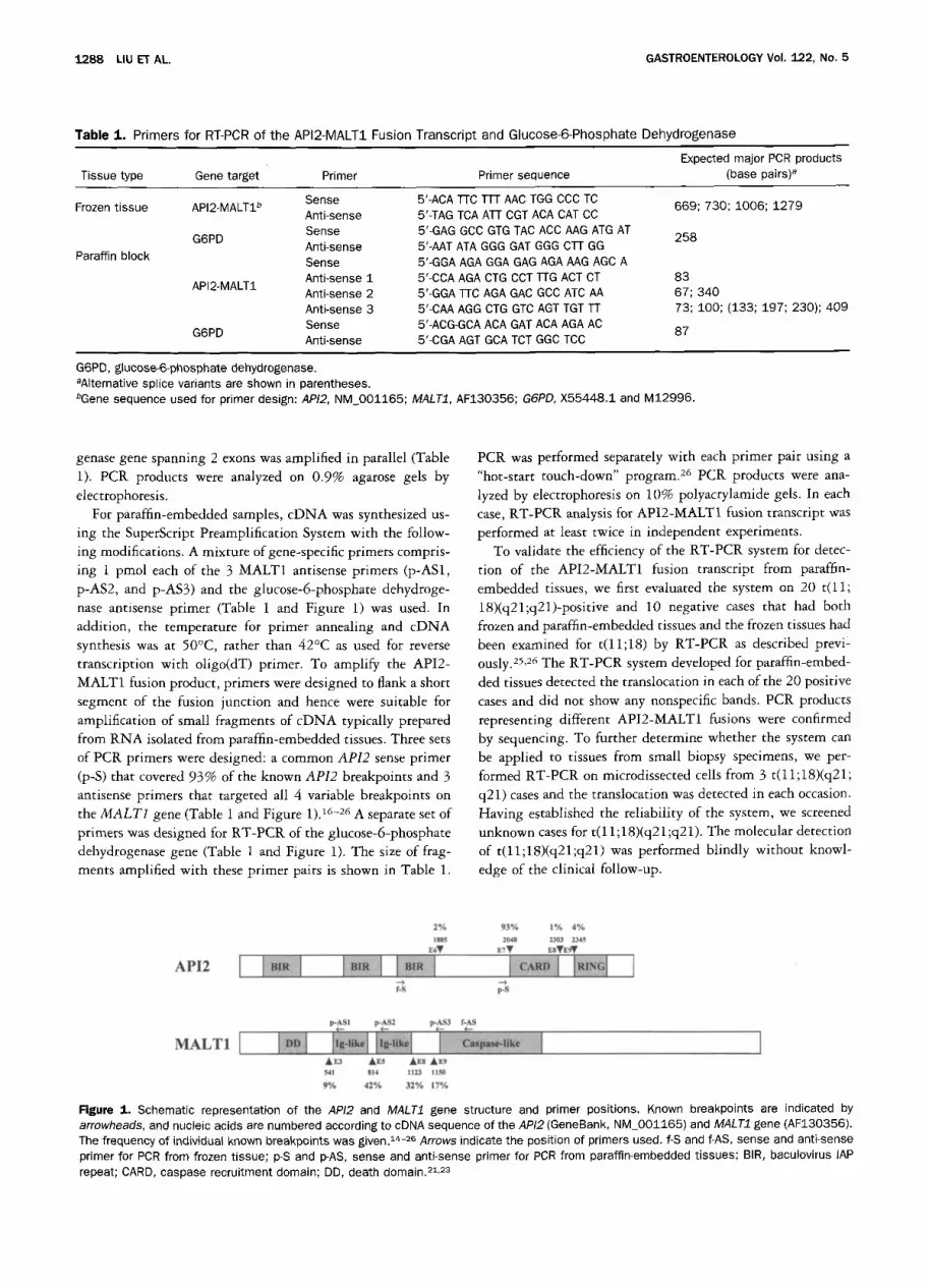
T a b l e 1. Primers for RT-PCR of the API2-MALT1 Fusion Transcript and Glucose-6-Phosphate Dehydrogenase
Expected major PCR products Tissue type Gene target Primer Primer sequence (base pairs) a
Sense 5'-ACA T[C TTT AAC TGG CCC TC Frozen tissue API2-MALT1 b 669; 730; 1006; 1279
Anti-sense 5'-TAG TCA AT[ CGT ACA CAT CC Sense 5'-GAG GCC GTG TAC ACC AAG ATG AT
G6PD 258 Anti-sense 5'-AAT ATA GGG GAT GGG CEI GG Sense 5'-GGA AGA GGA GAG AGA AAG AGC A Anti-sense 1 5'-CCA AGA CTG CCT BG ACT CT 83 Anti-sense 2 5'-GGA i-rc AGA GAC GCC ATC AA 67; 340 Anti-sense 3 5'-CAA AGG CTG GTC AGT TGT TT 73; 100; (133; 197; 230); 409 Sense 5'-ACG-GCA ACA GAT ACA AGA AC
G6PD 87 Anti-sense 5'-CGA AGT GCA TCT GGC TCC
Paraffin block
API2-MALT1
G6PD, glucose-6-phosphate dehydrogenase. aAIternative splice variants are shown in parentheses. bGene sequence used for primer design: API2, NM_001165; MALT1, AF130356; G6PD, X55448.1 and M12996.
genase gene spanning 2 exons was amplified in parallel (Table 1). PCR products were analyzed on 0.9% agarose gels by electrophoresis.
For paraffin-embedded samples, cDNA was synthesized us- ing the SuperScript Preamplification System with the follow- ing modifications. A mixture of gene-specific primers compris- ing 1 pmol each of the 3 MALT1 antisense primers (p-ASl, p-AS2, and p-AS3) and the glucose-6-phosphate dehydroge- nase antisense primer (Table 1 and Figure 1) was used. In addition, the temperature for primer annealing and cDNA synthesis was at 50°C, rather than 42°C as used for reverse transcription with oligo(dT) primer. To amplify the API2- MALT1 fusion product, primers were designed to flank a short segment of the fusion junction and hence were suitable for amplification of small fragments of cDNA typically prepared from RNA isolated from paraffin-embedded tissues. Three sets of PCR primers were designed: a common API2 sense primer (p-S) that covered 93% of the known API2 breakpoints and 3 antisense primers that targeted all 4 variable breakpoints on the MALT1 gene (Table 1 and Figure 1). 16-26 A separate set of primers was designed for RT-PCR of the glucose-6-phosphate dehydrogenase gene (Table 1 and Figure 1). The size of frag- ments amplified with these primer pairs is shown in Table 1.
PCR was performed separately with each primer pair using a "hot-start touch-down" program. 26 PCR products were ana- lyzed by electrophoresis on 10% polyacrylamide gels. In each case, RT-PCR analysis for API2-MALT1 fusion transcript was performed at least twice in independent experiments.
To validate the efficiency of the RT-PCR system for detec- tion of the API2-MALT1 fusion transcript from paraffin- embedded tissues, we first evaluated the system on 20 t ( l l ; 18)(q21;q21)-positive and 10 negative cases that had both frozen and paraffin-embedded tissues and the frozen tissues had been examined for t(11;18) by RT-PCR as described previ- ously. 25,26 The RT-PCR system developed for paraffin-embed- ded tissues detected the translocation in each of the 20 positive cases and did not show any nonspecific bands. PCR products representing different API2-MALT1 fusions were confirmed by sequencing. To further determine whether the system can be applied to tissues from small biopsy specimens, we per- formed RT-PCR on microdissected ceils from 3 t(11;18)(q21; q21) cases and the translocation was detected in each occasion. Having established the reliability of the system, we screened unknown cases for t(11 ; 18)(q21 ;q21). The molecular detection of t(11; 18)(q21 ;q21) was performed blindly without knowl- edge of the clinical follow-up.
2 % 93% 1% 4 %
180'$ 2048 2303 2345 z~V E7 • r~VzgV
AFa AEs A ~ Az9 f*41 814 1123 1150
9% 42% 32% 17%
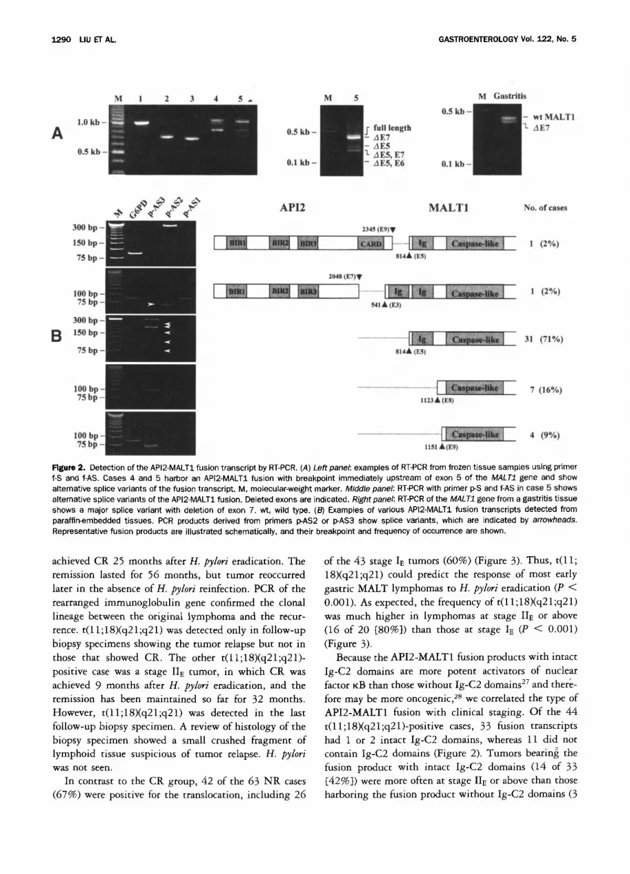
Figure 1. Schematic representation of the API2 and MALT1 gene structure and primer positions. Known breakpoints are indicated by arrowheads, and nucleic acids are numbered according to cDNA sequence of the API2 (GeneBank, NM_001165) and MALT1 gene (AF130356). The frequency of individual known breakpoints was given. 14-26 Arrows indicate the position of primers used. f-S and f-AS, sense and anti-sense primer for PCR from frozen tissue; p-S and p-AS, sense and anti-sense primer for PCR from paraffin-embedded tissues; BIR, baculovirus lAP repeat; CARD, caspase recruitment domain; DD, death domain. 21,23

May 2002 T(11;18)-GASTRIC MALT LYMPHOMA RESISTS H. PYLORI ERADICATION 1289
Sequencing of PCR Products
Where indicated, PCR products were gel purified (QIAquick Gel Extraction Kit; Qiagen) and sequenced in both directions using dRhodamine dye terminators on an ABI Prism 377 sequencer (PE Applied Biosystems, Foster City, CA).
Statistical Analysis
X 2 and Fisher exact tests were used to analyze the correlation between the response of MALT lymphomas to H. pylori eradication and clinical staging or t(ll;18)(q21;q21) s t a t u s .
R e s u l t s
Clinical Staging Predicts Treatment Failure to H. pylori Eradication in Stage lie or Above but Not Stage I E Gastric MALT Lymphoma
A total of 111 patients with gastric MALT lym- phoma were included in the present multicenter study (67 men and 44 women; mean age, 58 years [range, 2 5 - 8 8 years]). H. pylori infection was successfully cured in all cases as confirmed by histology and culture of gastric biopsy specimens taken after completion of the antibiotic therapy. After H. pylori eradication, patients were followed up by repeated endoscopy and biopsy. The mean period between H. pylori eradication and achieve- ment of CR or commencement of other treatment in N R patients was 12 months (range, 1-75 months), and the mean follow-up period to date is 35 months (range, 9 - 8 5 months). During follow-up, 48 cases showed CR, whereas 63 cases displayed NR. There is no difference in age and sex between the CR and NR groups. Both groups had similar length of follow-up. Histologically, focal transformed high-grade components were seen in 3 N R but not in any of the CR cases. In the CR group, 2 of 48 cases showed tumor relapse; in both cases, the lymphoma harbored t(11;18) (detailed in the next sec- tion).
Among the 48 CR cases, 47 were at stage I E and 1 at stage II> The stage IIE CR case is 1 of 2 that showed lymphoma relapse. Of the 63 NR cases, 20 were at stage IIE or above and the remaining 43 cases were at stage IE. Despite the fact that most of the lymphomas at stage IIs or above (20 of 21 [95%]) did not respond to H. priori eradication (P < 0.001), almost one half of stage Is tumors also did not respond to H. pylori eradication (43 of 90 [48%], P > 0.05). Therefore, the staging failed to predict the response of stage IE gastric MALT lymphoma to H. pylori eradication. Among cases with stage Is lymphoma, there was no difference in age, sex, and
Table 2. Clinical and Histopathologic Features of Stage IE Gastric MALT Lymphomas and Their Responses to H. pylori Eradication Therapy
Complete No regression regression
Number of patients 47 43 Age (yr)
Mean 60 57 Range 25-85 30-88
Sex M 29 21 F 18 22
Histology with high grade component 0 3 Stages by endoscopic ultrasonography
IE1 29 30 IE2 3 2
Follow-up period (too) Intervalsa
Mean 8.2 15 Range 1-26 5-75
Follow-up to date Mean 38 30 Range 10-82 9-85
aThe time between H. pylori eradication and complete regress/on or commencement of other treatment in NR cases.
follow-up periods between the CR and N R groups (P > 0.05) (Table 2). The extent of lymphoma invasion within the gastric wall was assessed by endoscopic ultrasonog- raphy in 64 cases with stage IE lymphoma. There was no difference in the response of gastric MALT lymphoma to H. pylori eradication between cases showing stage IE1 and
stage Is2 disease (P > 0.05) (Table 2).
t(11;18)(q21;q21) Is a Marker for Gastric MALT Lymphomas That Do Not Respond to H. pylori Eradication, Including Those at Stage IE
All cases presented in this study showed success- ful RT-PCR of the reference gene glucose-6-phosphate dehydrogenase. The API2-MALT1 fusion PCR product varied in size depending on the breakpoints and primer sets used, but accurate sizing of the PCR product on polyacrylamide gels and the characteristic PCR patterns allowed detection of t(11 ;18)(q21;q21) with high confi- dence (Figure 2). In 13 cases, PCR bands were weak and sequencing confirmation was performed. Overall, t ( l l ; 18) was positive in 40% (44 of 111) of cases detected. The positivity of t(11 ;18) detected from frozen tissues (9 of 22 cases [41%]) was similar to that from paraffin- embedded tissues (35 of 89 cases [39%]) (P > 0.05). The combined results from both frozen and paraffin-embed- ded tissues are summarized as follows.
Of the 48 CR cases, 2 were t(11; 18)(q21 ;q21) positive (Figure 3). One of these 2 cases, a stage IE tumor,
1290 LIU ET AL. GASTROENTEROLOGY Vol. 122, No. 5
1.0 kb -
A 0.5 k b -
M 1 2 3 4 5 . M 5
0.5 kb - full length A E 7 AE5 AES, E7
0.1 kb - AES, E6
0.5 kb
0.I kb
M Gastritis
wt MALT1 AE7
B
300 bp -
150 bp -
75 bp -
100 75
300 bp
150 bp
75 bp
I
API2 MALT1
2345 (E9)V
....... ~ ~ 1 ( 2 °,6)
814A ~
2o4s (E~V
.................... I m m ~ 1 (2%) 541 • (Fa)
Sl4A (ES)
No. of eases
31 (71%)
7 (16%) nT~A (Es)
100 bp - . . . . . . . . . . . . . . . . . . . . . . . . . . . . . . . . . . . . . . . . . . . . . . . . . . . . . . . . ~ 4 (9%) 75 bp l lSl Ak (E9)
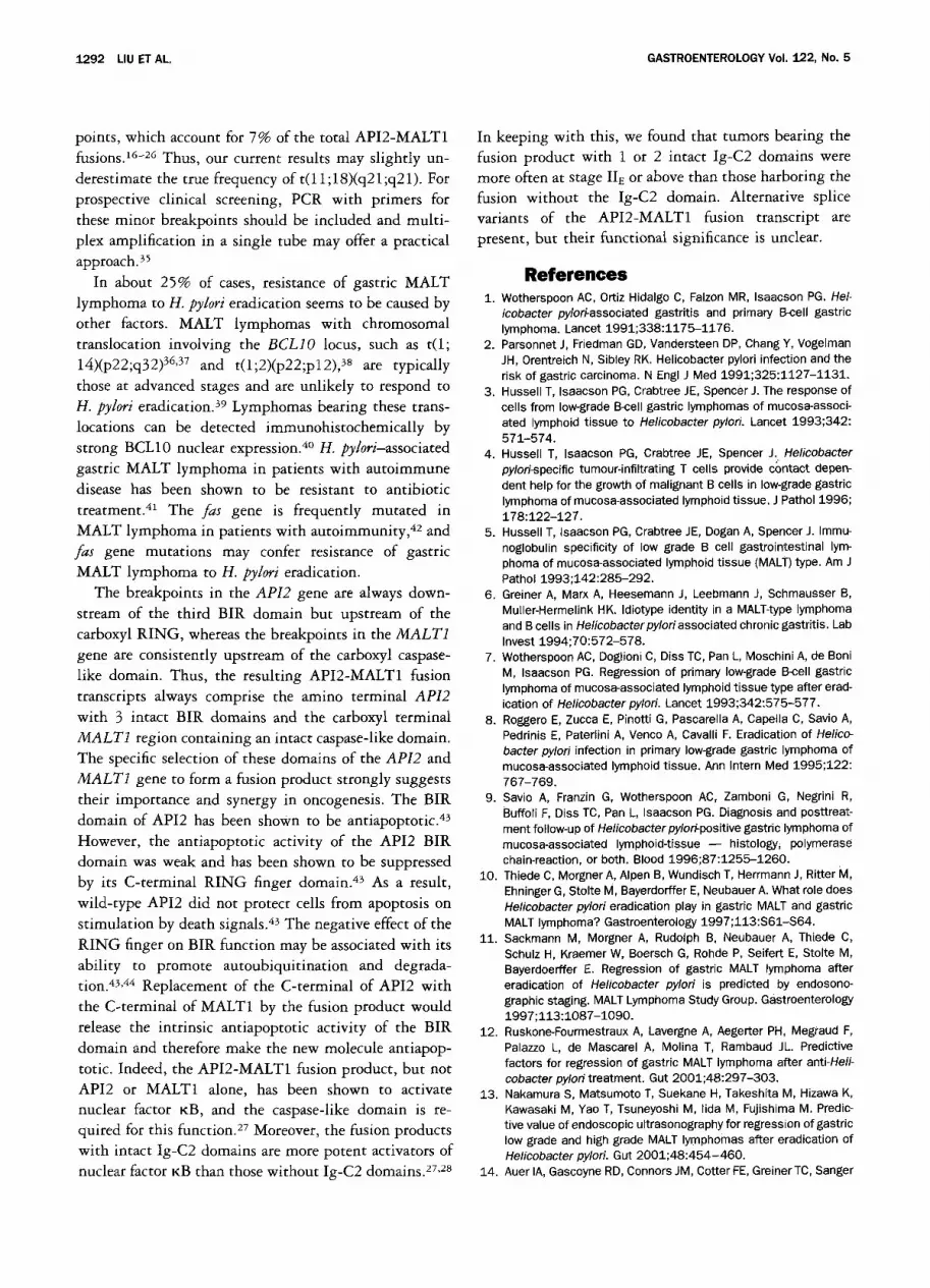
glgu~ 2. Detection of the API2-MALT1 fusion transcript by RT-PCR. (A) Leftpanel: examples of RT-PCR from frozen tissue samples using primer f-S and f-AS. Cases 4 and 5 harbor an API2-MALT1 fusion with breakpoint immediately upstream of exon 5 of the MALT1 gene and show alternative splice variants of the fusion transcript. M, molecular-weight marker. Middle panel: RT-PCR with primer p-S and f-AS in case 5 shows alternative splice variants of the API2-MALT1 fusion. Deleted exons are indicated. Right panel: RT-PCR of the MALT1 gene from a gastritis tissue shows a major splice variant with deletion of exon 7. wt, wild type. (B) Examples of various API2-MALT1 fusion transcripts detected from paraffin-embedded tissues. PCR products derived from primers p-AS2 or p-AS3 show splice variants, which are indicated by arrowheads. Representative fusion products are illustrated schematically, and their breakpoint and frequency of occurrence are shown.
achieved CR 25 months after H. pylori eradication. The remission lasted for 56 months, but tumor reoccurred later in the absence of H. pylori reinfection. PCR of the rearranged immunoglobulin gene confirmed the clonal lineage between the original lymphoma and the recur- rence, t(ll;18)(q21;q21) was detected only in follow-up biopsy specimens showing the tumor relapse but not in those that showed CR. The other t(ll;18)(q21;q21)- positive case was a stage IIE tumor, in which CR was achieved 9 months after H. pylori eradication, and the remission has been maintained so far for 32 months. However, t(11;18)(q21;q21) was detected in the last follow-up biopsy specimen. A review of histology of the biopsy specimen showed a small crushed fragment of lymphoid tissue suspicious of tumor relapse. H, pylori was not seen.
In contrast to the CR group, 42 of the 63 NR cases (67%) were positive for the translocation, including 26
of the 43 stage I~ tumors (60%) (Figure 3). Thus, t(11; 18)(q21;q21) could predict the response of most early gastric MALT lymphomas to H. pylori eradication (P < 0.001). As expected, the frequency of t(11; 18)(q21 ;q21) was much higher in lymphomas at stage II~ or above (16 of 20 [80%]) than those at stage IE (P < 0.001) (Figure 3).
Because the API2-MALT1 fusion products with intact Ig-C2 domains are more potent activators of nuclear factor KB than those without Ig-C2 domains 2v and there- fore may be more oncogenic, 28 we correlated the type of API2-MALT1 fusion with clinical staging. Of the 44 t(11;18)(q21;q21)-positive cases, 33 fusion transcripts had 1 or 2 intact Ig-C2 domains, whereas 11 did not contain Ig-C2 domains (Figure 2). Tumors bearin~ the fusion product with intact Ig-C2 domains (14 of 33 [42%]) were more often at stage IIE or above than those harboring the fusion product without Ig-C2 domains (3

May 2002 T(11;18)-GASTRIC MALT LYMPHOMA RESISTS H. PYLORI ERADICATION 1291
50
40
30
20
10
0
46
m t ( l l ;18) negative
1 t( l l ;18) positive
26
16
1 1
IE -> II E I E __ II~
Complete regression No regression
Figure 3. Correlation between response of gastric MALT lymphoma to H. pylori eradication therapy and clinical staging and presence of t(11;18)(q21;q21). Clinical staging has little value in predication of the response of stage IE gastric MALT lymphoma to H. pylori eradica- tion therapy. In contrast, the translocation can predict 60% of H. pylori therapy NR cases at stage I E.
of 11 [27%]), although statistical analysis did not show any significant difference (P > 0.05).
Alternative Splice Variants of the API2-MALT1 Fusion Transcript
The breakpoint in the MALT1 gene occurred variably immediately upstream of 4 exons (3, 5, 8, and 9), whereas the breakpoint in the API2 gene was always immediately downstream of exon 7 with an exception in 1 case that occurred after exon 9. Various API2-MALT1 fusions gave different PCR patterns (Figure 2). When the breakpoint occurred immediately before exons 8 and 9 of the MALT1 gene, RT-PCR showed only a single band. When the breakpoint occurred before exon 5, 1 expected band together with 4 additional smaller bands were seen. These additional PCR bands were of variable intensity but weaker than the expected fusion product. Sequencing of these bands confirmed that they were API2-MALT1 fusions identical to the corresponding fusion product in each case but with deletion of 1 or more exons of the MALT1 gene. It is most likely that these additional PCR bands represent alternative splice variants of the fusion transcript.
The splice variants of the API2-MALT1 fusion prod- uct are best shown by PCR from frozen tissues with the sense primer positioned just upstream of the API2 break- point (p-S), which yielded smaller fusion products and hence gave better separation on gels. Case 5, which harbored an API2-MALT1 fusion transcript with break- point immediately before exon 5 of the MALT1 gene, showed 4 additional bands (Figure 2). They represented alternative splice variants with deletion of exon 7, exon 5, exons 5 and 7, and exons 5 and 6 of the MALT1 gene. The variant without exon 7 did not alter the amino acid
reading frame and represented the major splice variant, whereas other splice species were minor and those with deletion of exon 5 or both exons 5 and 7 introduced a stop codon.
To examine whether these alternative splice events occur in wild-type MALTI, RT-PCR of the MALTI gene was performed. In contrast to the API2-MALT1 fusion, only a splice variant with exon 7 deletion was found in MALT1 (Figure 2). However, in keeping with the splice variants of the fusion transcript, the MALT1 transcript with exon 7 deletion was a major type (Figure 2). 34
Discussion
H. pylori eradication leads to complete regression of gastric MALT lymphoma in 75% of cases and is widely accepted as the first-line treatment for this tu- mor. v-13 One of the major dilemmas in clinical manage- ment of patients with this disease is the identification of those that will not respond t ° H . pylori eradication and require chemotherapy or radiotherapy. At present, this requires prolonged follow-up with repeated endoscopy and gastric biopsy. Clinical staging is helpful in predict- ing the response because lymphomas at stage lie or above rarely respond to H. pylori eradication. However, the predictive value of clinical staging for stage IE tumors is iimited ll-13 and better prognostic markers are needed. We have shown that t(ll;18)(q21;q21) is a marker for NR gastric MALT lymphomas, including those at stage I> In the stage Is cases, the translocation allows this prediction in 60% of NR cases. None of the CR cases were positive for t(ll;18)(q21;q21), with the exception of the 2 equivocal cases described.
Our findings indicate that t(11; 18)(q21 ;q21)-positive gastric MALT lymphomas do not undergo regression after H. pylori eradication and require other conventional therapies up front. Nevertheless, H. pylori should be eradicated in all cases because this not only eliminates reactive lymphoid infiltrates but most likely has an ad- juvant effect because in vitro experiments have shown that H. pylori stimulates t(11; 18)(q21 ;q21)-positive lym- phoma cells to proliferate via T-cell help. 3,4 Moreover, eradication of H. pylori and reactive lymphoid infiltrates may reduce the risk of developing secondary tumors in the stomach.
Among the NR cases, 33% failed to show t ( l l ; 18)(q21;q21) by RT-PCR. Our RT-PCR strategy for frozen tissues would theoretically detect 100% of known breakpoints in both the API2 and MALTI genes. How- ever, the RT-PCR methodology for paraffin-embedded tissues would miss some of the 3 minor API2 break-
1292 LIU El" AL. GASTROENTEROLOGY Vol. 122, No. 5
points, which account for 7% of the total API2-MALT1 fusions. 16-26 Thus, our current results may slightly un- derestimate the true frequency of t(11; 18)(q21 ;q21). For prospective clinical screening, PCR with primers for these minor breakpoints should be included and multi- plex amplification in a single tube may offer a practical approach) 5
In about 25% of cases, resistance of gastric MALT lymphoma to H. pylori eradication seems to be caused by other factors. MALT lymphomas with chromosomal translocation involving the BCLIO locus, such as r(1; 14)(p22;q32) 36'3v and t(1;2)(p22;p12), 38 are typically those at advanced stages and are unlikely to respond to H. pylori eradication) 9 Lymphomas bearing these trans- locations can be detected immunohistochemically by strong BCL10 nuclear expression, a° H. pylori-associated gastric MALT lymphoma in patients with autoimmune disease has been shown to be resistant to antibiotic treatment. 41 The fas gene is frequently mutated in MALT lymphoma in patients with autoimmunity, 42 and fas gene mutations may confer resistance of gastric MALT lymphoma to H. pylori eradication.
The breakpoints in the API2 gene are always down- stream of the third BIR domain but upstream of the carboxyl RING, whereas the breakpoints in the MALT1 gene are consistently upstream of the carboxyl caspase- like domain. Thus, the resulting API2-MALT1 fusion transcripts always comprise the amino terminal API2 with 3 intact BIR domains and the carboxyl terminal MALT1 region containing an intact caspase-like domain. The specific selection of these domains of the API2 and MALT1 gene to form a fusion product strongly suggests their importance and synergy in oncogenesis. The BIR domain of API2 has been shown to be antiapoptotic. 43 However, the antiapoptotic activity of the API2 BIR domain was weak and has been shown to be suppressed by its C-terminal RING finger domain. 43 As a result, wild-type API2 did not protect cells from apoptosis on stimulation by death signals. 43 The negative effect of the RING finger on BIR function may be associated with its ability to promote autoubiquitination and degrada- tion. 43,44 Replacement of the C-terminal of API2 with the C-terminal of MALT1 by the fusion product would release the intrinsic antiapoptotic activity of the BIR domain and therefore make the new molecule antiapop- totic. Indeed, the API2-MALT1 fusion product, but not API2 or MALT1 alone, has been shown to activate nuclear factor KB, and the caspase-like domain is re- quired for this f unc t i ons Moreover, the fusion products with intact Ig-C2 domains are more potent activators of nuclear factor KB than those without Ig-C2 domains. 2v,28
In keeping with this, we found that tumors bearing the fusion product with 1 or 2 intact Ig-C2 domains were more often at stage IIE or above than those harboring the fusion without the Ig-C2 domain. Alternative splice variants of the API2-MALT1 fusion transcript are present, but their functional significance is unclear.
References 1. Wotherspoon AC, Ortiz Hidalgo C, Falzon MR, Isaacson PG. Hel-
icobacter pylori-associated gastritis and primary B-cell gastric lymphoma. Lancet 1991;338:1175-1176.
2. Parsonnet J, Friedman GD, Vandersteen DP, Chang Y, Vogelman JH, Orentreich N, Sibley RK. Helicobacter pylori infection and the risk of gastric carcinoma. N Engl J Med 1991;325:1127-1131.
3. Hussell T, Isaacson PG, Crabtree JE, Spencer J. The response of cells from low-grade B-cell gastric lymphomas of mucosa-associ- ated lymphoid tissue to Helicobacter pylori. Lancet 1993;342: 571-574.
4. Hussell T, Isaacson PG, Crabtree JE, Spencer J. Helicobacter pylori-specific tumour-infiltrating T cells provide c°ntact depen- dent help for the growth of malignant B cells in low-grade gastric lymphoma of mucosa-associated lymphoid tissue. J Pathol 1996; 178:122-127.
5. Hussell T, Isaacson PG, Crabtree JE, Dogan A, Spencer J. Immu- noglobulin specificity of low grade B cell gastrointestinal lym- phoma of mucosa-associated lymphoid tissue (MALT) type. Am J Pathol 1993;142:285-292.
6. Greiner A, Marx A, Heesemann J, Leebmann J, Schmausser B, Muller-Hermelink HK. Idiotype identity in a MALT-type lymphoma and B cells in Helicobacterpylori associated chronic gastritis. Lab Invest 1994;70:572-578.
7. Wotherspoon AC, Doglioni C, Diss TC, Pan L, Moschini A, de Boni M, Isaacson PG. Regression of primary low-grade B-cell gastric lymphoma of mucosa-associated lymphoid tissue type after erad- ication of Helicobacter pylori. Lancet 1993;342:575-577.
8. Roggero E, Zucca E, Pinotti G, Pascarella A, Capella C, Savio A, Pedrinis E, Paterlini A, Venco A, Cavalli F. Eradication of Helico- bacter pylori infection in primary low-grade gastric lymphoma of mucosa-associated lymphoid tissue. Ann Intern Med 1995;122: 767-769.
9. Savio A, Franzin G, Wotherspoon AC, Zamboni G, Negrini R, Buffoli F, Diss TC, Pan L, Isaacson PG. Diagnosis and posttreat- ment follow-up of Helicobacter pylori-positive gastric lymphoma of mucosa-associated lymphoid-tissue - - histology, polymerase chain-reaction, or both. Blood 1996;87:1255-1260.
10. Thiede C, Morgner A, Alpen B, Wundisch T, Herrmann J, Ritter M, Ehninger G, Stolte M, Bayerdorffer E, Neubauer A. What role does Helicobacter pylori eradication play in gastric MALT and gastric MALT lymphoma? Gastroenterology 1997;113:S61-$64.
11. Sackmann M, Morgner A, Rudolph B, Neubauer A, Thiede C, Schulz H, Kraemer W, Boersch G, Rohde P, Seifert E, Stolte M, Bayerdoerffer E. Regression of gastric MALT lymphoma after eradication of Helicobacter pylori is predicted by endosono- graphic staging. MALT Lymphoma Study Group. Gastroenterology 1997; 113:1087-1090.
12. Ruskone-Fourmestraux A, Lavergne A, Aegerter PH, Megraud F, Palazzo L, de Mascarel A, Molina T, Rambaud JL. Predictive factors for regression of gastric MALT lymphoma after anti-Heft- cobacter pylori treatment. Gut 2001;48:297-303.
13. Nakamura S, Matsumoto T, Suekane H, Takeshita M, Hizawa K, Kawasaki M, Yao T, Tsuneyoshi M, lida M, Fujishima M. Predic- tive value of endoscopic ultrasonography for regression of gastric low grade and high grade MALT lymphomas after eradication of Helicobacter pylori. Gut 2001;48:454-460.
14. Auer IA, Gascoyne RD, Connors JM, Cotter FE, Greiner TC, Sanger

May 2002 T(11;18)-GASTRIC MALT LYMPHOMA RESISTS H. PYLORI ERADICATION 1293
WG, Horsman DE. t(11;18)(q21;q21) is the most common trans- location in MALT lymphomas. Ann Oncol 1997;8:979-985.
15. Ott G, Katzenberger T, Greiner A, Kalla J, Rosenwald A, Heinrich U, Ott MM, Muller Hermelink HK. The t(11;18)(q21;q21) chromo- some translocation is a frequent and specific aberration in low- grade but not highgrade malignant non-Hodgkin's lymphomas of the mucosa-associated lymphoid tissue (MALT-) type. Cancer Res 1997;57:3944-3948.
16. Dierlamm J, Baens M, Wlodarska I, Stefanova-Ouzounova M, Hernandez JM, Hossfeld DK, De Wolf-Peeters C, Hagemeijer A, Van den Berghe H, Marynen P, The apoptosis inhibitor gene API2 and a novel 18q gene, MLT, are recurrently rearranged in the t(11;18)(q21;q21) associated with mucosa-associated lymphoid tissue lymphomas. Blood 1999;93:3601-3609.
17. Akagi T, Motegi M, Tamura A, Suzuki R, Hosokawa Y, Suzuki H, Ota H, Nakamura S, Morishima Y, Taniwaki M, Seto M. A novel gene, MALT1 at 18q21, is involved in t(11;18) (q21;q21) found in low-grade B-cell lymphoma of mucosa-associated lymphoid tissue. Oncogene 1999;18:5785-5794.
18. Morgan JA, Yin Y, Borowsky AD, Kuo F, Nourmand N, Koontz JI, Reynolds C, Soreng L, Griffin CA, Graeme-Cook F, Harris NL, Weisenburger D, Pinkus GS, Fletcher JA, Sklar J. Breakpoints of the t(11;18)(q21;q21) in mucosa-associated lymphoid tissue (MALT) lymphoma lie within or near the previously undescribed gene MALT1 in chromosome 18. Cancer Res 1999;59:6205- 6213.
19. Remstein ED, James CD, Kurtin PJ. Incidence and subtype spec- ificity of API2-MALT1 fusion translocations in extranodal, nodal, and splenic marginal zone lymphomas. Am J Pathol 2000;156: 1183-1188.
20. Baens M, Maes B, Steyls A, Geboes K, Marynen P, De Wolf- Peeters C. The product of the t(11;18), an API2-MLT fusion, marks nearly half of gastric MALT type lymphomas without large cell proliferation. Am J Pathol 2000;156:1433-1439.
21. Dierlamm J, Baens M, Stefanova-Ouzounova M, Hinz K, Wlodar- ska I, Maes B, Steyls A, Driessen A, Verhoef G, Gaulard P, Hagemeijer A, Hossfeld DK, De Wolf-Peeters C, Marynen P. De- tection of t(11;18)(q21;q21) by interphase fluorescence in situ hybridization using API2 and MLT specific probes. Blood 2000; 96:2215-2218.
22. Motegi M, Yonezumi M, Suzuki H, Suzuki R, Hosokawa Y, Hosaka S, Kodera Y, Morishima Y, Nakamura S, Seto M. API2-MALT1 chimeric transcripts involved in mucosa-associated lymphoid tis- sue type lymphoma predict heterogeneous products. Am J Pathol 2000;156:807-812.
23. Nakamura T, Nakamura S, Yonezumi M, Suzuki T, Matsuura A, Yatabe Y, Yokoi T, Ohashi K, Seto M. Helicobacter pylori and the t(11;18)(q21;q21) translocation in gastric low-grade B-cell lym- phoma of mucosa-associated lymphoid tissue type. Jpn J Cancer Res 2000;91:301-309.
24. Kalla J, Stilgenbauer S, Schaffner C, Wolf S, Ott G, Greiner A, Rosenwald A, Dohner H, Muller-Hermelink HK, Lichter P. Hetero- geneity of the API2-MALT1 gene rearrangement in MALT-type lymphoma. Leukemia 2000;14:1967-1974.
25. Liu H, Ruskone-Fourmestraux A, Lavergne-Slove A, Ye H, Molina T, Bouhnik Y, Hamoudi RA, Diss TC, Dogan A, Megraud F, Ram- baud JC, Du M-Q, Isaacson PG. Resistance of t(11;18) positive gastric mucosa-associated lymphoid tissue lymphoma to Helico- bacter pylori eradication therapy. Lancet 2000;357:39-40.
26. Liu H, Ye H, Dogan A, Ranaldi R, Hamoudi RA, Bearzi I, Isaacson PG, Du M-Q. T(11;18)(q21;q21) is associated with more ad- vanced MALT lymphoma that expresses nuclear BCLIO. Blood 2001;98:1182-1187.
27. Uren GA, O'Rourke K, Aravind L, Pisabarro TM, Seshagiri S, Koonin VE, Dixit MV. Identification of paracaspases and meta- caspases: two ancient families of caspase-like proteins, one of
which plays a key role in MALT lymphoma. Mol Cell 2000;6:961- 967.
28. Lucas PC, Yonezumi M, Inohara N, McAIlister-Lucas LM, Abazeed ME, Chen FF, Yamaoka S, Seto M, Nunez G. BcllO and MALT1, independent targets of chromosomal translocation in malt lym- phoma, cooperate in a novel NF-kappa B signaling pathway. J Biol Chem 2001;276:19012-19019.
29. Bours V, Bentires-AIj M, Hellin AC, Viatour P, Robe P, Delhalle S, Benoit V, Merville MP. Nuclear factor-kappa B, cancer, and apo- ptosis. Biochem Pharmacol 2000;60:1085-1089.
30. Alpen B, Neubauer A, Dieriamm J, Marynen P, Thiede C, Bayer- doffer E, Stolte M. Translocation t(11;18) absent in early gastric marginal zone B-cell lymphoma of MALT type responding to erad- ication of Helicobacter pylori infection. Blood 2000;95:4014- 4015.
31. Sugiyama T, Asaka M, Nakamura S, Yonezumi S, Seto M. API2- MALT1 chimeric transcript is a predictive marker for the respon- siveness of H. pylori eradication treatment in low-grade gastric MALT lymphoma. Gastroenterology 2001;120:1884-1885.
32. Isaacson PG, Norton 4J. Extranodal lymphomas. Edinburgh: Churchill Livingstone, 1994.
33. Musshoff K. Klinische stadieneinteilung der nicht-Hodgkin-lym- phoma. Strahlentherapie 1977;153:218-221.
34. Baens M, Steyls A, Dierlamm J, De Wolf-Peeters C, Marynen P. Structure of the MLT gene and molecular characterization of the genomic breakpoint junctions in the t(11;18)(q21;q21) of mar- ginal zone B-cell lymphomas of MALT type. Genes Chromosomes Cancer 2000;29:281-291.
35. Inagaki H, Okabe M, Seto M, Nakamura S, Ueda R, Eimoto T. API2-MALT1 fusion transcripts involved in mucosa-associated lymphoid tissue lymphoma: multiplex RT-PCR detection using formalin-fixed paraffin-embedded specimens. Am J Pathol 2001; 158:699-706.
36. Willis TG, Jadayel DM, Du MQ, Peng H, Perry AR, AbduI-Rauf M, Price H, Karran L, Majekodunmi O, Wlodarska I, Pan L, Crook T, Hamoudi R, Isaacson PG, Dyer MJ. BcllO is involved in t(1; 14)(p22;q32) of MALT B cell lymphoma and mutated in multiple tumor types. Cell 1999;96:35-45.
37. Zhang Q, Siebert R, Yah M, Hinzmann B, Cui X, Xue L, Rakestraw KM, Naeve CW, Beckmann G, Weisenburger DD, Sanger WG, Nowotny H, Vesely M, Callet-Bauchu E, Salles G, Dixit VM, Rosenthal A, Schlegelberger B, Morris SW. Inactivating muta- tions and overexpression of BCLIO, a caspase recruitment do- main-containing gene, in MALT lymphoma with t(1;14)(p22;q32). Nat Genet 1999;22:63-68.
38. Achuthan R, Bell SM, Leek JP, Roberts P, Horgan K, Markham AF, Selby P J, MacLennan KA. Novel translocation of the BCLIO gene in a case of mucosa associated lymphoid tissue lymphoma. Genes Chromosomes Cancer 2000;29:347-349.
39. Du M-Q, Peng HZ, Liu H, Hamoudi RA, Diss TC, Willis TG, Ye HT, Dogan A, Wotherspoon AC, Dyer MJS, Isaacson PG. BCLIO mu- tation in lymphoma. Blood 2000;95:3885-3890.
40. Ye H, Dogan A, Karran L, Willis TG, Chen L, Wlodarska I, Dyer M J, Isaacson PG, Du MQ. BCLIO expression in normal and neoplastic lymphoid tissue: nuclear localization in MALT lymphoma. Am J Pathol 2000;157:1147-1154.
41. Raderer M, Osterreicher C, Machold K, Formanek M, Fiebiger W, Penz M, Dragosics B, Chott A. Impaired response of gastric MALT-lymphoma to Helicobacter pylori eradication in patients with autoimmune disease. Ann Oncol 2001;12:937-939.
42. Gronbaek K, Straten PT, Ralfkiaer E, Ahrenkiel V, Andersen MK, Hansen NE, Zeuthen J, Hou-Jensen K, Guldberg P. Somatic Fas mutations in non-Hodgkin's lymphoma: association with extra- nodal disease and autoimmunity. Blood 1998;92:3018-3024.
43. Clem R J, Sheu TT, Richter BW, He W-W, Thornberry NA, Duckett CS, Hardwick JM. C-lAP1 is cleaved by caspases to produce a

1294 LIUETAL GASTROENTEROLOGY VoI. 122, No. 5
pro-apoptotic c-terminal fragment. J Biol Chem 2001;276:7602- 7608.
44. Yang Y, Fang S, Jensen JP, Weissman AM, Ashwell JD. Ubiquitin protein ligase activity of lAPs and their degradation in protea- somes in response to apoptotic stimuli. Science 2000;288: 8 7 4 - 8 7 7 .
Received October 24, 2001. Accepted January 24, 2002. Address requests for reprints to: Ming-Qing Du, Ph.D., Department of
Histopathology, Royal Free and University College Medical School, University College London, Rockefeller Building, University Street, Lon-
don WCIE 6J J, England. e-mail: [email protected]; fax: (44) 20-7387- 3674.
Supported by research grants from the Leukemia Research Fund, Cancer Research Campaign, D~l~gation & la Recherche Clinique, As- sistance Publique-H6pitaux de Paris, Associazioine Italiana per la Ricerca sul Cancro, and Deutsche Krebshilfe (grant 70-2251).
H.L. and H.Y. contributed equally to this work. The authors thank J. Audouin, L. Bedenne, O. Bouche, Y. Bouhnik, J.
Fournet, A. De Mascarel, Ph. Moreau, J. Lafon, and A. Pariente of the Groupe d'Etude des Lymphomes Digestifs, France, for contribution of part of the specimens used for this study, Dr. Tim C. Diss for critical reading of the manuscript, and M. Sisnaki for technical assistance.
Copyright © 2022 FDOKUMEN