T-cell epitope mapping of ORF2 and ORF3 proteins of human hepatitis E virus
17
T-cell epitope mapping of ORF2 and ORF3 proteins of human hepatitis E virus R. Aggarwal 1 , R. Shukla 1 , S. Jameel 2 , S. Agrawal 3 , P. Puri 4 , V. K. Gupta 4 , A. P. Patil 5 , and S. Naik 6 1 Department of Gastroenterology, Sanjay Gandhi Postgraduate Institute of Medical Sciences, Lucknow, India 2 Virology Group, International Centre for Genetic Engineering and Biotechnology, New Delhi, India 3 Department of Medical Genetics, Sanjay Gandhi Postgraduate Institute of Medical Sciences, Lucknow, India 4 Department of Gastroenterology, Command Hospital (Central Command), Lucknow, India 5 Department of Medicine, Army Hospital, Bareilly, India 6 Department of Immunology, Sanjay Gandhi Postgraduate Institute of Medical Sciences, Lucknow, India SUMMARY Little data are available on cellular immune responses during infection with hepatitis E virus (HEV). We therefore mapped CD4 T-cell epitopes in open reading frame (ORF)2 and ORF3 proteins of HEV using lymphocyte proliferation assays and overlapping peptide libraries. Proliferation of peripheral blood mononuclear cells from 40 patients with acute hepatitis E and 21 healthy controls with recombinant HEV ORF2 protein or pools of overlapping HEV ORF2/ORF3 peptides was measured. HLA-DQB1 and HLA-DRB1 alleles were also determined. Mononuclear cells from patients with hepatitis E more often showed significant proliferation on stimulation with recombinant ORF2 protein than controls (32/40 vs 7/21), and had higher median (range) stimulation indices [2.6 (0.9–15.2) vs 1.3 (0.6–12.9)]. Peptide pools corresponding to amino acids 73–156, 289–372, 361–444 and 505–588 of HEV ORF2 protein were associated with significant proliferation. Individual peptides in these pools did not show a clear pattern of stimulation. HEV ORF3 peptide pools did not induce proliferative responses. Lymphocyte proliferation in response to the peptide pool corresponding to amino acids 289–372 of HEV ORF2 protein was associated with presence of HLA-DRB1 allele 010X. These data on mapping of T-cell epitopes in HEV proteins may prove useful for designing HEV vaccines and for studying the immunopathogenesis of hepatitis E. Keywords cellular immune response; immunopathogenesis; lymphocytes; lymphocyte proliferation assay; peripheral blood mononuclear cells; vaccination © 2006 Blackwell Publishing Ltd Correspondence: Dr Rakesh Aggarwal, Department of Gastroenterology, All India Institute of Medical Sciences, New Delhi 110029, India. E-mail: [email protected]. Europe PMC Funders Group Author Manuscript J Viral Hepat. Author manuscript; available in PMC 2008 June 27. Published in final edited form as: J Viral Hepat. 2007 April ; 14(4): 283–292. doi:10.1111/j.1365-2893.2006.00796.x. Europe PMC Funders Author Manuscripts Europe PMC Funders Author Manuscripts
-
Upload
independent -
Category
Documents
-
view
2 -
download
0
Transcript of T-cell epitope mapping of ORF2 and ORF3 proteins of human hepatitis E virus
T-cell epitope mapping of ORF2 and ORF3 proteins of humanhepatitis E virus
R. Aggarwal1, R. Shukla1, S. Jameel2, S. Agrawal3, P. Puri4, V. K. Gupta4, A. P. Patil5, and S.Naik6
1Department of Gastroenterology, Sanjay Gandhi Postgraduate Institute of Medical Sciences,Lucknow, India2Virology Group, International Centre for Genetic Engineering and Biotechnology, New Delhi,India3Department of Medical Genetics, Sanjay Gandhi Postgraduate Institute of Medical Sciences,Lucknow, India4Department of Gastroenterology, Command Hospital (Central Command), Lucknow, India5Department of Medicine, Army Hospital, Bareilly, India6Department of Immunology, Sanjay Gandhi Postgraduate Institute of Medical Sciences,Lucknow, India
SUMMARYLittle data are available on cellular immune responses during infection with hepatitis E virus(HEV). We therefore mapped CD4 T-cell epitopes in open reading frame (ORF)2 and ORF3proteins of HEV using lymphocyte proliferation assays and overlapping peptide libraries.Proliferation of peripheral blood mononuclear cells from 40 patients with acute hepatitis E and 21healthy controls with recombinant HEV ORF2 protein or pools of overlapping HEV ORF2/ORF3peptides was measured. HLA-DQB1 and HLA-DRB1 alleles were also determined. Mononuclearcells from patients with hepatitis E more often showed significant proliferation on stimulation withrecombinant ORF2 protein than controls (32/40 vs 7/21), and had higher median (range)stimulation indices [2.6 (0.9–15.2) vs 1.3 (0.6–12.9)]. Peptide pools corresponding to amino acids73–156, 289–372, 361–444 and 505–588 of HEV ORF2 protein were associated with significantproliferation. Individual peptides in these pools did not show a clear pattern of stimulation. HEVORF3 peptide pools did not induce proliferative responses. Lymphocyte proliferation in responseto the peptide pool corresponding to amino acids 289–372 of HEV ORF2 protein was associatedwith presence of HLA-DRB1 allele 010X. These data on mapping of T-cell epitopes in HEVproteins may prove useful for designing HEV vaccines and for studying the immunopathogenesisof hepatitis E.
Keywordscellular immune response; immunopathogenesis; lymphocytes; lymphocyte proliferation assay;peripheral blood mononuclear cells; vaccination
© 2006 Blackwell Publishing Ltd
Correspondence: Dr Rakesh Aggarwal, Department of Gastroenterology, All India Institute of Medical Sciences, New Delhi 110029,India. E-mail: [email protected].
Europe PMC Funders GroupAuthor ManuscriptJ Viral Hepat. Author manuscript; available in PMC 2008 June 27.
Published in final edited form as:J Viral Hepat. 2007 April ; 14(4): 283–292. doi:10.1111/j.1365-2893.2006.00796.x.
Europe PM
C Funders A
uthor Manuscripts
Europe PM
C Funders A
uthor Manuscripts
INTRODUCTIONHepatitis E virus (HEV) infection is a major cause of acute viral hepatitis in severaldeveloping countries, in particular those in South and Southeast Asia, North Africa, theMiddle East, etc. [1,2]. The infection is responsible for large outbreaks of viral hepatitis,each affecting several hundred to several thousand subjects [3], and a large proportion ofsporadic hepatitis E cases in endemic regions. Transmission is predominantly faecal–oral,usually through contaminated drinking water [1,2]. Person-to-person transmission isuncommon [4,5]. The disease is characterized by a high attack rate among young adults, arelative sparing of children, and a particularly high attack rate and mortality (up to 15–20%)among pregnant women [1,2]. Infection is usually self-limiting and chronicity is not known.
The HEV virions are small, nonenveloped, 32–34 nm diameter particles with icosahedralsymmetry. The viral genome is approximately 7.2-kb long, single-stranded, positive-sense,polyadenylated RNA that contains three open reading frames (ORF) [6]. The ORF1 codesfor nonstructural proteins and ORF2 for a 660-amino acid viral capsid protein. The ORF3encodes a 123-amino acid phosphoprotein that appears to associate with the cytoskeleton[7], as well as the viral capsid protein [8]; this protein appears to play a role in modulation ofcell signalling [9] and in the assembly of viral nucleocapsid [8].
Humoral immune responses against HEV have been studied in detail [10-16]. These studiesshow prominent antibody response directed against immunodominant antigenic epitopes inthe ORF2 and ORF3 proteins [10-15]. In addition, several B-cell epitopes have also beenidentified in the ORF1 protein [16]. An IgM anti-HEV response appears during the earlyphase of clinical illness and disappears over 4–5 months [10]. The IgG antibodies follow afew days later, are longer lived, persisting for several years at least in some patients [17].Humoral immune responses have been used for the diagnosis of acute infection or priorexposure to HEV.
Attempts have been made to use recombinant proteins corresponding to the viral capsidproteins as candidate vaccines against HEV infection. Animals vaccinated with theseproteins develop anti-HEV antibodies, and are protected from disease, although not againstinfection [18,19]. However, such protection appears to be short lasting [20].
In contrast to humoral immune responses, only limited data [21,22] are available on cellularimmune responses during HEV infection. Information on cellular immune responses in HEVinfection may have several implications. First, T-cell immune responses may be helpful inproviding protection against viruses. Thus, viral proteins that contain T-cell epitopes mayprove useful as potential vaccines, by stimulating T-cell immunity and by providing T-cellhelp for antibody production. Further, activation of T-cell immune responses maydownregulate viral replication through a cytokine-mediated, noncytolytic pathway, as hasbeen shown to occur in hepatitis B and C virus infections [23]. Secondly, cellular immuneresponses may play a role in host cell injury. Thus, information on cellular responses mayhelp us better understand the pathogenetic mechanisms in HEV infection. We havepreviously shown proliferation of lymphocytes from patients with acute hepatitis E inresponse to HEV peptides [21], and differences in cellular immune responses in pregnantand nonpregnant women with this infection [22]. In this report, we present our results on T-cell epitope mapping of HEV ORF2 and ORF3 proteins using overlapping peptidesencompassing the entire lengths of these proteins.
Aggarwal et al. Page 2
J Viral Hepat. Author manuscript; available in PMC 2008 June 27.
Europe PM
C Funders A
uthor Manuscripts
Europe PM
C Funders A
uthor Manuscripts
METHODSPatients and controls
Patients with acute hepatitis E, who had a characteristic clinical picture and biochemicalevidence of acute hepatitis, and who tested positive for serum IgM anti-HEV antibodies andnegative for HBsAg and anti-HCV antibody, were included in the study. Healthy adultsubjects with no recent history of acute hepatitis and testing negative for serum IgG anti-HEV antibodies were studied as controls. Serum IgM and IgG anti-HEV antibodies weredetected using commercial enzyme immunoassays (Genelabs Diagnostics, Singapore,Singapore).
An Institutional Ethics Committee approved the study protocol and all study subjectsprovided informed consent.
Hepatitis E virus open reading frame 2 protein and peptidesOverlapping 20-mer peptides corresponding to the amino acid sequences of the ORF2 andORF3 proteins of Burmese strain of HEV (GenBank accession number M73218) weresynthesized (Mimotopes, Clayton, Vic., Australia). These peptides were offset from eachother by eight amino acids; thus, consecutive peptides had an overlap of 12 amino acids. Atotal of 81 and 14 peptides corresponding to ORF2 and ORF3 proteins, respectively, weresynthesized (Appendix 1).
The ORF2 protein was expressed in insect Tn5 cells using a recombinant baculovirus vectorsystem and purified as described elsewhere [24]. When expressed in this manner, the full-length ORF2 protein is processed into an approximately 56 kDa form that encompassesamino acids 112–607 of this protein.
Lymphocyte proliferation studies [25]Peripheral blood mononuclear cells (PBMCs) were separated from heparinized (10 U/mL)venous blood by density gradient centrifugation using Histopaque (Sigma, St Louis, MO,USA); washed in phosphate-buffered saline and resuspended in complete RPMI-1640containing 10% heat-inactivated foetal calf serum (Sigma), 2 mM glutamine and antibioticand antimycotic mixture (Invitrogen, Carlsbad, CA, USA).
Triplicate 200-μL cultures were set up in flat-bottom 96-well tissue culture plates (Nunc,Roskilde, Denmark) using 105 cells per well and either phytohaemagglutinin (PHA, 1:200;Gibco-BRL, Rockville, MD, USA) or purified recombinant ORF2 protein of HEV (2 μg/well) or a pool of peptides corresponding to the HEV ORF2 or ORF3 protein (2 μg/well ofeach peptide dissolved in DMSO). A total of 11 pools were used (Appendix 1); nine of thesepools (1–9) contained nine consecutive peptides each corresponding to the ORF2 protein (10μg/mL of each peptide) and two (10 and 11) contained seven peptides each corresponding tothe ORF3 protein.
Cells were cultured at 37 °C in a 5% CO2 atmosphere for 3 days in case of PHA and for 5days in case of HEV protein or HEV peptide pools. The concentration of DMSO in cultureswas ≤1:100. Tritiated thymidine (0.5 μCi/well) was added during the last 18 h of culture,cells were harvested and radioactivity measured by liquid scintillation counting. The resultswere expressed as stimulation indices (SI) where SI = count per min for test culture/countper min for unstimulated control culture. A significant proliferation response was defined asSI ≥ 2.0. For experiments in which controls showed reactivity, additional analysis wascarried out, considering SI values exceeding the 95th percentile of those in controls assignificant.
Aggarwal et al. Page 3
J Viral Hepat. Author manuscript; available in PMC 2008 June 27.
Europe PM
C Funders A
uthor Manuscripts
Europe PM
C Funders A
uthor Manuscripts
In case of some patients who showed reactivity to peptide pools and consented to providingrepeat blood specimens, proliferation assays were set up using PBMCs and individualpeptides contained in these pools.
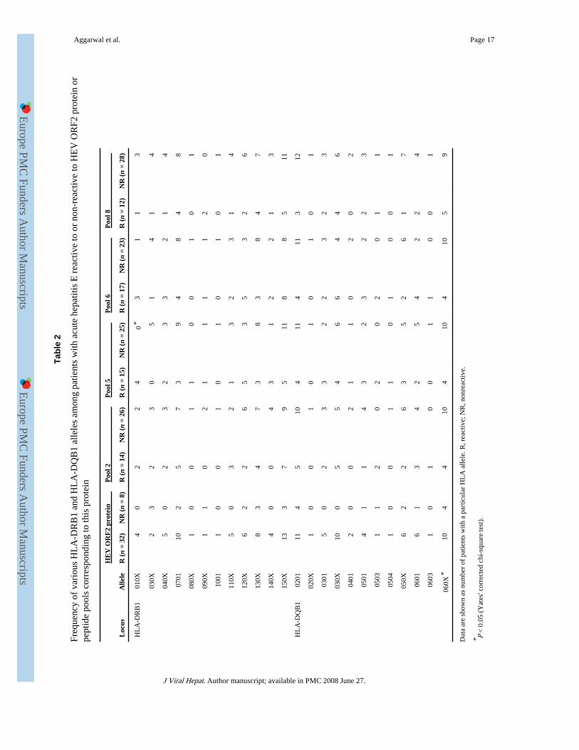
HLA-DR genotype determinationGenomic DNA was extracted using the salting-out method [26]. HLA-DRB1 and DQB1low-resolution typing was carried out using sequence-specific primer kits (Deutsche Dynal,Hamburg, Germany), as per the manufacturer’s protocol. The frequencies of various HLAalleles among patients showing reactivity to HEV ORF2 protein or to specific peptide poolswere compared with those not showing such reactivity.
Statistical methodsFor intergroup comparisons of categorical data, chi-square test (with Yates’ correction,where applicable) was used. For continuous data, Wilcoxon’s rank sum test or Student’s t-test was used, depending on whether the data were normally distributed or not. P-valuesbelow 0.05 were taken as significant.
RESULTSStudy groups
Forty patients with acute hepatitis E, aged 18-76 (median 23) were studied. The timeduration from the onset of acute hepatitis symptoms to the time of specimen collection was6–46 days (median 18). The serum bilirubin and alanine aminotransferase levels rangedfrom 3.0 to 30.0 (median 10.6) mg/dL and from 234 to 2321 (579) IU/L, respectively. Of the40 patients, two had fulminant hepatic failure and 38 had acute uncomplicated illness. Thecontrol subjects (n = 21) were aged 19–54 (median 28) years.
Lymphocyte proliferation assays using recombinant full-length hepatitis E virus openreading frame 2 protein
On stimulation with recombinant HEV ORF2 protein (10 μg/mL), 32 of the 40 patients(80%) showed significant stimulation. In contrast, only 7 (33%) of the 21 controls showedsimilar stimulation (P < 0.001). The SI values observed in patients (range 0.9–15.2; median2.6) were higher than those observed in healthy controls (range 0.6–12.9; median 1.3; P =0.002; Wilcoxon’s rank sum test) (Fig. 1). However, only 5 of 40 (13%) patients and 1 of 21(5%; P = ns) controls had SI values exceeding 3.8 (95th percentile values of amongcontrols).
All the patients with acute hepatitis E showed good stimulation with PHA, with median SIof 163 (range 15–599); the corresponding median SI in healthy controls was 161 (26–462)(P = ns; Wilcoxon’s rank sum test).
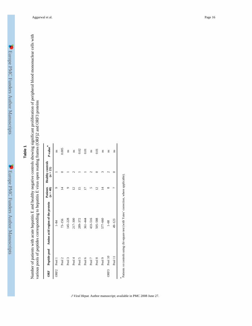
Lymphocyte proliferation using peptide poolsTable 1 shows the number of patients and controls in whom significant stimulation wasobserved with each of the peptide pools studied. For four of the nine peptide poolscorresponding to HEV ORF2 protein (pools 2, 5, 6 and 8; corresponding to amino acids 73–156, 289–372, 361–444 and 505–588, respectively, of the ORF2 protein), reactivity wasobserved more often among patients with hepatitis E than among controls. Figure 2 showsthe SI values observed among patients and controls with these peptide pools. The median(range) SI values among patients and controls were as follows: pool 2–1.7 (0.3–4.4) vs 0.8(0.4–1.8); pool 5–1.5 (0.3–4.9) vs 0.9 (0.4–2.0); pool 6–1.8 (0.5–6.3) vs 1.0 (0.3–3.6) andpool 8–1.3 (0.9–11.5) vs 0.9 (0.3–1.8). Reactivity to pools 4 and 9 was also common among
Aggarwal et al. Page 4
J Viral Hepat. Author manuscript; available in PMC 2008 June 27.
Europe PM
C Funders A
uthor Manuscripts
Europe PM
C Funders A
uthor Manuscripts
patients; however, the difference between the proportions of patients and controls showingreactivity to these pools was not statistically significant (P = ns; chi-square test with Yates’correction).
There was no difference in the proportion of patients and healthy controls who showedlymphoproliferative response to the peptide pools corresponding to HEV ORF3 protein(pools 10 and 11; Fig. 3).
Lymphocyte proliferation using individual peptidesLymphocyte proliferation assays were carried out with individual peptides contained inpeptide pools 2, 5 and 8 in four, six and two patients, respectively (Fig. 4). In theseexperiments, no consistent pattern of reactivity to individual peptides was observed.
HLA allelesTable 2 shows the frequency of various HLA-DR and -DQ alleles among patients whoshowed proliferation in response to ORF2 protein and to various peptide pools and thosewho did not show such proliferation. The only significant association observed was betweenthe reactivity to peptide pool 5 and the presence of HLA-DRB1 allele 010X.
DISCUSSIONOur data show that patients with acute hepatitis E mount a proliferative T-cell immuneresponse to the ORF2 protein of HEV. The immunodominant T-cell epitopes of HEV ORF2protein were located in the regions encompassed by amino acids 73–156, 289–444 and 505–588. Except for association of reactivity with HLA-DRB1 allele 010X with pool 5 (aminoacids 289–372), immune response to various regions of ORF2 protein did not show anyrelationship with the presence of a specific HLA allele. In comparison to ORF2, there wasno significant lymphoproliferative response to peptides corresponding to the HEV ORF3protein.
HEV infection is common in several parts of the world [1,2]. Antibody responses to thisinfection have been well characterized [10-16]. These responses develop early during thecourse of the infection and are directed against several epitopes distributed over all the threeviral proteins [10-16]. Based on this information, diagnostic tests have been developed usingrecombinant HEV proteins and peptides [10,14]. In contrast, little data are availableregarding the presence and nature of T-cell responses during HEV infection.
We have previously reported the presence of lymphoproliferative responses in nearly half ofthe adult patients with acute hepatitis E, including pregnant women with this disease[21,22]. However, these studies used only a limited panel of seven synthetic peptidescorresponding to HEV proteins that had originally been chosen to study humoral immuneresponses [15,27]. Our current data are based on the use of peptide libraries that covered theentire lengths of HEV ORF2 and ORF3 proteins and a recombinant ORF2 protein. In thecurrent study, we found lymphocyte proliferation in nearly 80% of patients with acutehepatitis E using the recombinant ORF2 protein. This higher frequency may represent thepresence of additional T-cell stimulatory regions in the ORF2 protein beyond the peptidesused in our previous study. In addition, the doses of peptides used in the current study (2 μg/well) were much higher than those used previously (1–100 ng/well). The current dataindicate that activation of T-cell responses is common during acute hepatitis E. However, itmay be worthwhile to point out that the SI values exceeded 4.0 in only five of the 32patients who showed a proliferative response. This is partly because of the presence ofproliferative responses in our control population, an issue that is discussed later. It may also
Aggarwal et al. Page 5
J Viral Hepat. Author manuscript; available in PMC 2008 June 27.
Europe PM
C Funders A
uthor Manuscripts
Europe PM
C Funders A
uthor Manuscripts
reflect sequestration of antigen-specific cells into liver, the major site of inflammation,leading to a relatively lower level of detectable immune reactivity in the peripheral blood.
In our previous study [21], proliferation responses were more frequent among patients withhepatitis E than among controls against a peptide corresponding to amino acids 613–627 ofHEV ORF2. In the current study, 14 of the 40 patients with acute hepatitis E hadproliferative responses to peptide pool 9 that included this region; however, this frequencywas not different from that among healthy subjects, as some (3/21) of the latter group alsoshowed proliferative responses to this pool.
We observed lymphoproliferative responses to three widely scattered regions in the HEVORF2 protein in patients with acute hepatitis E. The ongoing attempts to develop a vaccineagainst HEV have mostly included recombinant proteins corresponding to parts of the HEVORF2 protein [18-20]. A candidate vaccine, which includes amino acids 112–607 of thisprotein, has been shown to prevent liver injury but not viraemia and faecal excretion of thevirus in animal model [28,29]; however, the protection appears to be short lasting [20]. Thisvaccine includes two of the three immunostimulatory regions in entirety but does not includea part (amino acids 73–111) of the third T-cell stimulatory region identified in the currentstudy. It is possible that inclusion of additional T-cell epitopes corresponding to this thirdimmunostimulatory region may improve the efficacy of this vaccine and increase theduration of protection provided by it. As truncation of amino acids 1–111 appears to beimportant for the assembly of ORF2 protein as virus-like particles [30], this may implyinclusion of peptides or proteins corresponding to this region of ORF2 either as anadditional constituent of the vaccine or as a booster, or use of DNA-based vaccines, attemptsfor which have already been made [31,32].
Recently, a neutralization epitope that spans from amino acids 458–607 of the HEV ORF2has been identified [33]; it includes one of the three regions (505–588) of the HEV ORF2protein to which proliferative responses were found in our study. This suggests that thisregion of ORF2 may be particularly important in protection against HEV, as it may provideT-cell help for the development of protective antibodies.
In hepatitis B and hepatitis C virus infections, cellular immune responses, althoughimportant for elimination of the virus from the host, have also been shown to mediate hostcell injury [23,34,35]. It is thus important to determine whether cytotoxic T-cell responsesdirected against HEV proteins could play a role in the induction of host liver cell injury.This would be particularly important for those epitopes that induce proliferative responsesand are therefore likely candidates for inclusion in future vaccines. Further work is alsoneeded to identify the CD8+ cytotoxic T-cell responses to HEV.
We found association between only one HLA-DR allele and reactivity to a particular peptidepool. This association will need confirmation in future studies. Our failure to find morewidespread associations between HLA alleles and proliferative responses may be relatedeither to small sample size or to the use of peptide pools instead of specific peptides.
An important observation in our study was that while lymphoproliferative responses toORF2 peptide pools were marked, those to ORF3 peptide pools were virtually nonexistent.Although we did not use the recombinant ORF3 protein, it appears that T-cell responses aremore often directed against the viral capsid protein than against the ORF3 protein. This isparticularly interesting, as humoral immune responses are directed against both ORF2 andORF3, especially the immunodominant C-terminal end of the ORF3 protein.
Our study is limited by the fact that our control subjects were residents of an HEV-endemicregion and may have had prior subclinical exposure to HEV. Despite being negative for anti-
Aggarwal et al. Page 6
J Viral Hepat. Author manuscript; available in PMC 2008 June 27.
Europe PM
C Funders A
uthor Manuscripts
Europe PM
C Funders A
uthor Manuscripts
HEV antibodies, 7 of our 21 healthy control subjects showed proliferative responses toORF2 protein, suggesting that anti-HEV screening may not be adequate to exclude priorHEV exposure. In our previous studies too, we found reactivity to HEV peptides in aproportion of healthy subjects [21]. This is supported by the previous observation ofdisappearance of anti-HEV with time in at least half of the subjects infected during anoutbreak [17].
To conclude, we have shown activation of T-cell responses in patients with acute hepatitis Eand mapped the regions of HEV ORF2 protein that might contain lymphoproliferative T-cellepitopes. Further work is needed to narrow down these T-cell epitopes to identify therelationship of specific HLA alleles with these epitopes and to determine the role of theseepitopes in the development of a vaccine and in better understanding the mechanism of livercell damage during HEV infection.
AcknowledgmentsThis work was supported by a research grant from the Indian Council of Medical Research, New Delhi to RA, andalso received partial support from the Wellcome Trust, UK through a Senior Research Fellowship to SJ. Theauthors thank Mr Srikant Srivastava for help in specimen collection.
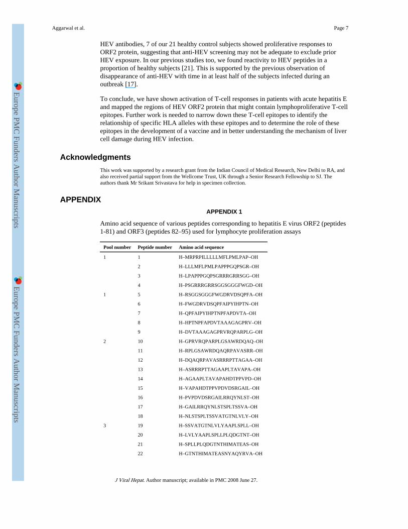
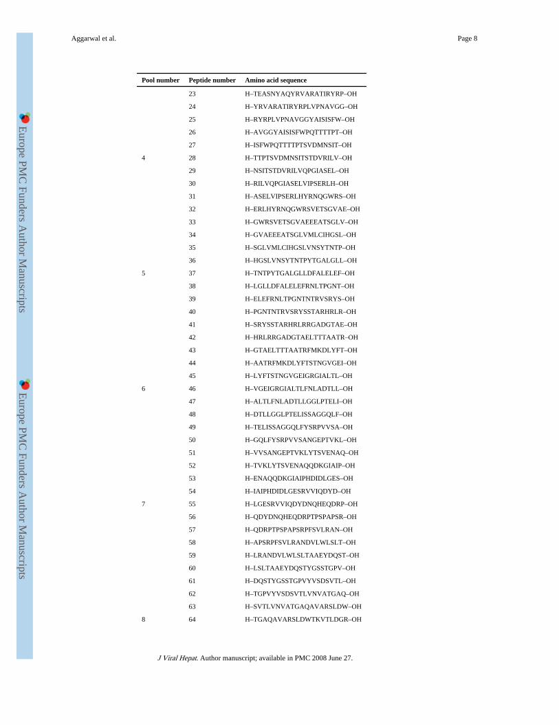
APPENDIXAPPENDIX 1
Amino acid sequence of various peptides corresponding to hepatitis E virus ORF2 (peptides1-81) and ORF3 (peptides 82–95) used for lymphocyte proliferation assays
Pool number Peptide number Amino acid sequence
1 1 H–MRPRPILLLLLMFLPMLPAP–OH
2 H–LLLMFLPMLPAPPPGQPSGR–OH
3 H–LPAPPPGQPSGRRRGRRSGG–OH
4 H–PSGRRRGRRSGGSGGGFWGD–OH
1 5 H–RSGGSGGGFWGDRVDSQPFA–OH
6 H–FWGDRVDSQPFAIPYIHPTN–OH
7 H–QPFAIPYIHPTNPFAPDVTA–OH
8 H–HPTNPFAPDVTAAAGAGPRV–OH
9 H–DVTAAAGAGPRVRQPARPLG–OH
2 10 H–GPRVRQPARPLGSAWRDQAQ–OH
11 H–RPLGSAWRDQAQRPAVASRR–OH
12 H–DQAQRPAVASRRRPTTAGAA–OH
13 H–ASRRRPTTAGAAPLTAVAPA–OH
14 H–AGAAPLTAVAPAHDTPPVPD–OH
15 H–VAPAHDTPPVPDVDSRGAIL–OH
16 H–PVPDVDSRGAILRRQYNLST–OH
17 H–GAILRRQYNLSTSPLTSSVA–OH
18 H–NLSTSPLTSSVATGTNLVLY–OH
3 19 H–SSVATGTNLVLYAAPLSPLL–OH
20 H–LVLYAAPLSPLLPLQDGTNT–OH
21 H–SPLLPLQDGTNTHIMATEAS–OH
22 H–GTNTHIMATEASNYAQYRVA–OH
Aggarwal et al. Page 7
J Viral Hepat. Author manuscript; available in PMC 2008 June 27.
Europe PM
C Funders A
uthor Manuscripts
Europe PM
C Funders A
uthor Manuscripts
Pool number Peptide number Amino acid sequence
23 H–TEASNYAQYRVARATIRYRP–OH
24 H–YRVARATIRYRPLVPNAVGG–OH
25 H–RYRPLVPNAVGGYAISISFW–OH
26 H–AVGGYAISISFWPQTTTTPT–OH
27 H–ISFWPQTTTTPTSVDMNSIT–OH
4 28 H–TTPTSVDMNSITSTDVRILV–OH
29 H–NSITSTDVRILVQPGIASEL–OH
30 H–RILVQPGIASELVIPSERLH–OH
31 H–ASELVIPSERLHYRNQGWRS–OH
32 H–ERLHYRNQGWRSVETSGVAE–OH
33 H–GWRSVETSGVAEEEATSGLV–OH
34 H–GVAEEEATSGLVMLCIHGSL–OH
35 H–SGLVMLCIHGSLVNSYTNTP–OH
36 H–HGSLVNSYTNTPYTGALGLL–OH
5 37 H–TNTPYTGALGLLDFALELEF–OH
38 H–LGLLDFALELEFRNLTPGNT–OH
39 H–ELEFRNLTPGNTNTRVSRYS–OH
40 H–PGNTNTRVSRYSSTARHRLR–OH
41 H–SRYSSTARHRLRRGADGTAE–OH
42 H–HRLRRGADGTAELTTTAATR–OH
43 H–GTAELTTTAATRFMKDLYFT–OH
44 H–AATRFMKDLYFTSTNGVGEI–OH
45 H–LYFTSTNGVGEIGRGIALTL–OH
6 46 H–VGEIGRGIALTLFNLADTLL–OH
47 H–ALTLFNLADTLLGGLPTELI–OH
48 H–DTLLGGLPTELISSAGGQLF–OH
49 H–TELISSAGGQLFYSRPVVSA–OH
50 H–GQLFYSRPVVSANGEPTVKL–OH
51 H–VVSANGEPTVKLYTSVENAQ–OH
52 H–TVKLYTSVENAQQDKGIAIP–OH
53 H–ENAQQDKGIAIPHDIDLGES–OH
54 H–IAIPHDIDLGESRVVIQDYD–OH
7 55 H–LGESRVVIQDYDNQHEQDRP–OH
56 H–QDYDNQHEQDRPTPSPAPSR–OH
57 H–QDRPTPSPAPSRPFSVLRAN–OH
58 H–APSRPFSVLRANDVLWLSLT–OH
59 H–LRANDVLWLSLTAAEYDQST–OH
60 H–LSLTAAEYDQSTYGSSTGPV–OH
61 H–DQSTYGSSTGPVYVSDSVTL–OH
62 H–TGPVYVSDSVTLVNVATGAQ–OH
63 H–SVTLVNVATGAQAVARSLDW–OH
8 64 H–TGAQAVARSLDWTKVTLDGR–OH
Aggarwal et al. Page 8
J Viral Hepat. Author manuscript; available in PMC 2008 June 27.
Europe PM
C Funders A
uthor Manuscripts
Europe PM
C Funders A
uthor Manuscripts

Pool number Peptide number Amino acid sequence
65 H–SLDWTKVTLDGRPLSTIQQY–OH
66 H–LDGRPLSTIQQYSKTFFVLP–OH
67 H–IQQYSKTFFVLPLRGKLSFW–OH
68 H–FVLPLRGKLSFWEAGTTKAG–OH
69 H–LSFWEAGTTKAGYPYNYNTT–OH
70 H–TKAGYPYNYNTTASDQLLVE–OH
71 H–YNTTASDQLLVENAAGHRVA–OH
72 H–LLVENAAGHRVAISTYTTSL–OH
9 73 H–HRVAISTYTTSLGAGPVSIS–OH
74 H–TTSLGAGPVSISAVAVLAPH–OH
75 H–VSISAVAVLAPHSALALLED–OH
76 H–LAPHSALALLEDTLDYPARA–OH
77 H–LLEDTLDYPARAHTFDDFCP–OH
78 H–PARAHTFDDFCPECRPLGLQ–OH
79 H–DFCPECRPLGLQGCAFQSTV–OH
80 H–LGLQGCAFQSTVAELQRLKM–OH
81 H–QSTVAELQRLKMKVGKTREL–OH
10 82 H–MNNMSFAAPMGSRPCALGLF–OH
83 H–PMGSRPCALGLFCCCSSCFC–OH
84 H–LGLFCCCSSCFCLCCPRHRP–OH
85 H–SCFCLCCPRHRPVSRLAAVV–OH
86 H–RHRPVSRLAAVVGGAAAVPA–OH
87 H–AAVVGGAAAVPAVVSGVTGL–OH
88 H–AVPAVVSGVTGLILSPSQSP–OH
11 89 H–VTGLILSPSQSPIFIQPTPS–OH
90 H–SQSPIFIQPTPSPPMSPLRP–OH
91 H–PTPSPPMSPLRPGLDLVFAN–OH
92 H–PLRPGLDLVFANPPDHSAPL–OH
93 H–VFANPPDHSAPLGVTRPSAP–OH
94 H–SAPLGVTRPSAPPLPHVVDL–OH
95 H–RPSAPPLPHVVDLPQLGPRR–OH
Abbreviations
HEV hepatitis E virus
PBMC peripheral blood mononuclear cells
ORF open reading frame
PHA phytohaemagglutinin
SI stimulation indices
Aggarwal et al. Page 9
J Viral Hepat. Author manuscript; available in PMC 2008 June 27.
Europe PM
C Funders A
uthor Manuscripts
Europe PM
C Funders A
uthor Manuscripts
REFERENCES1. Aggarwal R, Krawczynski K. Hepatitis E: an overview and recent advances in clinical and
laboratory research. J Gastroenterol Hepatol. 2000; 15:9–20. [PubMed: 10719741]
2. Krawczynski, K.; Aggarwal, R. Hepatitis E. In: Schiff, ER.; Sorrell, MF.; Maddrey, WC., editors.Schiff’s Diseases of the Liver. 9th edn.. Philadelphia, PA: Lippincott-Raven; 2002. p. 877-889.
3. Naik SR, Aggarwal R, Salunke PN, Mehrotra NN. A large waterborne viral hepatitis E epidemic inKanpur, India. Bull World Health Organ. 1992; 70:597–604. [PubMed: 1464145]
4. Aggarwal R, Naik SR. Hepatitis E: intrafamilial transmission versus waterborne spread. J Hepatol.1994; 22:718–723. [PubMed: 7890884]
5. Somani SK, Aggarwal R, Naik SR, Srivastava S, Naik S. A serological study of intra-familial spreadfrom patients with sporadic hepatitis E virus infection. J Viral Hepat. 2003; 10:446–449. [PubMed:14633178]
6. Tam AW, Smith MM, Guerra ME, et al. Hepatitis E virus (HEV): molecular cloning and sequencingof the full-length viral genome. Virology. 1991; 185:120–131. [PubMed: 1926770]
7. Zafrullah M, Ozdener MH, Panda SK, Jameel S. The ORF3 protein of hepatitis E virus is aphosphoprotein that associates with the cytoskeleton. J Virol. 1997; 71:9045–9053. [PubMed:9371561]
8. Tyagi S, Korkaya H, Zafrullah M, Jameel S, Lal SK. The phosphorylated form of the ORF3 proteinof hepatitis E virus interacts with its non-glycosylated form of the major capsid protein, ORF2. JBiol Chem. 2002; 277:22759–22767. [PubMed: 11934888]
9. Korkaya H, Jameel S, Gupta D, et al. The ORF3 protein of hepatitis E virus binds to Src homology3 domains and activates MAPK. J Biol Chem. 2001; 276:42389–42400. [PubMed: 11518702]
10. Favorov MO, Fields HA, Purdy MA, et al. Serologic identification of hepatitis E virus infections inepidemic and endemic settings. J Med Virol. 1992; 36:246–250. [PubMed: 1578218]
11. Khudyakov YE, Khudyakova NS, Fields HA, et al. Epitope mapping in proteins of hepatitis Evirus. Virology. 1993; 194:89–96. [PubMed: 7683162]
12. Khudyakov YE, Lopareva EN, Jue DL, Crews TK, Thyagarajan SP, Fields HA. Antigenic domainsof the open reading frame 2-encoded protein of hepatitis E virus. J Clin Microbiol. 1999; 37:2863–2871. [PubMed: 10449466]
13. Riddell MA, Li F, Anderson DA. Identification of immunodominant and conformational epitopesin the capsid protein of hepatitis E virus by using monoclonal antibodies. J Virol. 2000; 74:8011–8017. [PubMed: 10933710]
14. Dawson GJ, Chau KH, Cabal CM, Yarbough PO, Reyes GR, Mushahwar IK. Solid-phase enzyme-linked immunosorbent assay for hepatitis E virus IgG and IgM antibodies utilizing recombinantantigens and synthetic peptides. J Virol Methods. 1992; 38:175–186. [PubMed: 1644893]
15. Coursaget P, Buisson Y, Depril N, et al. Mapping of linear B cell epitopes on open reading frame2- and 3-encoded proteins of hepatitis E virus using synthetic peptides. FEMS Microbiol Lett.1993; 109:251–255. [PubMed: 7687968]
16. Kaur M, Hyams KC, Purdy MA, et al. Human linear B-cell epitopes encoded by the hepatitis Evirus include determinants in the RNA-dependent RNA polymerase. Proc Natl Acad Sci USA.1992; 89:3855–3858. [PubMed: 1373890]
17. Khuroo MS, Kamili S, Dar MY, Moecklii R, Jameel S. Hepatitis E and long-term antibody status.Lancet. 1993; 341:1355–1355. [PubMed: 8098491]
18. Tsarev SA, Tsareva TS, Emerson SU, et al. Recombinant vaccine against hepatitis E: doseresponse and protection against heterologous challenge. Vaccine. 1997; 15:1834–1838. [PubMed:9413090]
19. Purdy MA, McCaustland KA, Krawczynski K, Spelbring J, Reyes GR, Bradley DW. Preliminaryevidence that a trp-EHEV fusion protein protects cynomolgus macaques against challenge withwild-type hepatitis E virus (HEV). J Med Virol. 1993; 41:90–94. [PubMed: 8228944]
20. Zhang M, Emerson SU, Nguyen H, et al. Recombinant vaccine against hepatitis E: duration ofprotective immunity in rhesus macaques. Vaccine. 2002; 20:3285–3291. [PubMed: 12213398]
21. Naik S, Aggarwal R, Naik SR, et al. Evidence for activation of cellular immune responses inpatients with acute hepatitis E. Indian J Gastroenterol. 2002; 21:149–152. [PubMed: 12385543]
Aggarwal et al. Page 10
J Viral Hepat. Author manuscript; available in PMC 2008 June 27.
Europe PM
C Funders A
uthor Manuscripts
Europe PM
C Funders A
uthor Manuscripts
22. pal R, Aggarwal R, Naik SR, Das V, Das S, Naik SR. Immunological alterations in pregnantwomen with acute hepatitis E. J Gastroenterol Hepatol. 2005; 20:1094–1101. [PubMed:15955220]
23. Ferrari C, Missale G, Boni C, Urbani S. Immunopathogenesis of hepatitis B. J Hepatol. 2003;39(Suppl. 1):S36–S42. [PubMed: 14708676]
24. Zafrullah M, Khursheed Z, Yadav S, Sehgal D, Jameel S, Ahmad F. Acid pH enhances structureand structural stability of the capsid protein of hepatitis E virus. Biochem Biophys Res Commun.2003; 313:67–73. [PubMed: 14672699]
25. Fernandez-Botran, R.; Vetvicka, V. Methods in Cellular Immunology. 2nd edn.. Boca Raton, FL:CRC Press; 2002. p. 53-57.
26. Miller SA, Dykes DD, Polesky HF. A simple salting out procedure for extracting DNA fromhuman nucleated cells. Nucleic Acids Res. 1988; 16:1215–1218. [PubMed: 3344216]
27. Coursaget P, Depril N, Buisson Y, Molinie C, Roue R. Hepatitis type E in a French population:detection of anti-HEV by a synthetic peptide-based enzyme-linked immunosorbent assay. ResVirol. 1994; 145:51–57. [PubMed: 8023016]
28. Zhang M, Emerson SU, Nguyen H, et al. Immunogenicity and protective efficacy of a vaccineprepared from 53 kDa truncated hepatitis E virus capsid protein expressed in insect cells. Vaccine.2001; 20:853–857. [PubMed: 11738749]
29. Purcell RH, Nguyen H, Shapiro M, et al. Pre-clinical immunogenicity and efficacy trial of arecombinant hepatitis E vaccine. Vaccine. 2003; 21:2607–2615. [PubMed: 12744897]
30. Li TC, Yamakawa Y, Suzuki K, et al. Expression and self-assembly of empty virus-like particlesof hepatitis E virus. J Virol. 1997; 71:7207–7213. [PubMed: 9311793]
31. Kamili S, Spelbring J, Carson D, Krawczynski K. Protective efficacy of hepatitis E virus DNAvaccine administered by gene gun in the cynomolgus macaque model of infection. J Infect Dis.2004; 189:258–264. [PubMed: 14722891]
32. He J, Hayes CG, Binn LN, et al. Hepatitis E virus DNA vaccine elicits immunologic memory inmice. J Biomed Sci. 2001; 8:223–226. [PubMed: 11287754]
33. Zhou Y-H, Purcell RH, Emerson SU. An ELISA for putative neutralizing antibodies to hepatitis Evirus detects antibodies to genotypes 1, 2, 3 and 4. Vaccine. 2004; 22:2578–2585. [PubMed:15193383]
34. Jung MC, Pape GR. Immunology of hepatitis B infection. Lancet Infect Dis. 2002; 2:43–50.[PubMed: 11892495]
35. Rosen HR. Hepatitis C pathogenesis: mechanisms of viral clearance and liver injury. LiverTranspl. 2003; 9:S35–S43. [PubMed: 14586893]
Aggarwal et al. Page 11
J Viral Hepat. Author manuscript; available in PMC 2008 June 27.
Europe PM
C Funders A
uthor Manuscripts
Europe PM
C Funders A
uthor Manuscripts
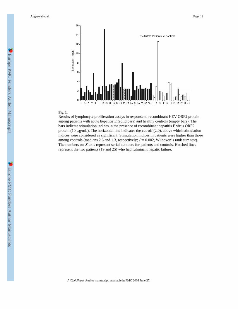
Fig. 1.Results of lymphocyte proliferation assays in response to recombinant HEV ORF2 proteinamong patients with acute hepatitis E (solid bars) and healthy controls (empty bars). Thebars indicate stimulation indices in the presence of recombinant hepatitis E virus ORF2protein (10 μg/mL). The horizontal line indicates the cut-off (2.0), above which stimulationindices were considered as significant. Stimulation indices in patients were higher than thoseamong controls (medians 2.6 and 1.3, respectively; P = 0.002, Wilcoxon’s rank sum test).The numbers on X-axis represent serial numbers for patients and controls. Hatched linesrepresent the two patients (19 and 25) who had fulminant hepatic failure.
Aggarwal et al. Page 12
J Viral Hepat. Author manuscript; available in PMC 2008 June 27.
Europe PM
C Funders A
uthor Manuscripts
Europe PM
C Funders A
uthor Manuscripts
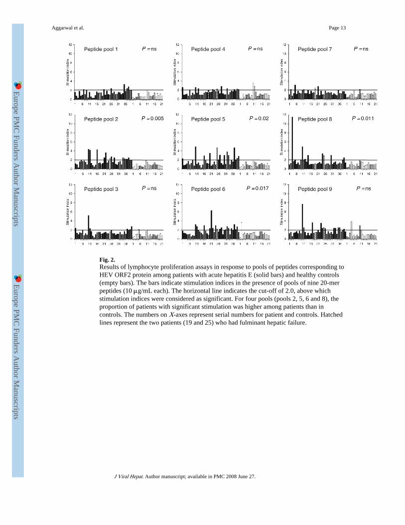
Fig. 2.Results of lymphocyte proliferation assays in response to pools of peptides corresponding toHEV ORF2 protein among patients with acute hepatitis E (solid bars) and healthy controls(empty bars). The bars indicate stimulation indices in the presence of pools of nine 20-merpeptides (10 μg/mL each). The horizontal line indicates the cut-off of 2.0, above whichstimulation indices were considered as significant. For four pools (pools 2, 5, 6 and 8), theproportion of patients with significant stimulation was higher among patients than incontrols. The numbers on X-axes represent serial numbers for patient and controls. Hatchedlines represent the two patients (19 and 25) who had fulminant hepatic failure.
Aggarwal et al. Page 13
J Viral Hepat. Author manuscript; available in PMC 2008 June 27.
Europe PM
C Funders A
uthor Manuscripts
Europe PM
C Funders A
uthor Manuscripts
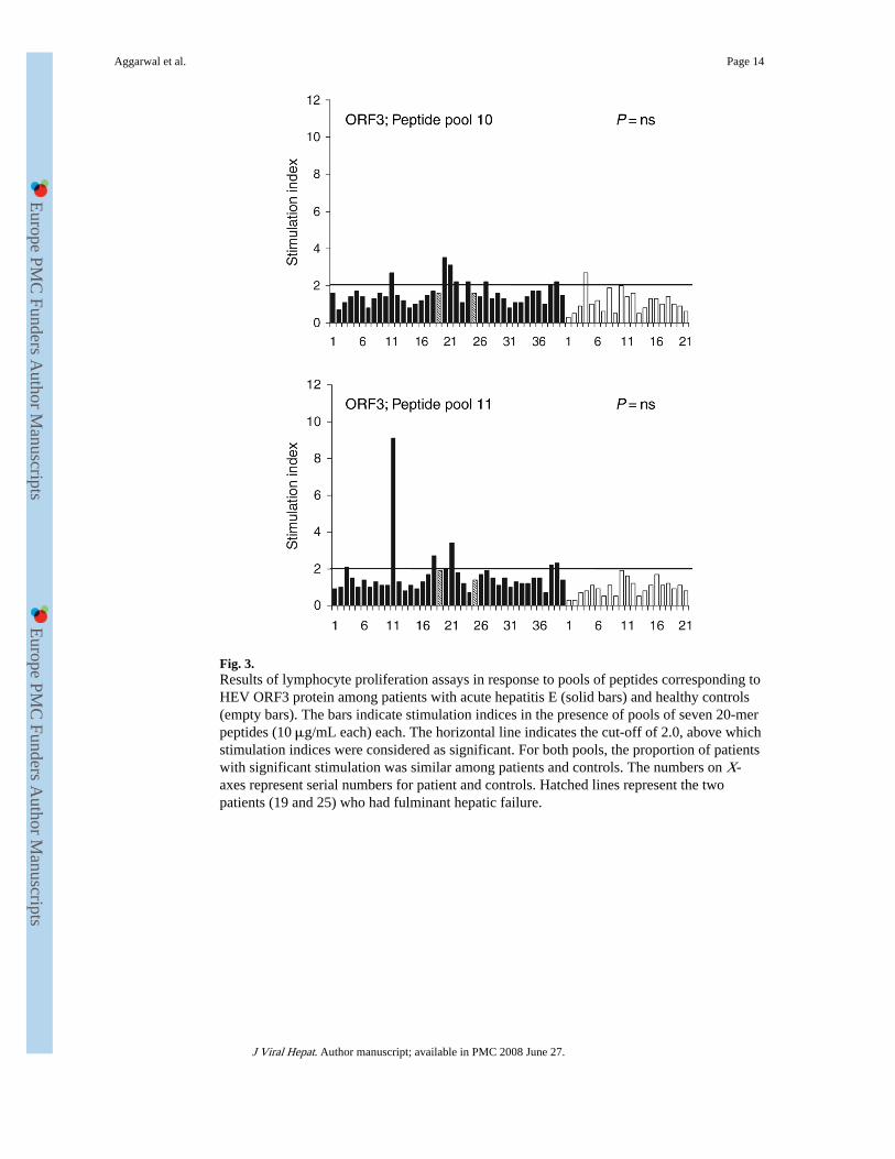
Fig. 3.Results of lymphocyte proliferation assays in response to pools of peptides corresponding toHEV ORF3 protein among patients with acute hepatitis E (solid bars) and healthy controls(empty bars). The bars indicate stimulation indices in the presence of pools of seven 20-merpeptides (10 μg/mL each) each. The horizontal line indicates the cut-off of 2.0, above whichstimulation indices were considered as significant. For both pools, the proportion of patientswith significant stimulation was similar among patients and controls. The numbers on X-axes represent serial numbers for patient and controls. Hatched lines represent the twopatients (19 and 25) who had fulminant hepatic failure.
Aggarwal et al. Page 14
J Viral Hepat. Author manuscript; available in PMC 2008 June 27.
Europe PM
C Funders A
uthor Manuscripts
Europe PM
C Funders A
uthor Manuscripts
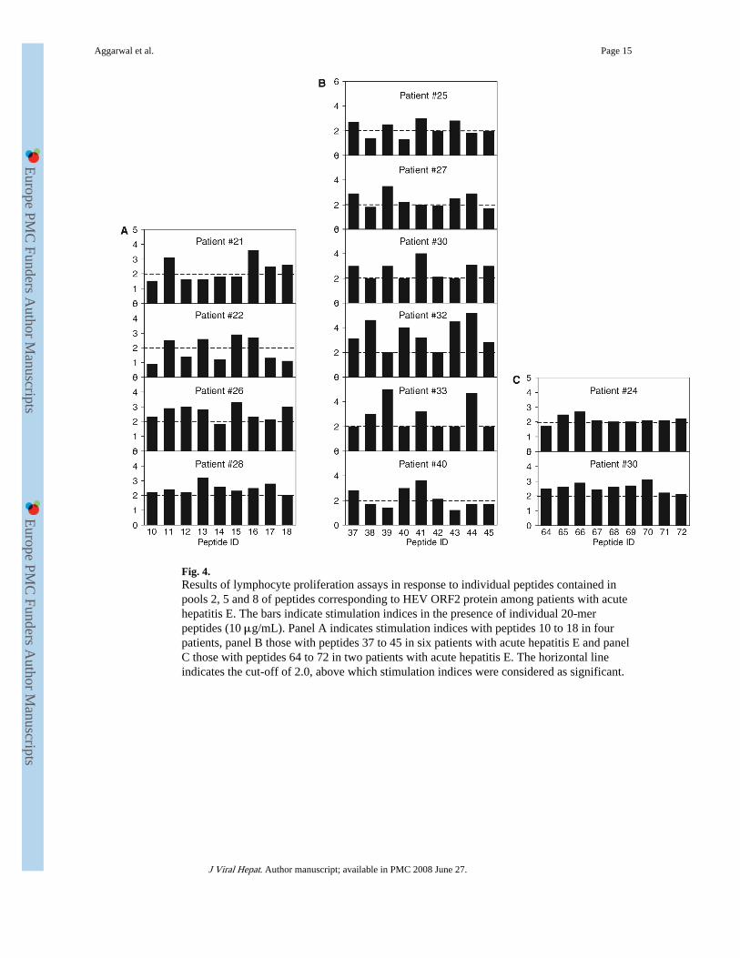
Fig. 4.Results of lymphocyte proliferation assays in response to individual peptides contained inpools 2, 5 and 8 of peptides corresponding to HEV ORF2 protein among patients with acutehepatitis E. The bars indicate stimulation indices in the presence of individual 20-merpeptides (10 μg/mL). Panel A indicates stimulation indices with peptides 10 to 18 in fourpatients, panel B those with peptides 37 to 45 in six patients with acute hepatitis E and panelC those with peptides 64 to 72 in two patients with acute hepatitis E. The horizontal lineindicates the cut-off of 2.0, above which stimulation indices were considered as significant.
Aggarwal et al. Page 15
J Viral Hepat. Author manuscript; available in PMC 2008 June 27.
Europe PM
C Funders A
uthor Manuscripts
Europe PM
C Funders A
uthor Manuscripts
Europe PM
C Funders A
uthor Manuscripts
Europe PM
C Funders A
uthor Manuscripts
Aggarwal et al. Page 16
Tabl
e 1
Num
ber
of p
atie
nts
with
acu
te h
epat
itis
E a
nd h
ealth
y ne
gativ
e co
ntro
ls s
how
ing
sign
ific
ant p
rolif
erat
ion
of p
erip
hera
l blo
od m
onon
ucle
ar c
ells
with
vari
ous
pool
s of
pep
tides
cor
resp
ondi
ng to
hep
atiti
s E
vir
us o
pen
read
ing
fram
es (
OR
F)2
and
OR
F3 p
rote
ins
OR
FP
epti
de p
ool
Am
ino
acid
reg
ion
of t
he p
rote
inP
atie
nts
(n =
40)
Hea
lthy
con
trol
s(n
= 2
1)P
-val
ue*
OR
F2Po
ol 1
1–84
91
ns
Pool
273
–156
140
0.00
5
Pool
314
5–22
89
1ns
Pool
421
7–30
012
2ns
Pool
528
9–37
215
10.
02
Pool
636
1–44
417
10.
01
Pool
743
3–51
65
2ns
Pool
850
5–58
812
00.
01
Pool
957
7–66
014
3ns
OR
F3Po
ol 1
01–
688
2ns
Pool
11
49–1
237
0ns
* Patie
nts
vs c
ontr
ols
usin
g ch
i-sq
uare
test
(w
ith Y
ates
’ co
rrec
tion,
whe
re a
pplic
able
).
J Viral Hepat. Author manuscript; available in PMC 2008 June 27.
Europe PM
C Funders A
uthor Manuscripts
Europe PM
C Funders A
uthor Manuscripts
Aggarwal et al. Page 17
Tabl
e 2
Freq
uenc
y of
var
ious
HL
A-D
RB
1 an
d H
LA
-DQ
B1
alle
les
amon
g pa
tient
s w
ith a
cute
hep
atiti
s E
rea
ctiv
e to
or
non-
reac
tive
to H
EV
OR
F2 p
rote
in o
rpe
ptid
e po
ols
corr
espo
ndin
g to
this
pro
tein
HE
V O
RF
2 pr
otei
nP
ool 2
Poo
l 5P
ool 6
Poo
l 8
Loc
usA
llele
R (
n =
32)
NR
(n
= 8)
R (
n =
14)
NR
(n
= 26
)R
(n
= 15
)N
R (
n =
25)
R (
n =
17)
NR
(n
= 23
)R
(n
= 12
)N
R (
n =
28)
HL
A-D
RB
101
0X4
02
24
0*3
11
3
030X
23
23
05
14
14
040X
50
23
23
32
14
0701
102
57
39
48
48
080X
10
01
10
01
01
090X
11
02
11
11
20
1001
10
01
01
01
01
110X
50
32
13
23
14
120X
62
26
53
53
26
130X
83
47
38
38
47
140X
40
04
31
22
13
150X
133
79
511
88
511
HL
A-D
QB
102
0111
45
104
114
113
12
020X
10
01
01
01
01
0301
50
23
32
23
23
030X
100
55
46
64
46
0401
20
02
11
02
02
0501
41
14
32
32
23
0503
11
20
20
20
11
0504
10
01
10
10
01
050X
62
26
35
26
17
0601
61
34
25
42
24
0603
10
10
01
10
01
060X
*10
44
104
104
105
9
Dat
a ar
e sh
own
as n
umbe
r of
pat
ient
s w
ith a
par
ticul
ar H
LA
alle
le. R
, rea
ctiv
e; N
R, n
onre
activ
e.
* P <
0.0
5 (Y
ates
' cor
rect
ed c
hi-s
quar
e te
st).
J Viral Hepat. Author manuscript; available in PMC 2008 June 27.