PEGylated galactosylated cationic liposomes for hepatocytic gene delivery
Upload
independentCategory
view
0download
0
e u r o p e a n j o u r n a l o f p h a r m a c e u t i c a l s c i e n c e s 3 3 ( 2 0 0 8 ) 424–433
avai lab le at www.sc iencedi rec t .com
journa l homepage: www.e lsev ier .com/ locate /e jps
Systemic and mucosal immune response induced bytranscutaneous immunization using Hepatitis B surfaceantigen-loaded modified liposomes
Dinesh Mishraa, Pradyumna Kumar Mishrab, Vaibhav Dubeya, Manoj Nahara,Sunil Dabadghaob, N.K. Jaina,∗
a Pharmaceutics Research Laboratory, Department of Pharmaceutical Sciences, Dr. Hari Singh Gour University, Sagar 470003, Indiab Bhopal Memorial Hospital and Research Center, Bhopal 462038, India
a r t i c l e i n f o
Article history:
Received 17 October 2007
Received in revised form
14 December 2007
Accepted 27 January 2008
Published on line 12 February 2008
Keywords:
Hepatitis B vaccine
Ethosomes
Transcutaneous immunization
Dendritic cells
a b s t r a c t
We have evaluated the efficiency of novel modified liposomes (ethosomes) for transcuta-
neous immunization (TCI) against Hepatitis B. Antigen-loaded ethosomes were prepared
and characterized for shape, lamellarity, fluidity, size distribution, and entrapment effi-
ciency. Spectral bio-imaging and flow cytometric studies showed efficient uptake of Hepatitis
B surface antigen (HBsAg)-loaded ethosomes by murine dendritic cells (DCs) in vitro, reach-
ing a peak by 180 min. Transcutaneous delivery potential of the antigen-loaded system
using human cadaver skin demonstrated a much higher skin permeation of the anti-
gen in comparison to conventional liposomes and soluble antigen preparation. Topically
applied HBsAg-loaded ethosomes in experimental mice showed a robust systemic and
mucosal humoral immune response compared to intramuscularly administered alum-
adsorbed HBsAg suspension, topically applied plain HBsAg solution and hydroethanolic
(25%) HBsAg solution. The ability of the antigen-pulsed DCs to stimulate autologous periph-
eral blood lymphocytes was demonstrated by BrdU assay and a predominantly TH1 type of
immune response was observed by multiplex cytometric bead array analysis. HBsAg-loaded
ethosomes are able to generate a protective immune response and their ability to traverse
and target the immunological milieu of the skin may find a potential application in the
development of a transcutaneous vaccine against Hepatitis B virus (HBV).
and perhaps the need for a stringent cold chain for vaccine
1. Introduction
Vaccination is one of the most powerful tools available in theongoing battle against infectious agents. Virtually all recom-mended immunizations require parenteral administration,and many require a series of injections, therefore, new vac-
cine delivery methods, specifically alternatives to injections,are being sought. Topically applied (transcutaneous) vaccines,transgenic edible plants that contain genes for human vaccine∗ Corresponding author. Tel.: +91 7582264712; fax: +91 7582264712.E-mail addresses: [email protected] (D. Mishra), jnarendr@ya
0928-0987/$ – see front matter © 2008 Elsevier B.V. All rights reserved.doi:10.1016/j.ejps.2008.01.015
© 2008 Elsevier B.V. All rights reserved.
antigens, and controlled delivery depot systems with vaccineantigens encapsulated in biodegradable polymers are possi-bilities currently under study (Tacket et al., 1998; Gupta et al.,1998) Such new delivery methods could decrease reliance onrepeated injections, the need for trained healthcare workers,
hoo.co.in (N.K. Jain).
storage (Poland et al., 2002).Transcutaneous immunization (TCI) offers a new method
for the delivery of vaccines, that relies on the application

t i c a
oasmTtrvsow(
taetscl(tsib2
apfwmHreu(rm(
stEsIspi
2
2
PoIVr
e u r o p e a n j o u r n a l o f p h a r m a c e u
f antigen with adjuvant onto the outer layer of the skinnd subsequent delivery to underlying Langerhans cells thaterve as antigen-presenting cells (Glenn et al., 1999). It hasany merits compared to injectable routes of administration.
his needle-free method of vaccine delivery could decreasehe risk of needle-borne diseases, reduce the complicationselated to physical skin penetration, and improve access toaccination by eliminating the need for trained personnel andterile equipment. As an initial step towards the developmentf a carrier system for TCI against Hepatitis B virus (HBV),e recently reported elastic liposome-based HBsAg vaccine
Mishra et al., 2006, 2007).Ethosomes are interesting and innovative vesicular carriers
hat have appeared in the fields of pharmaceutical technologynd drug delivery in recent years (Touitou et al., 2000). Thesethanol-containing liposomes present an ample opportunityo transport active substances more efficaciously through thetratum corneum into the deeper layers of the skin thanonventional liposomes. In fact, ethosomes are soft, mal-eable vesicles tailored for enhanced delivery of active agentsDubey et al., 2007a). This aspect is of great importance forhe design of carriers to be applied topically both for local andystemic administration. Furthermore, the ethosomal carriers also able to provide an effective intracellular delivery ofoth hydrophilic and lipophilic molecules (Dayan and Touitou,000).
Hepatitis B is a serious global public health problem withbout 2 billion people infected with the virus. 360 million peo-le have chronic infection and 600,000 deaths occur each yearrom HBV related disease or hepatocellular carcinoma world-ide (Shepard et al., 2006). Vaccination is the most effectiveeasure to prevent HBV infection. Although available anti-epatitis B vaccines produce good and sustained immune
esponse, the effectiveness falls with age and they are lessffective in special groups like patients with chronic renal fail-re, liver transplant recipients and HIV-affected individuals
Akbar et al., 2006). Also, the elimination of the disease wouldequire different vaccination efforts with improved delivery
odules in the population living with chronic HBV infectionInchauspe and Michel, 2007).
The aim of present work was to incorporate HBsAg in etho-omes further exploring its immunoadjuvant property for TCI,o induce both systemic and mucosal immune responses.thosomes were prepared and characterized for vesicularize, shape, lamellarity, entrapment efficiency and fluidity.n vitro cellular uptake and internalization studies of etho-omal carriers by dendritic cells (DCs) and fibroblasts wereerformed using flow cytometric analysis and spectral bio-
maging, respectively.
. Materials and methods
.1. Materials
urified HBsAg (source-genetically modified yeast cells) was
btained as gift sample from the National Institute ofmmunology, New Delhi, India. Bovine albumin (fraction), soya phosphatidylcholine (SPC), sephadex G-150, fluo-escein isothiocyanate (FITC) and Rhodamine were obtained
l s c i e n c e s 3 3 ( 2 0 0 8 ) 424–433 425
from Sigma–Aldrich, St. Louis, MO, USA. All reagents usedin ELISA were purchased from Genei, Bangalore, India andImx HBsAg (V2) kit was obtained from Abbott, Wiesbaden,Germany. RPMI-1640, alamar blue, foetal calf serum (FCS)and sodium azide were procured from Hyclone, Logan,Utah, USA. Annexin-V-Fluos staining kit and BrdU wereobtained from Roche Applied Sciences, Mannheim, Germany.Fluorescence-activated cell sorting (FACS) buffer, granulo-cyte macrophage colony stimulating factor (GM-CSF), antiCD 11c mAb-FITC, IL-4 and BD CBA Mouse TH1/TH2 kitwere purchased from BD Biosciences, NJ, USA. Dulbecco’s-modified eagles medium (DMEM) was purchased from GibcoBRL, Carlsbad, CA, USA. Reagents for SDS-PAGE were obtainedfrom Bio-Rad, Hercules, CA, USA. Anti-BrdU monoclonalantibody-phycoerythrin conjugated from Santacruz Biotech-nology Inc., USA and anti-CD3-monoclonal antibody-FITCconjugated from Chemicon International, USA were procured.All other solvents were of HPLC grade and triple distilled waterwas used wherever required.
2.2. Preparation and characterization of vesicularsystem
The ethosomes were prepared by classical mechanical dis-persion method as reported elsewhere (Lopez-Pinto et al.,2005; Dubey et al., 2007a). Ethosomes investigated here com-posed of 2.0% (w/v) SPC, 25% (v/v) of ethanol, antigen (HBsAg,10 �g/ml) or probe (Rhodamine BSA, 0.03% (w/w)). Briefly, SPCwas dissolved in chloroform:methanol (3:1) in a clean, dryround bottom flask. Organic solvents were then removed byrotary vacuum evaporator above the lipid main phase transi-tion temperature (Rotary Evaporator, Superfit, Ambala, India).Finally, the traces of solvent were removed from the depositedlipid film under vacuum overnight. The lipid film was thenhydrated with phosphate buffer saline (PBS, pH 6.5) contain-ing 25% ethanol and HBsAg (10 �g/ml) by rotation (60 rpm, 1 h)at the corresponding temperature. The preparation was thensonicated at 4 ◦C using probe sonicator at 40 W (Imeco, Ultra-sonics, Mumbai, India) in three cycles of 5 min with 5 minrest between the cycles. Similarly FITC-BSA and RhodamineB-BSA-loaded ethosomes were prepared by classic dispersionmethod. Conventional liposomes were prepared by cast filmmethod taking 2% (w/v) SPC. The deposited film in this casewas hydrated with PBS (pH 6.5) (Bangham and Horn, 1964).
Pure HBsAg was identified through sodium dodecyl sulfatepolyacrylamide gel electrophoresis (SDS-PAGE) using Mini-Protean 3 Cell System, Bio-Rad, Hercules, CA, USA underreducing condition using known concentration of HBsAg in5% stacking gel and 15% separation (resolving) gel with silverstaining as well as coomassie brilliant blue (CBB) staining.
The vesicles size and size distribution were determined byDynamic Light Scattering method (DLS), in a multimodal modeusing a computerized inspection system (Malvern Zetamas-ter, ZEM 5002, Malvern, UK). For vesicles size measurement,vesicular suspension was mixed with the appropriate medium(PBS, pH 6.5) and the measurements were conducted in tripli-
cate.Transmission electron microscope (TEM) (Philips CM12Electron Microscope, Eindhoven, Netherlands) was used as avisualizing aid for ethosomal vesicles. Samples were dried on

u t i c
426 e u r o p e a n j o u r n a l o f p h a r m a c ecarbon-coated grid and negatively stained with aqueous solu-tion of phosphotungustic acid. After drying, the specimen wasviewed under the microscope at 10–100 k-fold enlargements atan accelerating voltage of 100 kV.
Scanning electron microscopy (SEM) was conducted tocharacterize the surface morphology of the ethosomal vesi-cles. One drop of ethosomal system was mounted on clearglass stub, air-dried and coated with Polaron E 5100 Sputtercoater (Polaron, UK) and visualized under Scanning ElectronMicroscope (Leo-435 VP, Cambridge, UK).
For determination of entrapment efficiency the unen-trapped antigen in ethosomal formulation was separated byusing sephadex G-150 minicolumn centrifugation method (Fryet al., 1978; Sorensen et al., 1977). The amount of antigenentrapped in the vesicles was then determined by disruptingthe vesicles using 0.1% Triton X-100 followed by filtration andamount of antigen was determined by Imx HBsAg (V2) kit.
The main phase transition temperature (Tm) of vesicularlipids was measured in triplicate using modulated Differen-tial scanning calorimetry (DSC, TA Instrument 2910, USA) withprogrammed heating rate of 10 ◦C/min, temperature mod-ulation amplitude from ±0.01 ◦C to ±10 ◦C, under constantnitrogen stream within a range of −50 to +50 ◦C. Sampleweights were 20 ± 5 mg (Dubey et al., 2007b).
The ability of vesicles to retain the antigen was assessedby keeping the ethosomal suspensions in sealed vials (10 mlcapacity) after flushing with nitrogen at different time periods(1, 20, 40, 60, 80 and 120 days). Samples were withdrawn peri-odically and analyzed for residual antigen. The initial antigencontent was considered as 100%.
2.3. Isolation and culture of dendritic and fibroblastcells
DCs were generated from murine bone marrow cells asreported elsewhere (Lutz et al., 1999; Coester et al., 2006;Wischke and Borchert, 2006). Briefly, 2 × 10⁶ cells/well wereplated in six-well tissue culture plates (BD Discovery Labware,USA) in RPMI-1640 supplemented with 10% FCS. After incuba-tion for 1 h at 37 ◦C/5% CO2, non-adherent cells were removedby gentle washing and adherent cells were cultured with RPMI-1640 supplemented with 10% FCS enriched with 500 U/ml,GM-CSF and 8 ng/ml IL-4 to generate DCs. The culture mediawas replaced on days 3 and 6.
NIH3T3 cells (mouse embryonic fibroblast cells) wereobtained from American Type Culture Collection (ATCC), USAand were grown in DMEM with 4 mM l-glutamine adjustedto contain 1.5 g/l sodium bicarbonate and 4.5 g/l glucose, 90%;bovine calf serum, 10%; air 95%; CO2 5%.
2.4. Evaluation of uptake kinetics by FACS
On day 7 of culture, 100 �l (∼=1 �g of HBsAg) of ethosomal for-mulation containing HBsAg (Rhodamine labeled) was added towells. Kinetics of uptake of the system by the dendritic cellswas studied at 0, 15, 30, 60, 120, 180 and 360 min time inter-
vals. Phagocytosis was measured by flow cytometer (BD FACSCalibur, USA). Cell associated Rhodamine was analyzed by CellQuest Software (BD-IS, USA) and in some cases the cells weredouble labeled with a FITC conjugated antibody (anti-CD 11ca l s c i e n c e s 3 3 ( 2 0 0 8 ) 424–433
mAb) to confirm the gating for DC (Copland et al., 2003; Mishraet al., 2007). Mock-treated DCs (DCs pulsed with ethosomes notloaded with antigen) was taken as control.
2.5. Internalization studies by spectral bio-imaging
For spectral bio-imaging, following day 7 of culture,2 × 105 DCs/300 �l were transferred to LabTek chamberedslides (Nalge Nunc International, USA). The cells were treatedwith 100 �l (∼=1 �g of HBsAg) of FITC-BSA-loaded ethosomalformulation. The investigations were carried out by using anupright fluorescence microscope (Axioscope, Carl Zeiss, Ger-many) (Huth et al., 2006; Mishra et al., 2007). Internalizationstudies were also performed in NIH3T3 cells using similar pro-tocol. Mock-treated cells pulsed with ethosomes not loadedwith antigen were taken as control.
2.6. Cell growth and apoptosis, necrosis andcytotoxicity evaluation
Three-day post-culture with HBsAg-loaded ethosomes, DCswere seeded into 96-well plates at high density of 10,000cells/well. After 24 and 48 h cell growth assays were per-formed with Alamar Blue, which incorporates a colorimetricgrowth indicator, based on detection of metabolic activity.At the same time-point cells were harvested and an apop-tosis/necrosis assay was performed using Annexin-V-Fluosstaining kit according to the manufacturer’s recommenda-tions (Roche Diagnostics, 2006; Mishra et al., 2007).
2.7. T cell proliferative response assay
Mononuclear cells were isolated from the murine blood col-lected by cardiac puncture method by Ficoll density gradientcentrifugation (Ficoll-plaque plus, GE Healthcare, USA). T-cell proliferation was assessed by BrdU incorporation method(Roche Diagnostics, 2006; Mishra et al., 2007). Mock-treatedDCs were treated as control. Detection of BrdU incorporationwas studied following standard labeling protocol with anti-BrdU monoclonal antibody-phycoerythrin conjugated anddual labeling with FITC conjugated anti-CD3-monoclonal anti-body.
2.8. Cytokine estimation by multiplex cytometric beadarray
Supernatants collected from cultures were subjected for mea-suring TH1/TH2 mediated response by determining levels ofcytokines, i.e. TH1 (IL-2, IFN-�, TNF-�) and TH2 (IL-4, IL-6, IL-10)using BD CBA Mouse TH1/TH2 kit and the assay was performedas per the manufacturer’s instructions. Data acquisition andanalysis were carried out on a flow cytometric platform usingBD CBA software.
2.9. Skin permeation study
The in vitro skin permeation of antigen-loaded ethosomalformulations were studied using locally fabricated Franz dif-fusion cell with an effective permeation area and receptorcell volume of 1.0 cm2 and 10 ml, respectively. The tempera-
t i c a l s c i e n c e s 3 3 ( 2 0 0 8 ) 424–433 427
tcmDatpttcpoaTqH
2
FwdoMf1ttacitsn1SretCMI
2
SwpptofPatdaPbw
nature of the prepared HBsAg formulation was better justi-fied by obtaining an optimum polydispersity of 0.082 ± 0.026(Table 1). Entrapment efficiency is the percentage fraction ofthe total antigen incorporated into that system. In this case,
e u r o p e a n j o u r n a l o f p h a r m a c e u
ure was maintained at 32 ± 1 ◦C. The receptor compartmentontained 10 ml PBS (pH 6.5) and was constantly stirred byagnetic stirrer (Expo India Ltd., Mumbai, India) at 100 rpm.ermatomed (500 �m thickness) human cadaver skin frombdominal areas (from District Hospital, Sagar, India) wasaken after checking and then mounted on a receptor com-artment with the stratum corneum side facing upward intohe donor compartment. The ethosomal formulation con-aining 10 �g of HBsAg was applied on the skin in donorompartment. Samples were withdrawn through the sam-ling port of the diffusion cell at predetermined time intervalsver 24 h and analyzed. The receptor phase was immedi-tely replenished with equal volume of fresh diffusion buffer.riplicate experiments were conducted for each study. Theuantitative estimation of antigen was performed using ImxBsAg (V2) kit.
.10. Immunization
ive groups, each comprising of six animals (Balb/c mice),ere used for this study. The animals were kept under stan-ardized condition at the animal facility of the Departmentf Pharmaceutical Sciences, Dr. H.S. Gour University, Sagar,.P., India. They were carefully shaved on dorsum and rested
or 24–48 h. For topical immunization ethosomes (loaded with0 �g of HBsAg), plain antigen solution, hydroethanolic solu-ion of antigen-loaded with 10 �g of HBsAg were applied onhe shaved skin over a 1.0 cm2 area. Immune response waslso compared with alum-adsorbed HBsAg given intramus-ularly. One group of animals served as control. Secondarymmunization was done on day 14 with formulation con-aining same dose of HBsAg. The pre-immune samples oferum and secretions were collected on day 0 before immu-ization. Post-immunization collections were made on days4, 28, 42, 56 and 90 (Mishra et al., 2006; Saraf et al., 2006).amples were allowed to clot and then centrifuged to sepa-ate serum, which was stored at −20 ◦C until analyzed (Saraft al., 2006). The study was carried out in accordance withhe guidelines issued by the Committee for the Purpose ofontrol and Supervision of Experiments on Animals (CPCSEA,inistry of Social Empowerment and Justice, Government of
ndia).
.11. Estimation of antibody titre by ELISA
pecific anti-HBsAg IgG & IgA antibody level in the serumas determined by ELISA. 100 �l of HBsAg (10 �g/ml in PBS,H 7.4) was coated to each well of Nunc immuno plate. Thelate was incubated at 4 ◦C overnight which was then washedhree times with PBS-T (0.05% (v/v) Tween 20 in PBS). 100 �lf 2% BSA was added in each well and plate was incubatedor 2 h at room temperature and washed three times withBS-T. 100 �l of diluted serum sample was added to each wellnd incubated for 2 h at room temperature. Plate was washedhree times with PBS-T. 100 �l of diluted horseradish peroxi-ase conjugated goat antimouse-IgG was added to each well
nd incubated for 2 h. Plate was again washed three times withBS-T and then 100 �l of substrate solution 3,3,5,5-tetramethylenzidine containing hydrogen peroxide was added to eachell. Plate was incubated in darkness at room temperatureFig. 1 – TEM of ethosomes (X1, 10,000).
for 15 min. The reaction was stopped by adding 50 �l of 2 MH2SO4 to each well. The absorbance was measured at 450 nmusing a microplate ELISA reader (Lab System Multiscan, Fin-land). Similar procedure was followed for determination ofIgA antibodies in serum and salivary washes using goatantimouse-IgA.
3. Results
3.1. Preparation and characterization
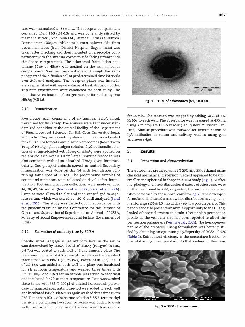
The ethosomes prepared with 2% SPC and 25% ethanol usingclassical mechanical dispersion method appeared to be unil-amellar and spherical in shape in a TEM study (Fig. 1). Surfacemorphology and three-dimensional nature of ethosomes werefurther confirmed by SEM, suggesting the vesicular character-istics possessed by these novel carriers (Fig. 2). The developedformulation indicated a narrow size distribution having nano-metric range (153 ± 8.5 nm) with a very low polydispersity. Thisnanometric size presents an ample opportunity to the HBsAg-loaded ethosomal system to attain a better skin permeationprofile, as the vesicular size has been reported to affect thepermeation parameters (Verma et al., 2003). The homogenous
Fig. 2 – SEM of ethosomes.
428 e u r o p e a n j o u r n a l o f p h a r m a c e u t i c a l s c i e n c e s 3 3 ( 2 0 0 8 ) 424–433
Table 1 – Characterization of HBsAg-loaded formulations
S. no. Parameters Ethosomes Conventional liposomes Plain antigen solution
1. Mean vesicular size (nm) 153 ± 8.5 202 ± 13 –2. Polydispersity index 0.082 ± 0.026 0.088 ± 0.024 –
4ellar,.2
3. % entrapment efficiency 57 ± 3.44. Vesicular shape and lamellarity Unilam5. % cumulative release in 24 h (via human skin) 67.2 ± 2
the maximum entrapment efficiency obtained was 57 ± 3.44%for HBsAg-loaded ethosomal formulation.
DSC study conducted to evaluate the fluidity of the for-mulations showed the transition temperature (Tm) of −6.44
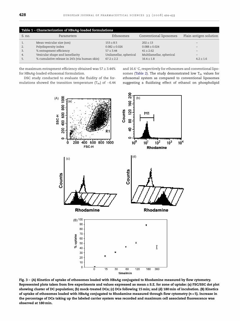
Fig. 3 – (A) Kinetics of uptake of ethosomes loaded with HBsAg cRepresented plots taken from five experiments and values expreshowing cluster of DC population; (b) mock-treated DCs; (c) DCs fof uptake of ethosomes loaded with HBsAg conjugated to Rhodathe percentage of DCs taking up the labeled carrier system was robserved at 180 min.
41 ± 2.62 –spherical Multilamellar, spherical –
16.4 ± 1.8 4.2 ± 1.6
and 16.6 ◦C, respectively for ethosomes and conventional lipo-somes (Table 2). The study demonstrated low Tm values forethosomal system as compared to conventional liposomessuggesting a fluidizing effect of ethanol on phospholipid
onjugated to Rhodamine measured by flow cytometry.ssed as mean ± S.E. for zone of uptake: (a) FSC/SSC dot plotollowing 15 min; and (d) 180 min of incubation. (B) Kineticsmine measured through flow cytometry (n = 5). Increase inecorded and maximum cell associated fluorescence was
e u r o p e a n j o u r n a l o f p h a r m a c e u t i c a
Table 2 – DSC scans of ethosomes and conventionalliposomes
Formulation Transition temperature, Tm (◦C)
bl
3
Ia2wais
3
IBtii(m
FHl
Ethosomes −6.44Conventional liposomes 16.62
ilayers, which could further be beneficial for fluidizing skinipids.
.2. Stability studies
n SDS-PAGE, clearly visible bands for pure as well as extractedntigens were obtained (data not shown) for HBsAg at around4-kDa. On storage at 25 ± 1 ◦C, around 12% of antigen leakageas detected from the system whereas at 4 ± 1 ◦C only 6% of
ntigen leaked after 120 days. No change in lamellarity andnsignificant difference in vesicular size was observed aftertorage of HBsAg-loaded ethosomal formulation for 120 days.
.3. Flow cytometric analysis
n the present study, cells were incubated with Rhodamine-SA-loaded ethosomal formulations and analyzed at differentime intervals (0, 15, 30, 60, 120, 180 and 360 min). A steady
ncrease in the uptake percentage (%) was recorded and max-mum cell associated fluorescence was observed at 180 min88.33 ± 1.01%). The kinetics of uptake has been presented withean fluorescence intensity (MFI) versus counts by FACS anal-
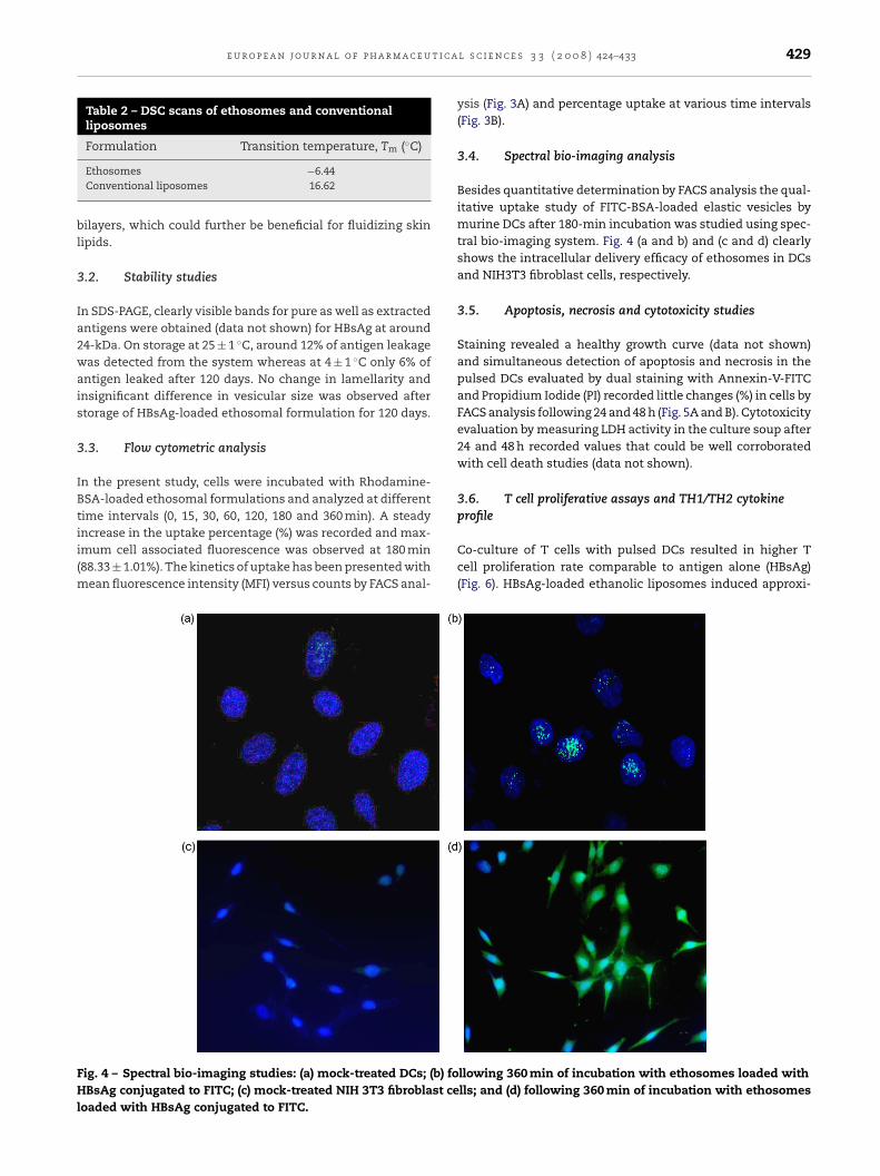
ig. 4 – Spectral bio-imaging studies: (a) mock-treated DCs; (b) foBsAg conjugated to FITC; (c) mock-treated NIH 3T3 fibroblast ce
oaded with HBsAg conjugated to FITC.
l s c i e n c e s 3 3 ( 2 0 0 8 ) 424–433 429
ysis (Fig. 3A) and percentage uptake at various time intervals(Fig. 3B).
3.4. Spectral bio-imaging analysis
Besides quantitative determination by FACS analysis the qual-itative uptake study of FITC-BSA-loaded elastic vesicles bymurine DCs after 180-min incubation was studied using spec-tral bio-imaging system. Fig. 4 (a and b) and (c and d) clearlyshows the intracellular delivery efficacy of ethosomes in DCsand NIH3T3 fibroblast cells, respectively.
3.5. Apoptosis, necrosis and cytotoxicity studies
Staining revealed a healthy growth curve (data not shown)and simultaneous detection of apoptosis and necrosis in thepulsed DCs evaluated by dual staining with Annexin-V-FITCand Propidium Iodide (PI) recorded little changes (%) in cells byFACS analysis following 24 and 48 h (Fig. 5A and B). Cytotoxicityevaluation by measuring LDH activity in the culture soup after24 and 48 h recorded values that could be well corroboratedwith cell death studies (data not shown).
3.6. T cell proliferative assays and TH1/TH2 cytokineprofile
Co-culture of T cells with pulsed DCs resulted in higher Tcell proliferation rate comparable to antigen alone (HBsAg)(Fig. 6). HBsAg-loaded ethanolic liposomes induced approxi-
llowing 360 min of incubation with ethosomes loaded withlls; and (d) following 360 min of incubation with ethosomes

430 e u r o p e a n j o u r n a l o f p h a r m a c e u t i c a l s c i e n c e s 3 3 ( 2 0 0 8 ) 424–433
Fig. 5 – (A) Flow cytometric analysis for apoptosis and necrosis: (a) FSC/SSC plot showing the population of DCs; (b)non-pulsed DCs; (c) pulsed DCs following 6 h; (d) 12 h; (e) 24 h; and (f) 48 h of incubation. (B) Flow cytometric analysis forapoptosis and necrosis. Apoptotic index is the sum of the percentage of cells that are positive for Annexin-V-FITC alone andcells positive for both Annexin-V-FITC and propidium iodide (PI) within a population of cells. Cells negative for both stains
s neto no
are considered healthy and living and stained only with PI apulsed DCs was significantly higher (p < 0.001) as compared
mately two- to threefold increase in IL-2 levels, four- to fivefoldincrease in IFN-� levels and twofold increase in TNF-� lev-els as compared to control (p < 0.001) (Table 3). Levels of TH2cytokines though recorded an increase but the data did notdelineate a clear skewing effect. Thus the significant effect ofHBsAg-loaded ethosomes in inducing TH1 response could beobserved (Table 3).
3.7. Skin permeation studies
Skin permeation studies investigated by determining the %cumulative release of antigen across human cadaver skin viadifferent formulations revealed better permeation profile ofHBsAg-loaded in ethosomal formulation as compared to other
crotic. Data represent 48 h time point. Apoptotic index ofn-pulsed DCs.
formulations. The % cumulative skin permeation of antigenfrom ethosomes, conventional liposomes and plain antigensolution were found to be 67.2 ± 2.2, 16.4 ± 1.8 and 4.2 ± 1.6,respectively (Table 1).
3.8. Immunization
IgG titre values measured after immunization of Balb/c micewith HBsAg-loaded ethosomes and intramuscular HBsAginjection, resulted into comparable (P < 0.05) IgG levels (Fig. 7).
The booster on day 14th produced a higher durable IgG titrelevels as compared to that of primary immunization. Similarly,in case of IgA titre values a significantly higher systemic andmucosal IgA levels were obtained from the topically applied
e u r o p e a n j o u r n a l o f p h a r m a c e u t i c a l s c i e n c e s 3 3 ( 2 0 0 8 ) 424–433 431
Fig. 6 – T cell proliferation analysis by BrdU assay.Represented plots taken from five experiments and valuesexpressed as mean ± S.E. Proliferation index in DCs pulsedwith ethosomes loaded with HBsAg was significantlyhigher (p < 0.001) compared to mock-treated DCs.
Table 3 – Multiplex cytometric bead array analysis ofTH1/TH2 cytokines in the supernatant following 5 daysco-culture of mock-treated DCs (control) and DCs pulsedwith elastic liposomes loaded with HBsAg (treated)
Control (pg/ml) Treated (pg/ml)
TH1IL-2 134.63 ± 10.89 363.16 ± 6.544*
IFN-� 83.83 ± 2.98 199.73 ± 0.90*
TNF-� 46.33 ± 6.34 278.96 ± 6.12*
TH2IL-4 144.0 ± 4.56 149.43 ± 9.46IL-6 84.63 ± 5.67 85.2 ± 7.10IL-10 243.86 ± 3.95 253.33 ± 6.63
∗ p < 0.001; values represent mean ± S.D. (n = 3).
Fig. 7 – IgG level in serum (values represent mean ± S.D.,n = 6).
Fig. 8 – (A) IgA level in serum (values represent
mean ± S.D., n = 6). (B) IgA level in salivary washes (valuesrepresent mean ± S.D., n = 6).HBsAg-loaded ethanolic liposomes in comparison to other for-mulations (Fig. 8A and B).
4. Discussion
This is the first report of testing a novel preparation for elicit-ing an immune response against Hepatitis B which has usedHBsAg-loaded ethosomes as antigens and DCs as antigen-presenting cells. The immune response was studied in amurine system. The antigen-loaded ethanolic liposomes wereefficiently internalized by the DCs as evidenced by flow cytom-etry and spectral bio-imaging analysis. The antigen-pulsedDCs were able to initiate an immune response predominantlyof TH1 type. However, a good systemic and mucosal humoralimmune response of IgG and IgA type was observed which wascomparable to that obtained by intramuscular administrationof soluble HBsAg in experimental mice. Although, the anti-
genic formulation appears to have some apoptogenic potentialin the treated DCs, this was evident only after 24 h of antigenicexposure and at 48 h reached the apoptotic index of about 17%suggesting that the preparation is relatively non-toxic.
u t i c
r
432 e u r o p e a n j o u r n a l o f p h a r m a c e
The protective immune response for the prevention of Hep-atitis B infection is humoral as well as cellular. While theformer is necessary for primary prevention, the latter maybe necessary for resolution of established infection and pre-vention of re-infection. Hepatitis B has not been convincinglyshown to be transmitted via feaco-oral route; therefore, therelevance of the observed mucosal immune response is debat-able. But such a response may be useful in other forms ofviral Hepatitis (A, E, etc.) which are predominantly transmittedfeco-orally (Shaw-Stiffel, 2000).
The key feature of this ethosomal formulation in com-parison to liposomal and other delivery systems is theembodiment of greater concentration of ethanol providingbetter transdermal permeation profile. In this case ethosomeswere selected as carrier for TCI for delivery of HBsAg. Thereports relating to the stability of this antigen suggest thedegradation of antigen at about 70–80% ethanol concentrationand below 50% the antigen was quite stable (Ito et al., 2002).In our case, 25% ethanol was present in a dispersed mannerin the ethosomal carriers that might have accounted only forenhancing vesicular and skin fluidity and not for deteriorat-ing antigen, as evident in our SDS-PAGE and stability studies.Further, in our opinion, ethanol present in ethosomes mightbe intercalated in lipid bilayers and hence did not exist in freeform to degrade the entrapped antigen.
Ethanol interacts with lipid molecules in the polar headgroup region, resulting in a reduction in the Tm of the stra-tum corneum lipids, increasing their fluidity. The intercalationof ethanol into the polar headgroup environment can resultin an increase in the membrane permeability. In additionto the effects of ethanol on stratum corneum structure, theethosome itself may interact with the stratum corneum bar-rier. Ethanol may also provide the vesicles with soft flexiblecharacteristics, which allow them to more easily penetrateinto deeper layers of the skin. The interdigitated, malleableethosome vesicles can forge paths in the disordered stratumcorneum. The release of antigen in the deep layers of the skinand its transdermal absorption could then be the result offusion of ethosomes with skin lipids and its release at vari-ous points along the penetration pathway (Touitou et al., 2000;Dubey et al., 2007b). We observed that the antigen-loadedethosomes were highly permeable through the skin comparedto conventional liposomes and soluble antigen, suggestingtheir potential for TCI.
In conclusion, in the present study HBsAg-loaded etho-somes showed greater entrapment efficiency, optimal sizerange, and a unilamellar, spherical shape in comparisonto conventional liposomes. Rapid internalization of HBsAg-loaded ethosomes by murine DCs and fibroblasts as well asstrong immunogenicity demonstrated by in vitro T cell prolif-eration and induction of TH1 type of immune response clearlysuggests the potential of this novel carrier system in develop-ment a DCs based vaccine against Hepatitis B.
Acknowledgements
The authors are indebted to Dr. A.K. Panda, National Instituteof Immunology, New Delhi, India for providing pure HBsAg andthe All India Institute of Medical Sciences, New Delhi, India for
a l s c i e n c e s 3 3 ( 2 0 0 8 ) 424–433
providing facility for Electron Microscopy. Mr. Dinesh Mishrais a recipient of Senior Research Fellowship of the UniversityGrants Commission (UGC), New Delhi, India.
e f e r e n c e s
Akbar, S.M.F., Horiike, N., Onji, M., 2006. Immune therapyincluding dendritic cell based therapy in chronic Hepatitis Bvirus infection. World J. Gastroenterol. 12 (18), 2876–2883.
Apoptosis, Cell Death and Proliferation. Roche DiagnosticsCorporation 2006. 3rd ed., 2004/2005.
Bangham, A.D., Horn, T.N., 1964. Negative staining ofphospholipids and their structural modification bysurface-active agents as observed in the electron microscope.J. Mol. Biol. 8, 660.
Coester, C., Nayyar, P., Samuel, J., 2006. In vitro uptake of gelatinnanoparticles by murine dendritic cells and their intracellularlocalisation. Eur. J. Pharm. Biopharm. 62, 306–314.
Copland, M.J., Baird, M.A., Rades, T., McKenzie, J.L., Becker, B.,Reck, F., Tyler, P.C., Davies, N.M., 2003. Liposomal delivery ofantigen to human dendritic cells. Vaccine 21 (9–10), 883–890.
Dayan, N., Touitou, E., 2000. Carriers for skin delivery oftrihexyphenidyl HCl: ethosomes vs. liposomes. Biomaterials21, 1879–1885.
Dubey, V., Mishra, D., Dutta, T., Nahar, M., Saraf, D.K., Jain, N.K.,2007a. Dermal and transdermal delivery of an antipsoriaticagent via ethanolic liposomes. J. Control. Rel. 123,148–155.
Dubey, V., Mishra, D., Jain, N.K., 2007b. Melatonin loaded ethanolicliposomes: physicochemical characterization and enhancedtransdermal delivery. Eur. J. Pharm. Biopharm. 67 (2), 398–405.
Fry, D.W., White, J.C., Goldman, I.D., 1978. Rapid separation of lowmolecular weight solutes from liposomes without dilution. J.Anal. Biochem. 90, 809–815.
Glenn, G.M., Scharton-Kersten, T., Alving, C.R., 1999. Advances invaccine delivery: transcutaneous immunization. Exp. Opin.Invest. Drugs 8 (6), 797–805.
Gupta, R.K., Chang, A.C., Siber, G.R., 1998. Biodegradable polymermicrospheres as vaccine adjuvants and delivery systems. Dev.Biol. Stand. 92, 63–78.
Huth, U.S., Schubert, R., Peschka-Suss, R., 2006. Investigating theuptake and intracellular fate of pH-sensitive liposomes byflow cytometry and spectral bio-imaging. J. Control. Rel. 110(3), 490–504.
Inchauspe, G., Michel, M.L., 2007. Vaccines and immunotherapiesagainst Hepatitis B and hepatitis C viruses. J. Viral Hepat. 14(Suppl. 1), 97–103.
Ito, K., Kajiura, T., Abe, K., 2002. Effect of ethanol on antigenicityof Hepatitis B virus envelope proteins. Jpn. J. Infect. Dis. 55,117–121.
Lopez-Pinto, J.M., Gonzalez-Rodrıguez, M.L., Rabasco, A.M., 2005.Effect of cholesterol and ethanol on dermal delivery fromDPPC liposomes. Int. J. Pharm. 298, 1–12.
Lutz, M.B., Kutsch, N., Ogilvie, A.L.J., Rosser, S., Koch, F., Romani,N., 1999. An advanced culture method for generating largequantities of highly pure dendritic cells from mouse bonemarrow. J. Immunol. Methods 223, 77–92.
Mishra, D., Dubey, V., Asthana, A., Jain, N.K., 2006. Elasticliposomes mediated transcutaneous immunization againstHepatitis B. Vaccine 24, 4847–4855.
Mishra, D., Mishra, P.K., Dubey, V., Dabadghao, S., Jain, N.K., 2007.
Evaluation of uptake and generation of immune response bymurine dendritic cells pulsed with Hepatitis B surface antigenloaded elastic liposomes. Vaccine 25 (39–40), 6939–6944.Poland, G.A., Murray, D., BonillaGuerrero, R., 2002. New vaccinedevelopment. BMJ 324, 1315–1319.
t i c a
S
S
S
S
e u r o p e a n j o u r n a l o f p h a r m a c e u
araf, S., Mishra, D., Asthana, A., Jain, R., Singh, S., Jain, N.K.,2006. Lipid microparticles for mucosal immunization againstHepatitis B. Vaccine 24 (1), 45–56.
haw-Stiffel, T.A., 2000. Chronic hepatitis. In: Mandell, G.L.,Bennett, J.E., Dolin, R. (Eds.), Principles and Practice ofInfectious Diseases. Churchill Livingstone, Philadelphia, pp.1297–1332.
hepard, C.W., Simard, E.P., Finelli, L., Fiore, A.E., Bell, B.P., 2006.Hepatitis B virus infection: epidemiology and vaccination.
Epidemiol. Rev. 28, 112–125.orensen, E.N., Weisman, G., Vidaver, G.A., 1977. A Sephadexcolumn for measuring uptake and loss of low molecularweight solutes from small vesicles. Anal. Biochem. 82,376–384.
l s c i e n c e s 3 3 ( 2 0 0 8 ) 424–433 433
Tacket, C.O., Mason, H.S., Losonsky, G., Clements, J.D., Levine,M.M., Arntzen, C.J., 1998. Immunogenicity in humans of arecombinant bacterial antigen delivered in a transgenicpotato. Nat. Med. 4 (5), 607–609.
Touitou, E., Dayan, N., Bergelson, L., Godin, B., Eliaz, M., 2000.Ethosomes-novel vesicular carriers for enhanced delivery:characterization and skin penetration properties. J. Control.Rel. 65, 403–418.
Verma, D.D., Verma, S., Blume, G., Fahr, A., 2003. Particle size of
liposomes influences dermal delivery of substances into skin.Int. J. Pharm. 258, 141–151.Wischke, C., Borchert, H.H., 2006. Fluorescein isothiocyanatelabelled bovine serum albumin (FITC-BSA) as a model proteindrug: opportunities and drawbacks. Pharmazie 61 (9), 770–774.
Copyright © 2022 FDOKUMEN