Efficacy of Imiquimod-Based Transcutaneous Immunization Using a Nano-Dispersed Emulsion Gel...
9
Efficacy of Imiquimod-Based Transcutaneous Immunization Using a Nano-Dispersed Emulsion Gel Formulation Pamela Stein 1. , Karsten Gogoll 2. , Stefan Tenzer 1 , Hansjo ¨ rg Schild 1 , Stefan Stevanovic 3 , Peter Langguth 2 , Markus P. Radsak 4 * 1 Institute for Immunology, Johannes Gutenberg-University Medical Center, Mainz, Germany, 2 Biopharmaceutics and Pharmaceutical Technology, Johannes Gutenberg- University, Mainz, Germany, 3 Department of Immunology, Eberhard Karls-University Tu ¨ bingen, Tu ¨ bingen, Germany, 4 Third Department of Medicine, Johannes Gutenberg-University Medical Center, Mainz, Germany Abstract Background: Transcutaneous immunization (TCI) approaches utilize skin associated lymphatic tissues to elicit specific immune responses. In this context, the imidazoquinoline derivative imiquimod formulated in Aldara applied onto intact skin together with a cytotoxic T lymphocyte (CTL) epitope induces potent CTL responses. However, the feasibility and efficacy of the commercial imiquimod formulation Aldara is limited by its physicochemical properties as well as its immunogenicity. Methodology/Principal Findings: To overcome these obstacles, we developed an imiquimod-containing emulsion gel (IMI- Gel) and characterized it in comparison to Aldara for rheological properties and in vitro mouse skin permeation in a Franz diffusion cell system. Imiquimod was readily released from Aldara, while IMI-Gel showed markedly decreased drug release. Nevertheless, comparing vaccination potency of Aldara or IMI-Gel-based TCI in C57BL/6 mice against the model cytotoxic T- lymphocyte epitope SIINFEKL, we found that IMI-Gel was equally effective in terms of the frequency of peptide-specific T- cells and in vivo cytolytic activity. Importantly, transcutaneous delivery of IMI-Gel for vaccination was clearly superior to the subcutaneous or oral route of administration. Finally, IMI-Gel based TCI was at least equally effective compared to Aldara- based TCI in rejection of established SIINFEKL-expressing E.G7 tumors in a therapeutic setup indicated by enhanced tumor rejection and survival. Conclusion/Significance: In summary, we developed a novel imiquimod formulation with feasible pharmaceutical properties and immunological efficacy that fosters the rational design of a next generation transcutaneous vaccination platform suitable for the treatment of cancer or persistent virus infections. Citation: Stein P, Gogoll K, Tenzer S, Schild H, Stevanovic S, et al. (2014) Efficacy of Imiquimod-Based Transcutaneous Immunization Using a Nano-Dispersed Emulsion Gel Formulation. PLoS ONE 9(7): e102664. doi:10.1371/journal.pone.0102664 Editor: Hiroshi Shiku, Mie University Graduate School of Medicine, Japan Received December 10, 2013; Accepted June 20, 2014; Published July 15, 2014 Copyright: ß 2014 Stein et al. This is an open-access article distributed under the terms of the Creative Commons Attribution License, which permits unrestricted use, distribution, and reproduction in any medium, provided the original author and source are credited. Funding: This work was supported by grants from the Deutsche Forschungsgemeinschaft KFO 183 to M.P.R. and H.S. (Ra988/4-2), CI3 Cluster (BMBF 131A035A, the GRK 1043 International Graduate School of Immunotherapy (H.S.), ‘‘Forschungszentrum Immunologie (FZI)’’ of the University Medical Center Mainz. The funders had no role in study design, data collection and analysis, decision to publish, or preparation of the manuscript. Competing Interests: The authors have declared that no competing interests exist. * Email: [email protected] . These authors contributed equally to this work. Introduction Transcutaneous immunization (TCI) approaches are increas- ingly gaining interest by vaccine developers since they incorporate all desirable properties of an ideal cancer vaccine, in terms of defined antigen specificities, targeting of specific APC populations and well-defined adjuvants [1,2]. Besides the advantage of patient self-medication, such easy-to-use vaccines lack the need for injections. In particular, needle-borne accidents in both medical personnel and patients will be circumvented. This is a priority issue conceded by the WHO based on the medical and socioeconomic consequences of needle injuries [3,4]. From an immunological point of view the skin is an attractive target for shaping immune responses: the delivery of antigens is controlled and specifically targeted to skin resident APC [5] in direct conjunction with an adjuvant eliciting potent adaptive immune responses as pioneered by Glenn and coworkers [6]. While parenteral (subcutaneous or intramuscular) vaccine delivery systems are not as well controlled in terms of drug release and targeting of specific immune organs, the drug dosage and targeting to the skin in transcutaneous approaches can be more easily and specifically achieved, e. g. by specific modifications of the vaccine as recently demonstrated for a nanoparticle-formulated DNA vaccine [7]. We have previously shown that the concurrent administration of a cytotoxic T lymphocyte (CTL) epitope together with the TLR7 agonist imiquimod onto intact skin elicits potent primary CTL responses [8]. While imiquimod-based TCI has been successfully applied to experimental rodent models by us and others [5,9–11], the clinical efficacy of topical imiquimod in patients with HPV PLOS ONE | www.plosone.org 1 July 2014 | Volume 9 | Issue 7 | e102664
Transcript of Efficacy of Imiquimod-Based Transcutaneous Immunization Using a Nano-Dispersed Emulsion Gel...

Efficacy of Imiquimod-Based TranscutaneousImmunization Using a Nano-Dispersed Emulsion GelFormulationPamela Stein1., Karsten Gogoll2., Stefan Tenzer1, Hansjorg Schild1, Stefan Stevanovic3,
Peter Langguth2, Markus P. Radsak4*
1 Institute for Immunology, Johannes Gutenberg-University Medical Center, Mainz, Germany, 2 Biopharmaceutics and Pharmaceutical Technology, Johannes Gutenberg-
University, Mainz, Germany, 3Department of Immunology, Eberhard Karls-University Tubingen, Tubingen, Germany, 4 Third Department of Medicine, Johannes
Gutenberg-University Medical Center, Mainz, Germany
Abstract
Background: Transcutaneous immunization (TCI) approaches utilize skin associated lymphatic tissues to elicit specificimmune responses. In this context, the imidazoquinoline derivative imiquimod formulated in Aldara applied onto intact skintogether with a cytotoxic T lymphocyte (CTL) epitope induces potent CTL responses. However, the feasibility and efficacy ofthe commercial imiquimod formulation Aldara is limited by its physicochemical properties as well as its immunogenicity.
Methodology/Principal Findings: To overcome these obstacles, we developed an imiquimod-containing emulsion gel (IMI-Gel) and characterized it in comparison to Aldara for rheological properties and in vitro mouse skin permeation in a Franzdiffusion cell system. Imiquimod was readily released from Aldara, while IMI-Gel showed markedly decreased drug release.Nevertheless, comparing vaccination potency of Aldara or IMI-Gel-based TCI in C57BL/6 mice against the model cytotoxic T-lymphocyte epitope SIINFEKL, we found that IMI-Gel was equally effective in terms of the frequency of peptide-specific T-cells and in vivo cytolytic activity. Importantly, transcutaneous delivery of IMI-Gel for vaccination was clearly superior to thesubcutaneous or oral route of administration. Finally, IMI-Gel based TCI was at least equally effective compared to Aldara-based TCI in rejection of established SIINFEKL-expressing E.G7 tumors in a therapeutic setup indicated by enhanced tumorrejection and survival.
Conclusion/Significance: In summary, we developed a novel imiquimod formulation with feasible pharmaceuticalproperties and immunological efficacy that fosters the rational design of a next generation transcutaneous vaccinationplatform suitable for the treatment of cancer or persistent virus infections.
Citation: Stein P, Gogoll K, Tenzer S, Schild H, Stevanovic S, et al. (2014) Efficacy of Imiquimod-Based Transcutaneous Immunization Using a Nano-DispersedEmulsion Gel Formulation. PLoS ONE 9(7): e102664. doi:10.1371/journal.pone.0102664
Editor: Hiroshi Shiku, Mie University Graduate School of Medicine, Japan
Received December 10, 2013; Accepted June 20, 2014; Published July 15, 2014
Copyright: � 2014 Stein et al. This is an open-access article distributed under the terms of the Creative Commons Attribution License, which permitsunrestricted use, distribution, and reproduction in any medium, provided the original author and source are credited.
Funding: This work was supported by grants from the Deutsche Forschungsgemeinschaft KFO 183 to M.P.R. and H.S. (Ra988/4-2), CI3 Cluster (BMBF 131A035A,the GRK 1043 International Graduate School of Immunotherapy (H.S.), ‘‘Forschungszentrum Immunologie (FZI)’’ of the University Medical Center Mainz. Thefunders had no role in study design, data collection and analysis, decision to publish, or preparation of the manuscript.
Competing Interests: The authors have declared that no competing interests exist.
* Email: [email protected]
. These authors contributed equally to this work.
Introduction
Transcutaneous immunization (TCI) approaches are increas-
ingly gaining interest by vaccine developers since they incorporate
all desirable properties of an ideal cancer vaccine, in terms of
defined antigen specificities, targeting of specific APC populations
and well-defined adjuvants [1,2]. Besides the advantage of patient
self-medication, such easy-to-use vaccines lack the need for
injections. In particular, needle-borne accidents in both medical
personnel and patients will be circumvented. This is a priority
issue conceded by the WHO based on the medical and
socioeconomic consequences of needle injuries [3,4]. From an
immunological point of view the skin is an attractive target for
shaping immune responses: the delivery of antigens is controlled
and specifically targeted to skin resident APC [5] in direct
conjunction with an adjuvant eliciting potent adaptive immune
responses as pioneered by Glenn and coworkers [6]. While
parenteral (subcutaneous or intramuscular) vaccine delivery
systems are not as well controlled in terms of drug release and
targeting of specific immune organs, the drug dosage and targeting
to the skin in transcutaneous approaches can be more easily and
specifically achieved, e. g. by specific modifications of the vaccine
as recently demonstrated for a nanoparticle-formulated DNA
vaccine [7].
We have previously shown that the concurrent administration of
a cytotoxic T lymphocyte (CTL) epitope together with the TLR7
agonist imiquimod onto intact skin elicits potent primary CTL
responses [8]. While imiquimod-based TCI has been successfully
applied to experimental rodent models by us and others [5,9–11],
the clinical efficacy of topical imiquimod in patients with HPV
PLOS ONE | www.plosone.org 1 July 2014 | Volume 9 | Issue 7 | e102664

infections [12] or skin cancers [13] makes it very likely that novel
imiquimod-based vaccination concepts may also be effective in
humans. This assumption is further supported by the finding that
imiquimod efficiently enhances T-cell responses when used as an
adjuvant in humans in pilot studies with melanoma patients
immunized against NY-ESO-1 protein [14] or patients with
prostate cancer treated with a multi peptide vaccine [15].
CTL responses induced by TCI with imiquimod based on the
commercial formulation in Aldara rapidly fade away, resulting in
poor memory formation and only partial tumor protection [5,9].
However, T-cell responses and tumor protection can be rescued
by additional stimuli, e.g. by CD40 ligation [5,9] or low dose UV-
B irradiation [5]. This highlights the importance of ‘‘high-quality’’
T cell responses that confer effective memory responses to achieve
anti-tumor immunity and furthermore suggests that imiquimod
formulated in Aldara is not an ideal preparation for TCI purposes.
We have also recently shown that various imiquimod formulations
considerably differ in their release of imiquimod and their potency
to induce CTL responses in TCI [16].
Based on this, we hypothesized that for effective TCI it is not
necessary for imiquimod to be solubilized e. g. in isostearic acid,
which is responsible for Aldara-induced TLR7-independent
adverse skin reactions [17]. We therefore generated an imiqui-
mod-containing emulsion gel (IMI-Gel) in which the crystalline
imiquimod is nano-dispersed in a hydrophilic polyacrylate gel
containing a lipophilic dispersed phase. We compared IMI-Gel to
Aldara in terms of drug permeation across mouse skin. Not
surprisingly, imiquimod release over time occurred significantly
faster from Aldara compared to IMI-Gel. However, when IMI-
Gel was compared to Aldara-based TCI, both imiquimod
formulations showed equal potency in the induction of primary
CTL responses. More importantly, IMI-Gel was also at least as
effective as Aldara induced TCI in a therapeutic tumor
vaccination model, indicating the induction of high quality
memory T cell responses. Collectively, these results support the
notion that Aldara is not an ideal formulation for vaccination
purposes and that the development of revised preparations and a
deeper understanding of the underlying mechanisms may harbour
the key for the rational design of a next-generation TCI platform
that can be used for the treatment of cancer.
Results
Physicochemical properties of imiquimod formulationIMI-GelBased on our previous finding that despite the presence of
crystalline imiquimod various imiquimod formulations can induce
CTL responses in TCI [16] we hypothesized that solubilisation of
imiquimod is not required for successful TCI. We therefore
manufactured a hydrophilic polyacrylate gel in which imiquimod
is nano-dispersed in a lipophilic phase (IMI-Gel). Imiquimod
crystals were dispersed within the gel formulation (Fig. 1A) and
had an average mean size of 335 nm679 nm post manufacturing.
Martin diameter of imiquimod crystals ranges from approximately
100 nm–500 nm. The particle size remained stable after 9 months
of storage at room temperature (286 nm612 nm calculated as
average mean of three DLS z-average values), excluding potential
Ostwald ripening processes (Fig. 1B), and there was no phase
separation during the storage period. In summary, this indicates
an accurate stability of the IMI-Gel formulation.
As shown in Fig. 1C, rheologic measurements demonstrated a
thin fluid consistency of Aldara, similar to common dermally
applied lotions. In contrast, for IMI-Gel we detected a high
viscosity resulting in an approx. 20-fold increased shear stress at
comparable shear rates. Taken together, imiquimod formulated in
IMI-Gel was pharmaceutically different compared to Aldara, but
was stable with acceptable pharmaceutical quality properties and
therefore suitable for further functional evaluation.
Imiquimod formulated in IMI-Gel shows lowerpermeation through murine skin compared to AldaraTCI approaches offer the advantage of gaining direct access to
skin-resident DC and therefore facilitate their stimulation,
migration and subsequent antigen presentation in draining lymph
nodes. To this end, imiquimod as the active component or
adjuvant in our TCI approach should be targeted to the skin and
remain there for optimal DC activation. To address this, we
explored the skin permeation of both imiquimod formulations and
placed skin samples in a Franz-diffusion-cell system (Fig. 2A),
either applying Aldara or IMI-Gel on isolated mouse skin.
Subsequently, we analyzed the concentration of imiquimod in
the acceptor medium over time. As depicted in Fig. 2B, 11.5% of
imiquimod was released by Aldara (grey line), while only 1%
imiquimod permeated across the skin when formulated in IMI-Gel
(black line). To determine the amount of imiquimod within the
skin, we treated mice either with IMI-Gel or Aldara on an area of
6 cm2 on the shaved backs. After 3 hours, the amounts of the
respective formulations remaining on the skin were harvested
using gauze. Afterwards 1 cm2 of skin was prepared and fat was
carefully removed. Samples were collected in extraction buffer and
then hackled with an ultra turrax. The amount of imiquimod
found in the supernatant of centrifuged gauze or skin samples was
analyzed by HPLC. As shown in Fig. 2C (left panel) 40% of
imiquimod applied with IMI-Gel remained on the skin surface 3
hours after treatment, whereas 19% were recovered after Aldara
treatment (statistically not significant). Analyzing the amount of
imiquimod within the skin (Fig. 2C right panel) revealed that
slightly more imiquimod was found in the skin of IMI-Gel treated
(14 mg/cm263 mg/cm2) compared to Aldara treated (11 mg/cm261.7 mg/cm2) mice (statistically not significant).
These results indicate that imiquimod formulated in Aldara was
taken up across the skin more effectively allowing systemic
exposure of the drug, while imiquimod formulated in IMI-Gel
appeared to remain in and on top of the skin.
IMI-Gel and Aldara are equally potent in inducingpeptide specific CTL responsesSo far, our results indicated a lower drug permeation of
imiquimod when formulated in IMI-Gel. We asked whether or not
this is of relevance for TCI efficacy since imiquimod should
specifically activate skin DCs [5,10,18]. Therefore, a lower skin
permeation of IMI-Gel imiquimod may not necessarily be a
disadvantage for TCI induced immune responses. To directly
address this, we compared the potency of IMI-Gel or Aldara
treatment regarding the induction of peptide-specific CTL
responses. This was done either by administering Aldara together
with the major histocompatibility complex (MHC) class-I-restrict-
ed T cell epitope OVA257–264 (SIINFEKL, SIIN) or IMI-Gel
followed by the additional treatment with OVA257–264 in officinal
cremor basalis on two consecutive days on the dorsal region of
C57BL/6 mice. Analyzing the frequency of peptide-specific T cells
via MHC I tetramer stainings of blood samples revealed
comparable amounts in both immunized groups (Fig. 3A). We
then determined the lytic capacity of cytotoxic T cells with an
in vivo cytotoxicity assay (Fig. 3B) and found that the majority of
target cells (75%) was specifically lyzed after Aldara-induced TCI,
as described previously [5,9]. Interestingly, the CTL response
IMI-Gel for Transcutaneous Immunization
PLOS ONE | www.plosone.org 2 July 2014 | Volume 9 | Issue 7 | e102664

induced by TCI with IMI-Gel was comparable and indistinguish-
able from the Aldara-induced response despite a .10 fold less
drug permeation across mouse skin. These results suggest that it is
important for CTL induction in imiquimod-based TCI that the
drug is targeted to the skin and that it does not need to be
delivered systemically.
Transcutaneous delivery of imiquimod induces superiorCTL responses compared to s. c. or oral applicationIn case it is indeed important for imiquimod-based TCI that the
drug remains locally within the skin, then an imiquimod-based
vaccination would be less effective if the skin was passed by via s. c.
injection. To clarify this, we vaccinated mice as before with IMI-
Gel-based TCI. As important controls and to exclude the
induction of CTL responses was due to skin irritations by the
shaving procedure or independent of the CTL epitope, we
included a group treated with IMI-Gel without the CTL epitope
SIINFEKL or a group treated with the SIINFEKL (in officinal
cremor basalis) without IMI-Gel. For comparison, we added low
amounts of sterile water to IMI-Gel and the CTL epitope to allow
aspiration in a syringe and injected this s. c. in the dorsal neck
region. As depicted in Fig. 4, the IMI-Gel s. c. administration did
neither induce specific T cells nor specific cytotoxicity. Also, the
treatments with IMI-Gel or the CTL epitope alone were unable to
elicit CTL responses. To furthermore exclude the possibility that
IMI-Gel is orally taken up, e. g. by grooming post topical
application on the back, we performed additional experiments
where a group of mice was fed with IMI-Gel and peptide using an
oral gavage on two consecutive days. As shown in Fig. 4, the oral
delivery of IMI-Gel did not elicit a CTL response. In contrast,
IMI-Gel-based TCI was again effective in terms of frequency of
specific CTL and cytolytic activity. Importantly, we used the
identical pharmaceutically active components (IMI-Gel and
peptide) at the same doses.
Figure 1. In vitro characterization of IMI-Gel. To characterize IMI-Gel and Aldara, imiquimod containing formulations were analyzed in terms ofA) presence of imiquimod crystals in IMI-Gel using electron microscopy, B) sizes (mean+SD) of imiquimod-particles in IMI-Gel immediately or 9months after manufacturing (under room conditions) and C) flow curves defining rheological characteristics.doi:10.1371/journal.pone.0102664.g001
IMI-Gel for Transcutaneous Immunization
PLOS ONE | www.plosone.org 3 July 2014 | Volume 9 | Issue 7 | e102664

These results clearly show that the transcutaneous delivery of
imiquimod is important for vaccine efficacy and superior to the
parenteral route of application.
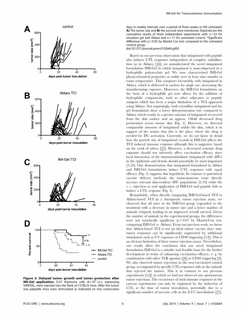
IMI-Gel induced TCI increases tumor protection in atherapeutic modelAfter having demonstrated comparable primary immune
responses via Aldara or IMI-Gel-based TCI (Fig. 3), we wanted
to evaluate the potency of both imiquimod formulations regarding
the induction of high-quality memory CTL responses as required
for tumor protection. We therefore tested both TCI treatments in
a therapeutic tumor model, in which mice were inoculated s. c.
with E.G7 thymoma cells, expressing ovalbumin as a surrogate
tumor antigen. After the tumors became palpable, mice were left
untreated or vaccinated as described before in weekly intervals
over a period of three weeks. Unimmunized control mice showed a
durable tumor growth in 12 of 17 animals (Fig. 5A). In the five
animals with spontaneous tumor rejection, we detected up to 2%
peptide-specific CTL (data not shown), indicating potential
endogenous immunogenicity of the E.G7 tumor cells. Importantly,
in the 12 animals with growing solid tumors no specific CTL
responses were detectable and the mice had to be sacrificed in the
end (overall survival 29.4%; median survival 25 days).
In the Aldara-based TCI group, 21 out of 23 animals responded
to the treatment, as shown by a reduction in tumor size. However,
we observed a tumor relapse in seven mice, leaving in a durable
protection in 14 of 23 mice (overall survival 60.8%, median
survival not reached) (Fig. 5B). In contrast, all animals treated with
IMI-Gel-based TCI initially responded to vaccination treatment
with decreasing tumor sizes (Fig. 5C). However, the tumors
relapsed in six mice, leading to an overall survival of 73.9%
(median survival not reached, Fig. 5D). While there was a trend for
higher survival for IMI-Gel-treated mice compared the Aldara
Figure 2. Imiquimod passes mouse skin in vitro more rapidly when formulated in Aldara than in IMI-Gel. A) Release of imiquimod asthe active component was detected with a modified Franz-diffusion-cell model. B) Shaved skin of C57BL/6 mice (n = 6) was ablated and afterwardstreated with Aldara or IMI-Gel (each 50 mg). The imiquimod concentration in the acceptor medium was determined after various time points asindicated using HPLC (UV absorption 245 nm). *Significant difference with p,0.05 by Wilcoxon signed rank test. C) C57BL/6 mice (n = 3) were withAldara or IMI-Gel (each 50 mg/6 cm2) on. After 3 hours mice were sacrificed and remaining formulation was removed with gauze. 1 cm2 of treatedskin was prepared, fat removed and subsequently hackled with an ultra turrax. The amount of imiquimod recovered from the skin surface (left panel)and within the skin (right panel) was determined using HPLC.doi:10.1371/journal.pone.0102664.g002
IMI-Gel for Transcutaneous Immunization
PLOS ONE | www.plosone.org 4 July 2014 | Volume 9 | Issue 7 | e102664

group, this did not reach statistical significance (p= 0.07 by
Mantel-Cox test).
In summary, our results suggest that the vaccination potency of
IMI-Gel-based TCI is at least as effective as Aldara-based TCI in
the induction of tumor specific immunity despite lower imiquimod
permeation through the skin.
Discussion
Although standard vaccination approaches are effective in a
considerable number of infectious diseases [19], vaccination
strategies against tumors have only recently begun to prove
successful [20]. Despite the limited efficacy of Sipuleucel-T, the
FDA has recently approved this DC vaccine for the treatment of
advanced prostate cancer, further underlining the medical need
for therapeutic cancer vaccines [21]. Moreover, peptide-based
cancer vaccine approaches are effective, e. g. in renal cancer with
GM-CSF as adjuvant [22]. While classical prophylactic vaccina-
tion approaches are suitable for inducing protective antibody
responses against virus infections, they may not be ideal for the
induction of therapeutic T-cell responses. In this context,
transcutaneous vaccination approaches may be an attractive way
to deliver antigens and adjuvants. The proof-of-concept has been
demonstrated already years ago [6,8,10]. We have been following
a TCI approach based on a CTL epitope and the TLR7 agonist
imiquimod formulated in Aldara. While we observed primary
CTL responses [8,11] and anti-tumor effects of Aldara-based TCI
[5,9], this particular TCI method is limited by inefficient memory
CTL generation. Aldara-based TCI efficiency can be increased by
costimulatory signals like CD40 ligation [9] or UV-B irradiation
[5] leading to enhanced memory formation and improved tumor
protection. An important limitation of our transcutaneous
vaccination approach concerns the use of a mouse model, which
on the one hand allows the evaluation of this novel treatment
approach in a preclinical animal model. On the other hand, the
usage of mouse models does not account for differences between
human and mouse skin, in the latter the stratum corneum being
significantly thinner and better permeable for transcutaneous drug
delivery [23]. In addition, differences of murine and human APCs
in TLR responsiveness or antigen presentation need to be taken
into account [1,24] and limit applicability and the direct
translation of our results to the application in humans. On the
other hand, the topical application of imiquimod has proven
efficacy for the treatment of patients with HPV infections [12] and
skin cancers [13] as well as when used as an adjuvant for
vaccination [14,15] suggesting that this approach may be also
feasible and effective in humans.
Figure 3. IMI-Gel and Aldara are equally potent in inducing primary CTL-responses. C57BL/6 mice were shaved on their backs andafterwards immunized on two consecutive days with either Aldara (50 mg) together with SIINFEKL (100 mg) or IMI-Gel (50 mg) and officinal cremorbasalis together with SIINFEKL or left untreated (untreated control). A) The frequency of peptide-specific CD8+ T cells in the blood (mean and SD) andB) in vivo cytolytic activity 24 hours (mean and SD) after transfer of peptide-loaded target cells was assessed. Depicted are the cumulative results oftwo independent experiments with n = 6 for the tetramer staining and three independent experiments with n = 9 for the cytotoxicity assay.*Significant difference with p,0.05 by one-way ANOVA with Bonferroni’s posttest.doi:10.1371/journal.pone.0102664.g003
Figure 4. The route of IMI-Gel application influences vaccina-tion efficacy. C57BL/6 mice were shaved on their backs and receivedthe following treatments: untreated (untreated control), IMI-Gel withSIINFEKL (100 mg) (IMI-Gel TCI) on two consecutive days, IMI-Gel alone(IMI-Gel without SIIN) on two consecutive days, oral IMI-Gel (50 mg)with SIINFEKL (100 mg) on two consecutive days (IMI-Gel p. o.) or s. c.once with IMI-Gel (100 mg) diluted with SIINFEKL (200 mg) and distilledwater into the neck. A) The frequency of peptide-specific CD8+ T cells inthe blood (mean and SD) and B) in vivo cytolytic activity 24 hours(mean and SD) after transfer of peptide-loaded target cells wasassessed. Depicted are the cumulative results of two independentexperiments with n = 7 for IMI-Gel treated groups and n=4 for controls.*Significant difference with p,0.05 by Students t test.doi:10.1371/journal.pone.0102664.g004
IMI-Gel for Transcutaneous Immunization
PLOS ONE | www.plosone.org 5 July 2014 | Volume 9 | Issue 7 | e102664
Based on our previous observation that imiquimod with peptide
also induces CTL responses independent of complete solubilisa-
tion (as in Aldara) [16], we manufactured the novel imiquimod
formulation IMI-Gel in which imiquimod is nano-dispersed in a
hydrophilic polyacrylate gel. We now characterized IMI-Gel
physicochemical properties as stable over at least nine months at
room temperature. This compares favourably with imiquimod in
Aldara, which is delivered in sachets for single use, increasing the
manufacturing expenses. Moreover, the IMI-Gel formulation on
the basis of a hydrophilic gel now allows for the addition of
hydrophilic components, such as other adjuvants or peptide
antigens which has been a major limitation of a TCI approach
using Aldara. Not surprisingly, both crystalline imiquimod and the
gel formulation show a lower skin-permeation rate compared to
Aldara which results in a greater amount of imiquimod recovered
from the skin surface and an approx. 10fold decreased drug
permeation across mouse skin (Fig. 2). However, we detected
comparable amounts of imiquimod within the skin, which is in
support of the notion that this is the place where the drug is
needed for DC activation. Currently, we do not know in detail
how the particle size of imiquimod crystals in IMI-Gel affects the
TCI induced immune response although this is suggestive based
on the work of others [25]. However, a decreased systemic drug
exposure should not adversely affect vaccination efficacy since
local interaction of the immunostimulator imiquimod with APCs
in the epidermis and dermis should potentially be most important
[5,10]. Our demonstration that imiquimod formulated in Aldara
and IMI-Gel formulations induce CTL responses with equal
efficacy (Fig. 3) supports this hypothesis. In contrast to parenteral
vaccine delivery methods, the transcutaneous route directly
accesses relevant skin-resident APC populations [5,10] while the
s. c. injection or oral application of IMI-Gel and peptide fails to
induce a CTL response (Fig. 4).
Remarkably, when directly comparing IMI-Gel-based TCI to
Aldara-based TCI in a therapeutic tumor rejection assay, we
observed that all mice in the IMI-Gel group responded to the
treatment with a decrease in tumor size and a lower number of
animals relapsed, leading to an improved overall survival. Given
the number of animals in the experimental groups, the differences
were not statistically significant (p = 0.07 by Mantel-Cox test,
comparing IMI-Gel vs. Aldara). From our previous work, we know
that Aldara-based TCI is not an ideal tumor vaccine since anti-
tumor responses can be significantly augmented by additional
stimulation such as UV exposure or CD40 triggering [5,9]. This is
an obvious limitation of these tumor rejection assays. Nevertheless,
our results allow the conclusion that our novel imiquimod
formulation IMI-Gel is a suitable and feasible basis for the further
development in terms of enhancing vaccination efficacy, e. g. by
combination with other TLR agonists [26] or CD40 triggering [9].
We also observed tumor rejections in the non-vaccinated control
group, accompanied by specific CTL responses only in the animals
that rejected the tumors. This is in contrast to our previous
experiments [5,9], in which we had not observed any spontaneous
tumor rejections. The occurrence of such immune responses in the
current experiments can only be explained by the induction of
CTL at the time of tumor inoculation, potentially due to a
significant number of necrotic cells in the E.G7 inoculations [27].
Figure 5. Delayed tumor growth and tumor protection afterIMI-Gel application. E.G7 thymoma cells (46105 s.c.), expressingSIINFEKL, were injected into the flank of C57BL/6 mice. After the tumorwas palpable mice were immunized as indicated on two consecutive
days in weekly intervals over a period of three weeks or left untreated.A) The tumor size and B) the survival were monitored. Depicted are thecumulative results of three independent experiments with n= 23 foremulsion gel and Aldara and n= 17 for untreated control. *Significantdifference with p,0.05 by Mantel Cox test compared to the untreatedcontrol group.doi:10.1371/journal.pone.0102664.g005
IMI-Gel for Transcutaneous Immunization
PLOS ONE | www.plosone.org 6 July 2014 | Volume 9 | Issue 7 | e102664

Spontaneous tumor rejections should not compromise the validity
of the experimental system since (i) it can be assumed that they
occurred equally over all experimental groups and (ii) CTL
responses were absent in tumor-bearing mice. Therefore, it is safe
to conclude that TCI with IMI-Gel is at least as effective as TCI
with Aldara despite the 10fold lower skin permeation of
imiquimod.
Taken together, we developed a novel imiquimod-based
formulation with interesting pharmaceutical and immunological
properties that should be used for the development of TCI
approaches. A deeper understanding of the underlying mecha-
nisms may harbour the key for a next-generation vaccination
platform that can be used for the treatment of persistent infections
and cancer.
Materials and Methods
Ethics StatementAll animal studies were conducted according to the national
guidelines and were reviewed and confirmed by an institutional
review board/ethics committee headed by the local animal welfare
officer (Prof. Dr. O. Kempski) of the University Medical Center
(Mainz, Germany). The National Investigation Office Rheinland-
Pfalz (Koblenz, Germany) finally approved the animal experi-
ments (AZ 23 177-07/G11-1-034 and 23 177-07/G13-1-012).
MaterialsImiquimod and Azone were obtained from Chemos GmbH
(Regenstauf, Germany). Jojoba wax, polysorbate 80, polyacrylic
acid, sodium hydroxide, sodium acetate trihydrate, and glacial
acetic acid were supplied by Carl Roth GmbH (Karlsruhe,
Germany). Trifluoric acid was provided by Sigma-Aldrich
(Steinheim, Germany). Acetonitrile and ethanol, both HPLC
grade, were obtained from VWR (Darmstadt, Germany). Officinal
cremor basalis according to DAC was obtained from Caesar and
Loretz GmbH (Hilden, Germany).
Production of IMI-GelFor pre-homogenization, imiquimod was added to a 9 mg/ml
aqueous solution of polysorbate 80 in a grinding vessel. Milling
was performed for six cycles to ensure an appropriate pre-
homogenization as well as a particle size below 30 mm to establish
a safe and reliable function during the subsequent high-pressure
homogenization (HPH) process.
After sieving, polysorbate 80 was added again to ensure a stable
emulsion. Moreover, Azone and jojoba wax were added (2.5%
and 42.5%, w/w, respectively). HPH was performed by an
Emulsiflex C3 (Avestin) for five cycles at 500 bar and ten cycles at
1000 bar, respectively. The resulting oil in water emulsion with
suspended imiquimod nanoparticles appeared as a yellow fluid. In
order to create a semisolid formulation, a previously prepared and
precisely neutralized polyacrylic acid gel was added. Additionally,
we investigated whether a storage period of nine months under
room conditions affects imiquimod particle size and dispersity
under measurement settings described below.
Electron micrographsSamples were spread on a glass slide and then adsorbed onto a
continuous carbon grid. They were washed three times with 20 mlH2O distilled prior to staining with 5 ml of a 1% uranyl acetate
solution. After the staining solution was blotted off, grids were air-
dried. They were transferred to an electron microscope and
images were recorded using a 4 k64 k TemCam-F416 (TVIPS,
Munich, Germany).
Rheologic studiesRheograms were obtained and analyzed as described previously
[16] using a Haake Rheostress 1 viscosimeter with a cone and
plate device PiT-L 35 and RheoWin 4 data manager software.
Flow curves display shear stress vs. shear rate and were obtained at
a predefined shear rate of 0–200 s21, recording the resulting shear
stress. Thixotropy was determined by a controlled shear rate with
a rate of 0–1000 s21 for 60 s, following a 1000 s21 phase for 30 s
and subsequent reduction of the shear rate from 1000 to 0 s21 for
a further 60 s. The experiments were performed at 23.0uC.
Particle size determinationVisualization of imiquimod particles embedded in the emulsion
gel matrix (IMI-Gel) was performed by electron microscopy. To
determine particle sizes within the formulation, dynamic light
scattering (DLS) measurements were performed using a Zeta Sizer
nano (Malvern Instruments GmbH, Herrenberg, Germany).
Briefly, 50 ml of IMI-Gel was diluted with 4 ml double distilled
water. Then, 100 ml of the resulting suspension was additionally
diluted with 1.3 ml of double distilled water in a disposable
cuvette. Measurements were performed directly after the manu-
facturing process or nine months post manufacturing to assess the
occurrence of Ostwald-ripening. Each sample was prepared three
times. Z-average values were measured by Zeta Sizer nano. Then,
particle sizes were calculated as the mean value of three z-average
measurements. In order to give an overview of particle size range,
martin diameter of 25crystals from the electron microscopic
specimen was determined by open source ImageJ software.
Permeation assaysMale or female C57BL/6 mice at 6–8 weeks were obtained
from the local animal facility of the University of Mainz. The
animals were sacrificed and dorsal hair was removed with electric
clippers. Skin samples were prepared and fat was removed using a
scalpel. Skin (0.79 cm2) was treated with either 50 mg Aldara or
IMI-Gel, both containing 5% imiquimod. Afterwards skin samples
were placed in an EDC-07 Franz-diffusion-cell model (Labswiss,
Muttenz, Switzerland). The acceptor chamber contained a blend
of sodium acetate trihydrate/glacial acetic acid buffer (20 mM) and
ethanol (7/3 v/v) with a resulting pH of 3.6, previously degassed
by an ultra sound bath for appropriate duration. Temperature was
set at 32uC and a magnetic stirrer constantly agitated acceptor
media. Samples were collected after 1, 2, 3, 5, 7, 8.5 and 24 hours.
To quantify imiquimod concentrations, a 300 C8 5 mm reversed
phase RP 250*4.6 mm column (Mainz Analysentechnik, Mainz,
Germany) was used. Mobile phase comprises double distilled
water, trifluoric acid and acetonitril (70:0.0125:30 V/V). This
mobile phase provided a pH value of 2.8 to avoid imiquimod
precipitation in the column. Flow rate was 1.0 ml/min ensured by
a Jasco PU-980 Intelligent HPLC pump (JASCO Germany
GmbH, Gross-Umstadt, Germany). For quantitative API detec-
tion UV absorbance was determined by a UV detector (JASCO
Germany GmbH, Grob-Umstadt, Germany) at 245 nm wave-
length. Imiquimod eluted after 5.5 minutes. Detectable concen-
tration ranges was from 35–1790 ng/ml. Jasco-Borwin HSS-2000
software was used to analyze revealed peaks.
Quantification of imiquimod in the skinMice were treated with either 50 mg Aldara or IMI-Gel on the
shaved dorsum (6 cm2). Quantification of imiquimod from skin
samples and skin surface was performed after 3 hours using
reversed phase HPLC, using a modified method similar to De
Paula et al. [28].
IMI-Gel for Transcutaneous Immunization
PLOS ONE | www.plosone.org 7 July 2014 | Volume 9 | Issue 7 | e102664

Briefly, skin (1 cm2) was homogenized and extracted with 3 ml
extraction buffer (7:3 (v/v) MeOH:acetate buffer (pH 4.0,
100 mM).
To determine residual imiquimod on the skin surface, skin was
wiped thoroughly with gauze and the gauze pad extracted with
3 ml of extraction buffer.
HPLC was performed using a Dionex Ultimate 3000 HPLC
equipped with a UV-Vis detector. Detection wavelength was
242 nm. Buffer A was 100 mM Acetate Buffer pH4.0 Buffer B was
100% Acetonitrile. Samples were analysed on a 2 mm650 mm
Phenomenex Max RP (C-12) column using a gradient from 5% B
to 80% B in 7 min.
Column was reequilibrated to starting conditions for 5 min.
The flow rate was 250 ml/min. Column compartment was
thermostatted at 50uC. Under these experimental conditions,
imiquimod eluted at 5.25 min. Total run time was 12 min and the
injection volume was 5 ml.
Transcutaneous immunizationsTranscutaneous immunizations were described previously [5].
Briefly, dorsal hair was removed with electric clippers. For TCI,
mice were anesthetized by i. p. injection with ketamine
(Ratiopharm, Ulm, Germany; 71.2 mg per mouse) and Rompun
2% (Bayer Health Care, Leverkusen, Germany;0.2 mg per
mouse). 50 mg Aldara containing 5% imiquimod (Meda Pharma,
Wangen-Bruttisellen, Switzerland) together with peptide
(OVA257–264, SIINFEKL, 100 mg in DMSO, provided by S.
Stevanovic, Department of Immunology, Institute for Cell
Biology, University of Tubingen, Germany) were applied on the
shaved dorsum (approx. 3 cm65 cm) on days 0 and 1. Alterna-
tively mice were treated with 50 mg of IMI-Gel also containing
5% imiquimod followed by application of officinal cremor basalis
together with 100 mg SIINFEKL.
For immunizations s. c. IMI-Gel (100 mg per mouse) was
diluted in 80 ml distilled water. After adding 200 mg SIINFEKL
peptide, the solution was injected in the dorsal neck region of mice
on day 1.
For oral immunization, IMI-Gel (50 mg per mouse) was diluted
with 40 ml distilled water and 100 mg SIINFEKL peptide. Mice
were fed on days 0 and 1.
Tumor rejection assayTumors were inoculated by s. c. injection of E.G7 thymoma
cells (46105, from ATCC) as described previously [5]. Tumors
were allowed to grow until palpable before treatments started.
Mice were immunized in weekly intervals over a period of three
weeks. Tumor size was monitored every other or third day at least
three times per week with a caliper in two dimensions. During the
observation times the mice had no indication of discomfort as
indicated by lethargy or piloerection. To further avoid animal
suffering, mice were sacrificed when tumor size exceeded 20 mm
in length and width by CO2 asphyxiation. The day subsequent to
euthanasia was defined as the day of death.
Flow cytometric analyses and in vivo cytotoxicity assayThe following mAbs were used for flow cytometric analyses:
Pacific Blue-conjugated anti-CD8 (clone 53–6.7), APC-conjugated
anti-CD44 (clone IM7) and FITC-conjugated anti-CD62L (clone
MEL-14), all from eBioscience, Frankfurt, Germany. Blood
samples were collected after tail vein incision. After a hypotonic
lysis step samples were incubated with mAbs on ice. H2-Kb-
SIINFEKL-specific T cells were detected by H2-Kb tetramer
staining as described previously [11]. In vivo cytolytic activity was
assessed by transfer of 26107 target cells labelled with either 4 mM(CFSEhigh) or 0.4 mM (CFSElow) carboxyfluorescein diacetate
succinimidyl ester. The CFSElow cells were additionally loaded
with SIINFEKL (2 mM). Both populations were transferred i.v. in
1:1 ratio. Splenocytes of immunized and control mice were
analysed by flow cytometry. All analyses were performed with a
LSRII Flow Cytometer and FACSDiva software (BD Pharmingen,
Hamburg, Germany).
Statistical analysisAll statistical analyses were performed using GraphPad Prism
(version 5.0a for Mac OS X, GraphPad Software, San Diego
California USA, www.graphpad.com). Survival analyses were
performed by the Mantel-Cox test. For comparison between two
groups a two-tailed Student’s t test was used. Comparisons of
multiple groups were performed by one-way ANOVA with
Bonferroni’s posttest. For all analyses, p,0.05 was considered as
statistically significant.
Acknowledgments
The authors express their gratitude to Annekatrin Klaric, Andrea Drescher
and Giusy Carlino for excellent technical assistance. We thank Dr. Markus
Munder for critical review of the manuscript.
Author Contributions
Conceived and designed the experiments: HS SS PL MPR. Performed the
experiments: PS KG ST. Analyzed the data: PS KG HS PL ST MPR.
Contributed reagents/materials/analysis tools: SS ST. Wrote the paper:
PS HS PL MPR.
References
1. Bal SM, Ding Z, van Riet E, Jiskoot W, Bouwstra JA (2010) Advances in
transcutaneous vaccine delivery: do all ways lead to Rome? J Control Release148: 266–282. doi:10.1016/j.jconrel.2010.09.018.
2. Mikszta JA, Laurent PE (2008) Cutaneous delivery of prophylactic andtherapeutic vaccines: historical perspective and future outlook. Exp Rev
Vaccines 7: 1329–1339.
3. Kermode M (2004) Unsafe injections in low-income country health settings:need for injection safety promotion to prevent the spread of blood-borne viruses.
Health Promot Int 19: 95–103.4. Pisani E, Miller MA (1999) The cost of unsafe injections. Bulletin of the World
Health Organization 77: 808.5. Stein P, Rechtsteiner G, Warger T, Bopp T, Fuhr T, et al. (2011) UV exposure
boosts transcutaneous immunization and improves tumor immunity: cytotoxic
T-cell priming through the skin. J Invest Dermatol 131: 211–219. doi:10.1038/jid.2010.254.
6. Glenn GM, Rao M, Matyas GR, Alving CR (1998) Skin immunization madepossible by cholera toxin. Nature 391: 851. doi:10.1038/36014.
7. van den Berg JH, Oosterhuis K, Hennink WE, Storm G, van der Aa LJ, et al.
(2010) Shielding the cationic charge of nanoparticle-formulated dermal DNA
vaccines is essential for antigen expression and immunogenicity. Journal of
Controlled Release 141: 234–240. doi:10.1016/j.jconrel.2009.09.005.
8. Rechtsteiner G, Warger T, Osterloh P, Schild H, Radsak MP (2005) Cutting
edge: priming of CTL by transcutaneous peptide immunization with imiquimod.
J Immunol 174: 2476–2480.
9. Warger T, Rechtsteiner G, Schmid B, Osterloh P, Schild H, et al. (2007)
Transcutaneous immunization with imiquimod is amplified by CD40 ligation
and results in sustained cytotoxic T-lymphocyte activation and tumor protection.
Clinic Rev Allerg Immunol 32: 57–66.
10. Stoitzner P, Green LK, Jung JY, Price KM, Tripp CH, et al. (2008) Tumor
immunotherapy by epicutaneous immunization requires langerhans cells.
J Immunol 180: 1991–1998.
11. Stein P, Weber M, Prufer S, Schmid B, Schmitt E, et al. (2011) Regulatory T
Cells and IL-10 Independently Counterregulate Cytotoxic T Lymphocyte
Responses Induced by Transcutaneous Immunization. PLoS ONE 6: e27911.
12. Edwards L, Ferenczy A, Eron L, Baker D, Owens ML, et al. (1998) Self-
administered topical 5% imiquimod cream for external anogenital warts. Arch
Dermatol 134: 25–30.
IMI-Gel for Transcutaneous Immunization
PLOS ONE | www.plosone.org 8 July 2014 | Volume 9 | Issue 7 | e102664
13. Navi D, Huntley A (2004) Imiquimod 5 percent cream and the treatment of
cutaneous malignancy. Dermatol Online J 10: 4.
14. Adams S, O’Neill DW, Nonaka D, Hardin E, Chiriboga L, et al. (2008)
Immunization of malignant melanoma patients with full-length NY-ESO-1
protein using TLR7 agonist imiquimod as vaccine adjuvant. J Immunol 181:
776–784.
15. Feyerabend S, Stevanovic S, Gouttefangeas C, Wernet D, Hennenlotter J, et al.
(2009) Novel multi-peptide vaccination in Hla-A2+ hormone sensitive patients
with biochemical relapse of prostate cancer. Prostate 69: 917–927. doi:10.1002/
pros.20941.
16. Gogoll K, Stein P, Wei H, Schild H, Radsak M, et al. (2012) Comparative
transcutaneous immunization with imiquimod-containing ointments and
potential of in vitro methods to predict effects. Biopharm Drug Dispos 33:
218–228. doi:10.1002/bdd.1787.
17. Walter A, Schafer M, Cecconi V, Matter C, Urosevic-Maiwald M, et al. (2013)
Aldara activates TLR7-independent immune defence. Nat Comms 4: 1560.
doi:10.1038/ncomms2566.
18. Romani N, Koide S, Crowley M, Witmer-Pack M, Livingstone AM, et al. (1989)
Presentation of exogenous protein antigens by dendritic cells to T cell clones.
Intact protein is presented best by immature, epidermal Langerhans cells. J Exp
Med 169: 1169–1178. Available: http://eutils.ncbi.nlm.nih.gov/entrez/eutils/
elink.fcgi?dbfrom=pubmed&id = 2522497&retmode= ref&cmd=prlinks.
19. Hilleman MR (2000) Vaccines in historic evolution and perspective: a narrative
of vaccine discoveries. Vaccine 18: 1436–1447.
20. Sharma P, Wagner K, Wolchok JD, Allison JP (2011) Novel cancer
immunotherapy agents with survival benefit: recent successes and next steps.
Nat Rev Cancer 11: 805–812. doi:10.1038/nrc3153.
21. Kantoff PW, Higano CS, Shore ND, Berger ER, Small EJ, et al. (2010)
Sipuleucel-T Immunotherapy for Castration-Resistant Prostate Cancer.N Engl J Med 363: 411–422. doi:10.1056/NEJMoa1001294.
22. Walter S, Weinschenk T, Stenzl A, Zdrojowy R, Pluzanska A, et al. (2012)
Multipeptide immune response to cancer vaccine IMA901 after single-dosecyclophosphamide associates with longer patient survival. Nat Med.
doi:10.1038/nm.2883.23. Matsuo K, Ishii Y, Quan Y-S, Kamiyama F, Mukai Y, et al. (2011)
Characterization of Transcutaneous Protein Delivery by a Hydrogel Patch in
Animal, Human, and Tissue-Engineered Skin Models. Biol Pharm Bull 34: 586–589. doi:10.1248/bpb.34.586.
24. Matsuo K, Hirobe S, Okada N, Nakagawa S (2013) Frontiers of transcutaneousvaccination systems: novel technologies and devices for vaccine delivery. Vaccine
31: 2403–2415. doi:10.1016/j.vaccine.2013.03.022.25. Knorr F, Lademann J, Patzelt A, Sterry W, Blume-Peytavi U, et al. (2009)
Follicular transport route – Research progress and future perspectives. European
Journal of Pharmaceutics and Biopharmaceutics 71: 173–180. doi:10.1016/j.ejpb.2008.11.001.
26. Warger T, Osterloh P, Rechtsteiner G, Fassbender M, Heib V, et al. (2006)Synergistic activation of dendritic cells by combined Toll-like receptor ligation
induces superior CTL responses in vivo. Blood 108: 544–550. doi:10.1182/
blood-2005–10–4015.27. Nestle FO, Alijagic S, Gilliet M, Sun Y, Grabbe S, et al. (1998) Vaccination of
melanoma patients with peptide- or tumorlysate-pulsed dendritic cells. Nat Med4: 328–332. doi:10.1038/nm0398–328.
28. Paula DD, Martins CA, Bentley MVLB (2008) Development and validation ofHPLC method for imiquimod determination in skin penetration studies. Biomed
Chromatogr 22: 1416–1423. doi:10.1002/bmc.1075.
IMI-Gel for Transcutaneous Immunization
PLOS ONE | www.plosone.org 9 July 2014 | Volume 9 | Issue 7 | e102664